Article

Implication of p16 Promoter Methylation, the BRAFV600E Mutation, and ETS1 Expression Determination on Papillary Thyroid Carcinoma Prognosis and High-Risk Patients’ Selection

by
Stefana Stojanović Novković
1,
Sonja Šelemetjev
1,
Milena Krajnović
2,
Ana Božović
2,
Bojana Kožik
2,
Uršula Prosenc Zmrzljak
3 and
Tijana Išić Denčić
1,*
1
Department of Endocrinology and Radioimmunology, Institute for the Application of Nuclear Energy—INEP, University of Belgrade, Banatska 31b, Zemun, 11080 Belgrade, Serbia
2
Laboratory for Radiobiology and Molecular Genetics, Vinča Institute of Nuclear Sciences, National Institute of the Republic of Serbia, University of Belgrade, Mike Petrovića Alasa 12–14, Vinča, 11351 Belgrade, Serbia
3
Molecular Biology Laboratory, BIA Separations CRO—Labena d.o.o, 1000 Ljubljana, Slovenia
*
Author to whom correspondence should be addressed.
Biomedicines 2025, 13(7), 1583; https://doi.org/10.3390/biomedicines13071583
Submission received: 30 May 2025 / Revised: 25 June 2025 / Accepted: 26 June 2025 / Published: 27 June 2025
(This article belongs to the Special Issue State-of-the-Art Endocrine Cancer Biology and Oncology)

Abstract

Background/Objectives: Papillary thyroid carcinoma (PTC) is the most common malignancy of the endocrine system, characterized by various molecular alterations. This study evaluates the relationship between p16 promoter methylation status, BRAFV600E mutation presence, and ETS1 (E26 transformation-specific) expression, aiming to better understand their clinical significance and to enhance the risk stratification of PTC patients. Methods: p16 promoter methylation was analyzed by methylation-specific PCR (MSP), BRAFV600E by mutant allele-specific PCR amplification (MASA), ETS1 mRNA expression by quantitative PCR (qPCR), ETS1 protein expression by immunohistochemistry (IHC), and Western blot. All tested factors were further associated with the occurrence of unfavorable clinicopathological data of the patients. Results: While p16 methylation did not correlate with adverse clinical parameters or BRAFV600E mutation presence, it was significantly associated with the increased ETS1 mRNA levels. Combined p16 methylation with high ETS1 protein levels was significantly associated with advanced pT and pTNM stages. BRAFV600E-mutated PTC cases with p16 methylation showed increased mRNA and protein ETS1 expression. Conclusions: Therefore, although p16 methylation could not be used as a standalone prognostic marker, its association with elevated ETS1 levels points to its potential involvement in tumor progression and adverse clinical outcomes, particularly in BRAFV600E-mutated PTCs. Deeper insights into these interactions may enhance PTC prognosis and the selection of high-risk patients.

1. Introduction

Papillary thyroid carcinoma (PTC), which accounts for 80–85% of all thyroid cancers, has distinct clinical and molecular features [1,2,3]. Despite its generally favorable prognosis, a subset of PTC patients develops recurrence and metastasis, underscoring the need for refined diagnostic and prognostic tools to guide personalized treatment strategies [4,5,6]. PTC is characterized by a wide variety of cytomorphological and pathological features, caused by a complex interaction of genetic and epigenetic changes that may drive its onset and progression [1,3,7,8]. This complex molecular landscape involves genetic mutations, epigenetic modifications, and altered gene expression patterns, each contributing to the heterogeneity observed in clinical behavior [1,2,7,8,9].
Epigenetic modifications, such as DNA methylation, play a pivotal role in regulating gene expression. Promoter methylation of tumor suppressor genes leads to their inactivation, disrupting cell cycle control and promoting uncontrolled proliferation, and has been observed in various cancers, including PTC [9,10,11]. One significant epigenetic alteration occurring in PTC is the hypermethylation of the p16 promoter, which has been demonstrated to contribute to tumor progression by inactivating the p16 tumor suppressor pathway [12,13,14]. Studies indicate that p16 promoter methylation can lead to the silencing of genes that would otherwise inhibit tumor progression, facilitating a more aggressive phenotype in PTC patients [14]. Moreover, promoter hypermethylation is considered an early event in thyroid carcinogenesis, facilitating the transition of precancerous states to invasive carcinoma [13].
In addition to p16 methylation, the BRAFV600E mutation has emerged as a prevalent genetic alteration in PTC, being detected in approximately 40–75% of cases [2,15]. This mutation not only plays a crucial role in enhancing tumor aggressiveness [16], but also interacts with other epigenetic modifications, such as methylation, potentially amplifying the oncogenic pathways involved in the progression of PTC [17]. The mutation is usually absent in other types of differentiated thyroid carcinomas, underscoring its specificity to PTC and its diagnostic relevance [7,17,18]. The interplay between the BRAFV600E mutation and p16 methylation might not only highlight the intricate mechanisms behind the PTC development but also elucidate potential therapeutic targets for managing this malignancy [19].
Furthermore, the expression levels of ETS1, both at the mRNA and protein levels, have emerged as significant factors in PTC development and progression [20,21]. ETS1 is a transcription factor related to cell differentiation and proliferation [22,23]. Therefore, it has been implicated in various processes commonly associated with tumor expansion, such as tumor growth, invasion and angiogenesis [24,25]. Research has demonstrated that altered ETS1 expressions may be indicative of PTC aggressiveness and may correlate with various clinicopathological features, such as lymph node metastasis and disease recurrence [26,27]. It has been shown that ETS1 is essential for the BRAFV600E-induced signaling pathway that promotes tumor survival and progression of PTC [28]. Furthermore, in human ovarian cancer ETS1 regulates its target genes mainly by DNA methylation [29].
Therefore, the interplay between p16 promoter methylation, the BRAFV600E mutation, and ETS1 expression may represent a significant aspect of PTC biology. By understanding these molecular alterations and their patterns, we could enhance our comprehension of PTC’s aggressive nature development and potentially contribute to the improvement of prognostic or therapeutic strategies in managing this prevalent malignancy.

2. Materials and Methods

2.1. Patients and Tissue Samples

The patients with PTC underwent surgical treatment at the Clinic for Endocrine Surgery, Clinical Center of Serbia, Belgrade. The resected tissue sample from each patient was split into two pieces: one half was immediately frozen in liquid nitrogen and stored at −80 °C for further use, while the other was fixed in formalin and embedded in paraffin (FFPE sample preparation) [30]. All participants provided informed consent. The study was conducted in accordance with the Declaration of Helsinki and was approved by the Ethics Committee of the Faculty of Medicine, University Clinical Center of Serbia.
A pathologist made a histopathological diagnosis based on recognized cytohistopathological criteria [31]. Final diagnosis and clinical information on patient age and sex, tumor dimensions, its dissemination through the gland (intraglandular dissemination, ID), outside of the gland (extrathyroidal invasion, Ei), local (lymph node metastases, Lnm), and distant metastases were obtained by reviewing pathology reports. Carcinomas were graded according to pathologic tumor-node-metastasis (pTNM) stage [32]. All cases were subsequently classified according to the degree of tumor infiltration (DTI) [33]: 1—fully encapsulated tumors; 2—non-encapsulated tumors with no invasion of thyroid capsule; 3—tumors with invasion of thyroid capsule; and 4—tumors with Ei.
The total number of PTC patients included in this study was 57, of which 41 were females (71.9%) and 16 were males (28.1%). The patients’ ages ranged from 14 to 89 years at diagnosis (53.0 ± 16.8 years, mean ± SD). Tumor sizes ranged from 6 to 120 mm (32.5 ± 18.5 mm, mean ± SD). ID was observed in 32 (56.1%), Lnm in 11 (19.3%) and Ei in 17 cases (29.8%). In our PTC sample series, no patient had a known distant metastasis.

2.2. RNA and DNA Isolation

The isolation of nucleic acids was performed as explained previously [30]. Shortly, TRIzol reagent (Invitrogen, Carlsbad, CA, USA) was used for RNA extraction, while DNA was extracted by Proteinase K (Sigma-Aldrich, Taufkirchen, Germany). The concentration and quality of the isolated RNA/DNA were determined with an Epoch microplate spectrophotometer (BioTek, Winooski, VT, USA).

2.3. p16 Methylation Analysis

Methylation-specific PCR (MSP) was used to assess DNA methylation patterns in the p16 promoter CpG islands. The EZ DNA Methylation-Lightning™ kit (Zymo Research, Orange, CA, USA) was used to convert genomic DNA (100–500 ng) to sodium bisulfite, following the manufacturer’s instructions. To perform MSP reactions, 1 μL of bisulfite-modified DNA was utilized per 10 μL. In a final volume of 25 μL, the PCR mixture included 1× PCR buffer (16 mmol/L ammonium sulfate, 67 mmol/L Tris-HCL, pH 8.8, 10 mmol/L 2-mercaptoethanol), 6.7 mmol/L MgCl2, dNTP (each at 1.25 mmol/L), primers (300 ng each per reaction), 5% DMSO, and 0.4 mg/mL bovine serum albumin (BSA). Reactions were hot-started at 95 °C for 5 min before adding 1 unit of Taq polymerase (Thermo Fisher Scientific, Rockford, IL, USA). Amplification was performed in an Applied Biosystems 2720 temperature cycler (Applied Biosystems, Foster City, CA, USA) for 40 cycles (45 s at 95 °C, 45 s at the annealing temperature unique to each primer set, and 60 s at 72 °C, followed by a final extension of 4 min at 72 °C). The primer sequences for each reaction are reported in Table 1. DNA from a healthy donor’s peripheral blood lymphocytes served as a negative control for the methylation alleles. The same leukocyte DNA was methylated in vitro with excess SssI methyltransferase (New England Biolabs, Ipswich, MA, USA) to generate completely methylated DNA at all CpG sites and used as a positive control for all genes. PCR products were separated by electrophoresis on 6% acrylamide gels, stained with silver nitrate and sodium carbonate.

2.4. The BRAFV600E Mutational Analysis

To detect the presence of the BRAFV600E point mutation we performed mutant allele-specific PCR amplification (MASA), as previously described [34]. The sequences of the primers utilized (two forward primers, one for wild type and one for the BRAFV600E mutant, and one reverse primer) are shown in Table 1.

2.5. Reverse Transcription and Quantitative PCR of ETS1 mRNA

To synthesize cDNA, 1 μg of extracted RNA was reverse transcribed using the iScript Select cDNA Synthesis Kit (Bio-Rad Laboratories, Inc., Hercules, CA, USA) and random hexamer primers, following the manufacturer’s instructions and as previously described [26]. The level of ETS1 mRNA expression was normalized to the GAPDH level.
Table 1 lists the primer sequences that were employed.

2.6. Quantification of ETS1 Protein Levels

The level of ETS1 protein expression was determined immunohistochemically (IHC) in 54 PTC cases and by Western blot in 30 PTC cases. The anti-ETS1 monoclonal antibody (clone JM92-32, Cat #MA5-32732, Thermo Fisher Scientific, Rockford, IL, USA) was applied in a 1:100 dilution for IHC and a 1:1000 dilution for Western blot. The IHC score was gained by determining both the intensity of staining and the distribution of staining (the percentage of ETS1-positive cells), as previously described in detail [26]. In Western blot analysis, the level of ETS1 expression in PTC was normalized to the level of β-actin as an endogenous control (ETS1 relative expression). The anti-β-actin antibody (clone AC-15, Cat #MA1-91399, RRID: AB_2273656, Thermo Fisher Scientific, Rockford, IL, USA) was applied in a 1:2000 dilution for Western blot. In addition, ETS1 expression was also determined in the matched nonmalignant thyroid tissue (NMT) resected from the opposite lobe of the patient. The fold change of ETS1 was calculated as the relationship of ETS1 relative expression in PTC vs. ETS1 relative expression in matched NMT. All values of ETS1 fold change greater than 1 indicate cases with upregulated ETS1 expression, which means that the levels of ETS1 are higher in malignant tissue compared to matched NMT. The protocols were previously explained in detail [26].

2.7. Statistical Analysis

All variables determined in this study were first tested for the distribution type by the Shapiro-Wilk test. None of them followed normal distributions; therefore, the non-parametric tests were applied for further analysis. Spearman’s correlation was applied for testing the correlation of p16 methylation, BRAFV600E occurrence, and ETS1 expression with the clinicopathological data of the patients. The χ2-test was applied for testing the associations of tested parameters with the occurrence of unfavorable clinical parameters of PTC patients, as well as to test the group differences through the examined categories. The Mann-Whitney U test and median test were applied for testing the difference between two groups of samples. The results were statistically significant at p < 0.05. Statistical analysis was carried out using SPSS software (SPSS 16.0, Chicago, IL, USA).

3. Results

3.1. The p16 Promoter Methylation

There were 57 PTC patients included in this study, of which 57.9% (33/57) samples had the p16 promoter methylated. The p16 promoter methylation had no statistically significant association (all p > 0.05, χ2-test, Supplementary Table S1) with any of the tested unfavorable clinical parameters in PTC patients.

3.2. The p16 Promoter Methylation and the BRAFV600E Mutation Status

From the 57 PTC cases tested in this study, 70.2% (40/57) had the BRAFV600E mutation and 38.6% (22/57) had mutually methylated p16 promoter and the BRAFV600E mutation present (Table 2). There was no significant association of the p16 promoter methylation and the BRAFV600E mutation (p = 0.497, tested by χ2-test) in the tested PTC cohort.

3.3. The p16 Promoter Methylation and ETS1

The p16 promoter methylation was further associated with the ETS1 mRNA levels, relative ETS1 protein levels and ETS1 protein fold change (defined as the ratio of the ETS1 protein levels in PTC and matched nonmalignant thyroid tissue, NMT). All variables were tested for the distribution type and none showed normal distribution. Therefore, a non-parametric set of tests was further applied. Figure 1 summarizes the number of PTC patients included in each analysis. P16 methylation and BRAFV600E were determined in all tested cases. mRNA expression of ETS1 was determined in 41/57 PTC cases included in the study due to the sample availability and the poor quality of the mRNA extraction. ETS1 protein expression was determined in 54/57 PTC cases by IHC, as in 3 cases, due to the small size of the tumor (microcarcinoma), the PTC tissue was “spent” for FFPE slides to obtain a definitive diagnosis. Finally, ETS1 was determined by Western blot in 30/57 PTC cases as in 30 cases the matched nonmalignant thyroid tissue (NMT) resected from the opposite lobe of the patient was available.
The methylation status of the p16 promoter correlated significantly with the level of ETS1 mRNA (r = 378, p = 0.015, Spearman’s test of correlation) in the tested PTC. Furthermore, the distribution (p = 0.017, tested by Mann-Whitney U test) and medians (p = 0.038, tested by Median test) of ETS1 mRNA levels differed significantly across the p16 methylation categories in the tested PTC cohort. Therefore, the level of ETS1 mRNA is significantly higher in PTCs with methylated p16 promoter than in the p16 nonmethylated group (Figure 2A).
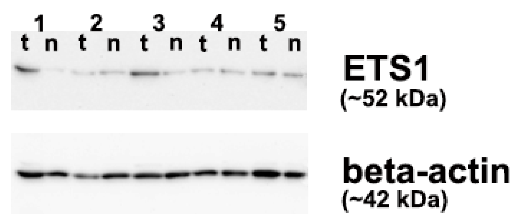
Although there was a noticeable increase in the relative levels of ETS1 protein expression (Figure 2B), as well as in ETS1 fold change (Figure 2C), in PTCs with methylated p16 vs. PTC cases with unmethylated p16, the difference did not achieve statistical significance (p > 0.05, tested by Mann-Whitney U test). Western blot analysis of ETS1 protein expression in PTC tissue and adjacent NMT is shown in Figure 3.
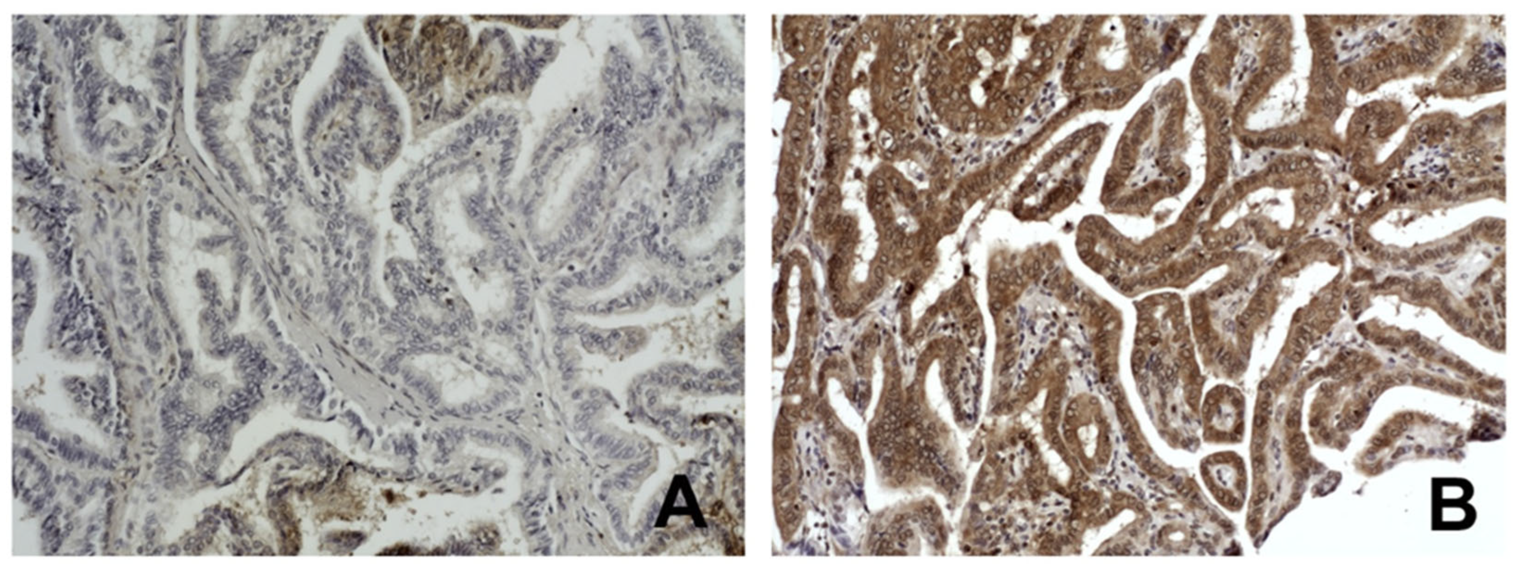
The mutual distribution of p16 methylation and the level of ETS1 protein expression were additionally determined by immunohistochemistry in 54 of 57 PTC cases included in the study. The tested group was then split into two subgroups according to the median value of ETS1 protein levels in the total PTC sample: high expressing subgroup included samples with ETS1 protein levels equal or higher than median ETS1 value in total sample and low expressing group included PTC samples with ETS1 protein levels lower than the same median value. The representative micrographs of IHC staining of ETS1 in PTC are shown in Figure 4. The results were again associated with the p16 methylation status and the unfavorable clinical parameters of the patients (Table 3). The combination of p16 methylation and the high levels of ETS1 protein was significantly associated with the pT and pTNM stage of the PTC patients (p < 0.05, tested by χ2-test).

3.4. The BRAFV600E Mutation Status and ETS1

There was no significant difference in the distribution or medians of ETS1 levels (mRNA or protein) across categories which included the BRAFV600E presence (Figure 5). In particular, the distribution of mRNA ETS1 levels was the same in BRAFV600E mutated and unmutated PTC cases (p = 0.151, tested by Mann-Whitney U test), as well as were the medians (p = 0.724, tested by the Median test). Although there was an increase in the relative levels of ETS1 protein expression (Figure 5B) and ETS1 fold change (Figure 5C) in PTCs with BRAFV600E compared to PTC cases without the mutation, the difference was not statistically significant (all p > 0.05, Mann-Whitney U test and Median test).

3.5. The Combination of the p16 Promoter Methylation, the BRAFV600E Mutation and ETS1 Levels

There was a significant difference in the distribution of ETS1 protein levels across categories which included the combination of p16 methylation and the BRAFV600E presence (p = 0.010, tested by χ2-test, Table 4). The BRAFV600E mutated PTC cases with methylated p16 promoter correlated significantly to the higher ETS1 mRNA expression (p = 0.003, r = 0.456, Sperman’s test of correlation), higher ETS1 protein expression (p = 0.005, r = 0.775, Sperman’s test of correlation), as well as to the ETS1 protein upregulation (p = 0.021, r = 0.609, Sperman’s test of correlation). On the other hand, the mutual presence of methylation in the p16 promoter with the BRAFV600E mutation and high ETS1 protein expression in PTC did not associate with the higher pT (0.067, tested by χ2-test) and pTNM stage (0.129, tested by χ2-test).

4. Discussion

The present study investigates the interplay between p16 promoter methylation status, the BRAFV600E mutation, and ETS1 gene and protein expression in patients with papillary thyroid carcinoma (PTC). Our findings shed light on the complex interactions that may exist within this malignancy. While p16 promoter methylation does not correlate significantly with unfavorable clinical parameters or BRAFV600E mutation status, it is associated with increased ETS1 mRNA expression in PTC patients. This intriguing observation may have implications for understanding the tumor behavior.
According to our results, the methylation status of the p16 promoter does not correlate with adverse clinical parameters typically associated with PTC. This lack of correlation implies that p16 methylation alone may not serve as a biomarker for predicting aggressiveness or metastatic potential of PTC. This finding isn’t consistent to the previous findings in PTC, as Wang et al. (2013) [14] showed that p16 methylation is associated with thyroid cancer metastasis and AMES (Age, Metastases, Extent and Size) stratification of PTC patients. Therefore, while p16 is an important tumor suppressor gene whose methylation status could be used as a diagnostic marker of PTC [12,13], its methylation status may not be a stand-alone factor critical for PTC progression and may not have a straightforward relationship with the clinical outcomes in patients with PTC.
Moreover, we found no correlation between p16 promoter methylation and BRAFV600E mutation in our PTC cohort. As long as BRAFV600E is prevalent in aggressive forms of PTC and contributes to disease progression [15], the association between BRAFV600E and methylation alterations is not consistently confirmed [17,35,36]. This distinction may indicate that p16 methylation could represent an epigenetic alteration that occurs without directly influencing the oncogenic activity of BRAFV600E and that maybe p16 methylation operates through alternate pathways that do not significantly overlap with the oncogenic effects of BRAFV600E mutations in PTC. In other words, this lack of correlation reinforces the notion that these two alterations may operate via distinct pathways in PTC. Previous research also failed to establish a correlation between p16 methylation and specific oncogenic mutations in thyroid cancers, indicating that p16 methylation may represent an event that occurs independently of other genetic alterations commonly explored in PTC [12,13]. This independence raises questions about the pathways involved in PTC development, suggesting a more complex interplay between genetic mutations and epigenetic modifications in thyroid tumors. It is also possible that maybe methylation of other tumor suppressor genes or alternative pathways contribute more significantly to the clinical outcomes in PTC than p16 status alone. This is noteworthy, particularly as some studies have found correlations between BRAF mutations and increased methylation in other carcinoma types [17,19,35,36].
In contrast, our results show that ETS1 mRNA levels were significantly higher in PTCs with a methylated p16 promoter than in those without, which presents an intriguing dimension to the role of ETS1 in thyroid carcinogenesis. Our findings suggest that p16 epigenetic silencing may result in enhanced transcriptional activity of ETS1, which is consistent with prior research indicating ETS1 as a modulator of carcinogenic processes [24,25,29]. However, it is crucial to note that despite the apparent rise in ETS1 expression at the mRNA level, there was no statistically significant change in the corresponding protein expression levels in this investigation. Namely, while fold changes in ETS1 protein levels were noted between the groups, the statistical significance was not achieved in this study. This implies a nuanced role for ETS1 in PTC where mRNA elevation does not necessarily translate to protein abundance with immediate clinical relevance [20,26]. Although this discrepancy might be attributed to the small sample size or variability in protein expression, which can often result in difficulties achieving clear statistical distinctions, it might also reflect post-transcriptional regulation, a concept well known in cellular biology and frequently associated with cancer development [37,38,39]. It assumes that mRNA levels do not always translate linearly to protein levels. Therefore, while transcriptional upregulation of ETS1 occurs, there could exist additional regulatory mechanisms, such as post-transcriptional silencing by microRNAs, post-translational modifications, protein stability or interactions with other signaling molecules, which influence ETS1 protein translation rate and warrant further investigation [37,38,39].
ETS1 often promotes oncogenic pathways by interacting with key transcription factors and signal transduction pathways. It is a downstream target of Ras/Raf/MEK signaling, which is frequently activated in PTC due to the BRAFV600E mutation [40,41]. Our observation that elevated ETS1 expression is particularly pronounced in the presence of the BRAFV600E mutation supports the hypothesis that these two factors may synergistically enhance tumor progression by over-activating downstream signaling pathways linked to cellular proliferation and migration. Furthermore, it was shown that ETS1 can be activated by MAPK-mediated phosphorylation [28,29,42].
An essential finding of our study is the significant association between the combination of p16 methylation and high levels of ETS1 protein with advanced pT and pTNM stages of PTC. This emphasizes the importance of understanding how these molecular alterations collectively impact tumor progression. While p16 methylation alone may not directly influence clinical outcomes, its interaction with ETS1 could still play a role in tumor advancement and may contribute to more aggressive tumor behavior. This highlights the potential utility of assessing both p16 methylation and ETS1 expression in predicting clinical outcomes and affirms their potential as indicators of PTC severity. Given the evolving nature of PTC research, these markers may serve as a composite biomarker to guide prognosis, especially in patients at increased risk for aggressive disease. This perspective aligns with the evolving paradigm, focusing not only on genetic mutations but also on epigenetic modifications and their effects on tumor behavior [9,11,12,13,14,43]. Moreover, we observed that within the BRAFV600E-mutated subset of PTC cases, methylated p16 was correlated with heightened ETS1 mRNA expression, increased ETS1 protein expression, and protein upregulation. Furthermore, although insignificant, some tendencies could be noticed in PTC cases with all three tested parameters positive (p16 methylated, BRAFV600E mutated, and ETS1 protein levels high) to the higher pT and pTNM stages. This relationship suggests that co-targeting this pathway could be a novel approach in precision medicine for PTC patients displaying these specific molecular profiles. While this necessitates further exploration into the functional roles of these molecules within the tumor development, the correlation of BRAFV600E-mutated PTC cases with methylated p16 and elevated ETS1 expression highlights a subtype of PTC that may benefit from more targeted therapeutic strategies. This supports the argument for stratifying PTC patients based on molecular profiles, which could ultimately guide individualized treatment regimens and predict response to therapy.
Nonetheless, our study has limitations that must be noted. The sample size, while adequate for initial explorations, may restrict the generalizability of our findings. Additionally, the focus on mRNA and protein expression without corresponding functional studies, gene silencing, or pathway inhibition experiments limits our capacity to draw definitive mechanistic conclusions about how p16 methylation or the BRAFV600E mutation influences ETS1 activity within the context of PTC. With all the limitations mentioned, most notably methodological limitations, a small sample size, and a lack of functional validation, the findings of this study should be regarded as a pilot study with substantial preliminary results that should be validated in further research. Future studies, possibly multicentric ones, incorporating larger cohorts and longitudinal follow-up (with recurrence and survival information), which would include a validation group of cases, as well as functional assays to dissect the roles of BRAFV600E, ETS1, and p16 methylation more precisely, will be essential to validate our findings and clarify the underlying mechanisms. Although the translational relevance of our results is still limited, restricting the applicability of this marker panel in clinical decision-making, the study’s findings are encouraging. Therefore, in the future, with a larger sample size, cell culture experiments, an adequate in vivo model system, external validation or replication in an independent cohort, and a tested marker panel, this combination might happen to be of great help for the risk stratification of PTC patients and for identifying patients who may benefit from more aggressive therapeutic strategies.

5. Conclusions

In summary, our investigation underscores the complexity of molecular interactions in PTC and highlights the multifactorial nature of its pathogenesis, which includes methylation, mutations, and expression changes. While p16 methylation could not be used as a standalone prognostic marker, its relationship with BRAFV600E and ETS1 could shed some light into PTC biology. Enhanced knowledge of these interactions may improve prognostic stratification of PTC patients and eventually adjust PTC patient management.

Supplementary Materials

The following supporting information can be downloaded at: https://www.mdpi.com/article/10.3390/biomedicines13071583/s1, Table S1: Association of p16 promoter methylation with the occurrence of unfavorable clinicopathological parameters of PTC patients.

Author Contributions

Conceptualization, T.I.D. and B.K.; methodology, S.S.N.,T.I.D., M.K., A.B. and B.K.; software, T.I.D. and B.K.; validation, B.K., S.Š. and U.P.Z.; formal analysis, T.I.D. and B.K.; investigation, S.S.N., M.K., A.B. and B.K.; writing—original draft preparation, T.I.D.; writing—review and editing, B.K., S.Š. and U.P.Z.; visualization, T.I.D.; supervision, T.I.D.; project administration, S.Š.; funding acquisition, S.Š. All authors have read and agreed to the published version of the manuscript.

Funding

This research was funded by the Ministry of Science, Technological Development and Innovation of the Republic of Serbia, Agreement No 451-03-136/2025-03/200019.

Institutional Review Board Statement

The study was conducted according to the Declaration of Helsinki and approved by the Ethics Committee of the Clinical Center of Serbia (reference no: 140/15, approved on 31 March 2022), Belgrade, Serbia.

Informed Consent Statement

Informed consent was obtained from all subjects involved in the study.

Data Availability Statement

The data presented in this study is available on request from the corresponding author.

Acknowledgments

We thank Katarina Taušanović (K.T.) from the Clinic for Endocrine Surgery, University Clinical Center of Serbia, Belgrade, Serbia for her help in obtaining resected tissue samples and clinicopathological data of the patients.

Conflicts of Interest

Uršula Prosenc Zmrzljak is an employee in BIA Separations CRO—Labena d.o.o. The remaining authors declare that the research was conducted in the absence of any commercial or financial relationships that could be construed as a potential conflict of interest.

Abbreviations

The following abbreviations are used in this manuscript:
PTCPapillary thyroid carcinoma
ETS1E26 transformation-specific
PCRpolymerase chain reaction
MSPmethylation-specific PCR
MASAmutant allele-specific PCR amplification
qPCRquantitative PCR
IHCimmunohistochemistry

References

  1. Chmielik, E.; Rusinek, D.; Oczko-Wojciechowska, M.; Jarzab, M.; Krajewska, J.; Czarniecka, A.; Jarzab, B. Heterogeneity of Thyroid Cancer. Pathobiology 2018, 85, 117–129. [Google Scholar] [CrossRef] [PubMed]
  2. Bhaijee, F.; Nikiforov, Y.E. Molecular Analysis of Thyroid Tumors. Endocr. Pathol. 2011, 22, 126–133. [Google Scholar] [CrossRef] [PubMed]
  3. Li, Y.; Su, Y.; Qin Dai, R.; Zhou, F.; Shen, C.; Su, Z.Y.; Yang, G.R. The Analysis of Clinical Biological and Pathological Features in Papillary Thyroid Carcinoma. Am. J. Biomed. Life Sci. 2021, 9, 292. [Google Scholar] [CrossRef]
  4. Pelizzo, M.R.; Boschin, I.M.; Toniato, A.; Piotto, A.; Pagetta, C.; Gross, M.D.; Al-Nahhas, A.; Rubello, D. Papillary Thyroid Carcinoma: 35-Year Outcome and Prognostic Factors in 1858 Patients. Clin. Nucl. Med. 2007, 32, 440–444. [Google Scholar] [CrossRef]
  5. Ito, Y.; Kudo, T.; Kobayashi, K.; Miya, A.; Ichihara, K.; Miyauchi, A. Prognostic Factors for Recurrence of Papillary Thyroid Carcinoma in the Lymph Nodes, Lung, and Bone: Analysis of 5,768 Patients with Average 10-year Follow-up. World J. surg. 2012, 36, 1274–1278. [Google Scholar] [CrossRef]
  6. Ito, Y.; Miyauchi, A.; Kihara, M.; Fukushima, M.; Higashiyama, T.; Miya, A. Overall Survival of Papillary Thyroid Carcinoma Patients: A Single-Institution Long-Term Follow-Up of 5897 Patients. World J. Surg. 2018, 42, 615–622. [Google Scholar] [CrossRef]
  7. Vu-Phan, D.; Koenig, R.J. Genetics and epigenetics of sporadic thyroid cancer. Mol. Cell. Endocrinol. 2014, 386, 55–66. [Google Scholar] [CrossRef]
  8. Asa, S.L.; Ezzat, S. The epigenetic landscape of differentiated thyroid cancer. Mol. Cell. Endocrinol. 2018, 469, 3–10. [Google Scholar] [CrossRef]
  9. Rodríguez-Rodero, S.; Fernández, A.F.; Fernández-Morera, J.L.; Castro-Santos, P.; Bayon, G.F.; Ferrero, C.; Urdinguio, R.G.; Gonzalez-Marquez, R.; Suarez, C.; Fernández-Vega, I.; et al. DNA Methylation Signatures Identify Biologically Distinct Thyroid Cancer Subtypes. J. Clin. Endocrinol. Metab. 2013, 98, 2811–2821. [Google Scholar] [CrossRef]
  10. Wu, W.; Zhang, L.; Lin, J.; Huang, H.; Shi, B.; Lin, X.; Huang, Z.; Wang, C.; Qiu, J.; Wei, X. Hypermethylation of the HIC1 Promoter and Aberrant Expression of HIC1/SIRT1 Contribute to the Development of Thyroid Papillary Carcinoma. Oncotarget 2016, 7, 84416–84427. [Google Scholar] [CrossRef]
  11. Salimi, F.; Asadikaram, G.; Ashrafi, M.R.; Zeynali Nejad, H.; Abolhassani, M.; Abbasi-Jorjandi, M.; Sanjari, M. Organochlorine Pesticides and Epigenetic Alterations in Thyroid Tumors. Front. Endocrinol. 2023, 14, 1130794. [Google Scholar] [CrossRef] [PubMed]
  12. Jiang, J.-L.; Tian, G.-L.; Chen, S.-J.; Xu, L.; Wang, H.-Q. Promoter Methylation of P16 and RASSF1A Genes May Contribute to the Risk of Papillary Thyroid Cancer: A Meta-Analysis. Exp. Ther. Med. 2015, 10, 1549–1555. [Google Scholar] [CrossRef]
  13. Mohammadi-asl, J.; Larijani, B.; Khorgami, Z.; Tavangar, S.M.; Haghpanah, V.; Kheirollahi, M.; Mehdipour, P. Qualitative and Quantitative Promoter Hypermethylation Patterns of the P16, TSHR, RASSF1A and RARβ2 Genes in Papillary Thyroid Carcinoma. Med. Oncol. 2011, 28, 1123–1128. [Google Scholar] [CrossRef] [PubMed]
  14. Wang, P.; Pei, R.; Lu, Z.; Rao, X.; Liu, B. Methylation of P16 CpG Islands Correlated with Metastasis and Aggressiveness in Papillary Thyroid Carcinoma. J. Chin. Med. Assoc. 2013, 76, 135–139. [Google Scholar] [CrossRef] [PubMed]
  15. Xing, M. BRAF Mutation in Thyroid Cancer. Endocr. Relat. Cancer 2005, 12, 245–262. [Google Scholar] [CrossRef]
  16. Smith, R.A.; Salajegheh, A.; Weinstein, S.; Nassiri, M.; Lam, A.K. Correlation between BRAF Mutation and the Clinicopathological Parameters in Papillary Thyroid Carcinoma with Particular Reference to Follicular Variant. Hum. Pathol. 2011, 42, 500–506. [Google Scholar] [CrossRef]
  17. Brait, M.; Loyo, M.; Rosenbaum, E.; Ostrow, K.L.; Markova, A.; Papagerakis, S.; Zahurak, M.; Goodman, S.M.; Zeiger, M.; Sidransky, D.; et al. Correlation between BRAF Mutation and Promoter Methylation of TIMP3, RARβ2 and RASSF1A in Thyroid Cancer. Epigenetics 2012, 7, 710–719. [Google Scholar] [CrossRef]
  18. Agrawal, N.; Akbani, R.; Aksoy, B.A.; Ally, A.; Arachchi, H.; Asa, S.L.; Auman, J.T.; Balasundaram, M.; Balu, S.; Baylin, S.B.; et al. Integrated Genomic Characterization of Papillary Thyroid Carcinoma. Cell 2014, 159, 676–690. [Google Scholar] [CrossRef]
  19. Kikuchi, Y.; Tsuji, E.; Yagi, K.; Matsusaka, K.; Tsuji, S.; Kurebayashi, J.; Ogawa, T.; Aburatani, H.; Kaneda, A. Aberrantly Methylated Genes in Human Papillary Thyroid Cancer and Their Association with BRAF/RAS Mutation. Front. Genet. 2013, 4, 271. [Google Scholar] [CrossRef]
  20. Nakayama, T.; Ito, M.; Ohtsuru, A.; Naito, S.; Nakashima, M.; Sekine, I. Expression of the Ets-1 Proto-Oncogene in Human Thyroid Tumor. Mod. Pathol. 1999, 12, 61–68. [Google Scholar]
  21. de Nigris, F.; Mega, T.; Berger, N.; Barone, M.V.; Santoro, M.; Viglietto, G.; Verde, P.; Fusco, A. Induction of ETS-1 and ETS-2 Transcription Factors Is Required for Thyroid Cell Transformation. Cancer Res. 2001, 61, 2267–2275. [Google Scholar] [PubMed]
  22. Plotnik, J.P.; Budka, J.A.; Ferris, M.W.; Hollenhorst, P.C. ETS1 Is a Genome-Wide Effector of RAS/ERK Signaling in Epithelial Cells. Nucleic Acids Res. 2014, 42, 11928–11940. [Google Scholar] [CrossRef] [PubMed]
  23. Dittmer, J. The biology of the Ets1 proto-oncogene. Mol Cancer. Mol. Cancer 2003, 2, 29. [Google Scholar] [CrossRef] [PubMed]
  24. Kar, A.; Gutierrez-Hartmann, A. Molecular Mechanisms of ETS Transcription Factor-Mediated Tumorigenesis. Crit. Rev. Biochem. Mol. Biol. 2013, 48, 522–543. [Google Scholar] [CrossRef]
  25. Wang, Y.; Huang, Z.; Sun, M.; Huang, W.; Xia, L. ETS Transcription Factors: Multifaceted Players from Cancer Progression to Tumor Immunity. Biochim. Biophys. Acta (BBA) Rev. Cancer 2023, 1878, 188872. [Google Scholar] [CrossRef]
  26. Novković, S.S.; Šelemetjev, S.; Miljuš, J.J.; Živaljević, V.; Dunđerović, D.; Milinković, M.; Denčić, T.I. ETS1 Protein Expression May Be Altered by the Complementarity of ETS1 mRNA Sequences with miR-203a-3p and miR-204-3p in Papillary Thyroid Carcinoma. Int. J. Mol. Sci. 2025, 26, 1253. [Google Scholar] [CrossRef]
  27. Peng, D.; Li, W.; Zhang, B.; Liu, X. Overexpression of lncRNA SLC26A4-AS1 inhibits papillary thyroid carcinoma progression through recruiting ETS1 to promote ITPR1-mediated autophagy. J. Cell. Mol. Med. 2021, 25, 8148–8158. [Google Scholar] [CrossRef]
  28. Peyret, V.; Nazar, M.; Martín, M.; Quintar, A.A.; Fernandez, E.A.; Geysels, R.C.; Fuziwara, C.S.; Montesinos, M.M.; Maldonado, C.A.; Santisteban, P.; et al. Functional Toll-like Receptor 4 Overexpression in Papillary Thyroid Cancer by MAPK/ERK–Induced ETS1 Transcriptional Activity. Mol. Cancer Res. 2018, 16, 833–845. [Google Scholar] [CrossRef]
  29. Wan, S.-M.; Peng, P.; Guan, T. Ets-1 Regulates Its Target Genes Mainly by DNA Methylation in Human Ovarian Cancer. J. Obstet. Gynaecol. 2013, 33, 877–881. [Google Scholar] [CrossRef]
  30. Stojanović, S.; Šelemetjev, S.; Đorić, I.; Janković Miljuš, J.; Tatić, S.; Živaljević, V.; Išić Denčić, T. BRAFV600E, BANCR, miR-203a-3p and miR-204-3p in Risk Stratification of PTC Patients. Biomedicines 2023, 11, 3338. [Google Scholar] [CrossRef]
  31. DeLellis, R.A.; Lloyd, R.V.; Heitz, P.U.; Eng, C. (Eds.) Pathology and Genetics of Tumours of Endocrine Organs: WHO Classification of Tumours, 3rd ed.; IARC Press: Lyon, France, 2004; ISBN 978-92-832-2416-7. [Google Scholar]
  32. Edge, S.B.; Compton, C.C. The American Joint Committee on Cancer: The 7th Edition of the AJCC Cancer Staging Manual and the Future of TNM. Ann. Surg. Oncol. 2010, 17, 1471–1474. [Google Scholar] [CrossRef] [PubMed]
  33. Basolo, F.; Torregrossa, L.; Giannini, R.; Miccoli, M.; Lupi, C.; Sensi, E.; Berti, P.; Elisei, R.; Vitti, P.; Baggiani, A.; et al. Correlation between the BRAF V600E Mutation and Tumor Invasiveness in Papillary Thyroid Carcinomas Smaller than 20 Millimeters: Analysis of 1060 Cases. J. Clin. Endocrinol. Metab. 2010, 95, 4197–4205. [Google Scholar] [CrossRef] [PubMed]
  34. Išić Denčić, T.; Bartolome, A.; Šelemetjev, S.; Đorić, I.; Tatić, S.; Živaljević, V.; Cvejić, D. High Expression and Localization of β-Catenin and Epidermal Growth Factor Receptor Identify High Risk Papillary Thyroid Carcinoma Patients. Exp. Mol. Pathol. 2018, 105, 181–189. [Google Scholar] [CrossRef] [PubMed]
  35. Bettington, M.; Brown, I.S.; Rosty, C. Serrated Lesions of the Appendix in Serrated Polyposis Patients. Pathology 2016, 48, 30–34. [Google Scholar] [CrossRef]
  36. Boissière-Michot, F.; Frugier, H.; Ho-Pun-Cheung, A.; Lopez-Crapez, E.; Duffour, J.; Bibeau, F. Immunohistochemical Staining for P16 and BRAFV600E Is Useful to Distinguish between Sporadic and Hereditary (Lynch Syndrome-Related) Microsatellite Instable Colorectal Carcinomas. Virchows Arch. 2016, 469, 135–144. [Google Scholar] [CrossRef]
  37. Louis, I.V.S.; Bohjanen, P.R. Post-Transcriptional Regulation of Cytokine and Growth Factor Signaling in Cancer. Cytokine Growth Factor. Rev. 2017, 33, 83–93. [Google Scholar] [CrossRef]
  38. Ali Syeda, Z.; Langden, S.S.S.; Munkhzul, C.; Lee, M.; Song, S.J. Regulatory Mechanism of MicroRNA Expression in Cancer. Int. J. Mol. Sci. 2020, 21, 1723. [Google Scholar] [CrossRef]
  39. Beňačka, R.; Szabóová, D.; Guľašová, Z.; Hertelyová, Z.; Radoňak, J. Non-Coding RNAs in Human Cancer and Other Diseases: Overview of the Diagnostic Potential. Int. J. Mol. Sci. 2023, 24, 16213. [Google Scholar] [CrossRef]
  40. Ros, J.; Baraibar, I.; Sardo, E.; Mulet, N.; Salvà, F.; Argilés, G.; Martini, G.; Ciardiello, D.; Cuadra, J.L.; Tabernero, J.; et al. BRAF, MEK and EGFR Inhibition as Treatment Strategies in BRAF V600E Metastatic Colorectal Cancer. Ther. Adv. Med. Oncol. 2021, 13, 1758835921992974. [Google Scholar] [CrossRef]
  41. Schubert, L.; Mariko, M.L.; Clerc, J.; Huillard, O.; Groussin, L. MAPK Pathway Inhibitors in Thyroid Cancer: Preclinical and Clinical Data. Cancers 2023, 15, 710. [Google Scholar] [CrossRef]
  42. Graves, B.J.; Petersen, J.M. Specificity within the Ets Family of Transcription Factors. Adv. Cancer Res. 1998, 75, 1–55. [Google Scholar] [CrossRef]
  43. Kozik, B.; Krajnovic, M.; Kokanov, N.; Jovanovic-Cupic, S.; Bozovic, A.; Todorovic, L.; Mandusic, V. Combined Analysis of KRAS Mutation and p16INK4a and p14ARF Methylation Status in Locally Advanced Rectal Carcinoma Treated with Preoperative Chemoradiotherapy. Arch. Biol. Sci. 2022, 74, 127–134. [Google Scholar] [CrossRef]
Figure 1. A diagram presenting the number of PTC cases included in each analysis. The numbers in the brackets represent the total number of PTC cases assessed for each analysis. The numbers presented below the total number of cases represent the additional number of PTC cases analyzed in each test, compared to the inner sample set (ellipsoid layer). The number of PTC cases with determined p16 and BRAFV600E status is indicated in orange. ETS1 expression is shown in blue. PTC: papillary thyroid carcinoma, WB: Western blot, IHC: immunohistochemistry.
Biomedicines 13 01583 g001
Figure 2. The relations between ETS1 expression and the p16 promoter methylation in papillary thyroid carcinoma. (A) The relation between ETS1 mRNA expression and the p16 promoter methylation, (B) The relation of ETS1 protein expression and the p16 promoter methylation, (C) The relation of ETS1 protein upregulation and the p16 promoter methylation. The reference line presents value 1 for ETS1 protein fold change. P16 promoter methylation was analyzed by methylation-specific PCR (MSP), relative ETS1 mRNA expression was determined by quantitative PCR, relative ETS1 protein expression and its fold change were determined by Western blot. The measurement data are expressed via box plots. The boxes represent the median value (the black horizontal line inside the box) and the interquartile range (upper and lower value); the symbols ‘o’ represent extreme values, and the symbols ‘*’ represent outliers. For details, see Section 2.
Biomedicines 13 01583 g002
Figure 3. Western blot analysis of 5 papillary thyroid carcinoma (PTC) cases using anti-ETS1 antibody. 1–5: case numbers, t: tumor (PTC), n: matched non-malignant thyroid tissue. Cases 1–3 are the BRAFV600E positive PTCs.
Biomedicines 13 01583 g003
Figure 4. Immunohistochemical staining of ETS1 in papillary thyroid carcinoma (PTC): (A) Low expression of ETS1 in PTC case with pT2, pTNM I, and non-methylated p16 promoter, Original magnifications ×10, (B) High expression of ETS1 in PTC case with pT3 and pTNM III, and methylated p16 promoter. Original magnifications ×10. For details, see Section 2.
Biomedicines 13 01583 g004
Figure 5. The relations between ETS1 expression and the BRAFV600E mutation in papillary thyroid carcinoma. (A) The relation between ETS1 mRNA expression and BRAFV600E, (B) The relation of ETS1 protein expression and BRAFV600E, (C) The relation of ETS1 protein upregulation and BRAFV600E. The reference line presents value 1 for ETS1 protein fold change. The BRAFV600E mutation was analyzed by mutant allele-specific PCR amplification (MASA), relative ETS1 mRNA expression was determined by quantitative PCR, relative ETS1 protein expression and its fold change were determined by Western blot. The measurement data are expressed via box plots. The boxes represent the median value (the black horizontal line inside the box) and the interquartile range (upper and lower value); the symbols ‘o’ represent extreme values, and the symbols ‘*’ represent outliers. For details, see Section 2.
Biomedicines 13 01583 g005
Table 1. Sequences of primers.
MethodPrimerSequence (5′-3′)
MASABRAF-f_wt (for wild type)GTGATTTTGGTCTAGCTACAGT
BRAF-f_mut (for BRAFV600E)GTGATTTTGGTCTAGCTACAGA
BRAF-rGGCCAAAAATTTAATCAGTGGA
MSPp16_U-fTTATTAGAGGGTGGGGTGGATTGT
p16_U-rCAACCCCAAACCACAACCATAA
p16_M-fTTATTAGAGGGTGGGGCGGATCGC
p16_M-rGACCCCGAACCGCGACCGTAA
qPCRETS1-fACAGGACTCCATGGCAAACG
ETS1-rATGAAGAAAGCCTGGTGCAGT
GAPDH-fGAAGGTGAAGGTCGGAGT
GAPDH-rGAAGATGGTGATGGGATTTC
PCR: Polymerase chain reaction, MASA: mutant allele-specific PCR amplification, MSP: methylation-specific PCR, qPCR: quantitative PCR. wt: wild type, mut: BRAFV600E mutant, f: forward primer, r: reverse primer, U: unmethylated (primer set for unmethylated modified DNA sequence), M: methylated (primer set for methylated modified DNA sequence).
Table 2. Association of the p16 promoter methylation and the BRAFV600E mutation in PTC.
PTCBRAFV600ETotal (n)
NoYes
p16 methylatedno61824
yes112233
Total (n)174057
PTC: Papillary thyroid carcinoma, n: number of cases.
Table 3. Association of p16 methylation and the ETS1 protein levels with the occurrence of unfavorable clinicopathological parameters of PTC patients.
Clinicopathological Parameterp16 Methylated and ETS1 Protein Levels High (n)p-Value
None or OneBoth
Gendermale1320.949
female2910
Intraglandular disseminationno1850.941
yes247
Lymph node metastasisno33100.718
yes92
Extrathyroid invasionno28100.265
yes142
Degree of tumor infiltration1960.251
2123
371
4142
pTT1810.032
T2149
T3191
T411
pTNMI1520.012
II98
III130
IV52
PTC: Papillary thyroid carcinoma, n: number of cases. p-value: statistical significance tested by χ2-test. Statistically significant results are bold.
Table 4. Association of the p16 promoter methylation, the BRAFV600E mutation presence and the ETS1 protein expression in PTC.
PTCThe Level of ETS1 Protein ExpressionTotal (n)
LowHigh
p16 methylated/
BRAFV600E mutated
None or one12719
Both3811
Total (n)151530
PTC: Papillary thyroid carcinoma, n: number of cases.
Disclaimer/Publisher’s Note: The statements, opinions and data contained in all publications are solely those of the individual author(s) and contributor(s) and not of MDPI and/or the editor(s). MDPI and/or the editor(s) disclaim responsibility for any injury to people or property resulting from any ideas, methods, instructions or products referred to in the content.