Article

Route of Application and Dose Evaluation of Dental Pulp Stem Cells for the Treatment of Sialadenitis Caused by Sjögren’s Syndrome: A Preclinical Study

1
Department of Oral and Maxillofacial Surgery, Peking University School and Hospital of Stomatology, No. 22, Zhongguancun South Avenue, Haidian District, Beijing 100081, China
2
Center Laboratory, Peking University School and Hospital of Stomatology & National Center for Stomatology & National Clinical Research Center for Oral Diseases & National Engineering Research Center of Oral Biomaterials and Digital Medical Devices & Beijing Key Laboratory of Digital Stomatology & NHC Key Laboratory of Digital Stomatology & NMPA Key Laboratory for Dental Materials, No. 22, Zhongguancun South Avenue, Haidian District, Beijing 100081, China
3
Department of Prosthodontics, North China University of Science and Technology, Stomatology, Tangshan Bay Ecological Zone No. 21 Bohai Avenue, Tangshan 063210, China
*
Authors to whom correspondence should be addressed.
These authors contributed equally to this work.
Biomedicines 2025, 13(5), 1068; https://doi.org/10.3390/biomedicines13051068
Submission received: 26 March 2025 / Revised: 22 April 2025 / Accepted: 24 April 2025 / Published: 28 April 2025
(This article belongs to the Special Issue Pathogenesis, Diagnostics, and Therapeutics for Rheumatic Diseases)

Abstract

:
Background: Sjögren’s syndrome (SS) is an autoimmune disorder characterized by sicca syndrome and/or systemic manifestations. In this study, non-obese diabetic (NOD) mice were used as an animal model for studying SS, to evaluate the optimal administration route and dose range of dental pulp stem cells (DPSCs) in the treatment of sialadenitis caused by SS. Methods: Different doses of DPSCs were transplanted into the submandibular glands (SMGs) of 14-week-old NOD mice through two different methods: injection or retrograde perfusion through the catheter orifice into the SMG. At 21 weeks of age, the saliva flow rate (SFR), ectopic lymphocytes, and CD4+ T-cell infiltration were measured. Tumor necrosis factor-alpha (TNF-α) and interferon-gamma (IFN-γ) in the glandular tissues were also quantitatively detected. Results: Compared with untreated and PBS-injected controls, different-dose groups of the two administration methods showed an increased saliva flow rate of NOD mice to varying degrees, reduced infiltration of lymphocytes and CD4+ T cells in the SMG, and decreased IFN-γ/TNF-α levels. Finally, we compared these two administration routes and found that the perfusion of 2 × 10⁵ DPSCs presents good therapeutic effects. Conclusions: DPSC perfusion through the catheter orifice is a simple and effective treatment method, which is worthy of further investigation through clinical trials.

1. Introduction

Sjögren’s syndrome (SS) is a chronic autoimmune disease characterized by infiltration of lymphocytes into exocrine glands. Clinically, 95% of SS patients have dry eyes and a dry mouth due to the ectopic infiltration of lymphocytes into the lacrimal and salivary glands. This dryness can last for more than 10 years, severely affecting patients’ quality of life. Additionally, approximately 60% of patients also have other autoimmune disorders, including rheumatoid arthritis and autoimmune thyroid disease, resulting in symptoms associated with multisystem damage [1,2,3]. The systemic extraglandular manifestations of this condition include polyarthralgia/polyarthritis, cutaneous vasculitis, bronchiolitis/pneumonia, tubulointerstitial nephritis, and peripheral neuropathy, as well as an increased risk of lymphoma [4]. These co-existing conditions can even be life-threatening.
Currently, the treatment of SS is still empirical. The available approaches focus mainly on relieving symptoms and preventing complications, and they are based on the treatment approaches used for other autoimmune diseases. Corticosteroids combined with immunosuppressants such as hydroxychloroquine is the first-line clinical treatment [5,6]. However, long-term use of glucocorticoids may lead to osteoporosis, hyperglycemia, central obesity, and other metabolic abnormalities.
Recent advances in the understanding of SS pathogenesis have prompted researchers to use new biological therapies. For example, B-cell-targeted therapies and T-cell-targeted therapies can be used. Rituximab is an anti-CD20 B lymphocyte depletion agent that has therapeutic effects on SS-related systemic manifestations (parotid gland swelling, lymphoma, and inflammatory arthritis) [5,7,8] and sicca manifestations (dry mouth) [7,9,10]. However, in other randomized controlled trials, insufficient evidence was found that rituximab improved dry mouth symptoms in SS patients [11,12]. T-cell-targeting medications include abatacept and alefacept, and cytokine-targeting agents include infliximab, tofacitinib, and IL-2 [13,14,15]. RO5459072, a cathepsin S inhibitor, was shown to attenuate CD4+ T-cell activation, but it did not confer a clinical benefit in SS patients [16]. These studies revealed that regardless of whether B-cell targeting or T-cell targeting is used, the therapeutic effect is nonselective and has limited benefits for exocrine function and sicca symptoms. Thus, identifying a therapeutic agent that targets CD4+ T cells in SS-damaged glands could significantly improve disease management.
Cell therapy, which involves the use of regenerative medicines designed to restore the function of damaged tissue, has gained significant traction for SS treatment. In previous studies, bone marrow-derived mesenchymal stem cells (BMMSCs) [17], umbilical cord-derived mesenchymal stem cells (UCMSCs) [18], and labial gland-derived mesenchymal stem cells (LGMSCs) [19] were used in non-obese diabetic (NOD) mice, a classic animal model with SS-like features. After administration of these cells, the saliva flow rate (SFR) improved, and glandular ectopic lymphocyte infiltration decreased. Compared with these MSCs, dental pulp stem cells (DPSCs) and stem cells derived from exfoliated deciduous teeth (SHEDs) originate from unique stem cell sources and are obtained using non-invasive techniques. These cells possess the remarkable ability to undergo multidirectional differentiation, including differentiation into adipocytes, odontoblasts, chondrocytes, osteoblasts, hepatocytes, neuronal cells, and myocytes [20,21,22,23,24], and they can exert immunoregulatory effects [25,26]. In a previous study, intravenous injection of SHEDs was shown to exert protective effects on saliva secretion in NOD mice by modulating the differentiation of CD4+ T cells and improving the inflammatory microenvironment [27]. Additionally, DPSCs are beneficial in the treatment of chronic inflammatory disease (traumatic brain injury, osteoarthritis) [28,29] and immunological diseases (rheumatoid arthritis, systemic lupus erythematosus) [30,31], which have shown promising results. However, there are no reports on the use of DPSCs for SS treatment. Whether DPSCs can target CD4+ T cells to treat sialadenitis caused by SS requires further investigation. Additionally, the route of administration and the optimal dose range of these cells should be established in a preclinical study.
Therefore, this preclinical study aimed to evaluate treatment parameters and outcomes for the administration of DPSCs to the submandibular gland (SMG) for the treatment of sialadenitis caused by SS.

2. Materials and Methods

2.1. Cell Culture

DPSCs were sourced from Beijing SH Biotechnology (Beijing, China), which were isolated and cultured with the established methods [32,33]. The reagents used for cell culture were from Gibco (Rockville, MD, USA). DPSCs from passages 2–7 were used to ensure reliability and consistency.

2.2. Animal Experiments

NOD mice are the classic animal model of SS [34,35,36]. Depending on the severity of sialadenitis, the NOD mice go through three stages: initial stage (the age of 7–8 weeks), early clinical stage (the age of 12–16 weeks), and late clinical stage (the age of 20–24 weeks) [37]. Correspondingly, NOD mice at the ages of 7, 14, and 21 weeks are commonly used to investigate SS progression [27,38,39]. In the present study, the 21-week-old NOD mice were used with and without DPSC treatment.

2.3. DPSC Treatment

This study was approved by the Peking University Institutional Review Board for the Care and Use of Laboratory Animals (LA2021581). Female NOD mice at 7, 14, and 21 weeks of age were purchased from Beijing Huafukang Biotechnology Co., Ltd. (Beijing, China). To minimize suffering, tribromoethanol was used to anesthetize the mice via intraperitoneal injection. NOD mice aged 7, 14, or 21 weeks that were not subjected to any treatments were used as an untreated group. For injection, 1 × 10⁵, 2.5 × 10⁵, 5 × 10⁵, or 1 × 10⁶ DPSCs (diluted in 20 μL of PBS) were injected directly into the SMG in 14-week-old NOD mice at multiple points. An equal volume of PBS was injected as a vehicle control. For intraductal perfusion, an intrathecal catheter device (32 g with 27 g needles) was inserted into the intraoral duct orifice of the SMG and used to perfuse 7.5 × 10⁴, 1.5 × 10⁵, 2 × 10⁵, 3 × 10⁵, or 4 × 10⁵ DPSCs (diluted in 20 μL of PBS). An equal volume of PBS was perfused as a vehicle control. The mice in both the injection group and the intraductal perfusion group were treated once at 14 weeks of age and sacrificed at 21 weeks of age for further experiments. For all animal experiments, there were 6 mice in each group.

2.4. DPSC Tracking

Before injection or perfusion, the DPSCs were stained with 1,1-dioctade-cyl-3,3,3,3-tetramethylindotricarbocyanine iodide (DiR) for 2 h for labeling. The DiR-labeled DPSCs were subsequently injected or perfused into NOD mice at 14 weeks of age. An in vivo imaging system (Caliper Life Sciences, Hopkinton, MA, USA) was used to observe the location of DiR-labeled DPSCs at 1 and 7 days after injection or perfusion.

2.5. Stimulated SFR Measurement

To stimulate salivary secretion, 21-week-old NOD mice were intraperitoneally injected with pilocarpine (5 μg per 10 g body weight) under anesthesia. Saliva was collected from the mouth for 10 min with a micropipette and weighed with an electronic balance.

2.6. Histological Evaluation of SMG

As described in our previous studies, the SMG tissues of NOD mice were used to prepare paraffin sections, and then hematoxylin–eosin (HE) staining was performed [27,40,41]. The lesion score (the number of lesions containing more than 50 monocytes in a 4 mm2 area) and lesion ratio (the ratio of the lesion area to the total tissue area) were subjected to quantitative analysis. Immunohistochemical staining was performed after the paraffin-embedded samples were deparaffinized, heat-induced antigen repair was performed, and the samples were stained with an anti-CD4 antibody (Abcam, Cambridge, MA, USA), as described previously [41]. The CD4 ratio index was defined as the ratio of CD4+ cells’ area to the lesion area.

2.7. Enzyme-Linked Immunosorbent Assay (ELISA)

SMG homogenates were collected from mice in each group. Then, TNF-α and IFN-γ levels were examined by TNF-α and IFN-γ ELISA kits (Neobioscience, Shenzhen, China) according to the manufacturer’s instructions.

2.8. Statistical Analysis

The experimental data are presented as the means ± standard deviations. Statistical analysis was performed via Student’s t test for comparisons between two groups or one-way analysis of variance followed by Bonferroni correction for comparisons among multiple groups using GraphPad Prism 8.0 software. A p value < 0.05 was regarded as indicating statistical significance.

3. Results

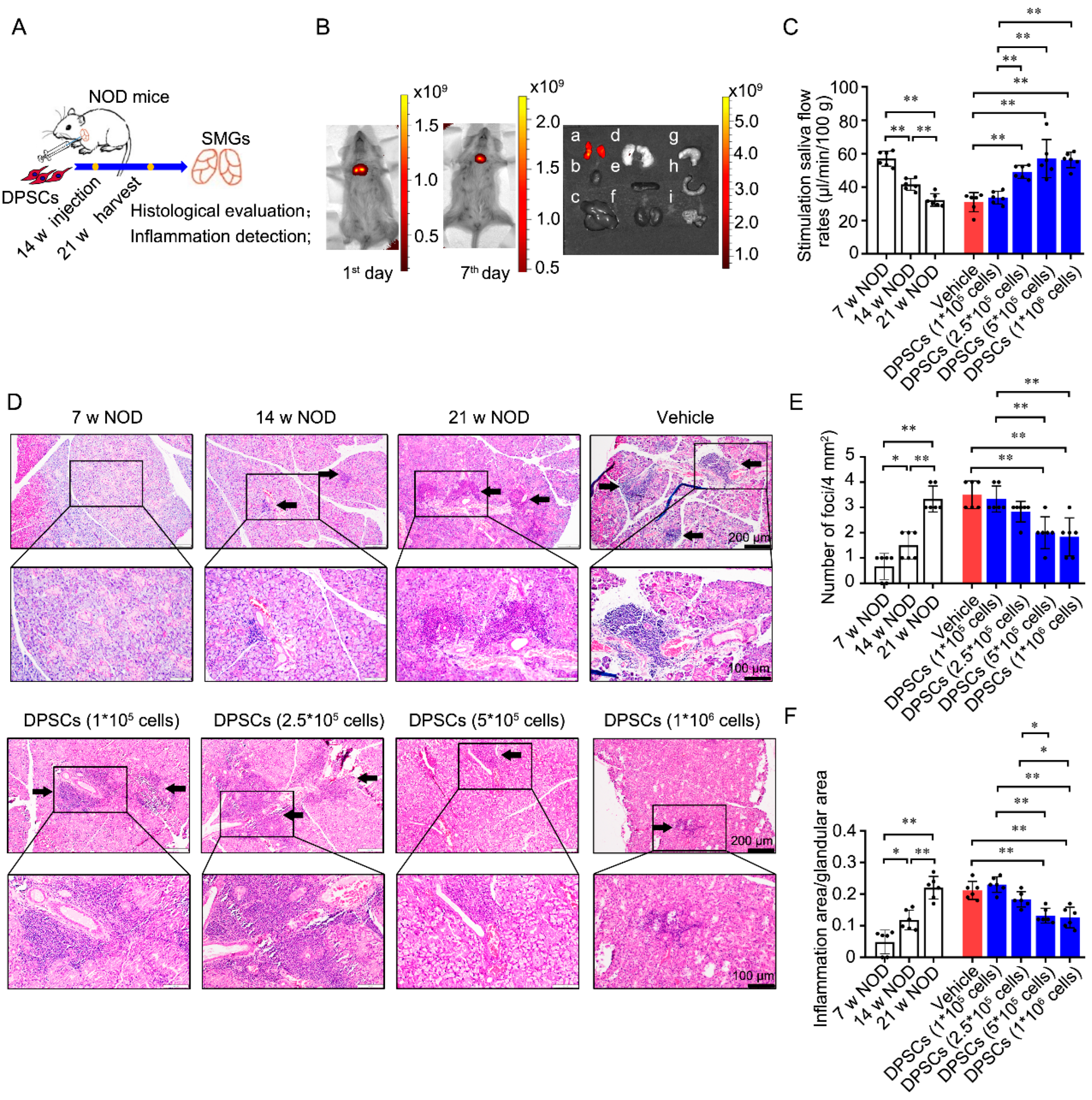
3.1. Injection of DPSCs into the SMG Promotes Saliva Secretion and Ameliorates Ectopic Lymphocyte Infiltration into the Glandular Tissues of NOD Mice

As defined cellular characteristics are critical for the clinical use of stem cells, we explored the phenotype of DPSCs and found that they expressed characteristic MSC markers, including CD44, CD73, and CD90 (Figure S1). To confirm the effect of DPSCs on SS-induced sialadenitis, we injected DPSCs into the SMG of NOD mice at 14 weeks of age and assessed the SFR and the degree of lymphatic infiltration at 21 weeks (Figure 1A). Initially, positive signals of DiR-labeled DPSCs were observed around the neck on the 1st and 7th days post-injection (Figure 1B, left and middle). Further observation on the 7th day after injection revealed that the positive signals were present in the SMG but not in other tissues (Figure 1B, right).
Compared with that in 7-week-old NOD mice, the SFR was significantly lower in 14-week-old NOD mice and further decreased in 21-week-old NOD mice in the untreated group. In the treated groups, injection of 1 × 10⁵ DPSCs did not change the SFR compared with that in the PBS-treated group. Compared with the vehicle control and injection of 1 × 10⁵ DPSCs, injection of 2.5 × 10⁵, 5 × 10⁵, or 1 × 10⁶ DPSCs significantly increased the SFR (Figure 1C).
Salivary gland epithelial cells are the main targets of immune attack. Ectopic lymphocyte infiltration into the salivary glands plays a crucial role in the development of hypersalivation caused by SS [42,43,44]. Histopathological evaluation revealed that lymphocyte infiltration foci appeared in the SMG of 14-week-old mice and were aggravated at 21 weeks. In the age-matched untreated control, vehicle control, and 1 × 10⁵ and 2.5 × 10⁵ DPSC-treated groups, there were still obvious lymphocyte infiltration foci in the glandular tissues. However, in the 5 × 10⁵ and 1 × 10⁶ DPSC-treated groups, the ectopic infiltration of lymphocytes was clearly alleviated (Figure 1D). Quantitative analysis further verified that both the lesion score and lesion ratio were increased in 14-week-old NOD mice and further increased at 21 weeks. Compared with the untreated and vehicle control groups, the groups injected with 5 × 10⁵ and 1 × 10⁶ DPSCs presented significantly lower lesion scores and lesion ratios (Figure 1E and Figure 1F, respectively).

3.2. Injection of DPSCs into the SMG Decreases CD4+ Cell Infiltration and the Levels of SS-Specific Inflammatory Cytokines in the Glandular Tissues of NOD Mice

Excessive activation of CD4+ T cells is a common hallmark of autoimmune diseases such as systemic lupus erythematosus and SS [44,45]. As shown in Figure 2A,B, the ratio of the area of positively stained CD4+ T cells to the total area was greater in the SMG of 14- and 21-week-old NOD mice than in that of 7-week-old NOD mice. Compared with the vehicle control, injection of 2.5 × 10⁵, 5 × 10⁵, and 1 × 10⁶ DPSCs significantly decreased the ratio of the CD4+ T-cell area to the total area. Compared with the groups injected with 1 × 10⁵ and 2.5 × 10⁵ DPSCs, the groups injected with 5 × 10⁵ and 1 × 10⁶ DPSCs presented decreased ratios of CD4+ T-cell area to the total area.
TNF-α and IFN-γ are crucial inflammatory cytokines that are highly expressed in the periphery and damaged glands of SS patients and NOD mice [46,47,48]. Compared with those in 7-week-old NOD mice, TNF-α levels were lower in the 2.5 × 10⁵ DPSCs group and further decreased in the 5 × 10⁵ and 1 × 10⁶ DPSC groups. The IFN-γ content in the SMG was increased in 21-week-old NOD mice. Compared with the vehicle control, injection of 5 × 10⁵ or 1 × 10⁶ DPSCs resulted in lower levels of IFN-γ. In particular, injection of 5 × 10⁵ DPSCs further decreased IFN-γ levels compared with those observed in the 1 × 10⁵ DPSC group and the 2.5 × 10⁵ DPSC group (Figure 2C, D). The above results are summarized in Table 1.
These data are consistent with the observed changes in the SFR and suggest that DPSC injection has a therapeutic effect on hyposalivation caused by SS. In particular, injection of 5 × 10⁵ DPSCs appears to be the optimal dose.

3.3. Intraductal Perfusion of DPSCs into the SMG Restores Saliva Secretion and Ameliorates Ectopic Lymphocyte Infiltration into the Glandular Tissues of NOD Mice

Stem cell-based cell therapies are typically delivered via intravenous or topical administration. For treatment of the salivary glands, intraductal perfusion is undoubtedly a better approach. Therefore, in parallel, we perfused DPSCs through the orifice of the SMG duct in 14-week-old NOD mice and detected the perfused cells at 21 weeks (Figure 3A). After perfusion, a DiR-DPSC-positive signal was detected around the mouth area on the 1st day and was then captured around the neck on the 7th day (Figure 3B left and middle). Further observation on the 7th day revealed that the positive signals were present in the SMG but not in other tissues (Figure 3B right). These results indicate that the perfused DPSCs passed through the ducts and migrated into the glandular tissue.
Compared with the vehicle control, perfusion of 7.5 × 10⁴ DPSCs did not change the SFR. As expected, 1.5 × 10⁵, 2 × 10⁵, 3 × 10⁵, or 4 × 10⁵ DPSC perfusion increased the SFR in 21-week-old NOD mice compared with the mice in the age-matched untreated group, vehicle control group, and 7.5 × 10⁴ DPSC perfusion group (Figure 3C). Similarly, perfusion of 7.5 × 10⁴ DPSCs did not suppress ectopic lymphocytic infiltration, whereas perfusion of 1.5 × 10⁵, 2 × 10⁵, 3 × 10⁵, or 4 × 10⁵ DPSCs significantly reduced both the lesion score and the lesion ratio in the SMG compared with those in the untreated group, vehicle control group, and 7.5 × 10⁴ DPSC perfusion group (Figure 3D–F).
CD4+ T-cell staining revealed that administration of 2 × 10⁵, 3 × 10⁵, and 4 × 10⁵ DPSCs reduced the ratio of the CD4+ T-cell area to the total area in the SMG compared with that in the untreated, vehicle control, and 7.5 × 10⁴ DPSC groups (Figure 4A,B). Furthermore, compared with perfusion of the vehicle control, perfusion of 1.5 × 10⁵ or 2 × 10⁵ DPSCs decreased TNF-α levels. Compared with perfusion of 7.5 × 10⁴ DPSCs, perfusion of 1.5 × 10⁵ DPSCs reduced TNF-α levels (Figure 4C). Perfusion of both 2 × 10⁵ and 3 × 10⁵ DPSCs decreased IFN-γ levels compared with those in the vehicle control and 7.5 × 10⁴ DPSC groups (Figure 4D). The above results are summarized in Table 2.
These results indicate that intraductal perfusion of DPSCs is indeed a good administration option for the treatment of SS-induced hyposalivation. The optimal dose for DPSC injection is 5 × 10⁵ cells.

3.4. Comparison of the Two Application Routes

Based on the above data, we surmised that the optimal dose of DPSC injection was 5 × 10⁵, and the optimal dose of intraductal perfusion was 2 × 10⁵. To reveal their therapeutic efficacy, we next compared these two application routes (Figure 5A). We found that 5×10⁵ injection and 2 × 10⁵ perfusion both promoted the SFR by 84% and 63% compared their vehicle control, respectively. And there was no difference between these two treated groups (57.07 ± 11.42 vs. 55.48 ± 3.87, p = 0.75; Figure 5B). Further, both administration methods can reduce the focus score and the ratio index in SMGs. The perfusion group showed the same inhibitory effect on the focus score (2.33 ± 0.52 vs. 1.67 ± 0.82, p = 0.12, Figure 5C). However, the ratio index of ectopic inflammatory infiltration notably declined in the 2 × 10⁵ perfusion group compared to the 5 × 10⁵ injection group (0.13 ± 0.02 vs. 0.07 ± 0.01, p < 0.01, Figure 5D). On the contrary, the 5 × 10⁵ injection group showed a better inhibiting effect on the CD4+ T-cell-positive staining ratio (0.24 ± 0.05 vs. 0.37 ± 0.05, p < 0.01, Figure 5E). The TNF-α level shown the same reduced effect in the two groups (13.00 ± 7.09 vs. 21.57 ± 12.46, p = 0.27, Figure 5F). However, the 5 × 10⁵ injection group showed the better inhibiting effect on IFN-γ (495.2 ± 113.83 vs. 767.2 ± 254.16, p = 0.04, Figure 5F). The above results are summarized in Table 3. Collectively, these findings suggest that 2 × 10⁵ DPSC perfusion is best suited for clinical application.

4. Discussion

In this article, we present preclinical results regarding the use of DPSCs as a potential cell therapy for sialadenitis caused by SS. We performed preclinical in vivo assessments of safety and therapeutic efficacy and determined the optimal route of administration and cell dose in an SS mouse model.
DPSCs were first derived from the third molars of humans by Gronthos in 2000 [20], and they can also be derived from orthodontic teeth and supernumerary teeth. They possess a stable self-renewal ability and multidirectional differentiation potential [49,50]. Compared with other types of MSCs, DPSCs exhibit stronger proliferative, anti-inflammatory, and antifibrotic effects [51,52] and do not exhibit tumor formation after transplantation like embryonic stem cells (ESCs) or induced pluripotent stem cells (iPSCs) [53]. These characteristics make DPSCs valuable for disease treatment and regenerative therapy [54,55]. In detail, DPSCs can exert immunosuppressive effects by interfering with activated T cells [56] and exert anti-inflammatory effects by inducing M2 polarization in macrophages [57]. Recent reports indicate that the secreted molecules within DPSCs contain diverse trophic factors and demonstrate efficacy in multiple disease models [58,59,60,61]. More importantly, cytokine production of DPSCs outperforms BMMSCs in reducing inflammation, promoting tissue repair, and combating infection [62]. In the present study, we confirmed that DPSCs exert therapeutic effects, promoting saliva production and alleviating inflammation in glandular tissues in NOD mice, via both injection and perfusion into the SMG. After injection or perfusion, the DPSCs were confined to glandular tissues, indicating the safety of local administration.
During the pathological process associated with SS, ectopic lymphocyte infiltration (mostly infiltration of CD4+ T cells) occurs; this is the first immune defense against injury or infection [63]. If the inflammatory process lasts too long, many activated cells are attracted to the site of injury, releasing excessive amounts of enzymes, chemokines, etc., which is detrimental to tissue repair and promotes further deterioration [64]. In this study, we evaluated the inflammatory condition of the SMG in NOD mice at 7, 14, and 21 weeks and found that, over time, the disease progressed, with intensified ectopic lymphatic infiltration, which was consistent with our previous studies [27,39]. However, after DPSC treatment, both the lesion score and the lesion ratio were related to lymphocyte infiltration, and the number of infiltrating CD4+ T cells was effectively reduced.
Additionally, the current consensus regarding SS pathogenesis is that diverse environmental factors act on susceptible individuals, leading to the disruption of autoimmune tolerance. Activated CD4+ T cells can secrete IL-2, TNF-α, IFN-γ, IL-10, and other inflammatory factors. Among these factors, TNF-α and IFN-γ are the most typical, and the levels of both are increased in SS-damaged tissues [27,65]. Single-cell RNA sequencing in combination with pathway enrichment analysis of peripheral blood mononuclear cells demonstrated the upregulation of genes associated with type I and II interferon signaling, TNF family signaling, and antigen processing and presentation processes in SS patients [66]. TNF-α can affect the local inflammatory immune microenvironment by stimulating the secretion of other proinflammatory factors and participating in the disruption of the tight junctions of glandular epithelial cells [48]. It can also increase the expression of two important autoantigens associated with SS, Ro/SSA and La/SSB [67], and may cause secretory dysfunction in SS by inducing apoptosis in glandular cells alone and with IFN-γ [68]. On the other hand, IFN-γ can induce the expression of adhesion molecules, causing a large number of inflammatory cells to aggregate and initiating local inflammatory damage [69]. Patients with the cluster characterized by the most robust IFN and inflammation gene signatures exhibited elevated ESSDAI scores, along with increased levels of anti-Ro/SSA and La/SSB autoantibodies [70]. FN-γ can also activate the innate immune system, increase the release of proinflammatory factors, increase antigen presentation, and damage the tight junction structure of salivary gland epithelial cells, resulting in persistent dysfunction of glandular secretion [71]. Previously, we verified that the expression of TNF-α and IFN-γ in 21-week-old NOD mice significantly increased with age. SHED administration via the tail vein can decrease TNF-α and IFN-γ levels in serum, SMG tissues, and saliva and, ultimately, alleviate hyposalivation caused by SS [27]. Here, after DPSC administration, both TNF-α and IFN-γ levels in the SMG were significantly reduced, which was consistent with the ectopic lymphatic infiltration and CD4+ T-cell staining results. Collectively, these results confirm that DPSC administration exerts a therapeutic effect on SS sialadenitis through the immunosuppression of CD4+ T cells.
Finally, compared with injection via the tail vein or direct injection into glandular tissue, intraductal perfusion is a good route of administration for clinical applications because the catheter system can guide reverse transport of DPSCs into the glandular tissue without additional damage to the glandular lobe. Previously, we performed intraductal perfusion of SHED exosomes into the SMG in 14-week-old NOD mice and confirmed the ability of the administered exosomes to increase the SFR and alleviate inflammatory infiltration [40]. In the present study, we further attempted to perform intraductal perfusion of DPSCs into the SMG and proved that DPSC perfusion has a therapeutic effect superior to that of injection, increasing the SFR and ameliorating the inflammatory response in SS. Furthermore, in terms of the translational feasibility, DPSC perfusion has the advantage of non-invasive administration, meaning it is more easily accepted by patients and doctors in clinical practice.
In future, the mechanism of DPSCs (whether DPSCs can interact with immune cells or glandular epithelium) requires further in-depth research. Further, the human disease pathology exhibits more complexity, mandating rigorous validation of pharmacokinetic profiles in humanized models, and the long-term efficacy and safety profiles also need further evaluation.

5. Conclusions

In the present study, various doses of DPSCs and the optimal route of administration for the treatment of sialadenitis caused by SS were assessed. Safety and efficacy tests were performed in this preclinical study. Both injection and perfusion of DPSCs into the SMG at various doses exhibited therapeutic effects in NOD mice via the targeting of CD4+ T cells. The perfusion of 2 × 10⁵ DPSCs is a convenient and effective therapeutic method. Overall, these findings provide a vital basis for designing experiments to optimize the route of administration and dose of DPSCs in clinical trials.

Supplementary Materials

The following supporting information can be downloaded at https://www.mdpi.com/article/10.3390/biomedicines13051068/s1. Figure S1. Characterization of DPSCs. (A) DPSCs showed long spindle-shaped adherent growth and uniform cell morphology under a phase contrast microscope. (B–D) DPSCs expressed MSC surface markers CD44, CD73, and CD90. (E,F) DPSCs were negative for hematopoietic stem cell surface markers CD34 and CD45.

Author Contributions

Conceptualization, G.Y. and C.D.; data curation, S.Z., R.Z., J.L. and X.L.; formal analysis, Z.D. and Y.Z.; funding acquisition, G.Y. and C.D.; investigation, X.P. and S.Z.; methodology, Z.D. and L.F.; project administration, G.Y. and C.D.; resources, X.P., S.Z., R.Z., J.L. and X.L.; validation, Z.D. and Y.Z.; visualization, L.F. and Y.Z.; writing—original draft, Z.D. and L.F.; writing—review and editing, Z.D., L.F. and X.P. All authors have read and agreed to the published version of the manuscript.

Funding

This work was supported by the National Natural Science Foundations of China (Grant Nos. 82270993 to Guangyan Yu and 81771088 to Chong Ding).

Institutional Review Board Statement

This study was approved by the Peking University Institutional Review Board for the Care and Use of Laboratory Animals (LA2021581 was approved at 13 December 2021).

Informed Consent Statement

Not applicable.

Data Availability Statement

All data generated or analyzed in this study are included in this published article. Any other data will be available on reasonable request from the corresponding author.

Conflicts of Interest

The authors declare no competing interests.

References

  1. Margaretten, M. Neurologic Manifestations of Primary Sjögren Syndrome. Rheum. Dis. Clin. N. Am. 2017, 43, 519–529. [Google Scholar] [CrossRef] [PubMed]
  2. Mariette, X.; Criswell, L.A. Primary Sjögren’s Syndrome. N. Engl. J. Med. 2018, 378, 931–939. [Google Scholar] [CrossRef] [PubMed]
  3. Xu, D.; Zhao, S.; Li, Q.; Wang, Y.H.; Zhao, J.L.; Li, M.T.; Zhao, Y.; Zeng, X.F. Characteristics of Chinese patients with primary Sjögren’s syndrome: Preliminary report of a multi-centre registration study. Lupus 2020, 29, 45–51. [Google Scholar] [CrossRef]
  4. Vivino, F.B.; Bunya, V.Y.; Massaro-Giordano, G.; Johr, C.R.; Giattino, S.L.; Schorpion, A.; Shafer, B.; Peck, A.; Sivils, K.; Rasmussen, A.; et al. Sjogren’s syndrome: An update on disease pathogenesis, clinical manifestations and treatment. Clin. Immunol. 2019, 203, 81–121. [Google Scholar] [CrossRef] [PubMed]
  5. Ramos-Casals, M.; Brito-Zerón, P.; Bombardieri, S.; Bootsma, H.; De Vita, S.; Dörner, T.; Fisher, B.A.; Gottenberg, J.E.; Hernandez-Molina, G.; Kocher, A.; et al. EULAR recommendations for the management of Sjögren’s syndrome with topical and systemic therapies. Ann. Rheum. Dis. 2020, 79, 3–18. [Google Scholar] [CrossRef]
  6. Srivastava, A.; Makarenkova, H.P. Innate Immunity and Biological Therapies for the Treatment of Sjögren’s Syndrome. Int. J. Mol. Sci. 2020, 21, 9172. [Google Scholar] [CrossRef]
  7. Carsons, S.E.; Vivino, F.B.; Parke, A.; Carteron, N.; Sankar, V.; Brasington, R.; Brennan, M.T.; Ehlers, W.; Fox, R.; Scofield, H.; et al. Treatment Guidelines for Rheumatologic Manifestations of Sjögren’s Syndrome: Use of Biologic Agents, Management of Fatigue, and Inflammatory Musculoskeletal Pain. Arthritis Care Res. 2017, 69, 517–527. [Google Scholar] [CrossRef]
  8. Price, E.J.; Rauz, S.; Tappuni, A.R.; Sutcliffe, N.; Hackett, K.L.; Barone, F.; Granata, G.; Ng, W.F.; Fisher, B.A.; Bombardieri, M.; et al. The British Society for Rheumatology guideline for the management of adults with primary Sjögren’s Syndrome. Rheumatology 2017, 56, 1643–1647. [Google Scholar] [CrossRef]
  9. Gottenberg, J.E.; Guillevin, L.; Lambotte, O.; Combe, B.; Allanore, Y.; Cantagrel, A.; Larroche, C.; Soubrier, M.; Bouillet, L.; Dougados, M.; et al. Tolerance and short term efficacy of rituximab in 43 patients with systemic autoimmune diseases. Ann. Rheum. Dis. 2005, 64, 913–920. [Google Scholar] [CrossRef]
  10. Meijer, J.M.; Meiners, P.M.; Vissink, A.; Spijkervet, F.K.; Abdulahad, W.; Kamminga, N.; Brouwer, E.; Kallenberg, C.G.; Bootsma, H. Effectiveness of rituximab treatment in primary Sjögren’s syndrome: A randomized, double-blind, placebo-controlled trial. Arthritis Rheum. 2010, 62, 960–968. [Google Scholar] [CrossRef]
  11. Bowman, S.J.; Everett, C.C.; O’Dwyer, J.L.; Emery, P.; Pitzalis, C.; Ng, W.F.; Pease, C.T.; Price, E.J.; Sutcliffe, N.; Gendi, N.S.T.; et al. Randomized Controlled Trial of Rituximab and Cost-Effectiveness Analysis in Treating Fatigue and Oral Dryness in Primary Sjögren’s Syndrome. Arthritis Rheumatol. 2017, 69, 1440–1450. [Google Scholar] [CrossRef] [PubMed]
  12. Devauchelle-Pensec, V.; Mariette, X.; Jousse-Joulin, S.; Berthelot, J.M.; Perdriger, A.; Puéchal, X.; Le Guern, V.; Sibilia, J.; Gottenberg, J.E.; Chiche, L.; et al. Treatment of primary Sjögren syndrome with rituximab: A randomized trial. Ann. Intern. Med. 2014, 160, 233–242. [Google Scholar] [CrossRef] [PubMed]
  13. de Wolff, L.; van Nimwegen, J.F.; Mossel, E.; van Zuiden, G.S.; Stel, A.J.; Majoor, K.I.; Olie, L.; Los, L.I.; Vissink, A.; Spijkervet, F.K.L.; et al. Long-term abatacept treatment for 48 weeks in patients with primary Sjögren’s syndrome: The open-label extension phase of the ASAP-III trial. Semin. Arthritis Rheum. 2022, 53, 151955. [Google Scholar] [CrossRef]
  14. Tan, Z.; Wang, L.; Li, X. Composition and regulation of the immune microenvironment of salivary gland in Sjögren’s syndrome. Front. Immunol. 2022, 13, 967304. [Google Scholar] [CrossRef] [PubMed]
  15. He, J.; Chen, J.; Miao, M.; Zhang, R.; Cheng, G.; Wang, Y.; Feng, R.; Huang, B.; Luan, H.; Jia, Y.; et al. Efficacy and Safety of Low-Dose Interleukin 2 for Primary Sjögren Syndrome: A Randomized Clinical Trial. JAMA Netw. Open 2022, 5, e2241451. [Google Scholar] [CrossRef]
  16. Bentley, D.; Fisher, B.A.; Barone, F.; Kolb, F.A.; Attley, G. A randomized, double-blind, placebo-controlled, parallel group study on the effects of a cathepsin S inhibitor in primary Sjögren’s syndrome. Rheumatology 2023, 62, 3644–3653. [Google Scholar] [CrossRef]
  17. Xu, J.; Wang, D.; Liu, D.; Fan, Z.; Zhang, H.; Liu, O.; Ding, G.; Gao, R.; Zhang, C.; Ding, Y.; et al. Allogeneic mesenchymal stem cell treatment alleviates experimental and clinical Sjögren syndrome. Blood 2012, 120, 3142–3151. [Google Scholar] [CrossRef]
  18. Liu, Y.; Li, C.; Wang, S.; Guo, J.; Guo, J.; Fu, J.; Ren, L.; An, Y.; He, J.; Li, Z. Human umbilical cord mesenchymal stem cells confer potent immunosuppressive effects in Sjögren’s syndrome by inducing regulatory T cells. Mod. Rheumatol. 2021, 31, 186–196. [Google Scholar] [CrossRef]
  19. Li, B.; Xing, Y.; Gan, Y.; He, J.; Hua, H. Labial gland-derived mesenchymal stem cells and their exosomes ameliorate murine Sjögren’s syndrome by modulating the balance of Treg and Th17 cells. Stem Cell Res. Ther. 2021, 12, 478. [Google Scholar] [CrossRef]
  20. Gronthos, S.; Mankani, M.; Brahim, J.; Robey, P.G.; Shi, S. Postnatal human dental pulp stem cells (DPSCs) in vitro and in vivo. Proc. Natl. Acad. Sci. USA 2000, 97, 13625–13630. [Google Scholar] [CrossRef]
  21. Liu, H.; Gronthos, S.; Shi, S. Dental pulp stem cells. Methods Enzymol. 2006, 419, 99–113. [Google Scholar] [CrossRef] [PubMed]
  22. Iohara, K.; Murakami, M.; Nakata, K.; Nakashima, M. Age-dependent decline in dental pulp regeneration after pulpectomy in dogs. Exp. Gerontol. 2014, 52, 39–45. [Google Scholar] [CrossRef] [PubMed]
  23. Iezzi, I.; Cerqueni, G.; Licini, C.; Lucarini, G.; Mattioli Belmonte, M. Dental pulp stem cells senescence and regenerative potential relationship. J. Cell. Physiol. 2019, 234, 7186–7197. [Google Scholar] [CrossRef]
  24. Osorio, R.; Rodríguez-Lozano, F.J.; Toledano, M.; Toledano-Osorio, M.; García-Bernal, D.; Murcia, L.; López-García, S. Mitigating lipopolysaccharide-induced impairment in human dental pulp stem cells with tideglusib-doped nanoparticles: Enhancing osteogenic differentiation and mineralization. Dent. Mater. Off. Publ. Acad. Dent. Mater. 2024, 40, 1591–1601. [Google Scholar] [CrossRef]
  25. Lee, S.; Zhang, Q.Z.; Karabucak, B.; Le, A.D. DPSCs from Inflamed Pulp Modulate Macrophage Function via the TNF-α/IDO Axis. J. Dent. Res. 2016, 95, 1274–1281. [Google Scholar] [CrossRef]
  26. Zayed, M.; Iohara, K. Immunomodulation and Regeneration Properties of Dental Pulp Stem Cells: A Potential Therapy to Treat Coronavirus Disease 2019. Cell Transplant. 2020, 29, 963689720952089. [Google Scholar] [CrossRef]
  27. Du, Z.H.; Ding, C.; Zhang, Q.; Zhang, Y.; Ge, X.Y.; Li, S.L.; Yu, G.Y. Stem cells  from exfoliated  deciduous  teeth alleviate hyposalivation caused by Sjögren syndrome. Oral Dis. 2019, 25, 1530–1544. [Google Scholar] [CrossRef]
  28. Földes, A.; Kádár, K.; Kerémi, B.; Zsembery, Á.; Gyires, K.; Zádori, Z.S.; Varga, G. Mesenchymal Stem Cells of Dental Origin-Their Potential for Antiinflammatory and Regenerative Actions in Brain and Gut Damage. Curr. Neuropharmacol. 2016, 14, 914–934. [Google Scholar] [CrossRef] [PubMed]
  29. Lo Monaco, M.; Gervois, P.; Beaumont, J.; Clegg, P.; Bronckaers, A.; Vandeweerd, J.M.; Lambrichts, I. Therapeutic Potential of Dental Pulp Stem Cells and Leukocyte- and Platelet-Rich Fibrin for Osteoarthritis. Cells 2020, 9, 980. [Google Scholar] [CrossRef]
  30. Ishikawa, J.; Takahashi, N.; Matsumoto, T.; Yoshioka, Y.; Yamamoto, N.; Nishikawa, M.; Hibi, H.; Ishigro, N.; Ueda, M.; Furukawa, K.; et al. Factors secreted from dental pulp stem cells show multifaceted benefits for treating experimental rheumatoid arthritis. Bone 2016, 83, 210–219. [Google Scholar] [CrossRef]
  31. Min, Q.; Yang, L.; Tian, H.; Tang, L.; Xiao, Z.; Shen, J. Immunomodulatory Mechanism and Potential Application of Dental Pulp-Derived Stem Cells in Immune-Mediated Diseases. Int. J. Mol. Sci. 2023, 24, 8068. [Google Scholar] [CrossRef] [PubMed]
  32. Hu, J.; Cao, Y.; Xie, Y.; Wang, H.; Fan, Z.; Wang, J.; Zhang, C.; Wang, J.; Wu, C.T.; Wang, S. Periodontal regeneration in swine after cell injection and cell sheet transplantation of human dental pulp stem cells following good manufacturing practice. Stem Cell Res. Ther. 2016, 7, 130. [Google Scholar] [CrossRef]
  33. Wang, S.Y.; Zhang, S.J.; Meng, H.F.; Xu, H.Q.; Guo, Z.X.; Yan, J.F.; Gao, J.L.; Niu, L.N.; Wang, S.L.; Jiao, K. DPSCs regulate epithelial-T cell interactions in oral submucous fibrosis. Stem Cell Res. Ther. 2024, 15, 113. [Google Scholar] [CrossRef]
  34. Humphreys-Beher, M.G.; Hu, Y.; Nakagawa, Y.; Wang, P.L.; Purushotham, K.R. Utilization of the non-obese diabetic (NOD) mouse as an animal model for the study of secondary Sjögren’s syndrome. Adv. Exp. Med. Biol. 1994, 350, 631–636. [Google Scholar] [CrossRef]
  35. Gao, Y.; Chen, Y.; Zhang, Z.; Yu, X.; Zheng, J. Recent Advances in Mouse Models of Sjögren’s Syndrome. Front. Immunol. 2020, 11, 1158. [Google Scholar] [CrossRef] [PubMed]
  36. Wang, S.; Yu, J.; Yang, J.; Ge, Y.; Tian, J. Effects of iguratimod on inflammatory factors and apoptosis of submandibular gland epithelial cells in NOD mice. Sci. Rep. 2023, 13, 18205. [Google Scholar] [CrossRef]
  37. Yamano, S.; Atkinson, J.C.; Baum, B.J.; Fox, P.C. Salivary gland cytokine expression in NOD and normal BALB/c mice. Clin. Immunol. 1999, 92, 265–275. [Google Scholar] [CrossRef]
  38. Su, Y.C.; Xiang, R.L.; Zhang, Y.; Ding, C.; Cong, X.; Guo, X.H.; Yang, N.Y.; Hua, H.; Wu, L.L.; Yu, G.Y. Decreased submandibular adiponectin is involved in the progression of autoimmune sialoadenitis in non-obese diabetic mice. Oral Dis. 2014, 20, 744–755. [Google Scholar] [CrossRef] [PubMed]
  39. Chu, W.X.; Ding, C.; Du, Z.H.; Wei, P.; Wang, Y.X.; Ge, X.J.; Yu, G.Y. SHED-exos promote saliva secretion by suppressing p-ERK1/2-mediated apoptosis in glandular cells. Oral Dis. 2024, 30, 3066–3080. [Google Scholar] [CrossRef]
  40. Du, Z.; Wei, P.; Jiang, N.; Wu, L.; Ding, C.; Yu, G. SHED-derived exosomes ameliorate hyposalivation caused by Sjögren’s syndrome via Akt/GSK-3β/Slug-mediated ZO-1 expression. Chin. Med. J. 2023, 136, 2596–2608. [Google Scholar] [CrossRef]
  41. Du, Z.H.; Chu, W.X.; Peng, X.; Wu, L.L.; Liu, Y.; Yu, G.Y.; Ding, C. SHED-Derived Exosomes Ameliorate Sjögren’s Syndrome-Induced Hyposalivation by Suppressing Th1 Cell Response via the miR-29a-3p/T-bet Axis. ACS Appl. Mater. Interfaces 2025, 17, 5752–5761. [Google Scholar] [CrossRef] [PubMed]
  42. Mitsias, D.I.; Kapsogeorgou, E.K.; Moutsopoulos, H.M. The role of epithelial cells in the initiation and perpetuation of autoimmune lesions: Lessons from Sjogren’s syndrome (autoimmune epithelitis). Lupus 2006, 15, 255–261. [Google Scholar] [CrossRef] [PubMed]
  43. Fox, R.I. The salivary gland epithelial cell in Sjogren’s Syndrome: What are the steps involved in wounding or killing their secretory function? J. Rheumatol. 2012, 39, 1117–1119. [Google Scholar] [CrossRef]
  44. Yao, Y.; Ma, J.F.; Chang, C.; Xu, T.; Gao, C.Y.; Gershwin, M.E.; Lian, Z.X. Immunobiology of T Cells in Sjögren’s Syndrome. Clin. Rev. Allergy Immunol. 2021, 60, 111–131. [Google Scholar] [CrossRef]
  45. Keller, C.W.; Adamopoulos, I.E.; Lünemann, J.D. Autophagy pathways in autoimmune diseases. J. Autoimmun. 2023, 136, 103030. [Google Scholar] [CrossRef]
  46. Xu, J.; Chen, C.; Yin, J.; Fu, J.; Yang, X.; Wang, B.; Yu, C.; Zheng, L.; Zhang, Z. Lactate-induced mtDNA Accumulation Activates cGAS-STING Signaling and the Inflammatory Response in Sjögren’s Syndrome. Int. J. Med. Sci. 2023, 20, 1256–1271. [Google Scholar] [CrossRef] [PubMed]
  47. Walsh, S.V.; Hopkins, A.M.; Nusrat, A. Modulation of tight junction structure and function by cytokines. Adv. Drug Deliv. Rev. 2000, 41, 303–313. [Google Scholar] [CrossRef]
  48. Ewert, P.; Aguilera, S.; Alliende, C.; Kwon, Y.J.; Albornoz, A.; Molina, C.; Urzúa, U.; Quest, A.F.; Olea, N.; Pérez, P.; et al. Disruption of tight junction structure in salivary glands from Sjögren’s syndrome patients is linked to proinflammatory cytokine exposure. Arthritis Rheum. 2010, 62, 1280–1289. [Google Scholar] [CrossRef]
  49. Janebodin, K.; Horst, O.V.; Ieronimakis, N.; Balasundaram, G.; Reesukumal, K.; Pratumvinit, B.; Reyes, M. Isolation and characterization of neural crest-derived stem cells from dental pulp of neonatal mice. PLoS ONE 2011, 6, e27526. [Google Scholar] [CrossRef]
  50. Yamazaki, H.; Tsuneto, M.; Yoshino, M.; Yamamura, K.; Hayashi, S. Potential of dental mesenchymal cells in developing teeth. Stem Cells 2007, 25, 78–87. [Google Scholar] [CrossRef]
  51. Sui, B.; Wu, D.; Xiang, L.; Fu, Y.; Kou, X.; Shi, S. Dental Pulp Stem Cells: From Discovery to Clinical Application. J. Endod. 2020, 46, S46–S55. [Google Scholar] [CrossRef] [PubMed]
  52. Ren, H.; Sang, Y.; Zhang, F.; Liu, Z.; Qi, N.; Chen, Y. Comparative Analysis of Human Mesenchymal Stem Cells from Umbilical Cord, Dental Pulp, and Menstrual Blood as Sources for Cell Therapy. Stem Cells Int. 2016, 2016, 3516574. [Google Scholar] [CrossRef]
  53. Gronthos, S.; Brahim, J.; Li, W.; Fisher, L.W.; Cherman, N.; Boyde, A.; DenBesten, P.; Robey, P.G.; Shi, S. Stem cell properties of human dental pulp stem cells. J. Dent. Res. 2002, 81, 531–535. [Google Scholar] [CrossRef]
  54. Racz, G.Z.; Kadar, K.; Foldes, A.; Kallo, K.; Perczel-Kovach, K.; Keremi, B.; Nagy, A.; Varga, G. Immunomodulatory and potential therapeutic role of mesenchymal stem cells in periodontitis. J. Physiol. Pharmacol. Off. J. Pol. Physiol. Soc. 2014, 65, 327–339. [Google Scholar]
  55. Mora, A.; García-Bernal, D.; Rodríguez-Lozano, F.J.; Ghilotti, J.; Lozano, A.; López-García, S. Biocompatibility and osteogenic potential of novel tricalcium silicate-based materials in human dental pulp stem cells: Advancing vital pulp therapies. Dent. Mater. Off. Publ. Acad. Dent. Mater. 2025, in press. [CrossRef]
  56. Zhao, Y.; Wang, L.; Jin, Y.; Shi, S. Fas ligand regulates the immunomodulatory properties of dental pulp stem cells. J. Dent. Res. 2012, 91, 948–954. [Google Scholar] [CrossRef] [PubMed]
  57. Anderson, S.; Prateeksha, P.; Das, H. Dental Pulp-Derived Stem Cells Reduce Inflammation, Accelerate Wound Healing and Mediate M2 Polarization of Myeloid Cells. Biomedicines 2022, 10, 1999. [Google Scholar] [CrossRef]
  58. Ogasawara, N.; Kano, F.; Hashimoto, N.; Mori, H.; Liu, Y.; Xia, L.; Sakamaki, T.; Hibi, H.; Iwamoto, T.; Tanaka, E.; et al. Factors secreted from dental pulp stem cells show multifaceted benefits for treating experimental temporomandibular joint osteoarthritis. Osteoarthr. Cartil. 2020, 28, 831–841. [Google Scholar] [CrossRef]
  59. Chen, Y.R.; Lai, P.L.; Chien, Y.; Lee, P.H.; Lai, Y.H.; Ma, H.I.; Shiau, C.Y.; Wang, K.C. Improvement of Impaired Motor Functions by Human Dental Exfoliated Deciduous Teeth Stem Cell-Derived Factors in a Rat Model of Parkinson’s Disease. Int. J. Mol. Sci. 2020, 21, 3807. [Google Scholar] [CrossRef]
  60. Chen, T.F.; Chen, K.W.; Chien, Y.; Lai, Y.H.; Hsieh, S.T.; Ma, H.Y.; Wang, K.C.; Shiau, C.Y. Dental Pulp Stem Cell-Derived Factors Alleviate Subarachnoid Hemorrhage-Induced Neuroinflammation and Ischemic Neurological Deficits. Int. J. Mol. Sci. 2019, 20, 3747. [Google Scholar] [CrossRef]
  61. Li, X.X.; Yuan, X.J.; Zhai, Y.; Yu, S.; Jia, R.X.; Yang, L.P.; Ma, Z.Z.; Zhao, Y.M.; Wang, Y.X.; Ge, L.H. Treatment with Stem Cells from Human Exfoliated Deciduous Teeth and Their Derived Conditioned Medium Improves Retinal Visual Function and Delays the Degeneration of Photoreceptors. Stem Cells Dev. 2019, 28, 1514–1526. [Google Scholar] [CrossRef]
  62. Delle Monache, S.; Pulcini, F.; Frosini, R.; Mattei, V.; Talesa, V.N.; Antognelli, C. Methylglyoxal-Dependent Glycative Stress Is Prevented by the Natural Antioxidant Oleuropein in Human Dental Pulp Stem Cells through Nrf2/Glo1 Pathway. Antioxidants 2021, 10, 716. [Google Scholar] [CrossRef] [PubMed]
  63. Su, Y.; Huang, T.; Sun, H.; Lin, R.; Zheng, X.; Bian, Q.; Zhang, J.; Chen, S.; Wu, H.; Xu, D.; et al. High Targeting Specificity toward Pulmonary Inflammation Using Mesenchymal Stem Cell-Hybrid Nanovehicle for an Efficient Inflammation Intervention. Adv. Healthc. Mater. 2023, 12, e2300376. [Google Scholar] [CrossRef]
  64. Wang, X.D.; Zhang, J.N.; Gan, Y.H.; Zhou, Y.H. Current understanding of pathogenesis and treatment of TMJ osteoarthritis. J. Dent. Res. 2015, 94, 666–673. [Google Scholar] [CrossRef]
  65. Chen, X.; Aqrawi, L.A.; Utheim, T.P.; Tashbayev, B.; Utheim, Ø.A.; Reppe, S.; Hove, L.H.; Herlofson, B.B.; Singh, P.B.; Palm, Ø.; et al. Elevated cytokine levels in tears and saliva of patients with primary Sjögren’s syndrome correlate with clinical ocular and oral manifestations. Sci. Rep. 2019, 9, 7319. [Google Scholar] [CrossRef] [PubMed]
  66. Hong, X.; Meng, S.; Tang, D.; Wang, T.; Ding, L.; Yu, H.; Li, H.; Liu, D.; Dai, Y.; Yang, M. Single-Cell RNA Sequencing Reveals the Expansion of Cytotoxic CD4+ T Lymphocytes and a Landscape of Immune Cells in Primary Sjögren’s Syndrome. Front. Immunol. 2020, 11, 594658. [Google Scholar] [CrossRef] [PubMed]
  67. Dörner, T.; Hucko, M.; Mayet, W.J.; Trefzer, U.; Burmester, G.R.; Hiepe, F. Enhanced membrane expression of the 52 kDa Ro(SS-A) and La(SS-B) antigens by human keratinocytes induced by TNF alpha. Ann. Rheum. Dis. 1995, 54, 904–909. [Google Scholar] [CrossRef]
  68. Baker, O.J.; Camden, J.M.; Redman, R.S.; Jones, J.E.; Seye, C.I.; Erb, L.; Weisman, G.A. Proinflammatory cytokines tumor necrosis factor-alpha and interferon-gamma alter tight junction structure and function in the rat parotid gland Par-C10 cell line. Am. J. physiology. Cell Physiol. 2008, 295, C1191–C1201. [Google Scholar] [CrossRef] [PubMed]
  69. Karabiyik, A.; Peck, A.B.; Nguyen, C.Q. The important role of T cells and receptor expression in Sjögren’s syndrome. Scand. J. Immunol. 2013, 78, 157–166. [Google Scholar] [CrossRef]
  70. James, J.A.; Guthridge, J.M.; Chen, H.; Lu, R.; Bourn, R.L.; Bean, K.; Munroe, M.E.; Smith, M.; Chakravarty, E.; Baer, A.N.; et al. Unique Sjögren’s syndrome patient subsets defined by molecular features. Rheumatology 2020, 59, 860–868. [Google Scholar] [CrossRef]
  71. Rizzo, C.; Grasso, G.; Destro Castaniti, G.M.; Ciccia, F.; Guggino, G. Primary Sjogren Syndrome: Focus on Innate Immune Cells and Inflammation. Vaccines 2020, 8, 272. [Google Scholar] [CrossRef] [PubMed]
Figure 1. DPSCs injected into the SMGs promote saliva secretion and ameliorate ectopic lymphocyte infiltration into glandular tissues of NOD mice. (A) Scheme of animal experiments. Different concentrations (1 × 10⁵, 2.5 × 10⁵, 5 × 10⁵, 1 × 10⁶) of DPSCs were injected into the SMGs of 14-week-old NOD. Further analyses were performed at 21 weeks. (B) Bioluminescence imaging was performed on the 1st and 7th days post-injection of DiR-DPSCs. Then, other organs were harvested on the 7th day. a: SMG; b: heart; c: liver; d: lung; e: spleen; f: kidney; g: stomach; h: intestines; i: pancreatic tissue. (C) Stimulated SFR of 7-, 14-, and 21-week-old NOD mice with or without DPSC injection. (D) Representative histological images of SMGs in above eight groups. Arrows indicate lymphocyte foci. (E,F) The degree of inflammatory infiltration in SMGs, assessed by focus score and ratio index. n = 6. * p < 0.05 and ** p < 0.01.
Biomedicines 13 01068 g001
Figure 2. DPSCs injected into the SMGs decrease the CD4+ cell infiltration and SS-specific inflammatory cytokines in glandular tissues of NOD mice. (A) Representative immunohistochemical staining images of CD4+ in SMG tissues across the eight groups. Arrows indicate CD4+ cell foci. (B) Quantitative immunohistochemical analysis. (C,D) IFN-γ and TNF-α levels in SMG tissues in above eight groups. n = 6. * p < 0.05 and ** p < 0.01.
Biomedicines 13 01068 g002
Figure 3. DPSC intraductal perfusion increases saliva secretion and ameliorates ectopic lymphocyte infiltration into SMGs of NOD mice. (A) Scheme of animal experiments. Different concentrations (7.5 × 10⁴, 1.5 × 10⁵, 2 × 10⁵, 3 × 10⁵, 4 × 10⁵) of DPSCs were intraductally perfused into the SMGs of 14-week-old NOD. Further analyses were performed at 21 weeks. (B) Bioluminescence imaging was performed on the 1st and 7th days post-injection of DiR-DPSCs. Then, organs were harvested and detected on the 7th day. a: SMG; b: heart; c: liver; d: lung; e: spleen; f: kidney; g: stomach; h: intestines; i: pancreatic tissue. (C) Stimulated SFR of 21-week-old NOD mice with or without DPSC perfusion. (D) Representative histological images of SMGs in these seven groups. Arrows indicate inflammatory cell foci. (E,F) The degree of inflammatory infiltration into gland tissues, assessed by focus score and ratio index. n = 6. * p < 0.05, ** p < 0.01 compared with 21 w untreated NOD group. # p < 0.05, ## p < 0.01 compared with vehicle group. && p < 0.01 compared with 7.5 × 10⁴ DPSC infusion group.
Biomedicines 13 01068 g003
Figure 4. DPSC perfusion into the SMGs decreases the CD4+ T-cell infiltration and TNF-α/IFN-γ levels in SMGs of NOD mice. (A) Representative immunohistochemical staining images of CD4+ in SMG tissues in above eight groups. Arrows indicate CD4+ cell foci. (B) Quantitative immunohistochemical analysis. (C,D) IFN-γ and TNF-α levels in SMG tissues in above eight groups. n = 6. * p < 0.05, ** p < 0.01 compared with 21 w untreated NOD group; # p < 0.05, ## p < 0.01 compared with vehicle group; & p < 0.05 compared with 7.5 × 10⁴ DPSC infusion group.
Biomedicines 13 01068 g004
Figure 5. Comparison of the two administration methods. (A) 5 × 10⁵ DPSC injection and 3 × 10⁵ DPSC perfusion were chosen to compare their therapeutic effects for hyposalivation caused by SS. (B) The stimulated SFR at 21 weeks. (C,D) The focus score and ratio index in SMG tissues. (E) Quantitative immunohistochemical analysis of CD4+ area ratio in SMG tissues. (F,G) IFN-γ and TNF-α levels in SMG tissues. Values represent the mean ± SD from 6 independent experiments. * p < 0.05, ** p < 0.01, ns means no significance.
Biomedicines 13 01068 g005
Table 1. The therapeutic effect of DPSC injection into the SMG in NOD mice (compared with vehicle control group).
DPSC Injection1 × 105 Cells2.5 × 105 Cells5 × 105 Cells1 × 106 Cells
Increase the stimulated SFRNoYesYesYes
Decrease the lesion scoreNoNoYesYes
Decrease the lesion ratioNoNoYesYes
Alleviate the infiltration of CD4+ cellsNoYesYesYes
Reduce the level of TNF-αNoYesYesYes
Reduce the level of IFN-γNoNoYesYes
Table 2. The therapeutic effect of DPSC perfusion into the SMG in NOD mice (compared with vehicle control group).
DPSC Perfusion7.5 × 104 Cells1 × 105 Cells2 × 105 Cells3 × 105 Cells4 × 105 Cells
Increase the stimulated SFRNoYesYesYesYes
Decrease the lesion scoreNoYesYesYesYes
Decrease the lesion ratioNoYesYesYesYes
Alleviate the infiltration of CD4+ cellsNoNoYesYesYes
Reduce the level of TNF-αNoYesYesNoNo
Reduce the level of IFN-γNoNoYesYesNo
Table 3. Comparison the therapeutic effects of the two application routes.
5 × 10⁵ injection vs. 2 × 10⁵ perfusion
Application routes’ advantage2 × 105 perfusion is non-invasive
Increase the stimulated SFRNo statistical difference
Decrease the lesion scoreNo statistical difference
Decrease the lesion ratio2 × 105 perfusion is better
Alleviate the infiltration of CD4+ cells5 × 105 injection is better
Reduce the level of TNF-αNo statistical difference
Reduce the level of IFN-γ5 × 10⁵ injection is better
Disclaimer/Publisher’s Note: The statements, opinions and data contained in all publications are solely those of the individual author(s) and contributor(s) and not of MDPI and/or the editor(s). MDPI and/or the editor(s) disclaim responsibility for any injury to people or property resulting from any ideas, methods, instructions or products referred to in the content.

