
The Utilization and Impact of Dopamine Transporter Imaging in Diagnosing Movement Disorders at a Tertiary Care Hospital in Greece

 ,
,  , and
, and 
Abstract
1. Introduction
2. Materials and Methods
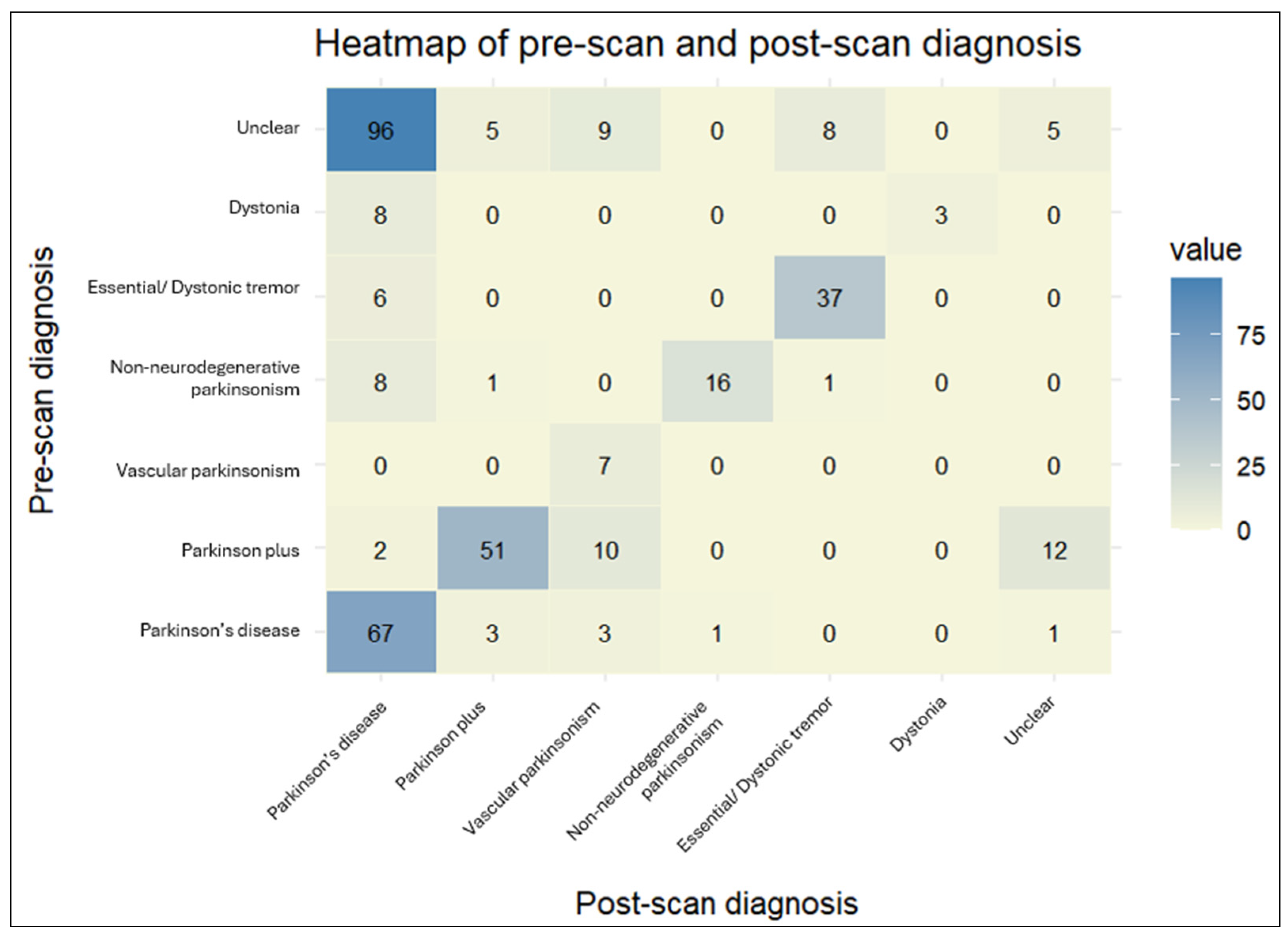
3. Results
4. Discussion
5. Conclusions
Supplementary Materials
Author Contributions
Funding
Institutional Review Board Statement
Informed Consent Statement
Data Availability Statement
Conflicts of Interest
Abbreviations
| DaTscan | Dopamine transporter scan |
| PD | Parkinson’s disease |
| CT | Computed tomography |
| MRI | Magnetic resonance imaging |
| SPECT | Single photon emission computed tomography |
| IV | Intravenous |
| EANM | European Association of Nuclear Medicine |
| OR | Odds Ratio |
| OSEM | Ordered subset expectation maximization |
| R | Right |
| L | Left |
| ROI | Region of interest |
| y | Years |
| n | Sample size |
| ET | Essential tremor |
| MSA | Multiple system atrophy |
| MIBG | I-123 meta-iodobenzylguanidine |
| IBZM | I-123 iodobenzamide |
| CBD | Corticobasal degeneration |
| PSP | Progressive supranuclear palsy |
References
- Bega, D.; Kuo, P.H.; Chalkidou, A.; Grzeda, M.T.; Macmillan, T.; Brand, C.; Sheikh, Z.H.; Antonini, A. Clinical utility of DaTscan in patients with suspected Parkinsonian syndrome: A systematic review and meta-analysis. NPJ Park. Dis. 2021, 7, 43. [Google Scholar] [CrossRef] [PubMed]
- Abdo, W.F.; van de Warrenburg, B.P.; Burn, D.J.; Quinn, N.P.; Bloem, B.R. The clinical approach to movement disorders. Nat. Rev. Neurol. 2010, 6, 29–37. [Google Scholar] [CrossRef] [PubMed]
- Booij, J.; Tissingh, G.; Boer, G.J.; Speelman, J.D.; Stoof, J.C.; Janssen, A.G.; Wolters, E.C.; van Royen, E.A. [123I]FP-CIT SPECT shows a pronounced decline of striatal dopamine transporter labelling in early and advanced Parkinson’s disease. J. Neurol. Neurosurg. Psychiatry 1997, 62, 133–140. [Google Scholar] [CrossRef]
- Jennings, D.; Siderowf, A.; Stern, M.; Seibyl, J.; Eberly, S.; Oakes, D.; Marek, K. Conversion to Parkinson Disease in the PARS Hyposmic and Dopamine Transporter-Deficit Prodromal Cohort. JAMA Neurol. 2017, 74, 933–940. [Google Scholar] [CrossRef]
- Pirker, W.; Djamshidian, S.; Asenbaum, S.; Gerschlager, W.; Tribl, G.; Hoffmann, M.; Brücke, T. Progression of dopaminergic degeneration in Parkinson’s disease and atypical parkinsonism: A longitudinal β-CIT SPECT study. Mov. Disord. 2002, 17, 45–53. [Google Scholar] [CrossRef]
- Quintas, S.; Sanles-Falagan, R.; Berbís, M. I(123)-FP-CIT (DaTSCAN) SPECT beyond the Most Common Causes of Parkinsonism: A Systematic Review. Mov. Disord. Clin. Pract. 2024, 11, 613–625. [Google Scholar] [CrossRef]
- Nabizadeh, F.; Pirahesh, K.; Ramezannezhad, E. Longitudinal striatal dopamine transporter binding and cerebrospinal fluid alpha-synuclein, amyloid beta, total tau, and phosphorylated tau in Parkinson’s disease. Neurol. Sci. 2023, 44, 573–585. [Google Scholar] [CrossRef] [PubMed]
- Yaghoobi, A.; Seyedmirzaei, H.; Ala, M. Genome- and Exome-Wide Association Studies Revealed Candidate Genes Associated with DaTscan Imaging Features. Park. Dis. 2023, 2023, 2893662. [Google Scholar] [CrossRef]
- Höglinger, G.U.; Adler, C.H.; Berg, D.; Klein, C.; Outeiro, T.F.; Poewe, W.; Postuma, R.; Stoessl, A.J.; Lang, A.E. A biological classification of Parkinson’s disease: The SynNeurGe research diagnostic criteria. Lancet Neurol. 2024, 23, 191–204. [Google Scholar] [CrossRef]
- Darcourt, J.; Booij, J.; Tatsch, K.; Varrone, A.; Vander Borght, T.; Kapucu, O.L.; Någren, K.; Nobili, F.; Walker, Z.; Van Laere, K. EANM procedure guidelines for brain neurotransmission SPECT using (123)I-labelled dopamine transporter ligands, version 2. Eur. J. Nucl. Med. Mol. Imaging 2010, 37, 443–450. [Google Scholar] [CrossRef]
- Morbelli, S.; Esposito, G.; Arbizu, J.; Barthel, H.; Boellaard, R.; Bohnen, N.I.; Brooks, D.J.; Darcourt, J.; Dickson, J.C.; Douglas, D.; et al. EANM practice guideline/SNMMI procedure standard for dopaminergic imaging in Parkinsonian syndromes 1.0. Eur. J. Nucl. Med. Mol. Imaging 2020, 47, 1885–1912. [Google Scholar] [CrossRef] [PubMed]
- R_Core_Team. R: A Language and Environment for Statistical Computing; R Foundation for Statistical Computing: Vienna, Austria, 2024. [Google Scholar]
- RStudio_Team. RStudio: Integrated Development for R; RStudio, PBC: Boston, MA, USA, 2020. [Google Scholar]
- Oh, M.; Oh, S.J.; Lee, S.J.; Oh, J.S.; Chung, S.J.; Kim, J.S. Diagnostic accuracy of (18)F-FP-CIT PET for clinically uncertain Parkinsonian syndrome. Sci. Rep. 2023, 13, 15069. [Google Scholar] [CrossRef]
- Constantinides, V.C.; Souvatzoglou, M.; Paraskevas, G.P.; Chalioti, M.; Stefanis, L.; Kapaki, E. Dopamine transporter SPECT imaging in Parkinson’s disease and atypical Parkinsonism: A study of 137 patients. Neurol. Sci. 2023, 44, 1613–1623. [Google Scholar] [CrossRef]
- Gerasimou, G.; Costa, D.C.; Papanastasiou, E.; Bostanjiopoulou, S.; Arnaoutoglou, M.; Moralidis, E.; Aggelopoulou, T.; Gotzamani-Psarrakou, A. SPECT study with I-123-Ioflupane (DaTSCAN) in patients with essential tremor. Is there any correlation with Parkinson’s disease? Ann. Nucl. Med. 2012, 26, 337–344. [Google Scholar] [CrossRef]
- Stathaki, M.; Koukouraki, S.; Simos, P.; Boura, I.; Papadaki, E.; Bourogianni, O.; Tsaroucha, A.; Kapsoritakis, N.; Mitsias, P.; Spanaki, C. Is There Any Clinical Value of Adding 123I-Metaiodobenzylguanidine Myocardial Scintigraphy to 123I-Ioflupane (DaTscan) in the Differential Diagnosis of Parkinsonism? Clin. Nucl. Med. 2020, 45, 588–593. [Google Scholar] [CrossRef]
- Louis, E.D.; Ferreira, J.J. How common is the most common adult movement disorder? Update on the worldwide prevalence of essential tremor. Mov. Disord. 2010, 25, 534–541. [Google Scholar] [CrossRef] [PubMed]
- Lees, A.J.; Hardy, J.; Revesz, T. Parkinson’s disease. Lancet 2009, 373, 2055–2066. [Google Scholar] [CrossRef] [PubMed]
- Louis, E.D.; Bares, M.; Benito-Leon, J.; Fahn, S.; Frucht, S.J.; Jankovic, J.; Ondo, W.G.; Pal, P.K.; Tan, E.K. Essential tremor-plus: A controversial new concept. Lancet Neurol. 2020, 19, 266–270. [Google Scholar] [CrossRef]
- Hopfner, F.; Deuschl, G. Managing Essential Tremor. Neurotherapeutics 2020, 17, 1603–1621. [Google Scholar] [CrossRef]
- Marshall, V.L.; Patterson, J.; Hadley, D.M.; Grosset, K.A.; Grosset, D.G. Two-year follow-up in 150 consecutive cases with normal dopamine transporter imaging. Nucl. Med. Commun. 2006, 27, 933–937. [Google Scholar] [CrossRef]
- Mirpour, S.; Turkbey, E.B.; Marashdeh, W.; El Khouli, R.; Subramaniam, R.M. Impact of DAT-SPECT on Management of Patients Suspected of Parkinsonism. Clin. Nucl. Med. 2018, 43, 710–714. [Google Scholar] [CrossRef] [PubMed]
- Marcucci, L.; Reggiani, C. Increase of resting muscle stiffness, a less considered component of age-related skeletal muscle impairment. Eur. J. Transl. Myol. 2020, 30, 8982. [Google Scholar] [CrossRef]
- Korczyn, A.D. Vascular parkinsonism—Characteristics, pathogenesis and treatment. Nat. Rev. Neurol. 2015, 11, 319–326. [Google Scholar] [CrossRef]
- Ma, K.K.Y.; Lin, S.; Mok, V.C.T. Neuroimaging in Vascular Parkinsonism. Curr. Neurol. Neurosci. Rep. 2019, 19, 102. [Google Scholar] [CrossRef] [PubMed]
- Contrafatto, D.; Mostile, G.; Nicoletti, A.; Dibilio, V.; Raciti, L.; Lanzafame, S.; Luca, A.; Distefano, A.; Zappia, M. [123I]FP-CIT-SPECT asymmetry index to differentiate Parkinson’s disease from vascular parkinsonism. Acta Neurol. Scand. 2012, 126, 12–16. [Google Scholar] [CrossRef]
- Antonini, A.; Vitale, C.; Barone, P.; Cilia, R.; Righini, A.; Bonuccelli, U.; Abbruzzese, G.; Ramat, S.; Petrone, A.; Quatrale, R.; et al. The relationship between cerebral vascular disease and parkinsonism: The VADO study. Park. Relat. Disord. 2012, 18, 775–780. [Google Scholar] [CrossRef]
- Hastings, A.; Cullinane, P.; Wrigley, S.; Revesz, T.; Morris, H.R.; Dickson, J.C.; Jaunmuktane, Z.; Warner, T.T.; De Pablo-Fernández, E. Neuropathologic Validation and Diagnostic Accuracy of Presynaptic Dopaminergic Imaging in the Diagnosis of Parkinsonism. Neurology 2024, 102, e209453. [Google Scholar] [CrossRef] [PubMed]
- Rizzo, G.; Copetti, M.; Arcuti, S.; Martino, D.; Fontana, A.; Logroscino, G. Accuracy of clinical diagnosis of Parkinson disease: A systematic review and meta-analysis. Neurology 2016, 86, 566–576. [Google Scholar] [CrossRef]
- Thiriez, C.; Itti, E.; Fénelon, G.; Evangelista, E.; Meignan, M.; Cesaro, P.; Remy, P. Clinical routine use of dopamine transporter imaging in 516 consecutive patients. J. Neurol. 2015, 262, 909–915. [Google Scholar] [CrossRef]
- Houot, M.; Arnaud, S.; Mongin, M.; Pop, G.; Soussan, M.; Lannuzel, A.; Degos, B. Relevance of (123)I-FP-CIT SPECT prescriptions for the diagnosis of parkinsonian syndromes. Sci. Rep. 2024, 14, 25088. [Google Scholar] [CrossRef]
- Amlang, C.J.; Trujillo Diaz, D.; Louis, E.D. Essential Tremor as a “Waste Basket” Diagnosis: Diagnosing Essential Tremor Remains a Challenge. Front. Neurol. 2020, 11, 172. [Google Scholar] [CrossRef] [PubMed]
- Jain, S.; Lo, S.E.; Louis, E.D. Common misdiagnosis of a common neurological disorder: How are we misdiagnosing essential tremor? Arch. Neurol. 2006, 63, 1100–1104. [Google Scholar] [CrossRef]
- Palermo, G.; Giannoni, S.; Bellini, G.; Siciliano, G.; Ceravolo, R. Dopamine Transporter Imaging, Current Status of a Potential Biomarker: A Comprehensive Review. Int. J. Mol. Sci. 2021, 22, 11234. [Google Scholar] [CrossRef] [PubMed]
- Yamamoto, T.; Asahina, M.; Yamanaka, Y.; Uchiyama, T.; Hirano, S.; Fuse, M.; Koga, Y.; Sakakibara, R.; Kuwabara, S. Postvoid residual predicts the diagnosis of multiple system atrophy in Parkinsonian syndrome. J. Neurol. Sci. 2017, 381, 230–234. [Google Scholar] [CrossRef] [PubMed]
- Erro, R.; Lazzeri, G.; Terranova, C.; Paparella, G.; Gigante, A.F.; De Micco, R.; Magistrelli, L.; Di Biasio, F.; Valentino, F.; Moschella, V.; et al. Comparing Essential Tremor with and without Soft Dystonic Signs and Tremor Combined with Dystonia: The TITAN Study. Mov. Disord. Clin. Pract. 2024, 11, 645–654. [Google Scholar] [CrossRef]
- Shetty, A.S.; Bhatia, K.P.; Lang, A.E. Dystonia and Parkinson’s disease: What is the relationship? Neurobiol. Dis. 2019, 132, 104462. [Google Scholar] [CrossRef]
- Piatkova, Y.; Doyen, M.; Heyer, S.; Tahmazov, A.; Frismand, S.; Hopes, L.; Imbert, L.; Verger, A. Effects of medication on dopamine transporter imaging using [(123)I]I-FP-CIT SPECT in routine practice. Eur. J. Nucl. Med. Mol. Imaging 2024, 51, 1323–1332. [Google Scholar] [CrossRef]
- Jenkins, P.O.; Roussakis, A.A.; De Simoni, S.; Bourke, N.; Fleminger, J.; Cole, J.; Piccini, P.; Sharp, D. Distinct dopaminergic abnormalities in traumatic brain injury and Parkinson’s disease. J. Neurol. Neurosurg. Psychiatry 2020, 91, 631–637. [Google Scholar] [CrossRef]
- Palermo, G.; Giannoni, S.; Depalo, T.; Frosini, D.; Volterrani, D.; Siciliano, G.; Bonuccelli, U.; Ceravolo, R. Negative DAT-SPECT in Old Onset Parkinson’s Disease: An Additional Pitfall? Mov. Disord. Clin. Pract. 2022, 9, 530–534. [Google Scholar] [CrossRef]
- Batla, A.; Erro, R.; Stamelou, M.; Schneider, S.A.; Schwingenschuh, P.; Ganos, C.; Bhatia, K.P. Patients with scans without evidence of dopaminergic deficit: A long-term follow-up study. Mov. Disord. 2014, 29, 1820–1825. [Google Scholar] [CrossRef]
- Sasaki, S. Long-term follow-up study of SWEDD patients with mild parkinsonian signs. BMJ Neurol. Open 2024, 6, e000600. [Google Scholar] [CrossRef] [PubMed]
- Marek, K.; Seibyl, J.; Eberly, S.; Oakes, D.; Shoulson, I.; Lang, A.E.; Hyson, C.; Jennings, D. Longitudinal follow-up of SWEDD subjects in the PRECEPT Study. Neurology 2014, 82, 1791–1797. [Google Scholar] [CrossRef]
- Camm, A.J.; Keith, A.A.F. Strengths and weaknesses of ‘real-world’ studies involving non-vitamin K antagonist oral anticoagulants. Open Heart 2018, 5, e000788. [Google Scholar] [CrossRef] [PubMed]
- Nicastro, N.; Nencha, U.; Burkhard, P.R.; Garibotto, V. Dopaminergic imaging in degenerative parkinsonisms, an established clinical diagnostic tool. J. Neurochem. 2023, 164, 346–363. [Google Scholar] [CrossRef] [PubMed]
- Louis, E.D.; Berry, D.; Ghanem, A.; Cosentino, S.A. Conversion Rate of Essential Tremor to Essential Tremor Parkinson Disease: Data From a Prospective Longitudinal Study. Neurol. Clin. Pract. 2023, 13, e200162. [Google Scholar] [CrossRef]
- Kang, U.J.; Xie, T. Diagnostic biomarkers of Parkinson’s disease: What gain at what cost? J. Neurol. Neurosurg. Psychiatry 2012, 83, 769. [Google Scholar] [CrossRef]
- Antonini, A.; Berto, P.; Lopatriello, S.; Tamma, F.; Annemans, L.; Chambers, M. Cost-effectiveness of 123I-FP-CIT SPECT in the differential diagnosis of essential tremor and Parkinson’s disease in Italy. Mov. Disord. 2008, 23, 2202–2209. [Google Scholar] [CrossRef]

| Age at Symptom Onset (y) | Time to DaTscan Referral (After Symptom Onset) (y) | |
|---|---|---|
| Overall (n = 360) | 56.9 ± 10.4 | 1.5 ± 1.1 |
| Primary referral symptom: | ||
| 49.7 ± 11.9 | 2.1 ± 2.4 |
| 63.1 ± 9.8 | 1.6 ± 1.1 |
| 71.3 ± 8.0 | 1.8 ± 3.1 |
| 60.6 ± 11.8 | 1.8 ± 1.1 |
| 39.1 ± 10.8 | 5.1 ± 7.5 ** |
| Pre-scan diagnosis | ||
| 62.5 ± 9.1 | 1.7 ± 1.3 |
| 70.1 ± 8.6 | 1.8 ± 3.1 |
| 74.1 ± 7.3 | 2.0 ± 2.3 |
| 56.8 ± 11.8 | 2.0 ± 0.9 |
| 40.3 ± 11.3 | 3.8 ± 3.7 ** |
| 38.6 ± 11.5 | 5.4 ± 8.3 ** |
| 54.7 ± 10.9 | 1.5 ± 1.0 |
| Change in diagnosis | ||
| 57.3 ± 12.0 | 1.4 ± 0.9 *** |
| 58.6 ± 15.7 | 2.5 ± 3.5 *** |
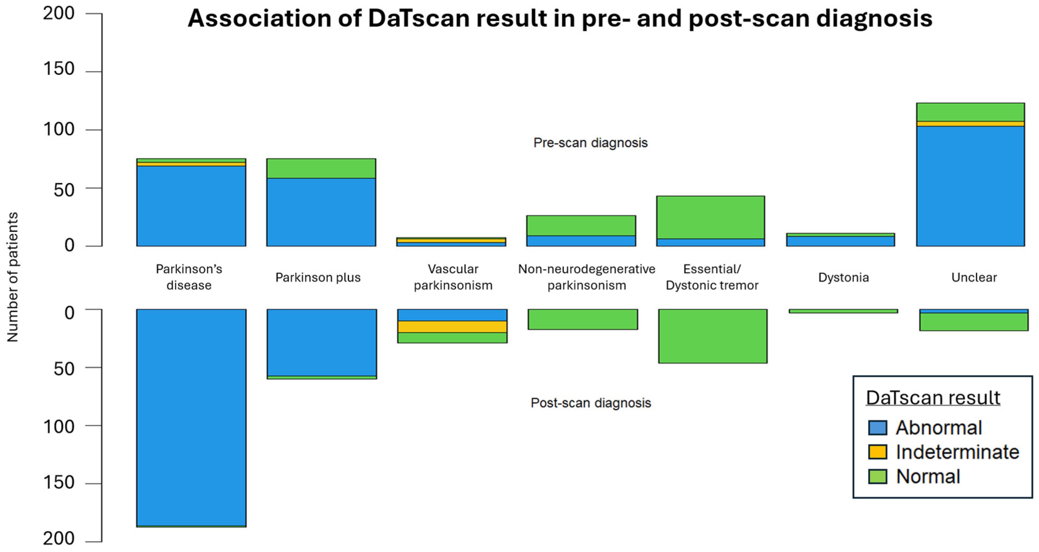
| Sensitivity | 92.7% |
| Specificity | 95.6% |
| Positive Predictive Value | 98.7% |
| Negative Predictive Value | 77.6% |
| False Positive Rate | 4.3% |
| False Negative Rate | 7.2% |
| Accuracy | 93.3% |
Disclaimer/Publisher’s Note: The statements, opinions and data contained in all publications are solely those of the individual author(s) and contributor(s) and not of MDPI and/or the editor(s). MDPI and/or the editor(s) disclaim responsibility for any injury to people or property resulting from any ideas, methods, instructions or products referred to in the content. |
© 2025 by the authors. Licensee MDPI, Basel, Switzerland. This article is an open access article distributed under the terms and conditions of the Creative Commons Attribution (CC BY) license (https://creativecommons.org/licenses/by/4.0/).
Share and Cite
Xiromerisiou, G.; Boura, I.; Barmpounaki, E.; Georgoulias, P.; Dardiotis, E.; Spanaki, C.; Valotassiou, V. The Utilization and Impact of Dopamine Transporter Imaging in Diagnosing Movement Disorders at a Tertiary Care Hospital in Greece. Biomedicines 2025, 13, 970. https://doi.org/10.3390/biomedicines13040970
Xiromerisiou G, Boura I, Barmpounaki E, Georgoulias P, Dardiotis E, Spanaki C, Valotassiou V. The Utilization and Impact of Dopamine Transporter Imaging in Diagnosing Movement Disorders at a Tertiary Care Hospital in Greece. Biomedicines. 2025; 13(4):970. https://doi.org/10.3390/biomedicines13040970
Chicago/Turabian StyleXiromerisiou, Georgia, Iro Boura, Eleni Barmpounaki, Panagiotis Georgoulias, Efthimios Dardiotis, Cleanthe Spanaki, and Varvara Valotassiou. 2025. "The Utilization and Impact of Dopamine Transporter Imaging in Diagnosing Movement Disorders at a Tertiary Care Hospital in Greece" Biomedicines 13, no. 4: 970. https://doi.org/10.3390/biomedicines13040970
APA StyleXiromerisiou, G., Boura, I., Barmpounaki, E., Georgoulias, P., Dardiotis, E., Spanaki, C., & Valotassiou, V. (2025). The Utilization and Impact of Dopamine Transporter Imaging in Diagnosing Movement Disorders at a Tertiary Care Hospital in Greece. Biomedicines, 13(4), 970. https://doi.org/10.3390/biomedicines13040970