The Effect of Naturally Acquired Immunity on Mortality Predictors: A Focus on Individuals with New Coronavirus
Abstract
1. Introduction
2. Materials and Methods
2.1. Data Source and Description
2.2. Machine Learning Methods
3. Results
4. Discussion
5. Conclusions
Author Contributions
Funding
Institutional Review Board Statement
Informed Consent Statement
Data Availability Statement
Acknowledgments
Conflicts of Interest
Abbreviations
| COVID-19 | Coronavirus Disease 19 |
| ICU | Intensive Care Unit |
| PCR | Polymerase Chain Reaction |
| AI | Artificial Intelligence |
| ML | Machine learning |
| RF | Random Forest |
| CURB | Confusion, Urea, Respiratory rate, Blood pressure |
| SOFA | Sequential Organ Failure Assessment |
| APACHE-II | Acute Physiology and Chronic Health Evaluation |
| MCV | Mean Corpuscular Volume |
| PT | Prothrombin time |
| INR | International normalized Ratio |
| aPTT | Activated Partial Thromboplastin Time |
| CKD-EPI | Chronic Kidney Disease Epidemiology Collaboration |
| ALT/GPT | Alanine Aminotransferase/Glutamate Pyruvate Transaminase |
| AST/GOT | Aspartate Aminotransferase/Glutamate Oxaloacetate Transaminase |
| GGT | Gamma-Glutamyl Transferase |
| LDH | Lactate Dehydrogenase |
| ROC | Receiver Operating Characteristic |
| AUC | Area Under the Curve |
| GNB | Gaussian Naïve Bayes |
| KNN | K-Nearest Neighbors |
| BLDA | Bayesian Linear Discriminant Analysis |
| SMV | Support Vector Machine |
| DT | Decision Tree |
| COPD | Chronic Obstructive Pulmonary Disease |
| IMV | Invasive Mechanical Ventilation |
| HFNC | High-Flow Nasal Cannula |
| NIV | Non-Invasive Mechanical Ventilation |
| ECMO | Extracorporeal Membrane Oxygenation |
| ARDS | Acute Respiratory Distress Syndrome |
| SD | Standard Deviation |
| CRP | C-Reactive Protein |
| SBP | Systolic Blood Pressure |
| DBP | Diastolic Blood Pressure |
| MCC | Mathew’s Correlation Coefficient |
| DYI | Degenerated Youden’s Index |
References
- Suryawanshi, Y.N.; Biswas, D.A. Herd Immunity to Fight Against COVID-19: A Narrative Review. Cureus 2023, 15. [Google Scholar] [CrossRef]
- Clemente-Suárez, V.J.; Hormeño-Holgado, A.; Jiménez, M.; Benitez-Agudelo, J.C.; Navarro-Jiménez, E.; Perez-Palencia, N.; Maestre-Serrano, R.; Laborde-Cárdenas, C.C.; Tornero-Aguilera, J.F. Dynamics of Population Immunity Due to the Herd Effect in the COVID-19 Pandemic. Vaccines 2020, 8, 236. [Google Scholar] [CrossRef] [PubMed]
- Fine, P.; Eames, K.; Heymann, D.L. “Herd Immunity”: A Rough Guide. Clin. Infect. Dis. 2011, 52, 911–916. [Google Scholar] [CrossRef] [PubMed]
- Hussain, A.; Yang, H.; Zhang, M.; Liu, Q.; Alotaibi, G.; Irfan, M.; He, H.; Chang, J.; Liang, X.-J.; Weng, Y.; et al. mRNA Vaccines for COVID-19 and Diverse Diseases. J. Control. Release 2022, 345, 314–333. [Google Scholar] [CrossRef]
- Zhang, M.; Hussain, A.; Yang, H.; Zhang, J.; Liang, X.-J.; Huang, Y. mRNA-Based Modalities for Infectious Disease Management. Nano Res. 2023, 16, 672–691. [Google Scholar] [CrossRef]
- Omer, S.B.; Yildirim, I.; Forman, H.P. Herd Immunity and Implications for SARS-CoV-2 Control. JAMA 2020, 324, 2095. [Google Scholar] [CrossRef]
- Mathieu, E.; Ritchie, H.; Rodés-Guirao, L.; Appel, C.; Gavrilov, D.; Giattino, C.; Hasell, J.; Macdonald, B.; Dattani, S.; Beltekian, D.; et al. Total COVID-19 Vaccine Doses Administered. In COVID-19 Pandemic; Data Adapted from Official Data Collated by Our World in Data; World Health Organization: Geneva, Switzerland, 2020. [Google Scholar]
- Ruan, Q.; Yang, K.; Wang, W.; Jiang, L.; Song, J. Clinical Predictors of Mortality Due to COVID-19 Based on an Analysis of Data of 150 Patients from Wuhan, China. Intensive Care Med. 2020, 46, 846–848. [Google Scholar] [CrossRef]
- Fajgenbaum, D.C.; June, C.H. Cytokine Storm. N. Engl. J. Med. 2020, 383, 2255–2273. [Google Scholar] [CrossRef]
- Iftimie, S.; López-Azcona, A.F.; Vallverdú, I.; Hernández-Flix, S.; De Febrer, G.; Parra, S.; Hernández-Aguilera, A.; Riu, F.; Joven, J.; Andreychuk, N.; et al. First and Second Waves of Coronavirus Disease-19: A Comparative Study in Hospitalized Patients in Reus, Spain. PLoS ONE 2021, 16, e0248029. [Google Scholar] [CrossRef]
- Chen, N.; Zhou, M.; Dong, X.; Qu, J.; Gong, F.; Han, Y.; Qiu, Y.; Wang, J.; Liu, Y.; Wei, Y.; et al. Epidemiological and Clinical Characteristics of 99 Cases of 2019 Novel Coronavirus Pneumonia in Wuhan, China: A Descriptive Study. Lancet 2020, 395, 507–513. [Google Scholar] [CrossRef]
- Soriano, V.; Ganado-Pinilla, P.; Sanchez-Santos, M.; Gómez-Gallego, F.; Barreiro, P.; De Mendoza, C.; Corral, O. Main Differences between the First and Second Waves of COVID-19 in Madrid, Spain. Int. J. Infect. Dis. 2021, 105, 374–376. [Google Scholar] [CrossRef]
- Buttenschøn, H.N.; Lynggaard, V.; Sandbøl, S.G.; Glassou, E.N.; Haagerup, A. Comparison of the Clinical Presentation across Two Waves of COVID-19: A Retrospective Cohort Study. BMC Infect. Dis. 2022, 22, 423. [Google Scholar] [CrossRef] [PubMed]
- The MO-COVID19 Working Group; Coloretti, I.; Farinelli, C.; Biagioni, E.; Gatto, I.; Munari, E.; Dall’Ara, L.; Busani, S.; Meschiari, M.; Tonelli, R.; et al. Critical COVID-19 Patients through First, Second, and Third Wave: Retrospective Observational Study Comparing Outcomes in Intensive Care Unit. J. Thorac. Dis. 2023, 15, 3218–3227. [Google Scholar] [CrossRef] [PubMed]
- San Martín-López, J.V.; Mesa, N.; Bernal-Bello, D.; Morales-Ortega, A.; Rivilla, M.; Guerrero, M.; Calderón, R.; Farfán, A.I.; Rivas, L.; Soria, G.; et al. Seven Epidemic Waves of COVID-19 in a Hospital in Madrid: Analysis of Severity and Associated Factors. Viruses 2023, 15, 1839. [Google Scholar] [CrossRef]
- Wu, Z.; Harrich, D.; Li, Z.; Hu, D.; Li, D. The Unique Features of SARS-CoV-2 Transmission: Comparison with SARS-CoV, MERS-CoV and 2009 H1N1 Pandemic Influenza Virus. Rev. Med. Virol. 2021, 31, e2171. [Google Scholar] [CrossRef]
- Zhou, H.; Yang, J.; Zhou, C.; Chen, B.; Fang, H.; Chen, S.; Zhang, X.; Wang, L.; Zhang, L. A Review of SARS-CoV2: Compared with SARS-CoV and MERS-CoV. Front. Med. 2021, 8, 628370. [Google Scholar] [CrossRef]
- Han, J.; Kamber, M.; Pei, J. Data Mining: Concepts and Techniques; Morgan Kaufmann Publishers: Burlington, VT, USA, 2022. [Google Scholar]
- Hesami, M.; Alizadeh, M.; Jones, A.M.P.; Torkamaneh, D. Machine Learning: Its Challenges and Opportunities in Plant System Biology. Appl. Microbiol. Biotechnol. 2022, 106, 3507–3530. [Google Scholar] [CrossRef]
- Haug, C.J.; Drazen, J.M. Artificial Intelligence and Machine Learning in Clinical Medicine, 2023. N. Engl. J. Med. 2023, 388, 1201–1208. [Google Scholar] [CrossRef] [PubMed]
- Wu, H.-T.; Liao, C.-C.; Peng, C.-F.; Lee, T.-Y.; Liao, P.-H. Exploring the Application of Machine Learning to Identify the Correlations between Phthalate Esters and Disease: Enhancing Nursing Assessments. Health Inf. Sci. Syst. 2024, 13, 10. [Google Scholar] [CrossRef]
- Rueda, R.; Fabello, E.; Silva, T.; Genzor, S.; Mizera, J.; Stanke, L. Machine Learning Approach to Flare-up Detection and Clustering in Chronic Obstructive Pulmonary Disease (COPD) Patients. Health Inf. Sci. Syst. 2024, 12, 50. [Google Scholar] [CrossRef]
- Barough, S.S.; Safavi-Naini, S.A.A.; Siavoshi, F.; Tamimi, A.; Ilkhani, S.; Akbari, S.; Ezzati, S.; Hatamabadi, H.; Pourhoseingholi, M.A. Generalizable Machine Learning Approach for COVID-19 Mortality Risk Prediction Using on-Admission Clinical and Laboratory Features. Sci. Rep. 2023, 13, 2399. [Google Scholar] [CrossRef]
- Kourmpanis, N.; Liaskos, J.; Zoulias, E.; Mantas, J. Predicting Mortality in COVID-19 Patients Using 6 Machine Learning Algorithms. In Studies in Health Technology and Informatics; Mantas, J., Gallos, P., Zoulias, E., Hasman, A., Househ, M.S., Charalampidou, M., Magdalinou, A., Eds.; IOS Press: Nieuwe Hemweg, Amsterdam, The Netherlands, 2023; ISBN 978-1-64368-400-0. [Google Scholar]
- Alballa, N.; Al-Turaiki, I. Machine Learning Approaches in COVID-19 Diagnosis, Mortality, and Severity Risk Prediction: A Review. Inform. Med. Unlocked 2021, 24, 100564. [Google Scholar] [CrossRef]
- Liu, L.; Song, W.; Patil, N.; Sainlaire, M.; Jasuja, R.; Dykes, P.C. Predicting COVID-19 Severity: Challenges in Reproducibility and Deployment of Machine Learning Methods. Int. J. Med. Inform. 2023, 179, 105210. [Google Scholar] [CrossRef]
- Sharifi-Kia, A.; Nahvijou, A.; Sheikhtaheri, A. Machine Learning-Based Mortality Prediction Models for Smoker COVID-19 Patients. BMC Med. Inform. Decis. Mak. 2023, 23, 129. [Google Scholar] [CrossRef]
- Navlakha, S.; Morjaria, S.; Perez-Johnston, R.; Zhang, A.; Taur, Y. Projecting COVID-19 Disease Severity in Cancer Patients Using Purposefully-Designed Machine Learning. BMC Infect. Dis. 2021, 21, 391. [Google Scholar] [CrossRef]
- Jung, Y.J.; Ahn, J.; Park, S.; Sun, J.-M.; Lee, S.-H.; Ahn, J.S.; Ahn, M.-J.; Cho, S.Y.; Jung, H.A. Machine Learning Prediction of the Case-Fatality of COVID-19 and Risk Factors for Adverse Outcomes in Patients with Non-Small Cell Lung Cancer. Transl. Cancer Res. 2024, 13, 2587–2595. [Google Scholar] [CrossRef]
- Subramanian, D.; Vittala, A.; Chen, X.; Julien, C.; Acosta, S.; Rusin, C.; Allen, C.; Rider, N.; Starosolski, Z.; Annapragada, A.; et al. Stratification of Pediatric COVID-19 Cases Using Inflammatory Biomarker Profiling and Machine Learning. J. Clin. Med. 2023, 12, 5435. [Google Scholar] [CrossRef]
- Cerbulescu, T.; Ignuta, F.; Rayudu, U.S.; Afra, M.; Rosca, O.; Vlad, A.; Loredana, S. Inflammatory Markers and Severity in COVID-19 Patients with Clostridioides Difficile Co-Infection: A Retrospective Analysis Including Subgroups with Diabetes, Cancer, and Elderly. Biomedicines 2025, 13, 227. [Google Scholar] [CrossRef]
- Schonlau, M.; Zou, R.Y. The Random Forest Algorithm for Statistical Learning. Stata J. Promot. Commun. Stat. Stata 2020, 20, 3–29. [Google Scholar] [CrossRef]
- Rhodes, J.S.; Cutler, A.; Moon, K.R. Geometry- and Accuracy-Preserving Random Forest Proximities. IEEE Trans. Pattern Anal. Mach. Intell. 2023, 45, 10947–10959. [Google Scholar] [CrossRef]
- Jayachitra, S.; Prasanth, A. Multi-Feature Analysis for Automated Brain Stroke Classification Using Weighted Gaussian Naïve Bayes Classifier. J. Circuit syst. Comp. 2021, 30, 2150178. [Google Scholar] [CrossRef]
- Balaji, V.R.; Suganthi, S.T.; Rajadevi, R.; Krishna Kumar, V.; Saravana Balaji, B.; Pandiyan, S. Skin Disease Detection and Segmentation Using Dynamic Graph Cut Algorithm and Classification through Naive Bayes Classifier. Measurement 2020, 163, 107922. [Google Scholar] [CrossRef]
- Uddin, S.; Haque, I.; Lu, H.; Moni, M.A.; Gide, E. Comparative Performance Analysis of K-Nearest Neighbour (KNN) Algorithm and Its Different Variants for Disease Prediction. Sci. Rep. 2022, 12, 6256. [Google Scholar] [CrossRef]
- Zhang, S.; Li, X.; Zong, M.; Zhu, X.; Wang, R. Efficient kNN Classification with Different Numbers of Nearest Neighbors. IEEE Trans. Neural Netw. Learn. Syst. 2018, 29, 1774–1785. [Google Scholar] [CrossRef] [PubMed]
- Zhou, W.; Liu, Y.; Yuan, Q.; Li, X. Epileptic Seizure Detection Using Lacunarity and Bayesian Linear Discriminant Analysis in Intracranial EEG. IEEE Trans. Biomed. Eng. 2013, 60, 3375–3381. [Google Scholar] [CrossRef] [PubMed]
- Yuan, S.; Zhou, W.; Li, J.; Wu, Q. Sparse Representation-Based EMD and BLDA for Automatic Seizure Detection. Med. Biol. Eng. Comput. 2017, 55, 1227–1238. [Google Scholar] [CrossRef]
- Pérez, A.; Larrañaga, P.; Inza, I. Supervised Classification with Conditional Gaussian Networks: Increasing the Structure Complexity from Naive Bayes. Int. J. Approx. Reason. 2006, 43, 1–25. [Google Scholar] [CrossRef]
- Patel, H.H.; Prajapati, P. Study and Analysis of Decision Tree Based Classification Algorithms. Int. J. Comput. Sci. Eng. 2018, 6, 74–78. [Google Scholar] [CrossRef]
- Charbuty, B.; Abdulazeez, A. Classification Based on Decision Tree Algorithm for Machine Learning. J. Appl. Sci. Technol. Trends 2021, 2, 20–28. [Google Scholar] [CrossRef]
- Jiang, C.; Lv, W.; Li, J. Protein-Protein Interaction Sites Prediction Using Batch Normalization Based CNNs and Oversampling Method Borderline-SMOTE. IEEE/ACM Trans. Comput. Biol. Bioinf. 2023, 20, 2190–2199. [Google Scholar] [CrossRef]
- Alshammari, T.S. Applying Machine Learning Algorithms for the Classification of Sleep Disorders. IEEE Access 2024, 12, 36110–36121. [Google Scholar] [CrossRef]
- Andrew, M.K.; Godin, J.; LeBlanc, J.; Boivin, G.; Valiquette, L.; McGeer, A.; McElhaney, J.E.; Hatchette, T.F.; ElSherif, M.; MacKinnon-Cameron, D.; et al. Older Age and Frailty Are Associated with Higher Mortality but Lower ICU Admission with COVID-19. Can. Geriatr. J. 2022, 25, 183–196. [Google Scholar] [CrossRef] [PubMed]
- Conway, B.J.; Kim, J.W.; Brousseau, D.C.; Conroy, M. Heart Disease, Advanced Age, Minority Race, and Hispanic Ethnicity Are Associated with Mortality in COVID-19 Patients. WMJ Off. Publ. State Med. Soc. Wis. 2021, 120, 152–155. [Google Scholar]
- Petrosillo, N.; Viceconte, G.; Ergonul, O.; Ippolito, G.; Petersen, E. COVID-19, SARS and MERS: Are They Closely Related? Clin. Microbiol. Infect. 2020, 26, 729–734. [Google Scholar] [CrossRef]
- Phillips, S.P.; Carver, L.F. Greatest Risk Factor for Death from COVID-19: Older Age, Chronic Disease Burden, or Place of Residence? Descriptive Analysis of Population-Level Canadian Data. Int. J. Environ. Res. Public Health 2023, 20, 7181. [Google Scholar] [CrossRef]
- Satici, C.; Demirkol, M.A.; Sargin Altunok, E.; Gursoy, B.; Alkan, M.; Kamat, S.; Demirok, B.; Surmeli, C.D.; Calik, M.; Cavus, Z.; et al. Performance of Pneumonia Severity Index and CURB-65 in Predicting 30-Day Mortality in Patients with COVID-19. Int. J. Infect. Dis. 2020, 98, 84–89. [Google Scholar] [CrossRef] [PubMed]
- Bradley, J.; Sbaih, N.; Chandler, T.R.; Furmanek, S.; Ramirez, J.A.; Cavallazzi, R. Pneumonia Severity Index and CURB-65 Score Are Good Predictors of Mortality in Hospitalized Patients with SARS-CoV-2 Community-Acquired Pneumonia. Chest 2022, 161, 927–936. [Google Scholar] [CrossRef]
- Alegría-Baños, J.A.; Rosas-Alvarado, M.A.; Jiménez-López, J.C.; Juárez-Muciño, M.; Méndez-Celis, C.A.; Enríquez-De Los Santos, S.T.; Valdez-Vázquez, R.R.; Prada-Ortega, D. Sociodemographic, Clinical and Laboratory Characteristics and Risk Factors for Mortality of Hospitalized COVID-19 Patients at Alternate Care Site: A Latin American Experience. Ann. Med. 2023, 55, 2224049. [Google Scholar] [CrossRef] [PubMed]
- Ilg, A.; Moskowitz, A.; Konanki, V.; Patel, P.V.; Chase, M.; Grossestreuer, A.V.; Donnino, M.W. Performance of the CURB-65 Score in Predicting Critical Care Interventions in Patients Admitted with Community-Acquired Pneumonia. Ann. Emerg. Med. 2019, 74, 60–68. [Google Scholar] [CrossRef]
- Kaal, A.G.; Op De Hoek, L.; Hochheimer, D.T.; Brouwers, C.; Wiersinga, W.J.; Snijders, D.; Rensing, K.L.; Van Dijk, C.E.; Steyerberg, E.W.; Van Nieuwkoop, C. Outcomes of Community-Acquired Pneumonia Using the Pneumonia Severity Index versus the CURB-65 in Routine Practice of Emergency Departments. ERJ Open Res. 2023, 9, 00051–02023. [Google Scholar] [CrossRef]
- Mohammadi, Z.; Faghih Dinevari, M.; Vahed, N.; Ebrahimi Bakhtavar, H.; Rahmani, F. Clinical and Laboratory Predictors of COVID-19-Related In-Hospital Mortality; a Cross-Sectional Study of 1000 Cases. Arch. Acad. Emerg. Med. 2022, 10, e49. [Google Scholar] [CrossRef] [PubMed]
- Al-Shajlawi, M.; Alsayed, A.R.; Abazid, H.; Awajan, D.; Al-Imam, A.; Basheti, I. Using Laboratory Parameters as Predictors for the Severity and Mortality of COVID-19 in Hospitalized Patients. Pharm. Pract. 2022, 20, 2721. [Google Scholar] [CrossRef]
- Singh, S.; Singh, K. Blood Urea Nitrogen/Albumin Ratio and Mortality Risk in Patients with COVID-19. Indian J. Crit. Care Med. 2022, 26, 626–631. [Google Scholar] [CrossRef] [PubMed]
- Yüksel, E.; Arac, S.; Deniz Altintaş, D. Predictors of Mortality in COVID-19 Induced Acute Kidney Injury. Ther. Apher. Dial. 2022, 26, 897–907. [Google Scholar] [CrossRef] [PubMed]
- Qureshi, M.A.; Toori, K.U. Raja Mobeen Ahmed Predictors of Mortality in COVID-19 Patients: An Observational Study. Pak. J. Med. Sci. 2022, 39. [Google Scholar] [CrossRef]
- Muñoz Lezcano, S.; Armengol De La Hoz, M.Á.; Corbi, A.; López, F.; García, M.S.; Reiz, A.N.; González, T.F.; Zlatkov, V.Y. Predictors of Mechanical Ventilation and Mortality in Critically Ill Patients with COVID-19 Pneumonia. Med. Intensiv. (Engl. Ed.) 2023, S2173572723001303. [Google Scholar] [CrossRef]
- Arunachala, S.; Parthasarathi, A.; Basavaraj, C.K.; Kaleem Ullah, M.; Chandran, S.; Venkataraman, H.; Vishwanath, P.; Ganguly, K.; Upadhyay, S.; Mahesh, P.A. The Validity of the ROX Index and APACHE II in Predicting Early, Late, and Non-Responses to Non-Invasive Ventilation in Patients with COVID-19 in a Low-Resource Setting. Viruses 2023, 15, 2231. [Google Scholar] [CrossRef]
- Vandenbrande, J.; Verbrugge, L.; Bruckers, L.; Geebelen, L.; Geerts, E.; Callebaut, I.; Gruyters, I.; Heremans, L.; Dubois, J.; Stessel, B. Validation of the Acute Physiology and Chronic Health Evaluation (APACHE) II and IV Score in COVID-19 Patients. Crit. Care Res. Pract. 2021, 2021, 1–9. [Google Scholar] [CrossRef]
- Onuk, S.; Sipahioğlu, H.; Karahan, S.; Yeşiltepe, A.; Kuzugüden, S.; Karabulut, A.; Beştepe Dursun, Z.; Akın, A. Cytokine Levels and Severity of Illness Scoring Systems to Predict Mortality in COVID-19 Infection. Healthcare 2023, 11, 387. [Google Scholar] [CrossRef]
- He, J.; Tang, D.; Liu, D.; Hong, X.; Ma, C.; Zheng, F.; Zeng, Z.; Chen, Y.; Du, J.; Kang, L.; et al. Serum Proteome and Metabolome Uncover Novel Biomarkers for the Assessment of Disease Activity and Diagnosing of Systemic Lupus Erythematosus. Clin. Immunol. 2023, 251, 109330. [Google Scholar] [CrossRef]
- Huang, Y.; Ma, S.-F.; Oldham, J.M.; Adegunsoye, A.; Zhu, D.; Murray, S.; Kim, J.S.; Bonham, C.; Strickland, E.; Linderholm, A.L.; et al. Machine Learning of Plasma Proteomics Classifies Diagnosis of Interstitial Lung Disease. Am. J. Respir. Crit. Care Med. 2024, 210, 444–454. [Google Scholar] [CrossRef]
- Safia, S.N. Prediction of Breast Cancer Through Random Forest. Curr. Med. Imaging 2023, 19, e300922209414. [Google Scholar] [CrossRef]
- Sidiropoulos, T.; Dovrolis, N.; Katifelis, H.; Michalopoulos, N.V.; Kokoropoulos, P.; Arkadopoulos, N.; Gazouli, M. Dysbiosis Signature of Fecal Microbiota in Patients with Pancreatic Adenocarcinoma and Pancreatic Intraductal Papillary Mucinous Neoplasms. Biomedicines 2024, 12, 1040. [Google Scholar] [CrossRef] [PubMed]
- Founta, K.; Dafou, D.; Kanata, E.; Sklaviadis, T.; Zanos, T.P.; Gounaris, A.; Xanthopoulos, K. Gene Targeting in Amyotrophic Lateral Sclerosis Using Causality-Based Feature Selection and Machine Learning. Mol. Med. 2023, 29, 12. [Google Scholar] [CrossRef] [PubMed]
- Kelly, J.; Moyeed, R.; Carroll, C.; Luo, S.; Li, X. Blood Biomarker-Based Classification Study for Neurodegenerative Diseases. Sci. Rep. 2023, 13, 17191. [Google Scholar] [CrossRef]
- Khamis, F.; Memish, Z.; Bahrani, M.A.; Dowaiki, S.A.; Pandak, N.; Bolushi, Z.A.; Salmi, I.A.; Al-Zakwani, I. Prevalence and Predictors of In-Hospital Mortality of Patients Hospitalized with COVID-19 Infection. J. Infect. Public Health 2021, 14, 759–765. [Google Scholar] [CrossRef]
- Aly, M.H.; Rahman, S.S.; Ahmed, W.A.; Alghamedi, M.H.; Al Shehri, A.A.; Alkalkami, A.M.; Hassan, M.H. Indicators of Critical Illness and Predictors of Mortality in COVID-19 Patients. Infect. Drug Resist. 2020, 13, 1995–2000. [Google Scholar] [CrossRef]
- Tian, W.; Jiang, W.; Yao, J.; Nicholson, C.J.; Li, R.H.; Sigurslid, H.H.; Wooster, L.; Rotter, J.I.; Guo, X.; Malhotra, R. Predictors of Mortality in Hospitalized COVID-19 Patients: A Systematic Review and Meta-analysis. J. Med. Virol. 2020, 92, 1875–1883. [Google Scholar] [CrossRef]


| Deceased (40 Patients, 11%) | Alive (323 Patients, 89%) | Global (363 Patients, 100%) | ||||
|---|---|---|---|---|---|---|
| (n) | (%) | (n) | (%) | (n) | (%) | |
| Sex | ||||||
| Men | 33 | 83 | 198 | 61 | 231 | 64 |
| Women | 7 | 18 | 125 | 39 | 132 | 36 |
| Comorbidities | ||||||
| Hypertension | 24 | 60 | 144 | 45 | 168 | 46 |
| Diabetes mellitus | 10 | 25 | 68 | 21 | 78 | 21 |
| Ischemic cardiopathy | 7 | 18 | 17 | 5 | 24 | 7 |
| Cardiac insufficiency | 1 | 3 | 6 | 2 | 7 | 2 |
| COPD (Chronic emphysema/bronchitis) | 1 | 3 | 17 | 5 | 18 | 5 |
| Asthma | 1 | 3 | 17 | 5 | 18 | 5 |
| Dyslipidemia | 16 | 40 | 100 | 31 | 116 | 32 |
| Coagulopathies | 7 | 18 | 34 | 11 | 41 | 11 |
| Renal insufficiency | 5 | 13 | 19 | 6 | 24 | 7 |
| Active tumors | 1 | 3 | 16 | 5 | 17 | 5 |
| Immune-mediated diseases | 2 | 5 | 38 | 12 | 40 | 11 |
| Overweight | 1 | 3 | 4 | 1 | 5 | 1 |
| Obesity | 3 | 8 | 30 | 9 | 33 | 9 |
| Chronic treatments | 32 | 80 | 183 | 57 | 215 | 59 |
| Antihypertensives | 21 | 53 | 115 | 36 | 136 | 37 |
| Beta-blockers | 10 | 25 | 38 | 12 | 48 | 13 |
| Diuretics | 8 | 20 | 37 | 11 | 45 | 12 |
| Antidiabetics | 8 | 20 | 58 | 18 | 66 | 18 |
| Anti-aggregants | 11 | 28 | 33 | 10 | 44 | 12 |
| Anticoagulants | 6 | 15 | 34 | 11 | 40 | 11 |
| Lipid lowering agents | 15 | 38 | 90 | 28 | 105 | 29 |
| Chemotherapy | 1 | 3 | 2 | 1 | 3 | 1 |
| Immunosuppressive chronic treatment | 3 | 8 | 21 | 7 | 24 | 7 |
| Antiretrovirals | 0 | 0 | 1 | 0 | 1 | 0 |
| Antivirals | 0 | 0 | 1 | 0 | 1 | 0 |
| Vaccination status | ||||||
| 1 dose | 2 | 5 | 15 | 5 | 17 | 5 |
| 2 doses | 0 | 0 | 6 | 2 | 6 | 2 |
| 3 doses | 0 | 0 | 0 | 0 | 0 | 0 |
| 4 doses | 0 | 0 | 0 | 0 | 0 | 0 |
| Deceased (40 Patients, 11%) | Alive (323 Patients, 89%) | Global (363 Patients) | ||||
|---|---|---|---|---|---|---|
| (n) | (%) | (n) | (%) | (n) | (%) | |
| Symptoms on admission | ||||||
| Dyspnea | 19 | 48 | 172 | 53 | 191 | 53 |
| Chest discomfort | 1 | 3 | 44 | 14 | 45 | 12 |
| Cough | 21 | 53 | 202 | 63 | 223 | 61 |
| Rhinorrhea | 0 | 0 | 10 | 3 | 10 | 3 |
| Loss of smell (anosmia) | 0 | 0 | 49 | 15 | 49 | 13 |
| Loss of taste (ageusia) | 2 | 5 | 48 | 15 | 50 | 14 |
| Odynophagia | 0 | 0 | 12 | 4 | 12 | 3 |
| Myalgia | 4 | 10 | 57 | 18 | 61 | 17 |
| Fever | 26 | 65 | 225 | 70 | 251 | 69 |
| Dysthermia | 3 | 8 | 27 | 8 | 30 | 8 |
| Headache | 2 | 5 | 33 | 10 | 35 | 10 |
| Nausea/Vomiting | 5 | 13 | 47 | 15 | 52 | 14 |
| Diarrhea | 3 | 8 | 65 | 20 | 68 | 19 |
| Asthenia | 7 | 18 | 101 | 31 | 108 | 30 |
| Confusion | 9 | 23 | 8 | 2 | 17 | 5 |
| Dizziness | 2 | 5 | 19 | 6 | 21 | 6 |
| Sputum | 6 | 15 | 45 | 14 | 51 | 14 |
| Diagnosis on admission | ||||||
| Respiratory distress | 6 | 15 | 21 | 7 | 27 | 7 |
| Acute respiratory insufficiency | 20 | 50 | 127 | 39 | 147 | 40 |
| Multiorgan failure | 2 | 5 | 0 | 0 | 2 | 1 |
| Deceased (40 Patients, 11%) | Alive (323 Patients, 89%) | Global (363 Patients) | ||||
|---|---|---|---|---|---|---|
| (n) | (%) | (n) | (%) | (n) | (%) | |
| ICU admission | 21 | 53 | 61 | 19 | 82 | 23 |
| Invasive mechanical ventilation (IMV) | 19 | 48 | 56 | 17 | 75 | 21 |
| High Flow Nasal Cannula (HFNC) | 4 | 10 | 29 | 9 | 33 | 9 |
| Non-invasive mechanical ventilation (NIV) | 12 | 30 | 30 | 9 | 42 | 12 |
| Prone sessions | 12 | 30 | 28 | 9 | 40 | 11 |
| Extracorporeal membrane oxygenation (ECMO) | 1 | 3 | 4 | 1 | 5 | 1 |
| At least one previous episode of COVID | 1 | 3 | 2 | 1 | 3 | 1 |
| Nosocomial infections | ||||||
| Viral | 0 | 0 | 2 | 1 | 2 | 1 |
| Bacterial | 11 | 28 | 30 | 9 | 41 | 11 |
| Fungal | 3 | 8 | 3 | 1 | 6 | 2 |
| Complications during hospital stay | ||||||
| Acute renal failure | 13 | 33 | 16 | 5 | 29 | 8 |
| Cardiac | 9 | 23 | 15 | 5 | 24 | 7 |
| Arrhythmias | 9 | 23 | 15 | 5 | 24 | 7 |
| Gastrointestinal | 5 | 13 | 22 | 7 | 27 | 7 |
| Increased transaminases | 5 | 13 | 10 | 3 | 15 | 4 |
| Ileus | 0 | 0 | 9 | 3 | 9 | 2 |
| Mesenteric ischemia | 0 | 0 | 1 | 0 | 1 | 0 |
| Subocclusion | 0 | 0 | 2 | 1 | 2 | 1 |
| Neurological | 3 | 8 | 19 | 6 | 22 | 6 |
| Delirium | 1 | 3 | 15 | 5 | 16 | 4 |
| Encephalopathy | 1 | 3 | 0 | 0 | 1 | 0 |
| Peripheral neuropathy | 1 | 3 | 4 | 1 | 5 | 1 |
| Coagulopathies | 9 | 23 | 14 | 4 | 23 | 6 |
| Deep vein thrombosis | 0 | 0 | 2 | 1 | 2 | 1 |
| Pulmonary thromboembolism | 3 | 8 | 7 | 2 | 10 | 3 |
| Stroke | 1 | 3 | 2 | 1 | 3 | 1 |
| Bleeding | 5 | 13 | 3 | 1 | 8 | 2 |
| Respiratory distress (ARDS) | 15 | 38 | 36 | 11 | 51 | 14 |
| Shock | 5 | 13 | 1 | 0 | 6 | 2 |
| Treatment for COVID-19 during hospital stay | ||||||
| Oxygen | 32 | 80 | 173 | 54 | 205 | 56 |
| Corticosteroids | 26 | 66 | 142 | 44 | 168 | 46 |
| Rendesivir | 2 | 5 | 26 | 8 | 28 | 8 |
| Ceftriaxone | 21 | 53 | 142 | 44 | 163 | 45 |
| Azithromycin | 15 | 38 | 111 | 34 | 126 | 35 |
| Heparin | 8 | 20 | 112 | 35 | 120 | 33 |
| Cause of death (if applicable) | ||||||
| Multiorgan failure | 17 | 43 | 0 | 0 | 17 | 5 |
| Respiratory distress | 1 | 3 | 0 | 0 | 1 | 0 |
| Respiratory failure | 11 | 28 | 0 | 0 | 11 | 3 |
| Deceased (40 Patients, 11%) | Alive (323 Patients, 89%) | Global (363 Patients) | ||||
|---|---|---|---|---|---|---|
| Mean | SD | Mean | SD | Mean | SD | |
| Age | 79.53 | 10.92 | 64.40 | 16.18 | 66.08 | 16.37 |
| Leucocytes (×103 µL) | 7.99 | 5.12 | 6.96 | 4.86 | 7.07 | 4.90 |
| Lymphocytes (×103 µL) | 0.71 | 0.36 | 1.53 | 5.60 | 1.43 | 5.28 |
| Neutrophils (×103 µL) | 6.42 | 5.23 | 5.05 | 3.80 | 5.20 | 4.00 |
| Monocytes (×103 µL) | 0.50 | 0.29 | 0.61 | 0.81 | 0.60 | 0.77 |
| Eosinophils (×103 µL) | 0.03 | 0.09 | 0.02 | 0.07 | 0.02 | 0.07 |
| Basophils (×103 µL) | 0.14 | 0.74 | 0.02 | 0.04 | 0.03 | 0.25 |
| Erythrocytes (×106 µL) | 4.40 | 0.68 | 4.72 | 0.70 | 4.69 | 0.70 |
| Hemoglobin (g/dL) | 13.41 | 1.87 | 13.79 | 1.83 | 13.75 | 1.83 |
| Hematocrit (%) | 39.62 | 7.00 | 41.52 | 5.36 | 41.30 | 5.59 |
| M.C.V. (fL) | 90.78 | 9.99 | 87.81 | 7.25 | 88.15 | 7.64 |
| Platelets (×103 µL) | 163.52 | 80.76 | 186.62 | 79.00 | 184.02 | 79.42 |
| Glucose (mg/dL) | 144.83 | 62.46 | 138.84 | 62.98 | 139.50 | 62.86 |
| Urea (mg/dL) | 71.72 | 40.20 | 43.57 | 26.37 | 46.77 | 29.60 |
| Creatinine (mg/dL) | 1.37 | 0.76 | 1.37 | 5.33 | 1.37 | 5.02 |
| Estimated glomerular filtrate (CKD-EPI 2009) (mL/min/1.73 m2) | 56.88 | 23.86 | 73.79 | 20.22 | 71.84 | 21.33 |
| Sodium (mmol/L) | 135.85 | 4.04 | 134.91 | 3.39 | 135.02 | 3.48 |
| Potassium (mmol/L) | 4.14 | 0.58 | 4.20 | 2.82 | 4.19 | 2.66 |
| Chloride (mmol/L) | 101.85 | 3.77 | 101.13 | 5.70 | 101.21 | 5.52 |
| Total bilirubin (mg/dL) | 0.76 | 0.42 | 1.64 | 9.60 | 1.54 | 9.05 |
| Aspartate aminotransferase (AST/GOT) (U/L) | 72.77 | 100.68 | 48.48 | 36.15 | 51.20 | 48.22 |
| Alanine aminotransferase (ALT/GPT) (U/L) | 51.83 | 75.99 | 42.17 | 40.45 | 43.21 | 45.59 |
| Lactate dehydrogenase (LDH) (U/L) | 411.46 | 201.74 | 325.42 | 128.12 | 335.40 | 140.97 |
| Albumin (g/dL) | 3.53 | 0.40 | 3.82 | 0.38 | 3.79 | 0.39 |
| C-reactive protein (mg/dL) | 115.47 | 70.71 | 78.23 | 69.85 | 82.22 | 70.79 |
| Procalcitonin (ng/mL) | 0.81 | 2.93 | 0.25 | 0.92 | 0.31 | 1.30 |
| D-dimer (ng/mL) | 1500.51 | 2568.30 | 1155.00 | 2318.82 | 1192.10 | 2345.08 |
| Fibrinogen (Derived) (mg/dL) | 688.53 | 171.63 | 667.34 | 161.70 | 669.71 | 162.71 |
| Ratio (TP) | 2.03 | 4.62 | 1.90 | 7.06 | 1.91 | 6.83 |
| Ratio (TTPA) | 29.83 | 3.87 | 30.78 | 5.24 | 30.67 | 5.11 |
| pH | 7.41 | 0.07 | 7.43 | 0.07 | 7.42 | 0.07 |
| pCO2 (mmHg) | 36.91 | 9.16 | 34.75 | 7.05 | 35.05 | 7.39 |
| pO2 (mmHg) | 64.20 | 24.83 | 60.80 | 23.28 | 61.27 | 23.48 |
| Bicarbonate (CO3H−) (mmol/L) | 23.01 | 4.61 | 23.07 | 3.10 | 23.06 | 3.34 |
| FIO2 (%) | 26.86 | 10.08 | 24.18 | 11.32 | 24.57 | 11.16 |
| pO2/FIO2 | 256.10 | 86.47 | 279.83 | 109.36 | 276.21 | 106.32 |
| Arterial/alveolar O2 gradient (mmHg) | 72.19 | 55.01 | 65.13 | 76.85 | 66.11 | 74.11 |
| Lactate (mmol/L) | 1.89 | 1.03 | 1.72 | 1.16 | 1.74 | 1.15 |
| Days in hospital | 20.59 | 17.44 | 14.92 | 18.34 | 15.54 | 18.30 |
| Days elapsed between PCR and hospital admission | 3.13 | 5.80 | 4.00 | 4.75 | 3.91 | 4.87 |
| Number of consolidations | 2.57 | 1.27 | 2.47 | 1.55 | 2.48 | 1.51 |
| Number of opacities | 3.41 | 1.76 | 3.18 | 1.69 | 3.20 | 1.69 |
| Curb 65 Scale value | 1.63 | 0.81 | 0.74 | 0.81 | 0.84 | 0.86 |
| Temperature (°C) | 36.87 | 1.08 | 36.70 | 0.94 | 36.72 | 0.96 |
| Systolic blood pressure (SBP) | 135.41 | 23.07 | 131.94 | 22.62 | 132.32 | 22.66 |
| Diastolic blood pressure (DBP) | 63.31 | 16.10 | 71.92 | 16.58 | 70.98 | 16.72 |
| Heart rate | 87.56 | 19.81 | 90.90 | 17.51 | 90.54 | 17.78 |
| Respiratory rate | 25.32 | 7.67 | 23.01 | 6.95 | 23.34 | 7.07 |
| Glasgow Coma Scale value | 14.43 | 1.32 | 14.86 | 0.78 | 14.81 | 0.87 |
| SOFA scale Value | 4.45 | 1.23 | 4.07 | 1.63 | 4.17 | 1.54 |
| APACHE-II scale value | 12.80 | 4.69 | 8.31 | 3.23 | 9.53 | 4.16 |
| Number of COVID-19 vaccine doses | 1.00 | 0.00 | 1.29 | 0.46 | 1.26 | 0.45 |
| Accuracy (%) | Recall | Precision | Specificity | Kappa | |
|---|---|---|---|---|---|
| SVM | 83.32 | 83.42 | 82.73 | 83.22 | 73.39 |
| BLDA | 80.37 | 80.47 | 79.78 | 80.27 | 71.41 |
| DT | 85.52 | 85.62 | 84.98 | 85.42 | 75.46 |
| GNB | 74.82 | 74.91 | 74.35 | 74.73 | 66.67 |
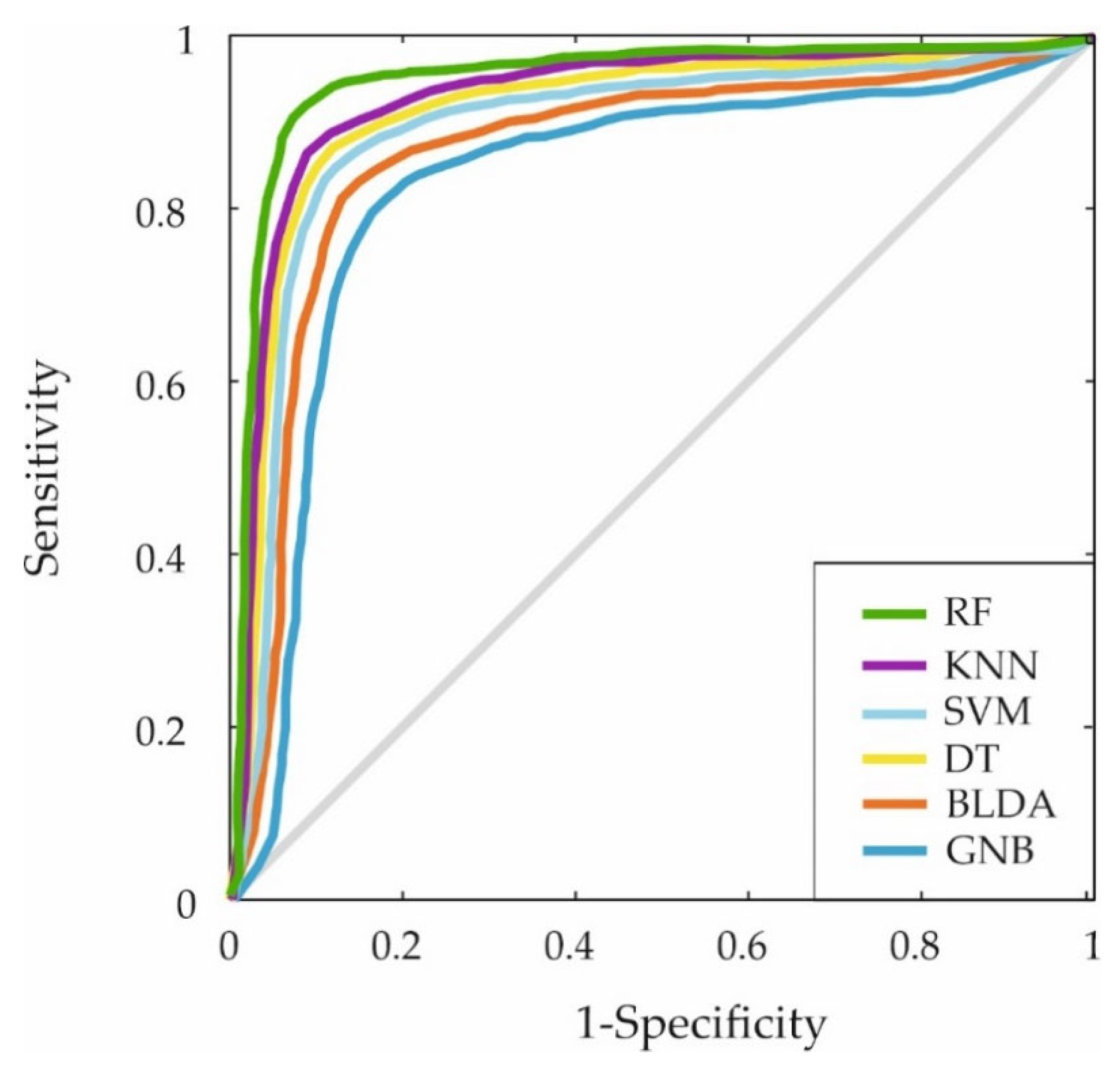
| KNN | 89.24 | 89.31 | 89.14 | 89.18 | 79.01 |
| RF | 95.83 | 95.92 | 95.15 | 95.73 | 86.32 |
| F1 Score | MCC | DYI | AUC | AUC (%) | |
|---|---|---|---|---|---|
| SVM | 83.07 | 73.93 | 83.32 | 82 | 0.82 |
| BLDA | 80.12 | 71.31 | 80.37 | 79 | 0.79 |
| DT | 85.29 | 75.94 | 85.52 | 84 | 0.84 |
| GNB | 74.63 | 65.83 | 74.82 | 74 | 0.74 |
| KNN | 89.23 | 79.48 | 89.24 | 89 | 0.89 |
| RF | 95.53 | 86.83 | 95.83 | 95 | 0.95 |
Disclaimer/Publisher’s Note: The statements, opinions and data contained in all publications are solely those of the individual author(s) and contributor(s) and not of MDPI and/or the editor(s). MDPI and/or the editor(s) disclaim responsibility for any injury to people or property resulting from any ideas, methods, instructions or products referred to in the content. |
© 2025 by the authors. Licensee MDPI, Basel, Switzerland. This article is an open access article distributed under the terms and conditions of the Creative Commons Attribution (CC BY) license (https://creativecommons.org/licenses/by/4.0/).
Share and Cite
Queipo, M.; Mateo, J.; Torres, A.M.; Barbado, J. The Effect of Naturally Acquired Immunity on Mortality Predictors: A Focus on Individuals with New Coronavirus. Biomedicines 2025, 13, 803. https://doi.org/10.3390/biomedicines13040803
Queipo M, Mateo J, Torres AM, Barbado J. The Effect of Naturally Acquired Immunity on Mortality Predictors: A Focus on Individuals with New Coronavirus. Biomedicines. 2025; 13(4):803. https://doi.org/10.3390/biomedicines13040803
Chicago/Turabian StyleQueipo, Mónica, Jorge Mateo, Ana María Torres, and Julia Barbado. 2025. "The Effect of Naturally Acquired Immunity on Mortality Predictors: A Focus on Individuals with New Coronavirus" Biomedicines 13, no. 4: 803. https://doi.org/10.3390/biomedicines13040803
APA StyleQueipo, M., Mateo, J., Torres, A. M., & Barbado, J. (2025). The Effect of Naturally Acquired Immunity on Mortality Predictors: A Focus on Individuals with New Coronavirus. Biomedicines, 13(4), 803. https://doi.org/10.3390/biomedicines13040803


