Article

Gender- and Grade-Dependent Activation of Androgen Receptor Signaling in Adult-Type Diffuse Gliomas: Epigenetic Insights from a Retrospective Cohort Study

1
Nervous System Medical Oncology Department, IRCCS Istituto delle Scienze Neurologiche di Bologna, Bellaria Hospital, 40139 Bologna, Italy
2
Department of Biomedical and Neuromotor Sciences (DIBINEM), University of Bologna, 40136 Bologna, Italy
3
IRCCS Istituto delle Scienze Neurologiche di Bologna, 40139 Bologna, Italy
4
Functional and Molecular Neuroimaging Unit, IRCCS Istituto delle Scienze Neurologiche di Bologna, Bellaria Hospital, via Altura 3, 40139 Bologna, Italy
5
Solid Tumor Molecular Pathology Laboratory, IRCCS Azienda Ospedaliero-Universitaria di Bologna, 40138 Bologna, Italy
6
Department of Medical and Surgical Sciences (DIMEC), University of Bologna, 40138 Bologna, Italy
7
Pathology Unit, IRCCS Azienda Ospedaliero-Universitaria di Bologna, 40138 Bologna, Italy
8
School of Medicine, Dipartimento di Scienze Mediche e Chirurgiche (DIMEC), University of Bologna, 40138 Bologna, Italy
9
Department of Biomedical and Neuromotor Sciences, Section of Anatomic Pathology at Bellaria Hospital, University of Bologna, 40138 Bologna, Italy
10
Functional and Molecular Neuroimaging Unit, IRCCS Istituto delle Scienze Neurologiche di Bologna, 40139 Bologna, Italy
*
Author to whom correspondence should be addressed.
These authors contributed equally to this work.
Biomedicines 2025, 13(10), 2379; https://doi.org/10.3390/biomedicines13102379
Submission received: 27 July 2025 / Revised: 24 September 2025 / Accepted: 25 September 2025 / Published: 28 September 2025
(This article belongs to the Special Issue Mechanisms and Novel Therapeutic Approaches for Gliomas)

Abstract

Background: The androgen receptor (AR) is a ligand-dependent transcription factor of the nuclear steroid receptor superfamily, implicated in the pathogenesis of various solid tumors. The AR gene, located on chromosome Xq11–12, is accompanied by several X-linked genes that modulate AR expression and function, including FLNA, UXT, and members of the melanoma antigen gene (MAGE) family (MAGEA1, MAGEA11, MAGEC1, MAGEC2). While the AR has been investigated in multiple tumor types, its role in adult-type diffuse gliomas remains largely unexplored. Here, we characterized AR protein expression and the promoter methylation status of the AR and associated regulatory genes in adult-type diffuse gliomas. Methods: A retrospective analysis was conducted on 50 patients with adult-type diffuse gliomas, including IDH-mutant gliomas (grades 2–4) and IDH-wildtype glioblastomas (GBMs), classified according to the 2021 WHO criteria. AR nuclear expression was assessed by immunohistochemistry (IHC). Methylation-specific PCR and quantitative DNA methylation analyses were employed to evaluate promoter methylation of the AR and selected co-regulatory genes. Results: AR nuclear positivity correlated significantly with male sex (p = 0.04) and higher tumor grade, with the highest expression in IDH-wildtype GBMs (p = 0.04). In IDH-mutant gliomas, AR immunoreactivity was more prevalent in astrocytomas than in 1p/19q codeleted oligodendrogliomas (p = 0.02). AR expression was associated with unmethylated MGMT promoter status (p = 0.02). DNA methylation analysis revealed AR gene hypomethylation in tumors displaying nuclear AR positivity and in IDH-wildtype GBMs (Kruskal–Wallis p < 0.05). Additionally, methylation patterns of AR co-regulators located on the X chromosome suggest epigenetic regulation of AR signaling in gliomas. Conclusions: The findings reveal distinct AR pathway activation patterns in adult-type diffuse gliomas, particularly IDH-wildtype GBMs, suggesting that further exploration of antiandrogen therapies is warranted.

1. Introduction

Adult-type diffuse gliomas are the most frequent tumors of the central nervous system (CNS), characterized by an extensive infiltrative growth with an overall survival that greatly varies depending on the specific subtype and histological grade, ranging from 7 to 15 years in Isocitrate Dehydrogenase (IDH)-mutant low-grade gliomas (LGGs) to 12–24 months in grade 4 IDH-wildtype gliomas [1,2]. Despite surgery, radiotherapy, and alkylating agents being the cornerstones of glioma treatment, effective treatments are lacking due to challenges such as the inability to fully resect infiltrative tumors, treatment resistance, difficulties delivering drugs across the blood–brain barrier (BBB), and tumor heterogeneity.
Although existing studies have tried different therapeutic methods [3], individually tailored strategies are needed to improve outcomes in subsets of patients [4,5,6,7].
A significant gender disparity exists in the incidence of glioblastoma (GBM), with a higher prevalence in males compared with females. Research suggests that this difference may be linked to the modulation of glioblastoma pathogenesis via the androgen receptor (AR) signaling pathway. Dysregulated activation of the AR signaling cascade, or cross-talk between the AR and other oncogenic signaling networks, appears to facilitate GBM progression. Consequently, AR inhibition or modulation presents a potential therapeutic strategy for GBM management [8].
Androgens interact with the AR to produce their biological effects, and they are essential for gender differentiation and male phenotypic development [9,10].
When androgens, primarily testosterone and dihydrotestosterone, bind to the AR, it becomes active. The inactivated AR is mainly located in the cytoplasm; the binding of the AR to androgens produces a conformational change in the receptor that dimerizes, migrates into the nucleus, and binds to specific hormone response elements to regulate the expression of androgen-responsive genes [11].
Several studies have shown that the AR is overexpressed in some human malignancies, such as prostate cancer, breast cancer, hepatocellular carcinoma, and bladder cancer [9,12,13]. However, to date, the same relationship is not as well understood in gliomas. Recent research suggests a role for the AR in gliomagenesis [14]; however, the expression level of the AR, its regulatory mechanisms, and its prognostic significance in gliomas remain unknown [15].
The AR gene is situated on the X chromosome at the Xq11–12 locus [16] and AR gene expression is modulated by regulators, predominantly located on the X chromosome, including filamin A (FLNA), Ubiquitously Expressed Prefoldin-Like Chaperone (UXT) and the genes belonging to melanoma antigen (MAGE) family genes, MAGEA1, MAGEA2, MAGEA3, MAGEA9, MAGEA11, MAGEC1, and MAGEC2 [17,18,19].
DNA methylation is a crucial epigenetic regulatory mechanism for gene transcription and represents a significant area of investigation in oncology. It is linked to aberrant gene silencing by sustaining a transcriptionally repressed state characterized by heterochromatin formation [20]. Methylation involves cytosine on the CpG sequences, which are abundant in the promoter regions of genes. The result is 5-methylcytosine, which produces transcriptional repression and silences gene expression [21].
In addition, the AR gene and its regulators, which map to the X chromosome, may exhibit copy number variations as a consequence of X-chromosome aneuploidy. At present, there is no available data regarding the variations in copy number of the X chromosome and AR in gliomas.
This study aimed to evaluate the AR nuclear expression detected at immunohistochemistry and to correlate AR expression with the data emerging from DNA sequencing, DNA methylation analysis, and X-chromosome copy number analysis. In particular, we assessed the methylation pattern of the AR gene and its regulatory genes such as FLNA, UXT, and MAGE family members, and analyzed X-chromosome copy number variations in a series of gliomas.

2. Materials and Methods

2.1. Study Design

This is a retrospective study aimed at assessing AR immunohistochemical expression, AR methylation patterns, and X-chromosome AR-related gene methylation patterns in adult-type diffuse gliomas.
We analyzed 50 tissue samples obtained from patients who underwent surgery at Bellaria Hospital in Bologna, between 1999 and 2021. All specimens were consistently preserved at the pathology department of our institution.
All cases were independently re-evaluated, classified, and graded by a neuropathologist (A.S.) according to currently available criteria (CNS WHO 2021).
Inclusion criteria were as follows:
(1)
age ≥ 18 years old;
(2)
morphological and immunohistochemical profile consistent with the diagnosis of adult-type diffuse glioma (IDH-mutant glioma, grade 2 or 3 or 4 and GBM IDH-wildtype grade 4 according to the fifth edition of CNS WHO 2021 [2]);
(3)
histological slides/formalin-fixed paraffin-embedded tissue tumor (FFPE) blocks from the archive available to perform immunohistochemical assessment of androgen receptor, in situ hybridization, and molecular tests.
Exclusion criteria were as follows:
(1)
histological diagnosis different from adult-type diffuse gliomas;
(2)
unavailability of histological slides/formalin-fixed paraffin-embedded tissue tumor (FFPE) blocks;
(3)
molecular GBM IDH-wildtype and gliomas IDH-mutant with homozygous cyclin-dependent kinase inhibitor (CDKN2A/B) gene deletion were excluded from our study;
(4)
patients undergoing antiandrogen therapy for another malignancy.
The primary objective of the study was to assess nuclear AR expression by immunohistochemistry on tissue samples, and how AR expression might be influenced by the DNA methylation pattern of the AR and its co-regulators and by X-chromosome copy number variations.
The secondary purpose of the study was to explore the prognostic value of the AR in adult-type diffuse gliomas.
The impact of categorical variables on time-dependent events such as overall survival (OS) and progression-free survival (PFS) has been estimated through the Kaplan–Meier method [22].

2.2. Ethical Statement

The study was approved by the Ethics Committee of the Azienda Sanitaria Locale di Bologna (protocol number CE09113, Bologna, Italy). This study was conducted in agreement with the most recently updated Declaration of Helsinki and all the international and local laws applied to clinical trials and patient protection. The study was conducted according to the principles of the ICH Harmonized Tripartite Guideline for Good Clinical Practice.

2.3. AR Immunohistochemistry

AR nuclear expression was evaluated in tissue sections using immunohistochemistry. Samples were fixed in 10% neutral-buffered formalin for 24 h and processed into formalin-fixed paraffin-embedded (FFPE) blocks following standard protocols. Immunostaining was conducted on an automated platform (Ventana BenchMark, Ventana Medical Systems Inc., Tucson, AZ, USA) utilizing a pre-diluted monoclonal anti-AR antibody (Cell Marque, Rocklin, CA, USA, clone SP107). Only nuclear immunoreactivity was considered for scoring. AR expression was assessed using a semi-quantitative four-point scoring system adapted from astrocytic tumor analyses [23], ranging from 0 to 3. Score 3 indicated strong, widespread (>50%) nuclear staining; score 2 denoted focal nuclear staining in approximately 50% of cells; score 1 was assigned for weak or focal nuclear staining in <50% of cells or faint staining not readily discernible at low magnification; and score 0 indicated absence of staining [23]. The highest AR staining intensity observed in adjacent non-neoplastic glial tissue served as the internal reference. In cases with heterogeneous staining, the predominant score was recorded. Scoring was performed independently by a neuropathologist (AS) blinded to clinical data. Tumors were classified as AR-positive (scores 1–3) or AR-negative (score 0).

2.4. Next-Generation Sequencing

Genomic DNA was extracted from formalin-fixed, paraffin-embedded (FFPE) tumor sections using the NucleoSpin® Tissue Kit (Macherey-Nagel, Dueren, Germany), following the manufacturer’s protocol. A board-certified neuropathologist (AS) reviewed the histological slides and identified the most representative tumor regions for analysis. Targeted mutational profiling was performed via next-generation sequencing (NGS), focusing on the IDH1, IDH2, TERT, and AR genes [24]. FASTQ data were filtered based on PHRED quality scores (>Q30) and processed using the Galaxy Project platform [25], incorporating alignment to the hg38 human reference genome with Bowtie2, local realignment with GATK, variant calling via HaplotypeCaller, and duplicate read removal using Picard MarkDuplicates. BAM files were visualized with the Integrative Genomics Viewer (IGV). Variants with a variant allele frequency (VAF) > 20% were retained. Functional predictions were assessed using PolyPhen-2 and cross-referenced with the COSMIC database [26]. All NGS assays were designed to achieve a minimum depth of coverage of 1000× with ≥1000 reads per region.
For DNA methylation analysis, bisulfite-converted NGS was employed to assess CpG methylation across multiple X-linked genes including the AR, MAGEA1, MAGEA11, MAGEC1, MAGEC2, UXT, and FLNA. Genomic DNA (50–500 ng) was treated with bisulfite using the EZ-96 DNA Methylation MagPrep Kit (Zymo Research, Irvine, CA, USA; Cat. D5040), per the manufacturer’s instructions. Specific CpG regions, including six within the AR gene (spanning from the first non-coding region to exon 1), were amplified using primers designed via MethPrimer (http://www.urogene.org/methprimer/, accessed on 24 September 2025). Libraries were generated using tagged primers and Phusion U DNA Polymerase (Thermo Fisher Scientific, Waltham, MA, USA, Cat. F555L), followed by a two-step PCR enrichment protocol utilizing the Nextera™ Index Kit (Illumina, San Diego, CA, USA) [27]. Sequencing was carried out on the MiSeq platform (Illumina), according to standard protocols. Methylation quantification was performed using BWAmeth for alignment and MethylDackel for methylation calling within the Galaxy environment. The methylation panel also included the TERT promoter and three specific regions of the MGMT gene.
Additionally, AR mutational analysis was extended to exons 4, 5, and 8—regions known to harbor frequent mutations associated with therapeutic resistance in prostate cancer.

2.5. FISH Analysis

FISH analysis was performed to assess the level of X-chromosome aneuploidy of the glioma samples.
A neuropathologist (AS) examined the tissue sections and selected the most representative tumor area on the histological slides.
Dual-color FISH was carried out according to a standard protocol summarized as follows: five-micron sections were obtained from each tumor block. One specific probe kit for the X chromosome (AR gene amplification probe, (AR(Xq12)/CEPX OACP IE LTD, Cork, Ireland), added with the Smart-ISH hybridization buffer (OACP IE LTD, Cork, Ireland), was applied [17,28]. The AR gene-specific probe length is stated as 281 Kb (AR gene amplification probe product datasheet OACP IE LTD, Cork, Ireland).

3. Results

Fifty patients were included, 26 (52%) were male and 24 (48%) were female. The median age at diagnosis was 50 years (range 26–77 years).
Histology included astrocytoma IDH-mutant grade 3 (n = 3), oligodendroglioma IDH-mutant and 1p19q codeleted grade 3 (n = 4), astrocytoma IDH-mutant grade 2 (n = 9), oligodendroglioma IDH-mutant and 1p19q codeleted grade 2 (n = 11), and GBM IDH-wt grade 4 (n = 23).
AR positivity was detected in 21 out of 50 samples (42%). Among different glioma subtypes, AR positivity was found in fourteen patients diagnosed with GBM IDH-wildtype grade 4 (66.7%), in four cases with astrocytoma IDH-mutant grade 2 (19%), in two cases of astrocytoma IDH-mutant grade 3 (9.5%) and in one case of oligodendroglioma IDH-mutant and 1p19q codeleted grade 2 (4.8%) (Table 1 and Figure 1). AR positivity was higher in males (71.4%; n = 15) than in females (28.6%, n = 6).
The mutational analysis of the AR gene from exon 4 to 8 revealed no mutations in all samples.
The clinicopathological characteristics of the cases are summarized in Table 1.

3.1. Gender and Pathologic Grade Are Associated with AR Expression Levels in Adult-Type Diffuse Gliomas

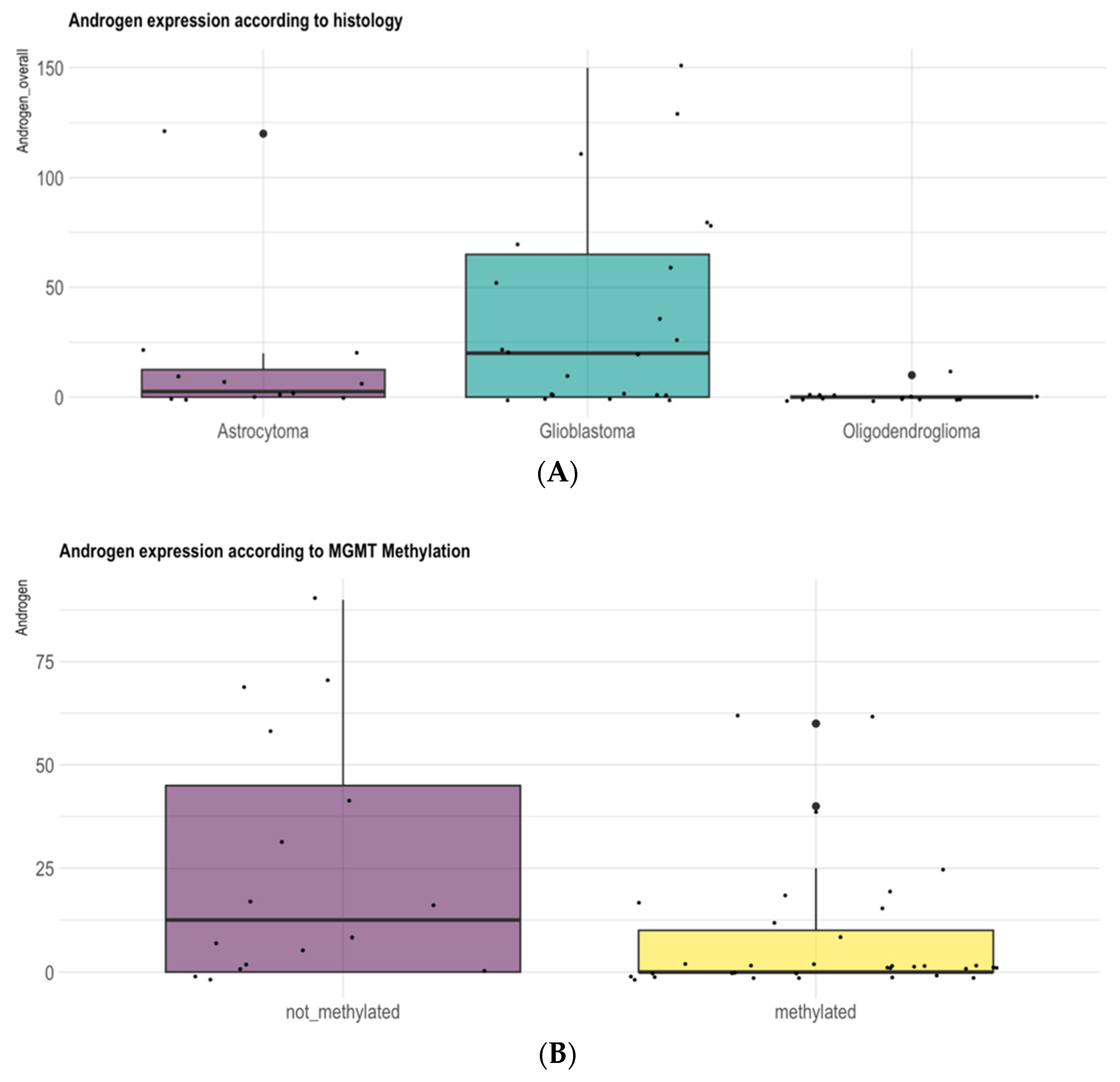
In our analysis AR expression was correlated with gender and with glioma grade. Immunohistochemical AR positivity was predominant in males (p = 0.04) and in patients with GB IDH-wildtype compared with low-grade IDH-mutant adult-type diffuse gliomas (p = 0.04) (Figure 2A). Among IDH-mutant LGGs, AR expression was more frequent in IDH-mutant astrocytomas compared with IDH-mutant oligodendrogliomas and 1p19q codeleted (p = 0.02) (Figure 1A). Moreover, AR expression was more frequent in MGMT-unmethylated gliomas compared with MGMT-methylated tumors (p = 0.02) (Figure 2B).

3.2. Prognostic Role of AR Expression Levels in Adult-Type Diffuse Gliomas

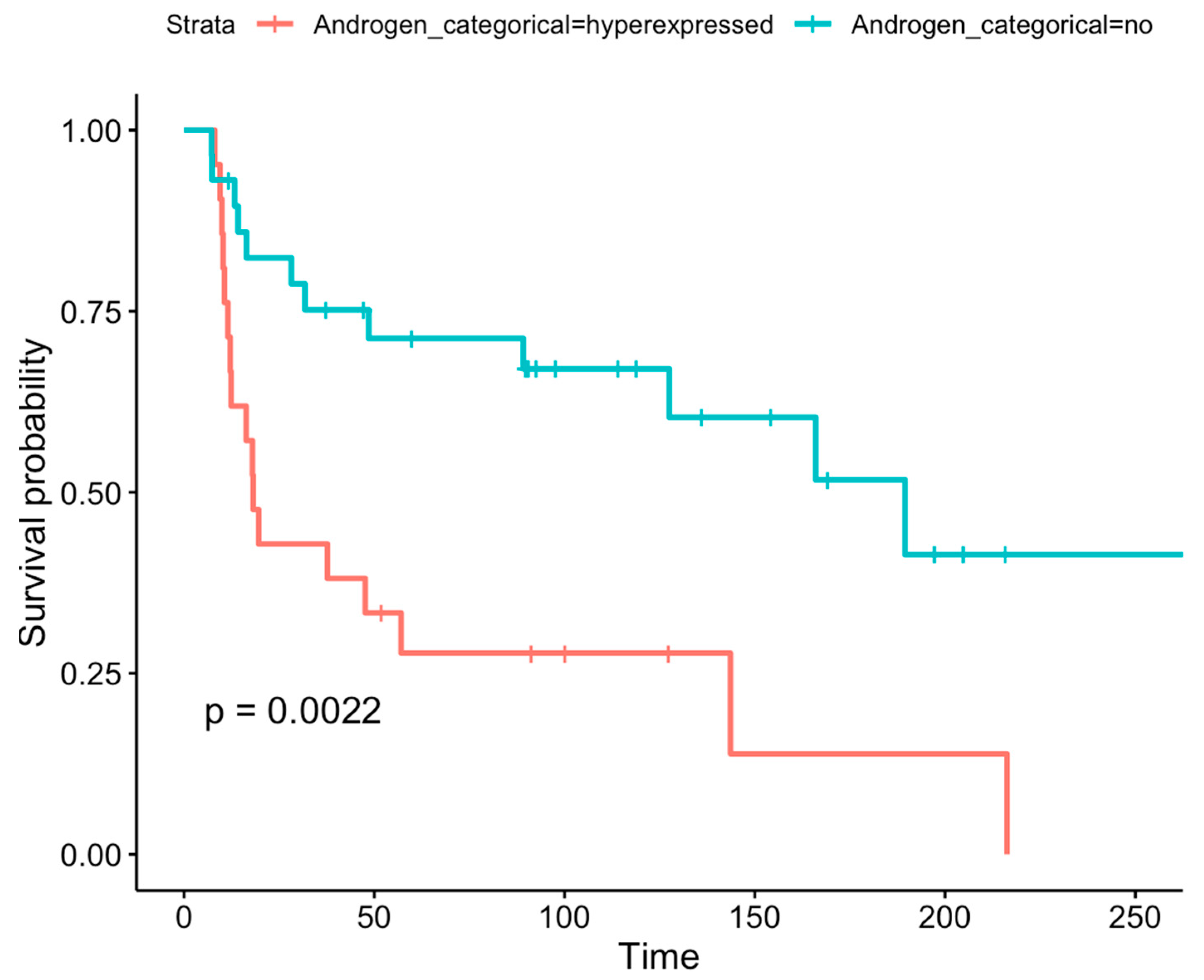
Immunohistochemically AR-positive gliomas showed a lower overall survival (OS) than AR-negative gliomas (p = 0.0022; HR = 3.1 (95%CI 1.45–6.6)) (Figure 3).
Among the different histological entities, we found the following survival outcomes:
In IDH-wt AR-positive GBMs, the median OS was 12.3 months (10.6–37.7; p-value 0.22; HR = 1.74 (95%CI 0.71–4.45)). In IDH-wt AR-positive and MGMT-methylated GBMs, the mOS was 18 months (9.4-NA; HR = 1.73 (95%CI 0.5–6.02)), while in IDH-wt AR-positive MGMT-unmethylated GBMs patients the mOS was 11.5 months (10.3-NA; p-value 0.31; HR = 2.1 (95%CI 0.5–8-84)). In IDH-wt AR-negative GBMs, the median OS was 16.4 months (13.3-NA). In IDH-wt AR-negative and MGMT-methylated GBMs, the mOS was 30 months (7.4-NA), while in IDH-wt AR-negative MGMT-unmethylated GBMs, the mOS was 14.2 months (13.3-NA). In IDH-mutant AR-positive grade 3 astrocytomas, the mOS was 137 months (p-value 0.81; HR = 0.71 (95%CI 0.04–11.8)), while in IDH-mutant AR-negative grade 3 astrocytomas, the mOS was 128 months. In IDH-mutant AR-positive grade 3 oligodendrogliomas (n = 1), the OS was 144 months, while in AR-negative grade 3 oligodendrogliomas, the mOS was not assessable, as the events had not yet been reached. In grade 2 IDH-mutant astrocytomas and oligodendrogliomas, the mOS was not assessable, as the events had not yet been reached.
We performed a Cox Regression Model investigating the prognostic impact of AR expression adjusted for MGMT-methylation status in the GBM IDH-wt group (Table 2). In this analysis, the MGMT-methylation status confirmed its prognostic role (p-value 0.04; HR = 0.34 (0.12–0.95)), while AR expression did not reach statistical significance (p-value 0.27; HR = 1.68 (0.67–4.26)), likely due to the small sample size. After adjusting the Cox Regression Model for MGMT-methylation status and gender, AR expression only showed a trend in impacting survival, without reaching statistical significance (p-value 0.12; HR = 2.25 (0.79–6.39)) (Table 3).

3.3. Differential DNA Methylation Analysis of AR Gene Promoter Region According to Immunohistochemical AR Expression

In our series we found a relation between immunohistochemical AR expression and methylation levels of different CpG sites in the AR promoter.
The AR expression was analyzed in the samples through immunohistochemistry. The samples were divided into two groups: positive AR expression (group 0, AR expression score 1, 2, and 3) or negative AR expression (group 1, AR expression score 0), based on the H-score being higher than or equal to 0.
Comparative analysis of DNA methylation in gliomas with high vs. low immunohistochemical expression of the AR involved four different regions of the AR gene body: AR CAG repeats (promoter) from coordinates 67,545,204 to 67,545,299; the AR region within CAG and CGG repeats (intra CAG-CGG) from 67,545,904 to 67,546,136; the AR region before CGG repeat from 67,546,381 to 67,546,405; and, finally, the AR region within the CGG repeats from 67,546,492 to 67,546,664.
Supplementary Figure S1A–D illustrate the levels of methylation for each genetic region compared between the two groups, positive vs. negative immunohistochemical expression of the AR: positive AR expression (group 0) or negative AR expression (group 1).
The AR was found hypomethylated, and therefore probably transcriptionally active, in cases with immunohistochemical positivity of the AR (Kruskal–Wallis < 0.05). In males, AR CAG repeats and AR INTRA CAG-CGG repeats resulted hypomethylated in AR-positive gliomas. In females, we observed the same trend; however, it was not statistically significant.

3.4. Differential Methylation Analysis of MAGEA1, MAGEA11, MAGEC1, MAGEC2, UXT, and FNLA Genes According to Immunohistochemical AR Expression

In males, MAGEA1 was found to be hypermethylated in cases negative for AR expression. In females, we observed a different trend: MAGEA1 was found to be hypomethylated in cases with low AR expression (Supplementary Figure S2A).
MAGEA11 showed a methylation profile similar to MAGEA1. In particular, in males MAGEA11 was hypermethylated in cases negative for AR expression. On the contrary, in females MAGEA11 was hypomethylated in cases with low AR expression (Supplementary Figure S2B).
The same trend was observed for the MAGEC1 gene. MAGEC1, in males, showed a tendency to be hypermethylated in AR-negative cases. In females, on the contrary, MAGEC1 showed a tendency to be hypomethylated in AR-negative cases (Supplementary Figure S2C).
MAGEC2 showed a tendency to be hypomethylated in cases negative for AR expression both in male and in female patients (Supplementary Figure S2D).
In males, UXT is hypermethylated in immune cases with positive AR (Kruskal–Wallis < 0.05). In females UXT shows the same trend, however it is not significant (Supplementary Figure S2F).
FLNA showed a tendency to be hypomethylated in AR-negative cases both in male and in female patients (Supplementary Figure S2E).

3.5. Differential Methylation Analysis of the MGMT Promoter According to Immunohistochemical Expression of AR

Regardless of gender, since MGMT is located on chromosome 10, we found that MGMT is hypermethylated in AR-positive cases (KR < 0.05) (Supplementary Figure S3).

3.6. Differential Methylation Analysis of the AR Gene Promoter Region According to Pathologic Grade

In our analysis, AR INTRA CAG-CGG repeats and AR CAG repeats were found to be hypermethylated in low-grade gliomas and hypomethylated in high-grade tumors (Supplementary Figure S4A–D).

3.7. Differential Methylation Analysis of MAGEA1, MAGEA11, MAGEC1, MAGEC2, UXT, and FNLA According to Pathologic Grade

MAGEA1 was found to be hypomethylated in low-grade tumors in male and in female patients (Supplementary Figure S5A).
MAGEA 11 showed a different behavior based on gender. In particular, in males MAGEA 11 was found to be hypermethylated in low-grade tumors; in females, on the contrary, it was found to be hypomethylated in low-grade tumors (Supplementary Figure S5B).
For MAGEC 1, in both males and females, we observed a trend towards hypermethylation in low-grade tumors (Supplementary Figure S5C).
Similarly, MAGEC 2 and FLNA also resulted hypermethylated in low-grade tumors (Supplementary Figure S5D,E). Conversely, UXT was found to be hypomethylated in low-grade tumors and hypermethylated in high-grade tumors (Supplementary Figure S5F).

3.8. Differential Methylation Analysis of MGMT and TERT According to Pathologic Grade

MGMT showed higher methylation in lower-grade gliomas and a lower methylation in higher-grade gliomas in the enhancer region, while the opposite trend was found for the exon1 region. This suggests that the methylation pattern of MGMT may vary depending on tumor grading. Higher-grade tumors tend to be MGMT hypomethylated in the enhancer region and hypermethylated in the exon1 region (Supplementary Figure S6A).
Finally, TERT resulted hypomethylated and therefore actively expressed in high-grade gliomas, which is consistent with TERT overexpression in more aggressive tumors (Supplementary Figure S6B).

3.9. X-Chromosome Copy Number Analysis in Glioma

We performed a FISH analysis to assess the level of X-chromosome aneusomy. Our analysis showed that X-chromosome monosomy is more frequent in women (p < 0.001), while X-chromosome polysomy is more frequent in men (p < 0.001).
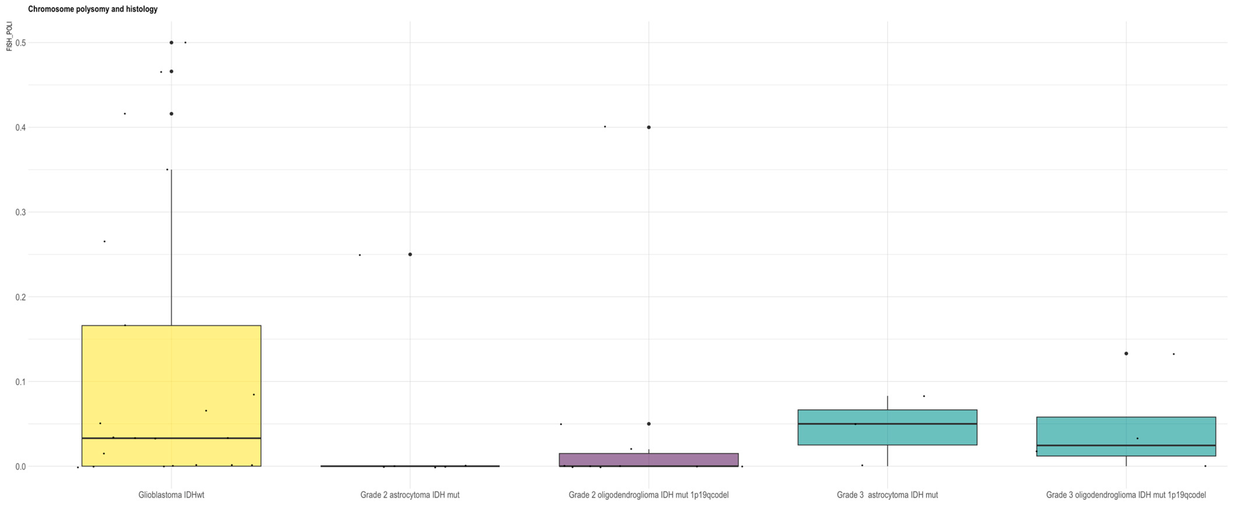
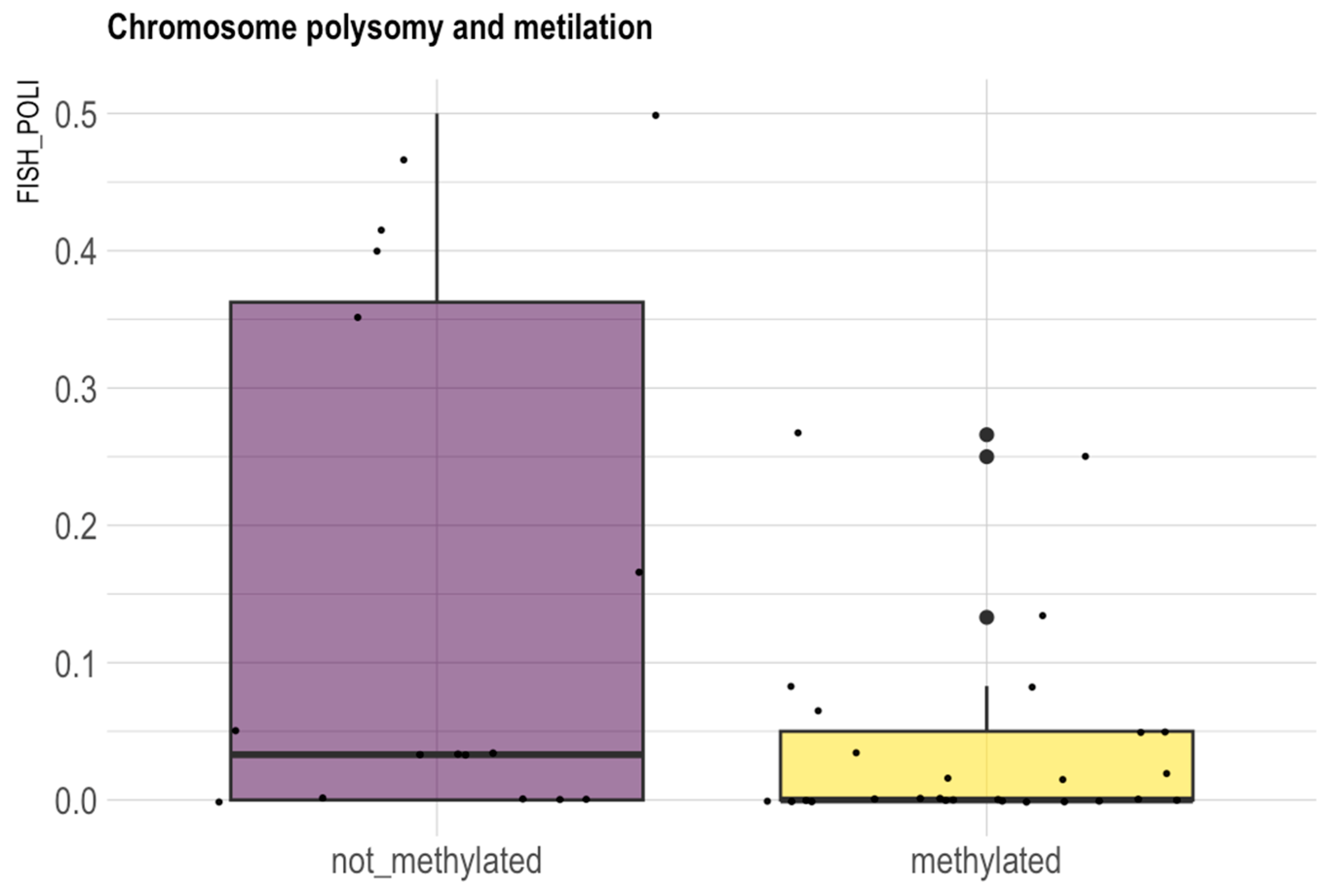
In our series polysomy resulted more frequently in GBM IDH-wt (p = 0.05) (Figure 4) and unmethylated tumors (p = 0.037) (Figure 5). In unmethylated patients there is also a trend of higher frequency of AR amplification (p = 0.06).
Table 1 shows the percentage of wildtype X chromosome, AR deletion, monoploidy of the X chromosome in female samples, amplification of the AR, and polyploidy of the X chromosome. Polyploidy means the presence of XX chromosomes in male samples and the presence of more than two X chromosomes in female samples.
The average polyploidy for females is 1.659%, while for males the average polyploidy is 15.745%.

3.10. Differential Methylation Analysis of AR Promoter Regions According to X-Chromosome Copy Number

We observed that a lower X-chromosome polyploidy was associated with intra CAG-CGG repeats hypermethylation (Supplementary Figure S7).

3.11. Differential Methylation Analysis of MAGEA1, MAGEA11, MAGEC1, MAGEC2, UXT, and FNLA Genes According to X-Chromosome Copy Number

We observed a correlation between a lower X-chromosome polyploidy and the hypomethylation status of MAGEA1, MAGEA11, and UXT genes (Supplementary Figure S8A–F).

4. Discussion

The oncogenic role of the AR in classical hormone-responsive tissues such as prostate and male breast is well established [10,17,29,30]. The AR is a targetable molecule widely used in the treatment of prostate cancer [31,32,33,34,35,36]; however, its role in gliomas is still not well understood.
In recent years, more attention has been given to understanding the pathogenic role of AR signaling in glioma biology [37,38,39,40]. AR overexpression has been observed in gliomas and their stem-like cell compartments, supporting the idea that AR signaling may affect the development of brain tumors [15,41,42]. However, due to conflicting and inconclusive data in the literature, the prognostic significance of AR overexpression in gliomas remains uncertain.
In this study, we aimed to investigate AR expression levels across gliomas of different histopathologic grades and molecular subtypes (Table 1) and to analyze the DNA methylation level of the AR and its co-regulators mapped on the X chromosome.
In our series AR expression positively correlated with glioma grading and with gender. Our analysis reports a higher expression of the AR in males (71.4%; n = 15) compared with females (28.6%, n = 6), (p = 0.04) and in higher-grade tumors, in particular in GBMs IDH-wt compared with low-grade IDH-mutant diffuse gliomas (p = 0.04) (Figure 1). Among IDH-mutant LGGs, AR expression was more frequent in IDH-mutant astrocytomas compared with IDH-mutant oligodendrogliomas and 1p19q codeleted (p= 0.02) (Figure 2A). Furthermore, AR expression was more frequent in MGMT-unmethylated gliomas compared with MGMT-methylated tumors (p = 0.02) (Figure 2B).
This aligns with data from previous research indicating that AR activation influences the proliferation of high-grade glioma cells and, primarily, of GBM cells [15,43]. Several studies suggest that AR expression is positively correlated with tumor pathological grade, with grade 4 tumors demonstrating the most elevated expression levels [15,38,44,45]. In particular, the AR is detected in GBM IDH-wt samples compared to normal human brain tissue and is associated with a worse prognosis in GBM IDH-wt patients [15,46]. Data from The Cancer Genome Atlas (TCGA) database indicate an upregulation of the AR at both mRNA and protein levels in human GBM IDH-wt specimens, particularly among male patients, in comparison with normal brain tissue [47]. Epidemiology indicates a gender disparity in GBMs [8,48,49,50]: the Central Brain Tumor Registry of the United States (CBTRUS) statistical report of central nervous system tumors (2017–2021) evidences an increased prevalence of GBM IDH-wt in males (1.6 times more prevalent) than in females, with a median survival duration of 15.0 months in male patients, versus 25.5 months in female patients [51,52]. In addition, experimental investigations have shown that GBM engrafted in animal models displays a reduced growth rate in female subjects compared with their male counterparts [38]. The exact mechanism underlying this pronounced epidemiology is unclear. It has been hypothesized that gender differences may be attributable to the AR signaling axis: the upregulation of the AR signaling pathway has been identified as a contributing factor to the increased male predominance of GBMs [53]. This mechanism appears to play a critical role in the proliferation, migration, and invasion of high-grade gliomas.
Despite decades of research, the identification of prognostic markers for gliomas is limited, with an even smaller subset that may serve as viable therapeutic targets [15,54,55]. We have performed Kaplan–Meier analyses [22] in patients with different AR expression levels to investigate the potential prognostic value of the AR. In our series, higher AR protein expression is associated with significantly poorer OS in glioma patients (p = 0.0022) (Figure 3). This finding is consistent with the prior literature indicating that elevated AR expression levels are correlated with poorer prognosis in both IDH-wildtype GBMs and LGGs [15,56] and that the exposure of GBM cells to androgens increases tumor aggressiveness [56].
In our study, we aimed to establish a correlation between AR expression, as assessed through immunohistochemistry, and the findings obtained from DNA sequencing, DNA methylation analysis, and X-chromosome copy number assessment.
Since mutations of the AR gene are associated with higher malignancy of prostate cancer, we investigated the mutational status of the AR to address any possible interactions also in glioma. Most mutations cause a substitution of one amino acid, thus allowing antiandrogens to act as AR agonists and favor cancer progression. The most common AR mutations and their associated drug resistance are p.F877L for enzalutamide and Apalutamide, p.T878A and p.H875Y for Abiraterone, p.W742C and p.V716T for bicalutamide and flutamide [57]. In our cohort, the mutational analysis of the AR gene, from exons 4 to 8, revealed no mutations in any of our samples.
We also performed a DNA methylation analysis of the AR and its co-regulatory genes to investigate whether epigenetic mechanisms might regulate AR expression, observing some differences in different classes of gliomas.
DNA methylation is a key epigenetic mechanism involved in gene expression regulation across multiple tumor types. Hypermethylation of CpG promoter regions recruits repressive chromatin modifiers, leading to stable silencing of genes, including tumor suppressors, thereby promoting tumorigenesis. In contrast, hypomethylation can reactivate gene expression, contribute to chromosomal instability, and promote oncogene or transposable element activation, further driving cancer progression [58,59,60,61,62,63].
The level of AR protein expression is determined by the transcriptional activity of the AR gene. As a result, the methylation status of AR gene regulators and variations in the copy number of the X chromosome could significantly impact AR protein expression. In our series, AR CAG repeats and “Intra CAG-CGG repeats” regions resulted hypermethylated in gliomas with low AR protein levels, as well as in LGGs (Supplementary Figures S1A–D and S4A–D). This indicates that the hypermethylation of the AR gene leads to transcriptional repression, resulting in low levels of AR expression, a phenomenon that is primarily evident in LGGs. Moreover, the methylation pattern of AR gene regulators, belonging to the MAGE family, revealed noteworthy insights. MAGEA11 and MAGEC1 were hypermethylated in AR-negative gliomas (Kruskal–Wallis < 0.05) as well as in IDH-mutant low-grade tumors. This suggests that the hypermethylation state of these genes may result in AR gene silencing and decreased AR protein expression, and this is primarily evident in IDH-mutant lower-grade tumors (Supplementary Figures S2A–F and S5A–F).
Furthermore, in our series, the X-chromosome polysomy (that is the presence of XX chromosomes in male samples and the presence of more than two X chromosomes in female samples) appeared more frequently in men (p < 0.001) and in GBM IDH-wt (p = 0.05) (Figure 4). X-chromosome polysomy resulted more frequently in MGMT-unmethylated tumors (p = 0.037). In MGMT-unmethylated patients, we also observed a trend of higher frequency of AR amplification (p = 0.06) (Figure 5).
X-chromosome polysomy can play a role in the AR gene copy number and, consequently, in AR protein expression in glioma cells, controlling neoplastic transformation, especially in GBM IDH-wt. It is conceivable that in high-grade glioma, the neoplastic cells acquire additional copies of the X chromosome with consequent AR gene amplification and AR protein overexpression.
Given their ability to cross the blood–brain barrier, AR antagonists may represent a promising therapeutic avenue for the treatment of GBMs. Currently, a variety of androgen antagonists are under investigation for their efficacy in AR-positive gliomas, particularly in IDH-wildtype GBMs [8,64]. Both enzalutamide and bicalutamide, which are approved for prostate cancer treatment, have shown concentration-dependent antitumor effects in A172, U87MG, and T98G GBM cell lines, with enzalutamide exhibiting a more pronounced impact [38].
Research has demonstrated that enzalutamide significantly decreases the growth of GBM cells in both laboratory and live animal studies. Furthermore, it has been demonstrated to diminish cancer stem cell populations, leading to a 50% improvement in survival rates in an orthotopic patient-derived xenograft model of GBM [38] and to induce apoptosis in GBM cells through the activation of a caspase cascade pathway [47]. The 5α-reductase inhibitor dutasteride, in combination with AR antagonists such as cyproterone or flutamide, exhibits significant antitumor effects in U87 GBM cell cultures [65]. The dutasteride–flutamide combination, notably, showed the strongest inhibition of tumor cell metabolism and proliferation. These findings support the potential of dual AR pathway inhibition as a promising therapeutic strategy for GBMs [65].
An increasing body of evidence indicates that the Epidermal Growth Factor Receptor (EGFR) pathway plays a role in the activation of the AR within GBM cells, as AR activation may also occur via ligand-independent signaling through the EGFR [14]. The pairing of enzalutamide with an EGFR-targeted agent, such as afatinib, may enhance antitumor efficacy in glioma cells by simultaneously targeting both signaling pathways. Afatinib has been used in conjunction with the AR antagonist enzalutamide in IDH-wildtype GBM cells, producing significant outcomes and underscoring the synergistic effects of this drug combination [8,14].
In addition to AR inhibitors, combination therapies incorporating AR-targeted agents alongside other treatment modalities, such as chemotherapy and radiation therapy, are presently under investigation [66]. The objective of these combination strategies is to enhance the effectiveness of AR inhibition and to surmount potential resistance mechanisms. The preliminary results from ongoing trials have demonstrated encouraging outcomes, including better overall survival (OS) rates [8].
The data of the present study suggest that gender differences in glioma patients are significant and could be attributed to the role of AR expression, which is controlled by DNA methylation and X-chromosome aneuploidy. AR nuclear expression, in addition, seems to have a close correlation with glioma grading, being more expressed in GBM IDH-wt, and appears to influence the prognosis of gliomas, reducing survival. The present results have some limitations, including the low case number and the retrospective nature of the study. Our findings do not reach statistical significance, likely due to the small sample size, and should be considered trends that require validation in larger cohorts.

5. Conclusions

In conclusion, in the present study, we found that the AR seems to be overexpressed in IDH-wildtype GBMs as well as in MGMT-unmethylated tumors and appears to be associated with worse survival. The AR could likely play a role in influencing the biological aggressiveness and the prognosis of adult diffuse gliomas. These points were investigated through immunohistochemical staining, which found further validation in DNA methylation and X-chromosome copy number analyses.
The AR is an ideal therapeutic target in oncology, as androgen deprivation therapy is well tolerated and cost-effective. The results shown here can be potentially interesting as the AR could be a therapeutically useful target for glioma treatment. Moreover, immunohistochemical analysis for the detection of AR positivity is an inexpensive and simple investigation, which can be easily performed in glioma patients.
However, more data are needed to confirm the therapeutic impact of this approach and androgen deprivation therapy warrants further investigation in clinical trials for adult diffuse gliomas. Further research is required to explore and address the resistance mechanisms, the heterogeneity of gliomas, and the potential off-target effects of AR inhibitors. Overall, advancements in targeted AR therapy for adult-type diffuse gliomas offer promising prospects for the development of new treatment approaches.

Supplementary Materials

The following supporting information can be downloaded at: https://www.mdpi.com/article/10.3390/biomedicines13102379/s1, Figure S1: DNA methylation for different AR gene promoter regions compared between two groups (positive vs. negative immunohistochemical expression of AR); Figure S2: DNA methylation analysis genetic of AR regulators on the X chromosome (FLNA, MAGEA1, MAGEA11, MAGEC1, MAGEC2, UXT); Figure S3: Differential methylation analysis of MGMT according to AR expression; Figure S4: Differential methylation analysis of the AR gene promoter region according to pathologic grade; Figure S5: Differential methylation analysis of MAGEA1, MAGEA11, MAGEC1, MAGEC2, UXT, and FNLA according to pathologic grade; Figure S6: Differential methylation analysis of MGMT and TERT according to pathologic grade; Figure S7: Polyploidy comparison: AR INTRA CAG-CGG REPEATS; Figure S8: Differential methylation analysis of MAGEA1, MAGEA11, MAGEC1, MAGEC2, UXT, and FNLA genes according to X-chromosome copy number.

Author Contributions

All authors contributed to the paper’s conception and design. Material preparation, data collection, and analysis were performed by L.G. (Lidia Gatto), S.A., L.M., E.D.O., V.D.N., L.G. (Lucia Griva), G.P., S.M., F.G., A.T., M.A., S.B., M.P.F., C.T., R.L., E.F. and F.C. V.D.N. and L.M. performed the statistical analysis. The draft of the manuscript was written by L.G. (Lidia Gatto), S.A. and L.M. All authors have read and agreed to the published version of the manuscript.

Funding

The study was funded by the private non-profit organization ‘Fondazione Cassa di Risparmio di Bologna’, Fondazione CARISBO Project, Bologna, Italy.

Institutional Review Board Statement

The study was approved by the Ethics Committee of the Azienda Sanitaria Locale di Bologna (protocol number CE09113, approval date: 29 September 2013). This study was conducted in agreement with the most recently updated Declaration of Helsinki and all the international and local laws applied to clinical trials and patient protection. The study was conducted ac-cording to the principles of the ICH Harmonized Tripartite Guideline for Good Clinical Practice.

Informed Consent Statement

Informed consent was obtained from all subjects involved in the study.

Data Availability Statement

The datasets used and/or analyzed during the current study are available from the corresponding author on reasonable request.

Conflicts of Interest

The authors declare that the research was conducted in the absence of any commercial or financial relationships that could be construed as a potential conflict of interest.

Abbreviations

BBB = blood–brain barrier; CNS = central nervous system; IDH = Isocitrate Dehydrogenase: GBM = glioblastoma; LGG = low-grade glioma; AR = androgen receptor; IHC = immunohistochemistry; CDKN2A/B = cyclin-dependent kinase inhibitor 2A/2B; MAGE = melanoma antigen gene; FLNA = filamin A; UXT = Ubiquitously Expressed Prefoldin-Like Chaperone; OS = overall survival; PFS = progression-free survival; FFPE = formalin-fixed paraffin-embedded; EGFR = Epidermal Growth Factor Receptor; TERT = telomerase reverse transcriptase; MGMT = O6-methylguanine-DNA methyltransferase; WT = wildtype; FISH = Fluorescence In Situ Hybridization; TCGA = The Cancer Genome Atlas; WHO = World Health Organization.

References

  1. Chakrabarti, I.; Mazumder, S. What Changed in CNS5? A Mini-Review on General Changes and Adult Diffuse Gliomas. Ann. Afr. Med. 2024, 23, 255–261. [Google Scholar] [CrossRef] [PubMed]
  2. Louis, D.N.; Perry, A.; Wesseling, P.; Brat, D.J.; Cree, I.A.; Figarella-Branger, D.; Hawkins, C.; Ng, H.K.; Pfister, S.M.; Reifenberger, G.; et al. The 2021 WHO Classification of Tumors of the Central Nervous System: A summary. Neuro Oncol. 2021, 23, 1231–1251. [Google Scholar] [CrossRef]
  3. Altinoz, M.A.; Yilmaz, A.; Taghizadehghalehjoughi, A.; Genc, S.; Yeni, Y.; Gecili, I.; Hacimuftuoglu, A. Ulipristal-temozolomide-hydroxyurea combination for glioblastoma: In-vitro studies. J. Neurosurg. Sci. 2024, 68, 468–481. [Google Scholar] [CrossRef]
  4. Weller, M.; Wen, P.Y.; Chang, S.M.; Dirven, L.; Lim, M.; Monje, M.; Reifenberger, G. Glioma. Nat. Rev. Dis. Primers 2024, 10, 33. [Google Scholar] [CrossRef]
  5. Shergalis, A.; Bankhead, A., III; Luesakul, U.; Muangsin, N.; Neamati, N. Current Challenges and Opportunities in Treating Glioblastoma. Pharmacol. Rev. 2018, 70, 412–445. [Google Scholar] [CrossRef]
  6. Richards, L.M.; Whitley, O.K.N.; MacLeod, G.; Cavalli, F.M.G.; Coutinho, F.J.; Jaramillo, J.E.; Svergun, N.; Riverin, M.; Croucher, D.C.; Kushida, M.; et al. Gradient of Developmental and Injury Response transcriptional states defines functional vulnerabilities underpinning glioblastoma heterogeneity. Nat. Cancer 2021, 2, 157–173. [Google Scholar] [CrossRef]
  7. Stupp, R.; Mason, W.P.; van den Bent, M.J.; Weller, M.; Fisher, B.; Taphoorn, M.J.; Belanger, K.; Brandes, A.A.; Marosi, C.; Bogdahn, U.; et al. Radiotherapy plus concomitant and adjuvant temozolomide for glioblastoma. N. Engl. J. Med. 2005, 352, 987–996. [Google Scholar] [CrossRef]
  8. Gan, X.; Liu, Y.; Wang, X. Targeting androgen receptor in glioblastoma. Crit. Rev. Oncol. Hematol. 2023, 191, 104142. [Google Scholar] [CrossRef]
  9. Bao, D.; Cheng, C.; Lan, X.; Xing, R.; Chen, Z.; Zhao, H.; Sun, J.; Wang, Y.; Niu, C.; Zhang, B.; et al. Regulation of p53wt glioma cell proliferation by androgen receptor-mediated inhibition of small VCP/p97-interacting protein expression. Oncotarget 2017, 8, 23142–23154. [Google Scholar] [CrossRef] [PubMed]
  10. Zeng, R.; Liu, Z.; Sun, Y.; Xu, C. Differential expression and function of AR isoforms in prostate cancer. Oncol. Rep. 2012, 27, 492–498. [Google Scholar] [CrossRef] [PubMed]
  11. Gao, W.; Bohl, C.E.; Dalton, J.T. Chemistry and structural biology of androgen receptor. Chem. Rev. 2005, 105, 3352–3370. [Google Scholar] [CrossRef]
  12. Jiang, L.; Shan, J.; Shen, J.; Wang, Y.; Yan, P.; Liu, L.; Zhao, W.; Xu, Y.; Zhu, W.; Su, L.; et al. Androgen/androgen receptor axis maintains and promotes cancer cell stemness through direct activation of Nanog transcription in hepatocellular carcinoma. Oncotarget 2016, 7, 36814–36828. [Google Scholar] [CrossRef] [PubMed]
  13. Jiang, H.S.; Kuang, X.Y.; Sun, W.L.; Xu, Y.; Zheng, Y.Z.; Liu, Y.R.; Lang, G.T.; Qiao, F.; Hu, X.; Shao, Z.M. Androgen receptor expression predicts different clinical outcomes for breast cancer patients stratified by hormone receptor status. Oncotarget 2016, 7, 41285–41293. [Google Scholar] [CrossRef]
  14. Zalcman, N.; Gutreiman, M.; Shahar, T.; Weller, M.; Lavon, I. Androgen Receptor Activation in Glioblastoma Can Be Achieved by Ligand-Independent Signaling through EGFR-A Potential Therapeutic Target. Int. J. Mol. Sci. 2021, 22, 10954. [Google Scholar] [CrossRef]
  15. Zhang, C.; Zhao, N.; Khan, R.; Hung, M.Y.; Zhang, C.; Wang, S.; Wang, T.J.C.; Lin, C. The prognostic significance of androgen receptor expression in gliomas. Sci. Rep. 2024, 14, 22122. [Google Scholar] [CrossRef]
  16. Brown, C.J.; Goss, S.J.; Lubahn, D.B.; Joseph, D.R.; Wilson, E.M.; French, F.S.; Willard, H.F. Androgen receptor locus on the human X chromosome: Regional localization to Xq11-12 and description of a DNA polymorphism. Am. J. Hum. Genet. 1989, 44, 264–269. [Google Scholar] [PubMed]
  17. Foschini, M.P.; Morandi, L.; Sanchez, A.M.; Santoro, A.; Mulè, A.; Zannoni, G.F.; Varga, Z.; Moskovszky, L.; Cucchi, M.C.; Moelans, C.B.; et al. Methylation Profile of X-Chromosome-Related Genes in Male Breast Cancer. Front. Oncol. 2020, 10, 784. [Google Scholar] [CrossRef]
  18. Minges, J.T.; Su, S.; Grossman, G.; Blackwelder, A.J.; Pop, E.A.; Mohler, J.L.; Wilson, E.M. Melanoma antigen-A11 (MAGE-A11) enhances transcriptional activity by linking androgen receptor dimers. J. Biol. Chem. 2013, 288, 1939–1952. [Google Scholar] [CrossRef] [PubMed]
  19. Lian, Y.; Meng, L.; Ding, P.; Sang, M. Epigenetic regulation of MAGE family in human cancer progression-DNA methylation, histone modification, and non-coding RNAs. Clin. Epigenetics 2018, 10, 115. [Google Scholar] [CrossRef] [PubMed]
  20. Das, P.M.; Singal, R. DNA methylation and cancer. J. Clin. Oncol. 2004, 22, 4632–4642. [Google Scholar] [CrossRef]
  21. Moore, L.D.; Le, T.; Fan, G. DNA methylation and its basic function. Neuropsychopharmacology 2013, 38, 23–38. [Google Scholar] [CrossRef]
  22. Stalpers, L.J.A.; Kaplan, E.L.; Edward, L. Kaplan and the Kaplan-Meier Survival Curve. BSHM Bull. J. Br. Soc. Hist. Math. 2018, 33, 109–135. [Google Scholar] [CrossRef]
  23. Liu, C.; Zhang, Y.; Zhang, K.; Bian, C.; Zhao, Y.; Zhang, J. Expression of estrogen receptors, androgen receptor and steroid receptor coactivator-3 is negatively correlated to the differentiation of astrocytic tumors. Cancer Epidemiol. 2014, 38, 291–297. [Google Scholar] [CrossRef]
  24. Gabusi, A.; Gissi, D.B.; Tarsitano, A.; Asioli, S.; Marchetti, C.; Montebugnoli, L.; Foschini, M.P.; Morandi, L. Intratumoral Heterogeneity in Recurrent Metastatic Squamous Cell Carcinoma of the Oral Cavity: New Perspectives Afforded by Multiregion DNA Sequencing and mtDNA Analysis. J. Oral Maxillofac. Surg. 2019, 77, 440–455. [Google Scholar] [CrossRef] [PubMed]
  25. Afgan, E.; Baker, D.; Batut, B.; van den Beek, M.; Bouvier, D.; Cech, M.; Chilton, J.; Clements, D.; Coraor, N.; Grüning, B.A.; et al. The Galaxy platform for accessible, reproducible and collaborative biomedical analyses: 2018 update. Nucleic Acids Res. 2018, 46, W537–W544. [Google Scholar] [CrossRef]
  26. Tate, J.G.; Bamford, S.; Jubb, H.C.; Sondka, Z.; Beare, D.M.; Bindal, N.; Boutselakis, H.; Cole, C.G.; Creatore, C.; Dawson, E.; et al. COSMIC: The Catalogue Of Somatic Mutations In Cancer. Nucleic Acids Res. 2019, 47, D941–D947. [Google Scholar] [CrossRef]
  27. Gissi, D.B.; Tarsitano, A.; Gabusi, A.; Rossi, R.; Attardo, G.; Lenzi, J.; Marchetti, C.; Montebugnoli, L.; Foschini, M.P.; Morandi, L. 13-gene DNA Methylation Analysis from Oral Brushing: A Promising Non Invasive Tool in the Follow-up of Oral Cancer Patients. J. Clin. Med. 2019, 8, 2107. [Google Scholar] [CrossRef]
  28. Di Oto, E.; Monti, V.; Cucchi, M.C.; Masetti, R.; Varga, Z.; Foschini, M.P. X chromosome gain in male breast cancer. Hum. Pathol. 2015, 46, 1908–1912. [Google Scholar] [CrossRef]
  29. Lonergan, P.E.; Tindall, D.J. Androgen receptor signaling in prostate cancer development and progression. J. Carcinog. 2011, 10, 20. [Google Scholar] [CrossRef]
  30. Di Zazzo, E.; Galasso, G.; Giovannelli, P.; Di Donato, M.; Di Santi, A.; Cernera, G.; Rossi, V.; Abbondanza, C.; Moncharmont, B.; Sinisi, A.A.; et al. Prostate cancer stem cells: The role of androgen and estrogen receptors. Oncotarget 2016, 7, 193–208. [Google Scholar] [CrossRef]
  31. Li, C.; Cheng, D.; Li, P. Androgen receptor dynamics in prostate cancer: From disease progression to treatment resistance. Front. Oncol. 2025, 15, 1542811. [Google Scholar] [CrossRef]
  32. Green, S.M.; Mostaghel, E.A.; Nelson, P.S. Androgen action and metabolism in prostate cancer. Mol. Cell Endocrinol. 2012, 360, 3–13. [Google Scholar] [CrossRef]
  33. He, Y.; Xu, W.; Xiao, Y.T.; Huang, H.; Gu, D.; Ren, S. Targeting signaling pathways in prostate cancer: Mechanisms and clinical trials. Signal Transduct. Target. Ther. 2022, 7, 198. [Google Scholar] [CrossRef] [PubMed]
  34. Sandhu, S.; Moore, C.M.; Chiong, E.; Beltran, H.; Bristow, R.G.; Williams, S.G. Prostate cancer. Lancet 2021, 398, 1075–1090. [Google Scholar] [CrossRef]
  35. Tran, C.; Ouk, S.; Clegg, N.J.; Chen, Y.; Watson, P.A.; Arora, V.; Wongvipat, J.; Smith-Jones, P.M.; Yoo, D.; Kwon, A.; et al. Development of a second-generation antiandrogen for treatment of advanced prostate cancer. Science 2009, 324, 787–790. [Google Scholar] [CrossRef] [PubMed]
  36. Wirth, M.P.; Hakenberg, O.W.; Froehner, M. Antiandrogens in the treatment of prostate cancer. Eur. Urol. 2007, 51, 306–313, discussion 314. [Google Scholar] [CrossRef]
  37. Sharma, A.; Sharma, S.; Sati, J.; Bhardwaj, V.; Kaur, T.; Dhawan, D.K.; Dhingra, N.; Chadha, V.D. Evaluation of Therapeutic Potential of a Selective 5α-reductase Inhibitor against Glioblastoma: Molecular Docking and In Vitro Insights. Cell Biochem. Biophys. 2025. [Google Scholar] [CrossRef]
  38. Zhao, N.; Wang, F.; Ahmed, S.; Liu, K.; Zhang, C.; Cathcart, S.J.; DiMaio, D.J.; Punsoni, M.; Guan, B.; Zhou, P.; et al. Androgen Receptor, Although Not a Specific Marker For, Is a Novel Target to Suppress Glioma Stem Cells as a Therapeutic Strategy for Glioblastoma. Front. Oncol. 2021, 11, 616625. [Google Scholar] [CrossRef]
  39. Alemán, O.R.; Quintero, J.C.; Camacho-Arroyo, I. The language of glioblastoma: A tale of cytokines and sex hormones communication. Neurooncol Adv. 2025, 7, vdaf017. [Google Scholar] [CrossRef] [PubMed]
  40. Yavuz, M.; Demircan, T. Exploring the potentials of S4, a selective androgen receptor modulator, in glioblastoma multiforme therapy. Toxicol. Appl. Pharmacol. 2024, 490, 117029. [Google Scholar] [CrossRef]
  41. Rossi, J.; Zedde, M.; Napoli, M.; Pascarella, R.; Pisanello, A.; Biagini, G.; Valzania, F. Impact of Sex Hormones on Glioblastoma: Sex-Related Differences and Neuroradiological Insights. Life 2024, 14, 1523. [Google Scholar] [CrossRef]
  42. Sharpe, M.A.; Baskin, D.S.; Jenson, A.V.; Baskin, A.M. Hijacking Sexual Immuno-Privilege in GBM-An Immuno-Evasion Strategy. Int. J. Mol. Sci. 2021, 22, 10983. [Google Scholar] [CrossRef] [PubMed]
  43. Rodríguez-Lozano, D.C.; Piña-Medina, A.G.; Hansberg-Pastor, V.; Bello-Alvarez, C.; Camacho-Arroyo, I. Testosterone Promotes Glioblastoma Cell Proliferation, Migration, and Invasion Through Androgen Receptor Activation. Front. Endocrinol. 2019, 10, 16. [Google Scholar] [CrossRef] [PubMed]
  44. Yu, X.; Jiang, Y.; Wei, W.; Cong, P.; Ding, Y.; Xiang, L.; Wu, K. Androgen receptor signaling regulates growth of glioblastoma multiforme in men. Tumour Biol. 2015, 36, 967–972. [Google Scholar] [CrossRef]
  45. Zalcman, N.; Canello, T.; Ovadia, H.; Charbit, H.; Zelikovitch, B.; Mordechai, A.; Fellig, Y.; Rabani, S.; Shahar, T.; Lossos, A.; et al. Androgen receptor: A potential therapeutic target for glioblastoma. Oncotarget 2018, 9, 19980–19993. [Google Scholar] [CrossRef]
  46. Chung, Y.G.; Kim, H.K.; Lee, H.K.; Lee, K.C. Expression of androgen receptors in astrocytoma. J. Korean Med. Sci. 1996, 11, 517–521. [Google Scholar] [CrossRef]
  47. Chang, C.Y.; Chen, J.T.; Chen, T.H.; Chen, R.M. Enzalutamide Induces Apoptotic Insults to Human Drug-Resistant and -Sensitive Glioblastoma Cells via an Intrinsic Bax-Mitochondrion-Cytochrome C Caspase Cascade Activation Pathway. Molecules 2022, 27, 6666. [Google Scholar] [CrossRef]
  48. Ostrom, Q.T.; Rubin, J.B.; Lathia, J.D.; Berens, M.E.; Barnholtz-Sloan, J.S. Females have the survival advantage in glioblastoma. Neuro Oncol. 2018, 20, 576–577. [Google Scholar] [CrossRef] [PubMed]
  49. Schiffgens, S.; Wilkens, L.; Brandes, A.A.; Meier, T.; Franceschi, E.; Ermani, M.; Hartmann, C.; Sandalcioglu, I.E.; Dumitru, C.A. Sex-specific clinicopathological significance of novel (Frizzled-7) and established (MGMT, IDH1) biomarkers in glioblastoma. Oncotarget 2016, 7, 55169–55180. [Google Scholar] [CrossRef]
  50. Franceschi, E.; Tosoni, A.; Minichillo, S.; Depenni, R.; Paccapelo, A.; Bartolini, S.; Michiara, M.; Pavesi, G.; Urbini, B.; Crisi, G.; et al. The Prognostic Roles of Gender and O6-Methylguanine-DNA Methyltransferase Methylation Status in Glioblastoma Patients: The Female Power. World Neurosurg. 2018, 112, e342–e347. [Google Scholar] [CrossRef]
  51. Weller, M.; van den Bent, M.; Preusser, M.; Le Rhun, E.; Tonn, J.C.; Minniti, G.; Bendszus, M.; Balana, C.; Chinot, O.; Dirven, L.; et al. EANO guidelines on the diagnosis and treatment of diffuse gliomas of adulthood. Nat. Rev. Clin. Oncol. 2021, 18, 170–186. [Google Scholar] [CrossRef]
  52. Price, M.; Ballard, C.; Benedetti, J.; Neff, C.; Cioffi, G.; Waite, K.A.; Kruchko, C.; Barnholtz-Sloan, J.S.; Ostrom, Q.T. CBTRUS Statistical Report: Primary Brain and Other Central Nervous System Tumors Diagnosed in the United States in 2017–2021. Neuro Oncol. 2024, 26, vi1–vi85. [Google Scholar] [CrossRef]
  53. Bello-Alvarez, C.; Camacho-Arroyo, I. Impact of sex in the prevalence and progression of glioblastomas: The role of gonadal steroid hormones. Biol. Sex. Differ. 2021, 12, 28. [Google Scholar] [CrossRef]
  54. Chistiakov, D.A.; Chekhonin, I.V.; Chekhonin, V.P. The EGFR variant III mutant as a target for immunotherapy of glioblastoma multiforme. Eur. J. Pharmacol. 2017, 810, 70–82. [Google Scholar] [CrossRef]
  55. Weller, M.; Butowski, N.; Tran, D.D.; Recht, L.D.; Lim, M.; Hirte, H.; Ashby, L.; Mechtler, L.; Goldlust, S.A.; Iwamoto, F.; et al. Rindopepimut with temozolomide for patients with newly diagnosed, EGFRvIII-expressing glioblastoma (ACT IV): A randomised, double-blind, international phase 3 trial. Lancet Oncol. 2017, 18, 1373–1385. [Google Scholar] [CrossRef]
  56. Fariña-Jerónimo, H.; Martín-Ramírez, R.; González-Fernández, R.; Medina, L.; de Vera, A.; Martín-Vasallo, P.; Plata-Bello, J. Androgen deficiency is associated with a better prognosis in glioblastoma. Eur. J. Med. Res. 2024, 29, 57. [Google Scholar] [CrossRef] [PubMed]
  57. Aurilio, G.; Cimadamore, A.; Mazzucchelli, R.; Lopez-Beltran, A.; Verri, E.; Scarpelli, M.; Massari, F.; Cheng, L.; Santoni, M.; Montironi, R. Androgen Receptor Signaling Pathway in Prostate Cancer: From Genetics to Clinical Applications. Cells 2020, 9, 2653. [Google Scholar] [CrossRef]
  58. Karpf, A.R.; Matsui, S. Genetic disruption of cytosine DNA methyltransferase enzymes induces chromosomal instability in human cancer cells. Cancer Res. 2005, 65, 8635–8639. [Google Scholar] [CrossRef] [PubMed]
  59. Watanabe, T.; Yokoo, H.; Yokoo, M.; Yonekawa, Y.; Kleihues, P.; Ohgaki, H. Concurrent inactivation of RB1 and TP53 pathways in anaplastic oligodendrogliomas. J. Neuropathol. Exp. Neurol. 2001, 60, 1181–1189. [Google Scholar] [CrossRef] [PubMed]
  60. Amatya, V.J.; Naumann, U.; Weller, M.; Ohgaki, H. TP53 promoter methylation in human gliomas. Acta Neuropathol. 2005, 110, 178–184. [Google Scholar] [CrossRef]
  61. Etcheverry, A.; Aubry, M.; de Tayrac, M.; Vauleon, E.; Boniface, R.; Guenot, F.; Saikali, S.; Hamlat, A.; Riffaud, L.; Menei, P.; et al. DNA methylation in glioblastoma: Impact on gene expression and clinical outcome. BMC Genom. 2010, 11, 701. [Google Scholar] [CrossRef] [PubMed]
  62. Stupp, R.; Hegi, M.E.; Mason, W.P.; van den Bent, M.J.; Taphoorn, M.J.; Janzer, R.C.; Ludwin, S.K.; Allgeier, A.; Fisher, B.; Belanger, K.; et al. Effects of radiotherapy with concomitant and adjuvant temozolomide versus radiotherapy alone on survival in glioblastoma in a randomised phase III study: 5-year analysis of the EORTC-NCIC trial. Lancet Oncol. 2009, 10, 459–466. [Google Scholar] [CrossRef] [PubMed]
  63. Gorlia, T.; van den Bent, M.J.; Hegi, M.E.; Mirimanoff, R.O.; Weller, M.; Cairncross, J.G.; Eisenhauer, E.; Belanger, K.; Brandes, A.A.; Allgeier, A.; et al. Nomograms for predicting survival of patients with newly diagnosed glioblastoma: Prognostic factor analysis of EORTC and NCIC trial 26981-22981/CE.3. Lancet Oncol. 2008, 9, 29–38. [Google Scholar] [CrossRef] [PubMed]
  64. Sharma, S.; Saini, S.; Chadha, V.D.; Saini, A.; Dhawan, D.K. A Computational and In Vitro Appraisal of Ostarine to Target Androgen Receptor in Glioma C6 Cells. Cell Biochem. Biophys. 2025. [Google Scholar] [CrossRef]
  65. Orozco, M.; Valdez, R.A.; Ramos, L.; Cabeza, M.; Segovia, J.; Romano, M.C. Dutasteride combined with androgen receptor antagonists inhibit glioblastoma U87 cell metabolism, proliferation, and invasion capacity: Androgen regulation. Steroids 2020, 164, 108733. [Google Scholar] [CrossRef]
  66. Díaz Méndez, A.B.; Di Giuliani, M.; Sacconi, A.; Tremante, E.; Lulli, V.; Di Martile, M.; Vari, G.; De Bacco, F.; Boccaccio, C.; Regazzo, G.; et al. Androgen receptor inhibition sensitizes glioblastoma stem cells to temozolomide by the miR-1/miR-26a-1/miR-487b signature mediated WT1 and FOXA1 silencing. Cell Death Discov. 2025, 11, 248. [Google Scholar] [CrossRef]
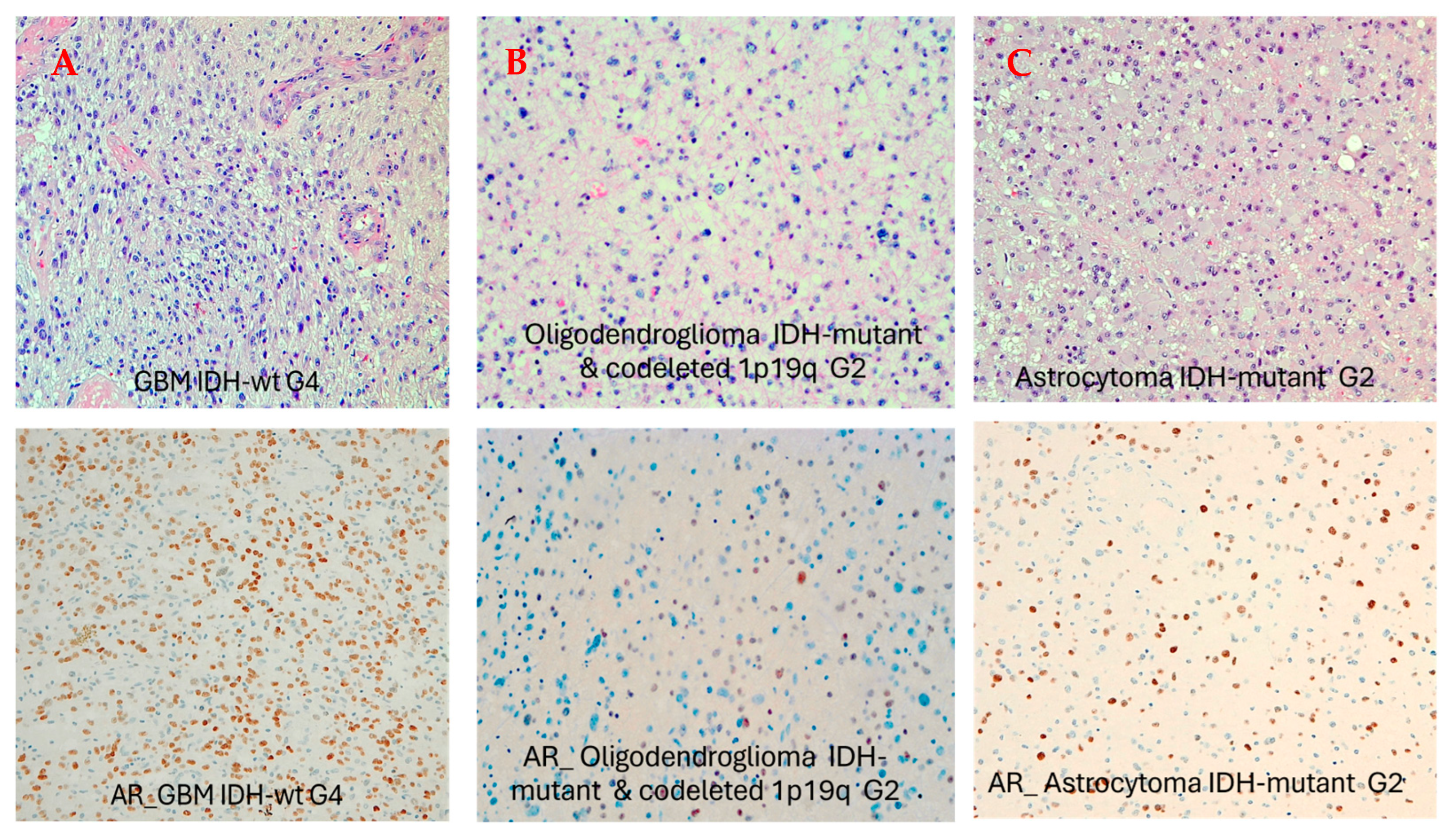
Figure 1. AR nuclear expression in gliomas according to histology and grading. (A) a case of AR-positive GBM IDH-wt (AR > 70%); (B) a case of grade 2 oligodendroglioma IDH-mutant with negative AR expression; (C) a case of grade 2 astrocytoma IDH-mutant with low AR positivity (focal positivity).
Biomedicines 13 02379 g001
Figure 2. (A,B) AR expression according to histology and to MGMT-methylation status. AR expression was more frequent in GBM IDH-wt and in MGMT-unmethylated gliomas compared with MGMT-methylated tumors (p = 0.02).
Biomedicines 13 02379 g002
Figure 3. AR expression and OS. In our series AR-positive gliomas exhibit a lower OS than AR-negative gliomas (p = 0.0022).
Biomedicines 13 02379 g003
Figure 4. X-chromosome copy number and histology. In our series X-chromosome polysomy resulted more frequently in GBM IDH-wt (p = 0.05).
Biomedicines 13 02379 g004
Figure 5. X-chromosome copy number and MGMT-methylation pattern. In our series X-chromosome polysomy resulted more frequently in MGMT-unmethylated tumors (p = 0.037).
Biomedicines 13 02379 g005
Table 1. Patients clinical-pathological characteristics.
AR-Negative
(n = 29)
AR-Positive
(n = 21)
p-Value
(Chi-Squared Test with Yates Continuity Correction)
Gender
      Male11 (37.9%)15 (71.4%)0.04
      Female18 (62.1%)6 (28.6%)
MGMT
      Unmethylated5 (17.2%)11 (52.4%)0.02
      Methylated24 (82.8%)10 (47.6%)
IDH1
      WT10 (34.4%)15 (71.4%)0.018
      Mutated19 (65.5%)6 (28.6%)
Age
      Mean (SD)48.0 54.0 0.126
      Median (Min, Max)46.3 (26.3, 69.1)53.7 (29.3, 76.7)
Histology
      Grade 3 astrocytoma IDH-mut1 (3.4%)2 (9.5%)0.018
      Grade 3 oligodendroglioma IDH-mut4 (13.8%)0 (0%)
      Grade 2 astrocytoma IDH-mut5 (17.2%)4 (19.0%)
      Grade 2 oligodendroglioma IDH-mut10 (34.5%)1 (4.8%)
      Glioblastoma IDHwt9 (31.0%)14 (66.7%)0.04
Table 2. Cox Regression Model adjusted for MGMT status in GBM IDH-wt group.
VariableHR (95%CI)p-Value
GBM MGMT-methylated vs. unmethylated0.34 (0.12–0.95)0.04
GBM AR-positive vs. negative1.68 (0.67–4.26)0.27
Table 3. Cox Regression Model adjusted for MGMT status and gender in GBM IDH-wt group.
VariableHR (95%CI)p-Value
GBM MGMT-methylated vs. unmethylated0.33 (0.11–0.93)0.04
GBM AR-positive vs. negative2.25 (0.79–6.39)0.12
Gender female vs. male1.9 (0.71–5.17)0.19
Disclaimer/Publisher’s Note: The statements, opinions and data contained in all publications are solely those of the individual author(s) and contributor(s) and not of MDPI and/or the editor(s). MDPI and/or the editor(s) disclaim responsibility for any injury to people or property resulting from any ideas, methods, instructions or products referred to in the content.