Review

The Role of NAD+ in Metabolic Regulation of Adipose Tissue: Implications for Obesity-Induced Insulin Resistance

by
Tatjana Ruskovska
1 and
David A. Bernlohr
2,*
1
Faculty of Medical Sciences, Goce Delcev University, 2000 Stip, North Macedonia
2
Department of Biochemistry, Molecular Biology and Biophysics, University of Minnesota—Twin Cities, Minneapolis, MN 55455, USA
*
Author to whom correspondence should be addressed.
Biomedicines 2023, 11(9), 2560; https://doi.org/10.3390/biomedicines11092560
Submission received: 10 August 2023 / Revised: 31 August 2023 / Accepted: 8 September 2023 / Published: 18 September 2023
(This article belongs to the Special Issue Adipose Tissue in Health and Diseases)

Abstract

:
Obesity-induced insulin resistance is among the key factors in the development of type 2 diabetes, atherogenic dyslipidemia and cardiovascular disease. Adipose tissue plays a key role in the regulation of whole-body metabolism and insulin sensitivity. In obesity, adipose tissue becomes inflamed and dysfunctional, exhibiting a modified biochemical signature and adipokine secretion pattern that promotes insulin resistance in peripheral tissues. An important hallmark of dysfunctional obese adipose tissue is impaired NAD+/sirtuin signaling. In this chapter, we summarize the evidence for impairment of the NAD+/sirtuin pathway in obesity, not only in white adipose tissue but also in brown adipose tissue and during the process of beiging, together with correlative evidence from human studies. We also describe the role of PARPs and CD38 as important NAD+ consumers and discuss findings from experimental studies that investigated potential NAD+ boosting strategies and their efficacy in restoring impaired NAD+ metabolism in dysfunctional obese adipose tissue. In sum, these studies suggest a critical role of NAD+ metabolism in adipose biology and provide a basis for the potential development of strategies to restore metabolic health in obesity.

1. Introduction

Obesity is one of the major health burdens worldwide, leading to increased morbidity and mortality [1,2], particularly during the COVID-19 pandemic [3]. Calorie-rich diets, along with a sedentary lifestyle and inadequate intake of health-promoting food bioactives, overwhelm the adaptive mechanisms resulting in cardiometabolic disorders. Obesity-induced insulin resistance has been recognized as one of the major causative factors for the development of type 2 diabetes (T2D), metabolic dysfunction-associated fatty liver disease, atherogenic dyslipidemia and cardiovascular disease [4,5,6]. Adipose tissue undergoes dramatic phenotypic, histological, and metabolic changes in obesity and has an essential role in the regulation of whole-body metabolism. Healthy, non-obese white adipose tissue (WAT) is insulin sensitive and contributes to whole-body insulin sensitivity, whereas the obese, inflamed, and dysfunctional WAT is insulin resistant and leads to the development of T2D and accompanying cardiometabolic disorders [7,8]. However, molecular mechanisms underlying the effects of obesity on metabolic disturbances in adipose tissue, which further influence whole-body metabolism, are still not completely clear. Therefore, studies directed toward understanding the molecular metabolism of adipose tissue in both the lean and obese states have the potential to provide essential information for the development of novel therapeutic strategies for patients with insulin resistance and metabolic syndrome. Among the plethora of regulators of cellular metabolism that are affected in obesity, sirtuins, which had already been identified as cellular energy sensors [9], have emerged as prominent players. Indeed, an important finding within the context of overall adipose tissue dysfunction in obesity is the down-regulation of the NAD+ (nicotinamide adenine dinucleotide, oxidized form)/SIRT (sirtuin) system [10]. In this chapter, we focus on the current evidence about the role of NAD+ (Figure 1) and NAD+-associated molecules in dysfunctional adipose tissue, exploring the potential strategies for restoring metabolic health in obesity. To this end, we conducted a systematic literature search on PubMed using the following search terms: 1. (nad) AND (adipo*); 2. (nad) AND (adipo*) AND (cd38); 3. (nad) AND (cd38) AND (sirt*); 4. (nad) AND (cd38); 5. (parp) AND (adipo*). Following the initial screening, we selected studies summarizing the role of NAD+ and NAD+-associated molecules in the metabolic regulation of adipose tissue.

2. Impairment of the NAD+/SIRT Pathway in the Obese Adipose Organ

In mammalian cells, four classes of lysine deacetylases have been described, of which class III consists of seven mammalian sirtuins (SIRT1-7) [11]. Sirtuins are an evolutionarily conserved class of NAD+-dependent deacetylases that also act as lysine deacylases and mono-ADP-ribosyltransferases. In particular, SIRT1-3 are strong deacetylases, whereas the others demonstrate a weak or hardly detectable deacetylase activity [12]. The first identified sirtuin was the NAD+-dependent histone deacetylase Sir2 (silent information regulator 2) from Saccharomyces cerevisiae [13,14], which promotes longevity [15]. In mammalian cells, sirtuins are ubiquitous and manifest their catalytic activity not only on histones, but also on a variety of non-histone proteins. Since their activity is dependent upon NAD+, they serve as cellular NAD+ sensors and translate their levels to the regulation of diverse cellular processes. Given their broad role in biology, many research groups have reported on their subcellular localization and functions, reviewed in detail elsewhere [12,16,17].
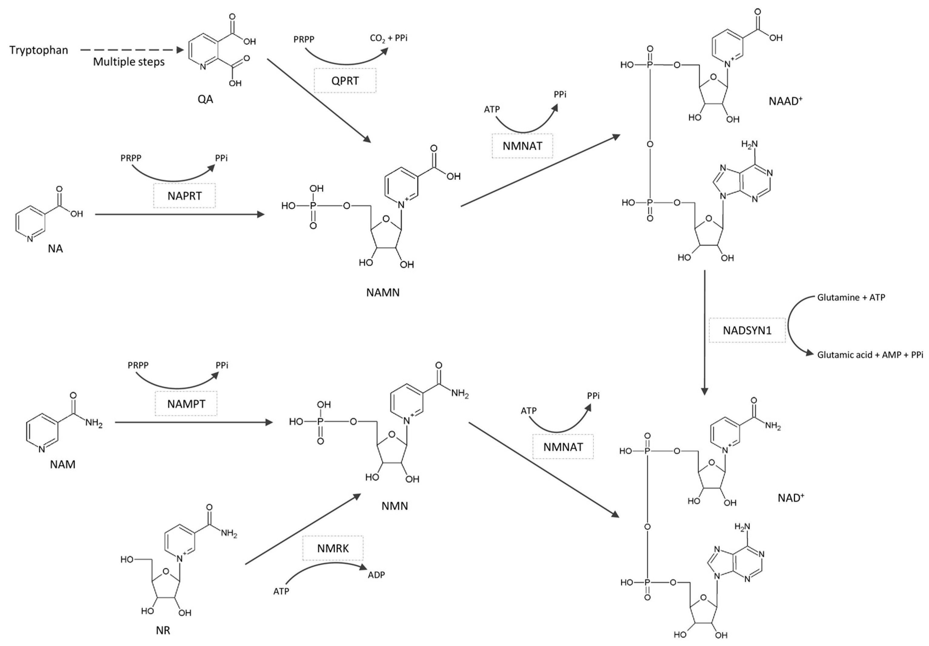
Since sirtuins use NAD+ as a co-substrate, they have been identified as NAD+ consumers. Within the deacetylation reaction, NAD+ is cleaved, producing nicotinamide (NAM), a deacetylated protein and 2′-O-acetyl-ADP-ribose [18] (Figure 2). In subsequent reactions, NAM is recycled for NAD+ biosynthesis (Figure 3), which is essential for the maintenance of cellular metabolism.

2.1. Animal Studies of NAD+/SIRT Pathway

In animal studies, there is strong experimental evidence that NAMPT (nicotinamide phosphoribosyltransferase), the key and rate-limiting enzyme in NAD+ biosynthesis, is down-regulated in obesity. Yoshino et al. (2011) have demonstrated a decrease of NAMPT protein in WAT of mice with high-fat diet (HFD)-induced T2D, with concomitant decrease of NAD+ concentration [19]. In addition, both NAMPT protein and NAD+ levels were decreased in the WAT of aged mice, which opened perspectives for studying the NAD+ metabolism in the context of age-related metabolic disorders [19]. Consistent with these findings, Song et al. (2014) reported up-regulation of NAMPT at both the mRNA and protein levels in visceral adipose tissue (VAT) of rats following calorie restriction (CR), with a concomitant increase of NAD+ concentration [20]. CR was accompanied by a decrease in oxidative stress and an increase in mitochondrial biogenesis in VAT. In addition, Song and coworkers used FK866 to inhibit NAMPT activity, which completely reversed the CR-induced decrease of oxidative stress and reduced the CR-induced increase of mitochondrial biogenesis. At the same time, NAMPT inhibition significantly decreased SIRT1 activity and deacetylation of its targets FOXO1 (forkhead box O1) and PGC1a (PPARg coactivator 1 alpha). Similarly, NAMPT inhibition significantly decreased SIRT3-mediated deacetylation of SOD2 (superoxide dismutase 2), which provides strong evidence for the role of the NAD+/SIRT pathway in the regulation of redox status in VAT.
Drew et al. (2016) further explored the NAD+/SIRT system in mice fed an HFD and demonstrated significant tissue specificity during both the early and late stages of development of obesity and glucose intolerance [21]. Twenty-five enzymes that are part of the NAD+/SIRT system were analyzed at the transcriptional level using a multiplex gene expression assay. In general, a more prominent response was identified in the liver compared to WAT and skeletal muscle. In the WAT, however, HFD modulated PNP (purine nucleoside phosphorylase), NNMT (nicotinamide n-methyltransferase), NMNAT2 (nicotinamide nucleotide adenylyltransferase 2), NMNAT3, SIRT4, SIRT5, and SIRT6. Interestingly, NNMT, the enzyme that catalyzes the reaction of methylation of NAM for its excretion, is up-regulated in the WAT in obesity and T2D. Moreover, NNMT knockdown in mice fed HFD protects against diet-induced obesity, with a concomitant increase of WAT NAD+ and modulated expression of several upstream and downstream genes, including SIRT1 targets [22]. In addition, dysregulation of the NAD+/SIRT system in obesity has profound effects on different fat depots [23].
To characterize the effect of adipocyte NAMPT on whole-body metabolism, an adipocyte-specific NAMPT deletion model (ANKO mouse) has been established [24]. ANKO mice develop severe multi-organ insulin resistance and adipose tissue inflammation. Plasma concentrations of free fatty acids and triglycerides increase, whereas plasma adiponectin and adipsin decrease. ANKO mice display an increased VAT PPARg (peroxisome proliferator-activated receptor gamma) Ser273 phosphorylation, a post-translation modification that impairs its insulin-sensitizing effects and modulated expression of most of its obesity-linked targets at the mRNA level. At the same time, phosphorylation of cyclin-dependent kinase 5, a critical regulator of PPARg Ser273 phosphorylation, is also increased. Importantly, ANKO mice display a concomitant increase in VAT PPARg acetylation. Since previous studies suggest that PPARg Lys293 hyperacetylation leads to increased Ser273 phosphorylation, it can be hypothesized that NAMPT’s influence on adipocyte and whole-body metabolism is mediated by SIRT1 and possibly other sirtuins. Treatment with rosiglitazone, a well-known PPARg agonist, NMN (nicotinamide mononucleotide) or NA (nicotinic acid), restores the described metabolic disturbances in ANKO mice [25]. Similarly, NR (nicotinamide riboside) improves metabolic flexibility in mice fed a mildly obesogenic diet [26].

2.2. In Vitro Studies on 3T3-L1 Adipocytes

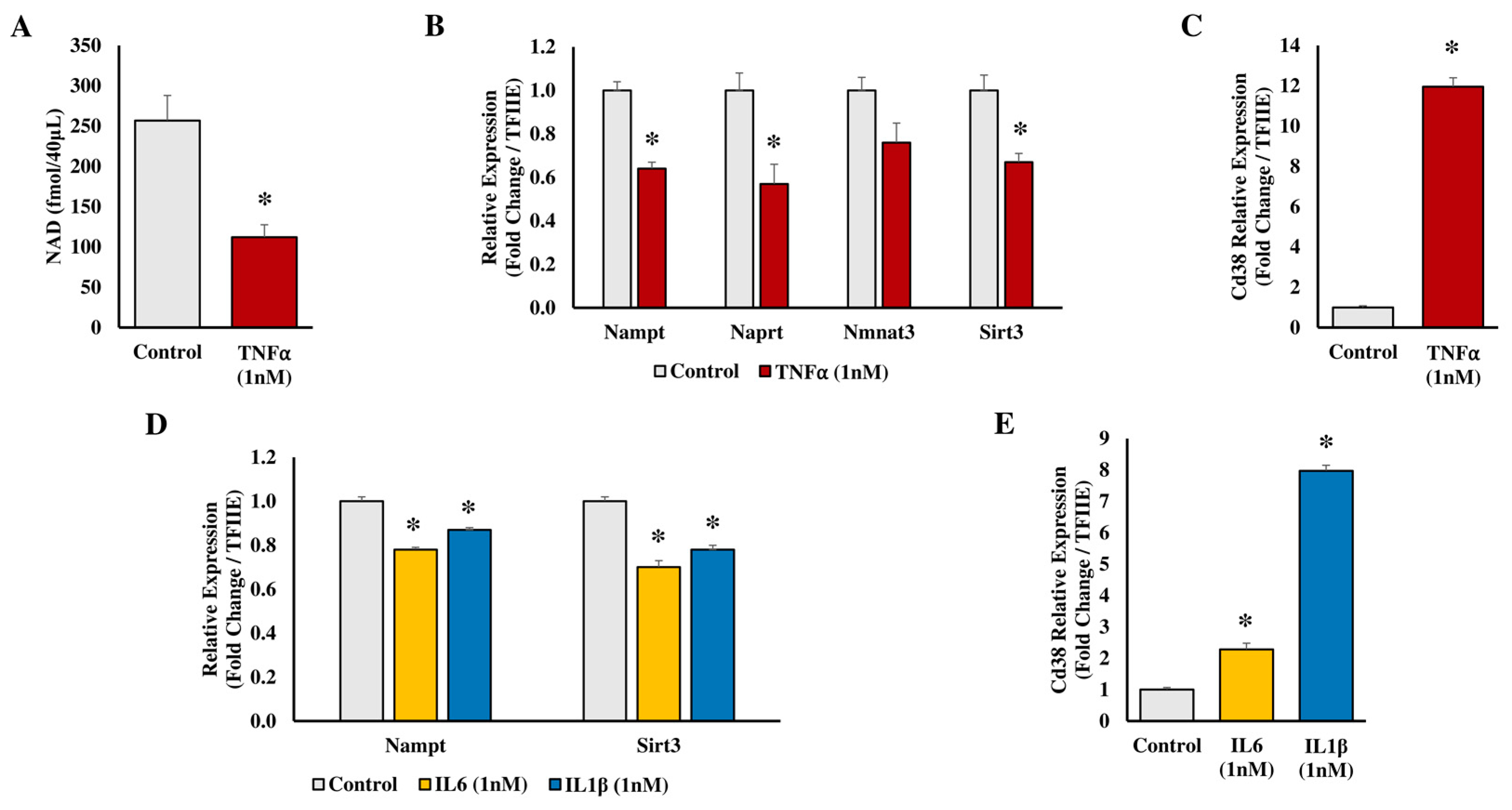
To evaluate disturbances of adipocyte metabolism in inflamed obese adipose tissue, the model of tumor necrosis factor alpha (TNFα)-treated 3T3-L1 adipocytes is widely used. Gouranton et al. (2014) demonstrated that TNFα treatment of cultured 3T3-L1 adipocytes leads to a time- and dose-dependent decrease of NAMPT mRNA [27]. In parallel, intracellular NAMPT protein and NAD+ concentration, CEBPa (CCAAT enhancer binding protein alpha) mRNA and protein, and SIRT1 activity also decrease, whereas SIRT1 mRNA remains unaffected. At the same time, PTP1B (protein-tyrosine phosphatase 1B), a negative regulator of insulin signaling, is up-regulated at both the mRNA and protein levels, which is mediated by the activity of SIRT1. It has also been demonstrated that NAMPT inhibition via either RNAi or FK866 induces an additional increase of PTP1B expression at both the mRNA and protein levels. TNFα is an important, but not the only, mediator of inflammation in dysfunctional obese adipose tissue [28,29]. Indeed, IL1β (interleukin 1 beta) similarly decreased NAMPT and SIRT3 mRNA expression, as well as significantly increased CD38 mRNA [30]. Extending this observation, TNFα treatment of mature 3T3-L1 adipocytes decreased intracellular NAD+ levels and modulated the mRNA expression of genes involved in NAD+ metabolism (Figure 4A–C). IL6 (interleukin 6) or IL1β similarly decreased NAMPT and SIRT3 mRNA expression and significantly increased CD38 mRNA (Figure 4D–E), indicating potential adipocyte metabolic and mitochondrial dysfunction [31]. Consistent with this, Zhang et al. (2020) used the model of SIRT3 overexpression in adipocytes and demonstrated that SIRT3 is a positive regulator of autophagy in mature adipocytes. As such, SIRT3 represents a promising target for the treatment of metabolic dysfunction in obesity [32]. Collectively, these data emphasize the role of TNFα, IL1β and IL6 as major modulators of the NAD+/SIRT axis and insulin signaling in mouse adipocytes.

2.3. Studies in Human Biology

Human studies on visceral adipose tissue metabolism are difficult to conduct, given the difficulty in obtaining samples for analysis. Therefore, most human studies report only results for subcutaneous adipose tissue (SAT). This is a serious shortcoming, given the significant differences in metabolic characteristics of adipose tissue from different fat depots [33]. Despite the limited sample availability, the regulation of NAD+ levels has been suggested as a major differentiator between adipose depots. Indeed, weight loss in response to bariatric surgery revealed a significant increase of NMNAT2 mRNA in SAT but not in VAT. At the same time, the expression of genes associated with mitochondrial energy metabolism was also increased, especially in SAT. Therefore, it has been hypothesized that the increase of intracellular NAD+ could boost adipose tissue mitochondrial activity [34]. Similarly, NAMPT protein levels are decreased in the visceral adipose tissue of patients with nonalcoholic fatty liver disease linked to the dysfunction of adipose tissue [35].
In 2016, the results of two human studies were published [10,36], the first of which involved young BMI-discordant monozygotic twins [10]. In this study, microarray analysis of SAT revealed decreased mRNA expression of SIRT1, SIRT3, SIRT5, NAMPT, NMNAT2, NMNAT3 and NMRK (nicotinamide riboside kinase) in heavier co-twins compared to their respective leaner co-twin. At the same time, total PARP (poly (ADP-ribose) polymerase) activity tended to be increased in heavier co-twins. In parallel, the expression of several genes that are involved in mitochondrial unfolded protein response (UPRmt) was significantly lower in heavier co-twins. Importantly, UPRmt is a downstream pathway of the NAD+/SIRT axis and promotes mitochondrial protein homeostasis against proteotoxic stress that is caused by misfolded proteins [37]. Therefore, reduced UPRmt activity in heavier co-twins suggests impairment of mitochondrial protein quality control in obesity, possibly due to a reduction in the NAD+/SIRT pathway activity. In the second study, Rappou et al. (2016) compared clinically healthy obese and lean subjects and monitored the effects of weight loss treatment [36]. Obese subjects had significantly lower NAMPT, SIRT1, SIRT3 and SIRT7 mRNA in SAT, whereas total PARP activity was significantly higher. Consistent with these findings, weight loss treatment caused an increase in SAT SIRT1 and NAMPT mRNAs, whereas total PARP activity decreased. In addition, weight loss was accompanied by a significant decrease in the expression of oxidative stress-related pathways. Importantly, the change in SAT SIRT1 mRNA during the weight loss treatment was positively correlated with the change in brown adipose tissue (BAT) glucose uptake. This finding sheds light on the metabolic function of BAT in adults and the possible involvement of the NAD+/SIRT pathway in energy dissipation.
Like white adipose tissue, which has been considered a metabolically inactive depot of triglycerides, the metabolic function of brown adipose tissue in humans has also been incompletely understood. The presence and the role of BAT had been described in newborns, but it was believed that it is completely absent in adults. The main function of brown adipocytes is the uncoupling of the respiratory chain and oxidative phosphorylation, which leads to energy dissipation and the generation of heat. This process is important for the regulation of body temperature in newborns. At the molecular level, the central role in energy dissipation is attributed to UCP1 (uncoupling protein 1), also known as thermogenin, a transmembrane protein located in the inner mitochondrial membrane. UCP1 increases the permeability of the inner mitochondrial membrane for the protons that have been translocated in the mitochondrial intermembrane space, thus reducing the proton motive force, decreasing ATP synthesis, and increasing heat production [38]. With the development of advanced imaging techniques, such as PET/CT scanning, the presence of various amounts of brown adipose tissue has also been detected in adults [39]. It has been demonstrated that a stay in a cold environment increases the levels of BAT within minutes. However, the activity of BAT is lower in men who are overweight or obese [40].
In parallel with the discovery of BAT in adult humans, the phenomenon of “beiging” of WAT has attracted much interest. Beiging of WAT is an adaptive and reversible response to environmental stimuli, such as cold exposure or sympathetic innervation [41]. Interestingly, it has been demonstrated that the gene expression pattern and immunohistochemical characteristics of adult human BAT are more like rodent beige adipose tissue than the classic BAT present in human newborns and rodents [42]. In comparison to the white adipocytes, beige adipocytes have an increased expression of several “brown genes”, which gives them attributes of energy-dissipating cells. Therefore, the beige adipose tissue is less effective in energy storage and has the potential to attenuate obesity and insulin resistance, making it a promising therapeutic target [43].

2.4. The Role of NAD+ in Regulating Brown Adipose Tissue

While human studies on the NAD+/SIRT pathway in VAT are rare, studies of BAT and/or beiging of WAT are similarly difficult. However, several comprehensive experimental studies provide strong evidence of the essential role of NAD+ in the metabolic function of BAT. To this end, Yamaguchi et al. (2019) used the previously established ANKO mouse, which lacks NAMPT in both WAT and BAT and demonstrated that ANKO mice have severe cold intolerance and impaired thermogenic responses to fasting and β-adrenergic stimulation [44]. It has also been demonstrated that the BAT of ANKO mice has extremely low NAD+ concentration and exhibits clear signs of hypertrophy and whitening. In the same study, Yamaguchi et al. also used a mouse model lacking NAMPT only in the BAT (i.e., BANKO mouse) and demonstrated normal thermogenic responses due to the essential role of NAMPT in catecholamine-induced lipolysis in the WAT, acting as a regulator of NAD+/SIRT1/Caveolin-1 axis to provide fatty acids as a fuel supply for BAT thermogenesis. Treatment of ANKO mice with NMN rescued the above-described metabolic abnormalities, thus confirming the indispensable role of NAD+ in the regulation of adaptive thermogenesis and whole-body energy metabolism [44]. Similarly, Crisol et al. (2018) have demonstrated that supplementation with NR induces thermogenesis in mice, which is accompanied by an increase in UCP1 protein and PGC1a mRNA in the BAT [45].
In addition to enzymes whose roles in NAD+ metabolism have been well defined, Nguyen et al. (2020) have characterized AIFM2 (apoptosis-inducing factor mitochondria associated 2) and its potential role as a regulator of NAD+ levels [46]. AIFM2 is highly expressed in mouse BAT as well as mouse inguinal WAT (iWAT) and human SAT. Upon cold exposure or β-adrenergic stimulation, AIFM2 translocates from the lipid droplets to the outer side of the mitochondrial inner membrane, where it acts to regenerate the cytosolic NAD+ and provide electrons to the electron transport chain, thus supporting the glycolysis that is needed for thermogenesis. Interestingly, although in the fasted state, BAT uses primarily the free fatty acids released from WAT for thermogenesis, Nguyen et al. propose that in the fed state with high circulating glucose, the glucose may serve as a major fuel for thermogenesis, with AIFM2 playing a critical role by supporting glycolysis. Indeed, AIFM2-knockout mice have impaired thermogenesis, alongside decreased BAT and iWAT NAD+/NADH, increased body weight and adiposity [46]. Therefore, understanding the role of AIFM2 in the adipose tissue may provide future therapeutic targets for diet-induced obesity and insulin resistance.
Wang et al. (2013) demonstrated that erythropoietin (EPO) has beneficial effects in obesity-induced insulin resistance in mice, and these effects are NAD+/SIRT1-mediated [47]. Erythropoietin receptor (EPOR) is highly expressed in the mouse WAT, with approximately 60% of its expression in hematopoietic tissue [48], suggesting that, in addition to its role in erythropoiesis, EPO also plays an important metabolic role. A mouse model of adipocyte-specific deletion of EPOR exhibits obesity, glucose intolerance and insulin resistance, especially when mice are challenged with a high-fat diet. Concordantly, treatment with EPO improves metabolic parameters in diet-induced obese wild-type mice. Aiming to reveal molecular mechanisms underlying the beneficial effects of EPO on metabolic health, Wang et al. demonstrated that EPO had virtually no effect on the BAT but significantly regulated SAT suggesting this depot as the main EPO target. Moreover, EPO induced an increase in the intracellular and tissue NAD+ needed for the activation of SIRT1 and the acquisition of brown adipocyte gene expression in white adipocytes.

3. The Role of Adipose PARPs and CD38 as NAD+ Consumers

The maintenance of intracellular NAD+ concentrations is critical for the proper activity of sirtuins, which have the capacity to ameliorate the negative effects of overnutrition and physical inactivity on mitochondrial function and metabolic health. Intracellular NAD+ is regulated by both NAD+ biosynthesis and NAD+ consumption, and in this section, we focus on two major NAD+ consumers, PARPs and CD38.

3.1. PARPs in Adipose Tissue

PARPs catalyze mono and poly-ADP-ribosylation, a post-translational protein modification where ADP-ribose is transferred to the target protein in a reaction that consumes NAD+ to produce NAM and ADP-ribose [49]. In addition to classical PARP functions that primarily include DNA repair, transcriptional regulation and apoptosis, recent studies point to a role in inflammation, mitochondrial function, cancer biology, circadian rhythm and aging. Focusing on the metabolic properties of PARPs, the interaction between PARP1 and SIRT1 emerges as an important regulatory mechanism. Using the same co-substrate, the balance between these two metabolic regulators is primarily determined by their Km values for NAD+. Interestingly, the PARP1 Km for NAD+ is lower than that for SIRT1, and upon its activation, PARP1 rapidly consumes the intracellular NAD+, thus disabling the SIRT1 activity. Conversely, SIRT1 can act as a negative regulator of PARP1 through the inhibition of transcription or deacetylation. Together, PARP1 and SIRT1 reciprocally regulate NF-κB, the pivotal mediator of inflammatory response, with PARP1 participating as a transcriptional co-activator of NF-κB, while SIRT1 inhibits NF-κB activity by deacetylating RelA/p65 [50].
The role of PARP1 in the metabolic function of adipose tissue has been studied in the mouse model of PARP1 deletion [51]. PARP1−/− mice are leaner and exhibit increased energy expenditure compared to their PARP1+/+ littermates. The analysis of BAT shows markedly increased NAD+ levels, increased SIRT1 activity (as demonstrated by reduced acetylation of its targets PGC1a and FOXO1) and increased mitochondrial biogenesis. At the transcriptional level, there is significant modulation of the genes involved in mitochondrial respiration, uncoupling, fatty acid oxidation and thyroid hormone activation. The metabolic involvement of PARP1 has also been demonstrated in the offspring of mice fed a high-calorie diet prior to and during gestation and lactation [52]. PARP1 protein level was increased in the SAT of offspring of mice fed a high-calorie diet, which was accompanied by complex metabolic disturbances, including decreased NAD+/NADH ratio, increased acetylation of FOXO1 indicative for decreased SIRT1 activity and impaired mitochondrial bioenergetics. Supplementation of the maternal diet with niacin partially reversed the metabolic disturbances resulting from the high-calorie diet, decreasing the PARP1 protein level and increasing the NAD+/NADH ratio and SIRT1 activity.

3.2. CD38 in Adipose Tissue

Besides its role as a co-substrate of sirtuins and PARPs, NAD+ also serves as a precursor for the biosynthesis of cyclic ADP-ribose (cADP-ribose) [53], a molecule that plays an important role in calcium signaling as a second messenger. The enzyme that catalyzes the reaction of biosynthesis of cADP-ribose was first discovered in the marine animal Aplysia and was named ADP-ribosyl cyclase [54]. This enzyme is a soluble protein, and therefore, the finding that CD38 is its mammalian homolog was rather surprising as CD38 is a transmembrane protein that was originally identified as a lymphocyte surface antigen. Further experiments have demonstrated the extraordinary multi-functionality of CD38. Indeed, CD38 has the ability to catalyze the conversion of NAD+ to both cADP-ribose (referred to as cyclase activity) and ADP-ribose (referred to as NADase activity) [55]. It has also been demonstrated that CD38 can hydrolyze the cADP-ribose to ADP-ribose (Figure 5). Consistent with CD38 as a major regulator of NAD+, Aksoy et al. (2006) demonstrated an increase in tissue NAD+ levels in CD38 knockout mice relative to wild-type animals [56].
CD38 has been extensively studied in the context of research, diagnostics and treatment of multiple myeloma [57], chronic lymphocytic leukemia [58], acute myeloid leukemia [59], autism spectrum disorder [60] and allergic airway disease [61]. However, there are only a handful of papers about the involvement of CD38 in metabolic disturbances in obesity. The San Antonio Family Heart Study was the first study that linked blood lipids (triglycerides and HDL) with CD38 and the risk for the development of metabolic syndrome [62]. Of note is the pioneering work of Barbosa et al. (2007), who demonstrated that CD38 knockout mice are resistant to high-fat diet-induced obesity, as they exhibit an enhanced energy expenditure. This phenomenon is mediated, at least in part, by the NAD+-dependent SIRT1/PGC1a axis that regulates mitochondrial biogenesis and energy homeostasis [63]. A follow-up study confirmed that CD38 knockout mice are protected from diet-induced obesity and demonstrated high NAD+ levels in their WAT and BAT on a high-fat, high-sucrose diet [64].
CD38 is among the genes that are significantly up-regulated in adipose tissue of high-fat diet-induced obese rats [65]. Consistent with this finding, high-fat-fed obese mice exhibit a 2-fold increase in CD38 mRNA in WAT [66]. Slightly discrepant results were reported by Drew et al. (2016), who demonstrated only a minor increase in CD38 mRNA in WAT [21]. CD38 mRNA in adipose tissue is up-regulated not only in animal models of diet-induced obesity but also in some genetic models. The publicly available database from the University of Wisconsin (http://diabetes.wisc.edu, accessed on 8 October 2022) provides information that CD38 mRNA is increased in adipose tissue of genetically obese mice of B6 strain in comparison to lean mice but not in genetically obese BTBR mice. In vitro experiments using cultured 3T3-L1 adipocytes demonstrated a marked increase in CD38 mRNA upon TNFα, IL6 or IL1β treatment [30,31].
Even though not analyzed in the context of diet-induced obesity, Benzi et al. (2021) demonstrated a marked down-regulation of CD38 at both mRNA and protein levels in the BAT of wild-type mice during cold exposure, accompanied by an increase in NAD+ levels. The effect of cold exposure was slightly different in WAT, where the significant down-regulation of CD38 was accompanied by a strong increase in NAD(P)H levels. Experiments conducted using CD38 knockout mice supported these findings, leading to the conclusion that CD38 can be considered a relevant molecular target to combat obesity-induced insulin resistance [67]. An overview of rodent studies on the role of NAD+ in adipose tissue is presented in Table 1.

4. Strategies for NAD+ Boosting in Adipose Tissue

Several research groups have directed their interest toward exploring NAD+-boosting strategies for regulating adipose energy metabolism. Calorie restriction [20] has been reported as a potential NAD+ boosting strategy but, in general, it is difficult to implement in humans. Even though some of the earlier studies have analyzed the metabolic effects of different NAD+ precursors on adipose tissue [25,26,44,45,52], latter studies of the role of NAD+ in the metabolic regulation of dysfunctional obese adipose tissue have primarily focused on the effects of supplementation with NAD+ precursors. For example, studying the metabolic effects of NAM supplementation in B57BL/6J male mice fed a high-fat diet, Méndez-Lara et al. (2021) demonstrated its capacity to reduce body weight gain and thus protect against diet-induced obesity. The mechanisms involved, among others, include increased global energy expenditure and induction of iWAT beiging, as well as increased mitochondrial β-oxidation of fatty acids and increased NAD+ levels in the iWAT [68]. In a similar experimental design, Luo et al. (2022) confirmed that NAM supplementation can ameliorate diet-induced obesity in B57BL/6J male mice. They also confirmed previously observed effects of NAM supplementation in adipose tissue, such as enhanced mitochondrial function and increased NAD+ levels. Surprisingly, this study also revealed that NAM supplementation can increase glutathione biosynthesis in the adipose tissue, which is of importance for maintaining cellular redox homeostasis [69].
Even though animal studies have clearly demonstrated the beneficial metabolic effects of NAD+ precursors on dysfunctional obese adipose tissue, translation of this experimental evidence to the human population has proven challenging [70,71]. Within these efforts, it will be essential to take into consideration the safety, efficacy and side effects of supplementation with NAD+ precursors in humans [71], different delivery routes [72], but also factors that determine interindividual variabilities [73,74], such as sex, age, comorbidities, medication and epigenetic factors.

5. Summary and Conclusions

NAD+ is one of the key non-protein molecules in cellular biology, which, since its discovery, has continuously surprised with new and previously unknown functions. NAD+ acts as a cofactor for several oxidoreductases that catalyze key biochemical reactions of the cellular energy metabolism and transports the reducing equivalents to the electron transport chain, thus being an important mediator in ATP biosynthesis. In addition, several non-redox functions of NAD+ have been reported, including being a donor of ADP-ribose in the reactions of ADP-ribosylation, a precursor of the second messenger cyclic ADP-ribose, and a co-substrate of sirtuins. The discovery of sirtuins and the elucidation of their versatile cellular regulatory functions were crucial for increased interest in this already well known “old” molecule.
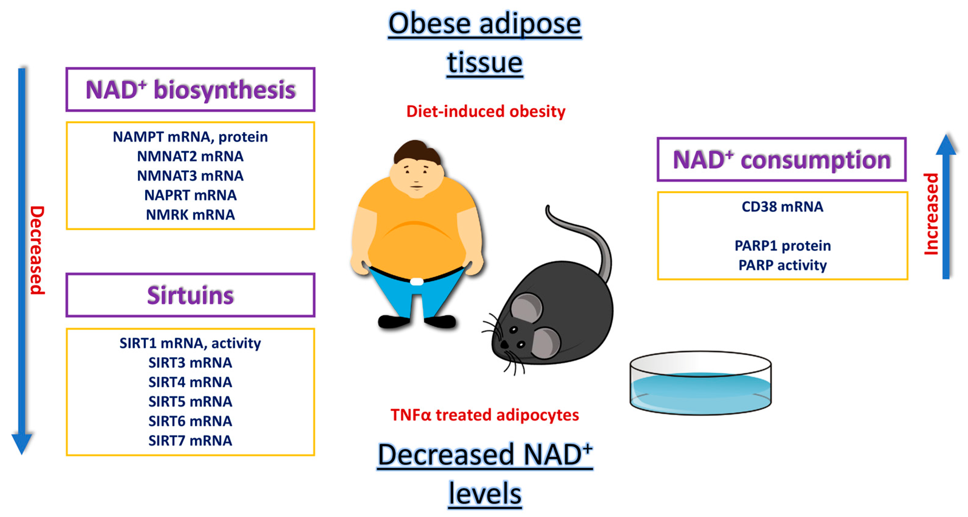
Compared to other metabolically active tissues, the non-redox functions of NAD+ in adipose tissue remain the least investigated and understood, mainly due to the inherent nature of adipose tissue, characterized by its remarkable dynamics and heterogeneity. Moreover, there is a significant involvement of NAD+ in the metabolic remodeling of adipose tissue, whether it is in the context of obesity-induced dysfunction or the process of beiging of white adipose tissue. Further investigation into the non-redox functions of NAD+ in adipose tissue represents one of the primary goals in adipose biology, particularly given the development of strategies for NAD+ boosting in the human population (Figure 6). The potential for the development of strategies for reversal of impaired NAD+ metabolism in obese adipose tissue and consequent improvement of metabolic health in humans seems achievable.

Author Contributions

T.R. conceptualized the chapter, wrote the drafts and carried out formal analysis. D.A.B. edited the chapter, validated the conclusions and supervised the writing. All authors have read and agreed to the published version of the manuscript.

Funding

This research was funded by NIH R01 DK053189 to D.A.B. and by a Fulbright Fellowship award to T.R.

Institutional Review Board Statement

Not applicable.

Informed Consent Statement

Not applicable.

Data Availability Statement

Not applicable.

Acknowledgments

T.R. was supported by the Fulbright grant No. 68140160, 2014/15, at the Department of Biochemistry, Molecular Biology and Biophysics, University of Minnesota, Minneapolis, USA. The authors thank Aleksandar Cvetkovski, Goce Delcev University—Stip, for his help in drawing the chemical structures used for the preparation of the figures and Timothy Walseth, University of Minnesota, for conducting NAD+ analysis.

Conflicts of Interest

The authors declare no conflict of interest.

Abbreviations

NAD+, nicotinamide adenine dinucleotide, oxidized form; NAM, nicotinamide; ADPR, ADP-ribose; QA, quinolinic acid; QPRT, quinolinate phosphoribosyltransferase; PRPP, phosphoribosyl pyrophosphate; NA, nicotinic acid; NAPRT, nicotinate phosphoribosyltransferase; NAMN, nicotinic acid mononucleotide; NMNAT, nicotinamide nucleotide adenylyltransferase; NAAD+, nicotinic acid adenine dinucleotide, oxidized form; NADSYN1, NAD synthetase 1; NAMPT, nicotinamide phosphoribosyltransferase; NMN, nicotinamide mononucleotide; NR, nicotinamide riboside; NMRK, nicotinamide riboside kinase 1 (NMRK1) and nicotinamide riboside kinase 2 (NMRK2).

References

  1. Abdelaal, M.; le Roux, C.W.; Docherty, N.G. Morbidity and mortality associated with obesity. Ann. Transl. Med. 2017, 5, 161. [Google Scholar] [CrossRef] [PubMed]
  2. Ghanta, R.K.; LaPar, D.J.; Zhang, Q.; Devarkonda, V.; Isbell, J.M.; Yarboro, L.T.; Kern, J.A.; Kron, I.L.; Speir, A.M.; Fonner, C.E.; et al. Obesity Increases Risk-Adjusted Morbidity, Mortality, and Cost Following Cardiac Surgery. J. Am. Heart Assoc. 2017, 6, e003831. [Google Scholar] [CrossRef] [PubMed]
  3. Tamara, A.; Tahapary, D.L. Obesity as a predictor for a poor prognosis of COVID-19: A systematic review. Diabetes Metab. Syndr. 2020, 14, 655–659. [Google Scholar] [CrossRef] [PubMed]
  4. Ormazabal, V.; Nair, S.; Elfeky, O.; Aguayo, C.; Salomon, C.; Zuñiga, F.A. Association between insulin resistance and the development of cardiovascular disease. Cardiovasc. Diabetol. 2018, 17, 122. [Google Scholar] [CrossRef]
  5. Di Pino, A.; DeFronzo, R.A. Insulin Resistance and Atherosclerosis: Implications for Insulin-Sensitizing Agents. Endocr. Rev. 2019, 40, 1447–1467. [Google Scholar] [CrossRef]
  6. Finck, B.N. Targeting Metabolism, Insulin Resistance, and Diabetes to Treat Nonalcoholic Steatohepatitis. Diabetes 2018, 67, 2485–2493. [Google Scholar] [CrossRef]
  7. Zatterale, F.; Longo, M.; Naderi, J.; Raciti, G.A.; Desiderio, A.; Miele, C.; Beguinot, F. Chronic Adipose Tissue Inflammation Linking Obesity to Insulin Resistance and Type 2 Diabetes. Front. Physiol. 2019, 10, 1607. [Google Scholar] [CrossRef]
  8. Ruskovska, T.; Bernlohr, D.A. Oxidative stress and protein carbonylation in adipose tissue-implications for insulin resistance and diabetes mellitus. J. Proteom. 2013, 92, 323–334. [Google Scholar] [CrossRef]
  9. Chalkiadaki, A.; Guarente, L. Sirtuins mediate mammalian metabolic responses to nutrient availability. Nat. Rev. Endocrinol. 2012, 8, 287–296. [Google Scholar] [CrossRef]
  10. Jukarainen, S.; Heinonen, S.; Rämö, J.T.; Rinnankoski-Tuikka, R.; Rappou, E.; Tummers, M.; Muniandy, M.; Hakkarainen, A.; Lundbom, J.; Lundbom, N.; et al. Obesity Is Associated with Low NAD(+)/SIRT Pathway Expression in Adipose Tissue of BMI-Discordant Monozygotic Twins. J. Clin. Endocrinol. Metab. 2016, 101, 275–283. [Google Scholar] [CrossRef]
  11. Li, P.; Ge, J.; Li, H. Lysine acetyltransferases and lysine deacetylases as targets for cardiovascular disease. Nat. Rev. Cardiol. 2020, 17, 96–115. [Google Scholar] [CrossRef] [PubMed]
  12. Poulose, N.; Raju, R. Sirtuin regulation in aging and injury. Biochim. Biophys. Acta 2015, 1852, 2442–2455. [Google Scholar] [CrossRef] [PubMed]
  13. Klar, A.J.; Fogel, S.; Macleod, K. MAR1-a Regulator of the HMa and HMalpha Loci in Saccharomyces cerevisiae. Genetics 1979, 93, 37–50. [Google Scholar] [CrossRef] [PubMed]
  14. Chang, H.C.; Guarente, L. SIRT1 and other sirtuins in metabolism. Trends Endocrinol. Metab. 2014, 25, 138–145. [Google Scholar] [CrossRef]
  15. Kaeberlein, M.; McVey, M.; Guarente, L. The SIR2/3/4 complex and SIR2 alone promote longevity in Saccharomyces cerevisiae by two different mechanisms. Genes. Dev. 1999, 13, 2570–2580. [Google Scholar] [CrossRef]
  16. Kupis, W.; Pałyga, J.; Tomal, E.; Niewiadomska, E. The role of sirtuins in cellular homeostasis. J. Physiol. Biochem. 2016, 72, 371–380. [Google Scholar] [CrossRef]
  17. Lee, I.H. Mechanisms and disease implications of sirtuin-mediated autophagic regulation. Exp. Mol. Med. 2019, 51, 1–11. [Google Scholar] [CrossRef]
  18. Shi, Y.; Zhou, Y.; Wang, S.; Zhang, Y. Sirtuin Deacetylation Mechanism and Catalytic Role of the Dynamic Cofactor Binding Loop. J. Phys. Chem. Lett. 2013, 4, 491–495. [Google Scholar] [CrossRef]
  19. Yoshino, J.; Mills, K.F.; Yoon, M.J.; Imai, S. Nicotinamide mononucleotide, a key NAD(+) intermediate, treats the pathophysiology of diet- and age-induced diabetes in mice. Cell Metab. 2011, 14, 528–536. [Google Scholar] [CrossRef]
  20. Song, J.; Ke, S.F.; Zhou, C.C.; Zhang, S.L.; Guan, Y.F.; Xu, T.Y.; Sheng, C.Q.; Wang, P.; Miao, C.Y. Nicotinamide phosphoribosyltransferase is required for the calorie restriction-mediated improvements in oxidative stress, mitochondrial biogenesis, and metabolic adaptation. J. Gerontol. A Biol. Sci. Med. Sci. 2014, 69, 44–57. [Google Scholar] [CrossRef]
  21. Drew, J.E.; Farquharson, A.J.; Horgan, G.W.; Williams, L.M. Tissue-specific regulation of sirtuin and nicotinamide adenine dinucleotide biosynthetic pathways identified in C57Bl/6 mice in response to high-fat feeding. J. Nutr. Biochem. 2016, 37, 20–29. [Google Scholar] [CrossRef] [PubMed]
  22. Kraus, D.; Yang, Q.; Kong, D.; Banks, A.S.; Zhang, L.; Rodgers, J.T.; Pirinen, E.; Pulinilkunnil, T.C.; Gong, F.; Wang, Y.C.; et al. Nicotinamide N-methyltransferase knockdown protects against diet-induced obesity. Nature 2014, 508, 258–262. [Google Scholar] [CrossRef] [PubMed]
  23. Wei, X.; Jia, R.; Wang, G.; Hong, S.; Song, L.; Sun, B.; Chen, K.; Wang, N.; Wang, Q.; Luo, X.; et al. Depot-specific regulation of NAD(+)/SIRTs metabolism identified in adipose tissue of mice in response to high-fat diet feeding or calorie restriction. J. Nutr. Biochem. 2020, 80, 108377. [Google Scholar] [CrossRef] [PubMed]
  24. Yoon, M.J.; Yoshida, M.; Johnson, S.; Takikawa, A.; Usui, I.; Tobe, K.; Nakagawa, T.; Yoshino, J.; Imai, S. SIRT1-Mediated eNAMPT Secretion from Adipose Tissue Regulates Hypothalamic NAD+ and Function in Mice. Cell Metab. 2015, 21, 706–717. [Google Scholar] [CrossRef]
  25. Stromsdorfer, K.L.; Yamaguchi, S.; Yoon, M.J.; Moseley, A.C.; Franczyk, M.P.; Kelly, S.C.; Qi, N.; Imai, S.; Yoshino, J. NAMPT-Mediated NAD(+) Biosynthesis in Adipocytes Regulates Adipose Tissue Function and Multi-organ Insulin Sensitivity in Mice. Cell Rep. 2016, 16, 1851–1860. [Google Scholar] [CrossRef]
  26. Shi, W.; Hegeman, M.A.; van Dartel, D.A.M.; Tang, J.; Suarez, M.; Swarts, H.; van der Hee, B.; Arola, L.; Keijer, J. Effects of a wide range of dietary nicotinamide riboside (NR) concentrations on metabolic flexibility and white adipose tissue (WAT) of mice fed a mildly obesogenic diet. Mol. Nutr. Food Res. 2017, 61, 1600878. [Google Scholar] [CrossRef]
  27. Gouranton, E.; Romier, B.; Marcotorchino, J.; Tourniaire, F.; Astier, J.; Peiretti, F.; Landrier, J.F. Visfatin is involved in TNFα-mediated insulin resistance via an NAD(+)/Sirt1/PTP1B pathway in 3T3-L1 adipocytes. Adipocyte 2014, 3, 180–189. [Google Scholar] [CrossRef]
  28. Shapouri-Moghaddam, A.; Mohammadian, S.; Vazini, H.; Taghadosi, M.; Esmaeili, S.A.; Mardani, F.; Seifi, B.; Mohammadi, A.; Afshari, J.T.; Sahebkar, A. Macrophage plasticity, polarization, and function in health and disease. J. Cell Physiol. 2018, 233, 6425–6440. [Google Scholar] [CrossRef]
  29. Engin, A.B. Adipocyte-Macrophage Cross-Talk in Obesity. Adv. Exp. Med. Biol. 2017, 960, 327–343. [Google Scholar] [CrossRef]
  30. Hertzel, A.V.; Yong, J.; Chen, X.; Bernlohr, D.A. Immune Modulation of Adipocyte Mitochondrial Metabolism. Endocrinology 2022, 163, bqac094. [Google Scholar] [CrossRef]
  31. Ruskovska, T.; Bernlohr, D.A. Effects of TNFα, IL6 and IL1β on 3T3-L1 Adipocytes in Culture. Department of Biochemistry, Molecular Biology and Biophysics, University of Minnesota-Twin Cities, Minneapolis, MN, USA. 2015; Unpublished work. [Google Scholar]
  32. Zhang, T.; Liu, J.; Tong, Q.; Lin, L. SIRT3 Acts as a Positive Autophagy Regulator to Promote Lipid Mobilization in Adipocytes via Activating AMPK. Int. J. Mol. Sci. 2020, 21, 372. [Google Scholar] [CrossRef] [PubMed]
  33. Kranendonk, M.E.; van Herwaarden, J.A.; Stupkova, T.; de Jager, W.; Vink, A.; Moll, F.L.; Kalkhoven, E.; Visseren, F.L. Inflammatory characteristics of distinct abdominal adipose tissue depots relate differently to metabolic risk factors for cardiovascular disease: Distinct fat depots and vascular risk factors. Atherosclerosis 2015, 239, 419–427. [Google Scholar] [CrossRef] [PubMed]
  34. Mardinoglu, A.; Heiker, J.T.; Gärtner, D.; Björnson, E.; Schön, M.R.; Flehmig, G.; Klöting, N.; Krohn, K.; Fasshauer, M.; Stumvoll, M.; et al. Extensive weight loss reveals distinct gene expression changes in human subcutaneous and visceral adipose tissue. Sci. Rep. 2015, 5, 14841. [Google Scholar] [CrossRef] [PubMed]
  35. Gaddipati, R.; Sasikala, M.; Padaki, N.; Mukherjee, R.M.; Sekaran, A.; Jayaraj-Mansard, M.; Rabella, P.; Rao-Guduru, V.; Reddy-Duvvuru, N. Visceral adipose tissue visfatin in nonalcoholic fatty liver disease. Ann. Hepatol. 2010, 9, 266–270. [Google Scholar] [CrossRef]
  36. Rappou, E.; Jukarainen, S.; Rinnankoski-Tuikka, R.; Kaye, S.; Heinonen, S.; Hakkarainen, A.; Lundbom, J.; Lundbom, N.; Saunavaara, V.; Rissanen, A.; et al. Weight Loss Is Associated with Increased NAD(+)/SIRT1 Expression but Reduced PARP Activity in White Adipose Tissue. J. Clin. Endocrinol. Metab. 2016, 101, 1263–1273. [Google Scholar] [CrossRef]
  37. Mouchiroud, L.; Houtkooper, R.H.; Moullan, N.; Katsyuba, E.; Ryu, D.; Cantó, C.; Mottis, A.; Jo, Y.S.; Viswanathan, M.; Schoonjans, K.; et al. The NAD(+)/Sirtuin Pathway Modulates Longevity through Activation of Mitochondrial UPR and FOXO Signaling. Cell 2013, 154, 430–441. [Google Scholar] [CrossRef]
  38. Heeren, J.; Münzberg, H. Novel aspects of brown adipose tissue biology. Endocrinol. Metab. Clin. N. Am. 2013, 42, 89–107. [Google Scholar] [CrossRef]
  39. Nedergaard, J.; Bengtsson, T.; Cannon, B. Unexpected evidence for active brown adipose tissue in adult humans. Am. J. Physiol. Endocrinol. Metab. 2007, 293, E444–E452. [Google Scholar] [CrossRef]
  40. van Marken Lichtenbelt, W.D.; Vanhommerig, J.W.; Smulders, N.M.; Drossaerts, J.M.; Kemerink, G.J.; Bouvy, N.D.; Schrauwen, P.; Teule, G.J. Cold-activated brown adipose tissue in healthy men. N. Engl. J. Med. 2009, 360, 1500–1508. [Google Scholar] [CrossRef]
  41. Tamucci, K.A.; Namwanje, M.; Fan, L.; Qiang, L. The dark side of browning. Protein Cell 2018, 9, 152–163. [Google Scholar] [CrossRef]
  42. Wu, J.; Boström, P.; Sparks, L.M.; Ye, L.; Choi, J.H.; Giang, A.H.; Khandekar, M.; Virtanen, K.A.; Nuutila, P.; Schaart, G.; et al. Beige adipocytes are a distinct type of thermogenic fat cell in mouse and human. Cell 2012, 150, 366–376. [Google Scholar] [CrossRef] [PubMed]
  43. Elattar, S.; Satyanarayana, A. Can Brown Fat Win the Battle Against White Fat? J. Cell Physiol. 2015, 230, 2311–2317. [Google Scholar] [CrossRef] [PubMed]
  44. Yamaguchi, S.; Franczyk, M.P.; Chondronikola, M.; Qi, N.; Gunawardana, S.C.; Stromsdorfer, K.L.; Porter, L.C.; Wozniak, D.F.; Sasaki, Y.; Rensing, N.; et al. Adipose tissue NAD(+) biosynthesis is required for regulating adaptive thermogenesis and whole-body energy homeostasis in mice. Proc. Natl. Acad. Sci. USA 2019, 116, 23822–23828. [Google Scholar] [CrossRef] [PubMed]
  45. Crisol, B.M.; Veiga, C.B.; Lenhare, L.; Braga, R.R.; Silva, V.R.R.; da Silva, A.S.R.; Cintra, D.E.; Moura, L.P.; Pauli, J.R.; Ropelle, E.R. Nicotinamide riboside induces a thermogenic response in lean mice. Life Sci. 2018, 211, 1–7. [Google Scholar] [CrossRef]
  46. Nguyen, H.P.; Yi, D.; Lin, F.; Viscarra, J.A.; Tabuchi, C.; Ngo, K.; Shin, G.; Lee, A.Y.; Wang, Y.; Sul, H.S. Aifm2, a NADH Oxidase, Supports Robust Glycolysis and Is Required for Cold- and Diet-Induced Thermogenesis. Mol. Cell 2020, 77, 600–617.e604. [Google Scholar] [CrossRef] [PubMed]
  47. Wang, L.; Teng, R.; Di, L.; Rogers, H.; Wu, H.; Kopp, J.B.; Noguchi, C.T. PPARα and Sirt1 mediate erythropoietin action in increasing metabolic activity and browning of white adipocytes to protect against obesity and metabolic disorders. Diabetes 2013, 62, 4122–4131. [Google Scholar] [CrossRef]
  48. Teng, R.; Gavrilova, O.; Suzuki, N.; Chanturiya, T.; Schimel, D.; Hugendubler, L.; Mammen, S.; Yver, D.R.; Cushman, S.W.; Mueller, E.; et al. Disrupted erythropoietin signalling promotes obesity and alters hypothalamus proopiomelanocortin production. Nat. Commun. 2011, 2, 520. [Google Scholar] [CrossRef]
  49. Szántó, M.; Bai, P. The role of ADP-ribose metabolism in metabolic regulation, adipose tissue differentiation, and metabolism. Genes. Dev. 2020, 34, 321–340. [Google Scholar] [CrossRef]
  50. Cantó, C.; Menzies, K.J.; Auwerx, J. NAD(+) Metabolism and the Control of Energy Homeostasis: A Balancing Act between Mitochondria and the Nucleus. Cell Metab. 2015, 22, 31–53. [Google Scholar] [CrossRef]
  51. Bai, P.; Cantó, C.; Oudart, H.; Brunyánszki, A.; Cen, Y.; Thomas, C.; Yamamoto, H.; Huber, A.; Kiss, B.; Houtkooper, R.H.; et al. PARP-1 inhibition increases mitochondrial metabolism through SIRT1 activation. Cell Metab. 2011, 13, 461–468. [Google Scholar] [CrossRef]
  52. Lettieri-Barbato, D.; D’Angelo, F.; Sciarretta, F.; Tatulli, G.; Tortolici, F.; Ciriolo, M.R.; Aquilano, K. Maternal high calorie diet induces mitochondrial dysfunction and senescence phenotype in subcutaneous fat of newborn mice. Oncotarget 2017, 8, 83407–83418. [Google Scholar] [CrossRef] [PubMed]
  53. Lee, H.C.; Walseth, T.F.; Bratt, G.T.; Hayes, R.N.; Clapper, D.L. Structural determination of a cyclic metabolite of NAD+ with intracellular Ca2+-mobilizing activity. J. Biol. Chem. 1989, 264, 1608–1615. [Google Scholar] [CrossRef] [PubMed]
  54. Lee, H.C.; Aarhus, R. ADP-ribosyl cyclase: An enzyme that cyclizes NAD+ into a calcium-mobilizing metabolite. Cell Regul. 1991, 2, 203–209. [Google Scholar] [CrossRef]
  55. Wei, W.; Graeff, R.; Yue, J. Roles and mechanisms of the CD38/cyclic adenosine diphosphate ribose/Ca(2+) signaling pathway. World J. Biol. Chem. 2014, 5, 58–67. [Google Scholar] [CrossRef] [PubMed]
  56. Aksoy, P.; White, T.A.; Thompson, M.; Chini, E.N. Regulation of intracellular levels of NAD: A novel role for CD38. Biochem. Biophys. Res. Commun. 2006, 345, 1386–1392. [Google Scholar] [CrossRef]
  57. McEllistrim, C.; Krawczyk, J.; O’Dwyer, M.E. New developments in the treatment of multiple myeloma-clinical utility of daratumumab. Biologics 2017, 11, 31–43. [Google Scholar] [CrossRef]
  58. Burgler, S. Role of CD38 Expression in Diagnosis and Pathogenesis of Chronic Lymphocytic Leukemia and Its Potential as Therapeutic Target. Crit. Rev. Immunol. 2015, 35, 417–432. [Google Scholar] [CrossRef]
  59. Ding, Y.; Gao, H.; Zhang, Q. The biomarkers of leukemia stem cells in acute myeloid leukemia. Stem Cell Investig. 2017, 4, 19. [Google Scholar] [CrossRef]
  60. Higashida, H.; Yuhi, T.; Akther, S.; Amina, S.; Zhong, J.; Liang, M.; Nishimura, T.; Liu, H.X.; Lopatina, O. Oxytocin release via activation of TRPM2 and CD38 in the hypothalamus during hyperthermia in mice: Implication for autism spectrum disorder. Neurochem. Int. 2018, 119, 42–48. [Google Scholar] [CrossRef]
  61. Deshpande, D.A.; Guedes, A.G.P.; Lund, F.E.; Subramanian, S.; Walseth, T.F.; Kannan, M.S. CD38 in the pathogenesis of allergic airway disease: Potential therapeutic targets. Pharmacol. Ther. 2017, 172, 116–126. [Google Scholar] [CrossRef]
  62. Cai, G.; Cole, S.A.; Freeland-Graves, J.H.; MacCluer, J.W.; Blangero, J.; Comuzzie, A.G. Principal component for metabolic syndrome risk maps to chromosome 4p in Mexican Americans: The San Antonio Family Heart Study. Hum. Biol. 2004, 76, 651–665. [Google Scholar] [CrossRef] [PubMed]
  63. Barbosa, M.T.; Soares, S.M.; Novak, C.M.; Sinclair, D.; Levine, J.A.; Aksoy, P.; Chini, E.N. The enzyme CD38 (a NAD glycohydrolase, EC 3.2.2.5) is necessary for the development of diet-induced obesity. Faseb J. 2007, 21, 3629–3639. [Google Scholar] [CrossRef] [PubMed]
  64. Chiang, S.H.; Harrington, W.W.; Luo, G.; Milliken, N.O.; Ulrich, J.C.; Chen, J.; Rajpal, D.K.; Qian, Y.; Carpenter, T.; Murray, R.; et al. Genetic Ablation of CD38 Protects against Western Diet-Induced Exercise Intolerance and Metabolic Inflexibility. PLoS ONE 2015, 10, e0134927. [Google Scholar] [CrossRef] [PubMed]
  65. Qiu, J.; Cheng, R.; Zhou, X.Y.; Zhu, J.G.; Zhu, C.; Qin, D.N.; Kou, C.Z.; Guo, X.R. Gene expression profiles of adipose tissue of high-fat diet-induced obese rats by cDNA microarrays. Mol. Biol. Rep. 2010, 37, 3691–3695. [Google Scholar] [CrossRef] [PubMed]
  66. Wang, L.F.; Miao, L.J.; Wang, X.N.; Huang, C.C.; Qian, Y.S.; Huang, X.; Wang, X.L.; Jin, W.Z.; Ji, G.J.; Fu, M.; et al. CD38 deficiency suppresses adipogenesis and lipogenesis in adipose tissues through activating Sirt1/PPARγ signaling pathway. J. Cell Mol. Med. 2018, 22, 101–110. [Google Scholar] [CrossRef]
  67. Benzi, A.; Sturla, L.; Heine, M.; Fischer, A.W.; Spinelli, S.; Magnone, M.; Sociali, G.; Parodi, A.; Fenoglio, D.; Emionite, L.; et al. CD38 downregulation modulates NAD(+) and NADP(H) levels in thermogenic adipose tissues. Biochim. Biophys. Acta Mol. Cell Biol. Lipids 2021, 1866, 158819. [Google Scholar] [CrossRef]
  68. Méndez-Lara, K.A.; Rodríguez-Millán, E.; Sebastián, D.; Blanco-Soto, R.; Camacho, M.; Nan, M.N.; Diarte-Añazco, E.M.G.; Mato, E.; Lope-Piedrafita, S.; Roglans, N.; et al. Nicotinamide Protects Against Diet-Induced Body Weight Gain, Increases Energy Expenditure, and Induces White Adipose Tissue Beiging. Mol. Nutr. Food Res. 2021, 65, e2100111. [Google Scholar] [CrossRef]
  69. Luo, C.; Yang, C.; Wang, X.; Chen, Y.; Liu, X.; Deng, H. Nicotinamide reprograms adipose cellular metabolism and increases mitochondrial biogenesis to ameliorate obesity. J. Nutr. Biochem. 2022, 107, 109056. [Google Scholar] [CrossRef]
  70. Nascimento, E.B.M.; Moonen, M.P.B.; Remie, C.M.E.; Gariani, K.; Jörgensen, J.A.; Schaart, G.; Hoeks, J.; Auwerx, J.; van Marken Lichtenbelt, W.D.; Schrauwen, P. Nicotinamide Riboside Enhances In Vitro Beta-adrenergic Brown Adipose Tissue Activity in Humans. J. Clin. Endocrinol. Metab. 2021, 106, 1437–1447. [Google Scholar] [CrossRef]
  71. Poljšak, B.; Kovač, V.; Milisav, I. Current Uncertainties and Future Challenges Regarding NAD+ Boosting Strategies. Antioxidants 2022, 11, 1637. [Google Scholar] [CrossRef]
  72. Sabbagh, F.; Kim, B.S. Ex Vivo Transdermal Delivery of Nicotinamide Mononucleotide Using Polyvinyl Alcohol Microneedles. Polymers 2023, 15, 2031. [Google Scholar] [CrossRef] [PubMed]
  73. Bañales-Luna, M.; Figueroa-Vega, N.; Marín-Aragón, C.I.; Perez-Luque, E.; Ibarra-Reynoso, L.; Gallardo-Blanco, H.L.; López-Aguilar, I.; Malacara, J.M. Associations of nicotidamide-N-methyltransferase, FTO, and IRX3 genetic variants with body mass index and resting energy expenditure in Mexican subjects. Sci. Rep. 2020, 10, 11478. [Google Scholar] [CrossRef] [PubMed]
  74. Gray, S.R.; Aird, T.P.; Farquharson, A.J.; Horgan, G.W.; Fisher, E.; Wilson, J.; Hopkins, G.E.; Anderson, B.; Ahmad, S.A.; Davis, S.R.; et al. Inter-individual responses to sprint interval training, a pilot study investigating interactions with the sirtuin system. Appl. Physiol. Nutr. Metab. 2018, 43, 84–93. [Google Scholar] [CrossRef] [PubMed]
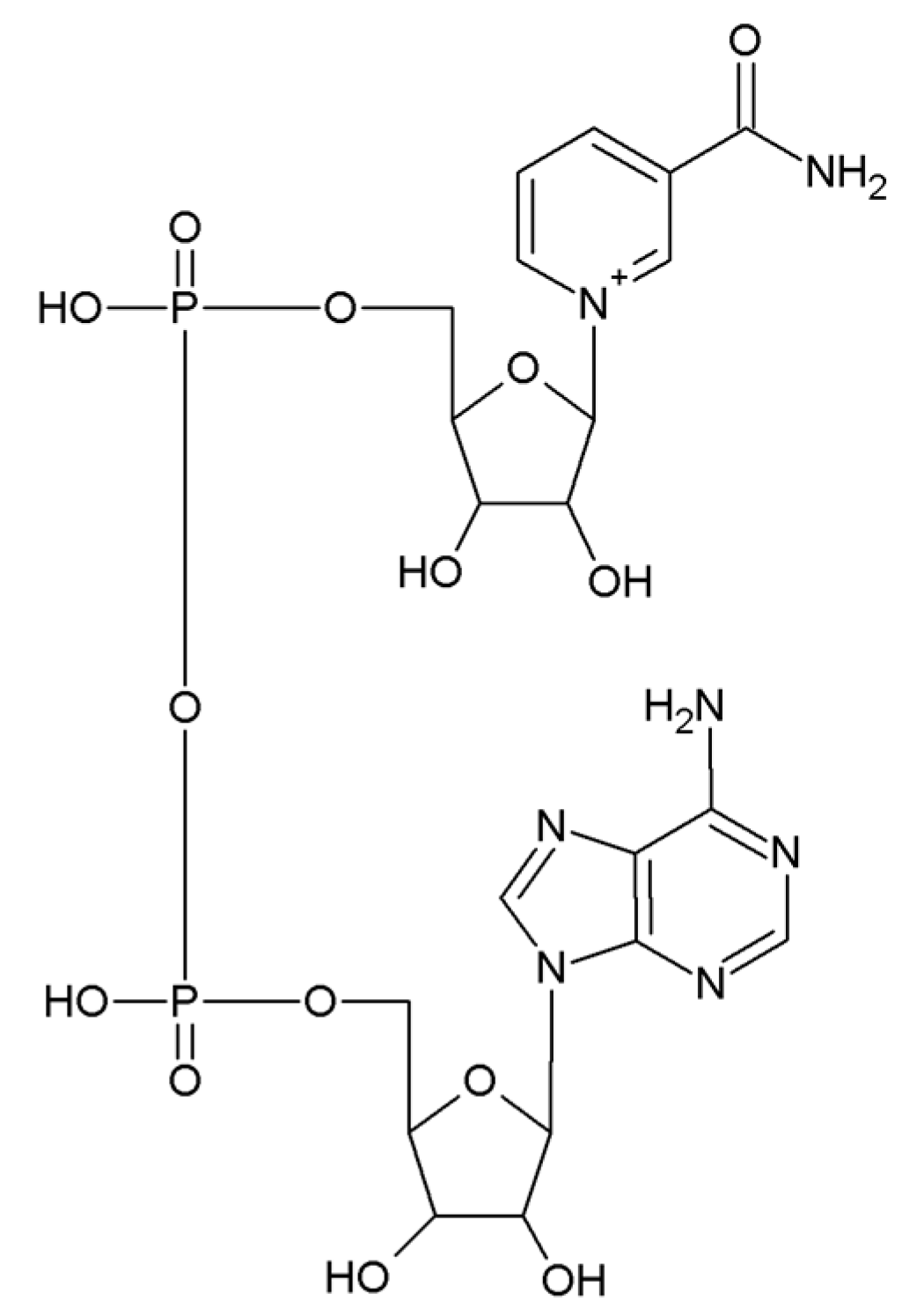
Figure 1. Chemical structure of NAD+.
Biomedicines 11 02560 g001
Figure 2. Sirtuin-mediated protein deacetylation.
Biomedicines 11 02560 g002
Figure 3. Pathways for NAD+ biosynthesis in mammalian cells.
Biomedicines 11 02560 g003
Figure 4. Effects of TNFα, IL6 and IL1β on 3T3-L1 adipocytes in culture. Differentiated 3T3-L1 adipocytes were treated with the indicated levels of inflammatory factors for 24 h, and extracts prepared for metabolite and mRNA qPCR analysis. (A) NAD+ in TNFα-treated 3T3-L1 adipocytes, (B) Nampt, Naprt, Nmnat3, and Sirt3 in TNFα-treated 3T3-L1 adipocytes, (C) Cd38 in TNFα-treated 3T3-L1 adipocytes, (D) Nampt and Sirt3 in IL6- or IL1β-treated 3T3-L1 adipocytes, (E) Cd38 in IL6- or IL1β-treated 3T3-L1 adipocytes. * p < 0.01.
Biomedicines 11 02560 g004
Figure 5. Multi-functionality of the CD38-catalyzed reaction.
Biomedicines 11 02560 g005
Figure 6. Summary of the evidence for impaired NAD+ metabolism in dysfunctional adipose tissue.
Biomedicines 11 02560 g006
Table 1. Overview of rodent studies on the role of NAD+ in adipose tissue.
ModelType of Adipose TissueMajor Metabolic Outcome/s
High-fat diet
(HFD)
White adipose tissue (WAT)Type 2 diabetes
Decreased NAD+ levels [19]
Obesity
Glucose intolerance
Altered regulation of the SIRT/NAD+ system [21]
Calorie restriction (CR)Visceral adipose tissue (VAT)Increased NAD+
Decreased oxidative stress
Increased mitogenesis [20]
Nnmt knockdown and high-fat dietWhite adipose tissue (WAT)Reduced diet-induced obesity
Enhanced insulin sensitivity
Increased energy expenditure
Increased NAD+ [22]
Adipocyte-specific Nampt knockout (ANKO)Visceral adipose tissue (VAT)
Subcutaneous adipose tissue (SubQ)
Multi-organ insulin resistance
Adipose tissue inflammation
Decrease of plasma adiponectin and adipsin [25]
Adipocyte-specific Nampt knockout (ANKO)Brown adipose tissue (BAT)Cold intolerance
Impaired thermogenic response
Decreased NAD+ in BAT
Hypertrophy and whitening of BAT [44]
High-fat diet supplemented with nicotinamide riboside (NR)Epididymal white adipose tissue (eWAT)Improved metabolic flexibility with NR addition [26]
Regular diet supplement with nicotinamide riboside (NR)Brown adipose tissue (BAT)Reduced abdominal visceral fat
Increased heat production [45]
Whole body Aifm2-knockoutBrown adipose tissue (BAT)
Inguinal white adipose tissue (iWAT)
Impaired thermogenesis
Decreased BAT and iWAT NAD+/NADH ratio
Increased body weight and adiposity [46]
Aifm2-overexpression in UCP1+ cellsBrown adipose tissue (BAT)
Inguinal white adipose tissue (iWAT)
Increased thermogenesis
Increased BAT and iWAT NAD+/NADH ratio
Decreased body weight and adiposity [46]
Adipocyte-specific deletion of erythropoietin receptorSubcutaneous WAT (SubQ)Obesity
Glucose intolerance
Insulin resistance
Decreased oxygen consumption
Decreased NAD+ in SubQ [47]
PARP1 knockout mouseBrown adipose tissue (BAT)Reduced fat accumulation
Higher energy expenditure
Higher NAD+ in BAT
Higher mitochondrial content in BAT [51]
Maternal high-calorie dietSubcutaneous WAT (SubQ) of newborn mice at the end of lactationIncreased SubQ mass
Increased adipocyte size
Decreased NAD+/NADH ratio
Increased oxidative damage to mitochondrial proteins
Impaired mitochondrial bioenergetics [52]
Maternal high-calorie diet supplemented with niacinSubcutaneous WAT (SubQ) of newborn mice at the end of lactationIncreased NAD+/NADH ratio in SubQ [52]
CD38 knockout mice on high-fat high-sucrose dietWhite adipose tissue (WAT)
Brown adipose tissue (BAT)
Higher levels of NAD+ in WAT and BAT
Improved metabolic flexibility [64]
CD38 knockout mice on high-fat dietWhite adipose tissue (WAT)Less body weight gain [66]
Cold exposureInterscapular brown adipose tissue (iBAT)Down-regulation of CD38 at both mRNA and protein levels in iBAT
Increased NAD+ in iBAT [67]
Cold exposure, CD38 knockout miceInterscapular brown adipose tissue (iBAT)Increased NAD+ in iBAT [67]
Disclaimer/Publisher’s Note: The statements, opinions and data contained in all publications are solely those of the individual author(s) and contributor(s) and not of MDPI and/or the editor(s). MDPI and/or the editor(s) disclaim responsibility for any injury to people or property resulting from any ideas, methods, instructions or products referred to in the content.