Article

Association between Low-Density Lipoprotein Cholesterol and Vascular Biomarkers in Primary Prevention

1
Department of Clinical and Experimental Medicine, University of Pisa, 56126 Pisa, Italy
2
Esaote SpA, 16152 Genova, Italy
3
Department of Surgical, Medical and Molecular Pathology and Critical Care Medicine, School of Medicine, University of Pisa, 56126 Pisa, Italy
4
Imaging Department, Fondazione Toscana G. Monasterio, 54100 Massa, Italy
5
Bioengineering and Deep Health Units, Fondazione Toscana G. Monasterio, 54100 Massa, Italy
*
Author to whom correspondence should be addressed.
Biomedicines 2023, 11(6), 1753; https://doi.org/10.3390/biomedicines11061753
Submission received: 18 May 2023 / Revised: 8 June 2023 / Accepted: 13 June 2023 / Published: 18 June 2023

Abstract

:
Several noninvasive vascular biomarkers have been proposed to improve risk stratification for atherothrombotic events. To identify biomarkers suitable for detecting intermediate-risk individuals who might benefit from lipid-lowering treatment in primary prevention, the present study tested the association of plasma LDL-cholesterol with coronary artery calcification (CAC) Agatston score, high carotid and femoral intima-media thickness (IMT), low carotid distensibility and high carotid-femoral pulse-wave velocity in 260 asymptomatic individuals at intermediate cardiovascular risk and without diabetes and lipid-lowering treatment. High or low vascular biomarkers were considered when their value was above the 95th or below the 5th percentile, respectively, of the distribution in the healthy or in the study population. LDL-cholesterol was independently associated with the CAC score = 0 (OR 0.67; 95%CI 0.48–0.92, p = 0.01), CAC score > 100 (1.59; 1.08–2.39, p = 0.01) and high common femoral artery (CFA) IMT (1.89; 1.19–3.06, p < 0.01), but not with other biomarkers. Our data confirm that in individuals at intermediate risk, lipid-lowering treatment can be avoided in the presence of a CAC score = 0, while it should be used with a CAC score > 100. CFA IMT could represent a useful biomarker for decisions regarding lipid-lowering treatment. However, sex- and age-specific reference values should be established in a large healthy population.

Graphical Abstract

1. Introduction

Low-density lipoprotein cholesterol (LDL-C) is the most abundant atherogenic lipoprotein in plasma, and its infiltration of the arterial wall is considered a key event in the initiation and progression of the atherosclerotic process. Increased plasma LDL-C levels are causally related to atherosclerotic cardiovascular (CV) disease [1] and lowering LDL-C values by lifestyle or therapeutic interventions has been shown to reduce the risk of CV events, both in primary and secondary prevention [1,2,3,4,5].
The 2019 ACC/AHA Guideline on the Primary Prevention of Cardiovascular Disease [6] suggests statin treatment not only in individuals with diabetes, very high LDL-C levels (≥190 mg/dL) or high 10-year risk of CV disease (≥20%), but even in individuals at intermediate CV risk (≥7.5–20%), in the presence of risk enhancers such as coronary artery calcification (CAC). In the absence of CAC, statin therapy can be avoided or withdrawn, while in the presence of the Agatston CAC score above 100, statins are recommended at any age.
CAC, a highly specific feature of coronary atherosclerosis, is the most predictive single CV risk marker in primary prevention. CAC scoring using the Agatston method was successfully incorporated into the coronary heart disease prediction model based on traditional risk factors [7,8,9,10]. However, the utility of the Agatston score for evaluating the effectiveness of statin therapy is limited because the score considers both the total area and the maximal density of coronary calcification, and as demonstrated by serial intravascular ultrasound, intensive statin therapy induces regression of atheroma volume but promotes its calcification [11,12]. This process leads to the stabilization of the atherosclerotic lesion but may increase the CAC score. Various studies have shown that statin treatment can accelerate coronary calcification [13,14], even as it reduces all-cause mortality and major vascular events among people without evidence of CV disease [15]. Therefore, the CAC score helps define individual CV risk. Still, repeated computed tomography to assess the response to statin therapy is probably inappropriate, considering also the cumulative radiation dose [16].
Other noninvasive vascular biomarkers, such as carotid and femoral intima-media thickness (IMT) and plaques or carotid and aortic stiffness, have been proposed for risk estimation and decision-making regarding statin treatment, as well as for assessing response to statin therapy. Previous studies have demonstrated that statins can slow the progression of carotid IMT and improve arterial stiffness [17,18,19,20], but this beneficial effect is not necessarily related to the main action of statins, i.e., lowering serum LDL-C levels. Statins have pleiotropic effects that include reducing inflammatory cytokines and reactive oxygen species, inhibiting smooth muscle cell proliferation, improving endothelial function and lowering blood pressure (BP) [21,22,23,24,25]. These mechanisms may be involved in reducing arterial wall thickness and stiffness.
The aim of this cross-sectional study was to evaluate the association of various noninvasive vascular biomarkers [26,27] with plasma LDL-C levels in order to identify biomarkers that could reflect the impact of LDL-C on vascular trees and could be used to select individuals suitable for lipid-lowering treatment and to monitor its effect. The study population included asymptomatic individuals at intermediate 10-year risk of CV disease, without diabetes and with LDL-C ≥ 190 mg/dL, i.e., individuals for whom additional risk assessment is recommended before starting statin therapy [6]. Vascular biomarkers tested were CAC score, high common carotid artery (CCA) or common femoral artery (CFA) IMT, low CCA distension and high carotid-femoral pulse wave velocity (cfPWV). Abnormally high or low vascular biomarkers were considered when their value was above the 95th percentile or below the 5th percentile [28], respectively, of distribution in a healthy population (if data in a healthy population were available) or in the study population (if data in healthy population were not available).

2. Materials and Methods

2.1. Study Population and Protocol

The study population is a part of the population enrolled in the prospective cohort study “MHeLP, Montignoso Heart-Lung Project”, aimed at defining the predictive value of CAC score for CV events in a community-based (the village of Montignoso, Tuscany, Italy) general population. The original population consisted of 638 individuals. For this study, we included only individuals aged 40–75 years, without CV symptoms, lipid-lowering therapy, and diabetes, with plasma LDL-C levels < 190 mg/dL and at intermediate 10-year risk of CV disease as estimated by Framingham Risk Score. The final population consists of 260 individuals.
All individuals underwent an examination protocol that included anthropometry, brachial BP measurements, a fasting blood test, ECG, a high-resolution carotid and femoral ultrasound and a computed tomography scan. Carotid-femoral pulse wave velocity was measured in 182 individuals. Hypertension was defined as systolic BP > 140 mmHg and/or diastolic BP  > 90 mmHg or hypertensive treatment [29].

2.2. Body Size and BP Measurement

Body weight and height were measured, and body mass index (BMI) was calculated. Waist circumference was measured as the narrowest circumference between the lower rib margin and anterior superior iliac crest. Brachial BP was measured at two visits by a validated digital electronic tensiometer (Omron, model 705cp, Kyoto, Japan) in participants seated for at least 10 min, using regular or large adult cuffs according to the arm circumference. Two measurements were taken at both visits, separated by 2-min intervals, and the average was calculated. The average of two separate visits was used to estimate BP (mmHg). Pulse pressure was calculated as the difference between systolic and diastolic BP.

2.3. Assessment of the 10-Year Risk of CV Disease

A 10-year risk of CV disease was estimated by the Framingham risk score prediction model that considers age, total cholesterol, high-density lipoprotein cholesterol (HDL-C), systolic brachial BP, ongoing treatment of hypertension, smoking and diabetes status [30]. The risk was classified as intermediate when the 10-year risk of CV disease ranged between 7.5 and 20% [6].

2.4. Assessment of LDL-C Plasma Level

Plasma LDL-C was measured by direct enzymatic colorimetric method (Beckman Coulter, Danaher Corporation, Sunnyvale, CA, USA). LDL-C levels were considered as optimal (<100 mg/dL), near-optimal (100–129 mg/dL), borderline high (130–159 mg/dL) and high (160–189 mg/dL) as recommended [31].

2.5. Coronary Calcium Score (CAC)

A low-dose radiation scan without contrast agent was done (120 KV, 60 mA) by a 64-detector scanner (Aquilion 64; Toshiba Medical Systems, Otawara, Japan) (2–3 mSv estimated dosimetry, with 1- and 3-mm collimation and reconstruction thickness, respectively). Prospective electrocardiographic triggering in sequential slice mode was used for scanning the heart. The CAC score was calculated by Agatston [32] with a dedicated program available (Vitrea 2.0; Vital Images Inc., Minnetonka, MN, USA). The Agatston score was determined by multiplying the area of calcification expressed in mm3 by the corresponding density number using the following density scale (1 = 130–199 Hounsfield units (HU), 2 = 200–299 HU, 3 = 300–399 HU, 4 = ≥400 HU). For this study, we identified individuals with a CAC score = 0 and a CAC score > 100.

2.6. Vascular Examination

All vascular examinations were carried out by the same operator (G.J.) in a quiet room with a stable temperature of 22 °C on individuals resting comfortably for at least 15 min in the supine position. All individuals were asked to abstain from cigarette smoking, caffeine and alcohol consumption and vigorous physical activity for 24 h.
Carotid ultrasound was performed on the right CCA using an ultrasound scanner equipped with a 10 MHz linear probe (MyLab 70, Esaote, Genova, Italy) and implemented with a previously validated radiofrequency-based tracking of the arterial wall (QIMT®, QAS®, Esaote, Genova, Italy) that allows an automatic and real-time determination of far-wall CCA IMT, CCA outer diameter and distension with a high spatial and temporal resolution (sampling rate of 550 Hz on 32 lines). CCA IMT, diameter and distension were measured within a rectangular 1-cm-long ROI placed approximately 1 cm before the flow divider. IMT was defined as the distance between the lumen-intima and media-adventitia interfaces of the far (posterior) wall. Distensibility coefficient (DC) was calculated from the distension curves as follows: DC = (∆A/A)/PP, where A = π ∗ (D/2)2, ∆A = π ∗ [(D + ∆D)/2]2—π ∗ (D/2)2, D = diastolic outer diameter and PP = Pulse Pressure [33].
Femoral ultrasound was performed on the right CFA using the same ultrasound scanner with radiofrequency-based tracking of the arterial wall. Far wall IMT was measured within a rectangular 1-cm-long ROI placed approximately 1 cm before the flow divider.
To identify individuals with high CCA IMT, the sex- and age-specific normality tables deriving from IMT measurements performed by the same radiofrequency-based system in 4234 healthy men and women were used [34], and a cutoff point of the 95th percentile for given sex and age was adopted [28]. To identify individuals with low CCA DC, the sex- and age-specific normality tables deriving from DC measurements performed by the same radiofrequency-based system in 3601 healthy men and women were used [33], and a cutoff point of the 5th percentile for given sex and age was adopted [28]. As no normalcy data were published for CFA IMT, a cutoff point of the 95th percentile of CFA IMT distribution in our population was adopted for defining high CFA IMT.
Radiofrequency-derived measures represent an average of over six consecutive cardiac beats. The mean of two acquisitions was used for statistical analysis. BP was measured at the left brachial artery (Omron, Kyoto, Japan) during each acquisition of the distension curves. Intra-individual variability of acquisitions was evaluated in 25 volunteers, in whom the acquisitions were performed in two sessions separated by 30 min. Brachial pulse pressure was comparable between the two acquisitions (p = 0.88). Intra-individual variability of CCA IMT, CCA distension and CFA IMT was 6.7 ± 4.2, 8.7 ± 6.4% and 8.1 ± 5.4%, respectively.
Carotid-femoral pulse wave velocity (cfPWV) was measured in 182 participants according to current guidelines [35] using the Complior device (Alam Medical, Vincennes, France). To identify individuals with high cfPWV, the sex-, age- and mean BP-specific normality tables deriving from cfPWV measurements performed in 2158 healthy men and women were used [36] and a cutoff point of the 95th percentile for given sex, age and mean BP was adopted [28]. Intra-individual variability of carotid-femoral PWV measurement was 4.3 ± 2.8%.

2.7. Statistical Analysis
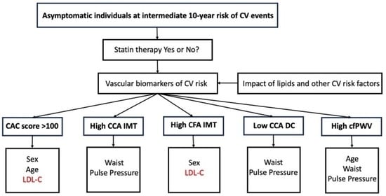
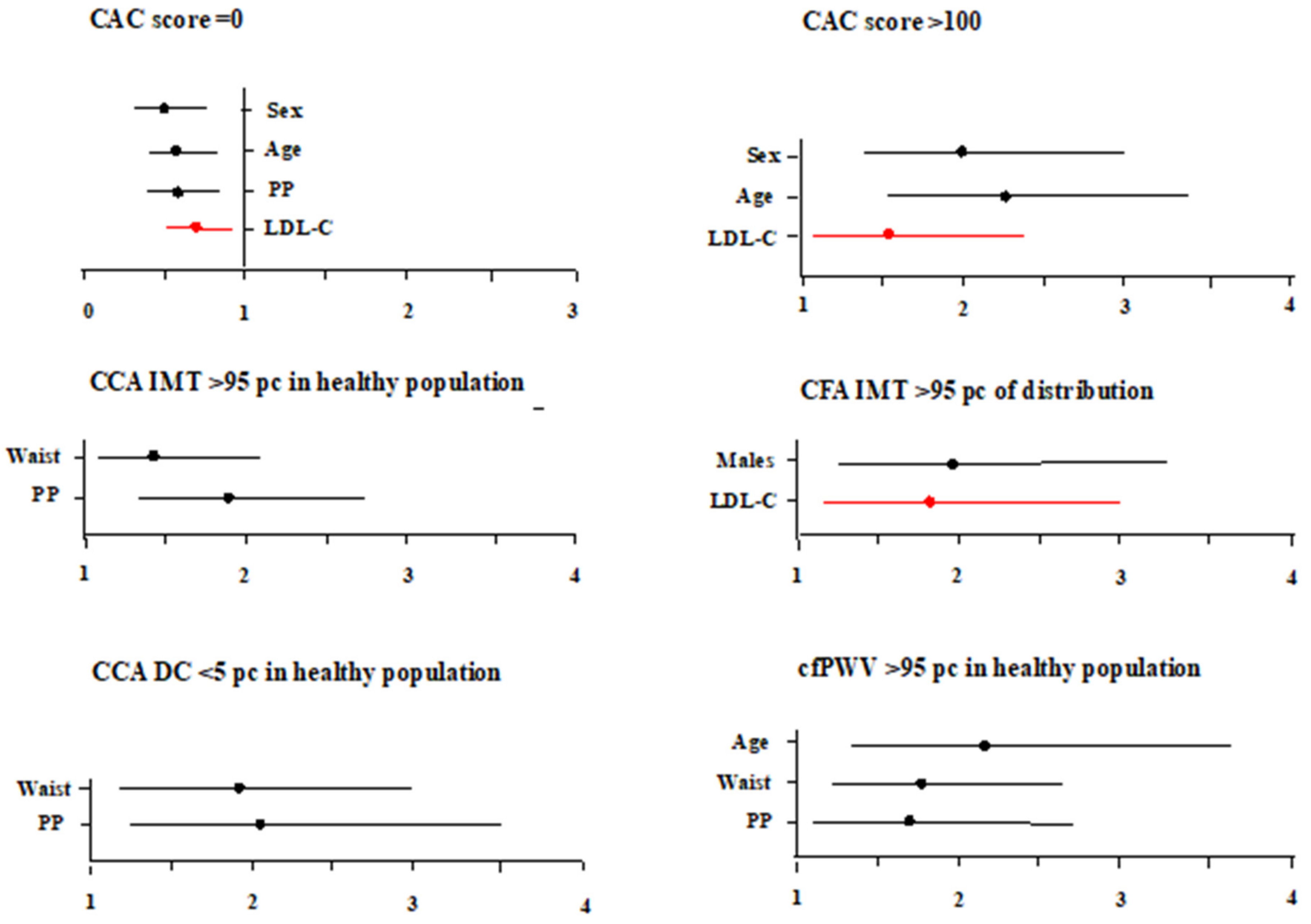
Data are expressed as mean ± SD, and categorical data as percentages. Variables with skewed distribution were summarized as median [interquartile range] and were logarithmically transformed for parametric statistical analysis. Multivariate logistic regression was performed to identify risk factors associated with CAC score = 0, CCA score > 100, high CCA IMT, high CFA IMT, low CCA DC and high cfPWV. Results are given as odds ratio (OR) and 95% confidence interval (CI). ORs were calculated for 1SD of the continuous variable. Statistical tests were two-sided, and significance was set at a value of p < 0.05.

3. Results

Characteristics of the study population are reported in Table 1. Twenty-nine individuals had optimal LDL-C levels, ninety-three near optimal levels, ninety-five borderline high and forty-three high levels.
Table 2 reports data on CAC score, CCA IMT and distensibility, CFA IMT and cfPWV. One hundred eighty-two individuals had CAC score = 0, 36 had CAC score > 100, 49 had high CCA IMT, 15 had low CCA DC, 25 had high CFA IMT, and 27 had high cfPWV.
Figure 1 reports risk factors associated with CAC score = 0, CAC score > 100, high CCA IMT or CFA IMT, low CCA DC and high cfPWV. LDL-C was independently associated with CAC score = 0, CAC score > 100 and high CFA IMT, but not with high CCA IMT, low CCA DC or high cfPWV. Neither HDL-C nor triglycerides were related to any vascular biomarker.
Odds ratios are calculated for 1SD of the continuous variable.

4. Discussion

In our asymptomatic population at intermediate CV risk, LDL-C was associated with coronary calcification and CFA IMT in the highest 5 percent distribution. Neither high CCA IMT, low carotid distensibility, nor high aortic stiffness was associated with plasma lipids.
These results confirm the association between LDL-C levels and coronary calcification in asymptomatic patients at intermediate CV risk. The 2019 ACC/AHA Guideline on the Primary Prevention of Cardiovascular Disease [6] does not recommend statin therapy in intermediate-risk individuals if the CAC score is 0, while it recommends starting statin therapy at any age if the CAC score is greater than 100. In our population, LDL-C was associated with a CAC score of 0 with OR = 0.67 and a CAC score above 100 with OR = 1.59.
The relationship between LDL-C exposure and vascular calcification has been demonstrated in both clinical and experimental studies. It reflects the fact that in the arterial wall are cells capable of osteoblastic differentiation and mineralization and that oxidized lipids accumulated in the subendothelial space induce differentiation of these cells and promote calcification [37,38]. Vascular smooth muscle cells, endothelial cells, fibroblasts, mesenchymal stem cells, and endothelial progenitor cells can transdifferentiate into osteoblast-like cells [39]. However, calcification in atherosclerotic lesions is associated with both progression of disease (microcalcification) and the healing of inflammation (macrocalcification), and statins may reduce arterial wall inflammation through a variety of mechanisms [40]. In the Heinz Nixdorf Recall Study, statin intake enhanced CAC progression, mostly in the less advanced stage of atherosclerosis, but this progression did not increase the risk for coronary events [14]. A meta-analysis of 7 studies suggested that in asymptomatic populations at high risk of CV diseases, statins do not reduce or enhance CAC score but slightly slow its progression, above all in individuals with CAC score > 400 [41]. CAC score can therefore help adjust individual CV risk and select suitable candidates for statin therapy, but its value for monitoring treatment efficacy is limited.
Among the other vascular biomarkers tested in our study, neither high CCA IMT, low CCA distensibility, nor high aortic stiffness was related to plasma LDL-C. On the other hand, all these biomarkers were independently associated with pulse pressure and waist circumference. Pulse pressure is considered a major mechanical determinant of arterial remodeling because repeated cyclic stress exerts a fatiguing effect on the load-bearing elements of the arterial media, above all on elastin, causing its fracture and degeneration and leading to luminal enlargement and wall stiffening. Luminal enlargement is accompanied by a further increase in circumferential wall stress, which activates intracellular signaling pathways promoting smooth muscle cell proliferation and migration, resulting in wall thickening and reducing stress [42]. Indeed, the results of various studies suggest that an increase in carotid IMT reflects, rather than atherosclerosis, a physiologic remodeling aimed to maintain stable circumferential wall stress when BP increases [43,44]. One of the pleiotropic effects of statins is BP reduction [25], above all in individuals with higher BP and in users of anti-hypertensive medication [45,46]. Thus, the reported deceleration of carotid IMT progression with statin therapy [17,18,19] could be partially explained by the decrease in BP and arterial wall stress and by the inhibition of smooth muscle cell proliferation and migration [24]. In a longitudinal community-based Taiwanese study evaluating the effect of changes in LDL-C and systolic BP on carotid IMT, baseline IMT was associated with both LDL-C and BP, but changes in IMT during a 6-year observational period were associated only with changes in BP [47].
Another independent determinant of high CCA IMT, low CCA distensibility and high aortic stiffness was waist circumference. Central obesity is related to low-grade systemic inflammation [48,49], and in hypercholesterolemic patients, low-grade systemic inflammation and abdominal fat were the main determinants of reduced arterial distensibility [50]. Similarly, in patients with type 2 diabetes mellitus, carotid IMT was associated with waist circumference and plasma inflammatory markers [51]. Therefore, anti-hypertensive and anti-inflammatory action of statins might explain the positive effect of stain therapy on elastic arteries structure and function, independent of LDL-C levels [21,25].
Unlike CCA IMT, high CFA IMT was independently associated with plasma LDL-C. Previous studies comparing the relationships between risk factors and IMT in the carotid and femoral arteries have not reported significant differences. In the AXA study [52], evaluating the association between large-artery wall thickness and risk factors in men and women 17 to 65 years old, both carotid and femoral IMT were associated with age, BMI, BP, total cholesterol and glucose, and in a study including normocholesterolemic and hypercholesterolemic subjects, carotid and femoral IMTs increased with total cholesterol (r = 0.35, p < 0.001 for both arteries) and LDL-C (r = 0.33, p < 0.001 and r = 0.34, p < 0.001, respectively) in a similar way [53]. Yet, when the impact of 2-year simvastatin treatment on carotid and femoral IMT was assessed in patients with familial hypercholesterolemia, a more important IMT regression was observed in the femoral than in the carotid artery (−0.283 vs. −0.053 mm). In addition, hypercholesterolemic patients with a history of CV disease compared with those without CV disease had significantly higher CFA IMT at baseline (2.31 ± 0.81 vs. 1.72 ± 0.63 mm, p < 0.001), whereas differences in CCA IMT were less significant (0.99 ± 0.17 vs. 0.90 ± 0.19 mm, p = 0.01) [54]. Based on these results, authors suggested that the femoral artery could be more sensitive to the reduction of LDL-C levels and that a greater regression of femoral IMT might be clinically relevant because the association between IMT and CV disease was better in the femoral compared to the carotid artery. Likewise, in the Regression Growth Evaluation Study (REGRESS), a double-blind, placebo-controlled, prospective study of 885 men with coronary artery disease, the most significant effect of pravastatin was seen in the CFA IMT. During the 2-year observational period, far wall IMT in CFA increased by 0.13 ± 0.05 (SE) mm in the placebo group but decreased by 0.06 ± 0.05 mm in the pravastatin group (p = 0.004). In contrast, the two groups did not differ for changes in far wall CCA IMT (0.05 ± 0.02 vs. 0.04 ± 0.02 mm; p = 0.67) [55].

4.1. Study Limitations

We cannot evaluate the association between LDL-C levels and carotid or femoral plaques because the prevalence of plaques in our asymptomatic participants at intermediate CV risk was very low. C-reactive protein or other markers of systemic inflammation were not assessed.

4.2. Conclusions

Among vascular biomarkers used in primary prevention of asymptomatic individuals at intermediate 10-year CV risk, only coronary calcification, and CFA IMT in the highest 5 percent of distribution were independently related to plasma LDL-C levels. To identify intermediate-risk individuals who can benefit from lipid-lowering therapy and to monitor its effect, age- and sex-specific CFA IMT reference values should be established in a large healthy population, as has been done for CCA IMT, CCA DC and cfPWV. High CCA IMT, low CCA distension and high aortic stiffness were not associated with blood lipids but with pulse pressure and waist circumference. The latter observation suggests that the positive effect of statins on the elastic artery wall thickness and stiffness, already described by others, could be independent of LDL-C and mediated by the anti-hypertensive and anti-inflammatory actions of statins. Overall, our data indicate that different vascular biomarkers may reflect the impact of different risk factors on CV systems and that assessing more than one biomarker could provide a more accurate estimate of CV risk.

Author Contributions

M.K., C.P. and D.C. contributed to the study design. D.C., C.M., G.J., S.C. and D.D.L. contributed to the acquisition and analysis of data. M.K. and C.P. contributed to the interpretation of data. All authors critically revised the paper and gave their final approval. All authors have read and agreed to the published version of the manuscript.

Funding

This research received no external funding.

Institutional Review Board Statement

The study protocol conformed to the ethical guidelines of the 1975 Declaration of Helsinki and was approved by the “Comitato Etico di Area Vasta Nord-Ovest” (CEAVNO); approval code number 2514; approval date 1 September 2008.

Informed Consent Statement

All individuals gave their informed consent to participate.

Data Availability Statement

The data presented in this study are available on request from the corresponding author. The data are not publicly available due to ethical and privacy reasons.

Acknowledgments

Authors sincerely thank Eng. Roberto Vagheggi, CEO Chlor Alkali division of Esseco Group, Altair Chimica, Saline di Volterra (Pisa, Italy), for his unconditioned support of this work.

Conflicts of Interest

M.K. is responsible for clinical studies in Esaote SpA; D.D.L. is affiliated with Terarecon Inc., Durham, NC 27703.

References

  1. von Birgelen, C.; Hartmann, M.; Mintz, G.S.; Baumgart, D.; Schmermund, A.; Erbel, R. Relation between progression and regression of atherosclerotic left main coronary artery disease and serum cholesterol levels as assessed with serial long-term (> or =12 months) follow-up intravascular ultrasound. Circulation 2003, 108, 2757–2762. [Google Scholar] [CrossRef] [PubMed] [Green Version]
  2. Nissen, S.E.; Nicholls, S.J.; Sipahi, I.; Libby, P.; Raichlen, J.S.; Ballantyne, C.M.; Davignon, J.; Erbel, R.; Fruchart, J.C.; Tardif, J.C.; et al. Effect of very high-intensity statin therapy on regression of coronary atherosclerosis: The ASTEROID trial. JAMA 2006, 295, 1556–1565. [Google Scholar] [CrossRef] [PubMed] [Green Version]
  3. Nicholls, S.J.; Tuzcu, E.M.; Sipahi, I.; Grasso, A.W.; Schoenhagen, P.; Hu, T.; Wolski, K.; Crowe, T.; Desai, M.Y.; Hazen, S.L.; et al. Statins, high-density lipoprotein cholesterol, and regression of coronary atherosclerosis. JAMA 2007, 297, 499–508. [Google Scholar] [CrossRef] [PubMed] [Green Version]
  4. Mach, F.; Baigent, C.; Catapano, A.L.; Koskinas, K.C.; Casula, M.; Badimon, L.; Chapman, M.J.; De Backer, G.G.; Delgado, V.; Ference, B.A.; et al. 2019 ESC/EAS Guidelines for the management of dyslipidaemias: Lipid modification to reduce cardiovascular risk. Eur. Heart J. 2020, 41, 111–188. [Google Scholar] [CrossRef] [Green Version]
  5. Jeong, S.M.; Choi, S.; Kim, K.; Kim, S.M.; Lee, G.; Park, S.Y.; Kim, Y.Y.; Son, J.S.; Yun, J.M.; Park, S.M. Effect of change in total cholesterol levels on cardiovascular disease among young adults. J. Am. Heart Assoc. 2018, 7, e008819. [Google Scholar] [CrossRef] [Green Version]
  6. Arnett, D.K.; Blumenthal, R.S.; Albert, M.A.; Buroker, A.B.; Goldberger, Z.D.; Hahn, E.J.; Himmelfarb, C.D.; Khera, A.; Lloyd-Jones, D.; McEvoy, J.W.; et al. 2019 ACC/AHA Guideline on the primary prevention of cardiovascular disease: A report of the American College of Cardiology/American Heart Association Task Force on Clinical Practice Guidelines. Circulation 2019, 140, e596–e646. [Google Scholar] [CrossRef]
  7. Sarwar, A.; Shaw, L.J.; Shapiro, M.D.; Blankstein, R.; Hoffmann, U.; Cury, R.C.; Abbara, S.M.; Brady, T.J.; Budoff, M.J.; Blumenthal, R.S.; et al. Diagnostic and prognostic value of absence of coronary artery calcification. JACC Cardiovasc. Imaging 2009, 2, 675–688. [Google Scholar] [CrossRef] [Green Version]
  8. Lo-Kioeng-Shioe, M.S.; Rijlaarsdam-Hermsen, D.; van Domburg, R.T.; Hadamitzky, M.; Lima, J.A.C.; Hoeks, S.E.; Deckers, J.W. Prognostic value of coronary artery calcium score in symptomatic individuals: A meta-analysis of 34,000 subjects. Int. J. Cardiol. 2020, 299, 56–62. [Google Scholar] [CrossRef]
  9. Greenland, P.; Blaha, M.J.; Budoff, M.J.; Erbel, R.; Watson, K.E. Coronary calcium score and cardiovascular risk. J. Am. Coll. Cardiol. 2018, 72, 434–447. [Google Scholar] [CrossRef]
  10. McClelland, R.L.; Jorgensen, N.W.; Budoff, M.; Blaha, M.J.; Post, W.S.; Kronmal, R.A.; Bild, D.E.; Shea, S.M.; Liu, K.M.; Watson, K.E.; et al. 10-Year Coronary Heart Disease Risk Prediction Using Coronary Artery Calcium and Traditional Risk Factors: Derivation in the MESA (Multi-Ethnic Study of Atherosclerosis) with Validation in the HNR (Heinz Nixdorf Recall) Study and the DHS (Dallas Heart Study). J. Am. Coll. Cardiol. 2015, 66, 1643–1653. [Google Scholar]
  11. Puri, R.; Nicholls, S.J.; Shao, M.; Kataoka, Y.; Uno, K.; Kapadia, S.R.; Tuzcu, E.M.; Nissen, S.E. Impact of statins on serial coronary calcification during atheroma progression and regression. J. Am. Coll. Cardiol. 2015, 65, 1273–1282. [Google Scholar] [CrossRef]
  12. Di Giovanni, G.; Nicholls, S.J. Intensive lipid lowering agents and coronary atherosclerosis: Insights from intravascular imaging. Am. J. Prev. Cardiol. 2022, 11, 100366. [Google Scholar] [CrossRef]
  13. Henein, M.; Granåsen, G.; Wiklund, U.; Schmermund, A.; Guerci, A.; Erbel, R.; Raggi, P. High dose and long-term statin therapy accelerate coronary artery calcification. Int. J. Cardiol. 2015, 184, 581–586. [Google Scholar] [CrossRef]
  14. Dykun, I.; Lehmann, N.; Kälsch, H.; Möhlenkamp, S.; Moebus, S.; Budde, T.; Seibel, R.; Grönemeyer, D.; Jöckel, K.H.; Erbel, R.; et al. Statin medication enhances progression of coronary artery calcification: The Heinz Nixdorf Recall Study. J. Am. Coll. Cardiol. 2016, 68, 2123–2125. [Google Scholar] [CrossRef] [PubMed]
  15. Taylor, F.; Huffman, M.D.; Macedo, A.F.; Moore, T.H.; Burke, M.; Davey Smith, G.; Ward, K.; Ebrahim, S. Statins for the primary prevention of cardiovascular disease. Cochrane Database Syst. Rev. 2013, 1, CD004816. [Google Scholar] [CrossRef] [PubMed]
  16. Kim, K.P.; Einstein, A.J.; Berrington de González, A. Coronary artery calcification screening: Estimated radiation dose and cancer risk. Arch. Intern. Med. 2009, 169, 1188–1194. [Google Scholar] [CrossRef] [Green Version]
  17. Crouse, J.R., 3rd; Raichlen, J.S.; Riley, W.A.; Evans, G.W.; Palmer, M.K.; O’Leary, D.H.; Grobbee, D.E.; Bots, M.L.; METEOR Study Group. Effect of rosuvastatin on progression of carotid intima-media thickness in low-risk individuals with subclinical atherosclerosis: The METEOR Trial. JAMA 2007, 297, 1344–1353. [Google Scholar] [CrossRef] [PubMed] [Green Version]
  18. Daida, H.; Nohara, R.; Hata, M.; Kaku, K.; Kawamori, R.; Kishimoto, J.; Kurabayashi, M.; Masuda, I.; Sakuma, I.; Yamazaki, T.; et al. Can intensive lipid-lowering therapy improve the carotid intima-media thickness in Japanese subjects under primary prevention for cardiovascular disease? The JART and JART extension subanalysis. J. Atheroscler. Thromb. 2014, 21, 739–754. [Google Scholar] [CrossRef] [Green Version]
  19. Smilde; Den Berkmortel, V.; Wollersheim; Langen, V.; Kastelein; Stalenhoef. The effect of cholesterol lowering on carotid and femoral artery wall stiffness and thickness in patients with familial hypercholesterolaemia. Eur. J. Clin. Investig. 2000, 30, 473–480. [Google Scholar] [CrossRef]
  20. Upala, S.; Wirunsawanya, K.; Jaruvongvanich, V.; Sanguankeo, A. Effects of statin therapy on arterial stiffness: A systematic review and meta-analysis of randomized controlled trial. Int. J. Cardiol. 2017, 227, 338–341. [Google Scholar] [CrossRef]
  21. Bu, D.X.; Griffin, G.; Lichtman, A.H. Mechanisms for the anti-inflammatory effects of statins. Curr. Opin. Lipidol. 2011, 22, 165–170. [Google Scholar] [CrossRef]
  22. Davignon, J.; Jacob, R.F.; Mason, R.P. The antioxidant effects of statins. Coron. Artery Dis. 2004, 15, 251–258. [Google Scholar] [CrossRef]
  23. Liao, J.K.; Laufs, U. Pleiotropic effects of statins. Annu. Rev. Pharmacol. Toxicol. 2005, 45, 89–118. [Google Scholar] [CrossRef] [PubMed] [Green Version]
  24. Bellosta, S.; Arnaboldi, L.; Gerosa, L.; Canavesi, M.; Parente, R.; Baetta, R.; Paoletti, R.; Corsini, A. Statins effect on smooth muscle cell proliferation. Semin. Vasc. Med. 2004, 4, 347–356. [Google Scholar] [CrossRef] [PubMed]
  25. Strazzullo, P.; Kerry, S.M.; Barbato, A.; Versiero, M.; D’Elia, L.; Cappuccio, F.P. Do statins reduce blood pressure? A meta-analysis of randomized, controlled trials. Hypertension 2007, 49, 792–798. [Google Scholar] [CrossRef] [PubMed]
  26. Vlachopoulos, C.; Xaplanteris, P.; Aboyans, V.; Brodmann, M.; Cífková, R.; Cosentino, F.; De Carlo, M.; Gallino, A.; Landmesser, U.; Laurent, S.; et al. The role of vascular biomarkers for primary and secondary prevention. A position paper from the European Society of Cardiology Working Group on peripheral circulation: Endorsed by the Association for Research into Arterial Structure and Physiology (ARTERY) Society. Atherosclerosis 2015, 241, 507–532. [Google Scholar] [PubMed] [Green Version]
  27. Tarkin, J.M.; Dweck, M.R.; Rudd, J.H.F. Imaging as a surrogate marker of drug efficacy in cardiovascular disease. Heart 2019, 105, 567–578. [Google Scholar] [CrossRef] [Green Version]
  28. Vasan, R.S. Biomarkers of cardiovascular disease: Molecular basis and practical considerations. Circulation 2006, 113, 2335–2362. [Google Scholar] [CrossRef]
  29. Mancia, G.; Fagard, R.; Narkiewicz, K.; Redón, J.; Zanchetti, A.; Böhm, M.; Christiaens, T.; Cifkova, R.; De Backer, G.; Dominiczak, A.; et al. 2013 ESH/ESC Guidelines for the management of arterial hypertension: The Task Force for the management of arterial hypertension of the European Society of Hypertension (ESH) and of the European Society of Cardiology (ESC). J. Hypertens. 2013, 31, 1281–1357. [Google Scholar] [CrossRef] [Green Version]
  30. D’Agostino, R.B., Sr.; Vasan, R.S.; Pencina, M.J.; Wolf, P.A.; Cobain, M.; Massaro, J.M.; Kannel, W.B. General cardiovascular risk profile for use in primary care: The Framingham Heart Study. Circulation 2008, 117, 743–753. [Google Scholar] [CrossRef] [PubMed] [Green Version]
  31. NCEP. National Cholesterol Education Program (NCEP) Expert Panel on Detection, Evaluation, and Treatment of High Blood Cholesterol in Adults (Adult Treatment Panel III). Third Report of the National Cholesterol Education Program (NCEP) Expert Panel on Detection, Evaluation, and Treatment of High Blood Cholesterol in Adults (Adult Treatment Panel III) final report. Circulation 2002, 106, 3143–3421. [Google Scholar]
  32. Agatston, A.S.; Janowitz, W.R.; Hildner, F.J.; Zusmer, N.R.; Viamonte, M., Jr.; Detrano, R. Quantification of coronary artery calcium using ultrafast computed tomography. J. Am. Coll. Cardiol. 1990, 15, 827–832. [Google Scholar] [CrossRef] [Green Version]
  33. Engelen, L.; Bossuyt, J.; Ferreira, I.; van Bortel, L.M.; Reesink, K.D.; Segers, P.; Stehouwer, C.D.; Laurent, S.; Boutouyrie, P. Reference Values for Arterial Measurements Collaboration. Reference values for local arterial stiffness. Part A: Carotid artery. J. Hypertens. 2015, 33, 1981–1996. [Google Scholar] [CrossRef]
  34. Engelen, L.; Ferreira, I.; Stehouwer, C.D.; Boutouyrie, P.; Laurent, S. Reference Values for Arterial Measurements Collaboration. Reference intervals for common carotid intima-media thickness measured with echotracking: Relation with risk factors. Eur. Heart J. 2013, 34, 2368–2380. [Google Scholar] [CrossRef] [Green Version]
  35. Van Bortel, L.M.; Laurent, S.; Boutouyrie, P.; Chowienczyk, P.; Cruickshank, J.K.; De Backer, T.; Filipovsky, J.; Huybrechts, S.; Mattace-Raso, F.U.; Protogerou, A.D.; et al. Expert consensus document on the measurement of aortic stiffness in daily practice using carotid-femoral pulse wave velocity. J. Hypertens. 2012, 30, 445–458. [Google Scholar] [CrossRef] [Green Version]
  36. Baldo, M.P.; Cunha, R.S.; Molina, M.D.C.B.; Chór, D.; Griep, R.H.; Duncan, B.B.; Schmidt, M.I.; Ribeiro, A.L.P.; Barreto, S.M.; Lotufo, P.A.; et al. Carotid-femoral pulse wave velocity in a healthy adult sample: The ELSA-Brasil study. Int. J. Cardiol. 2018, 251, 90–95. [Google Scholar] [CrossRef] [PubMed] [Green Version]
  37. Demer, L.L. Cholesterol in vascular and valvular calcification. Circulation 2001, 104, 1881–1883. [Google Scholar] [CrossRef] [PubMed] [Green Version]
  38. Parhami, F.; Morrow, A.D.; Balucan, J.; Leitinger, N.; Watson, A.D.; Tintut, Y.; Berliner, J.A.; Demer, L.L. Lipid oxidation products have opposite effects on calcifying vascular cell and bone cell differentiation: A possible explanation for the paradox of arterial calcification in osteoporotic patients. Arterioscler. Thromb. Vasc. Biol. 1997, 17, 680–687. [Google Scholar] [CrossRef] [PubMed]
  39. Jiang, W.; Zhang, Z.; Li, Y.; Chen, C.; Yang, H.; Lin, Q.; Hu, M.; Qin, X. The cell origin and role of osteoclastogenesis and osteoblastogenesis in vascular calcification. Front. Cardiovasc. Med. 2021, 8, 639740. [Google Scholar] [CrossRef]
  40. Shioi, A.; Ikari, Y. Plaque calcification during atherosclerosis progression and regression. J. Atheroscler. Thromb. 2018, 25, 294–303. [Google Scholar] [CrossRef] [Green Version]
  41. Lai, R.; Ju, J.; Lin, Q.; Xu, H. Coronary artery calcification under statin therapy and its effect on cardiovascular outcomes: A systematic review and meta-analysis. Front. Cardiovasc. Med. 2020, 7, 600497. [Google Scholar] [CrossRef]
  42. Laurent, S.; Boutouyrie, P. Arterial stiffness and hypertension in the elderly. Front. Cardiovasc. Med. 2020, 7, 544302. [Google Scholar] [CrossRef] [PubMed]
  43. Chiesa, S.T.; Charakida, M.; Georgiopoulos, G.; Dangardt, F.; Wade, K.H.; Rapala, A.; Bhowruth, D.J.; Nguyen, H.C.; Muthurangu, V.; Shroff, R.; et al. Determinants of intima-media thickness in the young: The ALSPAC Study. JACC Cardiovasc. Imaging 2021, 14, 468–478. [Google Scholar] [CrossRef] [PubMed]
  44. Kozakova, M.; Palombo, C.; Paterni, M.; Anderwald, C.H.; Konrad, T.; Colgan, M.P.; Flyvbjerg, A.; Dekker, J. Relationship between Insulin Sensitivity Cardiovascular Risk Investigators. Body composition and common carotid artery remodeling in a healthy population. J. Clin. Endocrinol. Metab. 2008, 93, 3325–3332. [Google Scholar] [CrossRef] [PubMed] [Green Version]
  45. Bautista, L.E. Blood pressure-lowering effects of statins: Who benefits? J. Hypertens. 2009, 27, 1478–1484. [Google Scholar] [CrossRef] [Green Version]
  46. Briasoulis, A.; Agarwal, V.; Valachis, A.; Messerli, F.H. Antihypertensive effects of statins: A meta-analysis of prospective controlled studies. J. Clin. Hypertens. 2013, 15, 310–320. [Google Scholar] [CrossRef] [Green Version]
  47. Chien, K.L.; Tu, Y.K.; Hsu, H.C.; Su, T.C.; Lin, H.J.; Chen, M.F.; Lee, Y.T. Differential effects of the changes of LDL cholesterol and systolic blood pressure on the risk of carotid artery atherosclerosis. BMC Cardiovasc. Disord. 2012, 12, 66. [Google Scholar] [CrossRef] [Green Version]
  48. Steene-Johannessen, J.; Kolle, E.; Reseland, J.E.; Anderssen, S.A.; Andersen, L.B. Waist circumference is related to low-grade inflammation in youth. Int. J. Pediatr. Obes. 2010, 5, 313–319. [Google Scholar] [CrossRef]
  49. Ackermann, D.; Jones, J.; Barona, J.; Calle, M.C.; Kim, J.E.; LaPia, B.; Volek, J.S.; McIntosh, M.; Kalynych, C.; Najm, W.; et al. Waist circumference is positively correlated with markers of inflammation and negatively with adiponectin in women with metabolic syndrome. Nutr. Res. 2011, 3, 197–204. [Google Scholar] [CrossRef]
  50. Pirro, M.; Schillaci, G.; Savarese, G.; Gemelli, F.; Vaudo, G.; Siepi, D.; Bagaglia, F.; Mannarino, E. Low-grade systemic inflammation impairs arterial stiffness in newly diagnosed hypercholesterolaemia. Eur. J. Clin. Investig. 2004, 34, 335–341. [Google Scholar] [CrossRef]
  51. Dahlén, E.M.; Tengblad, A.; Länne, T.; Clinchy, B.; Ernerudh, J.; Nystrom, F.H.; Östgren, C.J. Abdominal obesity and low-grade systemic inflammation as markers of subclinical organ damage in type 2 diabetes. Diabetes Metab. 2014, 40, 76–81. [Google Scholar] [CrossRef] [PubMed] [Green Version]
  52. Gariepy, J.; Salomon, J.; Denarié, N.; Laskri, F.; Mégnien, J.L.; Levenson, J.; Simon, A. Sex and topographic differences in associations between large-artery wall thickness and coronary risk profile in a French working cohort: The AXA Study. Arterioscler. Thromb. Vasc. Biol. 1998, 18, 584–590. [Google Scholar] [CrossRef] [PubMed] [Green Version]
  53. Gariepy, J.; Simon, A.; Massonneau, M.; Linhart, A.; Levenson, J. Wall thickening of carotid and femoral arteries in male subjects with isolated hypercholesterolemia. PCVMETRA Group. Prevention Cardio-Vasculaire en Medecine du Travail. Atherosclerosis 1995, 113, 141–151. [Google Scholar] [CrossRef] [PubMed]
  54. de Sauvage Nolting, P.R.; de Groot, E.; Zwinderman, A.H.; Buirma, R.J.; Trip, M.D.; Kastelein, J.J. Regression of carotid and femoral artery intima-media thickness in familial hypercholesterolemia: Treatment with simvastatin. Arch. Intern. Med. 2003, 163, 1837–1841. [Google Scholar] [CrossRef] [Green Version]
  55. de Groot, E.; Jukema, J.W.; Montauban van Swijndregt, A.D.; Zwinderman, A.H.; Ackerstaff, R.G.; van der Steen, A.F.; Bom, N.; Lie, K.I.; Bruschke, A.V. B-mode ultrasound assessment of pravastatin treatment effect on carotid and femoral artery walls and its correlations with coronary arteriographic findings: A report of the Regression Growth Evaluation Statin Study (REGRESS). J. Am. Coll. Cardiol. 1998, 31, 1561–1567. [Google Scholar] [CrossRef] [Green Version]
Figure 1. Risk factors associated with CACs score = 0, CAC score > 100, high CCA IMT, high CFA IMT, low CCA DC and high cfPWV.
Biomedicines 11 01753 g001
Table 1. Characteristics of Study Population.
Mean ± SD, Median [IR], n (%)Range
Male: Female119 (46):141 (54)
Age (years)63 ± 650–74
BMI (kg/m2)26.9 ± 3.917.3–51.7
Waist circumference (cm)95 ± 1167–147
Systolic BP (mmHg)136 ± 14105–180
Pulse pressure (mmHg)61 ± 1330–95
Total cholesterol (mmo/L)5.45 ± 0.783.28–6.88
LDL-cholesterol (mmo/L)3.42 ± 0.6 1.66–4.85
LDL-C O:NO:BH: H29 (11):93 (36):95 (36):43 (17)
HDL-cholesterol (mmo/L)1.58 ± 0.370.89–2.64
Triglycerides (mmo/L)0.89 [0.75]0.23–3.89
Fasting glucose (mmo/L)5.36 ± 0.494.44–6.70
Current smoking (yes)43 (17)
Hypertension (yes)113 (43)
Hypertensive treatment (yes)38 (15)
O: optimal; NO: near-optimal; BH: borderline high; H: high.
Table 2. CAC Score, CCA IMT and Distensibility, CFA IMT and Aortic Stiffness.
Mean ± SD, n (%)Range
CAC = 0182 (70)
CAC score > 10036 (14)
CCA IMT (microns)731 ± 135459–1202
CCA IMT > 95th percentile49 (19)
CCA DC (10−3kPa−1)14.2 ± 4.54.31–30.2
CCA DC < 5th percentile15 (6)
CFA IMT (microns)736 ± 202340–1671
CFA IMT > 95th percentile25 (10)
cfPWV (m/s) (n = 182)9.4 ± 2.44.1–23.6
cfPWV > 95th percentile (n = 182)27 (15)
Disclaimer/Publisher’s Note: The statements, opinions and data contained in all publications are solely those of the individual author(s) and contributor(s) and not of MDPI and/or the editor(s). MDPI and/or the editor(s) disclaim responsibility for any injury to people or property resulting from any ideas, methods, instructions or products referred to in the content.

