Article

Histologic Analysis of Idiopathic Pulmonary Fibrosis by Morphometric and Fractal Analysis

1
Morphologic and Molecular Pathology Unit, Sant’Andrea University Hospital, 00189 Rome, Italy
2
Department of Clinical and Molecular Medicine Sant’Andrea University Hospital, “Sapienza” University of Rome”, 00189 Rome, Italy
3
UOC Respiratory Disease, Sant’Andrea University Hospital, 00189 Rome, Italy
4
Radiology Unit, Sant’Andrea University Hospital, 00189 Rome, Italy
5
Thoracic Surgery Unit, Sant’Andrea University Hospital, “Sapienza” University of Rome, 00189 Rome, Italy
*
Author to whom correspondence should be addressed.
Biomedicines 2023, 11(5), 1483; https://doi.org/10.3390/biomedicines11051483

Submission received: 4 April 2023 / Revised: 15 May 2023 / Accepted: 18 May 2023 / Published: 19 May 2023
(This article belongs to the Special Issue Phenotypes and Endotypes in Interstitial Lung Diseases)

Abstract

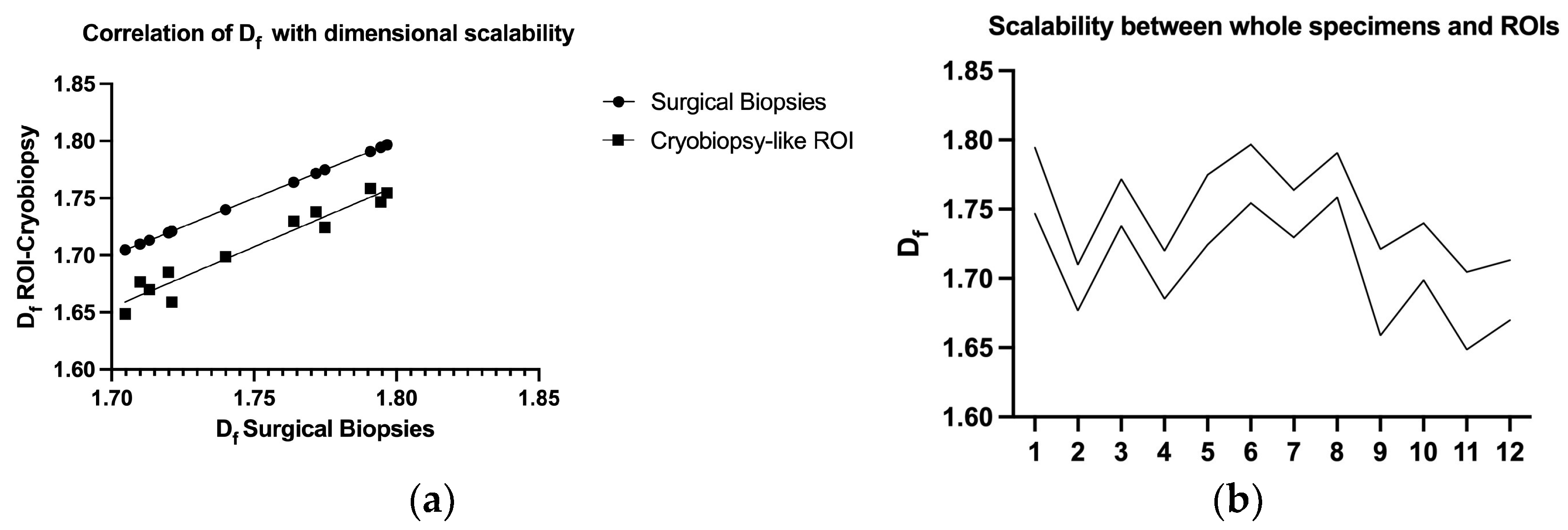
Idiopathic pulmonary fibrosis (IPF) is a chronic, progressive fibrotic lung disorder, ultimately leading to respiratory failure and death. Despite great research advances in understanding the mechanisms underlying the disease, its diagnosis, and its treatment, IPF still remains idiopathic without known biological or histological markers able to predict disease progression or response to treatment. The histologic hallmark of IPF is usual interstitial pneumonia (UIP), with its intricate architectural distortion and temporal inhomogeneity. We hypothesize that normal lung alveolar architecture can be compared to fractals, such as the Pythagoras tree with its fractal dimension (Df), and every pathological insult, distorting the normal lung structure, could result in Df variations. In this study, we aimed to assess the UIP histologic fractal dimension in relationship to other morphometric parameters in newly diagnosed IPF patients and its possible role in the prognostic stratification of the disease. Clinical data and lung tissue specimens were obtained from twelve patients with IPF, twelve patients with non-specific interstitial pneumonia (NSIP), and age-matched “healthy” control lung tissue from patients undergoing lung surgery for other causes. Histology and histomorphometry were performed to evaluate Df and lacunarity measures, using the box counting method on the FracLac ImageJ plugin. The results showed that Df was significantly higher in IPF patients compared to controls and fibrotic NSIP patients, indicating greater architectural distortion in IPF. Additionally, high Df values were associated with higher fibroblastic foci density and worse prognostic outcomes in IPF, suggesting that Df may serve as a potential novel prognostic marker for IPF. The scalability of Df measurements was demonstrated through repeated measurements on smaller portions from the same surgical biopsies, which were selected to mimic a cryobiopsy. Our study provides further evidence to support the use of fractal morphometry as a tool for quantifying and determining lung tissue remodeling in IPF, and we demonstrated a significant correlation between histological and radiological Df in UIP pattern, as well as a significant association between Df and FF density. Furthermore, our study demonstrates the scalability and self-similarity of Df measurements across different biopsy types, including surgical and smaller specimens.
Keywords: IPF; UIP; fractals; histomorphometry IPF; UIP; fractals; histomorphometry