Article

Association between SMAD4 Mutations and GATA6 Expression in Paired Pancreatic Ductal Adenocarcinoma Tumor Specimens: Data from Two Independent Molecularly-Characterized Cohorts

1
Rutgers New Jersey Medical School, Rutgers Health, Newark, NJ 07103, USA
2
Rutgers Cancer Institute of New Jersey, Rutgers Health, New Brunswick, NJ 08901, USA
3
Robert Wood Johnson Medical School, Rutgers University, New Brunswick, NJ 08901, USA
4
Cooperman Barnabas Medical Center, Livingston, NJ 07039, USA
5
Department of Surgical Oncology, University of Texas San Antonio, San Antonio, TX 78249, USA
*
Author to whom correspondence should be addressed.
Biomedicines 2023, 11(11), 3058; https://doi.org/10.3390/biomedicines11113058
Submission received: 7 October 2023 / Revised: 3 November 2023 / Accepted: 7 November 2023 / Published: 15 November 2023

Abstract

:
Several molecular biomarkers have been identified to guide induction treatment selection for localized pancreatic ductal adenocarcinoma (PDAC). SMAD4 alterations and low GATA6 expression/modified “Moffitt” basal-like phenotype have each been associated with inferior survival uniquely for patients receiving 5-FU-based therapies. SMAD4 may directly regulate the expression of GATA6 in PDAC, pointing to a common predictive biomarker. To evaluate the relationship between SMAD4 mutations and GATA6 expression in human PDAC tumors, patients with paired SMAD4 mutation and GATA6 mRNA expression data in the TCGA and CPTAC were identified. In 321 patients (TCGA: n = 180; CPTAC: n = 141), the rate of SMAD4 alterations was 26.8%. The rate of SMAD4 alteration did not vary per tertile of normalized GATA6 expression (TCGA: p = 0.928; CPTAC: p = 0.828). In the TCGA, SMAD4 alterations and the basal-like phenotype were each associated with worse survival (log rank p = 0.077 and p = 0.080, respectively), but their combined presence did not identify a subset with uniquely inferior survival (p = 0.943). In the CPTAC, the basal-like phenotype was associated with significantly worse survival (p < 0.001), but the prognostic value was not influenced by the combined presence of SMAD4 alterations (p = 0.960). SMAD4 alterations were not associated with poor clinico-pathological features such as poor tumor grade, advanced tumor stage, positive lymphovascular invasion (LVI), or positive perineural invasion (PNI), compared with SMAD4-wildtype. Given that SMAD4 mutations were not associated with GATA6 expression or Moffitt subtype in two independent molecularly characterized PDAC cohorts, distinct biomarker-defined clinical trials are necessary.

1. Introduction

Pancreatic ductal adenocarcinoma (PDAC) has one of the highest case-specific mortality rates of all cancers [1]. Although resection remains the only curative therapy for PDAC, improvements in long-term survival are attributable to advances in systemic treatment [2,3,4,5]. Currently, 5-fluorouracil-based (i.e., with irinotecan and oxaliplatin as FOLFIRINOX) or gemcitabine-based (with Nab-paclitaxel) chemotherapies are both used, with selection largely driven by patient-related factors such as age, comorbidity, and performance status. Amid the expanding options for systemic therapy and the mounting emphasis on administering such agents in the neoadjuvant setting, identification of biomarkers to guide first-line treatment selection remains a critical unmet need.
Previous work has identified genomic alterations in SMAD4 as predictive of unique resistance to FOLFIRINOX induction. SMAD4 alterations, primarily loss-of-function mutations [6], are present in approximately 20% of localized PDAC patients and may be linked to increased rates of metastatic progression and lower rates of surgical resection in patients receiving induction FOLFIRINOX but not receiving gemcitabine/nab-paclitaxel [7,8]. Separately, the modified Moffitt “basal-like” phenotype, marked by the loss of expression of GATA6, may also confer resistance to FOLFIRINOX chemotherapy [9,10,11,12,13]. In an exploratory analysis of the ESPAC-3 trial, low GATA6 expression was associated with inferior clinical outcomes uniquely for patients receiving 5-FU/LV, but not gemcitabine [9]. Similarly, in the COMPASS trial of patients with locally advanced or metastatic PDAC receiving FOLFIRINOX or gemcitabine/nab-paclitaxel, those with a modified “basal-like” phenotype had uniquely fewer responses and worse overall survival when treated with FOLFIRINOX [13]. Moreover, GATA6-low cell lines derived from patient-derived xenografts were particularly resistant to 5-FU but not gemcitabine [9].
Understanding the relationship between SMAD4 and GATA6 will be important for informing future studies of molecular biomarkers to guide induction chemotherapy selection and rational clinical trial design. SMAD4 is on chromosome 18q11, and 28.7 Mb from GATA6. GATA6 and SMAD4 are frequently co-altered [9]. Moreover, SMAD4 may directly regulate the expression of GATA6 [10]. Together, these data raise the possibility that SMAD4 alterations at the genome level and loss of GATA6 expression may represent a single molecular pathway that confers unique resistance to 5-FU-based therapies. In this study, representing the largest cohort of PDAC patients with paired DNA and RNA expression data, we examined the association between SMAD4 alterations and GATA6 expression to further characterize the translational potential of this molecular relationship. We hypothesized that SMAD4 alterations would be positively correlated with GATA6 expression and the basal-like subtype.

2. Methods

The study was deemed IRB exempt with use of publicly available and deidentified datasets. The cBioPortal platform for Cancer Genomics database is an open-access resource for exploration of multidimensional cancer genomics data. The platform was queried for PDAC samples with paired mutation and mRNA expression data, with identification of two datasets: the Cancer Genome Atlas (TCGA) and the Clinical Proteomic Tumor Analysis Consortium (CPTAC) [14].
SMAD4 was considered altered when there was either a mutation or copy number deletion. The modified Moffitt phenotypes were assigned as previously described [15], where the R package ConsensusClusterPlus27 [16] was employed to subtype PDAC samples according to the expression signatures defined in Moffitt et al. [12]. Briefly, the number of clusters was confirmed by examining cumulative distribution function, with the existence of well-separated clusters for Moffitt et al. classification based on tumor (basal-like and classical) and stroma signatures. GATA6 mRNA expression z-scores, as a surrogate biomarker of the modified Moffitt “basal-like” phenotype [13,17], were downloaded along with SMAD4 alteration calls and paired clinical data from the FireBrowse data portal (http://firebrowse.org) accessed on 12 August 2022 (TCGA data version 2016_01_28) and from cbioportal (https://www.cbioportal.org) (CPTAC data) [14].

Statistical Analysis

Descriptive statistics are presented as frequencies for categorical variables and median interquartile range (IQR) for continuous variables. Pearson’s χ2 and Wilcoxon rank-sum test were used to analyze categorical and continuous variables, respectively. GATA6 mRNA expression z-scores were analyzed by tertiles in each study to evaluate low, medium, and high expression as previously described [18]. The primary outcomes assessed were (1) rates of SMAD4 alterations for classical vs. basal-like subtypes and (2) rates of SMAD4 alterations for GATA6-low vs. GATA6-medium/high. The secondary outcomes were the impact of SMAD4 and molecular subtype on overall survival (OS), which was evaluated by Kaplan–Meier estimates. The variables associated with OS on univariable analysis (p < 0.10) were entered into a multivariable Cox regression model (TCGA: AJCC stage, nodal status, and tumor grade; CPTAC: AJCC stage). p-values ≤ 0.05 were considered statistically significant; all tests were two-sided. Analyses were carried out using SPSS v27.0 (IBM Corp., Armonk, NY, USA).

3. Results

3.1. Patient Cohort

In total, 321 patients with PDAC with paired DNA and RNA data were identified. The TCGA (n = 180) included 60 patients (33.3%) with SMAD4 alterations (Table 1). The median age was 65 (IQR 56-73) and most patients were male (n = 99; 55.0%) and white (n = 158; 87.8%). Nearly all patients (n = 152; 94.4%) had AJCC pathologic stage I–II disease. Patients with and without SMAD4 alterations were comparable with respect to age, sex, T-stage, N-stage, and tumor grade.
The CPTAC (n = 141) included 26 patients (18.4%) with SMAD4 mutations (Table 2). The median age was 65 (IQR 60–71) and most patients were male (n = 74; 52.5%). Nearly all patients (n = 125; 93.3%) had localized (AJCC pathologic stage I–III) disease. Patients with and without SMAD4 alterations were comparable with respect to age, T-stage, N-stage, and the presence of peri-neural or lymphovascular invasion.

3.2. Association between SMAD4 Alteration Status and Basal Subtype

In the TCGA cohort, the rate of SMAD4 alteration did not vary per tertile of normalized GATA6 expression (31.7% vs. 33.3% vs. 35.0%, p = 0.928) (Table 3). Likewise, the rate of SMAD4 alteration was not associated with Moffitt subtype (classical: 37.2% vs. basal-like: 39.4%, p = 0.783). In the CPTAC cohort, the rate of SMAD4 mutation did not vary per tertile of normalized GATA6 expression (17.0% vs. 17.0% vs. 21.3%, p = 0.828). Similarly, the rate of SMAD4 alteration was not associated with Moffitt subtype (classical: 22.5% vs. basal-like: 16.7%, p = 0.416).

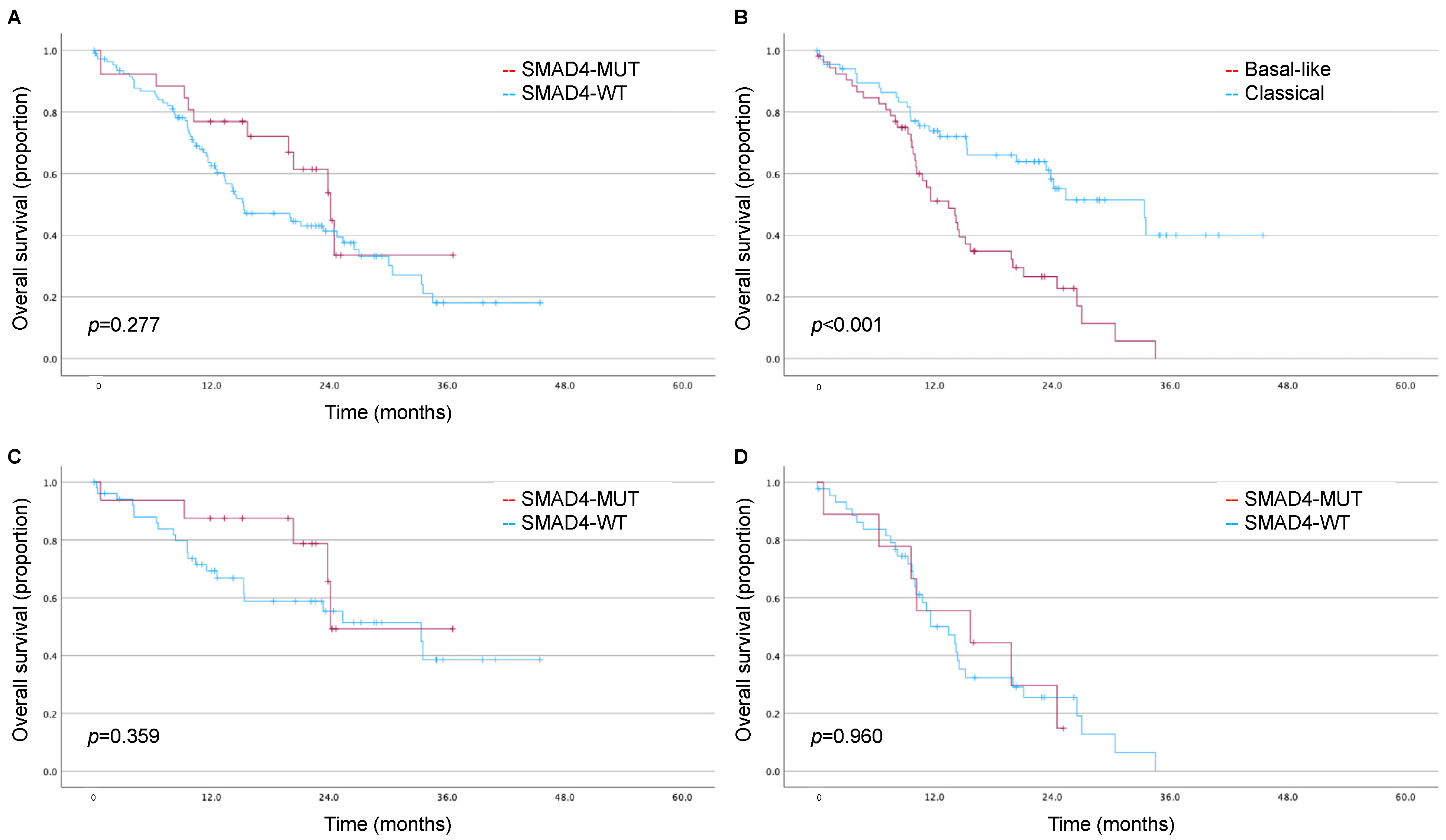
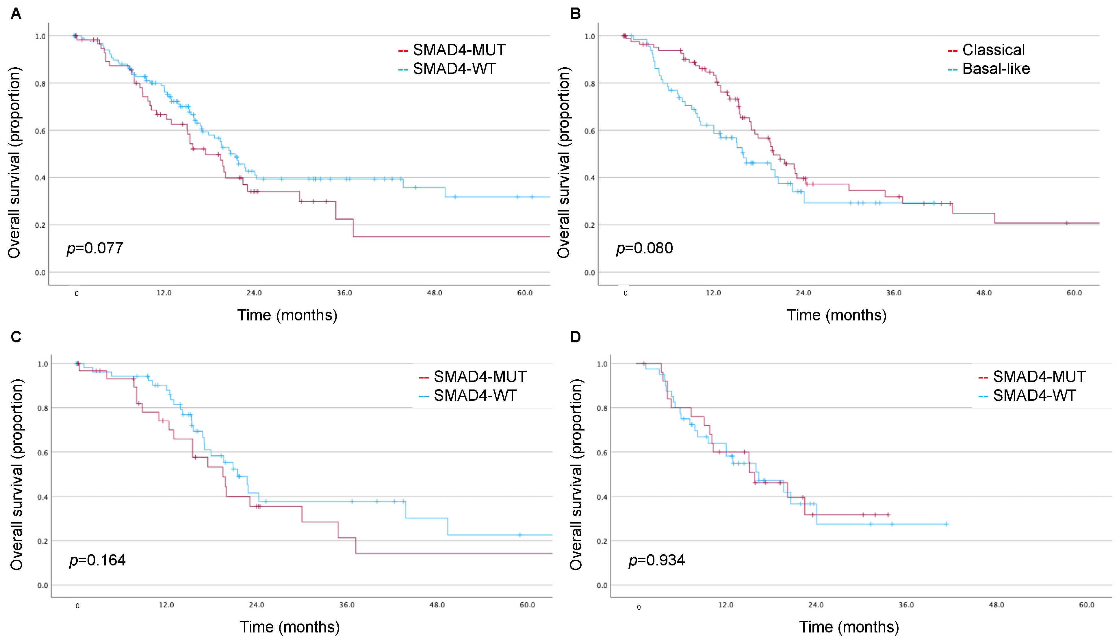
3.3. Impact of SMAD4 and Moffitt Subtype on Long-Term Survival

In the TCGA cohort, patients were followed for a median of 24.2 (IQR 15.2–42.3) months. SMAD4 alterations were not associated with significantly worse survival (estimated mean OS: 25.1 [95% CI 17.6–32.7] vs. 40.6 [95% CI 32.3–48.9] months, log rank p = 0.077; Figure 1A). Likewise, the basal-like phenotype was not associated with significantly worse survival (estimated mean OS: 20.4 [95% CI 16.3–24.5] vs. 30.8 [95% CI 24.4–37.2] months, log rank p = 0.080; Figure 1B). There were no trends in OS for SMAD4 alterations in the subsets of the classical subtype (log rank p = 0.164; Figure 1C) or basal-like subtype (log rank p = 0.934; Figure 1D). SMAD4 alterations were not associated with OS in a multivariable model accounting for AJCC stage, nodal status, and tumor grade (HR 1.25, 95% CI 0.82–1.91).
In the CPTAC cohort, patients were followed for a median of 23.9 (IQR 15.5–34.9) months. SMAD4 alterations were not associated with worse survival (estimated mean OS: 23.3 [95% CI 18.1–28.5] vs. 21.2 [95% CI 18.0–24.5] months, log rank p = 0.277; Figure 2A). The basal-like phenotype was associated with significantly worse survival (estimated mean OS: 15.3 [95% CI 12.4–18.1] vs. 28.2 [95% CI 23.7–32.8] months, log rank p < 0.001; Figure 2B). There were no trends in OS for SMAD4 alterations in the subsets of the classical subtype (log rank p = 0.359; Figure 2C) or basal-like subtype (log rank p = 0.960; Figure 2D). SMAD4 alterations were not associated with OS in a multivariable model accounting for AJCC stage (HR 0.75, 95% CI 0.39–1.42).

4. Discussion

Improving patient outcomes in pancreatic cancer hinges on precise chemotherapy selection to match tumor responsiveness. FOLFIRINOX has emerged as one of the most effective chemotherapeutic regimens for managing PDAC, demonstrating efficacy in both metastatic and adjuvant therapy settings [4,19]. As its application in the neoadjuvant setting continues to evolve, the integration of predictive biomarkers will be critical in refining treatment selection. SMAD4 mutations and the basal-like expression subtype (or GATAT6 low expression) have each emerged as potential biomarkers in this context. Herein, we present the largest cohort of PDAC patients evaluating the relationship between SMAD4 mutation, the modified Moffitt phenotype, and GATA6 expression in the context of clinical outcomes. Contrary to hypothesis, there was no correlation between SMAD4 alterations and the modified Moffitt phenotype or GATA6 expression. While in vitro data suggested that SMAD4 may regulate the expression of GATA6, these results are not confirmed in two human PDAC cohorts. Moreover, the combined presence of both biomarkers did not identify a patient subset with uniquely inferior outcomes. These data highlight the need for distinct biomarker-driven clinical trials and independent investigation of each biomarker in its potential mechanisms of treatment resistance.
SMAD4 serves as a mediator in the TGFB1 (TGF-β) signaling pathway and is recognized as a driver of the progression of pancreatic intraepithelial neoplasia to invasive adenocarcinoma [20,21]. Together with KRAS, TP53, and CDKN2A, SMAD4 is recognized as one of the four driver mutations in PDAC [17,20,22]. In these data, the rate of SMAD4 alterations ranged from 18.4% to 33.3%. The rate of SMAD4 in this study similarly compares to the previous literature’s rates of 20–33% [23,24]. This range likely represents inherent differences in study populations, where rates of SMAD4 alterations increase with greater tumor burden. Iacobuzio-Donahue et al. previously observed that locally advanced PDAC without metastatic disease uncommonly showed loss of SMAD4 (22%) as compared with carcinomas with extensive metastatic burden, where the rates of SMAD4 alteration approached 75% [25].
SMAD4 alteration has been reported to be an independent prognostic factor for recurrence-free survival and overall survival [26,27,28,29]. In a meta-analysis of eight studies with available data on SMAD4 status and patient survival, SMAD4 alterations conferred a pooled 40% increased risk of death [30]. Several studies have observed that the loss of SMAD4 is linked to distant metastases, which may explain its prognostic significance [25,31]. In our data, there was a trend to inferior survival in patients with SMAD4 alterations in the TCGA. However, a similar relationship was not observed in the CPTAC cohort. Notably, some studies have not identified SMAD4 as a prognostic biomarker [32,33]. Winter et al. reported that loss of SMAD4 expression showed no association with either recurrence or early mortality in resected PDAC patients [23]. These conflicting data highlight that disease-related survival is a complex interplay not solely driven by the function (or loss of) SMAD4, but likely driven by additional genetic, epigenetic, and environmental factors.
The lack of in vivo correlation between SMAD4 and the basal-like expression subtype (or GATAT6 low expression) warrants further discussion. Preclinical data suggested that SMAD4 can regulate the expression of GATA6. Using hTERT immortalized pancreatic ductal epithelial cells, suppressed SMAD4 (via small interfering RNA) reduced GATA6 expression. Conversely, FLAG-SMAD4 overexpression in PSN1 cells (which are SMAD4 deleted) resulted in re-establishment of GATA6 [10]. However, these findings did not translate to the human PDAC specimens included in our study. The absence of a direct molecular correlation between these two biomarkers diminishes the likelihood of a single, druggable target to address treatment resistance. Moreover, the clinical significance of the basal-like subtype, regardless of SMAD4 mutational status, further emphasizes the divergence of these molecular pathways. In these data, the basal-like subtype was associated with trends to inferior survival in the TCGA and significantly inferior survival in the CPTAC, and such associations were not impacted by the loss of SMAD4. While the association between basal-like subtype or low GATA6 and inferior survival are well established in the literature [7,8,10,13], the impact (or lack thereof) of concurrent SMAD4 alterations represents an additional complexity to this search for precision oncology.
GATA6, a member of the transcription factor family, binds to the (A/T)GATA(A/G) consensus sequence, influencing gene expression [34]. Crucial for cell differentiation, GATA6 is vital for maintaining the exocrine pancreas [35]. Recent studies propose a tumor-suppressive role of GATA6 in PDAC mouse models, influencing both differentiation and cancer-related transcriptional programs [36,37]. In human PDAC cells, GATA6 plays a pivotal role in inhibiting de-differentiation and epithelial–mesenchymal transition (EMT). The sequential regulation of EMT and mesenchymal–epithelial transition (MET) is crucial for effective tumor spreading [38,39]. In human PDAC samples, the loss of GATA6 in PDAC primary samples correlates with altered differentiation and shorter overall patient survival [40,41].
Our study is not without limitations. Conducting a retrospective analysis of clinical outcomes using a database inherently carries the risk of selection bias and potential inaccuracies in data reporting. Additionally, there is the possibility of sampling error in tumor biopsies, which could lead to mischaracterizations of SMAD4 mutational status or GATA6 mRNA expression. This is a particular limitation of genomic classification of PDAC given the large stromal component of many tumors. Third, certain analyses may have been underpowered given the limited sample sizes. The basal-like phenotype was associated with statistically inferior survival in the CPTAC cohort but not in the TCGA cohort; we believe this represents a type II error in the TCGA cohort. We methodologically avoided combining the TCGA and CPTAC datasets given the differences in tumor processing, which may have also contributing to some conflicting results. Fourth, the survival analyses may have been influenced by heterogeneity in the use of adjuvant therapy, for which data were not available. Additionally, analyses of recurrence-free survival were limited by the lack of such data in these cohorts.
Nonetheless, our study represents the largest cohort of PDAC patients with direct evaluation of the relationship between SMAD4 mutation and the basal-like subtype/GATA6 expression. Given the lack of correlation, distinct biomarker-driven clinical trials and individualized studies exploring the mechanistic basis of each biomarker are necessary for the advancement of precision oncology for this disease.

Author Contributions

Conceptualization, J.D.G. and B.L.E.; Data curation, J.D.G., C.M.C. and B.L.E.; Formal analysis, J.D.G., C.M.C. and B.L.E.; Investigation, J.D.G., C.M.C. and B.L.E.; Methodology, J.D.G. and B.L.E.; Project administration, B.L.E.; Resources, J.D.G. and B.L.E.; Software, J.D.G. and B.L.E.; Supervision, B.L.E.; Validation, J.D.G., C.M.C. and B.L.E.; Visualization, J.D.G. and B.L.E.; Writing—original draft, J.D.G. and B.L.E.; Writing—review & editing, J.D.G., W.E.A., H.R.A., T.B., M.F.E., M.S.G., T.J.K., R.C.L., J.C.M., S.D. and B.L.E. All authors have read and agreed to the published version of the manuscript.

Funding

This research received no external funding.

Institutional Review Board Statement

Study deemed IRB exempt given use of publicly available and deidentified datasets.

Informed Consent Statement

Informed consent was obtained from all subjects involved in the original studies from which these data were derived.

Data Availability Statement

Data are publicly available on cbioportal.

Acknowledgments

The results shown here are wholly or in part based upon data generated by the TCGA Research Network: https://www.cancer.gov/about-nci/organization/ccg/research/structural-genomics/tcga, accessed on 12 August 2022. Data used in this publication were generated by the Clinical Proteomic Tumor Analysis Consortium (NCI/NIH).

Conflicts of Interest

Russell C. Langan is a paid consultant of Eon.

References

  1. Rahib, L.; Smith, B.D.; Aizenberg, R.; Rosenzweig, A.B.; Fleshman, J.M.; Matrisian, L.M. Projecting cancer incidence and deaths to 2030: The unexpected burden of thyroid, liver, and pancreas cancers in the United States. Cancer Res. 2014, 74, 2913–2921. [Google Scholar] [CrossRef] [PubMed]
  2. Park, W.; Chawla, A.; O’Reilly, E.M. Pancreatic Cancer: A Review. JAMA 2021, 326, 851–862. [Google Scholar] [CrossRef] [PubMed]
  3. Ahmad, S.A.; Duong, M.; Sohal, D.P.S.; Gandhi, N.S.; Beg, M.S.; Wang-Gillam, A.; Wade, J.L., 3rd; Chiorean, E.G.; Guthrie, K.A.; Lowy, A.M.; et al. Surgical Outcome Results from SWOG S1505: A Randomized Clinical Trial of mFOLFIRINOX versus Gemcitabine/Nab-paclitaxel for Perioperative Treatment of Resectable Pancreatic Ductal Adenocarcinoma. Ann. Surg. 2020, 272, 481–486. [Google Scholar] [CrossRef] [PubMed]
  4. Conroy, T.; Hammel, P.; Hebbar, M.; Ben Abdelghani, M.; Wei, A.C.; Raoul, J.L.; Chone, L.; Francois, E.; Artru, P.; Biagi, J.J.; et al. FOLFIRINOX or Gemcitabine as Adjuvant Therapy for Pancreatic Cancer. N. Engl. J. Med. 2018, 379, 2395–2406. [Google Scholar] [CrossRef] [PubMed]
  5. Tempero, M.A.; Pelzer, U.; O’Reilly, E.M.; Winter, J.; Oh, D.Y.; Li, C.P.; Tortora, G.; Chang, H.M.; Lopez, C.D.; Bekaii-Saab, T.; et al. Adjuvant nab-Paclitaxel + Gemcitabine in Resected Pancreatic Ductal Adenocarcinoma: Results From a Randomized, Open-Label, Phase III Trial. J. Clin. Oncol. 2023, 41, 2007–2019. [Google Scholar] [CrossRef]
  6. Wang, F.; Xia, X.; Yang, C.; Shen, J.; Mai, J.; Kim, H.C.; Kirui, D.; Kang, Y.; Fleming, J.B.; Koay, E.J.; et al. SMAD4 Gene Mutation Renders Pancreatic Cancer Resistance to Radiotherapy through Promotion of Autophagy. Clin. Cancer Res. 2018, 24, 3176–3185. [Google Scholar] [CrossRef]
  7. Ecker, B.L.; Court, C.M.; Janssen, Q.P.; Tao, A.J.; D’Angelica, M.I.; Drebin, J.A.; Gonen, M.; O’Reilly, E.M.; Jarnagin, W.R.; Wei, A.C.; et al. Alterations in Somatic Driver Genes Are Associated with Response to Neoadjuvant FOLFIRINOX in Patients with Localized Pancreatic Ductal Adenocarcinoma. J. Am. Coll. Surg. 2022, 235, 342–349. [Google Scholar] [CrossRef]
  8. Ecker, B.L.; Tao, A.J.; Janssen, Q.P.; Walch, H.S.; Court, C.M.; Balachandran, V.P.; Crane, C.H.; D’Angelica, M.I.; Drebin, J.A.; Kingham, T.P.; et al. Genomic Biomarkers Associated with Response to Induction Chemotherapy in Patients with Localized Pancreatic Ductal Adenocarcinoma. Clin. Cancer Res. 2023, 29, 1368–1374. [Google Scholar] [CrossRef]
  9. Martinelli, P.; Carrillo-de Santa Pau, E.; Cox, T.; Sainz, B., Jr.; Dusetti, N.; Greenhalf, W.; Rinaldi, L.; Costello, E.; Ghaneh, P.; Malats, N.; et al. GATA6 regulates EMT and tumour dissemination, and is a marker of response to adjuvant chemotherapy in pancreatic cancer. Gut 2017, 66, 1665–1676. [Google Scholar] [CrossRef]
  10. Eyres, M.; Lanfredini, S.; Xu, H.; Burns, A.; Blake, A.; Willenbrock, F.; Goldin, R.; Hughes, D.; Hughes, S.; Thapa, A.; et al. TET2 Drives 5hmc Marking of GATA6 and Epigenetically Defines Pancreatic Ductal Adenocarcinoma Transcriptional Subtypes. Gastroenterology 2021, 161, 653–668.e6. [Google Scholar] [CrossRef]
  11. Williams, H.L.; Dias Costa, A.; Zhang, J.; Raghavan, S.; Winter, P.S.; Kapner, K.S.; Ginebaugh, S.P.; Väyrynen, S.A.; Väyrynen, J.P.; Yuan, C.; et al. Spatially Resolved Single-Cell Assessment of Pancreatic Cancer Expression Subtypes Reveals Co-expressor Phenotypes and Extensive Intratumoral Heterogeneity. Cancer Res. 2023, 83, 441–455. [Google Scholar] [CrossRef] [PubMed]
  12. Moffitt, R.A.; Marayati, R.; Flate, E.L.; Volmar, K.E.; Loeza, S.G.; Hoadley, K.A.; Rashid, N.U.; Williams, L.A.; Eaton, S.C.; Chung, A.H.; et al. Virtual microdissection identifies distinct tumor- and stroma-specific subtypes of pancreatic ductal adenocarcinoma. Nat. Genet. 2015, 47, 1168–1178. [Google Scholar] [CrossRef] [PubMed]
  13. O’Kane, G.M.; Grunwald, B.T.; Jang, G.H.; Masoomian, M.; Picardo, S.; Grant, R.C.; Denroche, R.E.; Zhang, A.; Wang, Y.; Lam, B.; et al. GATA6 Expression Distinguishes Classical and Basal-like Subtypes in Advanced Pancreatic Cancer. Clin. Cancer Res. 2020, 26, 4901–4910. [Google Scholar] [CrossRef] [PubMed]
  14. Cao, L.; Huang, C.; Cui Zhou, D.; Hu, Y.; Lih, T.M.; Savage, S.R.; Krug, K.; Clark, D.J.; Schnaubelt, M.; Chen, L.; et al. Proteogenomic characterization of pancreatic ductal adenocarcinoma. Cell 2021, 184, 5031–5052 e5026. [Google Scholar] [CrossRef]
  15. de Santiago, I.; Yau, C.; Heij, L.; Middleton, M.R.; Markowetz, F.; Grabsch, H.I.; Dustin, M.L.; Sivakumar, S. Immunophenotypes of pancreatic ductal adenocarcinoma: Meta-analysis of transcriptional subtypes. Int. J. Cancer 2019, 145, 1125–1137. [Google Scholar] [CrossRef]
  16. Wilkerson, M.D.; Hayes, D.N. ConsensusClusterPlus: A class discovery tool with confidence assessments and item tracking. Bioinformatics 2010, 26, 1572–1573. [Google Scholar] [CrossRef]
  17. Aung, K.L.; Fischer, S.E.; Denroche, R.E.; Jang, G.-H.; Dodd, A.; Creighton, S.; Southwood, B.; Liang, S.-B.; Chadwick, D.; Zhang, A.; et al. Genomics-Driven Precision Medicine for Advanced Pancreatic Cancer: Early Results from the COMPASS Trial. Clin. Cancer Res. 2018, 24, 1344–1354. [Google Scholar] [CrossRef]
  18. Zhou, X.; An, J.; Kurilov, R.; Brors, B.; Hu, K.; Peccerella, T.; Roessler, S.; Pfütze, K.; Schulz, A.; Wolf, S.; et al. Persister cell phenotypes contribute to poor patient outcomes after neoadjuvant chemotherapy in PDAC. Nat. Cancer 2023, 4, 1362–1381. [Google Scholar] [CrossRef]
  19. Conroy, T.; Desseigne, F.; Ychou, M.; Bouché, O.; Guimbaud, R.; Bécouarn, Y.; Adenis, A.; Raoul, J.L.; Gourgou-Bourgade, S.; de la Fouchardière, C.; et al. FOLFIRINOX versus gemcitabine for metastatic pancreatic cancer. N. Engl. J. Med. 2011, 364, 1817–1825. [Google Scholar] [CrossRef]
  20. Aguirre, A.J.; Nowak, J.A.; Camarda, N.D.; Moffitt, R.A.; Ghazani, A.A.; Hazar-Rethinam, M.; Raghavan, S.; Kim, J.; Brais, L.K.; Ragon, D.; et al. Real-time Genomic Characterization of Advanced Pancreatic Cancer to Enable Precision Medicine. Cancer Discov. 2018, 8, 1096–1111. [Google Scholar] [CrossRef]
  21. David, C.J.; Huang, Y.H.; Chen, M.; Su, J.; Zou, Y.; Bardeesy, N.; Iacobuzio-Donahue, C.A.; Massagué, J. TGF-β Tumor Suppression through a Lethal EMT. Cell 2016, 164, 1015–1030. [Google Scholar] [CrossRef]
  22. Grossberg, A.J.; Chu, L.C.; Deig, C.R.; Fishman, E.K.; Hwang, W.L.; Maitra, A.; Marks, D.L.; Mehta, A.; Nabavizadeh, N.; Simeone, D.M. Multidisciplinary standards of care and recent progress in pancreatic ductal adenocarcinoma. CA Cancer J. Clin. 2020, 70, 375–403. [Google Scholar] [CrossRef] [PubMed]
  23. Winter, J.M.; Tang, L.H.; Klimstra, D.S.; Liu, W.; Linkov, I.; Brennan, M.F.; D’Angelica, M.I.; DeMatteo, R.P.; Fong, Y.; Jarnagin, W.R.; et al. Failure patterns in resected pancreas adenocarcinoma: Lack of predicted benefit to SMAD4 expression. Ann. Surg. 2013, 258, 331–335. [Google Scholar] [CrossRef] [PubMed]
  24. McIntyre, C.A.; Lawrence, S.A.; Richards, A.L.; Chou, J.F.; Wong, W.; Capanu, M.; Berger, M.F.; Donoghue, M.T.A.; Yu, K.H.; Varghese, A.M.; et al. Alterations in driver genes are predictive of survival in patients with resected pancreatic ductal adenocarcinoma. Cancer 2020, 126, 3939–3949. [Google Scholar] [CrossRef] [PubMed]
  25. Iacobuzio-Donahue, C.A.; Fu, B.; Yachida, S.; Luo, M.; Abe, H.; Henderson, C.M.; Vilardell, F.; Wang, Z.; Keller, J.W.; Banerjee, P.; et al. DPC4 gene status of the primary carcinoma correlates with patterns of failure in patients with pancreatic cancer. J. Clin. Oncol. 2009, 27, 1806–1813. [Google Scholar] [CrossRef] [PubMed]
  26. Yokose, T.; Kitago, M.; Matsuda, S.; Sasaki, Y.; Masugi, Y.; Nakamura, Y.; Shinoda, M.; Yagi, H.; Abe, Y.; Oshima, G.; et al. Combination of KRAS and SMAD4 mutations in formalin-fixed paraffin-embedded tissues as a biomarker for pancreatic cancer. Cancer Sci. 2020, 111, 2174–2182. [Google Scholar] [CrossRef]
  27. Tascilar, M.; Skinner, H.G.; Rosty, C.; Sohn, T.; Wilentz, R.E.; Offerhaus, G.J.; Adsay, V.; Abrams, R.A.; Cameron, J.L.; Kern, S.E.; et al. The SMAD4 protein and prognosis of pancreatic ductal adenocarcinoma. Clin. Cancer Res. 2001, 7, 4115–4121. [Google Scholar]
  28. Biankin, A.V.; Morey, A.L.; Lee, C.S.; Kench, J.G.; Biankin, S.A.; Hook, H.C.; Head, D.R.; Hugh, T.B.; Sutherland, R.L.; Henshall, S.M. DPC4/Smad4 expression and outcome in pancreatic ductal adenocarcinoma. J. Clin. Oncol. 2002, 20, 4531–4542. [Google Scholar] [CrossRef]
  29. Oshima, M.; Okano, K.; Muraki, S.; Haba, R.; Maeba, T.; Suzuki, Y.; Yachida, S. Immunohistochemically detected expression of 3 major genes (CDKN2A/p16, TP53, and SMAD4/DPC4) strongly predicts survival in patients with resectable pancreatic cancer. Ann. Surg. 2013, 258, 336–346. [Google Scholar] [CrossRef]
  30. Gu, Y.; Ji, Y.; Jiang, H.; Qiu, G. Clinical Effect of Driver Mutations of KRAS, CDKN2A/P16, TP53, and SMAD4 in Pancreatic Cancer: A Meta-Analysis. Genet. Test. Mol. Biomark. 2020, 24, 777–788. [Google Scholar] [CrossRef]
  31. Crane, C.H.; Varadhachary, G.R.; Yordy, J.S.; Staerkel, G.A.; Javle, M.M.; Safran, H.; Haque, W.; Hobbs, B.D.; Krishnan, S.; Fleming, J.B.; et al. Phase II trial of cetuximab, gemcitabine, and oxaliplatin followed by chemoradiation with cetuximab for locally advanced (T4) pancreatic adenocarcinoma: Correlation of Smad4(Dpc4) immunostaining with pattern of disease progression. J. Clin. Oncol. 2011, 29, 3037–3043. [Google Scholar] [CrossRef] [PubMed]
  32. Shin, S.H.; Kim, H.J.; Hwang, D.W.; Lee, J.H.; Song, K.B.; Jun, E.; Shim, I.K.; Hong, S.M.; Kim, H.J.; Park, K.M.; et al. The DPC4/SMAD4 genetic status determines recurrence patterns and treatment outcomes in resected pancreatic ductal adenocarcinoma: A prospective cohort study. Oncotarget 2017, 8, 17945–17959. [Google Scholar] [CrossRef] [PubMed]
  33. Qian, Z.R.; Rubinson, D.A.; Nowak, J.A.; Morales-Oyarvide, V.; Dunne, R.F.; Kozak, M.M.; Welch, M.W.; Brais, L.K.; Da Silva, A.; Li, T.; et al. Association of Alterations in Main Driver Genes with Outcomes of Patients with Resected Pancreatic Ductal Adenocarcinoma. JAMA Oncol. 2018, 4, e173420. [Google Scholar] [CrossRef] [PubMed]
  34. Maeda, M.; Ohashi, K.; Ohashi-Kobayashi, A. Further extension of mammalian GATA-6. Dev. Growth Differ. 2005, 47, 591–600. [Google Scholar] [CrossRef]
  35. Martinelli, P.; Cañamero, M.; del Pozo, N.; Madriles, F.; Zapata, A.; Real, F.X. Gata6 is required for complete acinar differentiation and maintenance of the exocrine pancreas in adult mice. Gut 2013, 62, 1481–1488. [Google Scholar] [CrossRef]
  36. Martinelli, P.; Madriles, F.; Cañamero, M.; Pau, E.C.; Pozo, N.D.; Guerra, C.; Real, F.X. The acinar regulator Gata6 suppresses KrasG12V-driven pancreatic tumorigenesis in mice. Gut 2016, 65, 476–486. [Google Scholar] [CrossRef]
  37. Hermann, P.C.; Sancho, P.; Cañamero, M.; Martinelli, P.; Madriles, F.; Michl, P.; Gress, T.; de Pascual, R.; Gandia, L.; Guerra, C.; et al. Nicotine promotes initiation and progression of KRAS-induced pancreatic cancer via Gata6-dependent dedifferentiation of acinar cells in mice. Gastroenterology 2014, 147, 1119–1133.e4. [Google Scholar] [CrossRef]
  38. Ocaña, O.H.; Córcoles, R.; Fabra, A.; Moreno-Bueno, G.; Acloque, H.; Vega, S.; Barrallo-Gimeno, A.; Cano, A.; Nieto, M.A. Metastatic colonization requires the repression of the epithelial-mesenchymal transition inducer Prrx1. Cancer Cell 2012, 22, 709–724. [Google Scholar] [CrossRef]
  39. Tsai, J.H.; Donaher, J.L.; Murphy, D.A.; Chau, S.; Yang, J. Spatiotemporal regulation of epithelial-mesenchymal transition is essential for squamous cell carcinoma metastasis. Cancer Cell 2012, 22, 725–736. [Google Scholar] [CrossRef]
  40. Zhong, Y.; Wang, Z.; Fu, B.; Pan, F.; Yachida, S.; Dhara, M.; Albesiano, E.; Li, L.; Naito, Y.; Vilardell, F.; et al. GATA6 activates Wnt signaling in pancreatic cancer by negatively regulating the Wnt antagonist Dickkopf-1. PLoS ONE 2011, 6, e22129. [Google Scholar] [CrossRef]
  41. Neoptolemos, J.P.; Stocken, D.D.; Bassi, C.; Ghaneh, P.; Cunningham, D.; Goldstein, D.; Padbury, R.; Moore, M.J.; Gallinger, S.; Mariette, C.; et al. Adjuvant chemotherapy with fluorouracil plus folinic acid vs gemcitabine following pancreatic cancer resection: A randomized controlled trial. JAMA 2010, 304, 1073–1081. [Google Scholar] [CrossRef] [PubMed]
Figure 1. Kaplan–Meier estimates for the impact of SMAD4 and molecular subtype on overall survival in the TCGA cohort. (A) Association of SMAD4 alterations with overall survival; (B) association of Moffitt subtype with overall survival; (C) association of SMAD4 alterations with overall survival in the TCGA subset defined by the classical subtype; (D) association of SMAD4 alterations with overall survival in the TCGA subset defined by the basal-like subtype.
Biomedicines 11 03058 g001
Figure 2. Kaplan–Meier estimates for the impact of SMAD4 and molecular subtype on overall survival in the CPTAC cohort. (A) Association of SMAD4 alterations with overall survival; (B) association of Moffitt subtype with overall survival; (C) association of SMAD4 alterations with overall survival in the CPTAC subset defined by the classical subtype; (D) association of SMAD4 alterations with overall survival in the CPTAC subset defined by the basal-like subtype.
Biomedicines 11 03058 g002
Table 1. Clinicodemographics of TCGA Patient Cohort.
SMAD4-WTSMAD4-MUTp
(n = 120)(n = 60)
#%#%
Age 0.874
Median (IQR)65 (54–74) 65 (58–73)
Sex 0.751
Female5545.8%2643.3%
Male6554.2%3456.7%
Race a 0.041
White10690.6%5288.1%
Black65.1%00.0%
Asian54.3%711.9%
Ethnicity b 0.588
Hispanic32.5%23.3%
AJCC Stage c 0.412
I1613.6%610.0%
II9580.5%5388.3%
III43.4%00.0%
IV32.5%11.7%
T Stage 0.596
T154.2%35.0%
T21613.3%711.7%
T39478.3%5083.3%
T432.5%00.0%
TX21.7%00.0%
N Stage 0.839
N-negative3529.2%1525.0%
N-positive8167.5%4371.7%
NX43.3%23.3%
Grade 0.373
G12420.0%711.7%
G26150.8%3558.3%
G33327.5%1830.0%
GX21.7%00.0%
Abbreviations: IQR interquartile range; AJCC American Joint Committee on Cancer. a Race data unavailable for 4 patients. b Ethnicity data unavailable for 43 patients. c AJCC stage data unavailable for 2 patients.
Table 2. Clinicodemographics of CPTAC Patient Cohort.
SMAD4 Wild-TypeSMAD4 Mutantp
(n = 115)(n = 26)
#%#%
Age 0.147
Median (IQR)64 (59–71) 66 (63–72)
Sex 0.556
Female5648.7%1142.3%
Male5951.3%1557.7%
AJCC Stage a 0.491
I1816.4%520.8%
II4843.6%1250.0%
III3531.8%729.2%
IV98.2%00.0%
T Stage 0.799
pT176.1%311.5%
pT27262.6%17.257.7%
pT33328.7%19.530.8%
pT410.9%00.0%
pTX21.7%00.0%
N Stage 0.842
pN02420.9%726.9%
pN14438.2%1038.5%
pN24034.8%726.9%
pNX76.1%27.7%
LVI b 0.429
Absent3129.2%937.5%
Present7570.8%1562.5%
PNI c 0.471
Absent1513.8%28.3%
Present9486.2%2291.7%
Abbreviations: IQR interquartile range; AJCC American Joint Committee on Cancer; LVI lymphovascular invasion; PNI perineural invasion. a AJCC stage data unavailable for 7 patients. b LVI data unavailable for 11 patients. c PNI data unavailable for 8 patients.
Table 3. Association between SMAD4 alterations and GATA6 expression and Moffitt subtype.
SMAD4 Wild-TypeSMAD4 Mutantp
#%#%
TCGA
GATA6 Tertile 0.928
Low4134.2%1931.7%
Medium4033.3%2033.3%
High3932.5%2135.0%
Moffitt Subtype a 0.783
Classical5457.4%3255.2%
Basal-like4042.6%2644.8%
CPTAC
GATA6 Tertile 0.828
Low3931.5%830.8%
Medium3931.5%830.8%
High4637.0%1038.4%
Moffitt Subtype b 0.416
Classical5555.0%1664.0%
Basal-like4545.0%936.0%
a Moffitt subtype unavailable for 28 patients in TCGA cohort. b Moffitt subtype unavailable for 16 patients in CPTAC cohort.
Disclaimer/Publisher’s Note: The statements, opinions and data contained in all publications are solely those of the individual author(s) and contributor(s) and not of MDPI and/or the editor(s). MDPI and/or the editor(s) disclaim responsibility for any injury to people or property resulting from any ideas, methods, instructions or products referred to in the content.

