
Immunomodulatory Role of Neuropeptides in the Cornea

Abstract

1. Introduction
2. Corneal Immune Privilege
3. Corneal Innervation
4. Neuropeptides and Their Receptors in the Cornea
- Gq/11 Signaling: The neuropeptide receptors belonging to the Gq/11 family include the PACAP receptor (PAC1R) and the tachykinin receptors (NK1R, NK2R, and NK3R).
- Gi/o Signaling: The neuropeptide receptors belonging to the Gi/o family include the neuropeptide Y receptors (NPY1R, NPY2R, NPY4R, and NPY5R) and the somatostatin receptors (SST1R, SST2R, SST3R, SST4R, and SST5R).
4.1. Substance P (SP)
4.1.1. Transcriptional Regulation
4.1.2. Metabolism and Signaling
4.1.3. Immunomodulation and Inflammation
4.1.4. Role of Substance P in the Cornea
4.2. Calcitonin Gene-Related Peptide (CGRP)
4.2.1. Transcriptional Regulation
4.2.2. Metabolism and Signaling
4.2.3. Immunomodulation and Inflammation
4.2.4. Role of CGRP in the Cornea
4.3. Adrenomedullin (AM)
4.3.1. Transcriptional Regulation
4.3.2. Metabolism and Signaling
4.3.3. Immunomodulation and Inflammation
4.3.4. Role of Adrenomedullin in the Cornea
4.4. Vasoactive Intestinal Polypeptide (VIP)
4.4.1. Transcriptional Regulation
4.4.2. Metabolism and Signaling
4.4.3. Immunomodulation and Inflammation
4.4.4. Role of VIP in the Cornea
4.5. Pituitary Adenylate Cyclase-Activating Polypeptide (PACAP)
4.5.1. Transcriptional Regulation
4.5.2. Metabolism and Signaling
4.5.3. Immunomodulation and Inflammation
4.5.4. Role of PACAP in the Cornea
4.6. Neuropeptide Y (NPY)
4.6.1. Transcriptional Regulation
4.6.2. Metabolism and Signaling
4.6.3. Immunomodulation and Inflammation
4.6.4. Role of NPY in the Cornea
4.7. Somatostatin (SST)
4.7.1. Transcriptional Regulation
4.7.2. Metabolism and Signaling
4.7.3. Immunomodulation and Inflammation
4.7.4. Role of SST in the Cornea
4.8. α-Melanocyte Stimulating Hormone (α-MSH)
4.8.1. Transcriptional Regulation
4.8.2. Metabolism and Signaling
4.8.3. Immunomodulation and Inflammation
4.8.4. Role of α-MSH in the Cornea
4.9. Galanin (GAL)
4.9.1. Transcriptional Regulation
4.9.2. Metabolism and Signaling
4.9.3. Immunomodulation and Inflammation
4.9.4. Role of GAL in the Cornea
4.10. Methionine Enkephalin (Met-Enkephalin, MENK, [Met5]Enkephalin) or Opioid Growth Factor (OGF)
4.10.1. Transcriptional Regulation
4.10.2. Metabolism and Signaling
4.10.3. Immunomodulation and Inflammation
4.10.4. Role of MENK in the Cornea
4.11. Neurotensin (NT)
4.11.1. Transcriptional Regulation
4.11.2. Metabolism and Signaling
4.11.3. Immunomodulation and Inflammation
4.11.4. Role of NT in the Cornea
5. Neuropeptides as Therapeutic Targets/Drugs for Corneal Diseases
5.1. Corneal Wound Healing
5.2. Dry Eye Disease (DED)
5.3. Infectious Keratitis
5.4. Corneal Neovascularization
5.5. Corneal Transplantation
6. Conclusions
Author Contributions
Funding
Institutional Review Board Statement
Informed Consent Statement
Data Availability Statement
Acknowledgments
Conflicts of Interest
References
- DelMonte, D.W.; Kim, T. Anatomy and Physiology of the Cornea. J. Cataract Refract. Surg. 2011, 37, 588–598. [Google Scholar] [CrossRef] [PubMed]
- Hamrah, P.; Zhang, Q.; Liu, Y.; Dana, M.R. Novel Characterization of MHC Class II–Negative Population of Resident Corneal Langerhans Cell–Type Dendritic Cells. Investig. Ophthalmol. Vis. Sci. 2002, 43, 639–646. [Google Scholar]
- Notara, M.; Alatza, A.; Gilfillan, J.; Harris, A.R.; Levis, H.J.; Schrader, S.; Vernon, A.; Daniels, J.T. In Sickness and in Health: Corneal Epithelial Stem Cell Biology, Pathology and Therapy. Exp. Eye Res. 2010, 90, 188–195. [Google Scholar] [CrossRef] [PubMed]
- Waring, G.O.; Bourne, W.M.; Edelhauser, H.F.; Kenyon, K.R. The Corneal Endothelium. Normal and Pathologic Structure and Function. Ophthalmology 1982, 89, 531–590. [Google Scholar] [CrossRef]
- Bron, A.J. The Architecture of the Corneal Stroma. Br. J. Ophthalmol. 2001, 85, 379–381. [Google Scholar] [CrossRef]
- Espana, E.M.; Birk, D.E. Composition, Structure and Function of the Corneal Stroma. Exp. Eye Res. 2020, 198, 108137. [Google Scholar] [CrossRef]
- Hamrah, P.; Liu, Y.; Zhang, Q.; Dana, M.R. The Corneal Stroma Is Endowed with a Significant Number of Resident Dendritic Cells. Investig. Ophthalmol. Vis. Sci. 2003, 44, 581–589. [Google Scholar] [CrossRef]
- Jamali, A.; Hu, K.; Sendra, V.G.; Blanco, T.; Lopez, M.J.; Ortiz, G.; Qazi, Y.; Zheng, L.; Turhan, A.; Harris, D.L.; et al. Characterization of Resident Corneal Plasmacytoid Dendritic Cells and Their Pivotal Role in Herpes Simplex Keratitis. Cell Rep. 2020, 32, 108099. [Google Scholar] [CrossRef]
- Jamali, A.; Kenyon, B.; Ortiz, G.; Abou-Slaybi, A.; Sendra, V.G.; Harris, D.L.; Hamrah, P. Plasmacytoid Dendritic Cells in the Eye. Prog. Retin. Eye Res. 2021, 80, 100877. [Google Scholar] [CrossRef]
- Müller, L.J.; Marfurt, C.F.; Kruse, F.; Tervo, T.M.T. Corneal Nerves: Structure, Contents and Function. Exp. Eye Res. 2003, 76, 521–542. [Google Scholar] [CrossRef]
- Hori, J.; Yamaguchi, T.; Keino, H.; Hamrah, P.; Maruyama, K. Immune Privilege in Corneal Transplantation. Prog. Retin. Eye Res. 2019, 72, 100758. [Google Scholar] [CrossRef] [PubMed]
- Belmonte, C.; Aracil, A.; Acosta, M.C.; Luna, C.; Gallar, J. Nerves and Sensations from the Eye Surface. Ocul. Surf. 2004, 2, 248–253. [Google Scholar] [CrossRef]
- Belmonte, C.; Carmen Acosta, M.; Gallar, J. Neural Basis of Sensation in Intact and Injured Corneas. Exp. Eye Res. 2004, 78, 513–525. [Google Scholar] [CrossRef]
- Unanue, E.R. Perspective on Antigen Processing and Presentation. Immunol. Rev. 2002, 185, 86–102. [Google Scholar] [CrossRef]
- Cruzat, A.; Witkin, D.; Baniasadi, N.; Zheng, L.; Ciolino, J.B.; Jurkunas, U.V.; Chodosh, J.; Pavan-Langston, D.; Dana, R.; Hamrah, P. Inflammation and the Nervous System: The Connection in the Cornea in Patients with Infectious Keratitis. Investig. Ophthalmol. Vis. Sci. 2011, 52, 5136–5143. [Google Scholar] [CrossRef] [PubMed]
- Gao, N.; Lee, P.; Yu, F.-S. Intraepithelial Dendritic Cells and Sensory Nerves Are Structurally Associated and Functional Interdependent in the Cornea. Sci. Rep. 2016, 6, 36414. [Google Scholar] [CrossRef]
- Hamrah, P.; Seyed-Razavi, Y.; Yamaguchi, T. Translational Immunoimaging and Neuroimaging Demonstrate Corneal Neuroimmune Crosstalk. Cornea 2016, 35 (Suppl. 1), S20–S24. [Google Scholar] [CrossRef]
- Jamali, A.; Seyed-Razavi, Y.; Chao, C.; Ortiz, G.; Kenyon, B.; Blanco, T.; Harris, D.L.; Hamrah, P. Intravital Multiphoton Microscopy of the Ocular Surface: Alterations in Conventional Dendritic Cell Morphology and Kinetics in Dry Eye Disease. Front. Immunol. 2020, 11, 742. [Google Scholar] [CrossRef]
- Seyed-Razavi, Y.; Chinnery, H.R.; McMenamin, P.G. A Novel Association between Resident Tissue Macrophages and Nerves in the Peripheral Stroma of the Murine Cornea. Investig. Ophthalmol. Vis. Sci. 2014, 55, 1313–1320. [Google Scholar] [CrossRef]
- Harris, D.L.; Yamaguchi, T.; Hamrah, P. A Novel Murine Model of Radiation Keratopathy. Investig. Ophthalmol. Vis. Sci. 2018, 59, 3889–3896. [Google Scholar] [CrossRef]
- Wu, M.; Hill, L.J.; Downie, L.E.; Chinnery, H.R. Neuroimmune crosstalk in the cornea: The role of immune cells in corneal nerve maintenance during homeostasis and inflammation. Prog. Retin. Eye Res. 2022, 101105, Advance online publication. [Google Scholar] [CrossRef] [PubMed]
- Peters, E.M.J.; Ericson, M.E.; Hosoi, J.; Seiffert, K.; Hordinsky, M.K.; Ansel, J.C.; Paus, R.; Scholzen, T.E. Neuropeptide Control Mechanisms in Cutaneous Biology: Physiological and Clinical Significance. J. Investig. Dermatol. 2006, 126, 1937–1947. [Google Scholar] [CrossRef] [PubMed]
- Goetzl, E.J.; Sreedharan, S.P. Mediators of Communication and Adaptation in the Neuroendocrine and Immune Systems. FASEB J. 1992, 6, 2646–2652. [Google Scholar] [CrossRef] [PubMed]
- He, J.; Kakazu, A.H.; Russ, T.C.; Bazan, H.E. Differential Expression Of Neuropeptide Y (Npy) And Its Receptor Y2 In Corneal Cells. Investig. Ophthalmol. Vis. Sci. 2011, 52, 286. [Google Scholar]
- He, J.; Bazan, H.E.P. Neuroanatomy and Neurochemistry of Mouse Cornea. Investig. Ophthalmol. Vis. Sci. 2016, 57, 664–674. [Google Scholar] [CrossRef]
- He, J.; Pham, T.L.; Bazan, H.E.P. Neuroanatomy and Neurochemistry of Rat Cornea: Changes with Age. Ocul. Surf. 2021, 20, 86–94. [Google Scholar] [CrossRef]
- Medawar, P.B. Immunity to Homologous Grafted Skin. III. The Fate of Skin Homographs Transplanted to the Brain, to Subcutaneous Tissue, and to the Anterior Chamber of the Eye. Br. J. Exp. Pathol. 1948, 29, 58–69. [Google Scholar]
- Taylor, A.W. Ocular Immune Privilege. Eye 2009, 23, 1885–1889. [Google Scholar] [CrossRef]
- Taylor, A.W. Ocular Immune Privilege and Transplantation. Front. Immunol. 2016, 7, 37. [Google Scholar] [CrossRef]
- Forrester, J.; Xu, H. Good News–Bad News: The Yin and Yang of Immune Privilege in the Eye. Front. Immunol. 2012, 3, 338. [Google Scholar] [CrossRef]
- Hori, J.; Joyce, N.; Streilein, J.W. Epithelium-Deficient Corneal Allografts Display Immune Privilege beneath the Kidney Capsule. Investig. Ophthalmol. Vis. Sci. 2000, 41, 443–452. [Google Scholar]
- Niederkorn, J.; Paunicka, K.; Mellon, J. Penetrating Keratoplasty to One Eye Abolishes Immune Privilege and Promotes Corneal Allograft Rejection in the Opposite Eye, Even to Grafts from Unrelated Donors. Investig. Ophthalmol. Vis. Sci. 2013, 54, 2160. [Google Scholar]
- Stuart, P.M.; Griffith, T.S.; Usui, N.; Pepose, J.; Yu, X.; Ferguson, T.A. CD95 Ligand (FasL)-Induced Apoptosis Is Necessary for Corneal Allograft Survival. J. Clin. Investig. 1997, 99, 396–402. [Google Scholar] [CrossRef] [PubMed]
- Brissette-Storkus, C.S.; Reynolds, S.M.; Lepisto, A.J.; Hendricks, R.L. Identification of a Novel Macrophage Population in the Normal Mouse Corneal Stroma. Investig. Ophthalmol. Vis. Sci. 2002, 43, 2264–2271. [Google Scholar]
- Streilein, J.W.; Toews, G.B.; Bergstresser, P.R. Corneal Allografts Fail to Express Ia Antigens. Nature 1979, 282, 326–327. [Google Scholar] [CrossRef]
- Hamrah, P.; Dana, M.R. Corneal Antigen-Presenting Cells. Chem. Immunol. Allergy 2007, 92, 58–70. [Google Scholar] [CrossRef]
- Zheng, J.; Liu, Y.; Lau, Y.-L.; Tu, W. CD40-Activated B Cells Are More Potent than Immature Dendritic Cells to Induce and Expand CD4+ Regulatory T Cells. Cell. Mol. Immunol. 2010, 7, 44–50. [Google Scholar] [CrossRef]
- Asselin-Paturel, C.; Boonstra, A.; Dalod, M.; Durand, I.; Yessaad, N.; Dezutter-Dambuyant, C.; Vicari, A.; O’Garra, A.; Biron, C.; Brière, F.; et al. Mouse Type I IFN-Producing Cells Are Immature APCs with Plasmacytoid Morphology. Nat. Immunol. 2001, 2, 1144–1150. [Google Scholar] [CrossRef]
- Björck, P. Isolation and Characterization of Plasmacytoid Dendritic Cells from Flt3 Ligand and Granulocyte-Macrophage Colony-Stimulating Factor–Treated Mice. Blood 2001, 98, 3520–3526. [Google Scholar] [CrossRef]
- Nakano, H.; Burgents, J.E.; Nakano, K.; Whitehead, G.S.; Cheong, C.; Bortner, C.D.; Cook, D.N. Migratory Properties of Pulmonary Dendritic Cells Are Determined by Their Developmental Lineage. Mucosal. Immunol. 2013, 6, 678–691. [Google Scholar] [CrossRef]
- Lund, J.M.; Linehan, M.M.; Iijima, N.; Iwasaki, A. Cutting Edge: Plasmacytoid Dendritic Cells Provide Innate Immune Protection against Mucosal Viral Infection In Situ. J. Immunol. 2006, 177, 7510–7514. [Google Scholar] [CrossRef]
- Smit, J.J.; Rudd, B.D.; Lukacs, N.W. Plasmacytoid Dendritic Cells Inhibit Pulmonary Immunopathology and Promote Clearance of Respiratory Syncytial Virus. J. Exp. Med. 2006, 203, 1153–1159. [Google Scholar] [CrossRef] [PubMed]
- Wang, Z.; Larregina, A.T.; Shufesky, W.J.; Perone, M.J.; Montecalvo, A.; Zahorchak, A.F.; Thomson, A.W.; Morelli, A.E. Use of the Inhibitory Effect of Apoptotic Cells on Dendritic Cells for Graft Survival Via T-Cell Deletion and Regulatory T Cells. Am. J. Transplant. 2006, 6, 1297–1311. [Google Scholar] [CrossRef] [PubMed]
- Cervantes-Barragan, L.; Züst, R.; Weber, F.; Spiegel, M.; Lang, K.S.; Akira, S.; Thiel, V.; Ludewig, B. Control of Coronavirus Infection through Plasmacytoid Dendritic-Cell-Derived Type I Interferon. Blood 2007, 109, 1131–1137. [Google Scholar] [CrossRef] [PubMed]
- Reizis, B. Classical Dendritic Cells as a Unique Immune Cell Lineage. J. Exp. Med. 2012, 209, 1053–1056. [Google Scholar] [CrossRef][Green Version]
- Ochando, J.C.; Homma, C.; Yang, Y.; Hidalgo, A.; Garin, A.; Tacke, F.; Angeli, V.; Li, Y.; Boros, P.; Ding, Y.; et al. Alloantigen-Presenting Plasmacytoid Dendritic Cells Mediate Tolerance to Vascularized Grafts. Nat. Immunol. 2006, 7, 652–662. [Google Scholar] [CrossRef]
- Gautreau, L.; Chabannes, D.; Heslan, M.; Josien, R. Modulation of Regulatory T Cell-Th17 Balance by Plasmacytoid Dendritic Cells. J. Leukoc. Biol. 2011, 90, 521–527. [Google Scholar] [CrossRef]
- Nakamura, M.; Kawahara, M.; Nakata, K.; Nishida, T. Restoration of Corneal Epithelial Barrier Function and Wound Healing by Substance P and IGF-1 in Rats with Capsaicin-Induced Neurotrophic Keratopathy. Investig. Ophthalmol. Vis. Sci. 2003, 44, 2937–2940. [Google Scholar] [CrossRef]
- Chinnery, H.R.; Ruitenberg, M.J.; Plant, G.W.; Pearlman, E.; Jung, S.; McMenamin, P.G. The Chemokine Receptor CX3CR1 Mediates Homing of MHC Class II–Positive Cells to the Normal Mouse Corneal Epithelium. Investig. Ophthalmol. Vis. Sci. 2007, 48, 1568–1574. [Google Scholar] [CrossRef]
- Takayama, H.; Nishimura, K.; Tsujimura, A.; Nakai, Y.; Nakayama, M.; Aozasa, K.; Okuyama, A.; Nonomura, N. Increased Infiltration of Tumor Associated Macrophages Is Associated With Poor Prognosis of Bladder Carcinoma In Situ After Intravesical Bacillus Calmette-Guerin Instillation. J. Urol. 2009, 181, 1894–1900. [Google Scholar] [CrossRef]
- Gautier, E.L.; Shay, T.; Miller, J.; Greter, M.; Jakubzick, C.; Ivanov, S.; Helft, J.; Chow, A.; Elpek, K.G.; Gordonov, S.; et al. Gene-Expression Profiles and Transcriptional Regulatory Pathways That Underlie the Identity and Diversity of Mouse Tissue Macrophages. Nat. Immunol. 2012, 13, 1118–1128. [Google Scholar] [CrossRef]
- Chinnery, H.R.; Leong, C.M.; Chen, W.; Forrester, J.V.; McMenamin, P.G. TLR9 and TLR7/8 Activation Induces Formation of Keratic Precipitates and Giant Macrophages in the Mouse Cornea. J. Leukoc. Biol. 2015, 97, 103–110. [Google Scholar] [CrossRef] [PubMed]
- Li, Z.; Burns, A.R.; Smith, C.W. Two Waves of Neutrophil Emigration in Response to Corneal Epithelial Abrasion: Distinct Adhesion Molecule Requirements. Investig. Ophthalmol. Vis. Sci. 2006, 47, 1947–1955. [Google Scholar] [CrossRef] [PubMed]
- Li, Z.; Burns, A.R.; Rumbaut, R.E.; Smith, C.W. Γδ T Cells Are Necessary for Platelet and Neutrophil Accumulation in Limbal Vessels and Efficient Epithelial Repair after Corneal Abrasion. Am. J. Pathol. 2007, 171, 838–845. [Google Scholar] [CrossRef] [PubMed]
- Loi, J.K.; Alexandre, Y.O.; Senthil, K.; Schienstock, D.; Sandford, S.; Devi, S.; Christo, S.N.; Mackay, L.K.; Chinnery, H.R.; Osborne, P.B.; et al. Corneal Tissue-Resident Memory T Cells Form a Unique Immune Compartment at the Ocular Surface. Cell Rep. 2022, 39, 110852. [Google Scholar] [CrossRef]
- Liu, Q.; Smith, C.W.; Zhang, W.; Burns, A.R.; Li, Z. NK Cells Modulate the Inflammatory Response to Corneal Epithelial Abrasion and Thereby Support Wound Healing. Am. J. Pathol. 2012, 181, 452–462. [Google Scholar] [CrossRef]
- Niederkorn, J.Y.; Stern, M.E.; Pflugfelder, S.C.; Paiva, C.S.D.; Corrales, R.M.; Gao, J.; Siemasko, K. Desiccating Stress Induces T Cell-Mediated Sjögren’s Syndrome-Like Lacrimal Keratoconjunctivitis. J. Immunol. 2006, 176, 3950–3957. [Google Scholar] [CrossRef]
- Morgan, C.; DeGroat, W.C.; Jannetta, P.J. Sympathetic Innervation of the Cornea from the Superior Cervical Ganglion. An HRP Study in the Cat. J. Auton. Nerv. Syst. 1987, 20, 179–183. [Google Scholar] [CrossRef]
- Marfurt, C.F. Sympathetic Innervation of the Rat Cornea as Demonstrated by the Retrograde and Anterograde Transport of Horseradish Peroxidase-Wheat Germ Agglutinin. J. Comp. Neurol. 1988, 268, 147–160. [Google Scholar] [CrossRef]
- Marfurt, C.F.; Jones, M.A.; Thrasher, K. Parasympathetic Innervation of the Rat Cornea. Exp. Eye Res. 1998, 66, 437–448. [Google Scholar] [CrossRef]
- Gee, A.P.; Boyle, M.D.; Munger, K.L.; Lawman, M.J.; Young, M. Nerve Growth Factor: Stimulation of Polymorphonuclear Leukocyte Chemotaxis in Vitro. Proc. Natl. Acad. Sci. USA 1983, 80, 7215–7218. [Google Scholar] [CrossRef] [PubMed]
- Ambati, B.K.; Joussen, A.M.; Kuziel, W.A.; Adamis, A.P.; Ambati, J. Inhibition of Corneal Neovascularization by Genetic Ablation of CCR2. Cornea 2003, 22, 465–467. [Google Scholar] [CrossRef] [PubMed]
- Hu, K.; Harris, D.L.; Yamaguchi, T.; Von Andrian, U.H.; Hamrah, P. A Dual Role for Corneal Dendritic Cells in Herpes Simplex Keratitis: Local Suppression of Corneal Damage and Promotion of Systemic Viral Dissemination. PLoS ONE 2015, 10, e0137123. [Google Scholar] [CrossRef]
- Yamaguchi, T.; Turhan, A.; Harris, D.L.; Hu, K.; Prüss, H.; Von Andrian, U.; Hamrah, P. Bilateral Nerve Alterations in a Unilateral Experimental Neurotrophic Keratopathy Model: A Lateral Conjunctival Approach for Trigeminal Axotomy. PLoS ONE 2013, 8, e70908. [Google Scholar] [CrossRef]
- Seyed-Razavi, Y.; Lopez, M.J.; Mantopoulos, D.; Zheng, L.; Massberg, S.; Sendra, V.G.; Harris, D.L.; Hamrah, P. Kinetics of Corneal Leukocytes by Intravital Multiphoton Microscopy. FASEB J. 2019, 33, 2199–2211. [Google Scholar] [CrossRef] [PubMed]
- Hamrah, P.; Sendra, V.G.; Harris, D.L.; Puri, S.; Yamaguchi, T. Trigeminal Ganglia Sensory Neurons Alter the Expression of Vascular Adhesion Molecules on Endothelial Cells in a Neuropeptide-Dependent Fashion. Investig. Ophthalmol. Vis. Sci. 2022, 63, 421. [Google Scholar]
- Dunzendorfer, S.; Kaser, A.; Meierhofer, C.; Tilg, H.; Wiedermann, C.J. Cutting Edge: Peripheral Neuropeptides Attract Immature and Arrest Mature Blood-Derived Dendritic Cells. J. Immunol. 2001, 166, 2167–2172. [Google Scholar] [CrossRef]
- Chernova, I.; Lai, J.-P.; Li, H.; Schwartz, L.; Tuluc, F.; Korchak, H.M.; Douglas, S.D.; Kilpatrick, L.E. Substance P (SP) Enhances CCL5-Induced Chemotaxis and Intracellular Signaling in Human Monocytes, Which Express the Truncated Neurokinin-1 Receptor (NK1R). J. Leukoc. Biol. 2009, 85, 154–164. [Google Scholar] [CrossRef]
- Catania, A.; Rajora, N.; Capsoni, F.; Minonzio, F.; Star, R.A.; Lipton, J.M. The Neuropeptide Alpha-MSH Has Specific Receptors on Neutrophils and Reduces Chemotaxis in Vitro. Peptides 1996, 17, 675–679. [Google Scholar] [CrossRef]
- Maugeri, G.; D’Amico, A.G.; Amenta, A.; Saccone, S.; Federico, C.; Reibaldi, M.; Russo, A.; Bonfiglio, V.; Avitabile, T.; Longo, A.; et al. Protective Effect of PACAP against Ultraviolet B Radiation-Induced Human Corneal Endothelial Cell Injury. Neuropeptides 2020, 79, 101978. [Google Scholar] [CrossRef]
- Harrison, S.; Geppetti, P. Substance p. Int. J. Biochem. Cell Biol. 2001, 33, 555–576. [Google Scholar] [CrossRef]
- Chang, M.M.; Leeman, S.E.; Niall, H.D. Amino-Acid Sequence of Substance P. Nat. New Biol. 1971, 232, 86–87. [Google Scholar] [CrossRef] [PubMed]
- Gayen, A.; Goswami, S.K.; Mukhopadhyay, C. NMR Evidence of GM1-Induced Conformational Change of Substance P Using Isotropic Bicelles. Biochim. Biophys. Acta 2011, 1808, 127–139. [Google Scholar] [CrossRef] [PubMed]
- Fong, T.M.; Yu, H.; Huang, R.R.; Strader, C.D. The Extracellular Domain of the Neurokinin-1 Receptor Is Required for High-Affinity Binding of Peptides. Biochemistry 1992, 31, 11806–11811. [Google Scholar] [CrossRef]
- Lai, J.-P.; Ho, W.Z.; Kilpatrick, L.E.; Wang, X.; Tuluc, F.; Korchak, H.M.; Douglas, S.D. Full-Length and Truncated Neurokinin-1 Receptor Expression and Function during Monocyte/Macrophage Differentiation. Proc. Natl. Acad. Sci. USA 2006, 103, 7771–7776. [Google Scholar] [CrossRef]
- Lai, J.-P.; Lai, S.; Tuluc, F.; Tansky, M.F.; Kilpatrick, L.E.; Leeman, S.E.; Douglas, S.D. Differences in the Length of the Carboxyl Terminus Mediate Functional Properties of Neurokinin-1 Receptor. Proc. Natl. Acad. Sci. USA 2008, 105, 12605–12610. [Google Scholar] [CrossRef]
- Tuluc, F.; Lai, J.P.; Kilpatrick, L.E.; Evans, D.L.; Douglas, S.D. Neurokinin 1 Receptor Isoforms and the Control of Innate Immunity. Trends Immunol. 2009, 30, 271–276. [Google Scholar] [CrossRef]
- Conner, A.C.; Hay, D.L.; Howitt, S.G.; Kilk, K.; Langel, U.; Wheatley, M.; Smith, D.M.; Poyner, D.R. Interaction of Calcitonin-Gene-Related Peptide with Its Receptors. Biochem. Soc. Trans. 2002, 30, 451–455. [Google Scholar] [CrossRef]
- Breeze, A.L.; Harvey, T.S.; Bazzo, R.; Campbell, I.D. Solution Structure of Human Calcitonin Gene-Related Peptide by 1H NMR and Distance Geometry with Restrained Molecular Dynamics. Biochemistry 1991, 30, 575–582. [Google Scholar] [CrossRef]
- McLatchie, L.M.; Fraser, N.J.; Main, M.J.; Wise, A.; Brown, J.; Thompson, N.; Solari, R.; Lee, M.G.; Foord, S.M. RAMPs Regulate the Transport and Ligand Specificity of the Calcitonin-Receptor-like Receptor. Nature 1998, 393, 333–339. [Google Scholar] [CrossRef]
- Hagner, S.; Knauer, J.; Haberberger, R.; Göke, B.; Voigt, K.; McGregor, G.P. Calcitonin Receptor-like Receptor Is Expressed on Gastrointestinal Immune Cells. Digestion 2002, 66, 197–203. [Google Scholar] [CrossRef] [PubMed]
- Hoare, S.R.J. Allosteric Modulators of Class B G-Protein-Coupled Receptors. Curr. Neuropharmacol. 2007, 5, 168–179. [Google Scholar] [CrossRef] [PubMed][Green Version]
- Hoare, S.R.J. Mechanisms of Peptide and Nonpeptide Ligand Binding to Class B G-Protein-Coupled Receptors. Drug Discov. Today 2005, 10, 417–427. [Google Scholar] [CrossRef]
- Kitamura, K.; Kangawa, K.; Eto, T. Adrenomedullin and PAMP: Discovery, Structures, and Cardiovascular Functions. Microsc. Res. Tech. 2002, 57, 3–13. [Google Scholar] [CrossRef] [PubMed]
- Liang, Y.-L.; Belousoff, M.J.; Fletcher, M.M.; Zhang, X.; Khoshouei, M.; Deganutti, G.; Koole, C.; Furness, S.G.B.; Miller, L.J.; Hay, D.L.; et al. Structure and Dynamics of Adrenomedullin Receptors AM1 and AM2 Reveal Key Mechanisms in the Control of Receptor Phenotype by Receptor Activity-Modifying Proteins. ACS Pharmacol. Transl. Sci. 2020, 3, 263–284. [Google Scholar] [CrossRef] [PubMed]
- Gibbons, C.; Dackor, R.; Dunworth, W.; Fritz-Six, K.; Caron, K.M. Receptor Activity-Modifying Proteins: RAMPing up Adrenomedullin Signaling. Mol. Endocrinol. 2007, 21, 783–796. [Google Scholar] [CrossRef]
- Goetzl, E.J.; Voice, J.K.; Shen, S.; Dorsam, G.; Kong, Y.; West, K.M.; Morrison, C.F.; Harmar, A.J. Enhanced Delayed-Type Hypersensitivity and Diminished Immediate-Type Hypersensitivity in Mice Lacking the Inducible VPAC(2) Receptor for Vasoactive Intestinal Peptide. Proc. Natl. Acad. Sci. USA 2001, 98, 13854–13859. [Google Scholar] [CrossRef]
- Fry, D.C.; Madison, V.S.; Bolin, D.R.; Greeley, D.N.; Toome, V.; Wegrzynski, B.B. Solution Structure of an Analogue of Vasoactive Intestinal Peptide as Determined by Two-Dimensional NMR and Circular Dichroism Spectroscopies and Constrained Molecular Dynamics. Biochemistry 1989, 28, 2399–2409. [Google Scholar] [CrossRef]
- Ganea, D.; Hooper, K.M.; Kong, W. The Neuropeptide Vasoactive Intestinal Peptide: Direct Effects on Immune Cells and Involvement in Inflammatory and Autoimmune Diseases. Acta Physiol. 2015, 213, 442–452. [Google Scholar] [CrossRef]
- Dickson, L.; Finlayson, K. VPAC and PAC Receptors: From Ligands to Function. Pharmacol. Ther. 2009, 121, 294–316. [Google Scholar] [CrossRef]
- Delgado, M.; Pozo, D.; Ganea, D. The Significance of Vasoactive Intestinal Peptide in Immunomodulation. Pharmacol. Rev. 2004, 56, 249–290. [Google Scholar] [CrossRef] [PubMed]
- Gonzalez-Rey, E.; Anderson, P.; Delgado, M. Emerging Roles of Vasoactive Intestinal Peptide: A New Approach for Autoimmune Therapy. Ann. Rheum. Dis. 2007, 66 (Suppl. S3), iii70–iii76. [Google Scholar] [CrossRef] [PubMed]
- Martinez, C.; Abad, C.; Delgado, M.; Arranz, A.; Juarranz, M.G.; Rodriguez-Henche, N.; Brabet, P.; Leceta, J.; Gomariz, R.P. Anti-Inflammatory Role in Septic Shock of Pituitary Adenylate Cyclase-Activating Polypeptide Receptor. Proc. Natl. Acad. Sci. USA 2002, 99, 1053–1058. [Google Scholar] [CrossRef] [PubMed]
- Lauenstein, H.D.; Quarcoo, D.; Welte, T.; Braun, A.; Groneberg, D.A. Expression of VPAC1 in a Murine Model of Allergic Asthma. J. Occup. Med. Toxicol. 2013, 8, 28. [Google Scholar] [CrossRef][Green Version]
- Samarasinghe, A.E.; Hoselton, S.A.; Schuh, J.M. The Absence of VPAC2 Leads to Aberrant Antibody Production in Aspergillus Fumigatus Sensitized and Challenged Mice. Peptides 2011, 32, 131–137. [Google Scholar] [CrossRef]
- Vaudry, D.; Falluel-Morel, A.; Bourgault, S.; Basille, M.; Burel, D.; Wurtz, O.; Fournier, A.; Chow, B.K.C.; Hashimoto, H.; Galas, L.; et al. Pituitary Adenylate Cyclase-Activating Polypeptide and Its Receptors: 20 Years after the Discovery. Pharm. Rev. 2009, 61, 283–357. [Google Scholar] [CrossRef]
- Miyata, A.; Jiang, L.; Dahl, R.D.; Kitada, C.; Kubo, K.; Fujino, M.; Minamino, N.; Arimura, A. Isolation of a Neuropeptide Corresponding to the N-Terminal 27 Residues of the Pituitary Adenylate Cyclase Activating Polypeptide with 38 Residues (PACAP38). Biochem. Biophys. Res. Commun. 1990, 170, 643–648. [Google Scholar] [CrossRef]
- Braas, K.M.; May, V.; Harakall, S.A.; Hardwick, J.C.; Parsons, R.L. Pituitary Adenylate Cyclase-Activating Polypeptide Expression and Modulation of Neuronal Excitability in Guinea Pig Cardiac Ganglia. J. Neurosci. 1998, 18, 9766–9779. [Google Scholar] [CrossRef]
- Calupca, M.A.; Vizzard, M.A.; Parsons, R.L. Origin of Pituitary Adenylate Cyclase-Activating Polypeptide (PACAP)-Immunoreactive Fibers Innervating Guinea Pig Parasympathetic Cardiac Ganglia. J. Comp. Neurol. 2000, 423, 26–39. [Google Scholar] [CrossRef]
- Hirabayashi, T.; Nakamachi, T.; Shioda, S. Discovery of PACAP and Its Receptors in the Brain. J. Headache Pain 2018, 19, 28. [Google Scholar] [CrossRef]
- Harmar, T.; Lutz, E. Multiple Receptors for PACAP and VIP. Trends Pharm. Sci. 1994, 15, 97–99. [Google Scholar] [CrossRef]
- Arimura, A. Perspectives on Pituitary Adenylate Cyclase Activating Polypeptide (PACAP) in the Neuroendocrine, Endocrine, and Nervous Systems. Jpn. J. Physiol. 1998, 48, 301–331. [Google Scholar] [CrossRef] [PubMed]
- Braas, K.M.; May, V. Pituitary Adenylate Cyclase-Activating Polypeptides Directly Stimulate Sympathetic Neuron Neuropeptide Y Release through PAC(1) Receptor Isoform Activation of Specific Intracellular Signaling Pathways. J. Biol. Chem. 1999, 274, 27702–27710. [Google Scholar] [CrossRef] [PubMed][Green Version]
- Higuchi, H.; Yang, H.-Y.T.; Costa, E. Age-Related Bidirectional Changes in Neuropeptide Y Peptides in Rat Adrenal Glands, Brain, and Blood. J. Neurochem. 1988, 50, 1879–1886. [Google Scholar] [CrossRef] [PubMed]
- Fricker, L.D. Carboxypeptidase E and the Identification of Novel Neuropeptides as Potential Therapeutic Targets. Adv. Pharm. 2018, 82, 85–102. [Google Scholar] [CrossRef]
- Blundell, T.L.; Pitts, J.E.; Tickle, I.J.; Wood, S.P.; Wu, C.-W. X-ray Analysis (1. 4-Å Resolution) of Avian Pancreatic Polypeptide: Small Globular Protein Hormone. Proc. Natl. Acad. Sci. USA 1981, 78, 4175–4179. [Google Scholar] [CrossRef]
- Lerch, M.; Mayrhofer, M.; Zerbe, O. Structural Similarities of Micelle-Bound Peptide YY (PYY) and Neuropeptide Y (NPY) Are Related to Their Affinity Profiles at the Y Receptors. J. Mol. Biol. 2004, 339, 1153–1168. [Google Scholar] [CrossRef]
- Parker, M.S.; Sah, R.; Balasubramaniam, A.; Sallee, F.R.; Zerbe, O.; Parker, S.L. Non-Specific Binding and General Cross-Reactivity of Y Receptor Agonists Are Correlated and Should Importantly Depend on Their Acidic Sectors. Peptides 2011, 32, 258–265. [Google Scholar] [CrossRef]
- Starbäck, P.; Wraith, A.; Eriksson, H.; Larhammar, D. Neuropeptide Y Receptor Gene Y6: Multiple Deaths or Resurrections? Biochem. Biophys. Res. Commun. 2000, 277, 264–269. [Google Scholar] [CrossRef]
- Widdowson, P.S.; Upton, R.; Henderson, L.; Buckingham, R.; Wilson, S.; Williams, G. Reciprocal Regional Changes in Brain NPY Receptor Density during Dietary Restriction and Dietary-Induced Obesity in the Rat. Brain Res. 1997, 774, 1–10. [Google Scholar] [CrossRef]
- Burkhoff, A.; Linemeyer, D.L.; Salon, J.A. Distribution of a Novel Hypothalamic Neuropeptide Y Receptor Gene and It’s Absence in Rat. Brain Res. Mol. Brain Res. 1998, 53, 311–316. [Google Scholar] [CrossRef]
- Balasubramaniam, A. Clinical Potentials of Neuropeptide Y Family of Hormones. Am. J. Surg. 2002, 183, 430–434. [Google Scholar] [CrossRef]
- Larhammar, D.; Wraith, A.; Berglund, M.M.; Holmberg, S.K.; Lundell, I. Origins of the Many NPY-Family Receptors in Mammals. Peptides 2001, 22, 295–307. [Google Scholar] [CrossRef]
- Michel, M.C.; Beck-Sickinger, A.; Cox, H.; Doods, H.N.; Herzog, H.; Larhammar, D.; Quirion, R.; Schwartz, T.; Westfall, T. XVI. International Union of Pharmacology Recommendations for the Nomenclature of Neuropeptide Y, Peptide YY, and Pancreatic Polypeptide Receptors. Pharmacol. Rev. 1988, 50, 143–150. [Google Scholar]
- Patel, Y.C. Molecular Pharmacology of Somatostatin Receptor Subtypes. J. Endocrinol. Investig. 1997, 20, 348–367. [Google Scholar] [CrossRef]
- Patel, Y.C.; Srikant, C.B. Somatostatin Receptors. Trends Endocrinol. Metab. 1997, 8, 398–405. [Google Scholar] [CrossRef]
- Brazeau, P.; Vale, W.; Burgus, R.; Ling, N.; Butcher, M.; Rivier, J.; Guillemin, R. Hypothalamic Polypeptide That Inhibits the Secretion of Immunoreactive Pituitary Growth Hormone. Science 1973, 179, 77–79. [Google Scholar] [CrossRef]
- Pradayrol, L.; Jörnvall, H.; Mutt, V.; Ribet, A. N-Terminally Extended Somatostatin: The Primary Structure of Somatostatin-28. FEBS Lett. 1980, 109, 55–58. [Google Scholar] [CrossRef]
- Paragliola, R.M.; Salvatori, R. Novel Somatostatin Receptor Ligands Therapies for Acromegaly. Front. Endocrinol. 2018, 9, 78. [Google Scholar] [CrossRef]
- D’Agostino, G.; Diano, S. Alpha-Melanocyte Stimulating Hormone: Production and Degradation. J. Mol. Med. 2010, 88, 1195–1201. [Google Scholar] [CrossRef]
- Singh, M.; Mukhopadhyay, K. Alpha-Melanocyte Stimulating Hormone: An Emerging Anti-Inflammatory Antimicrobial Peptide. BioMed Res. Int. 2014, 2014, 874610. [Google Scholar] [CrossRef] [PubMed]
- Carotenuto, A.; Saviello, M.R.; Auriemma, L.; Campiglia, P.; Catania, A.; Novellino, E.; Grieco, P. Structure-Function Relationships and Conformational Properties of Alpha-MSH(6-13) Analogues with Candidacidal Activity. Chem. Biol. Drug Des. 2007, 69, 68–74. [Google Scholar] [CrossRef] [PubMed]
- Varshavsky, A. The N-End Rule Pathway and Regulation by Proteolysis. Protein Sci. 2011, 20, 1298–1345. [Google Scholar] [CrossRef] [PubMed]
- Getting, S.J. Melanocortin Peptides and Their Receptors: New Targets for Anti-Inflammatory Therapy. Trends Pharmacol. Sci. 2002, 23, 447–449. [Google Scholar] [CrossRef]
- Yang, Y. Structure, Function and Regulation of the Melanocortin Receptors. Eur. J. Pharmacol. 2011, 660, 125–130. [Google Scholar] [CrossRef]
- Wolf Horrell, E.M.; Boulanger, M.C.; D’Orazio, J.A. Melanocortin 1 Receptor: Structure, Function, and Regulation. Front. Genet. 2016, 7, 95. [Google Scholar] [CrossRef]
- Land, T.; Langel, O.; Löw, M.; Berthold, M.; Undén, A.; Bartfai, T. Linear and Cyclic N-Terminal Galanin Fragments and Analogs as Ligands at the Hypothalamic Galanin Receptor. Int. J. Pept. Protein Res. 1991, 38, 267–272. [Google Scholar] [CrossRef]
- Branchek, T.A.; Smith, K.E.; Gerald, C.; Walker, M.W. Galanin Receptor Subtypes. Trends Pharm. Sci. 2000, 21, 109–117. [Google Scholar] [CrossRef]
- Kakuyama, H.; Kuwahara, A.; Mochizuki, T.; Hoshino, M.; Yanaihara, N. Role of N-Terminal Active Sites of Galanin in Neurally Evoked Circular Muscle Contractions in the Guinea-Pig Ileum. Eur. J. Pharmacol. 1997, 329, 85–91. [Google Scholar] [CrossRef]
- Bedecs, K.; Langel, U.; Bartfai, T. Metabolism of Galanin and Galanin (1-16) in Isolated Cerebrospinal Fluid and Spinal Cord Membranes from Rat. Neuropeptides 1995, 29, 137–143. [Google Scholar] [CrossRef]
- Öhman, A.; Lycksell, P.-O.; Juréus, A.; Langel, Ü.; Bartfai, T.; Gräslund, A. NMR Study of the Conformation and Localization of Porcine Galanin in SDS Micelles. Comparison with an Inactive Analog and a Galanin Receptor Antagonist. Biochemistry 1998, 37, 9169–9178. [Google Scholar] [CrossRef] [PubMed]
- Morris, M.B.; Ralston, G.B.; Biden, T.J.; Browne, C.L.; King, G.F.; Iismaa, T.P. Structural and Biochemical Studies of Human Galanin: NMR Evidence for Nascent Helical Structures in Aqueous Solution. Biochemistry 1995, 34, 4538–4545. [Google Scholar] [CrossRef] [PubMed]
- Zachariou, V.; Georgescu, D.; Kansal, L.; Merriam, P.; Picciotto, M.R. Galanin Receptor 1 Gene Expression Is Regulated by Cyclic AMP through a CREB-Dependent Mechanism. J. Neurochem. 2001, 76, 191–200. [Google Scholar] [CrossRef]
- Hawes, J.J.; Brunzell, D.H.; Wynick, D.; Zachariou, V.; Picciotto, M.R. GalR1, but Not GalR2 or GalR3, Levels Are Regulated by Galanin Signaling in the Locus Coeruleus through a Cyclic AMP-Dependent Mechanism. J. Neurochem. 2005, 93, 1168–1176. [Google Scholar] [CrossRef]
- Smith, K.E.; Forray, C.; Walker, M.W.; Jones, K.A.; Tamm, J.A.; Bard, J.; Branchek, T.A.; Linemeyer, D.L.; Gerald, C. Expression Cloning of a Rat Hypothalamic Galanin Receptor Coupled to Phosphoinositide Turnover. J. Biol. Chem. 1997, 272, 24612–24616. [Google Scholar] [CrossRef]
- Bloomquist, B.T.; Beauchamp, M.R.; Zhelnin, L.; Brown, S.E.; Gore-Willse, A.R.; Gregor, P.; Cornfield, L.J. Cloning and Expression of the Human Galanin Receptor GalR2. Biochem. Biophys. Res. Commun. 1998, 243, 474–479. [Google Scholar] [CrossRef] [PubMed]
- Waters, S.M.; Krause, J.E. Distribution of Galanin-1, -2 and -3 Receptor Messenger RNAs in Central and Peripheral Rat Tissues. Neuroscience 2000, 95, 265–271. [Google Scholar] [CrossRef]
- Kolakowski, L.F., Jr.; O’Neill, G.P.; Howard, A.D.; Broussard, S.R.; Sullivan, K.A.; Feighner, S.D.; Sawzdargo, M.; Nguyen, T.; Kargman, S.; Shiao, L.-L.; et al. Molecular Characterization and Expression of Cloned Human Galanin Receptors GALR2 and GALR3. J. Neurochem. 1998, 71, 2239–2251. [Google Scholar] [CrossRef]
- Kimura, S.; Lewis, R.V.; Stern, A.S.; Rossier, J.; Stein, S.; Udenfriend, S. Probable Precursors of [Leu]Enkephalin and [Met]Enkephalin in Adrenal Medulla: Peptides of 3-5 Kilodaltons. Proc. Natl. Acad. Sci. USA 1980, 77, 1681–1685. [Google Scholar] [CrossRef]
- Kimura, T. Human Opioid Peptide Met-Enkephalin Binds to Anionic Phosphatidylserine in High Preference to Zwitterionic Phosphatidylcholine: Natural-Abundance 13C NMR Study on the Binding State in Large Unilamellar Vesicles. Biochemistry 2006, 45, 15601–15609. [Google Scholar] [CrossRef]
- Pasternak, G.W.; Pan, Y.-X. Mu Opioids and Their Receptors: Evolution of a Concept. Pharm. Rev. 2013, 65, 1257–1317. [Google Scholar] [CrossRef] [PubMed]
- Jordan, B.A.; Cvejic, S.; Devi, L.A. Opioids and Their Complicated Receptor Complexes. Neuropsychopharmacology 2000, 23, S5–S18. [Google Scholar] [CrossRef]
- Zagon, I.S.; Verderame, M.F.; McLaughlin, P.J. The Biology of the Opioid Growth Factor Receptor (OGFr). Brain Res. Rev. 2002, 38, 351–376. [Google Scholar] [CrossRef]
- Zagon, I.S.; Verderame, M.F.; Allen, S.S.; McLaughlin, P.J. Cloning, Sequencing, Chromosomal Location, and Function of CDNAs Encoding an Opioid Growth Factor Receptor (OGFr) in Humans1The Nucleotide Sequences of Human OGFr Have Been Deposited in GenBank under Accession Numbers AF172449, AF172450, AF172451, AF172452, and AF172453.1. Brain Res. 2000, 856, 75–83. [Google Scholar] [CrossRef] [PubMed]
- Carraway, R.; Leeman, S.E. The Amino Acid Sequence of a Hypothalamic Peptide, Neurotensin. J. Biol. Chem. 1975, 250, 1907–1911. [Google Scholar] [CrossRef]
- Sotty, F.; Brun, P.; Leonetti, M.; Steinberg, R.; Soubrié, P.; Renaud, B.; Suaud-Chagny, M.F. Comparative Effects of Neurotensin, Neurotensin(8-13) and [D-Tyr(11)]Neurotensin Applied into the Ventral Tegmental Area on Extracellular Dopamine in the Rat Prefrontal Cortex and Nucleus Accumbens. Neuroscience 2000, 98, 485–492. [Google Scholar] [CrossRef]
- Tanaka, K.; Masu, M.; Nakanishi, S. Structure and Functional Expression of the Cloned Rat Neurotensin Receptor. Neuron 1990, 4, 847–854. [Google Scholar] [CrossRef]
- Vita, N.; Laurent, P.; Lefort, S.; Chalon, P.; Dumont, X.; Kaghad, M.; Gully, D.; Le Fur, G.; Ferrara, P.; Caput, D. Cloning and Expression of a Complementary DNA Encoding a High Affinity Human Neurotensin Receptor. FEBS Lett. 1993, 317, 139–142. [Google Scholar] [CrossRef]
- Chalon, P.; Vita, N.; Kaghad, M.; Guillemot, M.; Bonnin, J.; Delpech, B.; Le Fur, G.; Ferrara, P.; Caput, D. Molecular Cloning of a Levocabastine-Sensitive Neurotensin Binding Site. FEBS Lett. 1996, 386, 91–94. [Google Scholar] [CrossRef]
- Mazella, J.; Botto, J.M.; Guillemare, E.; Coppola, T.; Sarret, P.; Vincent, J.P. Structure, Functional Expression, and Cerebral Localization of the Levocabastine-Sensitive Neurotensin/Neuromedin N Receptor from Mouse Brain. J. Neurosci. 1996, 16, 5613–5620. [Google Scholar] [CrossRef]
- Mazella, J.; Zsürger, N.; Navarro, V.; Chabry, J.; Kaghad, M.; Caput, D.; Ferrara, P.; Vita, N.; Gully, D.; Maffrand, J.P.; et al. The 100-KDa Neurotensin Receptor Is Gp95/Sortilin, a Non-G-Protein-Coupled Receptor. J. Biol. Chem. 1998, 273, 26273–26276. [Google Scholar] [CrossRef]
- Jacobsen, L.; Madsen, P.; Jacobsen, C.; Nielsen, M.S.; Gliemann, J.; Petersen, C.M. Activation and Functional Characterization of the Mosaic Receptor SorLA/LR11. J. Biol. Chem. 2001, 276, 22788–22796. [Google Scholar] [CrossRef] [PubMed]
- Binder, E.B.; Kinkead, B.; Owens, M.J.; Nemeroff, C.B. Neurotensin and Dopamine Interactions. Pharm. Rev. 2001, 53, 453–486. [Google Scholar] [PubMed]
- Tervo, T.; Tervo, K.; Eränkö, L. Ocular Neuropeptides. Med. Biol. 1982, 60, 53–60. [Google Scholar]
- Stone, R.A.; Kuwayama, Y. Substance P-like Immunoreactive Nerves in the Human Eye. Arch. Ophthalmol. 1985, 103, 1207–1211. [Google Scholar] [CrossRef]
- Stone, R.A.; McGlinn, A.M. Calcitonin Gene-Related Peptide Immunoreactive Nerves in Human and Rhesus Monkey Eyes. Investig. Ophthalmol. Vis. Sci. 1988, 29, 305–310. [Google Scholar]
- Ueda, S.; Rao, G.N.; LoCascio, J.A.; Del Cerro, M.; Aquavella, J.V. Corneal and Conjunctival Changes in Congenital Erythropoietic Porphyria. Cornea 1989, 8, 286–294. [Google Scholar] [CrossRef]
- Jones, M.A.; Marfurt, C.F. Peptidergic Innervation of the Rat Cornea. Exp. Eye Res. 1998, 66, 421–435. [Google Scholar] [CrossRef]
- Jones, M.A.; Marfurt, C.F. Calcitonin Gene-Related Peptide and Corneal Innervation: A Developmental Study in the Rat. J. Comp. Neurol. 1991, 313, 132–150. [Google Scholar] [CrossRef]
- Beckers, H.J.M.; Klooster, J.; Vrensen, G.F.J.M.; Lamers, W.P.M.A. Substance P in Rat Corneal and Iridal Nerves: An Ultrastructural Immunohistochemical Study. ORE 1993, 25, 192–200. [Google Scholar] [CrossRef]
- Ehinger, B. Distribution of Adrenergic Nerves in the Eye and Some Related Structures in the Cat. Acta Physiol. Scand. 1966, 66, 123–128. [Google Scholar] [CrossRef] [PubMed]
- Ehinger, B.; Sjöberg, N.-O. Development of the Ocular Adrenergic Nerve Supply in Man and Guinea-Pig. Z. Für Zellforsch. Mikrosk. Anat. 1971, 118, 579–592. [Google Scholar] [CrossRef] [PubMed]
- Uusitalo, H.; Lehtosalo, J.; Laakso, J.; Härkonen, M.; Palkama, A. Immunohistochemical and Biochemical Evidence for 5-Hydroxytryptamine Containing Nerves in the Anterior Part of the Eye. Exp. Eye Res. 1982, 35, 671–675. [Google Scholar] [CrossRef]
- Osborne, N.N. The Occurrence of Serotonergic Nerves in the Bovine Cornea. Neurosci. Lett. 1983, 35, 15–18. [Google Scholar] [CrossRef]
- Palkama, A.; Kaufman, H.; Uusitalo, R.; Uusitalo, H. Histochemistry of Adenylate Cyclase Activity in the Anterior Segment of the Eye: A Methodological Evaluation with Biochemical Background. Exp. Eye Res. 1986, 43, 1043–1056. [Google Scholar] [CrossRef]
- Osborne, N.N.; Tobin, A.B. Serotonin-Accumulating Cells in the Iris-Ciliary Body and Cornea of Various Species. Exp. Eye Res. 1987, 44, 731–745. [Google Scholar] [CrossRef]
- Too, H.P.; Todd, K.; Lightman, S.L.; Horn, A.; Unger, W.G.; Hanley, M.R. Presence and Actions of Vasopressin-like Peptides in the Rabbit Anterior Uvea. Regul. Pept. 1989, 25, 259–266. [Google Scholar] [CrossRef]
- Yamamoto, T.; Otake, H.; Hiramatsu, N.; Yamamoto, N.; Taga, A.; Nagai, N. A Proteomic Approach for Understanding the Mechanisms of Delayed Corneal Wound Healing in Diabetic Keratopathy Using Diabetic Model Rat. Int. J. Mol. Sci. 2018, 19, 3635. [Google Scholar] [CrossRef]
- Hegarty, D.M.; Tonsfeldt, K.; Hermes, S.M.; Helfand, H.; Aicher, S.A. Differential Localization of Vesicular Glutamate Transporters and Peptides in Corneal Afferents to Trigeminal Nucleus Caudalis. J. Comp. Neurol. 2010, 518, 3557–3569. [Google Scholar] [CrossRef]
- Kieselbach, G.F.; Ragaut, R.; Knaus, H.G.; König, P.; Wiedermann, C.J. Autoradiographic Analysis of Binding Sites for 125I-Bolton-Hunter-Substance P in the Human Eye. Peptides 1990, 11, 655–659. [Google Scholar] [CrossRef]
- Denis, P.; Fardin, V.; Nordmann, J.P.; Elena, P.P.; Laroche, L.; Saraux, H.; Rostene, W. Localization and Characterization of Substance P Binding Sites in Rat and Rabbit Eyes. Investig. Ophthalmol. Vis. Sci. 1991, 32, 1894–1902. [Google Scholar]
- Nakamura, M.; Sato, N.; Chikama, T.-I.; Hasegawa, Y.; Nishida, T. Hyaluronan Facilitates Corneal Epithelial Wound Healing in Diabetic Rats. Exp. Eye Res. 1997, 64, 1043–1050. [Google Scholar] [CrossRef] [PubMed]
- Green, D.P.; Limjunyawong, N.; Gour, N.; Pundir, P.; Dong, X. A Mast-Cell-Specific Receptor Mediates Neurogenic Inflammation and Pain. Neuron 2019, 101, 412–420.e3. [Google Scholar] [CrossRef] [PubMed]
- Morelli, A.E.; Sumpter, T.L.; Rojas-Canales, D.M.; Bandyopadhyay, M.; Chen, Z.; Tkacheva, O.; Shufesky, W.J.; Wallace, C.T.; Watkins, S.C.; Berger, A.; et al. Neurokinin-1 Receptor Signaling Is Required for Efficient Ca2+ Flux in T-Cell-Receptor-Activated T Cells. Cell Rep. 2020, 30, 3448–3465.e8. [Google Scholar] [CrossRef] [PubMed]
- Spitsin, S.; Meshki, J.; Winters, A.; Tuluc, F.; Benton, T.D.; Douglas, S.D. Substance P-Mediated Chemokine Production Promotes Monocyte Migration. J. Leukoc. Biol. 2017, 101, 967–973. [Google Scholar] [CrossRef]
- Janelsins, B.M.; Sumpter, T.L.; Tkacheva, O.A.; Rojas-Canales, D.M.; Erdos, G.; Mathers, A.R.; Shufesky, W.J.; Storkus, W.J.; Falo, L.D.; Morelli, A.E.; et al. Neurokinin-1 Receptor Agonists Bias Therapeutic Dendritic Cells to Induce Type 1 Immunity by Licensing Host Dendritic Cells to Produce IL-12. Blood 2013, 121, 2923–2933. [Google Scholar] [CrossRef] [PubMed]
- Mathers, A.R.; Tckacheva, O.A.; Janelsins, B.M.; Shufesky, W.J.; Morelli, A.E.; Larregina, A.T. In Vivo Signaling through the Neurokinin 1 Receptor Favors Transgene Expression by Langerhans Cells and Promotes the Generation of Th1- and Tc1-Biased Immune Responses. J. Immunol. 2007, 178, 7006–7017. [Google Scholar] [CrossRef]
- Heino, P.; Oksala, O.; Luhtala, J.; Uusitalo, H. Localization of Calcitonin Gene-Related Peptide Binding Sites in the Eye of Different Species. Curr. Eye Res. 1995, 14, 783–790. [Google Scholar] [CrossRef]
- Tran, M.T.; Ritchie, M.H.; Lausch, R.N.; Oakes, J.E. Calcitonin Gene-Related Peptide Induces IL-8 Synthesis in Human Corneal Epithelial Cells. J. Immunol. 2000, 164, 4307–4312. [Google Scholar] [CrossRef]
- Souza-Moreira, L.; Campos-Salinas, J.; Caro, M.; Gonzalez-Rey, E. Neuropeptides as Pleiotropic Modulators of the Immune Response. Neuroendocrinology 2011, 94, 89–100. [Google Scholar] [CrossRef]
- Mikami, N.; Watanabe, K.; Hashimoto, N.; Miyagi, Y.; Sueda, K.; Fukada, S.; Yamamoto, H.; Tsujikawa, K. Calcitonin Gene-Related Peptide Enhances Experimental Autoimmune Encephalomyelitis by Promoting Th17-Cell Functions. Int. Immunol. 2012, 24, 681–691. [Google Scholar] [CrossRef]
- Wallrapp, A.; Burkett, P.R.; Riesenfeld, S.J.; Kim, S.-J.; Christian, E.; Abdulnour, R.-E.E.; Thakore, P.I.; Schnell, A.; Lambden, C.; Herbst, R.H.; et al. Calcitonin Gene-Related Peptide Negatively Regulates Alarmin-Driven Type 2 Innate Lymphoid Cell Responses. Immunity 2019, 51, 709–723.e6. [Google Scholar] [CrossRef] [PubMed]
- Ma, W.; Quirion, R. Increased Calcitonin Gene-Related Peptide in Neuroma and Invading Macrophages Is Involved in the up-Regulation of Interleukin-6 and Thermal Hyperalgesia in a Rat Model of Mononeuropathy. J. Neurochem. 2006, 98, 180–192. [Google Scholar] [CrossRef] [PubMed]
- Mikami, N.; Sueda, K.; Ogitani, Y.; Otani, I.; Takatsuji, M.; Wada, Y.; Watanabe, K.; Yoshikawa, R.; Nishioka, S.; Hashimoto, N.; et al. Calcitonin Gene-Related Peptide Regulates Type IV Hypersensitivity through Dendritic Cell Functions. PLoS ONE 2014, 9, e86367. [Google Scholar] [CrossRef]
- Edvinsson, L.; Grell, A.-S.; Warfvinge, K. Expression of the CGRP Family of Neuropeptides and Their Receptors in the Trigeminal Ganglion. J. Mol. Neurosci. 2020, 70, 930–944. [Google Scholar] [CrossRef] [PubMed]
- Moreno, M.J.; Cohen, Z.; Stanimirovic, D.B.; Hamel, E. Functional Calcitonin Gene-Related Peptide Type 1 and Adrenomedullin Receptors in Human Trigeminal Ganglia, Brain Vessels, and Cerebromicrovascular or Astroglial Cells in Culture. J. Cereb. Blood Flow Metab. 1999, 19, 1270–1278. [Google Scholar] [CrossRef] [PubMed]
- Hoopes, S.L.; Willcockson, H.H.; Caron, K.M. Characteristics of Multi-Organ Lymphangiectasia Resulting from Temporal Deletion of Calcitonin Receptor-Like Receptor in Adult Mice. PLoS ONE 2012, 7, e45261. [Google Scholar] [CrossRef]
- Liverani, E.; McLeod, J.D.; Paul, C. Adrenomedullin Receptors on Human T Cells Are Glucocorticoid-Sensitive. Int. Immunopharmacol. 2012, 14, 75–81. [Google Scholar] [CrossRef]
- Rullé, S.; Ah Kioon, M.-D.; Asensio, C.; Mussard, J.; Ea, H.-K.; Boissier, M.-C.; Lioté, F.; Falgarone, G. Adrenomedullin, a Neuropeptide with Immunoregulatory Properties Induces Semi-Mature Tolerogenic Dendritic Cells. Immunology 2012, 136, 252–264. [Google Scholar] [CrossRef]
- Nakamachi, T.; Ohtaki, H.; Seki, T.; Yofu, S.; Kagami, N.; Hashimoto, H.; Shintani, N.; Baba, A.; Mark, L.; Lanekoff, I.; et al. PACAP Suppresses Dry Eye Signs by Stimulating Tear Secretion. Nat. Commun. 2016, 7, 12034. [Google Scholar] [CrossRef]
- Figueiredo, C.A.; Düsedau, H.P.; Steffen, J.; Gupta, N.; Dunay, M.P.; Toth, G.K.; Reglodi, D.; Heimesaat, M.M.; Dunay, I.R. Immunomodulatory Effects of the Neuropeptide Pituitary Adenylate Cyclase-Activating Polypeptide in Acute Toxoplasmosis. Front. Cell. Infect. Microbiol. 2019, 9, 154. [Google Scholar] [CrossRef] [PubMed]
- Sasaoka, A.; Ishimoto, I.; Kuwayama, Y.; Sakiyama, T.; Manabe, R.; Shiosaka, S.; Inagaki, S.; Tohyama, M. Overall Distribution of Substance P Nerves in the Rat Cornea and Their Three-Dimensional Profiles. Investig. Ophthalmol. Vis. Sci. 1984, 25, 351–356. [Google Scholar]
- Stone, R.A.; Kuwayama, Y.; Terenghi, G.; Polak, J.M. Calcitonin Gene-Related Peptide: Occurrence in Corneal Sensory Nerves. Exp. Eye Res. 1986, 43, 279–283. [Google Scholar] [CrossRef]
- Møller, H.U.; Ehlers, N.; Bojsen-Møller, M.; Ridgway, A.E. Differential Diagnosis between Granular Corneal Dystrophy Groenouw Type I and Paraproteinemic Crystalline Keratopathy. Acta Ophthalmol. 1993, 71, 552–555. [Google Scholar] [CrossRef]
- Wang, Z.Y.; Alm, P.; Håkanson, R. Distribution and Effects of Pituitary Adenylate Cyclase-Activating Peptide in the Rabbit Eye. Neuroscience 1995, 69, 297–308. [Google Scholar] [CrossRef]
- Kojima, M.; Ito, T.; Oono, T.; Hisano, T.; Igarashi, H.; Arita, Y.; Kawabe, K.; Coy, D.H.; Jensen, R.T.; Nawata, H. VIP Attenuation of the Severity of Experimental Pancreatitis Is Due to VPAC1 Receptor-Mediated Inhibition of Cytokine Production. Pancreas 2005, 30, 62–70. [Google Scholar]
- Makinde, T.O.; Steininger, R.; Agrawal, D.K. NPY and NPY Receptors in Airway Structural and Inflammatory Cells in Allergic Asthma. Exp. Mol. Pathol. 2013, 94, 45–50. [Google Scholar] [CrossRef]
- Oda, N.; Miyahara, N.; Taniguchi, A.; Morichika, D.; Senoo, S.; Fujii, U.; Itano, J.; Gion, Y.; Kiura, K.; Kanehiro, A.; et al. Requirement for Neuropeptide Y in the Development of Type 2 Responses and Allergen-Induced Airway Hyperresponsiveness and Inflammation. Am. J. Physiol. Lung Cell Mol. Physiol. 2019, 316, L407–L417. [Google Scholar] [CrossRef]
- Woods, T.A.; Du, M.; Carmody, A.; Peterson, K.E. Neuropeptide Y Negatively Influences Monocyte Recruitment to the Central Nervous System during Retrovirus Infection. J. Virol. 2015, 90, 2783–2793. [Google Scholar] [CrossRef]
- Lagraauw, H.M.; Westra, M.M.; Bot, M.; Wezel, A.; Van Santbrink, P.J.; Pasterkamp, G.; Biessen, E.A.L.; Kuiper, J.; Bot, I. Vascular Neuropeptide Y Contributes to Atherosclerotic Plaque Progression and Perivascular Mast Cell Activation. Atherosclerosis 2014, 235, 196–203. [Google Scholar] [CrossRef]
- Minsel, I.; Mentlein, R.; Sel, S.; Diebold, Y.; Bräuer, L.; Mühlbauer, E.; Paulsen, F.P. Somatostatin Actions via Somatostatin Receptors on the Ocular Surface Are Modulated by Inflammatory Processes. Endocrinology 2009, 150, 2254–2263. [Google Scholar] [CrossRef] [PubMed][Green Version]
- Tsai, P.S.; Evans, J.E.; Green, K.M.; Sullivan, R.M.; Schaumberg, D.A.; Richards, S.M.; Dana, M.R.; Sullivan, D.A. Proteomic Analysis of Human Meibomian Gland Secretions. Br. J. Ophthalmol. 2006, 90, 372–377. [Google Scholar] [CrossRef] [PubMed]
- Ferone, D.; Van Hagen, P.M.; Semino, C.; Dalm, V.A.; Barreca, A.; Colao, A.; Lamberts, S.W.J.; Minuto, F.; Hofland, L.J. Somatostatin Receptor Distribution and Function in Immune System. Dig. Liver Dis. 2004, 36, S68–S77. [Google Scholar] [CrossRef]
- Leiba, H.; Garty, N.B.; Schmidt-Sole, J.; Piterman, O.; Azrad, A.; Salomon, Y. The Melanocortin Receptor in the Rat Lacrimal Gland: A Model System for the Study of MSH (Melanocyte Stimulating Hormone) as a Potential Neurotransmitter. Eur. J. Pharmacol. 1990, 181, 71–82. [Google Scholar] [CrossRef]
- Tinsley, P.W.; Fridland, G.H.; Killmar, J.T.; Desiderio, D.M. Purification, Characterization, and Localization of Neuropeptides in the Cornea. Peptides 1988, 9, 1373–1379. [Google Scholar] [CrossRef]
- Andersen, G.N.; Hägglund, M.; Nagaeva, O.; Frängsmyr, L.; Petrovska, R.; Mincheva-Nilsson, L.; Wikberg, J.E.S. Quantitative Measurement of the Levels of Melanocortin Receptor Subtype 1, 2, 3 and 5 and pro-Opio-Melanocortin Peptide Gene Expression in Subsets of Human Peripheral Blood Leucocytes. Scand. J. Immunol. 2005, 61, 279–284. [Google Scholar] [CrossRef] [PubMed]
- Andersen, M.; Nagaev, I.; Meyer, M.K.; Nagaeva, O.; Wikberg, J.; Mincheva-Nilsson, L.; Andersen, G.N. Melanocortin 2, 3 and 4 Receptor Gene Expressions Are Downregulated in CD8+ T Cytotoxic Lymphocytes and CD19+ B Lymphocytes in Rheumatoid Arthritis Responding to TNF-α Inhibition. Scand. J. Immunol. 2017, 86, 31–39. [Google Scholar] [CrossRef]
- Guzman-Aranguez, A.; Gasull, X.; Diebold, Y.; Pintor, J. Purinergic Receptors in Ocular Inflammation. Mediat. Inflamm. 2014, 2014, 320906. [Google Scholar] [CrossRef]
- Strömberg, I.; Björklund, H.; Melander, T.; Rökaeus, A.; Hökfelt, T.; Olson, L. Galanin-Immunoreactive Nerves in the Rat Iris: Alterations Induced by Denervations. Cell Tissue Res. 1987, 250, 267–275. [Google Scholar] [CrossRef]
- Koller, A.; Bianchini, R.; Schlager, S.; Münz, C.; Kofler, B.; Wiesmayr, S. The Neuropeptide Galanin Modulates Natural Killer Cell Function. Neuropeptides 2017, 64, 109–115. [Google Scholar] [CrossRef]
- Locker, F.; Lang, A.A.; Koller, A.; Lang, R.; Bianchini, R.; Kofler, B. Galanin Modulates Human and Murine Neutrophil Activation in Vitro. Acta Physiol. 2015, 213, 595–602. [Google Scholar] [CrossRef] [PubMed]
- Koller, A.; Brunner, S.M.; Bianchini, R.; Ramspacher, A.; Emberger, M.; Locker, F.; Schlager, S.; Kofler, B. Galanin Is a Potent Modulator of Cytokine and Chemokine Expression in Human Macrophages. Sci. Rep. 2019, 9, 7237. [Google Scholar] [CrossRef] [PubMed]
- Severini, C.; Improta, G.; Falconieri-Erspamer, G.; Salvadori, S.; Erspamer, V. The Tachykinin Peptide Family. Pharm. Rev. 2002, 54, 285–322. [Google Scholar] [CrossRef] [PubMed]
- Krause, J.E.; Chirgwin, J.M.; Carter, M.S.; Xu, Z.S.; Hershey, A.D. Three Rat Preprotachykinin MRNAs Encode the Neuropeptides Substance P and Neurokinin A. Proc. Natl. Acad. Sci. USA 1987, 84, 881–885. [Google Scholar] [CrossRef]
- Nawa, H.; Hirose, T.; Takashima, H.; Inayama, S.; Nakanishi, S. Nucleotide Sequences of Cloned CDNAs for Two Types of Bovine Brain Substance P Precursor. Nature 1983, 306, 32–36. [Google Scholar] [CrossRef] [PubMed]
- Euler, V.U.S.; Gaddum, J.H. An Unidentified Depressor Substance in Certain Tissue Extracts. J. Physiol. 1931, 72, 74–87. [Google Scholar] [CrossRef] [PubMed]
- Chang, M.M.; Leeman, S.E. Isolation of a Sialogogic Peptide from Bovine Hypothalamic Tissue and Its Characterization as Substance P. J. Biol. Chem. 1970, 245, 4784–4790. [Google Scholar] [CrossRef]
- Kageyama, R.; Sasai, Y.; Nakanishi, S. Molecular Characterization of Transcription Factors That Bind to the CAMP Responsive Region of the Substance P Precursor Gene. CDNA Cloning of a Novel C/EBP-Related Factor. J. Biol. Chem. 1991, 266, 15525–15531. [Google Scholar] [CrossRef]
- Hilton, K.J.; Bateson, A.N.; King, A.E. Neurotrophin-Induced Preprotachykinin-A Gene Promoter Modulation in Organotypic Rat Spinal Cord Culture. J. Neurochem. 2006, 98, 690–699. [Google Scholar] [CrossRef]
- Davidson, S.; Miller, K.A.; Dowell, A.; Gildea, A.; Mackenzie, A. A Remote and Highly Conserved Enhancer Supports Amygdala Specific Expression of the Gene Encoding the Anxiogenic Neuropeptide Substance-P. Mol. Psychiatry 2006, 11, 410–421. [Google Scholar] [CrossRef]
- McGregor, G.P.; Bloom, S.R. Radioimmunoassay of Substance P and Its Stability in Tissue. Life Sci. 1983, 32, 655–662. [Google Scholar] [CrossRef]
- Rameshwar, P.; Zhu, G.; Donnelly, R.J.; Qian, J.; Ge, H.; Goldstein, K.R.; Denny, T.N.; Gascón, P. The Dynamics of Bone Marrow Stromal Cells in the Proliferation of Multipotent Hematopoietic Progenitors by Substance P: An Understanding of the Effects of a Neurotransmitter on the Differentiating Hematopoietic Stem Cell. J. Neuroimmunol. 2001, 121, 22–31. [Google Scholar] [CrossRef]
- Skidgel, R.A.; Erdös, E.G. Angiotensin Converting Enzyme (ACE) and Neprilysin Hydrolyze Neuropeptides: A Brief History, the Beginning and Follow-Ups to Early Studies. Peptides 2004, 25, 521–525. [Google Scholar] [CrossRef] [PubMed]
- Nyberg, F.; Le Greves, P.; Sundqvist, C.; Terenius, L. Characterization of Substance P(1-7) and (1-8) Generating Enzyme in Human Cerebrospinal Fluid. Biochem. Biophys. Res. Commun. 1984, 125, 244–250. [Google Scholar] [CrossRef]
- Garcia-Recio, S.; Gascón, P. Biological and Pharmacological Aspects of the NK1-Receptor. Biomed. Res. Int. 2015, 2015, 495704. [Google Scholar] [CrossRef] [PubMed]
- Mishra, A.; Lal, G. Neurokinin Receptors and Their Implications in Various Autoimmune Diseases. Curr. Res. Immunol. 2021, 2, 66–78. [Google Scholar] [CrossRef]
- Suvas, S. Role of Substance P Neuropeptide in Inflammation, Wound Healing, and Tissue Homeostasis. J. Immunol. 2017, 199, 1543–1552. [Google Scholar] [CrossRef]
- Christian, C.; Gilbert, M.; Payan, D.G. Stimulation of Transcriptional Regulatory Activity by Substance P. Neuroimmunomodulation 1994, 1, 159–164. [Google Scholar] [CrossRef]
- Derocq, J.M.; Ségui, M.; Blazy, C.; Emonds-Alt, X.; Le Fur, G.; Brelire, J.C.; Casellas, P. Effect of Substance P on Cytokine Production by Human Astrocytic Cells and Blood Mononuclear Cells: Characterization of Novel Tachykinin Receptor Antagonists. FEBS Lett. 1996, 399, 321–325. [Google Scholar] [CrossRef]
- Fiebich, B.L.; Schleicher, S.; Butcher, R.D.; Craig, A.; Lieb, K. The Neuropeptide Substance P Activates P38 Mitogen-Activated Protein Kinase Resulting in IL-6 Expression Independently from NF-Kappa B. J. Immunol. 2000, 165, 5606–5611. [Google Scholar] [CrossRef]
- Foldenauer, M.E.B.; McClellan, S.A.; Berger, E.A.; Hazlett, L.D. Mammalian Target of Rapamycin Regulates IL-10 and Resistance to Pseudomonas Aeruginosa Corneal Infection. J. Immunol. 2013, 190, 5649–5658. [Google Scholar] [CrossRef] [PubMed]
- Guo, C.J.; Lai, J.P.; Luo, H.M.; Douglas, S.D.; Ho, W.Z. Substance P Up-Regulates Macrophage Inflammatory Protein-1beta Expression in Human T Lymphocytes. J. Neuroimmunol. 2002, 131, 160–167. [Google Scholar] [CrossRef]
- Koizumi, H.; Yasui, C.; Fukaya, T.; Ueda, T.; Ohkawara, A. Substance P Induces Inositol 1,4,5-Trisphosphate and Intracellular Free Calcium Increase in Cultured Normal Human Epidermal Keratinocytes. Exp. Dermatol. 1994, 3, 40–44. [Google Scholar] [CrossRef] [PubMed]
- Koon, H.-W.; Zhao, D.; Zhan, Y.; Simeonidis, S.; Moyer, M.P.; Pothoulakis, C. Substance P-Stimulated Interleukin-8 Expression in Human Colonic Epithelial Cells Involves Protein Kinase Cdelta Activation. J. Pharm. Exp. 2005, 314, 1393–1400. [Google Scholar] [CrossRef] [PubMed]
- Lieb, K.; Fiebich, B.L.; Berger, M.; Bauer, J.; Schulze-Osthoff, K. The Neuropeptide Substance P Activates Transcription Factor NF-Kappa B and Kappa B-Dependent Gene Expression in Human Astrocytoma Cells. J. Immunol. 1997, 159, 4952–4958. [Google Scholar] [PubMed]
- Quinlan, K.L.; Naik, S.M.; Cannon, G.; Armstrong, C.A.; Bunnett, N.W.; Ansel, J.C.; Caughman, S.W. Substance P Activates Coincident NF-AT- and NF-Kappa B-Dependent Adhesion Molecule Gene Expression in Microvascular Endothelial Cells through Intracellular Calcium Mobilization. J. Immunol. 1999, 163, 5656–5665. [Google Scholar]
- Sun, J.; Ramnath, R.D.; Zhi, L.; Tamizhselvi, R.; Bhatia, M. Substance P Enhances NF-KappaB Transactivation and Chemokine Response in Murine Macrophages via ERK1/2 and P38 MAPK Signaling Pathways. Am. J. Physiol. Cell Physiol. 2008, 294, C1586–C1596. [Google Scholar] [CrossRef]
- Zhao, D.; Kuhnt-Moore, S.; Zeng, H.; Pan, A.; Wu, J.S.; Simeonidis, S.; Moyer, M.P.; Pothoulakis, C. Substance P-Stimulated Interleukin-8 Expression in Human Colonic Epithelial Cells Involves Rho Family Small GTPases. Biochem. J. 2002, 368, 665–672. [Google Scholar] [CrossRef]
- Nishimura, K.; Warabi, K.; Roush, E.D.; Frederick, J.; Schwinn, D.A.; Kwatra, M.M. Characterization of GRK2-Catalyzed Phosphorylation of the Human Substance P Receptor in Sf9 Membranes. Biochemistry 1998, 37, 1192–1198. [Google Scholar] [CrossRef]
- McConalogue, K.; Corvera, C.U.; Gamp, P.D.; Grady, E.F.; Bunnett, N.W. Desensitization of the Neurokinin-1 Receptor (NK1-R) in Neurons: Effects of Substance P on the Distribution of NK1-R, Galphaq/11, G-Protein Receptor Kinase-2/3, and Beta-Arrestin-1/2. Mol. Biol. Cell 1998, 9, 2305–2324. [Google Scholar] [CrossRef]
- Grady, E.F.; Garland, A.M.; Gamp, P.D.; Lovett, M.; Payan, D.G.; Bunnett, N.W. Delineation of the Endocytic Pathway of Substance P and Its Seven-Transmembrane Domain NK1 Receptor. Mol. Biol. Cell 1995, 6, 509–524. [Google Scholar] [CrossRef] [PubMed]
- Partridge, B.J.; Chaplan, S.R.; Sakamoto, E.; Yaksh, T.L. Characterization of the Effects of Gabapentin and 3-Isobutyl-Gamma-Aminobutyric Acid on Substance P-Induced Thermal Hyperalgesia. Anesthesiology 1998, 88, 196–205. [Google Scholar] [CrossRef] [PubMed]
- Mantyh, P.W. Neurobiology of Substance P and the NK1 Receptor. J. Clin. Psychiatry 2002, 63 (Suppl. S11), 6–10. [Google Scholar] [PubMed]
- Pedersen-Bjergaard, U.; Nielsen, L.B.; Jensen, K.; Edvinsson, L.; Jansen, I.; Olesen, J. Calcitonin Gene-Related Peptide, Neurokinin A and Substance P: Effects on Nociception and Neurogenic Inflammation in Human Skin and Temporal Muscle. Peptides 1991, 12, 333–337. [Google Scholar] [CrossRef]
- Ahluwalia, A.; De Felipe, C.; O’Brien, J.; Hunt, S.P.; Perretti, M. Impaired IL-1beta-Induced Neutrophil Accumulation in Tachykinin NK1 Receptor Knockout Mice. Br. J. Pharm. 1998, 124, 1013–1015. [Google Scholar] [CrossRef]
- Castellani, M.L.; Conti, P.; Felaco, M.; Vecchiet, J.; Ciampoli, C.; Cerulli, G.; Boscolo, P.; Theoharides, T.C. Substance P Upregulates LTB4 in Rat Adherent Macrophages from Granuloma Induced by KMnO4. Neurotox. Res. 2009, 15, 49–56. [Google Scholar] [CrossRef]
- Calvo, C.F.; Chavanel, G.; Senik, A. Substance P Enhances IL-2 Expression in Activated Human T Cells. J. Immunol. 1992, 148, 3498–3504. [Google Scholar]
- Nio, D.A.; Moylan, R.N.; Roche, J.K. Modulation of T Lymphocyte Function by Neuropeptides. Evidence for Their Role as Local Immunoregulatory Elements. J. Immunol. 1993, 150, 5281–5288. [Google Scholar]
- Payan, D.G.; Brewster, D.R.; Goetzl, E.J. Specific Stimulation of Human T Lymphocytes by Substance P. J. Immunol. 1983, 131, 1613–1615. [Google Scholar]
- Rameshwar, P.; Ganea, D.; Gascón, P. In Vitro Stimulatory Effect of Substance P on Hematopoiesis. Blood 1993, 81, 391–398. [Google Scholar] [CrossRef]
- Scicchitano, R.; Biennenstock, J.; Stanisz, A.M. In Vivo Immunomodulation by the Neuropeptide Substance P. Immunology 1988, 63, 733–735. [Google Scholar] [PubMed]
- Lambrecht, B.N.; Germonpré, P.R.; Everaert, E.G.; Carro-Muino, I.; De Veerman, M.; De Felipe, C.; Hunt, S.P.; Thielemans, K.; Joos, G.F.; Pauwels, R.A. Endogenously Produced Substance P Contributes to Lymphocyte Proliferation Induced by Dendritic Cells and Direct TCR Ligation. Eur. J. Immunol. 1999, 29, 3815–3825. [Google Scholar] [CrossRef]
- Marriott, I.; Bost, K.L. IL-4 and IFN-Gamma up-Regulate Substance P Receptor Expression in Murine Peritoneal Macrophages. J. Immunol. 2000, 165, 182–191. [Google Scholar] [CrossRef] [PubMed]
- Simeonidis, S.; Castagliuolo, I.; Pan, A.; Liu, J.; Wang, C.-C.; Mykoniatis, A.; Pasha, A.; Valenick, L.; Sougioultzis, S.; Zhao, D.; et al. Regulation of the NK-1 Receptor Gene Expression in Human Macrophage Cells via an NF-Kappa B Site on Its Promoter. Proc. Natl. Acad. Sci. USA 2003, 100, 2957–2962. [Google Scholar] [CrossRef]
- Weinstock, J.V.; Blum, A.; Metwali, A.; Elliott, D.; Bunnett, N.; Arsenescu, R. Substance P Regulates Th1-Type Colitis in IL-10 Knockout Mice. J. Immunol. 2003, 171, 3762–3767. [Google Scholar] [CrossRef]
- Beinborn, M.; Blum, A.; Hang, L.; Setiawan, T.; Schroeder, J.C.; Stoyanoff, K.; Leung, J.; Weinstock, J.V. TGF-Beta Regulates T-Cell Neurokinin-1 Receptor Internalization and Function. Proc. Natl. Acad. Sci. USA 2010, 107, 4293–4298. [Google Scholar] [CrossRef]
- Weinstock, J.V.; Blum, A.; Metwali, A.; Elliott, D.; Arsenescu, R. IL-18 and IL-12 Signal through the NF-Kappa B Pathway to Induce NK-1R Expression on T Cells. J. Immunol. 2003, 170, 5003–5007. [Google Scholar] [CrossRef]
- Serra, M.C.; Bazzoni, F.; Della Bianca, V.; Greskowiak, M.; Rossi, F. Activation of Human Neutrophils by Substance P. Effect on Oxidative Metabolism, Exocytosis, Cytosolic Ca2+ Concentration and Inositol Phosphate Formation. J. Immunol. 1988, 141, 2118–2124. [Google Scholar]
- Wozniak, A.; McLennan, G.; Betts, W.H.; Murphy, G.A.; Scicchitano, R. Activation of Human Neutrophils by Substance P: Effect on FMLP-Stimulated Oxidative and Arachidonic Acid Metabolism and on Antibody-Dependent Cell-Mediated Cytotoxicity. Immunology 1989, 68, 359–364. [Google Scholar]
- Bar-Shavit, Z.; Goldman, R.; Stabinsky, Y.; Gottlieb, P.; Fridkin, M.; Teichberg, V.I.; Blumberg, S. Enhancement of Phagocytosis—A Newly Found Activity of Substance P Residing in Its N-Terminal Tetrapeptide Sequence. Biochem. Biophys. Res. Commun. 1980, 94, 1445–1451. [Google Scholar] [CrossRef]
- Hartung, H.P.; Toyka, K.V. Activation of Macrophages by Substance P: Induction of Oxidative Burst and Thromboxane Release. Eur. J. Pharm. 1983, 89, 301–305. [Google Scholar] [CrossRef]
- Murris-Espin, M.; Pinelli, E.; Pipy, B.; Leophonte, P.; Didier, A. Substance P and Alveolar Macrophages: Effects on Oxidative Metabolism and Eicosanoid Production. Allergy 1995, 50, 334–339. [Google Scholar] [CrossRef] [PubMed]
- Cuesta, M.C.; Quintero, L.; Pons, H.; Suarez-Roca, H. Substance P and Calcitonin Gene-Related Peptide Increase IL-1 Beta, IL-6 and TNF Alpha Secretion from Human Peripheral Blood Mononuclear Cells. Neurochem. Int. 2002, 40, 301–306. [Google Scholar] [CrossRef]
- Lotz, M.; Vaughan, J.H.; Carson, D.A. Effect of Neuropeptides on Production of Inflammatory Cytokines by Human Monocytes. Science 1988, 241, 1218–1221. [Google Scholar] [CrossRef] [PubMed]
- Ansel, J.C.; Kaynard, A.H.; Armstrong, C.A.; Olerud, J.; Bunnett, N.; Payan, D. Skin-Nervous System Interactions. J. Investig. Dermatol. 1996, 106, 198–204. [Google Scholar] [CrossRef] [PubMed]
- Tancowny, B.P.; Karpov, V.; Schleimer, R.P.; Kulka, M. Substance P Primes Lipoteichoic Acid- and Pam3CysSerLys4-Mediated Activation of Human Mast Cells by up-Regulating Toll-like Receptor 2. Immunology 2010, 131, 220–230. [Google Scholar] [CrossRef] [PubMed]
- Guhl, S.; Lee, H.-H.; Babina, M.; Henz, B.M.; Zuberbier, T. Evidence for a Restricted Rather than Generalized Stimulatory Response of Skin-Derived Human Mast Cells to Substance P. J. Neuroimmunol. 2005, 163, 92–101. [Google Scholar] [CrossRef]
- Asadi, S.; Alysandratos, K.-D.; Angelidou, A.; Miniati, A.; Sismanopoulos, N.; Vasiadi, M.; Zhang, B.; Kalogeromitros, D.; Theoharides, T.C. Substance P (SP) Induces Expression of Functional Corticotropin-Releasing Hormone Receptor-1 (CRHR-1) in Human Mast Cells. J. Investig. Dermatol. 2012, 132, 324–329. [Google Scholar] [CrossRef]
- Shaik-Dasthagirisaheb, Y.B.; Varvara, G.; Murmura, G.; Saggini, A.; Potalivo, G.; Caraffa, A.; Antinolfi, P.; Tete’, S.; Tripodi, D.; Conti, F.; et al. Vascular Endothelial Growth Factor (VEGF), Mast Cells and Inflammation. Int. J. Immunopathol. Pharm. 2013, 26, 327–335. [Google Scholar] [CrossRef]
- Croitoru, K.; Ernst, P.B.; Bienenstock, J.; Padol, I.; Stanisz, A.M. Selective Modulation of the Natural Killer Activity of Murine Intestinal Intraepithelial Leucocytes by the Neuropeptide Substance P. Immunology 1990, 71, 196–201. [Google Scholar]
- Feistritzer, C.; Clausen, J.; Sturn, D.H.; Djanani, A.; Gunsilius, E.; Wiedermann, C.J.; Kähler, C.M. Natural Killer Cell Functions Mediated by the Neuropeptide Substance P. Regul. Pept. 2003, 116, 119–126. [Google Scholar] [CrossRef]
- Fu, W.X.; Qin, B.; Zhou, A.P.; Yu, Q.Y.; Huang, Q.J.; Liang, Z.F. Regulation of NK92-MI Cell Cytotoxicity by Substance P. Scand. J. Immunol. 2011, 74, 107–113. [Google Scholar] [CrossRef] [PubMed]
- Lai, J.P.; Douglas, S.D.; Rappaport, E.; Wu, J.M.; Ho, W.Z. Identification of a Delta Isoform of Preprotachykinin MRNA in Human Mononuclear Phagocytes and Lymphocytes. J. Neuroimmunol. 1998, 91, 121–128. [Google Scholar] [CrossRef]
- Słoniecka, M.; Le Roux, S.; Boman, P.; Byström, B.; Zhou, Q.; Danielson, P. Expression Profiles of Neuropeptides, Neurotransmitters, and Their Receptors in Human Keratocytes In Vitro and In Situ. PLoS ONE 2015, 10, e0134157. [Google Scholar] [CrossRef]
- Watanabe, M.; Nakayasu, K.; Iwatsu, M.; Kanai, A. Endogenous Substance P in Corneal Epithelial Cells and Keratocytes. Jpn. J. Ophthalmol. 2002, 46, 616–620. [Google Scholar] [CrossRef]
- Lasagni Vitar, R.M.; Barbariga, M.; Fonteyne, P.; Bignami, F.; Rama, P.; Ferrari, G. Modulating Ocular Surface Pain Through Neurokinin-1 Receptor Blockade. Investig. Ophthalmol. Vis. Sci. 2021, 62, 26. [Google Scholar] [CrossRef]
- Yamada, M.; Ogata, M.; Kawai, M.; Mashima, Y.; Nishida, T. Substance P and Its Metabolites in Normal Human Tears. Investig. Ophthalmol. Vis. Sci. 2002, 43, 2622–2625. [Google Scholar]
- Yamada, M.; Ogata, M.; Kawai, M.; Mashima, Y.; Nishida, T. Substance P in Human Tears. Cornea 2003, 22, S48–S54. [Google Scholar] [CrossRef]
- Kovács, I.; Ludány, A.; Koszegi, T.; Fehér, J.; Kovács, B.; Szolcsányi, J.; Pintér, E. Substance P Released from Sensory Nerve Endings Influences Tear Secretion and Goblet Cell Function in the Rat. Neuropeptides 2005, 39, 395–402. [Google Scholar] [CrossRef]
- Gaddipati, S.; Rao, P.; Jerome, A.D.; Burugula, B.B.; Gerard, N.P.; Suvas, S. Loss of Neurokinin-1 Receptor Alters Ocular Surface Homeostasis and Promotes an Early Development of Herpes Stromal Keratitis. J. Immunol. 2016, 197, 4021–4033. [Google Scholar] [CrossRef]
- Yang, L.; Sui, W.; Li, Y.; Qi, X.; Wang, Y.; Zhou, Q.; Gao, H. Substance P Inhibits Hyperosmotic Stress-Induced Apoptosis in Corneal Epithelial Cells through the Mechanism of Akt Activation and Reactive Oxygen Species Scavenging via the Neurokinin-1 Receptor. PLoS ONE 2016, 11, e0149865. [Google Scholar] [CrossRef] [PubMed]
- Araki-Sasaki, K.; Aizawa, S.; Hiramoto, M.; Nakamura, M.; Iwase, O.; Nakata, K.; Sasaki, Y.; Mano, T.; Handa, H.; Tano, Y. Substance P-Induced Cadherin Expression and Its Signal Transduction in a Cloned Human Corneal Epithelial Cell Line. J. Cell. Physiol. 2000, 182, 189–195. [Google Scholar] [CrossRef]
- Vitar, R.L.; Triani, F.; Barbariga, M.; Fonteyne, P.; Rama, P.; Ferrari, G. Substance P/Neurokinin-1 Receptor Pathway Blockade Ameliorates Limbal Stem Cell Deficiency by Modulating MTOR Pathway and Preventing Cell Senescence. Stem Cell Rep. 2022, 17, 849–863. [Google Scholar] [CrossRef]
- Barbariga, M.; Fonteyne, P.; Ostadreza, M.; Bignami, F.; Rama, P.; Ferrari, G. Substance P Modulation of Human and Murine Corneal Neovascularization. Investig. Ophthalmol. Vis. Sci. 2018, 59, 1305–1312. [Google Scholar] [CrossRef]
- Bignami, F.; Giacomini, C.; Lorusso, A.; Aramini, A.; Rama, P.; Ferrari, G. NK1 Receptor Antagonists as a New Treatment for Corneal Neovascularization. Investig. Ophthalmol. Vis. Sci. 2014, 55, 6783–6794. [Google Scholar] [CrossRef] [PubMed]
- Ferrari, G.; Bignami, F.; Giacomini, C.; Capitolo, E.; Comi, G.; Chaabane, L.; Rama, P. Ocular Surface Injury Induces Inflammation in the Brain: In Vivo and Ex Vivo Evidence of a Corneal–Trigeminal Axis. Investig. Ophthalmol. Vis. Sci. 2014, 55, 6289–6300. [Google Scholar] [CrossRef] [PubMed]
- Yang, L.; Di, G.; Qi, X.; Qu, M.; Wang, Y.; Duan, H.; Danielson, P.; Xie, L.; Zhou, Q. Substance P Promotes Diabetic Corneal Epithelial Wound Healing through Molecular Mechanisms Mediated via the Neurokinin-1 Receptor. Diabetes 2014, 63, 4262–4274. [Google Scholar] [CrossRef]
- McClellan, S.A.; Zhang, Y.; Barrett, R.P.; Hazlett, L.D. Substance P Promotes Susceptibility to Pseudomonas Aeruginosa Keratitis in Resistant Mice: Anti-Inflammatory Mediators Downregulated. Investig. Ophthalmol. Vis. Sci. 2008, 49, 1502–1511. [Google Scholar] [CrossRef]
- Lighvani, S.; Huang, X.; Trivedi, P.P.; Swanborg, R.H.; Hazlett, L.D. Substance P Regulates Natural Killer Cell Interferon-Gamma Production and Resistance to Pseudomonas Aeruginosa Infection. Eur. J. Immunol. 2005, 35, 1567–1575. [Google Scholar] [CrossRef]
- Lu, H.; Lu, Q.; Zheng, Y.; Li, Q. Notch Signaling Promotes the Corneal Epithelium Wound Healing. Mol. Vis. 2012, 18, 403–411. [Google Scholar]
- Chui, J.; Di Girolamo, N.; Wakefield, D.; Coroneo, M.T. The Pathogenesis of Pterygium: Current Concepts and Their Therapeutic Implications. Ocul. Surf. 2008, 6, 24–43. [Google Scholar] [CrossRef]
- Fujishima, H.; Toda, I.; Shimazaki, J.; Tsubota, K. Allergic Conjunctivitis and Dry Eye. Br. J. Ophthalmol. 1996, 80, 994–997. [Google Scholar] [CrossRef] [PubMed][Green Version]
- Paunicka, K.J.; Mellon, J.; Robertson, D.; Petroll, M.; Brown, J.R.; Niederkorn, J.Y. Severing Corneal Nerves in One Eye Induces Sympathetic Loss of Immune Privilege and Promotes Rejection of Future Corneal Allografts Placed in Either Eye. Am. J. Transplant. 2015, 15, 1490–1501. [Google Scholar] [CrossRef]
- Lee, S.J.; Im, S.-T.; Wu, J.; Cho, C.S.; Jo, D.H.; Chen, Y.; Dana, R.; Kim, J.H.; Lee, S.-M. Corneal Lymphangiogenesis in Dry Eye Disease Is Regulated by Substance P/Neurokinin-1 Receptor System through Controlling Expression of Vascular Endothelial Growth Factor Receptor 3. Ocul. Surf. 2021, 22, 72–79. [Google Scholar] [CrossRef] [PubMed]
- Twardy, B.S.; Channappanavar, R.; Suvas, S. Substance P in the Corneal Stroma Regulates the Severity of Herpetic Stromal Keratitis Lesions. Investig. Ophthalmol. Vis. Sci. 2011, 52, 8604–8613. [Google Scholar] [CrossRef] [PubMed]
- McClellan, S.A.; Huang, X.; Barrett, R.P.; Van Rooijen, N.; Hazlett, L.D. Macrophages Restrict Pseudomonas Aeruginosa Growth, Regulate Polymorphonuclear Neutrophil Influx, and Balance pro- and Anti-Inflammatory Cytokines in BALB/c Mice. J. Immunol. 2003, 170, 5219–5227. [Google Scholar] [CrossRef]
- Nishida, T. Neurotrophic Mediators and Corneal Wound Healing. Ocul. Surf. 2005, 3, 194–202. [Google Scholar] [CrossRef]
- Nishida, T.; Nakamura, M.; Ofuji, K.; Reid, T.W.; Mannis, M.J.; Murphy, C.J. Synergistic Effects of Substance P with Insulin-like Growth Factor-1 on Epithelial Migration of the Cornea. J. Cell. Physiol. 1996, 169, 159–166. [Google Scholar] [CrossRef]
- Ogoshi, M.; Inoue, K.; Naruse, K.; Takei, Y. Evolutionary History of the Calcitonin Gene-Related Peptide Family in Vertebrates Revealed by Comparative Genomic Analyses. Peptides 2006, 27, 3154–3164. [Google Scholar] [CrossRef]
- Ostrovskaya, A.; Hick, C.; Hutchinson, D.S.; Stringer, B.W.; Wookey, P.J.; Wootten, D.; Sexton, P.M.; Furness, S.G.B. Expression and Activity of the Calcitonin Receptor Family in a Sample of Primary Human High-Grade Gliomas. BMC Cancer 2019, 19, 157. [Google Scholar] [CrossRef]
- Jia, S.; Zhang, S.-J.; Wang, X.-D.; Yang, Z.-H.; Sun, Y.-N.; Gupta, A.; Hou, R.; Lei, D.-L.; Hu, K.-J.; Ye, W.-M.; et al. Calcitonin Gene-Related Peptide Enhances Osteogenic Differentiation and Recruitment of Bone Marrow Mesenchymal Stem Cells in Rats. Exp. Med. 2019, 18, 1039–1046. [Google Scholar] [CrossRef] [PubMed]
- Amara, S.G.; Jonas, V.; Rosenfeld, M.G.; Ong, E.S.; Evans, R.M. Alternative RNA Processing in Calcitonin Gene Expression Generates MRNAs Encoding Different Polypeptide Products. Nature 1982, 298, 240–244. [Google Scholar] [CrossRef] [PubMed]
- Maggi, C.A.; Giuliani, S.; Meini, S.; Santicioli, P. Calcitonin Gene Related Peptide as Inhibitory Neurotransmitter in the Ureter. Can. J. Physiol. Pharm. 1995, 73, 986–990. [Google Scholar] [CrossRef] [PubMed]
- Alevizaki, M.; Shiraishi, A.; Rassool, F.V.; Ferrier, G.J.; MacIntyre, I.; Legon, S. The Calcitonin-like Sequence of the Beta CGRP Gene. FEBS Lett. 1986, 206, 47–52. [Google Scholar] [CrossRef]
- Steenbergh, P.H.; Höppener, J.W.; Zandberg, J.; Visser, A.; Lips, C.J.; Jansz, H.S. Structure and Expression of the Human Calcitonin/CGRP Genes. FEBS Lett. 1986, 209, 97–103. [Google Scholar] [CrossRef]
- Bowen, E.J.; Schmidt, T.W.; Firm, C.S.; Russo, A.F.; Durham, P.L. Tumor Necrosis Factor-Alpha Stimulation of Calcitonin Gene-Related Peptide Expression and Secretion from Rat Trigeminal Ganglion Neurons. J. Neurochem. 2006, 96, 65–77. [Google Scholar] [CrossRef] [PubMed]
- Xu, H.; Ding, J.; Porter, C.B.M.; Wallrapp, A.; Tabaka, M.; Ma, S.; Fu, S.; Guo, X.; Riesenfeld, S.J.; Su, C.; et al. Transcriptional Atlas of Intestinal Immune Cells Reveals That Neuropeptide α-CGRP Modulates Group 2 Innate Lymphoid Cell Responses. Immunity 2019, 51, 696–708.e9. [Google Scholar] [CrossRef]
- Tverberg, L.A.; Russo, A.F. Cell-Specific Glucocorticoid Repression of Calcitonin/Calcitonin Gene-Related Peptide Transcription. Localization to an 18-Base Pair Basal Enhancer Element. J. Biol. Chem. 1992, 267, 17567–17573. [Google Scholar] [CrossRef]
- Bucknell, S.J.; Ator, M.A.; Brown, A.J.H.; Brown, J.; Cansfield, A.D.; Cansfield, J.E.; Christopher, J.A.; Congreve, M.; Cseke, G.; Deflorian, F.; et al. Structure-Based Drug Discovery of N-((R)-3-(7-Methyl-1H-Indazol-5-Yl)-1-Oxo-1-(((S)-1-Oxo-3-(Piperidin-4-Yl)-1-(4-(Pyridin-4-Yl)Piperazin-1-Yl)Propan-2-Yl)Amino)Propan-2-Yl)-2’-Oxo-1’,2’-Dihydrospiro[Piperidine-4,4’-Pyrido[2,3-d][1,3]Oxazine]-1-Carboxamide (HTL22562): A Calcitonin Gene-Related Peptide Receptor Antagonist for Acute Treatment of Migraine. J. Med. Chem. 2020, 63, 7906–7920. [Google Scholar] [CrossRef]
- Matteoli, M.; Haimann, C.; Torri-Tarelli, F.; Polak, J.M.; Ceccarelli, B.; De Camilli, P. Differential Effect of Alpha-Latrotoxin on Exocytosis from Small Synaptic Vesicles and from Large Dense-Core Vesicles Containing Calcitonin Gene-Related Peptide at the Frog Neuromuscular Junction. Proc. Natl. Acad. Sci. USA 1988, 85, 7366–7370. [Google Scholar] [CrossRef]
- Meng, J.; Wang, J.; Lawrence, G.; Dolly, J.O. Synaptobrevin I Mediates Exocytosis of CGRP from Sensory Neurons and Inhibition by Botulinum Toxins Reflects Their Anti-Nociceptive Potential. J. Cell Sci. 2007, 120, 2864–2874. [Google Scholar] [CrossRef] [PubMed]
- Brain, S.D.; Williams, T.J. Substance P Regulates the Vasodilator Activity of Calcitonin Gene-Related Peptide. Nature 1988, 335, 73–75. [Google Scholar] [CrossRef] [PubMed]
- Katayama, M.; Nadel, J.A.; Bunnett, N.W.; Di Maria, G.U.; Haxhiu, M.; Borson, D.B. Catabolism of Calcitonin Gene-Related Peptide and Substance P by Neutral Endopeptidase. Peptides 1991, 12, 563–567. [Google Scholar] [CrossRef]
- Nelson, M.T.; Huang, Y.; Brayden, J.E.; Hescheler, J.; Standen, N.B. Arterial Dilations in Response to Calcitonin Gene-Related Peptide Involve Activation of K+ Channels. Nature 1990, 344, 770–773. [Google Scholar] [CrossRef] [PubMed]
- Schaeffer, C.; Vandroux, D.; Thomassin, L.; Athias, P.; Rochette, L.; Connat, J.-L. Calcitonin Gene-Related Peptide Partly Protects Cultured Smooth Muscle Cells from Apoptosis Induced by an Oxidative Stress via Activation of ERK1/2 MAPK. Biochim. Biophys. Acta 2003, 1643, 65–73. [Google Scholar] [CrossRef] [PubMed]
- Drake, W.M.; Ajayi, A.; Lowe, S.R.; Mirtella, A.; Bartlett, T.J.; Clark, A.J. Desensitization of CGRP and Adrenomedullin Receptors in SK-N-MC Cells: Implications for the RAMP Hypothesis. Endocrinology 1999, 140, 533–537. [Google Scholar] [CrossRef]
- Drake, W.M.; Lowe, S.R.; Mirtella, A.; Bartlett, T.J.; Clark, A.J. Desensitisation of Calcitonin Gene-Related Peptide Responsiveness but Not Adrenomedullin Responsiveness in Vascular Smooth Muscle Cells. J. Endocrinol. 2000, 165, 133–138. [Google Scholar] [CrossRef]
- Pin, S.S.; Bahr, B.A. Protein Kinase C Is a Common Component of CGRP Receptor Desensitization Induced by Distinct Agonists. Eur. J. Pharm. 2008, 587, 8–15. [Google Scholar] [CrossRef]
- Hilairet, S.; Foord, S.M.; Marshall, F.H.; Bouvier, M. Protein-Protein Interaction and Not Glycosylation Determines the Binding Selectivity of Heterodimers between the Calcitonin Receptor-like Receptor and the Receptor Activity-Modifying Proteins. J. Biol. Chem. 2001, 276, 29575–29581. [Google Scholar] [CrossRef]
- Cottrell, G.S.; Padilla, B.; Pikios, S.; Roosterman, D.; Steinhoff, M.; Grady, E.F.; Bunnett, N.W. Post-Endocytic Sorting of Calcitonin Receptor-like Receptor and Receptor Activity-Modifying Protein 1. J. Biol. Chem. 2007, 282, 12260–12271. [Google Scholar] [CrossRef]
- Spekker, E.; Tanaka, M.; Szabó, Á.; Vécsei, L. Neurogenic Inflammation: The Participant in Migraine and Recent Advancements in Translational Research. Biomedicines 2022, 10, 76. [Google Scholar] [CrossRef] [PubMed]
- Brain, S.D. Sensory Neuropeptides: Their Role in Inflammation and Wound Healing. Immunopharmacology 1997, 37, 133–152. [Google Scholar] [CrossRef]
- Mikami, N.; Miyagi, Y.; Sueda, K.; Takatsuji, M.; Fukada, S.; Yamamoto, H.; Tsujikawa, K. Calcitonin Gene-Related Peptide and Cyclic Adenosine 5’-Monophosphate/Protein Kinase A Pathway Promote IL-9 Production in Th9 Differentiation Process. J. Immunol. 2013, 190, 4046–4055. [Google Scholar] [CrossRef] [PubMed]
- Russell, F.A.; King, R.; Smillie, S.-J.; Kodji, X.; Brain, S.D. Calcitonin Gene-Related Peptide: Physiology and Pathophysiology. Physiol. Rev. 2014, 94, 1099–1142. [Google Scholar] [CrossRef] [PubMed]
- Asahina, A.; Moro, O.; Hosoi, J.; Lerner, E.A.; Xu, S.; Takashima, A.; Granstein, R.D. Specific Induction of CAMP in Langerhans Cells by Calcitonin Gene-Related Peptide: Relevance to Functional Effects. Proc. Natl. Acad. Sci. USA 1995, 92, 8323–8327. [Google Scholar] [CrossRef]
- Ichinose, M.; Sawada, M. Enhancement of Phagocytosis by Calcitonin Gene-Related Peptide (CGRP) in Cultured Mouse Peritoneal Macrophages. Peptides 1996, 17, 1405–1414. [Google Scholar] [CrossRef]
- McGillis, J.P.; Humphreys, S.; Rangnekar, V.; Ciallella, J. Modulation of B Lymphocyte Differentiation by Calcitonin Gene-Related Peptide (CGRP). I. Characterization of High-Affinity CGRP Receptors on Murine 70Z/3 Cells. Cell. Immunol. 1993, 150, 391–404. [Google Scholar] [CrossRef]
- Umeda, Y.; Takamiya, M.; Yoshizaki, H.; Arisawa, M. Inhibition of Mitogen-Stimulated T Lymphocyte Proliferation by Calcitonin Gene-Related Peptide. Biochem. Biophys. Res. Commun. 1988, 154, 227–235. [Google Scholar] [CrossRef]
- Wang, F.; Millet, I.; Bottomly, K.; Vignery, A. Calcitonin Gene-Related Peptide Inhibits Interleukin 2 Production by Murine T Lymphocytes. J. Biol. Chem. 1992, 267, 21052–21057. [Google Scholar] [CrossRef]
- Antúnez, C.; Torres, M.J.; López, S.; Rodriguez-Pena, R.; Blanca, M.; Mayorga, C.; Santamaría-Babi, L.F. Calcitonin Gene-Related Peptide Modulates Interleukin-13 in Circulating Cutaneous Lymphocyte-Associated Antigen-Positive T Cells in Patients with Atopic Dermatitis. Br. J. Dermatol. 2009, 161, 547–553. [Google Scholar] [CrossRef]
- Arden, W.A.; Fiscus, R.R.; Wang, X.; Yang, L.; Maley, R.; Nielsen, M.; Lanzo, S.; Gross, D.R. Elevations in Circulating Calcitonin Gene-Related Peptide Correlate with Hemodynamic Deterioration during Endotoxic Shock in Pigs. Circ. Shock 1994, 42, 147–153. [Google Scholar] [PubMed]
- Arnalich, F.; De Miguel, E.; Perez-Ayala, C.; Martinez, M.; Vazquez, J.J.; Gijon-Banos, J.; Hernanz, A. Neuropeptides and Interleukin-6 in Human Joint Inflammation Relationship between Intraarticular Substance P and Interleukin-6 Concentrations. Neurosci. Lett. 1994, 170, 251–254. [Google Scholar] [CrossRef]
- Schou, W.S.; Ashina, S.; Amin, F.M.; Goadsby, P.J.; Ashina, M. Calcitonin Gene-Related Peptide and Pain: A Systematic Review. J. Headache Pain 2017, 18, 34. [Google Scholar] [CrossRef]
- Iyengar, S.; Ossipov, M.H.; Johnson, K.W. The Role of Calcitonin Gene–Related Peptide in Peripheral and Central Pain Mechanisms Including Migraine. Pain 2017, 158, 543–559. [Google Scholar] [CrossRef] [PubMed]
- Garcia-Hirschfeld, J.; Lopez-Briones, L.G.; Belmonte, C. Neurotrophic Influences on Corneal Epithelial Cells. Exp. Eye Res. 1994, 59, 597–605. [Google Scholar] [CrossRef]
- Cortina, M.S.; He, J.; Li, N.; Bazan, N.G.; Bazan, H.E.P. Recovery of Corneal Sensitivity, Calcitonin Gene-Related Peptide–Positive Nerves, and Increased Wound Healing Induced by Pigment Epithelial–Derived Factor Plus Docosahexaenoic Acid After Experimental Surgery. Arch. Ophthalmol. 2012, 130, 76–83. [Google Scholar] [CrossRef]
- Mikulec, A.A.; Tanelian, D.L. CGRP Increases the Rate of Corneal Re-Epithelialization in an In Vitro Whole Mount Preparation. J. Ocul. Pharmacol. Ther. 1996, 12, 417–423. [Google Scholar] [CrossRef] [PubMed]
- Al-Aqaba, M.A.; Dhillon, V.K.; Mohammed, I.; Said, D.G.; Dua, H.S. Corneal Nerves in Health and Disease. Prog. Retin. Eye Res. 2019, 73, 100762. [Google Scholar] [CrossRef]
- Lambiase, A.; Micera, A.; Sacchetti, M.; Cortes, M.; Mantelli, F.; Bonini, S. Alterations of Tear Neuromediators in Dry Eye Disease. Arch. Ophthalmol. 2011, 129, 981–986. [Google Scholar] [CrossRef]
- Kitamura, K.; Kangawa, K.; Kawamoto, M.; Ichiki, Y.; Nakamura, S.; Matsuo, H.; Eto, T. Adrenomedullin: A Novel Hypotensive Peptide Isolated from Human Pheochromocytoma. Biochem. Biophys. Res. Commun. 1993, 192, 553–560. [Google Scholar] [CrossRef]
- Geven, C.; Kox, M.; Pickkers, P. Adrenomedullin and Adrenomedullin-Targeted Therapy As Treatment Strategies Relevant for Sepsis. Front. Immunol. 2018, 9, 292. [Google Scholar] [CrossRef] [PubMed]
- Fischer, J.-P.; Els-Heindl, S.; Beck-Sickinger, A.G. Adrenomedullin—Current Perspective on a Peptide Hormone with Significant Therapeutic Potential. Peptides 2020, 131, 170347. [Google Scholar] [CrossRef] [PubMed]
- Ferrero, H.; Larrayoz, I.M.; Gil-Bea, F.J.; Martínez, A.; Ramírez, M.J. Adrenomedullin, a Novel Target for Neurodegenerative Diseases. Mol. Neurobiol. 2018, 55, 8799–8814. [Google Scholar] [CrossRef]
- Garayoa, M.; Bodegas, E.; Cuttitta, F.; Montuenga, L.M. Adrenomedullin in Mammalian Embryogenesis. Microsc. Res. Tech. 2002, 57, 40–54. [Google Scholar] [CrossRef] [PubMed]
- Zhang, Y.; Li, Y.; Shibahara, S.; Takahashi, K. Synergistic Activation of the Human Adrenomedullin Gene Promoter by Sp1 and AP-2alpha. Peptides 2008, 29, 465–472. [Google Scholar] [CrossRef]
- Frede, S.; Freitag, P.; Otto, T.; Heilmaier, C.; Fandrey, J. The Proinflammatory Cytokine Interleukin 1beta and Hypoxia Cooperatively Induce the Expression of Adrenomedullin in Ovarian Carcinoma Cells through Hypoxia Inducible Factor 1 Activation. Cancer Res. 2005, 65, 4690–4697. [Google Scholar] [CrossRef]
- Wang, X.; Peters, M.A.; Utama, F.E.; Wang, Y.; Taparowsky, E.J. The Adrenomedullin Gene Is a Target for Negative Regulation by the Myc Transcription Complex. Mol. Endocrinol. 1999, 13, 254–267. [Google Scholar] [CrossRef][Green Version]
- Ozawa, N.; Shichiri, M.; Fukai, N.; Yoshimoto, T.; Hirata, Y. Regulation of Adrenomedullin Gene Transcription and Degradation by the C-Myc Gene. Endocrinology 2004, 145, 4244–4250. [Google Scholar] [CrossRef][Green Version]
- Taylor, M.M.; Samson, W.K. Adrenomedullin and the Integrative Physiology of Fluid and Electrolyte Balance. Microsc. Res. Tech. 2002, 57, 105–109. [Google Scholar] [CrossRef]
- Meeran, K.; O’Shea, D.; Upton, P.D.; Small, C.J.; Ghatei, M.A.; Byfield, P.H.; Bloom, S.R. Circulating Adrenomedullin Does Not Regulate Systemic Blood Pressure but Increases Plasma Prolactin after Intravenous Infusion in Humans: A Pharmacokinetic Study. J. Clin. Endocrinol. Metab. 1997, 82, 95–100. [Google Scholar] [CrossRef]
- Blom, J.; Giove, T.J.; Pong, W.W.; Blute, T.A.; Eldred, W.D. Evidence for a Functional Adrenomedullin Signaling Pathway in the Mouse Retina. Mol. Vis. 2012, 18, 1339–1353. [Google Scholar] [PubMed]
- Huang, W.; Wang, L.; Yuan, M.; Ma, J.; Hui, Y. Adrenomedullin Affects Two Signal Transduction Pathways and the Migration in Retinal Pigment Epithelial Cells. Investig. Ophthalmol. Vis. Sci. 2004, 45, 1507–1513. [Google Scholar] [CrossRef] [PubMed][Green Version]
- Larráyoz, I.M.; Martínez-Herrero, S.; García-Sanmartín, J.; Ochoa-Callejero, L.; Martínez, A. Adrenomedullin and Tumour Microenvironment. J. Transl. Med. 2014, 12, 339. [Google Scholar] [CrossRef] [PubMed]
- Szokodi, I.; Kinnunen, P.; Tavi, P.; Weckström, M.; Tóth, M.; Ruskoaho, H. Evidence for CAMP-Independent Mechanisms Mediating the Effects of Adrenomedullin, a New Inotropic Peptide. Circulation 1998, 97, 1062–1070. [Google Scholar] [CrossRef] [PubMed]
- Kato, H.; Shichiri, M.; Marumo, F.; Hirata, Y. Adrenomedullin as an Autocrine/Paracrine Apoptosis Survival Factor for Rat Endothelial Cells*. Endocrinology 1997, 138, 2615–2620. [Google Scholar] [CrossRef] [PubMed]
- Xu, Y.; Krukoff, T.L. Adrenomedullin Stimulates Nitric Oxide Release from SK-N-SH Human Neuroblastoma Cells by Modulating Intracellular Calcium Mobilization. Endocrinology 2005, 146, 2295–2305. [Google Scholar] [CrossRef]
- Uzan, B.; Villemin, A.; Garel, J.-M.; Cressent, M. Adrenomedullin Is Anti-Apoptotic in Osteoblasts through CGRP1 Receptors and MEK-ERK Pathway. J. Cell. Physiol. 2008, 215, 122–128. [Google Scholar] [CrossRef]
- Yanagawa, B.; Nagaya, N. Adrenomedullin: Molecular Mechanisms and Its Role in Cardiac Disease. Amino Acids 2007, 32, 157–164. [Google Scholar] [CrossRef]
- Vázquez, R.; Riveiro, M.E.; Berenguer-Daizé, C.; O’Kane, A.; Gormley, J.; Touzelet, O.; Rezai, K.; Bekradda, M.; Ouafik, L. Targeting Adrenomedullin in Oncology: A Feasible Strategy With Potential as Much More Than an Alternative Anti-Angiogenic Therapy. Front. Oncol. 2021, 10, 2678. [Google Scholar] [CrossRef]
- Chute, J.P.; Muramoto, G.G.; Dressman, H.K.; Wolfe, G.; Chao, N.J.; Lin, S. Molecular Profile and Partial Functional Analysis of Novel Endothelial Cell-Derived Growth Factors That Regulate Hematopoiesis. Stem Cells 2006, 24, 1315–1327. [Google Scholar] [CrossRef]
- Larrue, C.; Guiraud, N.; Mouchel, P.-L.; Dubois, M.; Farge, T.; Gotanègre, M.; Bosc, C.; Saland, E.; Nicolau-Travers, M.-L.; Sabatier, M.; et al. Adrenomedullin-CALCRL Axis Controls Relapse-Initiating Drug Tolerant Acute Myeloid Leukemia Cells. Nat. Commun. 2021, 12, 422. [Google Scholar] [CrossRef] [PubMed]
- Harris, D.L.; Lopez, M.J.; Jamali, A.; Hamrah, P. Expression of the Neuropeptide Adrenomedullin and Its Receptors in Normal and Inflamed Murine Corneas. Investig. Ophthalmol. Vis. Sci. 2015, 56, 4038. [Google Scholar]
- Harris, D.L.; Jamali, A.; Abbouda, A.; Moein, H.-R.; Hamrah, P. The Neuropeptide Adrenomedullin as a New Target to Treat Corneal Angiogenesis. Investig. Ophthalmol. Vis. Sci. 2016, 57, 3521. [Google Scholar]
- Smalley, S.G.R.; Barrow, P.A.; Foster, N. Immunomodulation of Innate Immune Responses by Vasoactive Intestinal Peptide (VIP): Its Therapeutic Potential in Inflammatory Disease. Clin. Exp. Immunol. 2009, 157, 225–234. [Google Scholar] [CrossRef] [PubMed]
- Henning, R.J.; Sawmiller, D.R. Vasoactive Intestinal Peptide: Cardiovascular Effects. Cardiovasc. Res. 2001, 49, 27–37. [Google Scholar] [CrossRef]
- Hahm, S.H.; Eiden, L.E. Cis-Regulatory Elements Controlling Basal and Inducible VIP Gene Transcription. Ann. N. Y. Acad. Sci. 1998, 865, 10–26. [Google Scholar] [CrossRef]
- Liu, Y.-C.; Khawaja, A.M.; Rogers, D.F. Effect of Vasoactive Intestinal Peptide (VIP)-Related Peptides on Cholinergic Neurogenic and Direct Mucus Secretion in Ferret Trachea in Vitro. Br. J. Pharm. 1999, 128, 1353–1359. [Google Scholar] [CrossRef][Green Version]
- Loll, B.; Fabian, H.; Huser, H.; Hee, C.-S.; Ziegler, A.; Uchanska-Ziegler, B.; Ziegler, A. Increased Conformational Flexibility of HLA-B*27 Subtypes Associated With Ankylosing Spondylitis. Arthritis Rheumatol. 2016, 68, 1172–1182. [Google Scholar] [CrossRef]
- MacKenzie, C.J.; Lutz, E.M.; Johnson, M.S.; Robertson, D.N.; Holland, P.J.; Mitchell, R. Mechanisms of Phospholipase C Activation by the Vasoactive Intestinal Polypeptide/Pituitary Adenylate Cyclase-Activating Polypeptide Type 2 Receptor. Endocrinology 2001, 142, 1209–1217. [Google Scholar] [CrossRef]
- Izzo, R.S.; Scipione, R.A.; Pellecchia, C.; Lokchander, R.S. Binding and Internalization of VIP in Rat Intestinal Epithelial Cells. Regul. Pept. 1991, 33, 21–30. [Google Scholar] [CrossRef]
- Yang, K.; Trepanier, C.H.; Li, H.; Beazely, M.A.; Lerner, E.A.; Jackson, M.F.; MacDonald, J.F. Vasoactive intestinal peptide (vip) acts via multiple signal pathways to regulate hippocampal nmda receptors and synaptic transmission. Hippocampus 2009, 19, 779–789. [Google Scholar] [CrossRef] [PubMed]
- Fernández, M.; Sánchez-Franco, F.; Palacios, N.; Sánchez, I.; Cacicedo, L. IGF-I and Vasoactive Intestinal Peptide (VIP) Regulate CAMP-Response Element-Binding Protein (CREB)-Dependent Transcription via the Mitogen-Activated Protein Kinase (MAPK) Pathway in Pituitary Cells: Requirement of Rap1. J. Mol. Endocrinol. 2005, 34, 699–712. [Google Scholar] [CrossRef] [PubMed]
- Ibrahim, H.; Askar, B.; Barrow, P.; Foster, N. Dysregulation of JAK/STAT Genes by Vasoactive Intestinal Peptide (VIP) in Salmonella-Infected Monocytes May Inhibit Its Therapeutic Potential in Human Sepsis. Cytokine 2018, 105, 49–56. [Google Scholar] [CrossRef] [PubMed]
- Symes, A.; Gearan, T.; Eby, J.; Fink, J.S. Integration of Jak-Stat and AP-1 Signaling Pathways at the Vasoactive Intestinal Peptide Cytokine Response Element Regulates Ciliary Neurotrophic Factor-Dependent Transcription. J. Biol. Chem. 1997, 272, 9648–9654. [Google Scholar] [CrossRef]
- Ding, W.; Wagner, J.A.; Granstein, R.D. CGRP, PACAP, and VIP Modulate Langerhans Cell Function by Inhibiting NF-ΚB Activation. J. Investig. Dermatol. 2007, 127, 2357–2367. [Google Scholar] [CrossRef]
- Chorny, A.; Gonzalez-Rey, E.; Fernandez-Martin, A.; Pozo, D.; Ganea, D.; Delgado, M. Vasoactive Intestinal Peptide Induces Regulatory Dendritic Cells with Therapeutic Effects on Autoimmune Disorders. Proc. Natl. Acad. Sci. USA 2005, 102, 13562–13567. [Google Scholar] [CrossRef] [PubMed]
- Gonzalez-Rey, E.; Chorny, A.; Fernandez-Martin, A.; Ganea, D.; Delgado, M. Vasoactive Intestinal Peptide Generates Human Tolerogenic Dendritic Cells That Induce CD4 and CD8 Regulatory T Cells. Blood 2006, 107, 3632–3638. [Google Scholar] [CrossRef]
- Delgado, M.; Gonzalez-Rey, E.; Ganea, D. The Neuropeptide Vasoactive Intestinal Peptide Generates Tolerogenic Dendritic Cells. J. Immunol. 2005, 175, 7311–7324. [Google Scholar] [CrossRef]
- Pedrera, C.; Lucas, M.; Bellido, L.; López-González, M.A. Receptor-Independent Mechanisms Are Involved in the Priming of Neutrophil’s Oxidase by Vasoactive Intestinal Peptide. Regul. Pept. 1994, 54, 505–511. [Google Scholar] [CrossRef]
- Karnad, A.B.; Hartshorn, K.L.; Wright, J.; Myers, J.B.; Schwartz, J.H.; Tauber, A.I. Priming of Human Neutrophils with N-Formyl-Methionyl-Leucyl-Phenylalanine by a Calcium-Independent, Pertussis Toxin-Insensitive Pathway. Blood 1989, 74, 2519–2526. [Google Scholar] [CrossRef]
- Tunçel, N.; Gürer, F.; Aral, E.; Uzuner, K.; Aydin, Y.; Bayçu, C. The Effect of Vasoactive Intestinal Peptide (VIP) on Mast Cell Invasion/Degranulation in Testicular Interstitium of Immobilized + Cold Stressed and β-Endorphin-Treated Rats. Peptides 1996, 17, 817–824. [Google Scholar] [CrossRef]
- Xu, H.; Shi, X.; Li, X.; Zou, J.; Zhou, C.; Liu, W.; Shao, H.; Chen, H.; Shi, L. Neurotransmitter and Neuropeptide Regulation of Mast Cell Function: A Systematic Review. J. Neuroinflamm. 2020, 17, 356. [Google Scholar] [CrossRef] [PubMed]
- Qin, H.; Buckley, J.A.; Li, X.; Liu, Y.; Fox, T.H.; Meares, G.P.; Yu, H.; Yan, Z.; Harms, A.S.; Li, Y.; et al. Inhibition of the JAK/STAT Pathway Protects Against α-Synuclein-Induced Neuroinflammation and Dopaminergic Neurodegeneration. J. Neurosci. 2016, 36, 5144–5159. [Google Scholar] [CrossRef] [PubMed]
- Delgado, M.; Ganea, D. Inhibition of IFN-γ-Induced Janus Kinase-1-STAT1 Activation in Macrophages by Vasoactive Intestinal Peptide and Pituitary Adenylate Cyclase-Activating Polypeptide. J. Immunol. 2000, 165, 3051–3057. [Google Scholar] [CrossRef] [PubMed]
- Dunzendorfer, S.; Meierhofer, C.; Wiedermann, C.J. Signaling in Neuropeptide-Induced Migration of Human Eosinophils. J. Leukoc. Biol. 1998, 64, 828–834. [Google Scholar] [CrossRef]
- Foey, A.D.; Field, S.; Ahmed, S.; Jain, A.; Feldmann, M.; Brennan, F.M.; Williams, R. Impact of VIP and CAMP on the Regulation of TNF-α and IL-10 Production: Implications for Rheumatoid Arthritis. Arthritis Res. 2003, 5, R317. [Google Scholar] [CrossRef]
- Delgado, M.; Munoz-Elias, E.J.; Gomariz, R.P.; Ganea, D. Vasoactive Intestinal Peptide and Pituitary Adenylate Cyclase-Activating Polypeptide Enhance IL-10 Production by Murine Macrophages: In Vitro and in Vivo Studies. J. Immunol. 1999, 162, 1707–1716. [Google Scholar]
- Jiang, X.; McClellan, S.A.; Barrett, R.P.; Zhang, Y.; Foldenauer, M.E.; Hazlett, L.D. The Role of VIP in Cornea. Investig. Ophthalmol. Vis. Sci. 2012, 53, 7560–7566. [Google Scholar] [CrossRef]
- Jiang, X.; McClellan, S.A.; Barrett, R.P.; Berger, E.A.; Zhang, Y.; Hazlett, L.D. VIP and Growth Factors in the Infected Cornea. Investig. Ophthalmol. Vis. Sci. 2011, 52, 6154–6161. [Google Scholar] [CrossRef]
- Tuncel, N.; Yildirim, N.; Gurer, F.; Basmak, H.; Uzuner, K.; Sahinturk, V.; Gursoy, H. Effect of Vasoactive Intestinal Peptide on the Wound Healing of Alkali-Burned Corneas. Int. J. Ophthalmol. 2016, 9, 204–210. [Google Scholar] [CrossRef]
- Zhang, Y.; Gao, N.; Wu, L.; Lee, P.S.Y.; Me, R.; Dai, C.; Xie, L.; Yu, F.X. Role of VIP and Sonic Hedgehog Signaling Pathways in Mediating Epithelial Wound Healing, Sensory Nerve Regeneration, and Their Defects in Diabetic Corneas. Diabetes 2020, 69, 1549–1561. [Google Scholar] [CrossRef] [PubMed]
- Yang, L.W.Y.; Mehta, J.S.; Liu, Y.-C. Corneal Neuromediator Profiles Following Laser Refractive Surgery. Neural Regen. Res. 2021, 16, 2177–2183. [Google Scholar] [CrossRef] [PubMed]
- Koh, S.W.; Waschek, J.A. Corneal Endothelial Cell Survival in Organ Cultures under Acute Oxidative Stress: Effect of VIP. Investig. Ophthalmol. Vis. Sci. 2000, 41, 4085–4092. [Google Scholar]
- Koh, S.-W.M.; Gloria, D.; Molloy, J. Corneal Endothelial Autocrine VIP Enhances Its Integrity in Stored Human Donor Corneoscleral Explant. Investig. Ophthalmol. Vis. Sci. 2011, 52, 5632–5640. [Google Scholar] [CrossRef] [PubMed]
- Koh, S.-W.M.; Coll, T.; Gloria, D.; Sprehe, N. Corneal Endothelial Cell Integrity in Precut Human Donor Corneas Enhanced by Autocrine Vasoactive Intestinal Peptide. Cornea 2017, 36, 476–483. [Google Scholar] [CrossRef] [PubMed]
- Satitpitakul, V.; Sun, Z.; Suri, K.; Amouzegar, A.; Katikireddy, K.R.; Jurkunas, U.V.; Kheirkhah, A.; Dana, R. Vasoactive Intestinal Peptide Promotes Corneal Allograft Survival. Am. J. Pathol. 2018, 188, 2016–2024. [Google Scholar] [CrossRef]
- Berger, E.A.; Vistisen, K.S.; Barrett, R.P.; Hazlett, L.D. Effects of VIP on Corneal Reconstitution and Homeostasis Following Pseudomonas Aeruginosa Induced Keratitis. Investig. Ophthalmol. Vis. Sci. 2012, 53, 7432–7439. [Google Scholar] [CrossRef]
- Jiang, X.; McClellan, S.A.; Barrett, R.P.; Zhang, Y.; Hazlett, L.D. Vasoactive Intestinal Peptide Downregulates Proinflammatory TLRs While Upregulating Anti-Inflammatory TLRs in the Infected Cornea. J. Immunol. 2012, 189, 269–278. [Google Scholar] [CrossRef]
- Berger, E.A.; McClellan, S.A.; Barrett, R.P.; Hazlett, L.D. VIP Promotes Resistance in the Pseudomonas Aeruginosa-Infected Cornea by Modulating Adhesion Molecule Expression. Investig. Ophthalmol. Vis. Sci. 2010, 51, 5776–5782. [Google Scholar] [CrossRef]
- Li, C.; Liu, Y.-Y.; Zhao, G.-Q.; Lin, J.; Che, C.-Y.; Jiang, N.; Li, N.; Zhang, J.; He, K.; Peng, X.-D. Role of Vasoactive Intestinal Peptide in Aspergillus Fumigatus-Infected Cornea. Int. J. Ophthalmol. 2018, 11, 183–188. [Google Scholar] [CrossRef]
- Legradi, G.; Das, M.; Giunta, B.; Hirani, K.; Mitchell, E.A.; Diamond, D.M. Microinfusion of Pituitary Adenylate Cyclase-Activating Polypeptide into the Central Nucleus of Amygdala of the Rat Produces a Shift from an Active to Passive Mode of Coping in the Shock-Probe Fear/Defensive Burying Test. Neural Plast. 2007, 2007, e79102. [Google Scholar] [CrossRef] [PubMed]
- Tompkins, J.D.; Ardell, J.L.; Hoover, D.B.; Parsons, R.L. Neurally Released Pituitary Adenylate Cyclase-Activating Polypeptide Enhances Guinea Pig Intrinsic Cardiac Neurone Excitability. J. Physiol. 2007, 582, 87–93. [Google Scholar] [CrossRef] [PubMed]
- Hammack, S.E.; May, V. Pituitary Adenylate Cyclase Activating Polypeptide in Stress-Related Disorders: Data Convergence from Animal and Human Studies. Biol. Psychiatry 2015, 78, 167–177. [Google Scholar] [CrossRef]
- Hammack, S.E.; Roman, C.W.; Lezak, K.R.; Kocho-Shellenberg, M.; Grimmig, B.; Falls, W.A.; Braas, K.; May, V. Roles for Pituitary Adenylate Cyclase-Activating Peptide (PACAP) Expression and Signaling in the Bed Nucleus of the Stria Terminalis (BNST) in Mediating the Behavioral Consequences of Chronic Stress. J. Mol. Neurosci. 2010, 42, 327–340. [Google Scholar] [CrossRef]
- Ressler, K.J.; Mercer, K.B.; Bradley, B.; Jovanovic, T.; Mahan, A.; Kerley, K.; Norrholm, S.D.; Kilaru, V.; Smith, A.K.; Myers, A.J.; et al. Post-Traumatic Stress Disorder Is Associated with PACAP and the PAC1 Receptor. Nature 2011, 470, 492–497. [Google Scholar] [CrossRef]
- Hill, J.; Chan, S.-A.; Kuri, B.; Smith, C. Pituitary Adenylate Cyclase-Activating Peptide (PACAP) Recruits Low Voltage-Activated T-Type Calcium Influx under Acute Sympathetic Stimulation in Mouse Adrenal Chromaffin Cells. J. Biol. Chem. 2011, 286, 42459–42469. [Google Scholar] [CrossRef]
- Cho, J.-H.; Zushida, K.; Shumyatsky, G.P.; Carlezon, W.A.; Meloni, E.G.; Bolshakov, V.Y. Pituitary Adenylate Cyclase-Activating Polypeptide Induces Postsynaptically Expressed Potentiation in the Intra-Amygdala Circuit. J. Neurosci. 2012, 32, 14165–14177. [Google Scholar] [CrossRef] [PubMed]
- Stroth, N.; Kuri, B.A.; Mustafa, T.; Chan, S.-A.; Smith, C.B.; Eiden, L.E. PACAP Controls Adrenomedullary Catecholamine Secretion and Expression of Catecholamine Biosynthetic Enzymes at High Splanchnic Nerve Firing Rates Characteristic of Stress Transduction in Male Mice. Endocrinology 2013, 154, 330–339. [Google Scholar] [CrossRef]
- Fukuchi, M.; Tabuchi, A.; Tsuda, M. Activity-Dependent Transcriptional Activation and MRNA Stabilization for Cumulative Expression of Pituitary Adenylate Cyclase-Activating Polypeptide MRNA Controlled by Calcium and CAMP Signals in Neurons. J. Biol. Chem. 2004, 279, 47856–47865. [Google Scholar] [CrossRef]
- Hashimoto, R.; Hashimoto, H.; Shintani, N.; Chiba, S.; Hattori, S.; Okada, T.; Nakajima, M.; Tanaka, K.; Kawagishi, N.; Nemoto, K.; et al. Pituitary Adenylate Cyclase-Activating Polypeptide Is Associated with Schizophrenia. Mol. Psychiatry 2007, 12, 1026–1032. [Google Scholar] [CrossRef]
- Cummings, K.J.; Gray, S.L.; Simmons, C.J.T.; Kozak, C.A.; Sherwood, N.M. Mouse Pituitary Adenylate Cyclase-Activating Polypeptide (PACAP): Gene, Expression and Novel Splicing. Mol. Cell. Endocrinol. 2002, 192, 133–145. [Google Scholar] [CrossRef]
- Kobayashi, N.; Miyoshi, S.; Mikami, T.; Koyama, H.; Kitazawa, M.; Takeoka, M.; Sano, K.; Amano, J.; Isogai, Z.; Niida, S.; et al. Hyaluronan Deficiency in Tumor Stroma Impairs Macrophage Trafficking and Tumor Neovascularization. Cancer Res. 2010, 70, 7073–7083. [Google Scholar] [CrossRef]
- Vaudry, D.; Gonzalez, B.J.; Basille, M.; Yon, L.; Fournier, A.; Vaudry, H. Pituitary Adenylate Cyclase-Activating Polypeptide and Its Receptors: From Structure to Functions. Pharm. Rev. 2000, 52, 269–324. [Google Scholar] [PubMed]
- Deutsch, P.J.; Sun, Y. The 38-Amino Acid Form of Pituitary Adenylate Cyclase-Activating Polypeptide Stimulates Dual Signaling Cascades in PC12 Cells and Promotes Neurite Outgrowth. J. Biol. Chem. 1992, 267, 5108–5113. [Google Scholar] [CrossRef]
- Spengler, D.; Waeber, C.; Pantaloni, C.; Holsboer, F.; Bockaert, J.; Seeburg, P.H.; Journot, L. Differential Signal Transduction by Five Splice Variants of the PACAP Receptor. Nature 1993, 365, 170–175. [Google Scholar] [CrossRef]
- Pisegna, J.R.; Wank, S.A. Cloning and Characterization of the Signal Transduction of Four Splice Variants of the Human Pituitary Adenylate Cyclase Activating Polypeptide Receptor. Evidence for Dual Coupling to Adenylate Cyclase and Phospholipase C. J. Biol. Chem. 1996, 271, 17267–17274. [Google Scholar] [CrossRef]
- Barrie, A.P.; Clohessy, A.M.; Buensuceso, C.S.; Rogers, M.V.; Allen, J.M. Pituitary Adenylyl Cyclase-Activating Peptide Stimulates Extracellular Signal-Regulated Kinase 1 or 2 (ERK1/2) Activity in a Ras-Independent, Mitogen-Activated Protein Kinase/ERK Kinase 1 or 2-Dependent Manner in PC12 Cells. J. Biol. Chem. 1997, 272, 19666–19671. [Google Scholar] [CrossRef]
- Bouschet, T.; Perez, V.; Fernandez, C.; Bockaert, J.; Eychene, A.; Journot, L. Stimulation of the ERK Pathway by GTP-Loaded Rap1 Requires the Concomitant Activation of Ras, Protein Kinase C, and Protein Kinase A in Neuronal Cells. J. Biol. Chem. 2003, 278, 4778–4785. [Google Scholar] [CrossRef]
- May, V.; Lutz, E.; MacKenzie, C.; Schutz, K.C.; Dozark, K.; Braas, K.M. Pituitary adenylate cyclase-activating polypeptide (PACAP)/PAC1HOP1 receptor activation coordinates multiple neurotrophic signaling pathways: Akt activation through phosphatidylinositol 3-kinase γ and vesicle endocytosis for neuronal survival. J. Biol. Chem. 2010, 285, 9749–9761. [Google Scholar] [CrossRef]
- May, V.; Buttolph, T.R.; Girard, B.M.; Clason, T.A.; Parsons, R.L. PACAP-Induced ERK Activation in HEK Cells Expressing PAC1 Receptors Involves Both Receptor Internalization and PKC Signaling. Am. J. Physiol. Cell Physiol. 2014, 306, C1068–C1079. [Google Scholar] [CrossRef]
- Calebiro, D.; Nikolaev, V.O.; Gagliani, M.C.; De Filippis, T.; Dees, C.; Tacchetti, C.; Persani, L.; Lohse, M.J. Persistent CAMP-Signals Triggered by Internalized G-Protein–Coupled Receptors. PLoS Biol. 2009, 7, e1000172. [Google Scholar] [CrossRef]
- Calebiro, D.; Nikolaev, V.O.; Persani, L.; Lohse, M.J. Signaling by Internalized G-Protein-Coupled Receptors. Trends Pharm. Sci. 2010, 31, 221–228. [Google Scholar] [CrossRef] [PubMed]
- Ferrandon, S.; Feinstein, T.N.; Castro, M.; Wang, B.; Bouley, R.; Potts, J.T.; Gardella, T.J.; Vilardaga, J.-P. Sustained Cyclic AMP Production by Parathyroid Hormone Receptor Endocytosis. Nat. Chem. Biol. 2009, 5, 734–742. [Google Scholar] [CrossRef] [PubMed]
- Irannejad, R.; Tomshine, J.C.; Tomshine, J.R.; Chevalier, M.; Mahoney, J.P.; Steyaert, J.; Rasmussen, S.G.F.; Sunahara, R.K.; El-Samad, H.; Huang, B.; et al. Conformational Biosensors Reveal GPCR Signalling from Endosomes. Nature 2013, 495, 534–538. [Google Scholar] [CrossRef] [PubMed]
- Vilardaga, J.-P.; Jean-Alphonse, F.G.; Gardella, T.J. Endosomal Generation of CAMP in GPCR Signaling. Nat. Chem. Biol. 2014, 10, 700–706. [Google Scholar] [CrossRef]
- Tan, Y.-V.; Waschek, J.A. Targeting VIP and PACAP Receptor Signalling: New Therapeutic Strategies in Multiple Sclerosis. ASN Neuro 2011, 3, AN20110024. [Google Scholar] [CrossRef]
- Delgado, M. Vasoactive Intestinal Peptide and Pituitary Adenylate Cyclase-Activating Polypeptide Inhibit the MEKK1/MEK4/JNK Signaling Pathway in Endotoxin-Activated Microglia. Biochem. Biophys. Res. Commun. 2002, 293, 771–776. [Google Scholar] [CrossRef]
- Grafer, C.M.; Thomas, R.; Lambrakos, L.; Montoya, I.; White, S.; Halvorson, L.M. GnRH Stimulates Expression of PACAP in the Pituitary Gonadotropes via Both the PKA and PKC Signaling Systems. Mol. Endocrinol. 2009, 23, 1022–1032. [Google Scholar] [CrossRef]
- Toth, D.; Tamas, A.; Reglodi, D. The Neuroprotective and Biomarker Potential of PACAP in Human Traumatic Brain Injury. Int. J. Mol. Sci. 2020, 21, 827. [Google Scholar] [CrossRef]
- Toth, D.; Szabo, E.; Tamas, A.; Juhasz, T.; Horvath, G.; Fabian, E.; Opper, B.; Szabo, D.; Maugeri, G.; D’Amico, A.G.; et al. Protective Effects of PACAP in Peripheral Organs. Front. Endocrinol. 2020, 11, 377. [Google Scholar] [CrossRef]
- Hamelink, C.; Lee, H.-W.; Chen, Y.; Grimaldi, M.; Eiden, L.E. Coincident Elevation of CAMP and Calcium Influx by PACAP-27 Synergistically Regulates Vasoactive Intestinal Polypeptide Gene Transcription through a Novel PKA-Independent Signaling Pathway. J. Neurosci. 2002, 22, 5310–5320. [Google Scholar] [CrossRef] [PubMed]
- Ravni, A.; Vaudry, D.; Gerdin, M.J.; Eiden, M.V.; Falluel-Morel, A.; Gonzalez, B.J.; Vaudry, H.; Eiden, L.E. A CAMP-Dependent, Protein Kinase A-Independent Signaling Pathway Mediating Neuritogenesis through Egr1 in PC12 Cells. Mol. Pharm. 2008, 73, 1688–1708. [Google Scholar] [CrossRef] [PubMed]
- Delgado, M.; Sun, W.; Leceta, J.; Ganea, D. VIP and PACAP Differentially Regulate the Costimulatory Activity of Resting and Activated Macrophages Through the Modulation of B7.1 and B7.2 Expression. J. Immunol. 1999, 163, 4213–4223. [Google Scholar] [PubMed]
- Ganea, D.; Delgado, M. The Neuropeptides VIP/PACAP and T Cells: Inhibitors or Activators? Curr. Pharm. Des. 2003, 9, 997–1004. [Google Scholar] [CrossRef] [PubMed]
- Squillacioti, C.; Mirabella, N.; De Luca, A.; Paino, G. Expression of Pituitary Adenylate Cyclase-Activating Polypeptide in the Primary Lymphoid Organs of the Duck Anas Platyrhynchos. J. Anat. 2006, 209, 51–58. [Google Scholar] [CrossRef] [PubMed]
- Ma, Y.; Zhao, S.; Wang, X.; Shen, S.; Ma, M.; Xu, W.; Hong, A. A New Recombinant PACAP-Derived Peptide Efficiently Promotes Corneal Wound Repairing and Lacrimal Secretion. Investig. Ophthalmol. Vis. Sci. 2015, 56, 4336–4349. [Google Scholar] [CrossRef]
- Wang, Z.; Shan, W.; Li, H.; Feng, J.; Lu, S.; Ou, B.; Ma, M.; Ma, Y. The PACAP-Derived Peptide MPAPO Facilitates Corneal Wound Healing by Promoting Corneal Epithelial Cell Proliferation and Trigeminal Ganglion Cell Axon Regeneration. Int. J. Biol. Sci. 2019, 15, 2676–2691. [Google Scholar] [CrossRef]
- Maugeri, G.; Longo, A.; D’Amico, A.G.; Rasà, D.M.; Reibaldi, M.; Russo, A.; Bonfiglio, V.; Avitabile, T.; D’Agata, V. Trophic Effect of PACAP on Human Corneal Endothelium. Peptides 2018, 99, 20–26. [Google Scholar] [CrossRef]
- Maugeri, G.; D’Amico, A.G.; Castrogiovanni, P.; Saccone, S.; Federico, C.; Reibaldi, M.; Russo, A.; Bonfiglio, V.; Avitabile, T.; Longo, A.; et al. PACAP through EGFR Transactivation Preserves Human Corneal Endothelial Integrity. J. Cell. Biochem. 2019, 120, 10097–10105. [Google Scholar] [CrossRef]
- Larhammar, D. Evolution of Neuropeptide Y, Peptide YY and Pancreatic Polypeptide. Regul. Pept. 1996, 62, 1–11. [Google Scholar] [CrossRef]
- Tatemoto, K.; Carlquist, M.; Mutt, V. Neuropeptide Y—A Novel Brain Peptide with Structural Similarities to Peptide YY and Pancreatic Polypeptide. Nature 1982, 296, 659–660. [Google Scholar] [CrossRef] [PubMed]
- Nozdrachev, A.D.; Masliukov, P.M. Neuropeptide Y and Autonomic Nervous System. J. Evol. Biochem. Phys. 2011, 47, 121–130. [Google Scholar] [CrossRef]
- Diaz-delCastillo, M.; Woldbye, D.P.D.; Heegaard, A.M. Neuropeptide Y and Its Involvement in Chronic Pain. Neuroscience 2018, 387, 162–169. [Google Scholar] [CrossRef] [PubMed]
- Dietz, A.B.; Bulur, P.A.; Knutson, G.J.; Matasić, R.; Vuk-Pavlović, S. Maturation of Human Monocyte-Derived Dendritic Cells Studied by Microarray Hybridization. Biochem. Biophys. Res. Commun. 2000, 275, 731–738. [Google Scholar] [CrossRef]
- Ferreira, R.; Xapelli, S.; Santos, T.; Silva, A.P.; Cristóvão, A.; Cortes, L.; Malva, J.O. Neuropeptide Y Modulation of Interleukin-1β (IL-1β)-Induced Nitric Oxide Production in Microglia. J. Biol. Chem. 2010, 285, 41921–41934. [Google Scholar] [CrossRef]
- Sabol, S.L.; Higuchi, H. Transcriptional Regulation of the Neuropeptide Y Gene by Nerve Growth Factor: Antagonism by Glucocorticoids and Potentiation by Adenosine 3′,5′-Monophosphate and Phorbol Ester. Mol. Endocrinol. 1990, 4, 384–392. [Google Scholar] [CrossRef]
- Minth-Worby, C.A. Transcriptional Regulation of the Human Neuropeptide Y Gene by Nerve Growth Factor. J. Biol. Chem. 1994, 269, 15460–15468. [Google Scholar] [CrossRef]
- Higuchi, H. Molecular Analysis of Central Feeding Regulation by Neuropeptide Y (NPY) Neurons with NPY Receptor Small Interfering RNAs (SiRNAs). Neurochem. Int. 2012, 61, 936–941. [Google Scholar] [CrossRef]
- Muraoka, O.; Xu, B.; Tsurumaki, T.; Akira, S.; Yamaguchi, T.; Higuchi, H. Leptin-Induced Transactivation of NPY Gene Promoter Mediated by JAK1, JAK2 and STAT3 in the Neural Cell Lines. Neurochem. Int. 2003, 42, 591–601. [Google Scholar] [CrossRef]
- Shimizu, H.; Bray, G.A. Effects of Neuropeptide Y on Norepinephrine and Serotonin Metabolism in Rat Hypothalamus in Vivo. Brain Res. Bull. 1989, 22, 945–950. [Google Scholar] [CrossRef]
- Yang, Z.; Han, S.; Keller, M.; Kaiser, A.; Bender, B.J.; Bosse, M.; Burkert, K.; Kögler, L.M.; Wifling, D.; Bernhardt, G.; et al. Structural Basis of Ligand Binding Modes at the Neuropeptide Y Y1 Receptor. Nature 2018, 556, 520–524. [Google Scholar] [CrossRef] [PubMed]
- Herzog, H.; Hort, Y.J.; Ball, H.J.; Hayes, G.; Shine, J.; Selbie, L.A. Cloned Human Neuropeptide Y Receptor Couples to Two Different Second Messenger Systems. Proc. Natl. Acad. Sci. USA 1992, 89, 5794–5798. [Google Scholar] [CrossRef] [PubMed]
- Mullins, D.E.; Zhang, X.; Hawes, B.E. Activation of Extracellular Signal Regulated Protein Kinase by Neuropeptide Y and Pancreatic Polypeptide in CHO Cells Expressing the NPY Y1, Y2, Y4 and Y5 Receptor Subtypes. Regul. Pept. 2002, 105, 65–73. [Google Scholar] [CrossRef]
- Huang, Y.; Lin, X.; Lin, S. Neuropeptide Y and Metabolism Syndrome: An Update on Perspectives of Clinical Therapeutic Intervention Strategies. Front. Cell Dev. Biol. 2021, 9, 695623. [Google Scholar] [CrossRef]
- Li, C.; Wu, X.; Liu, S.; Zhao, Y.; Zhu, J.; Liu, K. Roles of Neuropeptide Y in Neurodegenerative and Neuroimmune Diseases. Front. Neurosci. 2019, 13, 869. [Google Scholar] [CrossRef]
- Pons, J.; Kitlinska, J.; Ji, H.; Lee, E.W.; Zukowska, Z. Mitogenic Actions of Neuropeptide Y in Vascular Smooth Muscle Cells: Synergetic Interactions with the Beta-Adrenergic System. Can. J. Physiol. Pharm. 2003, 81, 177–185. [Google Scholar] [CrossRef]
- Zukowska-Grojec, Z.; Neuropeptide, Y. A Novel Sympathetic Stress Hormone and More. Ann. N. Y. Acad. Sci. 1995, 771, 219–233. [Google Scholar] [CrossRef]
- Pons, J.; Kitlinska, J.; Jacques, D.; Perreault, C.; Nader, M.; Everhart, L.; Zhang, Y.; Zukowska, Z. Interactions of Multiple Signaling Pathways in Neuropeptide Y-Mediated Bimodal Vascular Smooth Muscle Cell Growth. Can. J. Physiol. Pharm. 2008, 86, 438–448. [Google Scholar] [CrossRef]
- Meltzer, J.C.; Grimm, P.C.; Greenberg, A.H.; Nance, D.M. Enhanced Immunohistochemical Detection of Autonomic Nerve Fibers, Cytokines and Inducible Nitric Oxide Synthase by Light and Fluorescent Microscopy in Rat Spleen. J. Histochem. Cytochem. 1997, 45, 599–610. [Google Scholar] [CrossRef]
- Straub, R.H.; Schaller, T.; Miller, L.E.; Von Hörsten, S.; Jessop, D.S.; Falk, W.; Schölmerich, J. Neuropeptide Y Cotransmission with Norepinephrine in the Sympathetic Nerve—Macrophage Interplay. J. Neurochem. 2000, 75, 2464–2471. [Google Scholar] [CrossRef]
- Vasamsetti, S.B.; Florentin, J.; Coppin, E.; Stiekema, L.C.A.; Zheng, K.H.; Nisar, M.U.; Sembrat, J.; Levinthal, D.J.; Rojas, M.; Stroes, E.S.G.; et al. Sympathetic Neuronal Activation Triggers Myeloid Progenitor Proliferation and Differentiation. Immunity 2018, 49, 93–106.e7. [Google Scholar] [CrossRef] [PubMed]
- Chen, W.; Liu, Y.; Liu, W.; Zhou, Y.; He, H.; Lin, S. Neuropeptide Y Is an Immunomodulatory Factor: Direct and Indirect. Front. Immunol. 2020, 11, 2624. [Google Scholar] [CrossRef] [PubMed]
- Jang, S.; Uematsu, S.; Akira, S.; Salgame, P. IL-6 and IL-10 Induction from Dendritic Cells in Response to Mycobacterium Tuberculosis Is Predominantly Dependent on TLR2-Mediated Recognition. J. Immunol. 2004, 173, 3392–3397. [Google Scholar] [CrossRef]
- Buttari, B.; Profumo, E.; Domenici, G.; Tagliani, A.; Ippoliti, F.; Bonini, S.; Businaro, R.; Elenkov, I.; Riganò, R. Neuropeptide Y Induces Potent Migration of Human Immature Dendritic Cells and Promotes a Th2 Polarization. FASEB J. 2014, 28, 3038–3049. [Google Scholar] [CrossRef] [PubMed]
- Bedoui, S.; Von Hörsten, S.; Gebhardt, T. A Role for Neuropeptide Y (NPY) in Phagocytosis: Implications for Innate and Adaptive Immunity. Peptides 2007, 28, 373–376. [Google Scholar] [CrossRef]
- Phan, T.A.; Taylor, A.W. The Neuropeptides α-MSH and NPY Modulate Phagocytosis and Phagolysosome Activation in RAW 264.7 Cells. J. Neuroimmunol. 2013, 260, 9–16. [Google Scholar] [CrossRef]
- Farzi, A.; Reichmann, F.; Holzer, P. The Homeostatic Role of Neuropeptide Y in Immune Function and Its Impact on Mood and Behaviour. Acta Physiol. 2015, 213, 603–627. [Google Scholar] [CrossRef]
- Elitsur, Y.; Luk, G.D.; Colberg, M.; Gesell, M.S.; Dosescu, J.; Moshier, J.A. Neuropeptide Y (NPY) Enhances Proliferation of Human Colonic Lamina Propria Lymphocytes. Neuropeptides 1994, 26, 289–295. [Google Scholar] [CrossRef]
- Wheway, J.; Mackay, C.R.; Newton, R.A.; Sainsbury, A.; Boey, D.; Herzog, H.; Mackay, F. A Fundamental Bimodal Role for Neuropeptide Y1 Receptor in the Immune System. J. Exp. Med. 2005, 202, 1527–1538. [Google Scholar] [CrossRef]
- Macia, L.; Yulyaningsih, E.; Pangon, L.; Nguyen, A.D.; Lin, S.; Shi, Y.C.; Zhang, L.; Bijker, M.; Grey, S.; Mackay, F.; et al. Neuropeptide Y1 Receptor in Immune Cells Regulates Inflammation and Insulin Resistance Associated With Diet-Induced Obesity. Diabetes 2012, 61, 3228–3238. [Google Scholar] [CrossRef]
- Zhang, Y.; Liu, C.-Y.; Chen, W.-C.; Shi, Y.-C.; Wang, C.-M.; Lin, S.; He, H.-F. Regulation of Neuropeptide Y in Body Microenvironments and Its Potential Application in Therapies: A Review. Cell Biosci. 2021, 11, 151. [Google Scholar] [CrossRef]
- Stone, R.A. Neuropeptide Y and the Innervation of the Human Eye. Exp. Eye Res. 1986, 42, 349–355. [Google Scholar] [CrossRef]
- Di Zazzo, A.; Coassin, M.; Micera, A.; Mori, T.; De Piano, M.; Scartozzi, L.; Sgrulletta, R.; Bonini, S. Ocular Surface Diabetic Disease: A Neurogenic Condition? Ocul. Surf. 2021, 19, 218–223. [Google Scholar] [CrossRef] [PubMed]
- Sacchetti, M.; Micera, A.; Lambiase, A.; Speranza, S.; Mantelli, F.; Petrachi, G.; Bonini, S.; Bonini, S. Tear Levels of Neuropeptides Increase after Specific Allergen Challenge in Allergic Conjunctivitis. Mol. Vis. 2011, 17, 47–52. [Google Scholar] [PubMed]
- Ekstrand, A.J.; Cao, R.; Björndahl, M.; Nyström, S.; Jönsson-Rylander, A.-C.; Hassani, H.; Hallberg, B.; Nordlander, M.; Cao, Y. Deletion of Neuropeptide Y (NPY) 2 Receptor in Mice Results in Blockage of NPY-Induced Angiogenesis and Delayed Wound Healing. Proc. Natl. Acad. Sci. USA 2003, 100, 6033–6038. [Google Scholar] [CrossRef]
- Low, M.J. Clinical Endocrinology and Metabolism. The Somatostatin Neuroendocrine System: Physiology and Clinical Relevance in Gastrointestinal and Pancreatic Disorders. Best Pract. Res. Clin. Endocrinol. Metab. 2004, 18, 607–622. [Google Scholar] [CrossRef]
- O’Toole, T.J.; Sharma, S. Physiology, Somatostatin. In StatPearls; StatPearls Publishing: Treasure Island, FL, USA, 2021. [Google Scholar]
- Miller, G.M.; Alexander, J.M.; Bikkal, H.A.; Katznelson, L.; Zervas, N.T.; Klibanski, A. Somatostatin Receptor Subtype Gene Expression in Pituitary Adenomas. J. Clin. Endocrinol. Metab. 1995, 80, 1386–1392. [Google Scholar] [CrossRef][Green Version]
- Patel, Y.C.; Panetta, R.; Escher, E.; Greenwood, M.; Srikant, C.B. Expression of Multiple Somatostatin Receptor Genes in AtT-20 Cells. Evidence for a Novel Somatostatin-28 Selective Receptor Subtype. J. Biol. Chem. 1994, 269, 1506–1509. [Google Scholar] [CrossRef]
- Capone, G.; Choi, C.; Vertifuille, J. Regulation of the Preprosomatostatin Gene by Cyclic-AMP in Cerebrocortical Neurons. Brain Res. Mol. Brain Res. 1998, 60, 247–258. [Google Scholar] [CrossRef]
- Schwartz, P.T.; Pérez-Villamil, B.; Rivera, A.; Moratalla, R.; Vallejo, M. Pancreatic Homeodomain Transcription Factor IDX1/IPF1 Expressed in Developing Brain Regulates Somatostatin Gene Transcription in Embryonic Neural Cells. J. Biol. Chem. 2000, 275, 19106–19114. [Google Scholar] [CrossRef]
- Ponna, S.K.; Ruskamo, S.; Myllykoski, M.; Keller, C.; Boeckers, T.M.; Kursula, P. Structural Basis for PDZ Domain Interactions in the Post-Synaptic Density Scaffolding Protein Shank3. J. Neurochem. 2018, 145, 449–463. [Google Scholar] [CrossRef] [PubMed]
- Hagemeister, A.L.; Sheridan, M.A. Somatostatin Inhibits Hepatic Growth Hormone Receptor and Insulin-like Growth Factor I MRNA Expression by Activating the ERK and PI3K Signaling Pathways. Am. J. Physiol.-Regul. Integr. Comp. Physiol. 2008, 295, R490–R497. [Google Scholar] [CrossRef] [PubMed][Green Version]
- Hanson, A.; Poudyal, D.; Hagemeister, A.; Reindl, K.M.; Sheridan, M.A. The ERK and PI3K Signaling Pathways Mediate Inhibition of Insulin-like Growth Factor-1 Receptor MRNA Expression by Somatostatin. Mol. Cell. Endocrinol. 2010, 315, 57–62. [Google Scholar] [CrossRef] [PubMed]
- Li, M.; Fisher, W.E.; Kim, H.J.; Wang, X.; Brunicardi, C.F.; Chen, C.; Yao, Q. Somatostatin, Somatostatin Receptors, and Pancreatic Cancer. World J. Surg. 2005, 29, 293–296. [Google Scholar] [CrossRef]
- Wang, H.; Muiznieks, L.D.; Ghosh, P.; Williams, D.; Solarski, M.; Fang, A.; Ruiz-Riquelme, A.; Pomès, R.; Watts, J.C.; Chakrabartty, A.; et al. Somatostatin Binds to the Human Amyloid β Peptide and Favors the Formation of Distinct Oligomers. eLife 2017, 6, e28401. [Google Scholar] [CrossRef]
- Theodoropoulou, M.; Stalla, G.K. Somatostatin Receptors: From Signaling to Clinical Practice. Front. Neuroendocr. 2013, 34, 228–252. [Google Scholar] [CrossRef]
- Vargas, S.H.; Kossatz, S.; Voss, J.; Ghosh, S.C.; Tran Cao, H.S.; Simien, J.; Reiner, T.; Dhingra, S.; Fisher, W.E.; Azhdarinia, A. Specific Targeting of Somatostatin Receptor Subtype-2 for Fluorescence-Guided Surgery. Clin. Cancer Res. 2019, 25, 4332–4342. [Google Scholar] [CrossRef]
- Hofland, L.J.; Liu, Q.; Van Koetsveld, P.M.; Zuijderwijk, J.; Van der Ham, F.; De Krijger, R.R.; Schonbrunn, A.; Lamberts, S.W.J. Immunohistochemical Detection of Somatostatin Receptor Subtypes Sst1 and Sst2A in Human Somatostatin Receptor Positive Tumors. J. Clin. Endocrinol. Metab. 1999, 84, 775–780. [Google Scholar] [CrossRef][Green Version]
- Aguila, M.C.; Dees, W.L.; Haensly, W.E.; McCann, S.M. Evidence That Somatostatin Is Localized and Synthesized in Lymphoid Organs. Proc. Natl. Acad. Sci. USA 1991, 88, 11485–11489. [Google Scholar] [CrossRef]
- Felten, D.L.; Felten, S.Y.; Carlson, S.L.; Olschowka, J.A.; Livnat, S. Noradrenergic and Peptidergic Innervation of Lymphoid Tissue. J. Immunol. 1985, 135, 755s–765s. [Google Scholar]
- Levite, M.; Cahalon, L.; Hershkoviz, R.; Steinman, L.; Lider, O. Neuropeptides, via Specific Receptors, Regulate T Cell Adhesion to Fibronectin. J. Immunol. 1998, 160, 993–1000. [Google Scholar] [PubMed]
- Sharma, K.; Srikant, C.B. Induction of Wild-Type P53, Bax, and Acidic Endonuclease during Somatostatin-Signaled Apoptosis in MCF-7 Human Breast Cancer Cells. Int. J. Cancer 1998, 76, 259–266. [Google Scholar] [CrossRef]
- Woltering, E.A. Development of Targeted Somatostatin-Based Antiangiogenic Therapy: A Review and Future Perspectives. Cancer Biother. Radiopharm. 2003, 18, 601–609. [Google Scholar] [CrossRef] [PubMed]
- Pyronnet, S.; Bousquet, C.; Najib, S.; Azar, R.; Laklai, H.; Susini, C. Antitumor Effects of Somatostatin. Mol. Cell. Endocrinol. 2008, 286, 230–237. [Google Scholar] [CrossRef] [PubMed]
- Kimata, H.; Yoshida, A.; Fujimoto, M.; Mikawa, H. Effect of Vasoactive Intestinal Peptide, Somatostatin, and Substance P on Spontaneous IgE and IgG4 Production in Atopic Patients. J. Immunol. 1993, 150, 4630–4640. [Google Scholar] [PubMed]
- Klisovic, D.D.; O’Dorisio, M.S.; Katz, S.E.; Sall, J.W.; Balster, D.; O’Dorisio, T.M.; Craig, E.; Lubow, M. Somatostatin Receptor Gene Expression in Human Ocular Tissues: RT-PCR and Immunohistochemical Study. Investig. Ophthalmol. Vis. Sci. 2001, 42, 2193–2201. [Google Scholar]
- Wu, P.-C.; Liu, C.-C.; Chen, C.; Kou, H.-K.; Shen, S.-C.; Lu, C.-Y.; Chou, W.-Y.; Sung, M.-T.; Yang, L.-C. Inhibition of Experimental Angiogenesis of Cornea by Somatostatin. Graefe’s Arch. Clin. Exp. Ophthalmol. 2003, 241, 63–69. [Google Scholar] [CrossRef]
- Hampel, U.; Frömmling, P.; Bräuer, L.; Schaefer, I.; Sel, S.; Holland, D.; Paulsen, F. Somatostatin Supports Corneal Wound Healing in Vivo. Ann. Anat. 2016, 205, 1–8. [Google Scholar] [CrossRef]
- Duque-Díaz, E.; Alvarez-Ojeda, O.; Coveñas, R. Enkephalins and ACTH in the Mammalian Nervous System. Vitam. Horm. 2019, 111, 147–193. [Google Scholar] [CrossRef]
- Harno, E.; Gali Ramamoorthy, T.; Coll, A.P.; White, A. POMC: The Physiological Power of Hormone Processing. Physiol. Rev. 2018, 98, 2381–2430. [Google Scholar] [CrossRef]
- Slominski, A.; Wortsman, J.; Luger, T.; Paus, R.; Solomon, S. Corticotropin Releasing Hormone and Proopiomelanocortin Involvement in the Cutaneous Response to Stress. Physiol. Rev. 2000, 80, 979–1020. [Google Scholar] [CrossRef] [PubMed]
- Jenks, B.G. Regulation of Proopiomelanocortin Gene Expression: An Overview of the Signaling Cascades, Transcription Factors, and Responsive Elements Involved. Ann. N. Y. Acad. Sci. 2009, 1163, 17–30. [Google Scholar] [CrossRef] [PubMed]
- Israeli, H.; Degtjarik, O.; Fierro, F.; Chunilal, V.; Gill, A.K.; Roth, N.J.; Botta, J.; Prabahar, V.; Peleg, Y.; Chan, L.F.; et al. Structure Reveals the Activation Mechanism of the MC4 Receptor to Initiate Satiation Signaling. Science 2021, 372, 808–814. [Google Scholar] [CrossRef] [PubMed]
- Wallingford, N.; Perroud, B.; Gao, Q.; Coppola, A.; Gyengesi, E.; Liu, Z.-W.; Gao, X.-B.; Diament, A.; Haus, K.A.; Shariat-Madar, Z.; et al. Prolylcarboxypeptidase Regulates Food Intake by Inactivating α-MSH in Rodents. J. Clin. Investig. 2009, 119, 2291–2303. [Google Scholar] [CrossRef]
- Catania, A.; Gatti, S.; Colombo, G.; Lipton, J.M. Targeting Melanocortin Receptors as a Novel Strategy to Control Inflammation. Pharm. Rev. 2004, 56, 1–29. [Google Scholar] [CrossRef]
- Ichiyama, T.; Sato, S.; Okada, K.; Catania, A.; Lipton, J.M. The Neuroimmunomodulatory Peptide α-MSH. Ann. N. Y. Acad. Sci. 2000, 917, 221–226. [Google Scholar] [CrossRef]
- Edling, A.E.; Gomes, D.; Weeden, T.; Dzuris, J.; Stefano, J.; Pan, C.; Williams, J.; Kaplan, J.; Perricone, M.A. Immunosuppressive Activity of a Novel Peptide Analog of Alpha-Melanocyte Stimulating Hormone (α-MSH) in Experimental Autoimmune Uveitis. J. Neuroimmunol. 2011, 236, 1–9. [Google Scholar] [CrossRef]
- Benque, I.J.; Xia, P.; Shannon, R.; Ng, T.F.; Taylor, A.W. The Neuropeptides of Ocular Immune Privilege, α-MSH and NPY, Suppress Phagosome Maturation in Macrophages. Immunohorizons 2018, 2, 314–323. [Google Scholar] [CrossRef]
- Weng, W.-T.; Wu, C.-S.; Wang, F.-S.; Wu, C.-Y.; Ma, Y.-L.; Chan, H.-H.; Wu, D.-C.; Wu, J.-C.; Chu, T.-H.; Huang, S.-C.; et al. α-Melanocyte-Stimulating Hormone Attenuates Neovascularization by Inducing Nitric Oxide Deficiency via MC-Rs/PKA/NF-ΚB Signaling. Int. J. Mol. Sci. 2018, 19, 3823. [Google Scholar] [CrossRef]
- Rajora, N.; Ceriani, G.; Catania, A.; Star, R.A.; Murphy, M.T.; Lipton, J.M. α-MSH Production, Receptors, and Influence on Neopterin in a Human Monocyte/Macrophage Cell Line. J. Leukoc. Biol. 1996, 59, 248–253. [Google Scholar] [CrossRef]
- Auriemma, M.; Brzoska, T.; Klenner, L.; Kupas, V.; Goerge, T.; Voskort, M.; Zhao, Z.; Sparwasser, T.; Luger, T.A.; Loser, K. α-MSH-Stimulated Tolerogenic Dendritic Cells Induce Functional Regulatory T Cells and Ameliorate Ongoing Skin Inflammation. J. Investig. Dermatol. 2012, 132, 1814–1824. [Google Scholar] [CrossRef] [PubMed]
- Loser, K.; Brzoska, T.; Oji, V.; Auriemma, M.; Voskort, M.; Kupas, V.; Klenner, L.; Mensing, C.; Hauschild, A.; Beissert, S.; et al. The Neuropeptide Alpha-Melanocyte-Stimulating Hormone Is Critically Involved in the Development of Cytotoxic CD8+ T Cells in Mice and Humans. PLoS ONE 2010, 5, e8958. [Google Scholar] [CrossRef] [PubMed]
- Taylor, A.W.; Lee, D. Applications of the Role of α-MSH in Ocular Immune Privilege. Adv. Exp. Med. Biol. 2010, 681, 143–149. [Google Scholar] [CrossRef] [PubMed]
- Nishida, T.; Taylor, A.W. Specific Aqueous Humor Factors Induce Activation of Regulatory T Cells. Investig. Ophthalmol. Vis. Sci. 1999, 40, 2268–2274. [Google Scholar]
- Denniston, A.K.; Kottoor, S.H.; Khan, I.; Oswal, K.; Williams, G.P.; Abbott, J.; Wallace, G.R.; Salmon, M.; Rauz, S.; Murray, P.I.; et al. Endogenous Cortisol and TGF-β in Human Aqueous Humor Contribute to Ocular Immune Privilege by Regulating Dendritic Cell Function. J. Immunol. 2011, 186, 305–311. [Google Scholar] [CrossRef]
- Biros, D.J.; Taylor, A.W. Suppression of Experimental Autoimmune Uveitis Using a Plasmid Encoding the Ocular Immunosuppressive Cytokine Alpha-Melanocyte Stimulating Hormone. Investig. Ophthalmol. Vis. Sci. 2003, 44, 4307. [Google Scholar]
- Luger, T.A.; Brzoska, T. A-MSH Related Peptides: A New Class of Anti-inflammatory and Immunomodulating Drugs. Ann. Rheum. Dis. 2007, 66, iii52–iii55. [Google Scholar] [CrossRef]
- Hamrah, P.; Haskova, Z.; Taylor, A.W.; Zhang, Q.; Ksander, B.R.; Dana, M.R. Local Treatment with Alpha-Melanocyte Stimulating Hormone Reduces Corneal Allorejection. Transplantation 2009, 88, 180–187. [Google Scholar] [CrossRef]
- Lužnik Marzidovšek, Z.; Blanco, T.; Sun, Z.; Alemi, H.; Ortiz, G.; Nakagawa, H.; Chauhan, S.K.; Taylor, A.W.; Jurkunas, U.V.; Yin, J.; et al. The Neuropeptide Alpha-Melanocyte-Stimulating Hormone Is Critical for Corneal Endothelial Cell Protection and Graft Survival after Transplantation. Am. J. Pathol. 2022, 192, 270–280. [Google Scholar] [CrossRef]
- Li, C.; Wu, M.; Gu, L.; Yin, M.; Li, H.; Wu, Y.; Lin, J.; Wang, Q.; Xu, Q.; Jiang, N.; et al. α- MSH Plays Anti-Inflammatory and Anti-Fungal Role in Aspergillus Fumigatus Keratitis. Curr. Eye Res. 2022, 47, 343–351. [Google Scholar] [CrossRef]
- Ru, Y.; Huang, Y.; Liu, H.; Du, J.; Meng, Z.; Dou, Z.; Liu, X.; Wei, R.H.; Zhang, Y.; Zhao, S. α-Melanocyte-Stimulating Hormone Ameliorates Ocular Surface Dysfunctions and Lesions in a Scopolamine-Induced Dry Eye Model via PKA-CREB and MEK-Erk Pathways. Sci. Rep. 2015, 5, 18619. [Google Scholar] [CrossRef] [PubMed]
- Chu, C.; Huang, Y.; Ru, Y.; Lu, X.; Zeng, X.; Liu, K.; Gan, L.; Zhang, Y.; Zhao, S. α-MSH Ameliorates Corneal Surface Dysfunction in Scopolamine-Induced Dry Eye Rats and Human Corneal Epithelial Cells via Enhancing EGFR Expression. Exp. Eye Res. 2021, 210, 108685. [Google Scholar] [CrossRef] [PubMed]
- Lang, R.; Gundlach, A.L.; Kofler, B. The Galanin Peptide Family: Receptor Pharmacology, Pleiotropic Biological Actions, and Implications in Health and Disease. Pharmacol. Ther. 2007, 115, 177–207. [Google Scholar] [CrossRef] [PubMed]
- Rökaeus, A.; Brownstein, M.J. Construction of a Porcine Adrenal Medullary CDNA Library and Nucleotide Sequence Analysis of Two Clones Encoding a Galanin Precursor. Proc. Natl. Acad. Sci. USA 1986, 83, 6287–6291. [Google Scholar] [CrossRef]
- Webling, K.E.B.; Runesson, J.; Bartfai, T.; Langel, Ü. Galanin Receptors and Ligands. Front. Endocrinol. 2012, 3, 146. [Google Scholar] [CrossRef]
- Kofler, B.; Evans, H.F.; Liu, M.L.; Falls, V.; Iismaa, T.P.; Shine, J.; Herzog, H. Characterization of the 5’-Flanking Region of the Human Preprogalanin Gene. DNA Cell Biol. 1995, 14, 321–329. [Google Scholar] [CrossRef] [PubMed]
- Anouar, Y.; Lee, H.W.; Eiden, L.E. Both Inducible and Constitutive Activator Protein-1-like Transcription Factors Are Used for Transcriptional Activation of the Galanin Gene by Different First and Second Messenger Pathways. Mol. Pharm. 1999, 56, 162–169. [Google Scholar] [CrossRef]
- Habert-Ortoli, E.; Amiranoff, B.; Loquet, I.; Laburthe, M.; Mayaux, J.F. Molecular Cloning of a Functional Human Galanin Receptor. Proc. Natl. Acad. Sci. USA 1994, 91, 9780–9783. [Google Scholar] [CrossRef]
- Parker, E.M.; Izzarelli, D.G.; Nowak, H.P.; Mahle, C.D.; Iben, L.G.; Wang, J.; Goldstein, M.E. Cloning and Characterization of the Rat GALR1 Galanin Receptor from Rin14B Insulinoma Cells. Mol. Brain Res. 1995, 34, 179–189. [Google Scholar] [CrossRef]
- Wang, S.; He, C.; Hashemi, T.; Bayne, M. Cloning and Expressional Characterization of a Novel Galanin Receptor: Identification of Different Pharmacophores within Galanin for the Three Galanin Receptor Subtypes. J. Biol. Chem. 1997, 272, 31949–31952. [Google Scholar] [CrossRef]
- Wang, S.; Hashemi, T.; Fried, S.; Clemmons, A.L.; Hawes, B.E. Differential Intracellular Signaling of the GalR1 and GalR2 Galanin Receptor Subtypes. Biochemistry 1998, 37, 6711–6717. [Google Scholar] [CrossRef] [PubMed]
- Fathi, Z.; Cunningham, A.M.; Iben, L.G.; Battaglino, P.B.; Ward, S.A.; Nichol, K.A.; Pine, K.A.; Wang, J.; Goldstein, M.E.; Iismaa, T.P.; et al. Cloning, Pharmacological Characterization and Distribution of a Novel Galanin Receptor. Mol. Brain Res. 1997, 51, 49–59. [Google Scholar] [CrossRef]
- Ding, X.; MacTavish, D.; Kar, S.; Jhamandas, J.H. Galanin Attenuates β-Amyloid (Aβ) Toxicity in Rat Cholinergic Basal Forebrain Neurons. Neurobiol. Dis. 2006, 21, 413–420. [Google Scholar] [CrossRef] [PubMed]
- Elliott-Hunt, C.R.; Pope, R.J.P.; Vanderplank, P.; Wynick, D. Activation of the Galanin Receptor 2 (GalR2) Protects the Hippocampus from Neuronal Damage. J. Neurochem. 2007, 100, 780–789. [Google Scholar] [CrossRef]
- Smith, K.E.; Walker, M.W.; Artymyshyn, R.; Bard, J.; Borowsky, B.; Tamm, J.A.; Yao, W.-J.; Vaysse, P.J.-J.; Branchek, T.A.; Gerald, C.; et al. Cloned Human and Rat Galanin GALR3 Receptors: Pharmacology and Activation of g-Protein Inwardly Rectifying k+ Channels. J. Biol. Chem. 1998, 273, 23321–23326. [Google Scholar] [CrossRef]
- Ethuin, F.; Gérard, B.; Benna, J.E.; Boutten, A.; Gougereot-Pocidalo, M.-A.; Jacob, L.; Chollet-Martin, S. Human Neutrophils Produce Interferon Gamma upon Stimulation by Interleukin-12. Lab. Investig. 2004, 84, 1363–1371. [Google Scholar] [CrossRef]
- Kerr, B.J.; Gupta, Y.; Pope, R.; Thompson, S.W.N.; Wynick, D.; McMahon, S.B. Endogenous Galanin Potentiates Spinal Nociceptive Processing Following Inflammation. Pain 2001, 93, 267–277. [Google Scholar] [CrossRef]
- Xu, Z.-Q.D.; Shi, T.-J.S.; Hökfelt, T. Galanin/GMAP- and NPY-like Immunoreactivities in Locus Coeruleus and Noradrenergic Nerve Terminals in the Hippocampal Formation and Cortex with Notes on the Galanin-R1 and -R2 Receptors. J. Comp. Neurol. 1998, 392, 227–251. [Google Scholar] [CrossRef]
- Sun, Y.-G.; Gu, X.-L.; Lundeberg, T.; Yu, L.-C. An Antinociceptive Role of Galanin in the Arcuate Nucleus of Hypothalamus in Intact Rats and Rats with Inflammation. Pain 2003, 106, 143–150. [Google Scholar] [CrossRef]
- Liu, H.; Hökfelt, T. Effect of Intrathecal Galanin and Its Putative Antagonist M35 on Pain Behavior in a Neuropathic Pain Model. Brain Res. 2000, 886, 67–72. [Google Scholar] [CrossRef]
- Flatters, S.J.L.; Fox, A.J.; Dickenson, A.H. In Vivo and in Vitro Effects of Peripheral Galanin on Nociceptive Transmission in Naive and Neuropathic States. Neuroscience 2003, 116, 1005–1012. [Google Scholar] [CrossRef]
- Blakeman, K.H.; Holmberg, K.; Hao, J.-X.; Xu, X.; Kahl, U.; Lendahl, U.; Bartfai, T.; Wiesenfeld-Hallin, Z.; Hökfelt, T. Mice Over-Expressing Galanin Have Elevated Heat Nociceptive Threshold. Neuroreport 2001, 12, 423–425. [Google Scholar] [CrossRef] [PubMed]
- Holmes, A.; Kinney, J.W.; Wrenn, C.C.; Li, Q.; Yang, R.J.; Ma, L.; Vishwanath, J.; Saavedra, M.C.; Innerfield, C.E.; Jacoby, A.S.; et al. Galanin GAL-R1 Receptor Null Mutant Mice Display Increased Anxiety-Like Behavior Specific to the Elevated Plus-Maze. Neuropsychopharmacology 2003, 28, 1031–1044. [Google Scholar] [CrossRef] [PubMed]
- Schrödl, F.; Kaser-Eichberger, A.; Trost, A.; Strohmaier, C.; Bogner, B.; Runge, C.; Bruckner, D.; Motloch, K.; Holub, B.; Kofler, B.; et al. Distribution of Galanin Receptors in the Human Eye. Exp. Eye Res. 2015, 138, 42–51. [Google Scholar] [CrossRef] [PubMed]
- Adeghate, E. Pattern of Distribution of Neuropeptides in the Camel Lacrimal Gland. Neuropeptides 1996, 30, 566–571. [Google Scholar] [CrossRef]
- Adeghate, E.; Singh, J. Immunohistochemical Identification of Galanin and Leucin-Enkephalin in the Porcine Lacrimal Gland. Neuropeptides 1994, 27, 285–289. [Google Scholar] [CrossRef]
- Hughes, J.; Kosterlitz, H.; Smith, T. The Distribution of Methionine-Enkephalin and Leucine-Enkephalin in the Brain and Peripheral Tissues. Br. J. Pharm. 1997, 120, 426–427. [Google Scholar] [CrossRef]
- Zhao, D.; Plotnikoff, N.; Griffin, N.; Song, T.; Shan, F. Methionine Enkephalin, Its Role in Immunoregulation and Cancer Therapy. Int. Immunopharmacol. 2016, 37, 59–64. [Google Scholar] [CrossRef]
- Legon, S.; Glover, D.M.; Hughes, J.; Lowry, P.J.; Rigby, P.W.; Watson, C.J. The Structure and Expression of the Preproenkephalin Gene. Nucleic Acids Res. 1982, 10, 7905–7918. [Google Scholar] [CrossRef][Green Version]
- Noda, M.; Teranishi, Y.; Takahashi, H.; Toyosato, M.; Notake, M.; Nakanishi, S.; Numa, S. Isolation and Structural Organization of the Human Preproenkephalin Gene. Nature 1982, 297, 431–434. [Google Scholar] [CrossRef]
- Weisinger, G. The Transcriptional Regulation of the Preproenkephalin Gene. Biochem. J. 1995, 307, 617–629. [Google Scholar] [CrossRef] [PubMed]
- Claff, T.; Yu, J.; Blais, V.; Patel, N.; Martin, C.; Wu, L.; Han, G.W.; Holleran, B.J.; Van der Poorten, O.; White, K.L.; et al. Elucidating the Active δ-Opioid Receptor Crystal Structure with Peptide and Small-Molecule Agonists. Sci. Adv. 2019, 5, eaax9115. [Google Scholar] [CrossRef] [PubMed]
- Breslin, M.B.; Lindberg, I.; Benjannet, S.; Mathis, J.P.; Lazure, C.; Seidah, N.G. Differential Processing of Proenkephalin by Prohormone Convertases 1(3) and 2 and Furin. J. Biol. Chem. 1993, 268, 27084–27093. [Google Scholar] [CrossRef]
- Zagon, I.S.; Donahue, R.; McLaughlin, P.J. Targeting the Opioid Growth Factor: Opioid Growth Factor Receptor Axis for Treatment of Human Ovarian Cancer. Exp. Biol. Med. 2013, 238, 579–587. [Google Scholar] [CrossRef] [PubMed]
- Kerr, M.A.; Kenny, A.J. The Purification and Specificity of a Neutral Endopeptidase from Rabbit Kidney Brush Border. Biochem. J. 1974, 137, 477–488. [Google Scholar] [CrossRef]
- Hambrook, J.M.; Morgan, B.A.; Rance, M.J.; Smith, C.F. Mode of Deactivation of the Enkephalins by Rat and Human Plasma and Rat Brain Homogenates. Nature 1976, 262, 782–783. [Google Scholar] [CrossRef]
- Petty, H.R.; Martin, S.M. Combinative Ligand-Receptor Interactions: Effects of CAMP, Epinephrine, and Met-Enkephalin on RAW264 Macrophage Morphology, Spreading, Adherence, and Microfilaments. J. Cell. Physiol. 1989, 138, 247–256. [Google Scholar] [CrossRef]
- McLaughlin, P.J.; Zagon, I.S. The Opioid Growth Factor-Opioid Growth Factor Receptor Axis: Homeostatic Regulator of Cell Proliferation and Its Implications for Health and Disease. Biochem. Pharm. 2012, 84, 746–755. [Google Scholar] [CrossRef]
- Cheng, F.; McLaughlin, P.J.; Verderame, M.F.; Zagon, I.S. The OGF-OGFr Axis Utilizes the P16INK4a and P21WAF1/CIP1 Pathways to Restrict Normal Cell Proliferation. Mol. Biol. Cell 2009, 20, 319–327. [Google Scholar] [CrossRef]
- Sizemore, R.C.; Dienglewicz, R.L.; Pecunia, E.; Gottlieb, A.A. Modulation of Concanavalin A-Induced, Antigen-Nonspecific Regulatory Cell Activity by Leu-Enkephalin and Related Peptides. Clin. Immunol. Immunopathol. 1991, 60, 310–318. [Google Scholar] [CrossRef]
- Kay, N.; Allen, J.; Morley, J.E. Endorphins Stimulate Normal Human Peripheral Blood Lymphocyte Natural Killer Activity. Life Sci. 1984, 35, 53–59. [Google Scholar] [CrossRef]
- Kraut, R.P.; Greenberg, A.H. Effects of Endogenous and Exogenous Opioids on Splenic Natural Killer Cell Activity. Nat. Immun. Cell Growth Regul. 1986, 5, 28–40. [Google Scholar]
- Srisuchart, B.; Fuchs, B.A.; Sikorski, E.E.; Munson, A.E.; Loveless, S.E. Antitumor Activity of Enkephalin Analogues in Inhibiting PYB6 Tumor Growth in Mice and Immunological Effects of Methionine Enkephalinamide. Int. J. Immunopharmacol. 1989, 11, 487–500. [Google Scholar] [CrossRef]
- Kowalski, J. Effect of Enkephalins and Endorphins on Cytotoxic Activity of Natural Killer Cells and Macrophages/Monocytes in Mice. Eur. J. Pharm. 1997, 326, 251–255. [Google Scholar] [CrossRef]
- Liang, X.; Liu, R.; Chen, C.; Ji, F.; Li, T. Opioid System Modulates the Immune Function: A Review. Transl. Perioper. Pain Med. 2016, 1, 5–13. [Google Scholar] [PubMed]
- Zagon, I.S.; Sassani, J.W.; McLaughlin, P.J. Cellular Dynamics of Corneal Wound Re-Epithelialization in the Rat: I. Fate of Ocular Surface Epithelial Cells Synthesizing DNA Prior to Wounding. Brain Res. 1999, 822, 149–163. [Google Scholar] [CrossRef]
- Sassani, J.W.; Mc Laughlin, P.J.; Zagon, I.S. The Yin and Yang of the Opioid Growth Regulatory System: Focus on Diabetes-The Lorenz, E. Zimmerman Tribute Lecture. J. Diabetes Res. 2016, 2016, 9703729. [Google Scholar] [CrossRef]
- Singh, V.K.; Bajpai, K.; Narayan, P.; Yadav, V.S.; Dhawan, V.C.; Haq, W.; Mathur, K.B.; Agarwal, S.S. Delta-Opioid Receptor Antagonist Inhibits Immunomodulation by Met-Enkephalin Analogs. Neuroimmunomodulation 1999, 6, 355–360. [Google Scholar] [CrossRef]
- Boules, M.; Li, Z.; Smith, K.; Fredrickson, P.; Richelson, E. Diverse Roles of Neurotensin Agonists in the Central Nervous System. Front. Endocrinol. 2013, 4, 36. [Google Scholar] [CrossRef]
- Carraway, R.; Leeman, S.E. The Isolation of a New Hypotensive Peptide, Neurotensin, from Bovine Hypothalami. J. Biol. Chem. 1973, 248, 6854–6861. [Google Scholar] [CrossRef]
- Kitabgi, P. Inactivation of Neurotensin and Neuromedin N by Zn Metallopeptidases. Peptides 2006, 27, 2515–2522. [Google Scholar] [CrossRef] [PubMed]
- Wang, C.; Xu, H.; Chen, H.; Li, J.; Zhang, B.; Tang, C.; Ghishan, F.K. Somatostatin Stimulates Intestinal NHE8 Expression via P38 MAPK Pathway. Am. J. Physiol. Cell Physiol. 2011, 300, C375–C382. [Google Scholar] [CrossRef] [PubMed]
- Wang, X.; Gulhati, P.; Li, J.; Dobner, P.R.; Weiss, H.; Townsend, C.M.; Evers, B.M. Characterization of Promoter Elements Regulating the Expression of the Human Neurotensin/Neuromedin N Gene. J. Biol. Chem. 2011, 286, 542–554. [Google Scholar] [CrossRef]
- Evers, B.M. Endocrine Gene Neurotensin: Molecular Mechanisms and a Model of Intestinal Differentiation. World J. Surg. 2002, 26, 799–805. [Google Scholar] [CrossRef] [PubMed]
- Egloff, P.; Hillenbrand, M.; Klenk, C.; Batyuk, A.; Heine, P.; Balada, S.; Schlinkmann, K.M.; Scott, D.J.; Schütz, M.; Plückthun, A. Structure of Signaling-Competent Neurotensin Receptor 1 Obtained by Directed Evolution in Escherichia Coli. Proc. Natl. Acad. Sci. USA 2014, 111, E655–E662. [Google Scholar] [CrossRef] [PubMed]
- St-Gelais, F.; Jomphe, C.; Trudeau, L.-E. The Role of Neurotensin in Central Nervous System Pathophysiology: What Is the Evidence? J. Psychiatry Neurosci. 2006, 31, 229–245. [Google Scholar] [PubMed]
- Geisler, S.; Zahm, D.S. Neurotensin Afferents of the Ventral Tegmental Area in the Rat: [1] Re-Examination of Their Origins and [2] Responses to Acute Psychostimulant and Antipsychotic Drug Administration. Eur. J. Neurosci. 2006, 24, 116–134. [Google Scholar] [CrossRef]
- Smith, D.J.; Hawranko, A.A.; Monroe, P.J.; Gully, D.; Urban, M.O.; Craig, C.R.; Smith, J.P.; Smith, D.L. Dose-Dependent Pain-Facilitatory and -Inhibitory Actions of Neurotensin Are Revealed by SR 48692, a Nonpeptide Neurotensin Antagonist: Influence on the Antinociceptive Effect of Morphine. J. Pharm. Exp. 1997, 282, 899–908. [Google Scholar]
- Zhao, D.; Zhan, Y.; Zeng, H.; Koon, H.-W.; Moyer, M.P.; Pothoulakis, C. Neurotensin Stimulates Interleukin-8 Expression through Modulation of IκBα Phosphorylation and P65 Transcriptional Activity: Involvement of Protein Kinase Cα. Mol. Pharm. 2005, 67, 2025–2031. [Google Scholar] [CrossRef]
- Da Silva, L.; Neves, B.M.; Moura, L.; Cruz, M.T.; Carvalho, E. Neurotensin Downregulates the Pro-Inflammatory Properties of Skin Dendritic Cells and Increases Epidermal Growth Factor Expression. Biochim. Biophys. Acta (BBA)-Mol. Cell Res. 2011, 1813, 1863–1871. [Google Scholar] [CrossRef]
- Kim, C.; Barbut, D.; Heinemann, M.H.; Pasternak, G.; Rosenblatt, M.I. Synthetic Neurotensin Analogues Are Nontoxic Analgesics for the Rabbit Cornea. Investig. Ophthalmol. Vis. Sci. 2014, 55, 3586–3593. [Google Scholar] [CrossRef] [PubMed]
- Nakamura, M.; Ofuji, K.; Chikama, T.; Nishida, T. Combined Effects of Substance P and Insulin-like Growth Factor-1 on Corneal Epithelial Wound Closure of Rabbit in Vivo. Curr. Eye Res. 1997, 16, 275–278. [Google Scholar] [CrossRef] [PubMed]
- Brown, S.M.; Lamberts, D.W.; Reid, T.W.; Nishida, T.; Murphy, C.J. Neurotrophic and Anhidrotic Keratopathy Treated With Substance P and Insulinlike Growth Factor 1. Arch. Ophthalmol. 1997, 115, 926–927. [Google Scholar] [CrossRef]
- Murphy, C.J.; Marfurt, C.F.; McDermott, A.; Bentley, E.; Abrams, G.A.; Reid, T.W.; Campbell, S. Spontaneous Chronic Corneal Epithelial Defects (SCCED) in Dogs: Clinical Features, Innervation, and Effect of Topical SP, with or without IGF-1. Investig. Ophthalmol. Vis. Sci. 2001, 42, 2252–2261. [Google Scholar]
- Lee, G.A.; Shah, P.; Cooling, R.J.; Dart, J.K.; Bunce, C. Penetrating Keratoplasty for Silicone Oil Keratopathy. Clin. Exp. Ophthalmol. 2001, 29, 303–306. [Google Scholar] [CrossRef]
- Chikama, T.; Fukuda, K.; Morishige, N.; Nishida, T. Treatment of Neurotrophic Keratopathy with Substance-P-Derived Peptide (FGLM) and Insulin-like Growth Factor I. Lancet 1998, 351, 1783–1784. [Google Scholar] [CrossRef]
- Nagano, T.; Nakamura, M.; Nakata, K.; Yamaguchi, T.; Takase, K.; Okahara, A.; Ikuse, T.; Nishida, T. Effects of Substance P and IGF-1 in Corneal Epithelial Barrier Function and Wound Healing in a Rat Model of Neurotrophic Keratopathy. Investig. Ophthalmol. Vis. Sci. 2003, 44, 3810–3815. [Google Scholar] [CrossRef]
- Kingsley, R.E.; Marfurt, C.F. Topical Substance P and Corneal Epithelial Wound Closure in the Rabbit. Investig. Ophthalmol. Vis. Sci. 1997, 38, 388–395. [Google Scholar]
- Yamada, N.; Matsuda, R.; Morishige, N.; Yanai, R.; Chikama, T.-i.; Nishida, T.; Ishimitsu, T.; Kamiya, A. Open Clinical Study of Eye-Drops Containing Tetrapeptides Derived from Substance P and Insulin-like Growth Factor-1 for Treatment of Persistent Corneal Epithelial Defects Associated with Neurotrophic Keratopathy. Br. J. Ophthalmol. 2008, 92, 896–900. [Google Scholar] [CrossRef]
- Chikamoto, N.; Chikama, T.; Yamada, N.; Nishida, T.; Ishimitsu, T.; Kamiya, A. Efficacy of Substance P and Insulin-like Growth Factor-1 Peptides for Preventing Postsurgical Superficial Punctate Keratopathy in Diabetic Patients. Jpn. J. Ophthalmol. 2009, 53, 464–469. [Google Scholar] [CrossRef]
- Semeraro, F.; Forbice, E.; Braga, O.; Bova, A.; Di Salvatore, A.; Azzolini, C. Evaluation of the Efficacy of 50% Autologous Serum Eye Drops in Different Ocular Surface Pathologies. Biomed. Res. Int. 2014, 2014, 826970. [Google Scholar] [CrossRef] [PubMed]
- Shigematsu, S.; Yamauchi, K.; Nakajima, K.; Iijima, S.; Aizawa, T.; Hashizume, K. IGF-1 Regulates Migration and Angiogenesis of Human Endothelial Cells. Endocr. J. 1999, 46, S59–S62. [Google Scholar] [CrossRef] [PubMed]
- Nakamura, M.; Chikama, T.; Nishida, T. Synergistic Effect with Phe-Gly-Leu-Met-NH2 of the C-Terminal of Substance P and Insulin-like Growth Factor-1 on Epithelial Wound Healing of Rabbit Cornea. Br. J. Pharm. 1999, 127, 489–497. [Google Scholar] [CrossRef] [PubMed]
- Baudouin, C.; Irkeç, M.; Messmer, E.M.; Benítez-Del-Castillo, J.M.; Bonini, S.; Figueiredo, F.C.; Geerling, G.; Labetoulle, M.; Lemp, M.; Rolando, M.; et al. Clinical Impact of Inflammation in Dry Eye Disease: Proceedings of the ODISSEY Group Meeting. Acta Ophthalmol. 2018, 96, 111–119. [Google Scholar] [CrossRef]
- Liu, L.; Dana, R.; Yin, J. Sensory Neurons Directly Promote Angiogenesis in Response to Inflammation via Substance P Signaling. FASEB J. 2020, 34, 6229–6243. [Google Scholar] [CrossRef]
- Yu, M.; Lee, S.-M.; Lee, H.; Amouzegar, A.; Nakao, T.; Chen, Y.; Dana, R. Neurokinin-1 Receptor Antagonism Ameliorates Dry Eye Disease by Inhibiting Antigen-Presenting Cell Maturation and T Helper 17 Cell Activation. Am. J. Pathol. 2020, 190, 125–133. [Google Scholar] [CrossRef]
- Taketani, Y.; Naderi, A.; Wang, S.; Blanco, T.; Yung, A.; Yin, J.; Dohlman, T.; Chen, Y.; Chauhan, S.; Dana, R. Neurokinin-1 Receptor Antagonism Ameliorates Ocular Pain and Immune Responses in Dry Eye Disease. Investig. Ophthalmol. Vis. Sci. 2021, 62, 1286. [Google Scholar]
- Taketani, Y.; Dohlman, T.; Chen, Y.; Dana, R. Restoration of Regulatory T Cell Function in Dry Eye Disease by Targeting Substance P/Neurokinin 1 Receptor. Investig. Ophthalmol. Vis. Sci. 2019, 60, 306. [Google Scholar]
- Zhou, X.; Shen, M.; Xie, J.; Wang, J.; Jiang, L.; Pan, M.; Qu, J.; Lu, F. The Development of the Refractive Status and Ocular Growth in C57BL/6 Mice. Investig. Ophthalmol. Vis. Sci. 2008, 49, 5208–5214. [Google Scholar] [CrossRef]
- Hazlett, L.D.; McClellan, S.A.; Barrett, R.P.; Liu, J.; Zhang, Y.; Lighvani, S. Spantide I Decreases Type I Cytokines, Enhances IL-10, and Reduces Corneal Perforation in Susceptible Mice after Pseudomonas Aeruginosa Infection. Investig. Ophthalmol. Vis. Sci. 2007, 48, 797–807. [Google Scholar] [CrossRef]
- Hong, H.S.; Lee, J.; Lee, E.; Kwon, Y.S.; Lee, E.; Ahn, W.; Jiang, M.H.; Kim, J.C.; Son, Y. A New Role of Substance P as an Injury-Inducible Messenger for Mobilization of CD29(+) Stromal-like Cells. Nat. Med. 2009, 15, 425–435. [Google Scholar] [CrossRef] [PubMed]
- Neelam, S.; Niederkorn, J.Y. Corneal Nerve Ablation Abolishes Ocular Immune Privilege by Downregulating CD103 on T Regulatory Cells. Investig. Ophthalmol. Vis. Sci. 2020, 61, 25. [Google Scholar] [CrossRef] [PubMed]
- Satitpitakul, V.; Taweekitikul, P.; Puangsricharern, V.; Kasetsuwan, N.; Reinprayoon, U.; Kittipibul, T. Alteration of Corneal Biomechanical Properties in Patients with Dry Eye Disease. PLoS ONE 2021, 16, e0254442. [Google Scholar] [CrossRef] [PubMed]


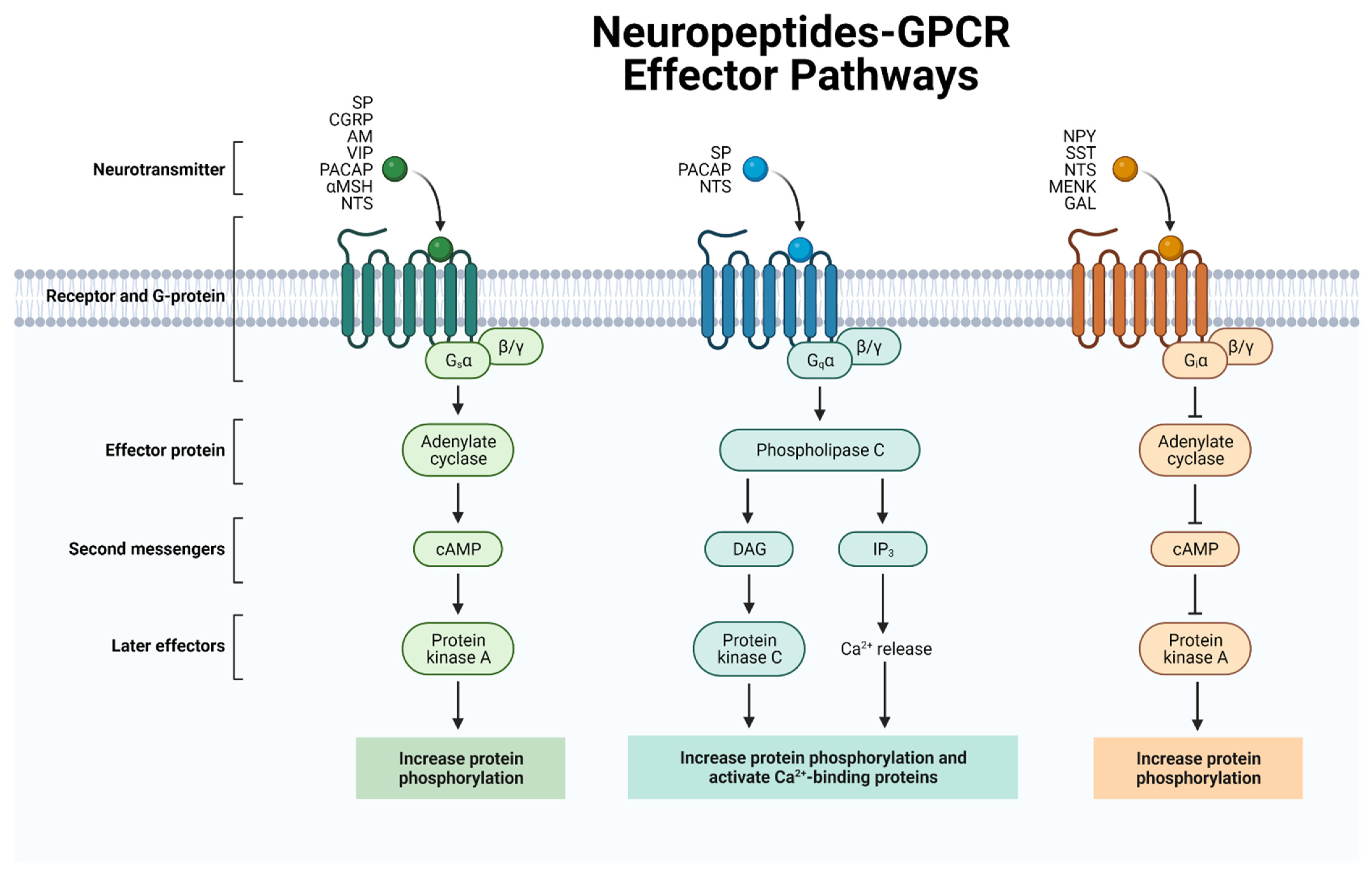
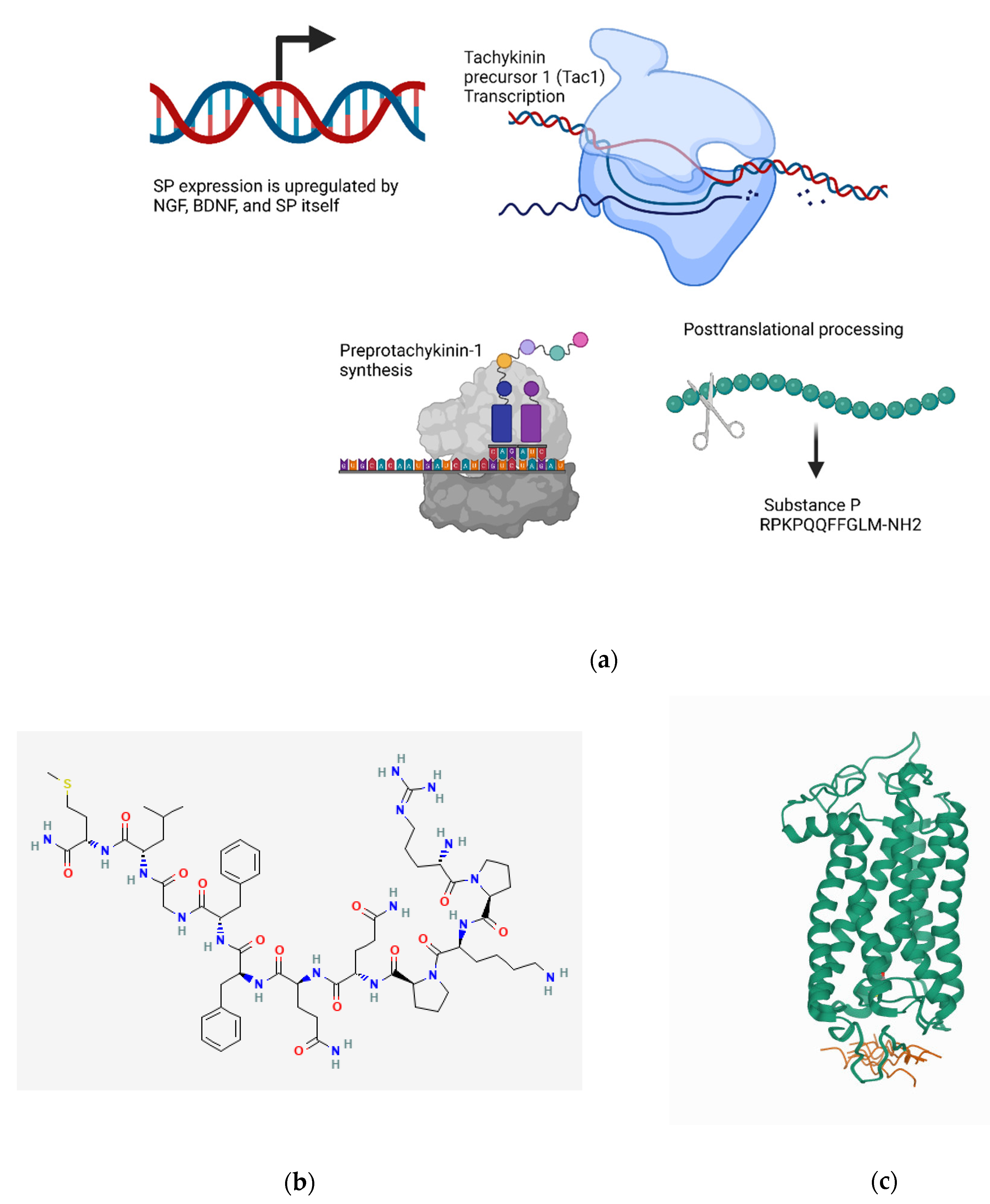
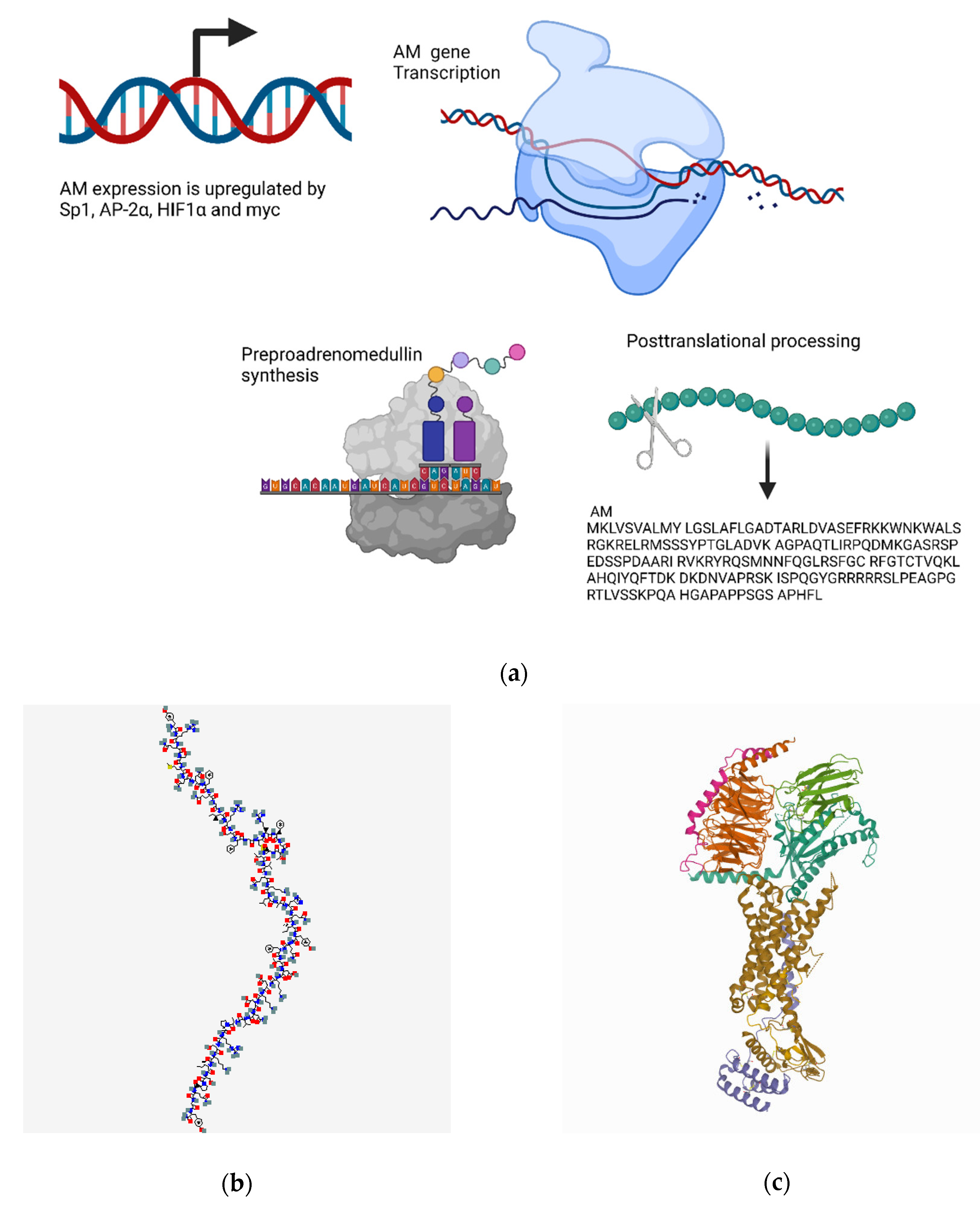
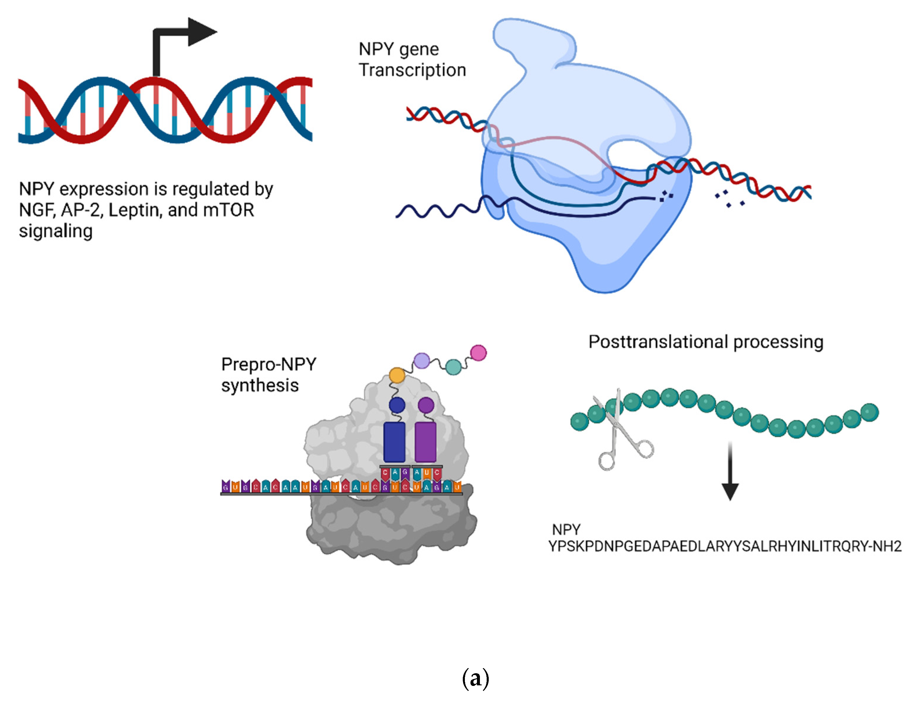
| Neuropeptides | Sequence | Receptors and Relative Affinity | References |
|---|---|---|---|
| Substance P | RPKPQQFFGLM | NK1R-F (Full) > NK1R-T (Truncated) >> NK2R, NK3R | [71,72,73,74,75,76,77] |
| CGRP | ACDTATCVTHRLALLSRSGG-VVKNNFVPTNVGSKAF | CLR/RAMP1 >> CLR/RAMP2 ≈ CLR/RAMP3 | [78,79,80,81,82,83] |
| Adrenomedullin | YRQSMNNFQGLRSFGCRFGTCTVQKLAHQIYGFTDKDKDNVAPRSKISPQGY | CLR/RAMP2 ≈ CLR/RAMP3 >> CLR/RAMP1 | [84,85,86] |
| VIP | HSDAVFTDNYTRLRKQMAVKKYLNSILN | VPAC1R > VPAC2R >> PAC1R | [87,88,89,90,91,92,93,94,95] |
| PACAP | HSDGIFTDSYSRYRKQMAVKKYLAAVLGKRYKQRVKNK | PAC1R >> VPAC1R ≈ VPAC2R | [87,96,97,98,99,100,101,102,103] |
| NPY | YPSKPDNPGEDAPAEDMARYYSALRHYINLITRQRY | Y1 ≈ Y2 ≈ Y5 >> Y4 | [104,105,106,107,108,109,110,111,112,113,114] |
| SST | SANSNPAMAPRERKAGCKNFFWKTFTSC | SST2 ≈ SST3 ≈ SST5 > SST1 ≈ SST4 | [115,116,117,118,119] |
| α-MSH | SYSMEHFRWGKPV | MC1R ≈ MC3R > MC4R > MC5R | [120,121,122,123,124,125,126] |
| Galanin | GWTLNSAGYLLGPHAVGNHRSFSDKNGLTS | GAL1R ≈ GAL2R > GAL3R | [127,128,129,130,131,132,133,134,135,136,137,138] |
| Opioid Growth Factor (OGF)/Met-enkephalin | YGGFM | μ >> OGFR > δ >> κ | [139,140,141,142,143,144] |
| Neurotensin | QLYENKPRRPYIL | NTS1R ≈ NTS2R | [145,146,147,148,149,150,151,152,153] |
| Neuropeptides (Tissue or Fluid) | Receptors (Tissue or Fluid) | References |
|---|---|---|
| SP (nerve fibers in corneal epithelium and stroma, normal tears) | NK1R (native and cultured corneal epithelial cells, mast cells, T cells, monocytes, conventional dendritic cells, and Langerhans cells) | [10,154,169,170,171,172,173,174,175,176,177] |
| CGRP (nerve fibers in corneal epithelium and stroma, normal tears) | CLR/RAMP1 (corneal and limbal epithelial cells, T cells, innate lymphoid cells, macrophages, conventional dendritic cells) | [158,159,169,178,179,180,181,182,183,184] |
| Adrenomedullin (corneal nerves) | CLR/RAMP2, CLR/RAMP3 (Corneal epithelium, stroma, and endothelium; lymphatic and vascular endothelium; T cells, dendritic cells) | [185,186,187,188,189] |
| VIP (corneal nerves in anterior stroma) | VPAC1-R, VPAC2-R (lacrimal glands—basal side of acinar cells and ducts, T cells, monocytes) | [158,180,190,191] |
| PACAP (corneal nerves, tears, lacrimal gland nerves, and acinar cells) | PAC1-R, VPAC1-R, VPAC2-R (lacrimal glands—basal side of acinar cells and ducts, T cells, monocytes) | [87,190,191,192,193,194,195,196] |
| NPY (corneal nerves in anterior stroma) | Y1, Y2, Y4, Y5, and y6 receptors (T cells, monocytes, mast cells) | [158,161,162,163,164,165,166,197,198,199,200] |
| SST (lacrimal gland, corneal nerves) | SST1R-SST5R (meibomian gland, T cells, B cells, monocytes) | [158,201,202,203] |
| α-MSH (cornea) | MC1R-MC5R (corneal endothelial cells, acinar cells in lacrimal glands, T cells, B cells, NK cells, monocytes, granulocytes) | [69,204,205,206,207] |
| Galanin (corneal and conjunctival sensory nerves) | GalR1, GalR2, and GalR3 (NK cells, neutrophils, macrophages) | [156,158,208,209,210,211,212] |
| Opioid Growth Factor (OGF)/Met-Enkephalin (Corneal nerves, corneal epithelium) | OGFr (corneal epithelial cells) | [158,205] |
| Neurotensin (corneal nerves) | Neurotensin receptor (cultured human corneal keratocytes) | [165,167,168] |
| Neuropeptide | Functions | References |
|---|---|---|
| Substance P | Pro-inflammatory. Promotes macrophage and neutrophil phagocytosis, increases pro-inflammatory cytokine secretion, activates mast cells and NK cells, and enhances T cell proliferation. Promotes tear secretion and anti-apoptotic functions on corneal epithelial cells. May maintain stemness of limbal stem cells and promotes corneal wound healing. Promotes corneal angiogenesis and lymphangiogenesis, as well as leukocyte recruitment to the cornea during inflammation. Also has a chief role in pain. | [68,179,195,227,258,259,260,261,262,263,264,265,266,267,268,269,270,271,272,273,274,275,276,277,278,279,280,281,282,283,284,285,286,287,288,289,290,291,292,293,294,295,296,297,298] |
| CGRP | Causes vasodilation and is pro-inflammatory. Enhances the pro-inflammatory activity of lymphocytes, cDCs, and macrophages. Promotes corneal wound healing through effects on corneal epithelial cells. Its levels correlate with lacrimal gland function. Also has a role in pain. | [322,323,324,325,326,327,328,329,330,331,332,334,335,336,337,338,339] |
| Adrenomedullin | Promotes the proliferation of CD34+ progenitor cells and hematopoietic stem cells. Elevated levels in models of corneal inflammation. Knockdown diminishes corneal angiogenesis. | [187,341,344,360,361,362,363] |
| VIP | Pro- and anti-inflammatory effects that may be context- or receptor-dependent. Primes the oxidative burst response in neutrophils, and causes histamine release in mast cells. Inhibits production of inflammatory cytokines and increases IL-10 production. Enhances corneal wound healing and corneal allograft survival. Promotes corneal nerve regeneration by regulating neurotrophic factors. Promotes survival of corneal endothelial cells. | [67,364,376,377,378,379,380,381,382,383,384,385,386,387,388,389,390,391,392,393,394,395,396] |
| PACAP | Pro- and anti-inflammatory effects mediated in a context-dependent manner. Inhibits secretion of pro-inflammatory cytokines from macrophages. Involved in T cell maturation and can skew towards a Th2 phenotype. Regulates tear secretion and may have utility as a treatment for dry eye disease. Enhances corneal nerve regeneration and sensitivity and accelerates corneal wound healing. | [70,190,429,430,431,432,433,434,435,436,437,438] |
| NPY | Pro- and anti-inflammatory effects. Increases chemotaxis in various immune cells. Inhibits the maturation of cDCs and proliferation of T cells. Promotes pro-inflammatory cytokine release from macrophages. Enhances corneal angiogenesis through the Y2 receptor. | [462,463,464,465,466,467,468,469,470,471,472,473,474,475] |
| SST | Pro- and anti-inflammatory effects. Correlates with activation state of immune cells. Regulates lymphocyte migration and macrophage/monocyte phagocytosis. Demonstrated to have antiangiogenic properties, including inhibiting corneal neovascularization. | [201,488,489,490,491,492,493,494,495,496,497,498,499] |
| α-MSH | Anti-inflammatory effects with widespread suppression of inflammation. Inhibits pro-inflammatory cytokine production and immune cell chemotaxis. Promotes the induction of regulatory T cells. Improves survival of corneal allografts and enhances survival of corneal endothelial cells. Increases tear secretion and goblet cell function in dry eye disease. | [69,121,124,502,507,508,509,510,511,512,513,519,520,521,522,523] |
| Galanin | Modulates neutrophil and NK cell functions. Present in the tear film, although its precise role in healthy and diseased corneas remains unclear. Also involved in pain signaling. | [210,212,524,537,538,539,540,541,542,543,544,545,546,547] |
| OGF/ Met-Enkephalin | Immunomodulatory effects on many immune cells, such as inhibiting regulatory T cells, enhancing NK cell activity, and increasing phagocytosis. Effects may be dependent on the presence of a potent immune stimulus. Suppresses corneal wound healing. | [549,555,561,562,563,564,565,566,567,568,569] |
| Neurotensin | Pro- and anti-inflammatory effects. Enhances chemotaxis and may stimulate or inhibit cytokine synthesis. Involved in pain signaling and has analgesic effects on the cornea. | [578,579,580,581,582] |
Publisher’s Note: MDPI stays neutral with regard to jurisdictional claims in published maps and institutional affiliations. |
© 2022 by the authors. Licensee MDPI, Basel, Switzerland. This article is an open access article distributed under the terms and conditions of the Creative Commons Attribution (CC BY) license (https://creativecommons.org/licenses/by/4.0/).
Share and Cite
Puri, S.; Kenyon, B.M.; Hamrah, P. Immunomodulatory Role of Neuropeptides in the Cornea. Biomedicines 2022, 10, 1985. https://doi.org/10.3390/biomedicines10081985
Puri S, Kenyon BM, Hamrah P. Immunomodulatory Role of Neuropeptides in the Cornea. Biomedicines. 2022; 10(8):1985. https://doi.org/10.3390/biomedicines10081985
Chicago/Turabian StylePuri, Sudan, Brendan M. Kenyon, and Pedram Hamrah. 2022. "Immunomodulatory Role of Neuropeptides in the Cornea" Biomedicines 10, no. 8: 1985. https://doi.org/10.3390/biomedicines10081985
APA StylePuri, S., Kenyon, B. M., & Hamrah, P. (2022). Immunomodulatory Role of Neuropeptides in the Cornea. Biomedicines, 10(8), 1985. https://doi.org/10.3390/biomedicines10081985