Article

Serotonin Receptor Gene Polymorphisms Are Associated with Cerebrospinal Fluid, Genetic, and Neuropsychological Biomarkers of Alzheimer’s Disease

1
Department of Neuroscience, Croatian Institute for Brain Research, University of Zagreb Medical School, 10000 Zagreb, Croatia
2
Department of Medical Biology, School of Medicine, University of Split, 21000 Split, Croatia
3
Department of Molecular Medicine, Institute Ruđer Bošković, 10000 Zagreb, Croatia
4
Laboratory for Neurobiochemistry, Department of Laboratory Diagnostics, University Hospital Centre Zagreb, 10000 Zagreb, Croatia
5
General Hospital Zabok, 49210 Zabok, Croatia
6
Department of Neurology, University Hospital Centre Zagreb, 10000 Zagreb, Croatia
7
Nash Family Department of Neuroscience, Friedman Brain Institute, Ronald M. Loeb Center for Alzheimer’s Disease, Icahn School of Medicine at Mount Sinai, New York, NY 10029, USA
*
Author to whom correspondence should be addressed.
Biomedicines 2022, 10(12), 3118; https://doi.org/10.3390/biomedicines10123118
Submission received: 8 November 2022 / Revised: 26 November 2022 / Accepted: 30 November 2022 / Published: 2 December 2022
(This article belongs to the Special Issue Genetic Architecture of Dementia)

Abstract

:
A decrease in serotonergic transmission throughout the brain is among the earliest pathological changes in Alzheimer’s disease (AD). Serotonergic receptors are also affected in AD. Polymorphisms in genes of serotonin (5HT) receptors have been mostly associated with behavioral and psychological symptoms of dementia (BPSD). In this study, we examined if AD patients carrying different genotypes in 5HTR1B rs13212041, 5HTR2A rs6313 (T102C), 5HTR2C rs3813929 (−759C/T), and 5HTR6 rs1805054 (C267T) polymorphisms have a higher risk of faster disease progression (assessed by neuropsychological testing), are more prone to develop AD-related pathology (reflected by levels of cerebrospinal fluid [CSF] AD biomarkers), or have an association with an apolipoprotein E (APOE) haplotype. This study included 115 patients with AD, 53 patients with mild cognitive impairment (MCI), and 2701 healthy controls. AD biomarkers were determined in the CSF of AD and MCI patients using enzyme-linked immunosorbent assays (ELISA), while polymorphisms were determined using either TaqMan SNP Genotyping Assays or Illumina genotyping platforms. We detected a significant decrease in the CSF amyloid β1–42 (Aβ1–42) and an increase in p-tau181/Aβ1–42 ratio in carriers of the T allele in the 5HTR2C rs3813929 (−759C/T) polymorphism. A significantly higher number of APOE ε4 allele carriers was observed among individuals carrying a TT genotype within the 5HTR2A T102C polymorphism, a C allele within the 5HTR1B rs13212041 polymorphism, and a T allele within the 5HTR6 rs1805054 (C267T) polymorphism. Additionally, individuals carrying the C allele within the 5HTR1B rs13212041 polymorphism were significantly more represented among AD patients and had poorer performances on the Rey–Osterrieth test. Carriers of the T allele within the 5HTR6 rs1805054 had poorer performances on the MMSE and ADAS–Cog. As all four analyzed polymorphisms of serotonin receptor genes showed an association with either genetic, CSF, or neuropsychological biomarkers of AD, they deserve further investigation as potential early genetic biomarkers of AD.

1. Introduction

The serotonergic system is severely affected in Alzheimer’s disease (AD) [1,2,3,4]. Indeed, serotonin (5-hydroxytryptamine, 5HT) is an indoleamine released by serotonergic neurons located in the brainstem raphe nuclei. These nuclei are divided into a rostral (B5–B9) and a caudal (B1–B3) raphe group [5,6,7,8]. The main serotonergic nucleus, the dorsal raphe nucleus (DRN, B7–B9), projects throughout the cerebral cortex (reviewed in [9]). Moreover, 5HT binds to serotonergic receptors. There are seven types of serotonergic receptors, with several subtypes (5HTR1A-F, 5HTR2A-C, 5HTR3A-E, 5HTR4, 5HTR5A-B, 5HTR6, 5HTR7). All 5HT receptors, except for 5HTR3, a ligand-gated ion channel, are G-protein-coupled receptors [10,11].
Loss of serotonergic innervation of the hippocampus and neocortex [2,11,12,13], decrease in the levels of 5HT and 5HT metabolites [14,15], and accumulation of AD pathological changes in serotonergic nuclei [16] have all been reported in AD. In addition, the loss of 5HT receptors and 5HT receptor binding was observed in AD [17,18,19]. Polymorphisms in genes for 5HT receptors have been associated with behavioral and psychological symptoms of dementia (BPSD) [20,21,22,23,24,25,26]. The 5HTR2A rs6313 (T102C) and 5HTR6 rs1805054 (C267T) polymorphisms were previously associated with AD, while the association of the 5HTR1B rs13212041 and 5HTR2C rs3813929 (−759C/T) polymorphisms with AD was not previously noticed. This study assessed whether the levels of cerebrospinal fluid (CSF) AD biomarkers, scores on neuropsychological tests, and genetic biomarkers of AD (apolipoprotein E (APOE) haplotype) differ between AD patients with various 5HTR1B rs13212041, 5HTR2A rs6313 (T102C), 5HTR2C rs3813929 (−759C/T), and 5HTR6 rs1805054 (C267T) polymorphisms. CSF AD biomarkers serve as endophenotypes of AD as they reflect AD pathological changes [27], while neuropsychological tests show potential in monitoring disease progression [28]. CSF amyloid β1–42 (Aβ1–42) is an index of amyloid plaque deposition [29], phosphorylated tau proteins reflect neurofibrillary tangles [30], and total tau (t-tau) and visinin-like protein 1 (VILIP-1) are markers of neurodegeneration [31,32]. We tested the potential of such polymorphisms as genetic biomarkers of AD and certain genotypes as representing a genetic predisposition to develop AD-related pathologies and faster disease progression.

2. Materials and Methods

2.1. Subjects

This study included 168 patients recruited at the University Hospital Center Zagreb and 2701 healthy controls (HC) from the “10,001 Dalmatians project” (part of the Croatian Biobank program [33]). AD was diagnosed using the criteria of the National Institutes on Aging–Alzheimer’s Association (NIA–AA) [34], while mild cognitive impairment (MCI) was diagnosed using the criteria of Petersen et al. [35] and Albert et al. [36]. Participants gave informed consent for participation in the study, and the Central Ethical Committee of the University of Zagreb Medical School (case no. 380-59-10106-18-111/126, class 641-01/18-02/01 from 20 June 2018), Ethical Committee of the Clinical Hospital Center Zagreb (case no. 02/21 AG, class 8.1-18/82-2 from 24 April 2018), and Ethical board of the University of Split, School of Medicine (case no. 2181-198-03-04-14-0031 and 2181-198-03-04-19-0022) approved all procedures. Additionally, all procedures performed within this study were in accord with the Helsinki Declaration [37]. Patients underwent neurological examination, examination of thyroid function, and serology for syphilis and Lyme disease. The levels of vitamin B12 and B9 (folic acid) were also determined in each patient. Table 1 summarizes information on biomarkers and demographic data, while Table 2 summarizes information on determined 5HTR and APOE genotypes.

2.2. Neuropsychological Testing

Patients were neuropsychologically tested using the Mini-Mental State Examination (MMSE), the Alzheimer’s Disease Assessment Scale–cognitive subscale (ADAS–Cog), the Clock Drawing Test (CDT), the Rey–Osterrieth complex figure test (ROCFT), and the Visual Association Test (VAT).

2.3. Analysis of CSF Biomarkers

CSF was collected in AD and MCI patients by lumbar puncture between intervertebral spaces L3/L4 or L4/L5. After the centrifuge at 2000× g for 10 min, CSF was stored at −80 °C in polypropylene tubes. AD biomarkers were determined by enzyme-linked immunosorbent assays (ELISA) using the following assays: Aβ1–42 (Innotest β-amyloid1–42, Fujirebio, Tokyo, Japan), VILIP-1 (VILIP-1 Human ELISA, BioVendor, Brno, Czech Republic), p-tau181 (Innotest Phospho-Tau [181P], Fujirebio, Tokyo, Japan), p-tau231 (Tau [pT231] Phospho-ELISA Kit, Human, Thermo Fisher Scientific, Waltham, MA, USA), p-tau199 (TAU [pS199] Phospho-ELISA Kit, Human, Thermo Fisher Scientific), and t-tau (Innotest hTau AG, Fujirebio, Tokyo, Japan) (Table 1).

2.4. Determination of Polymorphisms

The salting-out method was used for the isolation of DNA from the peripheral blood [38]. In the 168 patients recruited at the University Hospital Center Zagreb, single nucleotide polymorphisms (SNPs) were determined by ABI Prism 7300 Real-Time PCR System apparatus (Applied Biosystems, Foster City, CA, USA), using the following TaqMan SNP Genotyping Assays (Applied Biosystems): 5HTR1B rs13212041, 5HTR2A rs6313 (T102C), 5HTR2C rs3813929 (−759C/T), 5HTR6 rs1805054 (C267T), APOE rs7412, and rs429358. APOE SNPs were measured to determine APOE haplotypes (APOE ε2, ε3, and ε4) (rs429358 C allele and rs7412 C allele for ε4 variant, rs429358 T allele and rs7412 C allele for ε3 variant, and rs429358 T allele and rs7412 T allele for ε2 variant). SNPs were determined using Illumina genotyping platforms (CNV370v1, CNV370-Quadv3, and OmniExpressExome-8v1-2_A, Illumina, San Diego, CA, USA) in 2701 participants recruited from the “10,001 Dalmatians project”.

2.5. Statistical Analysis

Statistical analysis was performed with SPSS 19.0.1 (SPSS, Chicago, IL, USA). The level of statistical significance was set at α = 0.05. Levels of CSF biomarkers and scores on neuropsychological tests were compared between groups using the non-parametric Kruskal–Wallis test, while pairwise comparisons were conducted using a post-hoc non-parametric test (that corrects p values for multiple comparisons). The frequencies of different diagnoses and APOE genotypes among subjects with different 5HTR1B rs13212041, 5HTR2A rs6313 (T102C), 5HTR2C rs3813929 (−759C/T), and 5HTR6 rs1805054 (C267T) genotypes and alleles were analyzed using a χ2-test, with applied correction for pairwise comparisons. When analyzing frequencies of different diagnoses among subjects with different 5HTR genotypes, we included only HC of 70 years old and older (n = 461).

3. Results

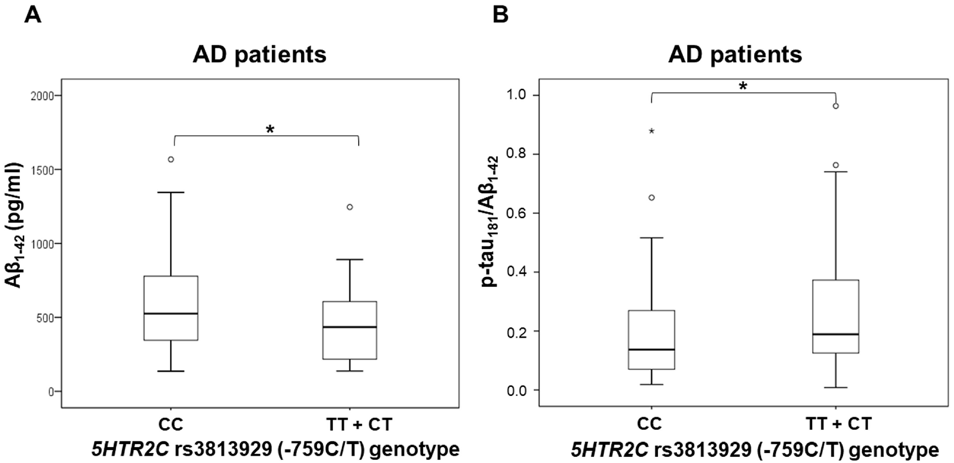
3.1. Polymorphisms in 5HT Receptor Genes and CSF Biomarkers

The CSF levels of Aβ1–42 were significantly decreased in AD patients with TT and CT genotypes compared to those with the CC 5HTR2C rs3813929 (-759C/T) genotype (U = 1080, Z = −2.063, p = 0.039) (Figure 1). P-tau181/Aβ1–42 ratio was significantly increased in AD patients with TT and CT genotypes compared to those with the CC 5HTR2C rs3813929 (-759C/T) genotype (U = 1056, Z = −2.121, p = 0.034) (Figure 1). There was no significant difference in the levels of CSF biomarkers (Aβ1–42, t-tau, p-tau181, p-tau199, p-tau231, VILIP-1, and p-tau181/Aβ1–42 ratio) between subjects with different 5HTR2A rs6313 (T102C), 5HTR1B rs13212041, and 5HTR6 rs1805054 (C267T) genotypes. No significant difference in t-tau, p-tau181, p-tau199, p-tau231, and VILIP-1 levels was observed between subjects with different 5HTR2C rs3813929 (-759C/T) genotypes.

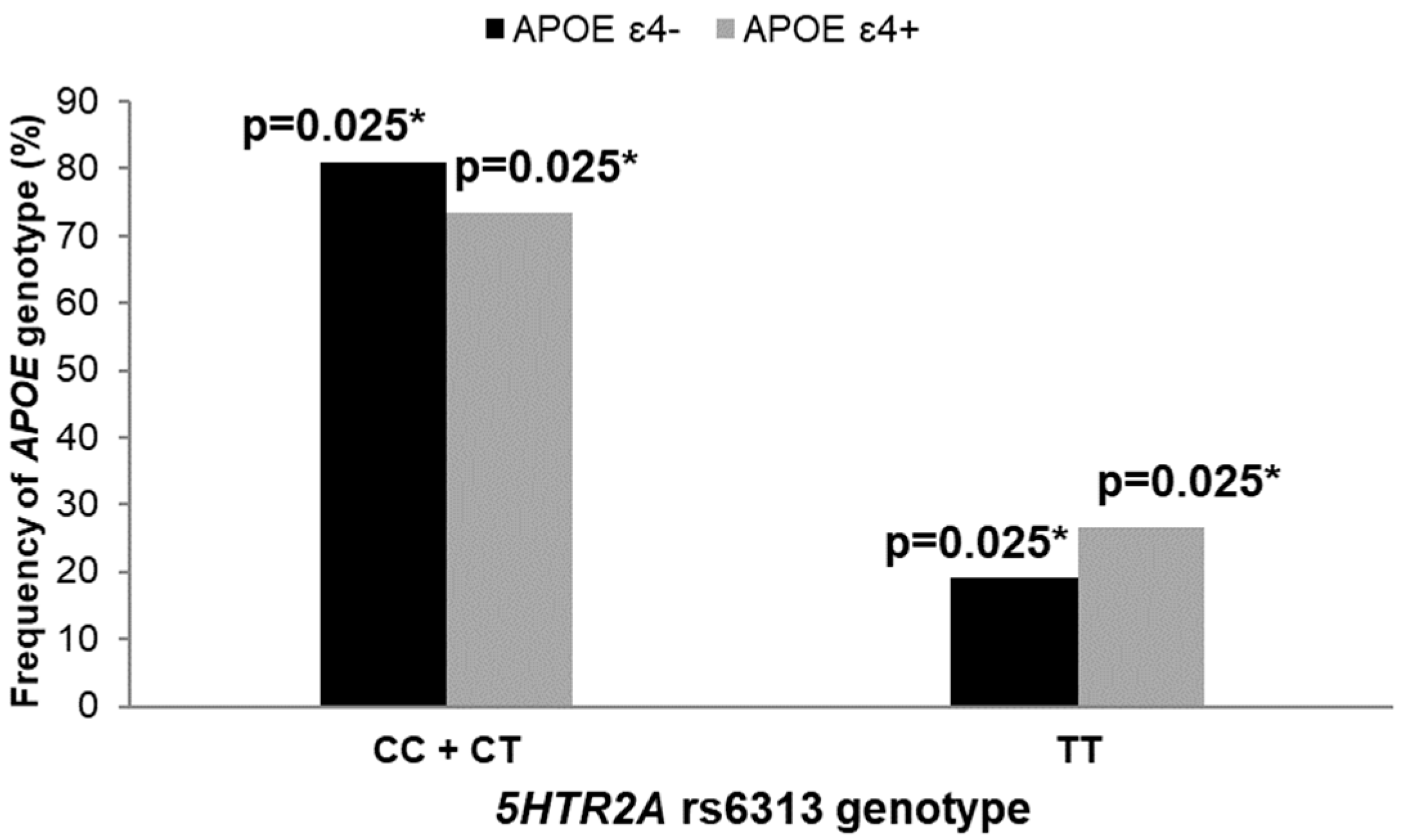
3.2. Polymorphisms in 5HT Receptor Genes, APOE Genotype, and AD Diagnosis

We observed a significantly higher number of APOE ε4 allele carriers among female patients with the TT genotype compared to carriers of the CC and CT genotypes within the 5HTR2A T102C polymorphism (χ2 = 7.453, df = 1; p = 0.006; Figure 2). This was also confirmed with logistic regression (β = 1.364, SE = 0.151, p = 0.040).
A significantly higher number of APOE ε4 allele carriers was also observed among male patients carrying the CC and CT genotypes compared to carriers of the TT genotype within the 5HTR1B rs13212041 polymorphism (χ2 = 7.064, df = 1; p = 0.008; Figure 3). Additionally, a significantly higher number of individuals carrying the C allele within the 5HTR1B rs13212041 polymorphism was observed among AD patients (χ2 = 6.973, df = 1; p = 0.008; Figure 3).
A significantly higher number of APOE ε4 allele carriers was also observed among individuals carrying the T allele within the 5HTR6 rs1805054 (C267T) polymorphism (χ2 = 6.425, df = 1; p = 0.011; Figure 4).

3.3. Polymorphisms in 5HT Receptors, Genes, and Neuropsychological Tests

AD patients carrying the C allele within the 5HTR1B rs13212041 polymorphism had poorer performances on the ROCFT test (U = 216.5, Z = −2.106, p = 0.035; Figure 3).
Carriers of the T allele within the 5HTR6 rs1805054 had poorer performances on the ADAS–Cog (in MCI patients; U = 80.5, Z = −1.985, p = 0.046; Figure 4) and MMSE (in AD patients; t = −2.015, df = 108, p = 0.046; Figure 4). In contrast, AD patients carrying the CC genotype within the 5HTR6 rs1805054 had poorer performances on the VAT test compared to TT and CT genotype carriers (U = 223, Z = −2.224, p = 0.026).

4. Discussion

The serotonergic system is highly affected in AD [1,2,3,4]. The main serotonergic nucleus that projects throughout the cortex, the dorsal raphe nucleus (DRN, B7-B9), is affected early by AD pathological changes, with neurofibrillary pathology in all of Braak stage I and more than 20% of Braak stage 0 cases [16]. In addition, altered activity of DRN neurons due to the accumulation of AD pathological changes is thought to cause BPSD in early AD [39,40,41], which is compatible with a reported decrease in the serotonergic innervation of the hippocampus and neocortex [2,11,12,13].
Changes in serotonergic receptors are also detected in AD. Loss of 5HT1B/1D and 5HT6 receptors was observed in the frontal and temporal cortex of AD patients [17]. Reduction in 5HT1A receptor binding [18] and loss of 5HT2A receptors [19] was observed in the AD brain using positron emission tomography (PET) imaging. Additionally, reduced binding to the 5HT1A receptor in the hippocampus and temporal neocortex, respectively, correlates with cognitive decline [42], and aggressive behavior [43]. Activation of 5HT4, 5HT6, and 5HT7 receptors in experimental models of AD resulted in a decrease in Aβ content [44,45,46,47], while injections of Aβ in the hippocampi of mouse models of AD [48,49] leads to a reduction in 5HT2A receptor expression. Interestingly, serotonergic receptors are potential targets for AD therapeutics [4] as their activation affects signaling pathways involved in the production of Aβ and hyperphosphorylated tau protein [3]. Activation of 5HTR4, 5HTR6, and 5HTR7 results in reduced production of Aβ (for details see [45]). Additionally, the activation of various 5HT receptors can modify tau phosphorylation. For example, the activation of 5HTR1A activates the phosphoinositide 3-kinase (PI3K), phosphoinositide-dependent kinase (PDK), and protein kinase B (AKT) cascade. AKT phosphorylates and consequently inactivates glycogen synthase kinase-3 (GSK3) that phosphorylates tau protein. 5HTR2 could modulate GSK3 phosphorylation through protein kinase C (PKC) [50] and β-arrestin-mediated signaling [51], while 5HTR4, 5HTR6, and 5HTR7 could modulate GSK3 phosphorylation through protein kinase A (PKA) [50]. Several studies also observed an association between APOE and 5HT receptors. Shinohara et al. showed that a 5HTR3 antagonist (ondansetron) increases apoE secretion through the liver X receptor (LXR) and ATB-binding cassette protein A1 (ABCA1) pathway [52]. Additionally, Chhibber and Zhao observed a significant difference in 5HT receptor expression levels in mice carrying different ApoE genotypes [53]. Specifically, 5HTR2A protein expression levels were higher in the cortexes of mice with human APOE4 gene-targeted replacement than in mice with ApoE2 and ApoE3 genotypes. However, 5HTR1A protein levels did not differ among mice with different ApoE genotypes [53].
In this study, we assessed whether the levels of CSF AD biomarkers, scores on neuropsychological tests, and genetic biomarkers of AD (APOE haplotype) differed between patients with various 5HTR1B rs13212041, 5HTR2A rs6313 (T102C), 5HTR2C rs3813929 (−759C/T), and 5HTR6 rs1805054 (C267T) polymorphisms. We observed a significantly higher number of APOE ε4 allele carriers among individuals carrying the TT genotype within the 5HTR2A T102C polymorphism, the C allele within the 5HTR1B rs13212041 polymorphism, and the T allele within the 5HTR6 rs1805054 (C267T) polymorphism. Additionally, individuals carrying the C allele within the 5HTR1B rs13212041 polymorphism were significantly more represented among AD patients and had poorer performances on the ROCFT test. Carriers of a T allele within the 5HTR6 rs1805054 had poorer performances on the MMSE and ADAS–Cog, while a significant decrease in the levels of CSF Aβ1–42 and an increase in the p-tau181/Aβ1–42 ratio was observed in carriers of a T allele in the 5HTR2C rs3813929 (−759C/T) polymorphism.
Our study shows that AD patients carrying a T allele in the 5HTR2C rs3813929 (−759C/T) polymorphism have pathological CSF Aβ1–42 levels. The 5HTR2C -759C/T polymorphism did not affect the expression levels of the 5HT2C receptor [54], and the effect of the 5HTR2C −759C/T polymorphism on 5HT2C receptor expression in different tissues is also not documented in the Genotype-Tissue Expression (GTEx) project database [55]. However, Buckland et al. observed that the C allele within the 5HTR2C −759C/T polymorphism shows less transcriptional activity compared to the T allele [56]. The association of the 5HTR2C -759C/T polymorphism with AD was not previously reported. However, in vitro [57] and in vivo [58] experiments showed that 5HT2C receptor activation stimulates the release of soluble amyloid precursor protein (sAPP). Our study reveals that carriers of the T allele in the 5HTR2C rs3813929 (-759C/T) polymorphism have pathological CSF Aβ1–42 levels, and Buckland et al.’s study showed that the T allele within the 5HTR2C -759C/T polymorphism increases transcriptional activity [56]. Thus, it is possible that this polymorphism indirectly affects the release of sAPP and the amount of produced Aβ1–42.
Additionally, this study shows that carriers of the T allele within the 5HTR6 rs1805054 (C267T) polymorphism have poorer performances on the MMSE and ADAS–Cog tests and that a higher number of APOE ε4 allele carriers is observed among these individuals. The 5HTR6 C267T polymorphism does not involve an amino acid change, but this silent mutation could affect the splicing process [59]. According to the GTEx portal [55], this SNP significantly affects the expression levels of the 5HT6 receptor, with carriers of the T allele within the 5HTR6 rs1805054 (C267T) polymorphism having a lower expression of 5HT6 receptor mRNA in whole blood. The 5HTR6 C267T polymorphism was previously associated with AD, albeit with conflicting results. Tsai et al. observed a higher frequency of the CC 5HTR6 C267T genotype in AD patients compared to controls [60], while Kan et al. observed an increased number of CT 5HTR6 C267T heterozygotes among AD patients [61]. Moreover, other authors did not find an association between 5HTR6 C267T polymorphism and AD [59,62,63]. Our study did not observe a difference in the distribution of 5HTR6 C267T genotypes between AD patients and controls, but this SNP elucidated an association between neuropsychological and genetic biomarkers of AD. The association between the 5HTR6 C267T polymorphism and cognitive decline in AD observed in this study is not surprising given that several studies elucidated an association between this receptor and AD (reviewed in [64]). In fact, the potential of 5HT6 receptor antagonists as therapeutics for AD has been tested in a number of studies [65].
Our study also revealedan association of the C allele within the 5HTR1B rs13212041 polymorphism with genetic and neuropsychological biomarkers of AD and AD diagnosis that has not been previously associated with AD. The effect of the 5HTR1B rs13212041 polymorphism on 5HT1B receptor expression in different tissues is also not documented in the GTEx portal [55], although Jensen et al. showed that carriers of the T allele within the 5HTR1B rs13212041 polymorphism show reduced 5HTR1B expression compared to carriers of the C allele [66].
Finally, we observed a significantly higher number of APOE ε4 allele carriers among individuals carrying the TT genotype within the 5HTR2A T102C polymorphism. According to the GTEx portal [55], this SNP does not affect the levels of 5HTR2A in the brain, although it significantly affected 5HTR2A expression in testes, muscles, and aortae. This polymorphism is located within the first exon of the 5HTR2A gene and, being near the promoter region, could be involved in gene regulation [67]. Li et al. recently showed that the 5HTR2A T102C polymorphism increases the risk of AD [68]. Interestingly, the 5HTR2A T102C polymorphism also showed an association with BPSD in AD [21,22,23,24,25,26], although inconsistently among studies [69,70,71,72].

5. Conclusions

In this study, we observed differences in the distribution of 5HT receptor gene genotypes and APOE genotypes between male and female participants. Gender difference in the distribution of both APOE genotypes and 5HT receptor gene genotypes was previously reported [73,74]. Namely, it was shown that elderly female APOE ε4 carriers are at higher risk of developing AD [75], show stronger cognitive decline [76], weaker brain connectivity (detected using functional magnetic resonance imaging (fMRI) in the precuneus and posterior cingulate cortex) [73], and lower brain metabolism [77] than males. In contrast, Cacciottolo et al. showed that elderly males diagnosed with AD or MCI carrying the APOE ε4 allele had a higher risk of brain microbleeds compared to females with the same genotype and condition [78]. Interestingly, a similar sex-dependent relationship between HTR2C gene variants and suicidal behavior [79] and HTR1B polymorphisms and schizophrenia [80] has been reported.
Our data reveal that all four analyzed polymorphisms of 5HT receptor genes had an association with either genetic, CSF, or neuropsychological biomarkers of AD. As such, considering the early involvement of the serotonergic systems in the progression of AD, these polymorphisms represent interesting diagnostic and therapeutic targets and deserve further investigation as potential early genetic biomarkers of AD.

Author Contributions

Conceptualization, G.Š. and M.B.L.; methodology, M.B.L., M.N.P., E.Š., N.P. (Nikolina Pleić), Ž.V., G.N.E., D.Š.Š., N.P. (Nela Pivac); validation, G.Š., N.P. (Nela Pivac), P.R.H., T.Z.; formal analysis, M.B.L., N.P. (Nikolina Pleić), M.N.P.; investigation, M.B.L., M.N.P., E.Š., N.P. (Nikolina Pleić), Ž.V., D.Š.Š., N.K., G.N.E., F.B., D.B., I.G.; resources, G.Š., T.Z.; data curation, M.B.L., M.N.P., N.P. (Nikolina Pleić); writing—original draft preparation, M.B.L.; writing—review and editing, G.Š., T.Z., M.N.P., I.G., N.P. (Nikolina Pleić), P.R.H.; visualization, M.B.L., G.Š.; supervision, G.Š.; project administration, G.Š.; funding acquisition, G.Š. All authors have read and agreed to the published version of the manuscript.

Funding

This work was funded by The Croatian Science Foundation grants IP-2019-04-3584 (“Role of the blood-brain barrier, innate immunity, and tau protein oligomerization in the pathogenesis of Alzheimer’s disease“) and IP-2014-09-9730 (“Tau protein hyperphosphorylation, aggregation, and trans-synaptic transfer in Alzheimer’s disease: cerebrospinal fluid analysis and assessment of potential neuroprotective compounds”) to G.Š., IP-2019-04-2593 (“Regulation of thyroid and parathyroid function and blood calcium homeostasis”) to T.Z., and by the Scientific Center of Excellence for Basic, Clinical, and Translational Neuroscience CoRE-NEURO (“Experimental and clinical research of hypoxic-ischemic damage in perinatal and adult brain”; GA KK01.1.1.01.0007 funded by the European Union through the European Regional Development Fund), and in part by the NIH grant P30 AG066514 to P.R.H.

Institutional Review Board Statement

The study was conducted in accordance with the Declaration of Helsinki, and it was approved by the Central Ethical Committee of the University of Zagreb Medical School (case no. 380-59-10106-18-111/126, class 641-01/18-02/01 from 20 June 2018), Ethical Committee of the Clinical Hospital Center Zagreb (case no. 02/21 AG, class 8.1-18/82-2 from 24 April 2018), and Ethical board of the University of Split, School of Medicine (case no. 2181-198-03-04-14-0031 from 30 May 2014 and 2181-198-03-04-19-0022 from 27 March 2019).

Informed Consent Statement

Informed consent was obtained from all subjects involved in this study. Written informed consent has been obtained from the patient(s) to publish this paper.

Data Availability Statement

All the data reported are available on request from the corresponding author.

Conflicts of Interest

The authors declare no conflict of interest. The funders had no role in the design of the study; in the collection, analyses, or interpretation of data; in the writing of the manuscript; or in the decision to publish the results.

Abbreviations

5HT: Serotonin; 5HTR, gene for 5HT receptor; Aβ, amyloid β; ABCA1, ATB-binding cassette protein A1; AD, Alzheimer’s disease; ADAS–Cog, Alzheimer’s Disease Assessment Scale–cognitive subscale; AKT, protein kinase B; APOE, apolipoprotein E; BPSD, behavioral and psychological symptoms of dementia; CDT, Clock Drawing Test; CSF, cerebrospinal fluid; DRN, dorsal raphe nucleus; ELISA, enzyme-linked immunosorbent assays; fMRI, functional magnetic resonance imaging; GSK3, glycogen synthase kinase-3; LP, lumbar puncture; LXR, liver X receptor; MCI, mild cognitive impairment; MMSE, Mini-Mental State Examination; NIA–AA, National Institutes on Aging–Alzheimer’s Association; PDK, phosphoinositide-dependent kinase; PET, positron emission tomography; PI3K, phosphoinositide 3-kinase; PKA, protein kinase A; PKC, protein kinase C; p-tau181, tau phosphorylated at Thr 181; p-tau199, tau phosphorylated at Ser 199; p-tau231, tau phosphorylated at Thr 231; ROCFT, Rey–Osterrieth complex figure test; sAPP, soluble amyloid precursor protein; SNP, single nucleotide polymorphisms; t-tau, total tau; VAT, Visual Association Test; VILIP-1, visinin-like protein 1.

References

  1. Šimić, G.; Stanić, G.; Mladinov, M.; Jovanov-Milošević, N.; Kostović, I.; Hof, P. Does Alzheimer’s disease begin in the brainstem? Annotation. Neuropathol. Appl. Neurobiol. 2009, 35, 532–554. [Google Scholar] [CrossRef] [PubMed] [Green Version]
  2. Trillo, L.; Das, D.; Hsieh, W.; Medina, B.; Moghadam, S.; Lin, B.; Dang, V.; Sanchez, M.M.; De Miguel, Z.; Ashford, J.W.; et al. Ascending monoaminergic systems alterations in Alzheimer’s disease. Translating basic science into clinical care. Neurosci. Biobehav. Rev. 2013, 37, 1363–1379. [Google Scholar] [CrossRef] [PubMed]
  3. Babić Leko, M.; Hof, P.R.; Šimić, G. Alterations and interactions of subcortical modulatory systems in Alzheimer’s disease. Prog. Brain Res. 2021, 261, 379–421. [Google Scholar] [CrossRef] [PubMed]
  4. Šimić, G.; Babić Leko, M.; Wray, S.; Harrington, C.R.; Delalle, I.; Jovanov-Milošević, N.; Bažadona, D.; Buée, L.; de Silva, R.; Di Giovanni, G.; et al. Monoaminergic neuropathology in Alzheimer’s disease. Prog. Neurobiol. 2017, 151, 101–138. [Google Scholar] [CrossRef] [Green Version]
  5. Takahashi, H.; Nakashima, S.; Ohama, E.; Takeda, S.; Ikuta, F. Distribution of serotonin-containing cell bodies in the brainstem of the human fetus determined with immunohistochemistry using antiserotonin serum. Brain Dev. 1986, 8, 355–365. [Google Scholar] [CrossRef]
  6. Halliday, G.M.; Törk, I. Serotonin-like immunoreactive cells and fibres in the rat ventromedial mesencephalic tegmentum. Brain Res. Bull. 1989, 22, 725–735. [Google Scholar] [CrossRef]
  7. Baker, K.; Halliday, G.; Törk, I. Cytoarchitecture of the human dorsal raphe nucleus. J. Comp. Neurol. 1990, 301, 147–161. [Google Scholar] [CrossRef]
  8. Nieuwenhuys, R.; Voogd, J.; van Huijzen, C. The Human Central Nervous System, 4th ed.; Springer: New York, NY, USA, 2008. [Google Scholar]
  9. Seyedabadi, M.; Fakhfouri, G.; Ramezani, V.; Mehr, S.E.; Rahimian, R. The role of serotonin in memory: Interactions with neurotransmitters and downstream signaling. Exp. Brain Res. 2014, 232, 723–738. [Google Scholar] [CrossRef]
  10. Darmon, M.; Al Awabdh, S.; Emerit, M.-B.; Masson, J. Insights into serotonin receptor trafficking: Cell membrane targeting and internalization. Prog. Mol. Biol. Transl. Sci. 2015, 132, 97–126. [Google Scholar] [CrossRef]
  11. Curcio, C.A.; Kemper, T. Nucleus raphe dorsalis in dementia of the Alzheimer type: Neurofibrillary changes and neuronal packing density. J. Neuropathol. Exp. Neurol. 1984, 43, 359–368. [Google Scholar] [CrossRef]
  12. Halliday, G.M.; McCann, H.L.; Pamphlett, R.; Brooks, W.S.; Creasey, H.; McCusker, E.; Cotton, R.G.; Broe, G.A.; Harper, C.G. Brain stem serotonin-synthesizing neurons in Alzheimer’s disease: A clinicopathological correlation. Acta Neuropathol. 1992, 84, 638–650. [Google Scholar] [CrossRef] [PubMed]
  13. Chen, C.P.L.-H.; Eastwood, S.L.; Hope, T.; McDonald, B.; Francis, P.T.; Esiri, M.M. Immunocytochemical study of the dorsal and median raphe nuclei in patients with Alzheimer’s disease prospectively assessed for behavioural changes. Neuropathol. Appl. Neurobiol. 2000, 26, 347–355. [Google Scholar] [CrossRef] [PubMed]
  14. Nazarali, A.J.; Reynolds, G.P. Monoamine neurotransmitters and their metabolites in brain regions in Alzheimer’s disease: A postmortem study. Cell. Mol. Neurobiol. 1992, 12, 581–587. [Google Scholar] [CrossRef] [PubMed]
  15. Garcia-Alloza, M.; Gil-Bea, F.J.; Diez-Ariza, M.; Chen, C.P.L.-H.; Francis, P.T.; Lasheras, B.; Ramirez, M.J. Cholinergic–serotonergic imbalance contributes to cognitive and behavioral symptoms in Alzheimer’s disease. Neuropsychologia 2005, 43, 442–449. [Google Scholar] [CrossRef] [PubMed] [Green Version]
  16. Grinberg, L.T.; Rüb, U.; Ferretti, R.E.L.; Nitrini, R.; Farfel, J.M.; Polichiso, L.; Gierga, K.; Jacob-Filho, W.; Heinsen, H. The dorsal raphe nucleus shows phospho-tau neurofibrillary changes before the transentorhinal region in Alzheimer’s disease. A precocious onset? Neuropathol. Appl. Neurobiol. 2009, 35, 406–416. [Google Scholar] [CrossRef]
  17. Garcia-Alloza, M.; Hirst, W.D.; Chen, C.P.L.-H.; Lasheras, B.; Francis, P.T.; Ramírez, M.J. Differential involvement of 5-HT1B/1D and 5-HT6 receptors in cognitive and non-cognitive symptoms in Alzheimer’s disease. Neuropsychopharmacology 2004, 29, 410–416. [Google Scholar] [CrossRef] [Green Version]
  18. Truchot, L.; Costes, N.; Zimmer, L.; Laurent, B.; Le Bars, D.; Thomas-Antérion, C.; Mercier, B.; Hermier, M.; Vighetto, A.; Krolak-Salmon, P. A distinct [18F]MPPF PET profile in amnestic mild cognitive impairment compared to mild Alzheimer’s disease. Neuroimage 2008, 40, 1251–1256. [Google Scholar] [CrossRef]
  19. Marner, L.; Frokjaer, V.G.; Kalbitzer, J.; Lehel, S.; Madsen, K.; Baaré, W.F.C.; Knudsen, G.M.; Hasselbalch, S.G. Loss of serotonin 2A receptors exceeds loss of serotonergic projections in early Alzheimer’s disease: A combined [11C]DASB and [18F]altanserin-PET study. Neurobiol. Aging 2012, 33, 479–487. [Google Scholar] [CrossRef]
  20. Holmes, C.; Arranz, M.; Powell, J.; Collier, D.; Lovestone, S. 5-HT2A and 5-HT2C receptor polymorphisms and psychopathology in late onset Alzheimer’s disease. Hum. Mol. Genet. 1998, 7, 1507–1509. [Google Scholar] [CrossRef] [Green Version]
  21. Holmes, C.; Arranz, M.; Collier, D.; Powell, J.; Lovestone, S. Depression in Alzheimer’s disease: The effect of serotonin receptor gene variation. Am. J. Med. Genet. B. Neuropsychiatr. Genet. 2003, 119B, 40–43. [Google Scholar] [CrossRef]
  22. Pritchard, A.L.; Harris, J.; Pritchard, C.W.; Coates, J.; Haque, S.; Holder, R.; Bentham, P.; Lendon, C.L. Role of 5HT 2A and 5HT 2C polymorphisms in behavioural and psychological symptoms of Alzheimer’s disease. Neurobiol. Aging 2008, 29, 341–347. [Google Scholar] [CrossRef] [PubMed]
  23. Assal, F.; Alarcón, M.; Solomon, E.C.; Masterman, D.; Geschwind, D.H.; Cummings, J.L. Association of the serotonin transporter and receptor gene polymorphisms in neuropsychiatric symptoms in Alzheimer’s disease. Arch. Neurol. 2004, 61, 1249–1253. [Google Scholar] [CrossRef] [PubMed] [Green Version]
  24. Lam, L.C.W.; Tang, N.L.S.; Ma, S.L.; Zhang, W.; Chiu, H.F.K. 5-HT2A T102C receptor polymorphism and neuropsychiatric symptoms in Alzheimer’s disease. Int. J. Geriatr. Psychiatry 2004, 19, 523–526. [Google Scholar] [CrossRef]
  25. Angelucci, F.; Bernardini, S.; Gravina, P.; Bellincampi, L.; Trequattrini, A.; Di Iulio, F.; Vanni, D.; Federici, G.; Caltagirone, C.; Bossù, P.; et al. Delusion symptoms and response to antipsychotic treatment are associated with the 5-HT2A receptor polymorphism (102T/C) in Alzheimer’s disease: A 3-year follow-up longitudinal study. J. Alzheimers. Dis. 2009, 17, 203–211. [Google Scholar] [CrossRef]
  26. Tang, L.; Wang, Y.; Chen, Y.; Chen, L.; Zheng, S.; Bao, M.; Xiang, J.; Luo, H.; Li, J.; Li, Y. The association between 5HT2A T102C and behavioral and psychological symptoms of dementia in Alzheimer’s disease: A meta-analysis. Biomed Res. Int. 2017, 2017, 5320135. [Google Scholar] [CrossRef] [PubMed] [Green Version]
  27. Babić Leko, M.; Willumsen, N.; Nikolac Perković, M.; Klepac, N.; Borovečki, F.; Hof, P.R.; Sonicki, Z.; Pivac, N.; de Silva, R.; Šimić, G. Association of MAPT haplotype-tagging polymorphisms with cerebrospinal fluid biomarkers of Alzheimer’s disease: A preliminary study in a Croatian cohort. Brain Behav. 2018, 8, e01128. [Google Scholar] [CrossRef] [PubMed] [Green Version]
  28. Boban, M.; Malojčić, B.; Mimica, N.; Vuković, S.; Zrilić, I.; Hof, P.R.; Šimić, G. The reliability and validity of the Mini-Mental State Examination in the elderly Croatian population. Dement. Geriatr. Cogn. Disord. 2012, 33, 385–392. [Google Scholar] [CrossRef] [Green Version]
  29. Grimmer, T.; Riemenschneider, M.; Förstl, H.; Henriksen, G.; Klunk, W.E.; Mathis, C.A.; Shiga, T.; Wester, H.-J.; Kurz, A.; Drzezga, A. Beta amyloid in Alzheimer’s disease: Increased deposition in brain is reflected in reduced concentration in cerebrospinal fluid. Biol. Psychiatry 2009, 65, 927–934. [Google Scholar] [CrossRef] [Green Version]
  30. Bürger, K.; Ewers, M.; Pirttila, T.; Zinkowski, R.; Alafuzoff, I.; Teipel, S.J.; DeBernardis, J.; Kerkman, D.; McCulloch, C.; Soininen, H.; et al. CSF phosphorylated tau protein correlates with neocortical neurofibrillary pathology in Alzheimer’s disease. Brain 2006, 129, 3035–3041. [Google Scholar] [CrossRef]
  31. Babić Leko, M.; Borovečki, F.; Dejanović, N.; Hof, P.R.; Šimić, G. Predictive value of cerebrospinal fluid visinin-like protein-1 levels for Alzheimer’s disease early detection and differential diagnosis in patients with mild cognitive impairment. J. Alzheimers Dis. 2016, 50, 765–778. [Google Scholar] [CrossRef]
  32. Babić Leko, M.; Krbot Skorić, M.; Klepac, N.; Borovečki, F.; Langer Horvat, L.; Vogrinc, Ž.; Sonicki, Z.; Hof, P.R.; Šimić, G. Event-related potentials improve the efficiency of cerebrospinal fluid biomarkers for differential diagnosis of Alzheimer’s disease. Curr. Alzheimer Res. 2018, 15, 1244–1260. [Google Scholar] [CrossRef] [PubMed]
  33. Rudan, I.; Marušić, A.; Janković, S.; Rotim, K.; Boban, M.; Lauc, G.; Grković, I.; Dogaš, Z.; Zemunik, T.; Vatavuk, Z.; et al. “10 001 Dalmatians:” Croatia launches its national biobank. Croat. Med. J. 2009, 50, 4–6. [Google Scholar] [CrossRef] [PubMed] [Green Version]
  34. McKhann, G.M.; Knopman, D.S.; Chertkow, H.; Hyman, B.T.; Jack, C.R.; Kawas, C.H.; Klunk, W.E.; Koroshetz, W.J.; Manly, J.J.; Mayeux, R.; et al. The diagnosis of dementia due to Alzheimer’s disease: Recommendations from the National Institute on Aging-Alzheimer’s Association workgroups on diagnostic guidelines for Alzheimer’s disease. Alzheimers Dement. 2011, 7, 263–269. [Google Scholar] [CrossRef] [PubMed] [Green Version]
  35. Petersen, R.C.; Smith, G.E.; Waring, S.C.; Ivnik, R.J.; Tangalos, E.G.; Kokmen, E. Mild cognitive impairment: Clinical characterization and outcome. Arch. Neurol. 1999, 56, 303–308. [Google Scholar] [CrossRef] [PubMed]
  36. Albert, M.S.; DeKosky, S.T.; Dickson, D.; Dubois, B.; Feldman, H.H.; Fox, N.C.; Gamst, A.; Holtzman, D.M.; Jagust, W.J.; Petersen, R.C.; et al. The diagnosis of mild cognitive impairment due to Alzheimer’s disease: Recommendations from the National Institute on Aging-Alzheimer’s Association workgroups on diagnostic guidelines for Alzheimer’s disease. Alzheimers Dement. 2011, 7, 270–279. [Google Scholar] [CrossRef] [PubMed] [Green Version]
  37. World Medical Association. World Medical Association Declaration of Helsinki: Ethical principles for medical research involving human subjects. JAMA 2013, 310, 2191–2194. [Google Scholar] [CrossRef] [Green Version]
  38. Miller, S.A.; Dykes, D.D.; Polesky, H.F. A simple salting out procedure for extracting DNA from human nucleated cells. Nucleic Acids Res. 1988, 16, 1215. [Google Scholar] [CrossRef] [Green Version]
  39. Borroni, B.; Costanzi, C.; Padovani, A. Genetic susceptibility to behavioral and psychological symptoms in Alzheimer’s disease. Curr. Alzheimer Res. 2010, 7, 158–164. [Google Scholar] [CrossRef]
  40. Martorana, A.; Di Lorenzo, F.; Esposito, Z.; Lo Giudice, T.; Bernardi, G.; Caltagirone, C.; Koch, G. Dopamine D2-agonist Rotigotine effects on cortical excitability and central cholinergic transmission in Alzheimer’s disease patients. Neuropharmacology 2013, 64, 108–113. [Google Scholar] [CrossRef]
  41. Stefani, A.; Olivola, E.; Liguori, C.; Hainsworth, A.H.; Saviozzi, V.; Angileri, G.; D’Angelo, V.; Galati, S.; Pierantozzi, M. Catecholamine-based treatment in AD patients: Expectations and delusions. Front. Aging Neurosci. 2015, 7, 67. [Google Scholar] [CrossRef]
  42. Kepe, V.; Barrio, J.R.; Huang, S.-C.; Ercoli, L.; Siddarth, P.; Shoghi-Jadid, K.; Cole, G.M.; Satyamurthy, N.; Cummings, J.L.; Small, G.W.; et al. Serotonin 1A receptors in the living brain of Alzheimer’s disease patients. Proc. Natl. Acad. Sci. USA 2006, 103, 702–707. [Google Scholar] [CrossRef] [PubMed] [Green Version]
  43. Lai, M.K.P.; Tsang, S.W.Y.; Francis, P.T.; Esiri, M.M.; Keene, J.; Hope, T.; Chen, C.P.L.H. Reduced serotonin 5-HT1A receptor binding in the temporal cortex correlates with aggressive behavior in Alzheimer’s disease. Brain Res. 2003, 974, 82–87. [Google Scholar] [CrossRef] [PubMed]
  44. Cho, S.; Hu, Y. Activation of 5-HT4 receptors inhibits secretion of β-amyloid peptides and increases neuronal survival. Exp. Neurol. 2007, 203, 274–278. [Google Scholar] [CrossRef] [PubMed]
  45. Fisher, J.R.; Wallace, C.E.; Tripoli, D.L.; Sheline, Y.I.; Cirrito, J.R. Redundant Gs-coupled serotonin receptors regulate amyloid-β metabolism in vivo. Mol. Neurodegener. 2016, 11, 45. [Google Scholar] [CrossRef] [PubMed] [Green Version]
  46. Baranger, K.; Giannoni, P.; Girard, S.D.; Girot, S.; Gaven, F.; Stephan, D.; Migliorati, M.; Khrestchatisky, M.; Bockaert, J.; Marchetti-Gauthier, E.; et al. Chronic treatments with a 5-HT 4 receptor agonist decrease amyloid pathology in the entorhinal cortex and learning and memory deficits in the 5xFAD mouse model of Alzheimer’s disease. Neuropharmacology 2017, 126, 128–141. [Google Scholar] [CrossRef] [PubMed]
  47. Tesseur, I.; Pimenova, A.A.; Lo, A.C.; Ciesielska, M.; Lichtenthaler, S.F.; De Maeyer, J.H.; Schuurkes, J.A.J.; D’Hooge, R.; De Strooper, B. Chronic 5-HT4 receptor activation decreases Aβ production and deposition in hAPP/PS1 mice. Neurobiol. Aging 2013, 34, 1779–1789. [Google Scholar] [CrossRef]
  48. Christensen, D.Z.; Kraus, S.L.; Flohr, A.; Cotel, M.-C.; Wirths, O.; Bayer, T.A. Transient intraneuronal Aβ rather than extracellular plaque pathology correlates with neuron loss in the frontal cortex of APP/PS1KI mice. Acta Neuropathol. 2008, 116, 647–655. [Google Scholar] [CrossRef] [PubMed] [Green Version]
  49. Holm, P.; Ettrup, A.; Klein, A.B.; Santini, M.A.; El-Sayed, M.; Elvang, A.B.; Stensbøl, T.B.; Mikkelsen, J.D.; Knudsen, G.M.; Aznar, S. Plaque deposition dependent decrease in 5-HT2A serotonin receptor in AβPPswe/PS1dE9 amyloid overexpressing mice. J. Alzheimers Dis. 2010, 20, 1201–1213. [Google Scholar] [CrossRef] [PubMed]
  50. Joshi, A.; Wang, D.-H.; Watterson, S.; McClean, P.L.; Behera, C.K.; Sharp, T.; Wong-Lin, K. Opportunities for multiscale computational modelling of serotonergic drug effects in Alzheimer’s disease. Neuropharmacology 2020, 174, 108118. [Google Scholar] [CrossRef]
  51. Polter, A.M.; Li, X. Glycogen synthase kinase-3 is an intermediate modulator of serotonin neurotransmission. Front. Mol. Neurosci. 2011, 4, 31. [Google Scholar] [CrossRef]
  52. Shinohara, M.; Shinohara, M.; Zhao, J.; Fu, Y.; Liu, C.C.; Kanekiyo, T.; Bu, G. 5-HT3 antagonist ondansetron increases apoE secretion by modulating the LXR-ABCA1 pathway. Int. J. Mol. Sci. 2019, 20, 1488. [Google Scholar] [CrossRef] [PubMed] [Green Version]
  53. Chhibber, A.; Zhao, L. ERβ and ApoE isoforms interact to regulate BDNF–5-HT2A signaling and synaptic function in the female brain. Alzheimers. Res. Ther. 2017, 9, 79. [Google Scholar] [CrossRef] [PubMed] [Green Version]
  54. Bundo, M.; Iwamoto, K.; Yamada, K.; Yoshikawa, T.; Kato, T. Mutation screening and assessment of the effect of genetic variations on expression and RNA editing of serotonin receptor 2C in the human brain. Psychiatry Clin. Neurosci. 2010, 64, 57–61. [Google Scholar] [CrossRef] [PubMed]
  55. Lonsdale, J.; Thomas, J.; Salvatore, M.; Phillips, R.; Lo, E.; Shad, S.; Hasz, R.; Walters, G.; Garcia, F.; Young, N.; et al. The Genotype-Tissue Expression (GTEx) project. Nat. Genet. 2013, 45, 580–585. [Google Scholar] [CrossRef]
  56. Buckland, P.R.; Hoogendoorn, B.; Guy, C.A.; Smith, S.K.; Coleman, S.L.; O’Donovan, M.C. Low gene expression conferred by association of an allele of the 5-HT2C receptor gene with antipsychotic-induced weight gain. Am. J. Psychiatry 2005, 162, 613–615. [Google Scholar] [CrossRef]
  57. Nitsch, R.M.; Deng, M.; Growdon, J.H.; Wurtman, R.J. Serotonin 5-HT2a and 5-HT2c receptors stimulate amyloid precursor protein ectodomain secretion. J. Biol. Chem. 1996, 271, 4188–4194. [Google Scholar] [CrossRef] [Green Version]
  58. Arjona, A.A.; Pooler, A.M.; Lee, R.K.; Wurtman, R.J. Effect of a 5-HT2C serotonin agonist, dexnorfenfluramine, on amyloid precursor protein metabolism in guinea pigs. Brain Res. 2002, 951, 135–140. [Google Scholar] [CrossRef]
  59. Orlacchio, A.; Kawarai, T.; Paciotti, E.; Stefani, A.; Orlacchio, A.; Sorbi, S.; St George-Hyslop, P.; Bernardi, G. Association study of the 5-hydroxytryptamine6 receptor gene in Alzheimer’s disease. Neurosci. Lett. 2002, 325, 13–16. [Google Scholar] [CrossRef]
  60. Tsai, S.; Liu, H.; Liu, T.; Wang, Y.; Hong, C. Association analysis of the 5-HT6 receptor polymorphism C267T in Alzheimer’s disease. Neurosci. Lett. 1999, 276, 138–139. [Google Scholar] [CrossRef]
  61. Kan, R.; Wang, B.; Zhang, C.; Yang, Z.; Ji, S.; Lu, Z.; Zheng, C.; Jin, F.; Wang, L. Association of the HTR6 polymorphism C267T with late-onset Alzheimer’s disease in Chinese. Neurosci. Lett. 2004, 372, 27–29. [Google Scholar] [CrossRef]
  62. Thome, J.; Retz, W.; Baader, M.; Pesold, B.; Hu, M.; Cowen, M.; Durany, N.; Adler, G.; Henn, F.; Rösler, M. Association analysis of HTR6 and HTR2A polymorphisms in sporadic Alzheimer’s disease. J. Neural Transm. 2001, 108, 1175–1180. [Google Scholar] [CrossRef] [PubMed]
  63. Alvarez-Alvarez, M.; Galdos, L.; Fernández-Martínez, M.; Gómez-Busto, F.; García-Centeno, V.; Arias-Arias, C.; Sánchez-Salazar, C.; Rodríguez-Martínez, A.B.; Zarranz, J.J.; de Pancorbo, M.M. 5-Hydroxytryptamine 6 receptor (5-HT6) receptor and apolipoprotein E (ApoE) polymorphisms in patients with Alzheimer’s disease in the Basque Country. Neurosci. Lett. 2003, 339, 85–87. [Google Scholar] [CrossRef] [PubMed]
  64. Khoury, R.; Grysman, N.; Gold, J.; Patel, K.; Grossberg, G.T. The role of 5 HT6-receptor antagonists in Alzheimer’s disease: An update. Expert Opin. Investig. Drugs 2018, 27, 523–533. [Google Scholar] [CrossRef] [PubMed]
  65. de Jong, I.E.M.; Mørk, A. Antagonism of the 5-HT6 receptor—Preclinical rationale for the treatment of Alzheimer’s disease. Neuropharmacology 2017, 125, 50–63. [Google Scholar] [CrossRef] [PubMed]
  66. Jensen, K.P.; Covault, J.; Conner, T.S.; Tennen, H.; Kranzler, H.R.; Furneaux, H.M. A common polymorphism in serotonin receptor 1B mRNA moderates regulation by miR-96 and associates with aggressive human behaviors. Mol. Psychiatry 2009, 14, 381–389. [Google Scholar] [CrossRef] [PubMed] [Green Version]
  67. Bortolato, M.; Pivac, N.; Mück Šeler, D.; Nikolac Perković, M.; Pessia, M.; Di Giovanni, G. The role of the serotonergic system at the interface of aggression and suicide. Neuroscience 2013, 236, 160–185. [Google Scholar] [CrossRef] [Green Version]
  68. Li, L.; Yang, Y.; Zhang, Q.; Wang, J.; Jiang, J. Use of deep-learning genomics to discriminate healthy individuals from those with Alzheimer’s disease or mild cognitive impairment. Behav. Neurol. 2021, 2021, 3359103. [Google Scholar] [CrossRef]
  69. Micheli, D.; Bonvicini, C.; Rocchi, A.; Ceravolo, R.; Mancuso, M.; Tognoni, G.; Gennarelli, M.; Siciliano, G.; Murri, L. No evidence for allelic association of serotonin 2A receptor and transporter gene polymorphisms with depression in Alzheimer disease. J. Alzheimers. Dis. 2006, 10, 371–378. [Google Scholar] [CrossRef]
  70. Fehér, Á.; Juhász, A.; László, A.; Pákáski, M.; Kálmán, J.; Janka, Z. Serotonin transporter and serotonin receptor 2A gene polymorphisms in Alzheimer’s disease. Neurosci. Lett. 2013, 534, 233–236. [Google Scholar] [CrossRef]
  71. Craig, D.; Donnelly, C.; Hart, D.; Carson, R.; Passmore, P. Analysis of the 5HT-2A T102C receptor polymorphism and psychotic symptoms in Alzheimer’s disease. Am. J. Med. Genet. B. Neuropsychiatr. Genet. 2007, 144B, 126–128. [Google Scholar] [CrossRef]
  72. Wilkosz, P.A.; Kodavali, C.; Weamer, E.A.; Miyahara, S.; Lopez, O.L.; Nimgaonkar, V.L.; DeKosky, S.T.; Sweet, R.A. Prediction of psychosis onset in Alzheimer disease: The role of depression symptom severity and the HTR2A T102C polymorphism. Am. J. Med. Genet. B. Neuropsychiatr. Genet. 2007, 144B, 1054–1062. [Google Scholar] [CrossRef] [PubMed] [Green Version]
  73. Damoiseaux, J.S.; Seeley, W.W.; Zhou, J.; Shirer, W.R.; Coppola, G.; Karydas, A.; Rosen, H.J.; Miller, B.L.; Kramer, J.H.; Greicius, M.D. Gender modulates the APOE ε4 eEffect in healthy older adults: Convergent evidence from functional brain connectivity and spinal fluid tau levels. J. Neurosci. 2012, 32, 8254–8262. [Google Scholar] [CrossRef] [PubMed] [Green Version]
  74. Perry, L.A.M.; Goldstein-Piekarski, A.N.; Williams, L.M. Sex differences modulating serotonergic polymorphisms implicated in the mechanistic pathways of risk for depression and related disorders: A mini-review: Sex Modulation of Genes in Depression. J. Neurosci. Res. 2017, 95, 737–762. [Google Scholar] [CrossRef] [PubMed] [Green Version]
  75. Payami, H.; Zareparsi, S.; Montee, K.R.; Sexton, G.J.; Kaye, J.A.; Bird, T.D.; Yu, C.E.; Wijsman, E.M.; Heston, L.L.; Litt, M.; et al. Gender difference in apolipoprotein E-associated risk for familial Alzheimer disease: A possible clue to the higher incidence of Alzheimer disease in women. Am. J. Hum. Genet. 1996, 58, 803–811. [Google Scholar] [PubMed]
  76. Mortensen, E.L.; Høgh, P. A gender difference in the association between APOE genotype and age-related cognitive decline. Neurology 2001, 57, 89–95. [Google Scholar] [CrossRef]
  77. Sampedro, F.; Vilaplana, E.; de Leon, M.J.; Alcolea, D.; Pegueroles, J.; Montal, V.; Carmona-Iragui, M.; Sala, I.; Sánchez-Saudinos, M.B.; Antón-Aguirre, S.; et al. APOE-by-sex interactions on brain structure and metabolism in healthy elderly controls. Oncotarget 2015, 6, 26663–26674. [Google Scholar] [CrossRef] [Green Version]
  78. Cacciottolo, M.; Christensen, A.; Moser, A.; Liu, J.; Pike, C.J.; Smith, C.; LaDu, M.J.; Sullivan, P.M.; Morgan, T.E.; Dolzhenko, E.; et al. The APOE4 allele shows opposite sex bias in microbleeds and Alzheimer’s disease of humans and mice. Neurobiol. Aging 2016, 37, 47–57. [Google Scholar] [CrossRef] [Green Version]
  79. Molina-Guzman, G.; González-Castro, T.B.; Hernández Díaz, Y.; Tovilla-Zárate, C.A.; Juárez-Rojop, I.E.; Guzmán-Priego, C.G.; Genis, A.; Pool García, S.; López-Narvaez, M.L.; Rodriguez-Perez, J.M. Gender differences in the association between HTR2C gene variants and suicidal behavior in a Mexican population: A case & ndash; control study. Neuropsychiatr. Dis. Treat. 2017, 13, 559–566. [Google Scholar] [CrossRef] [Green Version]
  80. Xia, X.; Ding, M.; Xuan, J.F.; Xing, J.X.; Pang, H.; Wang, B.J.; Yao, J. Polymorphisms in the human serotonin receptor 1B (HTR1B) gene are associated with schizophrenia: A case control study. BMC Psychiatry 2018, 18, 303. [Google Scholar] [CrossRef]
Figure 1. Levels of (A) Aβ1–42 and (B) p-tau181/Aβ1–42 ratio in AD patients with different 5HTR2C rs3813929 (−759C/T) genotypes. * p < 0.05.
Biomedicines 10 03118 g001
Figure 2. Frequency of APOE genotype in females younger than 65 years of age with different 5HTR2A rs6313 genotypes. * p < 0.05.
Biomedicines 10 03118 g002
Figure 3. Participants carrying the C allele within 5HTR1B rs13212041 polymorphism are (A) more represented among AD patients, (B) have higher frequency of APOE ε4 carriers (in males older than 65 years of age), and (C) show poorer performances on ROCFT test. * p < 0.05.
Biomedicines 10 03118 g003
Figure 4. Participants carrying the T allele within 5HTR6 rs1805054 (C267T) polymorphism (A) have higher frequency of APOE ε4 carriers (in individuals younger than 65 years of age), (B) have poorer performances on MMSE (shown in AD patients), and (C) have poorer performances on ADAS–Cog (shown in MCI patients). * p < 0.05.
Biomedicines 10 03118 g004
Table 1. Demographic data and biomarkers in different cohorts.
ADMCIHC
Measured biomarkersCSF++-
Genetic+++
Neuropsychological++
n 115532701
AgeMedian737055
(25–75th percentile)(67–77)(60–75)(43–66)
SexF/M62/5327/261714/987
MMSEMean ± SD19.6 ± 5.225.1 ± 3
1–42 (pg/mL)Mean ± SD536.9 ± 296.9723.4 ± 371.9
T-tau (pg/mL)520.0 ± 394.4246.4 ± 158.0
p-tau181 (pg/mL)80.0 ± 47.857.6 ± 30.9
p-tau199 (pg/mL)4.4 ± 3.53.4 ± 2.4
p-tau231 (U/mL)3.9 ± 5.51.8 ± 3.2
VILIP-1 (pg/mL)138.3 ± 88.594.9 ± 78.1
1–42, amyloid β1–42; AD, Alzheimer’s disease; CSF, cerebrospinal fluid; F, female; HC, healthy controls; M, male; MCI, mild cognitive impairment; MMSE, Mini-Mental State Examination; p-tau181, tau phosphorylated at Thr 181; p-tau199, tau phosphorylated at Ser 199; p-tau231, tau phosphorylated at Thr 231; t-tau, total tau; VILIP-1, visinin-like protein 1.
Table 2. Number of APOE and 5HTR genotypes in different cohorts.
ADMCIHC
APOEε2ε2 10
ε3ε291252
ε3ε358361966
ε4ε33614421
ε4ε47228
ε4ε25 24
5HTR2C rs3813929 (−759C/T) CC7937
CT2412
TT124
5HTR2A rs6313 CC4018911
CT56271267
TT198523
5HTR1B rs13212041 CC6187
CT3816648
TT71361966
5HTR6 rs1805054 (C267T)CC59281834
CT3318768
TT2199
5HTR2A, 5-hydroxytryptamine receptor 2A; 5HTR1B, 5-hydroxytryptamine receptor 1B; 5HTR2C, 5-hydroxytryptamine receptor 2C; 5HTR6, 5-hydroxytryptamine receptor 6; AD, Alzheimer’s disease; APOE, apolipoprotein E; HC, healthy controls; MCI, mild cognitive impairment.
Publisher’s Note: MDPI stays neutral with regard to jurisdictional claims in published maps and institutional affiliations.

