Review

Structural Determinant of β-Amyloid Formation: From Transmembrane Protein Dimerization to β-Amyloid Aggregates

by
Nicolas Papadopoulos
1,2,
Nuria Suelves
3,
Florian Perrin
4,
Devkee M. Vadukul
5,
Céline Vrancx
6,7,
Stefan N. Constantinescu
1,2,8,9 and
Pascal Kienlen-Campard
3,*
1
SIGN Unit, de Duve Institute, UCLouvain, 1200 Brussels, Belgium
2
Ludwig Institute for Cancer Research Brussels, 1348 Brussels, Belgium
3
Aging and Dementia Research Group, Cellular and Molecular (CEMO) Division, Institute of Neuroscience, UCLouvain, 1200 Brussels, Belgium
4
Memory Disorders Unit, Department of Neurology, Massachusetts General Hospital, Harvard Medical School, Boston, MA 02115, USA
5
Department of Chemistry, Molecular Sciences Research Hub, Imperial College London, London SW7 2BX, UK
6
Laboratory for Membrane Trafficking, VIB-Center for Brain and Disease Research, KU Leuven, 3000 Leuven, Belgium
7
Department of Neurosciences, KU Leuven, 3000 Leuven, Belgium
8
Walloon Excellence in Life Sciences and Biotechnology (WELBIO), 1300 Wavre, Belgium
9
Nuffield Department of Medicine, Ludwig Institute for Cancer Research, Oxford University, Oxford OX1 2JD, UK
*
Author to whom correspondence should be addressed.
Biomedicines 2022, 10(11), 2753; https://doi.org/10.3390/biomedicines10112753
Submission received: 7 September 2022 / Revised: 18 October 2022 / Accepted: 21 October 2022 / Published: 29 October 2022

Abstract

:
Most neurodegenerative diseases have the characteristics of protein folding disorders, i.e., they cause lesions to appear in vulnerable regions of the nervous system, corresponding to protein aggregates that progressively spread through the neuronal network as the symptoms progress. Alzheimer’s disease is one of these diseases. It is characterized by two types of lesions: neurofibrillary tangles (NFTs) composed of tau proteins and senile plaques, formed essentially of amyloid peptides (Aβ). A combination of factors ranging from genetic mutations to age-related changes in the cellular context converge in this disease to accelerate Aβ deposition. Over the last two decades, numerous studies have attempted to elucidate how structural determinants of its precursor (APP) modify Aβ production, and to understand the processes leading to the formation of different Aβ aggregates, e.g., fibrils and oligomers. The synthesis proposed in this review indicates that the same motifs can control APP function and Aβ production essentially by regulating membrane protein dimerization, and subsequently Aβ aggregation processes. The distinct properties of these motifs and the cellular context regulate the APP conformation to trigger the transition to the amyloid pathology. This concept is critical to better decipher the patterns switching APP protein conformation from physiological to pathological and improve our understanding of the mechanisms underpinning the formation of amyloid fibrils that devastate neuronal functions.

1. Introduction

Many neurodegenerative diseases are classified into the family of protein folding (or protein conformation) diseases, which are characterized by the abnormal accumulation of aberrantly folded or unfolded proteins [1,2,3]. Alzheimer’s disease (AD) is a typical example of protein folding disease where protein misfolding and aggregation are considered to play a causative role [1,2,3].
The concept of protein folding disorder has its origins in the mid-19th century, when, in 1854, Rudolf Virchow coined the term amyloid, from the Latin word “amylum” (starch), to describe a substance in the starchy brain bodies that exhibited a chemical reaction resembling that of cellulose [4]. Indeed, AD is associated with the formation of highly organized fibrillar aggregates called amyloid or senile plaques [5]. These aggregates are β-sheet-rich structures and are formed by the aggregation of amyloid beta (Aβ) peptides that are produced by the processing of the Amyloid Precursor Protein (APP) [6]. Remarkably, brain-derived Aβ fibrils are highly polymorphic [6,7] but share a common right-hand twist, unlike the left-hand twist usually observed in fibrils produced in vitro [6,7,8]. Each fibril usually consists of multiple twisted protofilaments that show a so-called cross-β structure, in which the strands of a β-sheet run perpendicular to the long axis of the fibril. Additionally, the formation of β-amyloid fibrils in AD is preceded by the generation of soluble Aβ oligomeric intermediates that have been shown to mediate cellular toxicity in AD [9].
The formation of Aβ fibrils and oligomers can be promoted through pathological conformational changes, the underlying mechanisms of which are not yet fully understood. Several risk factors have been demonstrated to increase the propensity of these vulnerable proteins to self-associate. For example, gene mutations have been linked to the onset of inherited forms of AD [10] and it has been proven that they can affect the conformational dynamics of Aβ. Changes in environmental conditions, such as temperature, pressure, pH, and concentration of organic solvents, can also affect the aggregation propensity of amyloid-forming proteins [5]. An imbalance in protein production or clearing mechanisms can also lead to the accumulation of the amyloidogenic protein/peptide, indicating that misfolding processes are protein-concentration dependent [11]. Although certain factors such as mutations are present in the individual at the outset, the disease only develops over time. Ageing is therefore a key factor that contributes to the context leading to protein misfolding and aggregation. This is illustrated by the progressive build-up of cellular stressors (such as reactive oxygen species or toxins) that might perturb the cell proteostasis and therefore increase the accumulation of misfolded or unfolded proteins [12].
Among neurodegenerative disorders, AD is the most prevalent and the most frequent form of amyloidosis. AD patients show a progressive loss of memory and other cognitive functions, and AD accounts for over 60 percent of dementia diagnoses. The disease is also characterized by the gradual appearance of brain dysfunction and neurodegeneration, and defining pathological features consisting in amyloid deposits and neurofibrillary tangles (NFTs), which are intraneuronal aggregates primarily composed of hyperphosphorylated tau proteins. The gradual progression of protein aggregates allowed a staging to be set that classifies the degree of pathology by correlating the density of lesions in specific brain regions and clinical symptoms [13,14]. From a molecular point of view, our knowledge of the mechanisms underpinning AD pathogeny is mostly rooted in the amyloid cascade hypothesis (ACH), which states that an early alteration in Aβ production, clearance, or deposition is central in the etiology of AD [15,16]. The debate around the ACH has only heated up in recent years [17], mainly because of the failure of compounds targeting amyloid pathology to restore cognitive function when tested in clinical trials. Still, the correlation between Aβ deposition and the progression of AD is supported by undeniable arguments combining biochemical, anatomopathological, and genetic evidence. AD causal mutations (responsible for autosomal-dominant AD or ADAD) are located in the genes encoding the Amyloid Precursor Protein (APP) or Presenilin 1 and 2 (PSEN1 and PSEN2, respectively) [18]. They trigger the accumulation of the Aβ peptide, which has a high propensity to aggregate, eventually forming the extracellular senile plaques. These insoluble assemblies gradually accumulate, explaining the characteristic progressive nature of the neurodegenerative process and the commonly late onset of clinical symptoms. In this review, we set to provide a structural perspective on the determinants that drive the generation of pathogenic Aβ peptides.

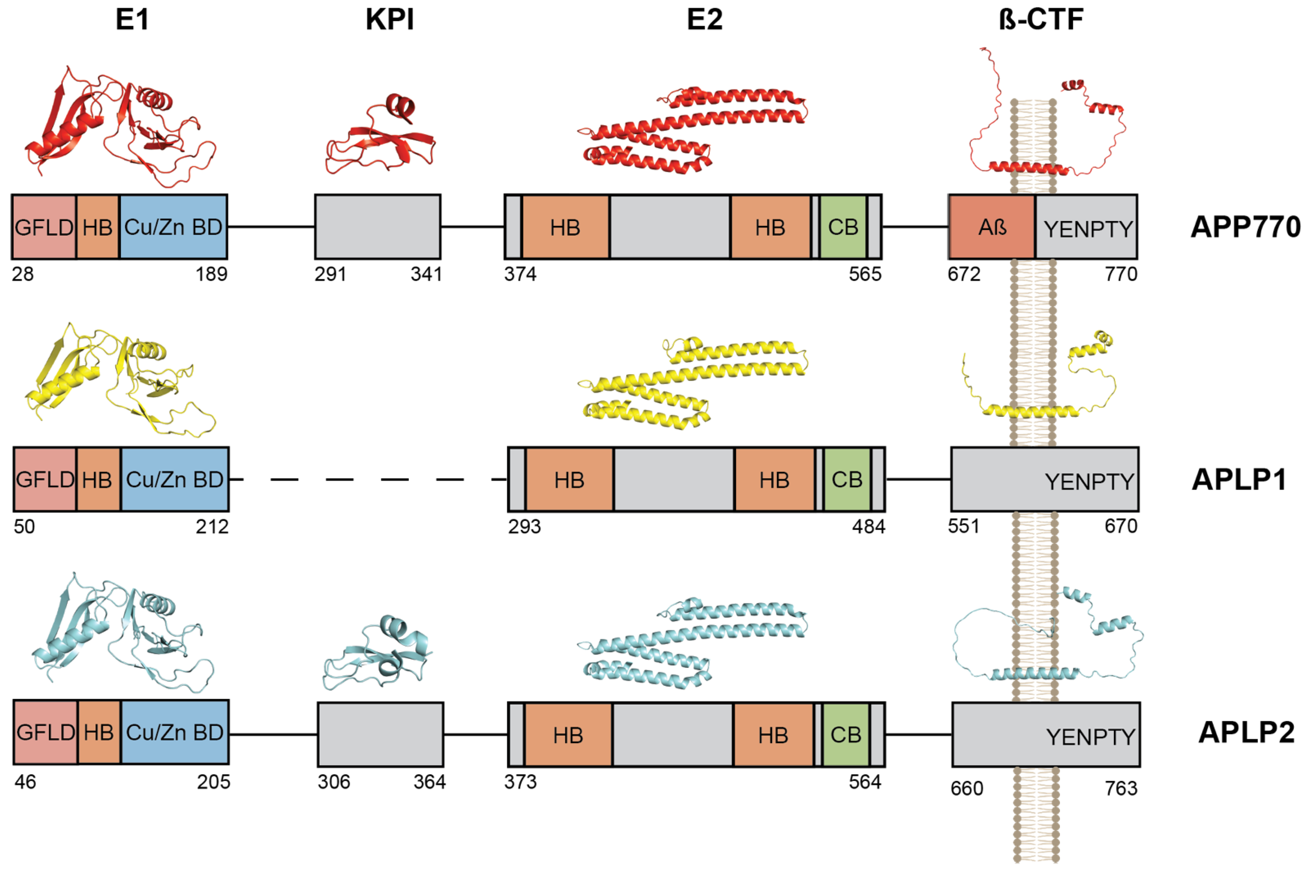
2. The Amyloid Precursor Protein Family and Aβ Production

The Amyloid Precursor Protein (APP) belongs to the APP protein family which includes APP, APLP1, and APLP2 in mammals [19,20,21,22], APPa and APPb in zebrafish [23], and APPL in drosophila [24]. The evolution of the APP gene family (for a review see [25]) indicates that the functions of APP, APLP1, and APLP2 have diverged to contribute distinctly to neuronal processes. After the discovery that Aβ was the main component of AD amyloid fibrils [26] and subsequent identification of APP [19,27] and APLPs [22,28,29] genes, extensive studies in knock-out models brought the assumption that APP, APLP1, and APLP2 are partially functionally redundant (for a review see [30]). The assumption that APP and APLPs have overlapping functions is also supported by the similarities in their expression pattern, biochemical processing, and structure properties. All members of the APP family possess a large ectodomain composed of the E1 and E2 domains. Besides the E2 domain [31,32,33], the structures of the APP protein family have only partially been solved by conventional techniques. Yet, the recent advances in structure prediction algorithm led by AlphaFold [34] allow a reasonable approximation of the respective structures of APP, APLP1, and APLP2. Figure 1 depicts the predicted structures of each segment of the APP and APLP proteins. Close analysis reveals that isolated fragments of the extracellular domains are almost identical in structure, with the inter-fragments predicted to be unstructured. The E1 domain is composed of two separate segments, corresponding to the Growth Factor-Like Domain (GFLD) and copper-binding domain (CuBD). GFLD is made of two β-sheets separated by an α-helix and the CuBD consists of a three-stranded antiparallel β-sheet [35]. The E2 domain is formed by two coiled-coil α-helices which are conserved in all three members of the APP family [32,33] (Figure 1). The Kunitz-type protease inhibitor (KPI) domain is specific to APLP2 and long isoforms of APP (APP770 and APP751) and not present in the neuronal isoform (APP695). It is composed of double-stranded β-sheets with a small α-helix [36]. Functionally, the extracellular domain of APP/APLPs binds components of the extracellular matrix such as heparin and collagen via the E1 and E2 domain [37,38]. Interaction with heparin plays a role in neurite outgrowth [39], APP/APLPs dimerization [32,38,40], and cell-cell adhesion [41]. The E1 and E2 domains are also reported to bind zinc and copper [42,43] via the E1 copper/zinc binding domain (Cu/Zn BD) (Figure 1) and less well-defined sites of the E2 domain. Binding of zinc and copper regulates APP and APLPs cis and trans-dimerization, possibly by regulating their interaction with heparin [44,45,46,47]. Interestingly, the major neuronal isoform of APP (APP695) [48] does not contain the KPI domain, whereas longer isoforms that contain the KPI are expressed in the periphery and, to a significant level, in platelets, in agreement with the reported role of APP KPI as an inhibitor of multiple coagulation factors [49].
The transmembrane (TM) domains of APP and APLPs are formed of a single α-helix and the intracellular domain is mostly unfolded with the exception of the C-terminus which is composed of two small α-helices (Figure 1). The similarity between APP/APLPs and Notch processing (see below) led some authors to hypothesize that the APP and APLPs [50] Intracellular C-terminal Domain (AICD) could possess signaling functions (see [51,52] for a review). Reporter gene assays based on Gal4-fusion constructs suggested that AICD could act as transcriptional regulator [53,54], but the controversy about the transcriptional activity of AICD and its target genes has only grown since then [55,56].

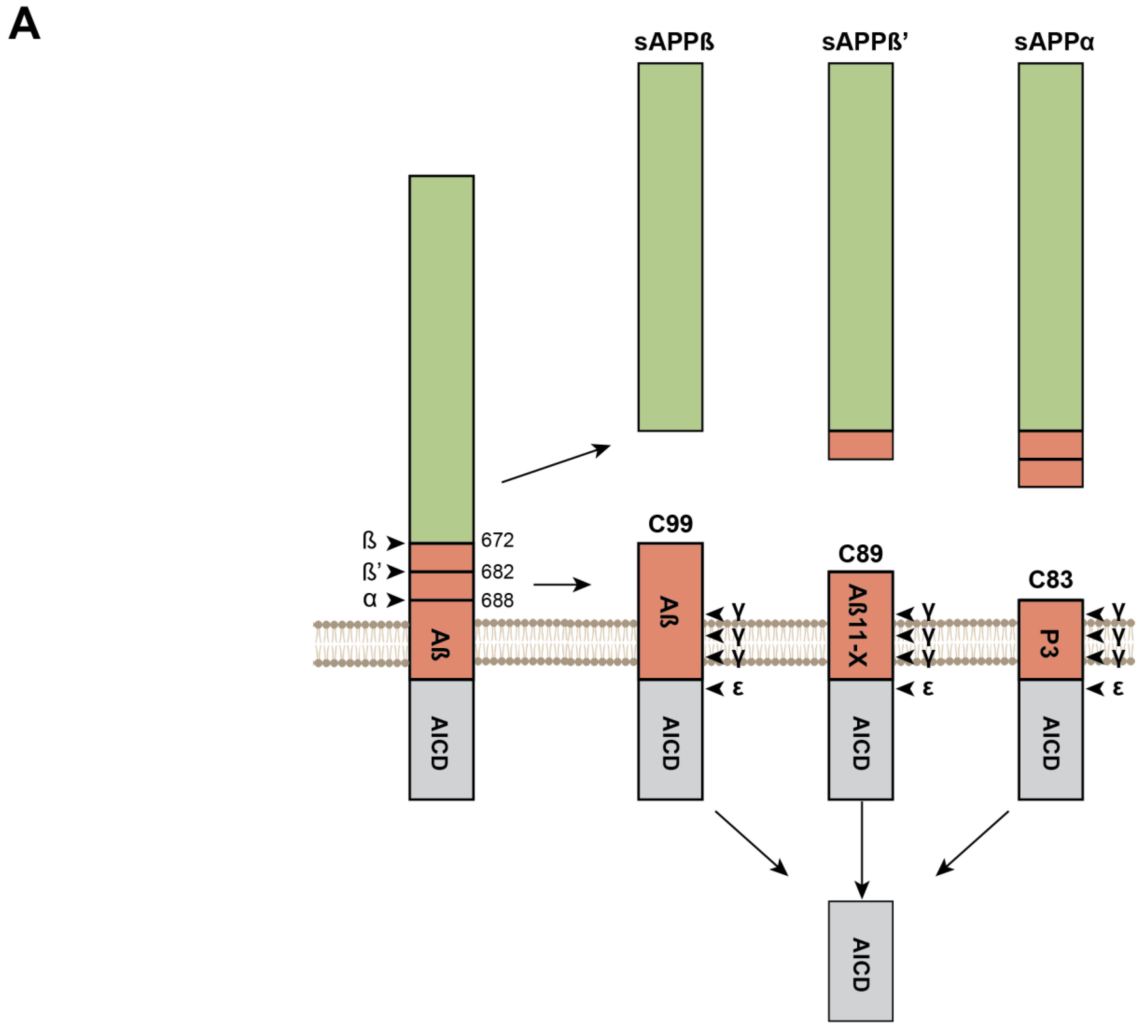
Sequential Processing of the APP and APLPs and Production of β-Amyloid Peptides

The structural similarities between APP family members goes hand in hand with the overlapping functions of APP, APLP1, and APLP2. Yet, APP is the only member of the family giving rise to the formation of pathogenic Aβ aggregates. Members of the APP family are processed via two different pathways (Figure 2A). The most common pathway was coined non-amyloidogenic or anti-amyloidogenic due to its inability to produce amyloidogenic Aβ fragments [57,58]. In the non-amyloidogenic pathway, APP processing begins with the shedding of the ectodomain by a membrane-bound α-secretase of the ADAM family [59]. The main α-secretase for APP is ADAM10 [60], while studies suggest that TACE is the preferred α-secretase of APLP2 [61]. The cleavage by the α-secretase is sequence-independent and occurs at a distance of 12-13 residues from the TM domain [62] to release the ectodomain (sAPPα in the case of APP) and the membrane-anchored C83 fragment. In the amyloidogenic pathway, a β-secretase (BACE1) always starts the cleavage a few amino acids upstream of the α-cleavage site to release sAPPβ. BACE1 is an aspartyl protease that possesses some level of sequence specificity, although large discrepancies do exist between cleavages sites (for a review see [63]). This low level of sequence specificity results in two possible β-cleavage sites of APP. The one after Glu682 produces C89 but only the cleavage after Asp672 can generate the C99 that enables production of amyloidogenic Aβ peptides [64]. Mutations responsible for inherited AD are reported to promote the cleavage at Asp672 (i.e., the APP Swedish or APP KM670/671NL mutation) by providing a more favorable environment for β-cleavage at Asp672 [65,66]. The two other members of the APP family, APLP1 and APLP2, are also processed by the β-secretase to produce C-terminal fragment (CTF) of 100 and 104 residues, respectively (Figure 2B). In both the amyloidogenic and non-amyloidogenic pathways, CTFs resulting from α- or β-cleavage are further processed by a γ-secretase complex. After initial ε-cleavage at position 48-49 (Aβ numbering in APP), which releases AICD and Aβ48-49 [67], the γ-secretase performs intramembrane successive cleavage of three (or four) residues at a time to produce C-terminal Aβ fragments ranging from 36 to 43 amino acids [68,69], of which Aβ42 and Aβ43 have the strongest oligomerization properties. This highlights the observation that the C-terminus of Aβ peptides is key for aggregation. Similarly, APLP1 and APLP2 are processed by γ-secretase complex but do not lead to the production of peptides with amyloidogenic properties [59,70], illustrating that Aβ-like oligomerization is a property specific to the APP sequence (i.e., transmembrane (TM) and juxtamembrane (JM) sequences) present in Aβ peptides. These sequences or motifs provide Aβ with fibrillogenic properties once it has been released upon γ-cleavage of C99. In addition to a specific C-terminal sequence, the pathologic properties of Aβ are also associated with the LVFF motif [71] (see below) present in the Aβ central region, which forms a β-hairpin and also plays a regulatory role in γ-secretase processing [72]. This motif is present only in APP and is not restricted to the C99 produced by β-secretase cleavage (Figure 2B), but is also present in C83. To date, the role of the N-terminus of Aβ sequence has been less investigated but it may impact the stabilization of Aβ aggregates [73]. In this respect, it is important to note that human and murine Aβ only differ by three amino acid residues in the N-terminal regions, and that murine Aβ is not amyloidogenic. Overall, growing evidence indicates that some peculiar motifs present in the human Aβ sequence (and not in related peptides generated by APLP1/APLP2 processing) may play a role in amyloid pathology both (i) by regulating Aβ production and (ii) by promoting or preventing the assembly of Aβ building blocks into amyloid fibrils.

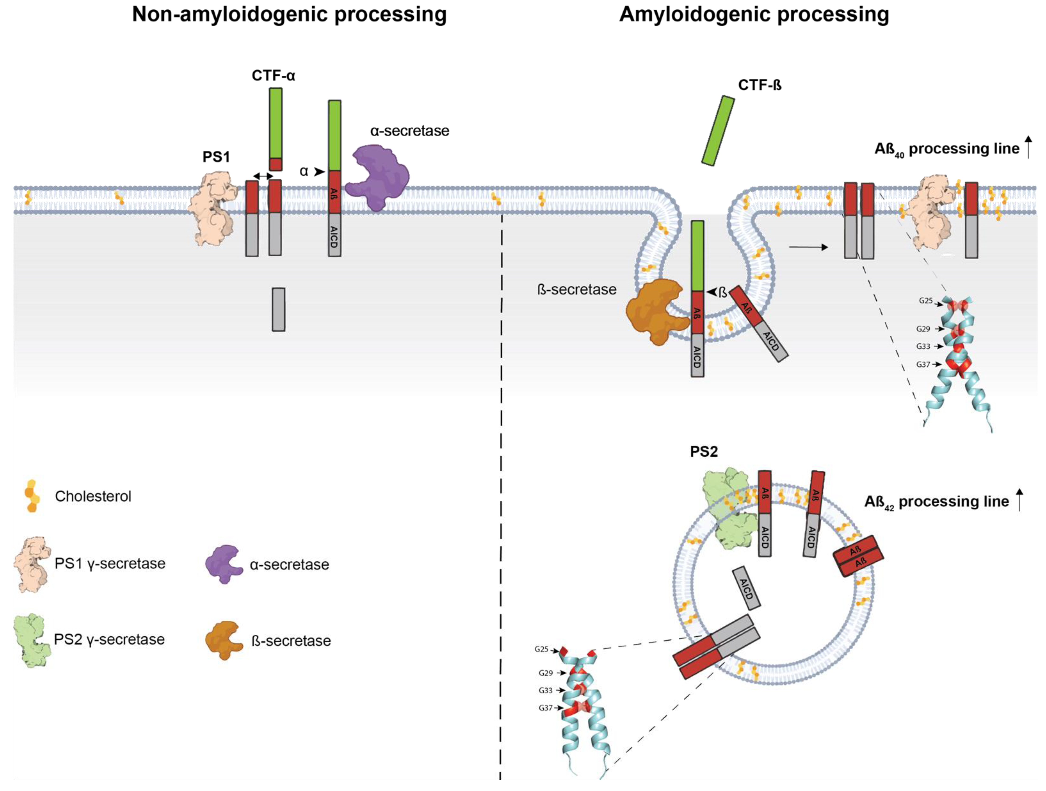
3. Transmembrane Interactions and Amyloidogenic Processing

The finding that APP forms dimers by homo- and hetero-interactions with other APP family members [41,74] was instrumental to the hypothesis that APP dimerization was a regulatory mechanism for its processing and function. Dimerization of APP was described early in the field and was first associated with its binding to heparin and collagen via the ectodomain [37]. Later, it was discovered that APP dimerization promoted the production of Aβ [74]. Homo-dimerization of APP was initially described between different segments of the ectodomain and for its role in cell-cell adhesion [41,75].
Transmembrane (TM) and juxtamembrane (JM) motifs involved in protein-protein interactions have attracted particular interest. Regarding the remarkable conservation of APP and APLPs TM sequences, these motifs may be involved in dimerization between all the members of the family. In addition to their involvement in dimerization, these motifs present in APP are found precisely in the Aβ sequence and in the vicinity of the APP cleavage sites releasing Aβ. Such motifs are GxxxG and GxxxG-like motifs, also known as Glycine zippers, and were described as key regulators of APP amyloidogenic processing [76,77]. GxxxG motifs were initially identified in the glycophorin A TM region [78,79,80,81]. The groove formed by an amino acid without a lateral chain (Gly) in an α-helix context allows close apposition of TM helices and packing into dimers by non-covalent interactions formed by amino acids surrounding the GxxxG sequence. Mutagenesis and structural analyses have revealed numerous examples in which the interaction between TM helices of single-pass membrane proteins is dependent on a GxxxG or (small)xxx(small) motif, where (small) designates amino acid residues with short side chains such as alanine. The interaction strength of motif-containing helices depends strongly on the sequence context and membrane properties. Several GxxxG-containing TM domains can interact via interfaces involving residues (hydrophobic, polar, aromatic) that are not organized in recognizable motifs. Importantly, in multipass membrane proteins, GxxxG motifs can be involved in protein folding, and not just oligomerization [82,83]. GxxxG and GxxxG-like motifs are enriched in protein TM regions, and strikingly, APP contains three in-register GxxxG motifs in its TM/JM region (Figure 3A), with an ADAD mutation (Flemish or A692G) adding a fourth motif in the JM sequence [84]. Such an occurrence of three GxxxG motifs is very rare (Figure 3B) and observed only in 17 human type I TM proteins (22 in mus musculus), and only one protein contains four GxxxG motifs in the region comprising the TM and six surrounding residues on each side. Intriguingly, the prion protein involved in prion disease by the misfolding of the endogenously expressed prion protein (PrPC) into an abnormal isoform (PrPSc) that has infectious properties, also contains a string of three in-register GxxxG motifs in its hydrophobic region [85]. However, the main differences are that PrPC is a membrane-anchored and not a TM protein, and that the hydrophobic regions—known as the hydrophobic tract—lie outside the α-helical C-terminal domain, and are therefore possibly in another conformation than the APP TM region. In the case of APP, GxxxG motifs are indeed involved in TM interactions once the bulky ectodomain has been removed by α- or β-secretase shedding (Figure 2A), and play key regulatory functions in the dimerization and processing of APP CTFs [75,76].
An important advance came from solid state Nuclear Magnetic Resonance (NMR) studies and other biophysical approaches applied to the JM-TM domains of APP using peptides resuspended in membrane-like bilayers, such as dimyristoyl-phosphocholine (DMPC): dimyristoylphosphoglycerol (DMPG) bilayers. Structural data indicated that the LVFF motif preceding the first GxxxG motif forms a β-sheet structure, that the GxxxG are likely helical, and that the helicity of the first GxxxG motif is enhanced by the A21G motif and by cholesterol [72,86]. Earlier NMR studies in detergent or detergent-lipid mixtures [87,88] showed a different picture for LVFF and GxxxG motifs that did not fit with processing. Careful titration of detergent by FTIR spectroscopy showed that detergent induced the helical structure of LVFF rather than the natural β-sheet. Interestingly, cholesterol appears to bind to the aromatic residues of the LVFF motif and influence the downstream structure [86]. Finally, introducing the Flemish mutation (A692G or A21G in Aβ numbering, see above) and adding cholesterol to the experimental context exerted additive effects on the helicity of the first GxxxG motif and on amyloidogenic processing [86]. Thus, APP processing at γ-sites might be controlled by specific orientations and interactions of its TM domain, with these processes (orientation and interaction) being controlled by the cellular context and, in particular, by the lipid composition of the membrane leaflets.
Figure 3. The role of C99 dimerization in Amyloid β generation. (A). Illustration of the role of GxxxG motifs in APP C99 dimerization. The figure illustrates the two dimerization interfaces described in [89]. The conformation shown on the left contains the 25GXXXG29 motif in the interface and was associated with production of SDS-resistant Aβ hexameric species. The conformation shown on the right interface contains the 33GXXXG37 motif in the interface and was linked to generation of AICD signaling properties and promotion of the Aβ42 processing line [89]. Glycines are shown in red, with the one in the interface shown as bubbles. (B). Occurrence of GxxxG motif in the TM-JM segments of type I TM proteins in the homo sapiens (black) and mus musculus (blue) proteome. Only one human protein contains 4 GXXXG in the TM-JM segments (not visible in the graph).
Biomedicines 10 02753 g003

3.1. Dimeric Conformation of C99 Regulates γ-Secretase Processing

Interest for the dimerization of APP C99 occurred later with the observation that a large proportion of familial AD (FAD) mutations occurred in the TM-JM domain where TM interaction motifs are located. The observation that GxxxG motifs and dimerization of APP regulated Aβ production provided strong arguments for an influence of TM dimerization in its processing by γ-secretase. The role of APP/C99 dimerization in the regulation of γ-secretase processing was extensively investigated [77,90,91,92,93,94,95,96] but led to two contradicting observations. On one hand, dimerization was found to increase generation of pathogenic Aβ. For example, Scheuermann and colleagues designed stable APP homodimers by introducing the K699C mutation (K624C in APP695 numbering) at the extracellular JM-TM section ahead of γ-secretase cleavage sites and showed that dimerization dramatically increased Aβ production [74]. In line with these findings, compounds that reduced Aβ42 generation were shown to act by interfering with APP TM dimerization [97]. On the other hand, different lines of evidence suggested that forced APP TM dimerization was not compatible with γ-secretase processing. Using a system in which dimerization is imposed from the intracellular domain, Eggert and colleagues observed that controlled dimerization resulted in decreased Aβ generation [98]. More recently, however, the acquisition of the cryo-EM structure of γ-secretase in complex with C83 hinted that processing of C-terminal fragments (CTF) could indeed not occur in their dimeric form [99], in line with our own observations using a cell-free γ-secretase processing assay [89]. A first answer to these apparently contradicting observations was provided by our finding that GxxxG motifs were critical in regulating the orientations of TM dimerization and that a specific interface was required for amyloidogenic processing [76]. Using a system of fusion proteins that force dimerization of TM helices in all possible orientations, we demonstrated that precise dimeric orientations of C99 controlled γ-secretase processing by regulating the initial ε-cleavage and therefore influencing AICD-dependent signaling and generation of Aβ42 [89]. Notably, the dimeric orientation resulting in enhanced Aβ42 production and AICD-dependent signaling contained GxxxG motifs in the interface, but unfolding is mandatory for γ-processing since covalently bound APP CTFs are not further processed [89].

3.2. APP Dimeric Conformation Controls Its Intracellular Localization and Aβ Generation

In addition to its role in the regulation of processing, multiple lines of evidence indicate that dimerization of C99 is both a regulator and a consequence of its subcellular localization. The impact of APP dimerization on its subcellular localization was only recently investigated. A link between dimerization and subcellular localization was first suggested by Ben Khalifa and colleagues who observed that APP dimers were mostly present in the secretory pathway [75]. Using a model of forced dimerization of full-length APP, Eggert and collaborators analyzed in more detail the role of APP dimerization on subcellular localization and observed that APP dimerization resulted in increased localization in the endoplasmic reticulum (ER) and in endosomes, where the generation of pathogenic Aβ is believed to occur, by modulating its interaction with LRP1 and SorLA [100] (reviewed in [101]). The role of dimerization in intracellular trafficking was extended by Perrin and colleagues who observed that different dimeric orientations of C99 resulted in distinct subcellular localization in primary neurons and that the Aβ42 processing line was favored by a dimeric conformation that resulted in decreased cell surface and increased intracellular localization [89].
To better understand the impact of subcellular localization on amyloidogenic processing (see Figure 4), one must keep in mind that γ-secretase is composed of four subunits, namely, Nicastrin, Aph1, Pen-2, and Presenilin 1/2 (PS1/2, encoded by the PSEN1 and PSEN2 genes, respectively) [102]. The catalytic core of γ-secretase is composed of either PS1 or PS2, which harbor two aspartate catalytic residues (e.g., Asp287 and Asp387 for PS1) [103,104]. The fact that mutations in PSs are the most common cause of FAD highlights their critical role in the pathology (for a review see [105]). The link between subcellular localization of APP and PSs was recently investigated by Sannerud and colleagues [106]. Using a wide variety of approaches, they elegantly demonstrated that PS1 and PS2 have distinct subcellular localizations that impact C99 processing. More specifically, they observed that PS2 was addressed to late endosomes/lysosomes by its N-terminal dileucine sorting motif E16RTSLM21 via interaction with AP-1 while PS1 was more ubiquitously expressed, notably at the plasma membrane. As suggested previously [107], secreted Aβ peptides were increased in the absence of PS2 but the intracellular Aβ and Aβ42/Aβ40 ratio was higher in the absence of PS1. They further observed that cleavage of C99 in late endosomes was associated with the formation of aggregation-prone Aβ peptides that were less efficiency secreted. Remarkably, these results correlated with those obtained in two of our recent studies. In Perrin et al., we found that the dimeric orientation of C99 that favored the Aβ42 processing line and localized in intracellular compartments was more efficiently processed by PS2 [89]. In Vrancx et al., we reported that specific hexameric-like Aβ42 assemblies were mainly produced by PS2 in vesicular compartments [108]. The fact that PS2 favors the Aβ42 processing line in late endosomes must also be put in perspective with the observation that BACE1, the limiting enzyme of APP amyloidogenic processing, is predominantly found in endosomal compartments [109,110,111,112]. Together, these observations provide strong arguments for a crosstalk between C99 dimerization, subcellular localization, and generation of pathogenic amyloid β. It is also interesting to note that TM interaction motifs discussed above are not restricted to APP, but are also found in PS1 and PS2 [113], and in the Aph-1 subunit of the γ-secretase [114], and that the hydrophilic loop of PS1 together with the APP GxxxG TM motif regulates γ-secretase function in generating pathogenic Aβ peptides [115]. These observations indicate that the role of TM interaction in amyloid pathology may not be restricted to the substrate (APP), but may act all along the processing line controlling the orientation of APP dimers, the docking and fitting in the γ-secretase complex, and, eventually, the formation of pathogenic Aβ species.

3.3. The Role of Lipids in C99 Dimerization and Amyloidogenic Processing

Cell membranes are a buoyant environment and are largely heterogeneous in their composition. The link between AD and the lipid composition of membranes was previously suggested by Alois Alzheimer who observed “lipoid granules” and “adipose inclusions” as a third hallmark of Alzheimer’s disease [116,117]. A large body of evidence has since confirmed that lipid composition is indeed a critical factor influencing AD pathogenesis (reviewed in [118,119]). Cholesterol was early found to be a major risk factor of Late Onset Alzheimer’s Disease (LOAD) [120,121,122]. This argument is reinforced by the identification of the allele ε4 of ApoE, a major lipoprotein involved in cholesterol transport, as the most important risk factor of non-familial AD [123,124,125]. Early studies also indicated that low cholesterol promotes α-cleavage and non-amyloidogenic processing [126,127] whereas β-cleavage and amyloidogenic processing is favored by high cholesterol content [128] found in lipid rafts [129]. The role of cholesterol in C99 dimerization remains largely controversial. Different lines of evidence support the observation that C99 directly interacts with cholesterol [87,130,131]. Nuclear Magnetic Resonance (NMR) analyses revealed that interaction between cholesterol and C99 involved essentially the N-terminal loop of C99 and residues Glu693 and Asn698. Interestingly, glycines of the first GxxxG motif also seemed to directly bind cholesterol [131]. In line with the observation that GxxxG motifs interact with cholesterol, Song and colleagues suggested that cholesterol binding to C99 competed with homodimerization [132], and others showed that lowering cholesterol inhibited Aβ production by promoting APP dimerization [133]. These results are intriguing since a large body of evidence linked C99 dimerization to amyloid β production. Yet, other investigations tend to show that rather than preventing dimerization, cholesterol modulates the conformation of C99 dimers. Studies of Tang and colleagues indicated that the 25GxxxG29 motif was in fact stabilized by cholesterol [86], and others suggested that the membrane micro-environment and cholesterol, rather than competing with homodimerization of C99, had a role in the regulation of C99 dimeric conformation [134,135,136]. These studies differ notably by the composition of the membrane environment used, not only in the percentage of cholesterol, but also in other lipids that may impact C99 dimerization. One plausible scenario is that a moderately increased level of cholesterol promotes C99 dimerization in a particular conformation related to increased amyloidogenic processing. This conformation may target C99 dimers to lipid rafts where the very high cholesterol content induces destabilization of C99 dimers, thereby allowing processing by γ-secretase and generation of Aβ peptides (Figure 4).
An important question, directly related to lipid composition, concerns the thickness of the membrane. A thicker membrane will embed more residues in the juxtamembrane region and change tilt and ionization of the residues bordering the TM domain. The same sequence can also fold as an α-helix, random coil, or β-strands in different environments. The use of lipid formulations close to that of the cell membrane allows the conclusion that the GxxxG motifs are helical [86], yet in less physiological environments they form other structures [88], which is relevant for post-cleavage events and formation of oligomers. Moreover, prediction by replica-exchange molecular dynamics simulations of wild-type (WT) and mutant APP dimer conformations in which Gly residues from GxxxG motifs were changed to Leu illustrated large conformational differences in a membrane context. Dimerization of the WT (GxxxG) is due to two hydrogen bonds between two APP fragments, whereas dimerization of the mutant (LxxxL) is due to hydrophobic interactions. In the mutant, each APP fragment is more tilted, and the γ-cleavage site is shifted toward the center of the membrane. This position produces a mismatch between the active site of γ-secretase and the γ-cleavage site of APP that might impair Aβ production [137].

3.4. Impact of Familial AD Mutations on APP TM Dimerization: A Link with Aβ Production?

The fact that over 20 FAD mutations are located in APP TM domain illustrates the importance of this region for pathogenic Aβ generation. Yet, their influence on Aβ production through C99 dimerization remains quite elusive. Using synthetic peptides, Gorman and colleagues found that the V717G, V717F (Indiana), and T714I (Austrian) mutations all decreased TM dimerization and concluded that TM dimerization was negatively associated with pathogenic Aβ generation [138]. Similarly, others concluded that FAD mutations in the 714TVIV717 TM segments induced lower dimerization [139]. Conversely, So and collaborators analyzed dimerization of full-length APP FAD mutants with techniques including biomolecular fluorescence complementation (BiFC) and live cell crosslinking, and found that FAD mutations did not significantly affect the overall level of dimerization [96]. These observations may suggest that APP FAD mutations do not act by increasing C99 dimerization levels but may rather induce modulation of the C99 dimeric interface. Consistently, we observed that the APP Flemish mutation (A692G) promotes dimerization in a conformation that puts the GxxxG motif in the interface and increases Aβ42 production [86]. Strikingly, however, results obtained with split protein assays indicated that mutations of Gly residues present in APP GxxxG motifs did not affect the number of dimers formed, but rather generated specific Aβ assemblies [91]. This led to a new hypothesis: the TM interaction motifs (i.e., GxxxG or GxxxG-like) may indeed not only control dimerization and processing, but also the processes involved in Aβ assembly that lead to toxic aggregates. This hypothesis was reinforced by our most recent findings showing that forcing the 25GxxxG29 interface in dimeric Aβ42 and Aβ43 assemblies resulted in the generation of SDS-resistant hexameric Aβ species, while forcing dimerization in the 33GxxxG37 interface did not allow generation of such oligomers, even in the context of highly amyloidogenic Aβ42 and Aβ43 [89].

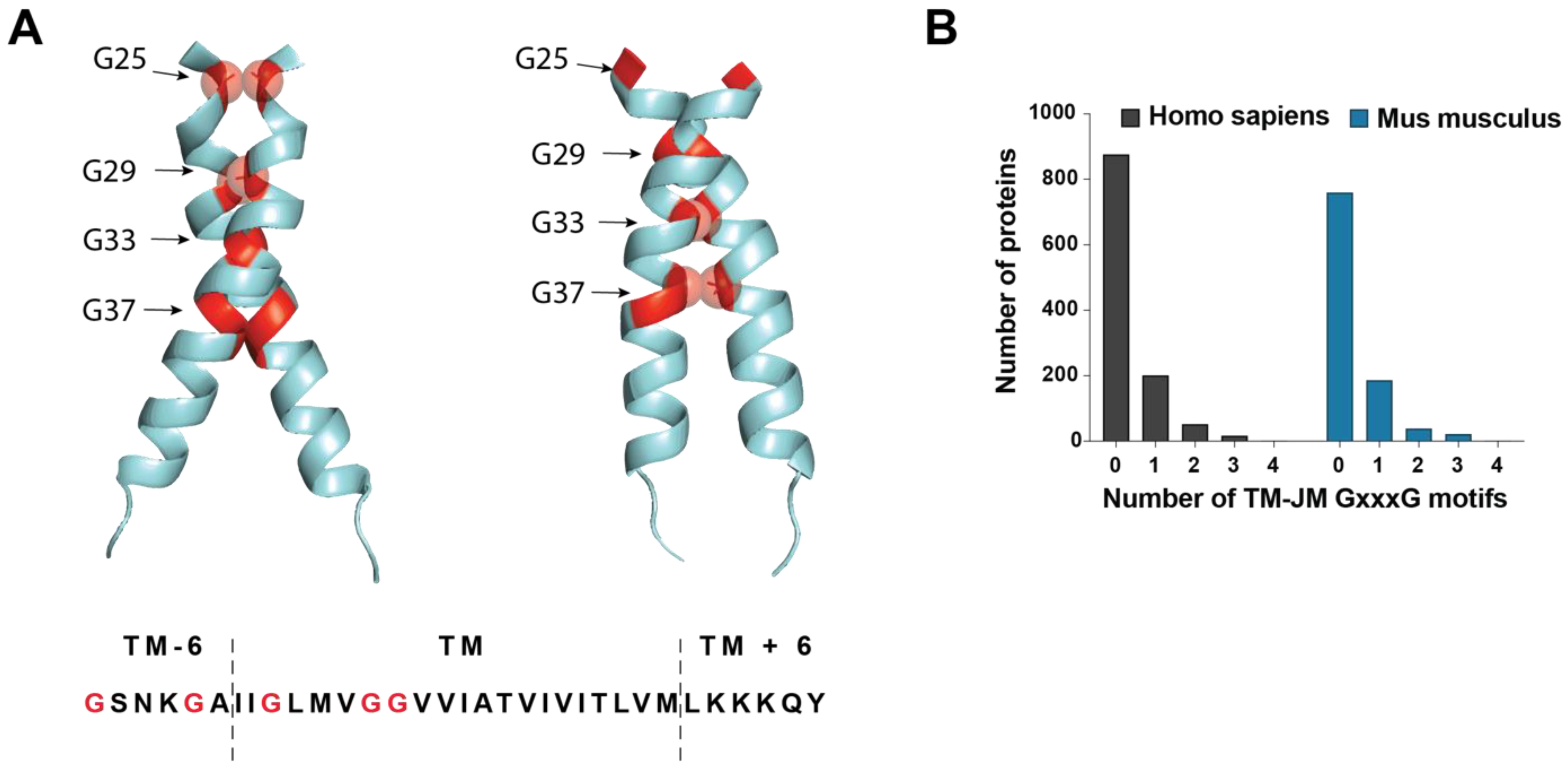
3.5. JM/TM Determinants Drive Aggregation Steps (Nucleation) Leading to Pathological Aβ Seeds

When the soluble Aβ (sAβ) peptides accumulate, they tend to spontaneously self-aggregate, eventually leading to the formation of fibrillar amyloid lesions. Over the past decades, a growing body of evidence indicated that Aβ intermediates, in particular soluble oligomers, are the primary cause of synaptic dysfunction and Aβ toxicity observed in AD. Indeed, while Aβ fibrils are large, insoluble materials aggregating into plaques, Aβ oligomers are soluble and may easily spread throughout the brain. Many oligomeric structures seem to play an important role in Aβ assembly and to have deleterious effects that may explain its related toxicity. For instance, dimers and trimers of Aβ have been consistently linked to long-term potentiation impairments [140,141,142]. Understanding the process of Aβ aggregation is crucial for identifying assembly steps that may be targeted by disease-modifying drugs.
Unlike the Tau protein, for instance, in which post-translational modifications (and especially phosphorylation) are closely associated with abnormal folding and aggregation, few post-translational modifications (PTMs) are described on Aβ and their possible role in Aβ aggregation remains uncertain. The Aβ peptide has been demonstrated to undergo several types of posttranslational modification, such as pyroglutamylation, N-terminal truncation, oxidation, glycosylation, nitration, isomerization, racemization, and phosphorylation [143,144]. However, whilst modified forms of Aβ are further investigated as potential markers of AD progression [145], such modifications are known to not be necessary for the induction of Aβ aggregation and subsequent deleterious effects.
One exception might be the phosphorylation of serine residue at position 8 and 26 (Aβ numbering), which has been reported to promote the formation of oligomeric Aβ assemblies or to stabilize them, respectively [146,147]. Thus, intrinsic determinants much more than PTMs are likely to be necessary for aggregation. This may be highly regulated by both (i) the nature of the C-terminal end and (ii) the presence of peculiar motifs that promote the folding and packaging of Aβ into oligomers and fibrils. The length of the Aβ peptide is correlated to its aggregation properties, with the longest forms (Aβ42 and Aβ43) being more hydrophobic and prone to assembling into oligomers and further elongating into fibrils. A faster rate of aggregation of Aβ42 in comparison to Aβ40 and a stable set of oligomers with a diameter distribution of ~7 to 9 nm was prevalently observed uniquely for Aβ42 even after fibril appearance [148]. Aβ assembly relies on a process called “nucleated polymerization”, involving three distinct phases: (i) a nucleation phase—or lag phase—during which several unfolded or partially folded monomers of Aβ come together to form an oligomeric nucleus; (ii) an elongation phase—or growth phase—in which the nuclei rapidly grow by further addition of monomers, giving rise to prefibrillar structures then ordered protofibrils; and (iii) a stationary phase—or equilibrium—where the concentration of monomers is low and constant, the assembly process reaches saturation, and protofibrils assemble into mature fibrils (Figure 5). The nucleation phase relies on Aβ monomers undergoing conformational changes and misfolding, which render it thermodynamically unfavorable, whereas the elongation phase is much more favorable and occurs rapidly. In primary nucleation, the initial formation of amyloidogenic nuclei occurs without contribution of pre-formed oligomers and constitutes the rate-limiting step. Amyloid fibril formation may also be seeded by the presence of preformed aggregates—oligomers, protofibrils, or fibrils [149]. As a consequence, a whole set of Aβ intermediate assemblies in Aβ fibril formation are present in the brain parenchyma. Some of them were shown to be readily formed in cells and having seeding properties when monomeric Aβ42 is available [108,150].
Peculiar motifs present in the Aβ sequence largely contribute to its complex aggregation properties. Strikingly, TM interaction motifs appear to play a key role not only in Aβ dimerization, but also in aggregation; in particular, the GxxxG motifs described above, which acquire aggregation properties after the transition from the α-helical structure found in the TM context to the β-sheet structure present in Aβ peptides. Glycines from GxxxG allow the turn in the β-helix and form a rigid and grooved structure in the β-sheet that allows stacking of amyloid fibrils [151]. It is tempting here to draw a parallel between GxxxG motifs involved in TM dimerization and their role in Aβ assembly by raising some key questions. Could Aβ stubs initially folded in an α-helical structure (corresponding to the APP TM structure) assemble into dimers that would initiate the nucleation process and promote the conversion to larger Aβ nuclei (Figure 5)? Some recent work supports this exciting hypothesis. First, specific APP TM orientations engaging a GxxxG interface lead to the production of specific (hexameric-like) Aβ assemblies. Remarkably, forcing a specific α-helical orientation of Aβ42 and Aβ43-like peptide dictates their ability to form oligomeric species [89]. Additionally, it has been recently reported that the modular Aβ C-terminal segment mediates rapid, non-nucleated formation of α-helical oligomers; as a result, high local concentration of tethered amyloidogenic segments within these α-oligomers facilitates transition to a β-oligomer population that ultimately generates mature amyloid [152]. Motifs located in the Aβ N-terminal segment are also involved in Aβ aggregation. This is particularly sound, since the N-terminal region of Aβ is present in the JM regions of its precursor (APP), which shows poor conservation across the APP family members. It therefore makes sense that intrinsic aggregation properties of Aβ directly relate to its N-terminal/central region. The LVFF motif (see above) plays a critical role in the Aβ assembly process. This LVFF sequence was initially proposed as an α-helix region that acts as a cholesterol sensor regulating APP processing [131,153]. Incorporation of cholesterol into model membranes enhances the structural changes induced by FAD mutations, suggesting a common link between familial mutations and the cellular environment. However, the LVFF motif was more recently shown to predominantly be a β-sheet in membrane bilayers that, when disrupted by changing lipid composition, increases Aβ production. In this β-sheet conformation, it can match hydrophobic motifs in Tau repeats and accelerate their aggregation via polymorphic states, appearing thus as a major interactor in both types of AD protein folding disorders [154].
Figure 5. Steps of amyloid β aggregation. Illustration of putative nucleation steps of Aβ adapted from [152]. γ-secretase cleaves monomers of C99 (see above) but released Aβ peptides may rapidly reform dimers centered on the GxxxG motifs. In the nucleation phase, dimer formation is the initial step leading to the formation of higher-order species (hexamers are represented) whose nucleation is mediated by the Aβ C-terminus. These oligomers usually form β-sheets but may go through a transient step to form α-oligomers that rapidly switch to more stable β-oligomers initiated by the LVFF motif. Elongation occurs by assembly of β-sheet oligomers into fibrils.
Biomedicines 10 02753 g005

4. Conclusions and Future Directions

Alzheimer’s disease is the first cause of dementia, and global ageing of the population is set to cause a dramatic rise in the number of cases, which is already very high. Despite recent controversy, the amyloid cascade hypothesis remains the most widely accepted in the scientific community given the robustness of its genetic and biochemical arguments. Yet, clinical trials targeting amyloid plaques have all failed to provide the expected benefits. These failures might be rooted in the fact that most trials have focused on targeting amyloid deposits rather than circulating Aβ oligomers, which are more believed to be the toxic species associated with the cognitive decline, although this hypothesis is also fiercely debated. Ultimately, the involvement of amyloid pathology in AD has been the subject of as much debate—if not more—as recent advances in knowledge.
Still, in recent years, important observations have advanced our understanding of the structural determinants that lead to the formation of toxic oligomers. The discovery that C99 dimerization and its dimeric orientations regulate its processing to generate pathogenic Aβ species was instrumental to understanding how amyloidogenic processing occurs. Recent advances highlight how different factors modulate the conformation of C99 dimers that, in turn, regulate amyloidogenic processing. Among them, membrane composition and, in particular, cholesterol content, seem to play a key role in the development of sporadic AD. The allele ε4 of ApoE, the main apolipoprotein involved in cholesterol homeostasis in the brain, is the principal risk factor for sporadic AD [123,155]. In addition, hypercholesterolemia was found to increase the production of Aβ oligomers in the brain of AD mouse models [156] and post-mortem analysis of the brain of AD patients demonstrated increased levels of cholesterol [157]. Despite this observation, the understanding of the role of membrane composition in the modulation of C99 dimeric conformations and generation of toxic amyloid species has been slowed and complicated by the heterogeneity of the methods used to recapitulate biological membranes.
Another critical question is whether the same motifs that regulate C99 processing and localization are also involved in the initial steps of Aβ oligomerization. Recent studies from two independent groups strongly suggest that early nucleation of Aβ could occur in a transient helical conformation, and that modulation of the dimeric orientation of helical Aβ-like peptides dictates the generation of Aβ oligomers [89,152]. This contrasts with the previous idea that Aβ oligomers are essentially formed of closely packed β-sheets. If this scenario holds true in future research, this would give additional weight to the role of GxxxG motifs and of membrane composition in the generation of toxic Aβ oligomers and the onset of Alzheimer’s disease. We propose that future studies should elaborate on these recent findings and assess how changes in membrane compositions of the ageing brain affect not only dimerization of C99, but also of Aβ peptides, and how this correlates with generation of toxic Aβ oligomers. Critically, it would be of major interest to assess how changes in lipid composition associated with ageing [158] affect the structural determinants of C99 processing and amyloid β oligomerization. We believe that understanding how the natural changes in membrane properties of ageing individuals associate with amyloidosis could indeed pave the way for a new array of therapeutic perspectives.

Author Contributions

Conceptualization, N.P. and P.K.-C.; methodology, N.P. and P.K.-C.; validation, N.P., N.S., P.K.-C. and S.N.C.; resources, P.K.-C. and S.N.C.; writing—original draft preparation, N.P. and P.K.-C.; writing—review and editing, N.P., P.K.-C., S.N.C., N.S., F.P., D.M.V. and C.V.; visualization, N.P.; supervision, S.N.C. and P.K.-C.; project administration, P.K.-C. and S.N.C.; funding acquisition, P.K.-C. and S.N.C. All authors have read and agreed to the published version of the manuscript.

Funding

N.P. has received an FSR PhD Fellowship from Université catholique de Louvain and an Aspirant PhD Fellowship from the FRS-FNRS (ASP/A 876), Belgium. N.S. is a postdoctoral fellow (Chargé de Recherches) from the FRS-FNRS and received a pilot grant from the Alzheimer Research Foundation (SAO-FRA/Stop Alzheimer Research Foundation). Funding to S.N.C. is acknowledged from Ludwig Institute for Cancer Research, Fondation contre le cancer, Salus Sanguinis and Fondation “Les avions de Sébastien”, projets Action de recherché concertée (ARC) 16/21-073, research projects from FNRS n°T.0043.21 and WelBio F 44/8/5—MCF/UIG—10955. C.V. is funded by an FWO postdoctoral fellowship 12B7423N and a grant from the consortium Interaction of KU Leuven C14/21/095. Funding to P.K.C. acknowledged from SAO-FRA/Stop Alzheimer Research Foundation, Fondation Louvain and Queen Elisabeth Medical Research Foundation, FNRS grant PDRT.0177.18, and UCLouvain Action de Reechrche Concertée ARC 21/26-114.

Institutional Review Board Statement

Not applicable.

Informed Consent Statement

Not applicable.

Conflicts of Interest

The authors declare no conflict of interest.

References

  1. Carrell, R.W.; Lomas, D.A. Conformational disease. Lancet 1997, 350, 134–138. [Google Scholar] [CrossRef]
  2. Walker, L.C.; LeVine, H. The cerebral proteopathies. Neurobiol. Aging 2000, 21, 559–561. [Google Scholar] [CrossRef]
  3. Ross, C.A.; Poirier, M.A. Protein aggregation and neurodegenerative disease. Nat. Med. 2004, 10, S10–S17. [Google Scholar] [CrossRef]
  4. Virchow, R. Cellular pathology. As based upon physiological and pathological histology. Lecture XVI–Atheromatous affection of arteries. 1858. Nutr. Rev. 1989, 47, 23–25. [Google Scholar] [CrossRef]
  5. Chiti, F.; Dobson, C.M. Protein misfolding, functional amyloid, and human disease. Annu. Rev. Biochem. 2006, 75, 333–366. [Google Scholar] [CrossRef] [Green Version]
  6. Lu, J.X.; Qiang, W.; Yau, W.M.; Schwieters, C.D.; Meredith, S.C.; Tycko, R. Molecular structure of β-amyloid fibrils in Alzheimer’s disease brain tissue. Cell 2013, 154, 1257–1268. [Google Scholar] [CrossRef] [Green Version]
  7. Kollmer, M.; Close, W.; Funk, L.; Rasmussen, J.; Bsoul, A.; Schierhorn, A.; Schmidt, M.; Sigurdson, C.J.; Jucker, M.; Fändrich, M. Cryo-EM structure and polymorphism of Aβ amyloid fibrils purified from Alzheimer’s brain tissue. Nat. Commun. 2019, 10, 4760. [Google Scholar] [CrossRef] [Green Version]
  8. Gremer, L.; Schölzel, D.; Schenk, C.; Reinartz, E.; Labahn, J.; Ravelli, R.B.G.; Tusche, M.; Lopez-Iglesias, C.; Hoyer, W.; Heise, H.; et al. Fibril structure of amyloid-β(1-42) by cryo-electron microscopy. Science 2017, 358, 116–119. [Google Scholar] [CrossRef] [Green Version]
  9. Haass, C.; Selkoe, D.J. Soluble protein oligomers in neurodegeneration: Lessons from the Alzheimer’s amyloid beta-peptide. Nat. Rev. Mol. Cell Biol. 2007, 8, 101–112. [Google Scholar] [CrossRef]
  10. Bertram, L.; Tanzi, R.E. The genetic epidemiology of neurodegenerative disease. J. Clin. Investig. 2005, 115, 1449–1457. [Google Scholar] [CrossRef]
  11. Goto, Y.; Noji, M.; Nakajima, K.; Yamaguchi, K. Supersaturation-Dependent Formation of Amyloid Fibrils. Molecules 2022, 27, 4588. [Google Scholar] [CrossRef]
  12. Morimoto, R.I.; Cuervo, A.M. Proteostasis and the aging proteome in health and disease. J. Gerontol. A Biol. Sci. Med. Sci. 2014, 69 (Suppl. S1), S33–S38. [Google Scholar] [CrossRef] [Green Version]
  13. Braak, H.; Braak, E. Neuropathological stageing of Alzheimer-related changes. Acta Neuropathol. 1991, 82, 239–259. [Google Scholar] [CrossRef]
  14. Thal, D.R.; Rub, U.; Orantes, M.; Braak, H. Phases of A beta-deposition in the human brain and its relevance for the development of AD. Neurology 2002, 58, 1791–1800. [Google Scholar] [CrossRef]
  15. Hardy, J. Alzheimer’s disease: The amyloid cascade hypothesis: An update and reappraisal. J. Alzheimers. Dis 2006, 9, 151–153. [Google Scholar] [CrossRef] [Green Version]
  16. Hardy, J.; Allsop, D. Amyloid deposition as the central event in the aetiology of Alzheimer’s disease. Trends Pharmacol. Sci. 1991, 12, 383–388. [Google Scholar] [CrossRef]
  17. Herrup, K. The case for rejecting the amyloid cascade hypothesis. Nat. Neurosci. 2015, 18, 794–799. [Google Scholar] [CrossRef]
  18. Tanzi, R.E. The genetics of Alzheimer disease. Cold Spring Harb. Perspect. Med. 2012, 2, a006296. [Google Scholar] [CrossRef]
  19. Kang, J.; Lemaire, H.G.; Unterbeck, A.; Salbaum, J.M.; Masters, C.L.; Grzeschik, K.H.; Multhaup, G.; Beyreuther, K.; Muller-Hill, B. The precursor of Alzheimer’s disease amyloid A4 protein resembles a cell-surface receptor. Nature 1987, 325, 733–736. [Google Scholar] [CrossRef]
  20. Slunt, H.H.; Thinakaran, G.; Von Koch, C.; Lo, A.C.; Tanzi, R.E.; Sisodia, S.S. Expression of a ubiquitous, cross-reactive homologue of the mouse beta-amyloid precursor protein (APP). J. Biol. Chem. 1994, 269, 2637–2644. [Google Scholar] [CrossRef]
  21. Tanzi, R.E.; Gusella, J.F.; Watkins, P.C.; Bruns, G.A.; St George-Hyslop, P.; Van Keuren, M.L.; Patterson, D.; Pagan, S.; Kurnit, D.M.; Neve, R.L. Amyloid beta protein gene: cDNA, mRNA distribution, and genetic linkage near the Alzheimer locus. Science 1987, 235, 880–884. [Google Scholar] [CrossRef] [PubMed]
  22. Wasco, W.; Bupp, K.; Magendantz, M.; Gusella, J.F.; Tanzi, R.E.; Solomon, F. Identification of a mouse brain cDNA that encodes a protein related to the Alzheimer disease-associated amyloid beta protein precursor. Proc. Natl. Acad. Sci. USA 1992, 89, 10758–10762. [Google Scholar] [CrossRef] [Green Version]
  23. Musa, A.; Lehrach, H.; Russo, V.A. Distinct expression patterns of two zebrafish homologues of the human APP gene during embryonic development. Dev. Genes Evol. 2001, 211, 563–567. [Google Scholar] [CrossRef]
  24. Rosen, D.R.; Martin-Morris, L.; Luo, L.Q.; White, K. A Drosophila gene encoding a protein resembling the human beta-amyloid protein precursor. Proc. Natl. Acad. Sci. USA 1989, 86, 2478–2482. [Google Scholar] [CrossRef] [Green Version]
  25. Shariati, S.A.; De Strooper, B. Redundancy and divergence in the amyloid precursor protein family. FEBS Lett. 2013, 587, 2036–2045. [Google Scholar] [CrossRef] [Green Version]
  26. Glenner, G.G.; Wong, C.W. Alzheimer’s disease: Initial report of the purification and characterization of a novel cerebrovascular amyloid protein. Biochem. Biophys. Res. Commun. 1984, 120, 885–890. [Google Scholar] [CrossRef]
  27. Tanaka, S.; Nakamura, S.; Ueda, K.; Kameyama, M.; Shiojiri, S.; Takahashi, Y.; Kitaguchi, N.; Ito, H. Three types of amyloid protein precursor mRNA in human brain: Their differential expression in Alzheimer’s disease. Biochem. Biophys. Res. Commun. 1988, 157, 472–479. [Google Scholar] [CrossRef]
  28. Sprecher, C.A.; Grant, F.J.; Grimm, G.; O'Hara, P.J.; Norris, F.; Norris, K.; Foster, D.C. Molecular cloning of the cDNA for a human amyloid precursor protein homolog: Evidence for a multigene family. Biochemistry 1993, 32, 4481–4486. [Google Scholar] [CrossRef]
  29. Wasco, W.; Gurubhagavatula, S.; Paradis, M.D.; Romano, D.M.; Sisodia, S.S.; Hyman, B.T.; Neve, R.L.; Tanzi, R.E. Isolation and characterization of APLP2 encoding a homologue of the Alzheimer’s associated amyloid beta protein precursor. Nat. Genet. 1993, 5, 95–100. [Google Scholar] [CrossRef]
  30. Müller, U.C.; Zheng, H. Physiological functions of APP family proteins. Cold Spring Harb. Perspect. Med. 2012, 2, a006288. [Google Scholar] [CrossRef]
  31. Wang, Y.; Ha, Y. The X-ray structure of an antiparallel dimer of the human amyloid precursor protein E2 domain. Mol. Cell 2004, 15, 343–353. [Google Scholar] [CrossRef] [PubMed]
  32. Lee, S.; Xue, Y.; Hu, J.; Wang, Y.; Liu, X.; Demeler, B.; Ha, Y. The E2 Domains of APP and APLP1 Share a Conserved Mode of Dimerization. Biochemistry 2011, 50, 5453–5464. [Google Scholar] [CrossRef] [Green Version]
  33. Roisman, L.C.; Han, S.; Chuei, M.J.; Connor, A.R.; Cappai, R. The crystal structure of amyloid precursor-like protein 2 E2 domain completes the amyloid precursor protein family. FASEB J. 2019, 33, 5076–5081. [Google Scholar] [CrossRef] [PubMed]
  34. Jumper, J.; Evans, R.; Pritzel, A.; Green, T.; Figurnov, M.; Ronneberger, O.; Tunyasuvunakool, K.; Bates, R.; Žídek, A.; Potapenko, A.; et al. Highly accurate protein structure prediction with AlphaFold. Nature 2021, 596, 583–589. [Google Scholar] [CrossRef] [PubMed]
  35. Dahms, S.O.; Hoefgen, S.; Roeser, D.; Schlott, B.; Gührs, K.H.; Than, M.E. Structure and biochemical analysis of the heparin-induced E1 dimer of the amyloid precursor protein. Proc. Natl. Acad. Sci. USA 2010, 107, 5381–5386. [Google Scholar] [CrossRef] [Green Version]
  36. Hynes, T.R.; Randal, M.; Kennedy, L.A.; Eigenbrot, C.; Kossiakoff, A.A. X-ray crystal structure of the protease inhibitor domain of Alzheimer’s amyloid beta-protein precursor. Biochemistry 1990, 29, 10018–10022. [Google Scholar] [CrossRef]
  37. Beher, D.; Hesse, L.; Masters, C.L.; Multhaup, G. Regulation of Amyloid Protein Precursor (APP) Binding to Collagen and Mapping of the Binding Sites on APP and Collagen Type I (∗). J. Biol. Chem. 1996, 271, 1613–1620. [Google Scholar] [CrossRef] [Green Version]
  38. Schubert, D.; LaCorbiere, M.; Saitoh, T.; Cole, G. Characterization of an amyloid beta precursor protein that binds heparin and contains tyrosine sulfate. Proc. Natl. Acad. Sci. USA 1989, 86, 2066–2069. [Google Scholar] [CrossRef] [Green Version]
  39. Small, D.H.; Nurcombe, V.; Reed, G.; Clarris, H.; Moir, R.; Beyreuther, K.; Masters, C.L. A heparin-binding domain in the amyloid protein precursor of Alzheimer’s disease is involved in the regulation of neurite outgrowth. J. Neurosci. 1994, 14, 2117–2127. [Google Scholar] [CrossRef] [Green Version]
  40. Hoefgen, S.; Coburger, I.; Roeser, D.; Schaub, Y.; Dahms, S.O.; Than, M.E. Heparin induced dimerization of APP is primarily mediated by E1 and regulated by its acidic domain. J. Struct. Biol. 2014, 187, 30–37. [Google Scholar] [CrossRef]
  41. Soba, P.; Eggert, S.; Wagner, K.; Zentgraf, H.; Siehl, K.; Kreger, S.; Löwer, A.; Langer, A.; Merdes, G.; Paro, R.; et al. Homo- and heterodimerization of APP family members promotes intercellular adhesion. EMBO J. 2005, 24, 3624–3634. [Google Scholar] [CrossRef]
  42. Bush, A.I.; Multhaup, G.; Moir, R.D.; Williamson, T.G.; Small, D.H.; Rumble, B.; Pollwein, P.; Beyreuther, K.; Masters, C.L. A novel zinc(II) binding site modulates the function of the beta A4 amyloid protein precursor of Alzheimer’s disease. J. Biol. Chem. 1993, 268, 16109–16112. [Google Scholar] [CrossRef]
  43. Multhaup, G.; Schlicksupp, A.; Hesse, L.; Beher, D.; Ruppert, T.; Masters, C.L.; Beyreuther, K. The amyloid precursor protein of Alzheimer’s disease in the reduction of copper(II) to copper(I). Science 1996, 271, 1406–1409. [Google Scholar] [CrossRef]
  44. Mayer, M.C.; Kaden, D.; Schauenburg, L.; Hancock, M.A.; Voigt, P.; Roeser, D.; Barucker, C.; Than, M.E.; Schaefer, M.; Multhaup, G. Novel zinc-binding site in the E2 domain regulates amyloid precursor-like protein 1 (APLP1) oligomerization. J. Biol. Chem. 2014, 289, 19019–19030. [Google Scholar] [CrossRef] [PubMed] [Green Version]
  45. Dienemann, C.; Coburger, I.; Mehmedbasic, A.; Andersen, O.M.; Than, M.E. Mutants of metal binding site M1 in APP E2 show metal specific differences in binding of heparin but not of sorLA. Biochemistry 2015, 54, 2490–2499. [Google Scholar] [CrossRef] [PubMed]
  46. Dahms, S.O.; Könnig, I.; Roeser, D.; Gührs, K.H.; Mayer, M.C.; Kaden, D.; Multhaup, G.; Than, M.E. Metal binding dictates conformation and function of the amyloid precursor protein (APP) E2 domain. J. Mol. Biol. 2012, 416, 438–452. [Google Scholar] [CrossRef] [Green Version]
  47. Noda, Y.; Asada, M.; Kubota, M.; Maesako, M.; Watanabe, K.; Uemura, M.; Kihara, T.; Shimohama, S.; Takahashi, R.; Kinoshita, A.; et al. Copper enhances APP dimerization and promotes Aβ production. Neurosci. Lett. 2013, 547, 10–15. [Google Scholar] [CrossRef] [PubMed]
  48. Rohan de Silva, H.A.; Jen, A.; Wickenden, C.; Jen, L.S.; Wilkinson, S.L.; Patel, A.J. Cell-specific expression of beta-amyloid precursor protein isoform mRNAs and proteins in neurons and astrocytes. Mol. Brain Res. 1997, 47, 147–156. [Google Scholar] [CrossRef]
  49. Mahdi, F.; Van Nostrand, W.E.; Schmaier, A.H. Protease nexin-2/amyloid beta-protein precursor inhibits factor Xa in the prothrombinase complex. J. Biol. Chem. 1995, 270, 23468–23474. [Google Scholar] [CrossRef] [Green Version]
  50. Scheinfeld, M.H.; Ghersi, E.; Laky, K.; Fowlkes, B.J.; D’Adamio, L. Processing of beta-amyloid precursor-like protein-1 and -2 by gamma-secretase regulates transcription. J. Biol. Chem. 2002, 277, 44195–44201. [Google Scholar] [CrossRef] [PubMed]
  51. Beckett, C.; Nalivaeva, N.N.; Belyaev, N.D.; Turner, A.J. Nuclear signalling by membrane protein intracellular domains: The AICD enigma. Cell Signal. 2012, 24, 402–409. [Google Scholar] [CrossRef] [PubMed]
  52. Pardossi-Piquard, R.; Checler, F. The physiology of the beta-amyloid precursor protein intracellular domain AICD. J. Neurochem. 2012, 120 (Suppl. S1), 109–124. [Google Scholar] [CrossRef] [PubMed]
  53. Cao, X.; Sudhof, T.C. A transcriptively active complex of APP with Fe65 and histone acetyltransferase Tip60. Science 2001, 293, 115–120. [Google Scholar] [CrossRef] [PubMed] [Green Version]
  54. Cao, X.; Sudhof, T.C. Dissection of amyloid-beta precursor protein-dependent transcriptional transactivation. J. Biol. Chem 2004, 279, 24601–24611. [Google Scholar] [CrossRef] [PubMed] [Green Version]
  55. Hebert, S.S.; Serneels, L.; Tolia, A.; Craessaerts, K.; Derks, C.; Filippov, M.A.; Muller, U.; De Strooper, B. Regulated intramembrane proteolysis of amyloid precursor protein and regulation of expression of putative target genes. EMBO Rep. 2006, 7, 739–745. [Google Scholar] [CrossRef] [Green Version]
  56. Opsomer, R.; Contino, S.; Perrin, F.; Gualdani, R.; Tasiaux, B.; Doyen, P.; Vergouts, M.; Vrancx, C.; Doshina, A.; Pierrot, N.; et al. Amyloid Precursor Protein (APP) Controls the Expression of the Transcriptional Activator Neuronal PAS Domain Protein 4 (NPAS4) and Synaptic GABA Release. eNeuro 2020, 7. [Google Scholar] [CrossRef] [Green Version]
  57. Sisodia, S.S.; Koo, E.H.; Beyreuther, K.; Unterbeck, A.; Price, D.L. Evidence that beta-amyloid protein in Alzheimer’s disease is not derived by normal processing. Science 1990, 248, 492–495. [Google Scholar] [CrossRef]
  58. Esch, F.S.; Keim, P.S.; Beattie, E.C.; Blacher, R.W.; Culwell, A.R.; Oltersdorf, T.; McClure, D.; Ward, P.J. Cleavage of amyloid beta peptide during constitutive processing of its precursor. Science 1990, 248, 1122–1124. [Google Scholar] [CrossRef]
  59. Eggert, S.; Paliga, K.; Soba, P.; Evin, G.; Masters, C.L.; Weidemann, A.; Beyreuther, K. The proteolytic processing of the amyloid precursor protein gene family members APLP-1 and APLP-2 involves alpha-, beta-, gamma-, and epsilon-like cleavages: Modulation of APLP-1 processing by n-glycosylation. J. Biol. Chem. 2004, 279, 18146–18156. [Google Scholar] [CrossRef] [Green Version]
  60. Kuhn, P.H.; Wang, H.; Dislich, B.; Colombo, A.; Zeitschel, U.; Ellwart, J.W.; Kremmer, E.; Rossner, S.; Lichtenthaler, S.F. ADAM10 is the physiologically relevant, constitutive alpha-secretase of the amyloid precursor protein in primary neurons. Embo J. 2010, 29, 3020–3032. [Google Scholar] [CrossRef]
  61. Jacobsen, K.T.; Adlerz, L.; Multhaup, G.; Iverfeldt, K. Insulin-like Growth Factor-1 (IGF-1)-induced Processing of Amyloid-β Precursor Protein (APP) and APP-like Protein 2 Is Mediated by Different Metalloproteinases*. J. Biol. Chem. 2010, 285, 10223–10231. [Google Scholar] [CrossRef] [PubMed] [Green Version]
  62. Sisodia, S.S. Beta-amyloid precursor protein cleavage by a membrane-bound protease. Proc. Natl. Acad. Sci. USA 1992, 89, 6075–6079. [Google Scholar] [CrossRef] [Green Version]
  63. Dislich, B.; Lichtenthaler, S.F. The Membrane-Bound Aspartyl Protease BACE1: Molecular and Functional Properties in Alzheimer’s Disease and Beyond. Front Physiol. 2012, 3, 8. [Google Scholar] [CrossRef] [PubMed] [Green Version]
  64. Deng, Y.; Wang, Z.; Wang, R.; Zhang, X.; Zhang, S.; Wu, Y.; Staufenbiel, M.; Cai, F.; Song, W. Amyloid-β protein (Aβ) Glu11 is the major β-secretase site of β-site amyloid-β precursor protein-cleaving enzyme 1(BACE1), and shifting the cleavage site to Aβ Asp1 contributes to Alzheimer pathogenesis. Eur. J. Neurosci. 2013, 37, 1962–1969. [Google Scholar] [CrossRef] [PubMed]
  65. Zhang, S.; Wang, Z.; Cai, F.; Zhang, M.; Wu, Y.; Zhang, J.; Song, W. BACE1 Cleavage Site Selection Critical for Amyloidogenesis and Alzheimer’s Pathogenesis. J. Neurosci. 2017, 37, 6915–6925. [Google Scholar] [CrossRef] [Green Version]
  66. Kimura, A.; Hata, S.; Suzuki, T. Alternative Selection of β-Site APP-Cleaving Enzyme 1 (BACE1) Cleavage Sites in Amyloid β-Protein Precursor (APP) Harboring Protective and Pathogenic Mutations within the Aβ Sequence. J. Biol. Chem. 2016, 291, 24041–24053. [Google Scholar] [CrossRef] [Green Version]
  67. Weidemann, A.; Eggert, S.; Reinhard, F.B.; Vogel, M.; Paliga, K.; Baier, G.; Masters, C.L.; Beyreuther, K.; Evin, G. A novel epsilon-cleavage within the transmembrane domain of the Alzheimer amyloid precursor protein demonstrates homology with Notch processing. Biochemistry 2002, 41, 2825–2835. [Google Scholar] [CrossRef]
  68. Takami, M.; Nagashima, Y.; Sano, Y.; Ishihara, S.; Morishima-Kawashima, M.; Funamoto, S.; Ihara, Y. γ-Secretase: Successive Tripeptide and Tetrapeptide Release from the Transmembrane Domain of β-Carboxyl Terminal Fragment. J. Neurosci. 2009, 29, 13042–13052. [Google Scholar] [CrossRef] [Green Version]
  69. Bolduc, D.M.; Montagna, D.R.; Seghers, M.C.; Wolfe, M.S.; Selkoe, D.J. The amyloid-beta forming tripeptide cleavage mechanism of γ-secretase. Elife 2016, 5, e17578. [Google Scholar] [CrossRef]
  70. Hogl, S.; Kuhn, P.H.; Colombo, A.; Lichtenthaler, S.F. Determination of the proteolytic cleavage sites of the amyloid precursor-like protein 2 by the proteases ADAM10, BACE1 and γ-secretase. PLoS ONE 2011, 6, e21337. [Google Scholar] [CrossRef]
  71. Liu, R.; McAllister, C.; Lyubchenko, Y.; Sierks, M.R. Residues 17-20 and 30-35 of beta-amyloid play critical roles in aggregation. J. Neurosci. Res. 2004, 75, 162–171. [Google Scholar] [CrossRef] [PubMed]
  72. Hu, Y.; Kienlen-Campard, P.; Tang, T.-C.; Perrin, F.; Opsomer, R.; Decock, M.; Pan, X.; Octave, J.-N.; Constantinescu, S.N.; Smith, S.O. β-Sheet Structure within the Extracellular Domain of C99 Regulates Amyloidogenic Processing. Sci. Rep. 2017, 7, 17159. [Google Scholar] [CrossRef] [PubMed] [Green Version]
  73. Söldner, C.A.; Sticht, H.; Horn, A.H.C. Role of the N-terminus for the stability of an amyloid-β fibril with three-fold symmetry. PLoS ONE 2017, 12, e0186347. [Google Scholar] [CrossRef] [Green Version]
  74. Scheuermann, S.; Hambsch, B.; Hesse, L.; Stumm, J.; Schmidt, C.; Beher, D.; Bayer, T.A.; Beyreuther, K.; Multhaup, G. Homodimerization of amyloid precursor protein and its implication in the amyloidogenic pathway of Alzheimer’s disease. J. Biol. Chem. 2001, 276, 33923–33929. [Google Scholar] [CrossRef] [PubMed] [Green Version]
  75. Ben Khalifa, N.; Tyteca, D.; Marinangeli, C.; Depuydt, M.; Collet, J.F.; Courtoy, P.J.; Renauld, J.C.; Constantinescu, S.; Octave, J.N.; Kienlen-Campard, P. Structural features of the KPI domain control APP dimerization, trafficking, and processing. FASEB J. 2012, 26, 855–867. [Google Scholar] [CrossRef] [PubMed]
  76. Kienlen-Campard, P.; Tasiaux, B.; Van Hees, J.; Li, M.; Huysseune, S.; Sato, T.; Fei, J.Z.; Aimoto, S.; Courtoy, P.J.; Smith, S.O.; et al. Amyloidogenic Processing but Not Amyloid Precursor Protein (APP) Intracellular C-terminal Domain Production Requires a Precisely Oriented APP Dimer Assembled by Transmembrane GXXXG Motifs. J. Biol. Chem. 2008, 283, 7733–7744. [Google Scholar] [CrossRef] [PubMed] [Green Version]
  77. Munter, L.M.; Voigt, P.; Harmeier, A.; Kaden, D.; Gottschalk, K.E.; Weise, C.; Pipkorn, R.; Schaefer, M.; Langosch, D.; Multhaup, G. GxxxG motifs within the amyloid precursor protein transmembrane sequence are critical for the etiology of Abeta42. EMBO J. 2007, 26, 1702–1712. [Google Scholar] [CrossRef] [Green Version]
  78. Adams, P.D.; Engelman, D.M.; Brunger, A.T. Improved prediction for the structure of the dimeric transmembrane domain of glycophorin A obtained through global searching. Proteins 1996, 26, 257–261. [Google Scholar] [CrossRef]
  79. Lemmon, M.A.; Treutlein, H.R.; Adams, P.D.; Brunger, A.T.; Engelman, D.M. A dimerization motif for transmembrane alpha-helices. Nat. Struct. Biol. 1994, 1, 157–163. [Google Scholar] [CrossRef]
  80. Russ, W.P.; Engelman, D.M. The GxxxG motif: A framework for transmembrane helix-helix association. J. Mol. Biol 2000, 296, 911–919. [Google Scholar] [CrossRef]
  81. Lemmon, M.A.; Flanagan, J.M.; Treutlein, H.R.; Zhang, J.; Engelman, D.M. Sequence specificity in the dimerization of transmembrane alpha-helices. Biochemistry 1992, 31, 12719–12725. [Google Scholar] [CrossRef] [PubMed]
  82. Teese, M.G.; Langosch, D. Role of GxxxG Motifs in Transmembrane Domain Interactions. Biochemistry 2015, 54, 5125–5135. [Google Scholar] [CrossRef] [PubMed]
  83. Kirrbach, J.; Krugliak, M.; Ried, C.L.; Pagel, P.; Arkin, I.T.; Langosch, D. Self-interaction of transmembrane helices representing pre-clusters from the human single-span membrane proteins. Bioinformatics 2013, 29, 1623–1630. [Google Scholar] [CrossRef] [PubMed] [Green Version]
  84. Hendriks, L.; van Duijn, C.M.; Cras, P.; Cruts, M.; Van Hul, W.; van Harskamp, F.; Warren, A.; McInnis, M.G.; Antonarakis, S.E.; Martin, J.J.; et al. Presenile dementia and cerebral haemorrhage linked to a mutation at codon 692 of the beta-amyloid precursor protein gene. Nat. Genet. 1992, 1, 218–221. [Google Scholar] [CrossRef]
  85. Harrison, C.F.; Lawson, V.A.; Coleman, B.M.; Kim, Y.S.; Masters, C.L.; Cappai, R.; Barnham, K.J.; Hill, A.F. Conservation of a glycine-rich region in the prion protein is required for uptake of prion infectivity. J. Biol. Chem. 2010, 285, 20213–20223. [Google Scholar] [CrossRef] [Green Version]
  86. Tang, T.-C.; Hu, Y.; Kienlen-Campard, P.; El Haylani, L.; Decock, M.; Van Hees, J.; Fu, Z.; Octave, J.-N.; Constantinescu, S.N.; Smith, S.O. Conformational changes induced by the A21G Flemish mutation in the amyloid precursor protein lead to increased Aβ production. Structure 2014, 22, 387–396. [Google Scholar] [CrossRef] [Green Version]
  87. Beel, A.J.; Mobley, C.K.; Kim, H.J.; Tian, F.; Hadziselimovic, A.; Jap, B.; Prestegard, J.H.; Sanders, C.R. Structural studies of the transmembrane C-terminal domain of the amyloid precursor protein (APP): Does APP function as a cholesterol sensor? Biochemistry 2008, 47, 9428–9446. [Google Scholar] [CrossRef] [Green Version]
  88. Nadezhdin, K.D.; Bocharova, O.V.; Bocharov, E.V.; Arseniev, A.S. Dimeric structure of transmembrane domain of amyloid precursor protein in micellar environment. FEBS Lett. 2012, 586, 1687–1692. [Google Scholar] [CrossRef] [Green Version]
  89. Perrin, F.; Papadopoulos, N.; Suelves, N.; Opsomer, R.; Vadukul, D.M.; Vrancx, C.; Smith, S.O.; Vertommen, D.; Kienlen-Campard, P.; Constantinescu, S.N. Dimeric Transmembrane Orientations of APP/C99 Regulate γ-Secretase Processing Line Impacting Signaling and Oligomerization. iScience 2020, 23, 101887. [Google Scholar] [CrossRef]
  90. Decock, M.; El, H.L.; Stanga, S.; Dewachter, I.; Octave, J.N.; Smith, S.O.; Constantinescu, S.N.; Kienlen-Campard, P. Analysis by a highly sensitive split luciferase assay of the regions involved in APP dimerization and its impact on processing. FEBS Open. Biol. 2015, 5, 763–773. [Google Scholar] [CrossRef]
  91. Decock, M.; Stanga, S.; Octave, J.N.; Dewachter, I.; Smith, S.O.; Constantinescu, S.N.; Kienlen-Campard, P. Glycines from the APP GXXXG/GXXXA Transmembrane Motifs Promote Formation of Pathogenic Abeta Oligomers in Cells. Front. Aging Neurosci. 2016, 8, 107. [Google Scholar] [CrossRef] [PubMed] [Green Version]
  92. Higashide, H.; Ishihara, S.; Nobuhara, M.; Ihara, Y.; Funamoto, S. Alanine substitutions in the GXXXG motif alter C99 cleavage by gamma-secretase but not its dimerization. J. Neurochem. 2017, 140, 955–962. [Google Scholar] [CrossRef] [PubMed] [Green Version]
  93. Munter, L.M.; Botev, A.; Richter, L.; Hildebrand, P.W.; Althoff, V.; Weise, C.; Kaden, D.; Multhaup, G. Aberrant amyloid precursor protein (APP) processing in hereditary forms of Alzheimer disease caused by APP familial Alzheimer disease mutations can be rescued by mutations in the APP GxxxG motif. J. Biol. Chem 2010, 285, 21636–21643. [Google Scholar] [CrossRef] [PubMed] [Green Version]
  94. Sagi, S.A.; Lessard, C.B.; Winden, K.D.; Maruyama, H.; Koo, J.C.; Weggen, S.; Kukar, T.L.; Golde, T.E.; Koo, E.H. Substrate sequence influences gamma-secretase modulator activity, role of the transmembrane domain of the amyloid precursor protein. J. Biol. Chem. 2011, 286, 39794–39803. [Google Scholar] [CrossRef] [Green Version]
  95. Sato, T.; Tang, T.C.; Reubins, G.; Fei, J.Z.; Fujimoto, T.; Kienlen-Campard, P.; Constantinescu, S.N.; Octave, J.N.; Aimoto, S.; Smith, S.O. A helix-to-coil transition at the epsilon-cut site in the transmembrane dimer of the amyloid precursor protein is required for proteolysis. Proc. Natl. Acad. Sci. USA 2009, 106, 1421–1426. [Google Scholar] [CrossRef] [Green Version]
  96. So, P.P.; Khodr, C.E.; Chen, C.D.; Abraham, C.R. Comparable dimerization found in wildtype and familial Alzheimer’s disease amyloid precursor protein mutants. Am. J. Neurodegener. Dis. 2013, 2, 15–28. [Google Scholar]
  97. Richter, L.; Munter, L.M.; Ness, J.; Hildebrand, P.W.; Dasari, M.; Unterreitmeier, S.; Bulic, B.; Beyermann, M.; Gust, R.; Reif, B.; et al. Amyloid beta 42 peptide (Abeta42)-lowering compounds directly bind to Abeta and interfere with amyloid precursor protein (APP) transmembrane dimerization. Proc. Natl. Acad. Sci. USA 2010, 107, 14597–14602. [Google Scholar] [CrossRef] [Green Version]
  98. Eggert, S.; Midthune, B.; Cottrell, B.; Koo, E.H. Induced dimerization of the amyloid precursor protein leads to decreased amyloid-beta protein production. J. Biol. Chem. 2009, 284, 28943–28952. [Google Scholar] [CrossRef] [Green Version]
  99. Zhou, R.; Yang, G.; Guo, X.; Zhou, Q.; Lei, J.; Shi, Y. Recognition of the amyloid precursor protein by human γ-secretase. Science 2019, 363. [Google Scholar] [CrossRef]
  100. Eggert, S.; Gonzalez, A.C.; Thomas, C.; Schilling, S.; Schwarz, S.M.; Tischer, C.; Adam, V.; Strecker, P.; Schmidt, V.; Willnow, T.E.; et al. Dimerization leads to changes in APP (amyloid precursor protein) trafficking mediated by LRP1 and SorLA. Cell. Mol. Life Sci. 2018, 75, 301–322. [Google Scholar] [CrossRef]
  101. Wong, C.-O. Endosomal-Lysosomal Processing of Neurodegeneration-Associated Proteins in Astrocytes. Int. J. Mol. Sci. 2020, 21, 5149. [Google Scholar] [CrossRef] [PubMed]
  102. Edbauer, D.; Winkler, E.; Regula, J.T.; Pesold, B.; Steiner, H.; Haass, C. Reconstitution of gamma-secretase activity. Nat. Cell Biol. 2003, 5, 486–488. [Google Scholar] [CrossRef] [PubMed]
  103. Wolfe, M.S.; Xia, W.; Ostaszewski, B.L.; Diehl, T.S.; Kimberly, W.T.; Selkoe, D.J. Two transmembrane aspartates in presenilin-1 required for presenilin endoproteolysis and gamma-secretase activity. Nature 1999, 398, 513–517. [Google Scholar] [CrossRef] [PubMed]
  104. Zhang, S.; Zhang, M.; Cai, F.; Song, W. Biological function of Presenilin and its role in AD pathogenesis. Transl. Neurodegener. 2013, 2, 15. [Google Scholar] [CrossRef] [PubMed] [Green Version]
  105. De Strooper, B. Loss-of-function presenilin mutations in Alzheimer disease. Talking Point on the role of presenilin mutations in Alzheimer disease. EMBO Rep. 2007, 8, 141–146. [Google Scholar] [CrossRef] [Green Version]
  106. Sannerud, R.; Esselens, C.; Ejsmont, P.; Mattera, R.; Rochin, L.; Tharkeshwar, A.K.; De Baets, G.; De Wever, V.; Habets, R.; Baert, V.; et al. Restricted Location of PSEN2/γ-Secretase Determines Substrate Specificity and Generates an Intracellular Aβ Pool. Cell 2016, 166, 193–208. [Google Scholar] [CrossRef] [Green Version]
  107. Bentahir, M.; Nyabi, O.; Verhamme, J.; Tolia, A.; Horré, K.; Wiltfang, J.; Esselmann, H.; De Strooper, B. Presenilin clinical mutations can affect gamma-secretase activity by different mechanisms. J. Neurochem. 2006, 96, 732–742. [Google Scholar] [CrossRef]
  108. Vrancx, C.; Vadukul, D.M.; Suelves, N.; Contino, S.; D’Auria, L.; Perrin, F.; van Pesch, V.; Hanseeuw, B.; Quinton, L.; Kienlen-Campard, P. Mechanism of Cellular Formation and In Vivo Seeding Effects of Hexameric beta-Amyloid Assemblies. Mol. Neurobiol. 2021, 58, 6647–6669. [Google Scholar] [CrossRef]
  109. Ye, X.; Cai, Q. Snapin-mediated BACE1 retrograde transport is essential for its degradation in lysosomes and regulation of APP processing in neurons. Cell Rep. 2014, 6, 24–31. [Google Scholar] [CrossRef] [Green Version]
  110. Huse, J.T.; Pijak, D.S.; Leslie, G.J.; Lee, V.M.; Doms, R.W. Maturation and endosomal targeting of beta-site amyloid precursor protein-cleaving enzyme. The Alzheimer’s disease beta-secretase. J. Biol. Chem. 2000, 275, 33729–33737. [Google Scholar] [CrossRef] [Green Version]
  111. Pastorino, L.; Ikin, A.F.; Nairn, A.C.; Pursnani, A.; Buxbaum, J.D. The carboxyl-terminus of BACE contains a sorting signal that regulates BACE trafficking but not the formation of total A(beta). Mol. Cell Neurosci. 2002, 19, 175–185. [Google Scholar] [CrossRef] [PubMed]
  112. Das, U.; Scott, D.A.; Ganguly, A.; Koo, E.H.; Tang, Y.; Roy, S. Activity-induced convergence of APP and BACE-1 in acidic microdomains via an endocytosis-dependent pathway. Neuron 2013, 79, 447–460. [Google Scholar] [CrossRef] [PubMed] [Green Version]
  113. Marinangeli, C.; Tasiaux, B.; Opsomer, R.; Hage, S.; Sodero, A.O.; Dewachter, I.; Octave, J.N.; Smith, S.O.; Constantinescu, S.N.; Kienlen-Campard, P. Presenilin transmembrane domain 8 conserved AXXXAXXXG motifs are required for the activity of the gamma-secretase complex. J. Biol. Chem. 2015, 290, 7169–7184. [Google Scholar] [CrossRef] [Green Version]
  114. Lee, S.F.; Shah, S.; Yu, C.; Wigley, W.C.; Li, H.; Lim, M.; Pedersen, K.; Han, W.; Thomas, P.; Lundkvist, J.; et al. A conserved GXXXG motif in APH-1 is critical for assembly and activity of the gamma-secretase complex. J. Biol. Chem. 2004, 279, 4144–4152. [Google Scholar] [PubMed] [Green Version]
  115. Liu, L.; Lauro, B.M.; Wolfe, M.S.; Selkoe, D.J. Hydrophilic loop 1 of Presenilin-1 and the APP GxxxG transmembrane motif regulate gamma-secretase function in generating Alzheimer-causing Abeta peptides. J. Biol. Chem. 2021, 296, 100393. [Google Scholar] [CrossRef] [PubMed]
  116. Alzheimer, A. über eigenartige Krankheitsfälle des späteren Alters. Z. Für Die Gesamte Neurol. Und Psychiatr. 1911, 4, 356. [Google Scholar] [CrossRef]
  117. Foley, P. Lipids in Alzheimer’s disease: A century-old story. Biochim. Biophys. Acta 2010, 1801, 750–753. [Google Scholar] [CrossRef]
  118. Morgado, I.; Garvey, M. Lipids in Amyloid-β Processing, Aggregation, and Toxicity. In Lipids in Protein Misfolding; Gursky, O., Ed.; Springer International Publishing: Cham, Switzerland, 2015; pp. 67–94. [Google Scholar]
  119. Grimm, M.O.W.; Mett, J.; Grimm, H.S.; Hartmann, T. APP Function and Lipids: A Bidirectional Link. Front. Mol. Neurosci. 2017, 10, 63. [Google Scholar] [CrossRef] [Green Version]
  120. Kivipelto, M.; Helkala, E.L.; Laakso, M.P.; Hänninen, T.; Hallikainen, M.; Alhainen, K.; Soininen, H.; Tuomilehto, J.; Nissinen, A. Midlife vascular risk factors and Alzheimer’s disease in later life: Longitudinal, population based study. BMJ 2001, 322, 1447–1451. [Google Scholar] [CrossRef] [Green Version]
  121. Kuo, Y.M.; Emmerling, M.R.; Bisgaier, C.L.; Essenburg, A.D.; Lampert, H.C.; Drumm, D.; Roher, A.E. Elevated low-density lipoprotein in Alzheimer’s disease correlates with brain abeta 1–42 levels. Biochem. Biophys. Res. Commun. 1998, 252, 711–715. [Google Scholar] [CrossRef]
  122. Sáiz-Vazquez, O.; Puente-Martínez, A.; Ubillos-Landa, S.; Pacheco-Bonrostro, J.; Santabárbara, J. Cholesterol and Alzheimer’s Disease Risk: A Meta-Meta-Analysis. Brain Sci. 2020, 10, 386. [Google Scholar] [CrossRef]
  123. Corder, E.H.; Saunders, A.M.; Strittmatter, W.J.; Schmechel, D.E.; Gaskell, P.C.; Small, G.W.; Roses, A.D.; Haines, J.L.; Pericak-Vance, M.A. Gene dose of apolipoprotein E type 4 allele and the risk of Alzheimer’s disease in late onset families. Science 1993, 261, 921–923. [Google Scholar] [CrossRef] [PubMed]
  124. Saunders, A.M.; Strittmatter, W.J.; Schmechel, D.; George-Hyslop, P.H.; Pericak-Vance, M.A.; Joo, S.H.; Rosi, B.L.; Gusella, J.F.; Crapper-MacLachlan, D.R.; Alberts, M.J.; et al. Association of apolipoprotein E allele epsilon 4 with late-onset familial and sporadic Alzheimer’s disease. Neurology 1993, 43, 1467–1472. [Google Scholar] [CrossRef] [PubMed] [Green Version]
  125. Strittmatter, W.J.; Saunders, A.M.; Schmechel, D.; Pericak-Vance, M.; Enghild, J.; Salvesen, G.S.; Roses, A.D. Apolipoprotein E: High-avidity binding to beta-amyloid and increased frequency of type 4 allele in late-onset familial Alzheimer disease. Proc. Natl. Acad. Sci. USA 1993, 90, 1977–1981. [Google Scholar] [CrossRef] [Green Version]
  126. Bodovitz, S.; Klein, W.L. Cholesterol modulates alpha-secretase cleavage of amyloid precursor protein. J. Biol. Chem. 1996, 271, 4436–4440. [Google Scholar] [CrossRef] [Green Version]
  127. Simons, M.; Keller, P.; De Strooper, B.; Beyreuther, K.; Dotti, C.G.; Simons, K. Cholesterol depletion inhibits the generation of beta-amyloid in hippocampal neurons. Proc. Natl. Acad. Sci. USA 1998, 95, 6460–6464. [Google Scholar] [CrossRef] [Green Version]
  128. Ghribi, O.; Larsen, B.; Schrag, M.; Herman, M.M. High cholesterol content in neurons increases BACE, beta-amyloid, and phosphorylated tau levels in rabbit hippocampus. Exp. Neurol. 2006, 200, 460–467. [Google Scholar] [CrossRef]
  129. Cordy, J.M.; Hussain, I.; Dingwall, C.; Hooper, N.M.; Turner, A.J. Exclusively targeting β-secretase to lipid rafts by GPI-anchor addition up-regulates β-site processing of the amyloid precursor protein. Proc. Natl. Acad. Sci. USA 2003, 100, 11735–11740. [Google Scholar] [CrossRef] [Green Version]
  130. Song, Y.; Kenworthy, A.K.; Sanders, C.R. Cholesterol as a co-solvent and a ligand for membrane proteins. Protein Sci. 2014, 23, 1–22. [Google Scholar] [CrossRef] [Green Version]
  131. Barrett, P.J.; Song, Y.; Van Horn, W.D.; Hustedt, E.J.; Schafer, J.M.; Hadziselimovic, A.; Beel, A.J.; Sanders, C.R. The amyloid precursor protein has a flexible transmembrane domain and binds cholesterol. Science 2012, 336, 1168–1171. [Google Scholar] [CrossRef] [Green Version]
  132. Song, Y.; Hustedt, E.J.; Brandon, S.; Sanders, C.R. Competition between homodimerization and cholesterol binding to the C99 domain of the amyloid precursor protein. Biochemistry 2013, 52, 5051–5064. [Google Scholar] [CrossRef]
  133. Langness, V.F.; van der Kant, R.; Das, U.; Wang, L.; Chaves, R.D.S.; Goldstein, L.S.B. Cholesterol-lowering drugs reduce APP processing to Aβ by inducing APP dimerization. Mol. Biol. Cell 2021, 32, 247–259. [Google Scholar] [CrossRef] [PubMed]
  134. Dominguez, L.; Foster, L.; Straub, J.E.; Thirumalai, D. Impact of membrane lipid composition on the structure and stability of the transmembrane domain of amyloid precursor protein. Proc. Natl. Acad. Sci. USA 2016, 113, E5281–E5287. [Google Scholar] [CrossRef] [Green Version]
  135. Li, C.-D.; Junaid, M.; Shan, X.; Wang, Y.; Wang, X.; Khan, A.; Wei, D.-Q. Effect of Cholesterol on C99 Dimerization: Revealed by Molecular Dynamics Simulations. Front. Mol. Biosci. 2022, 9, 872385. [Google Scholar] [CrossRef]
  136. Sun, F.; Chen, L.; Wei, P.; Chai, M.; Ding, X.; Xu, L.; Luo, S.-Z. Dimerization and Structural Stability of Amyloid Precursor Proteins Affected by the Membrane Microenvironments. J. Chem. Inf. Modeling 2017, 57, 1375–1387. [Google Scholar] [CrossRef]
  137. Miyashita, N.; Straub, J.E.; Thirumalai, D.; Sugita, Y. Transmembrane structures of amyloid precursor protein dimer predicted by replica-exchange molecular dynamics simulations. J. Am. Chem. Soc. 2009, 131, 3438–3439. [Google Scholar] [CrossRef] [Green Version]
  138. Gorman, P.M.; Kim, S.; Guo, M.; Melnyk, R.A.; McLaurin, J.; Fraser, P.E.; Bowie, J.U.; Chakrabartty, A. Dimerization of the transmembrane domain of amyloid precursor proteins and familial Alzheimer’s disease mutants. BMC Neurosci. 2008, 9, 17. [Google Scholar] [CrossRef] [Green Version]
  139. Yan, Y.; Xu, T.H.; Harikumar, K.G.; Miller, L.J.; Melcher, K.; Xu, H.E. Dimerization of the transmembrane domain of amyloid precursor protein is determined by residues around the γ-secretase cleavage sites. J. Biol. Chem. 2017, 292, 15826–15837. [Google Scholar] [CrossRef] [Green Version]
  140. Muller-Schiffmann, A.; Herring, A.; Abdel-Hafiz, L.; Chepkova, A.N.; Schable, S.; Wedel, D.; Horn, A.H.; Sticht, H.; de Souza Silva, M.A.; Gottmann, K.; et al. Amyloid-beta dimers in the absence of plaque pathology impair learning and synaptic plasticity. Brain 2016, 139, 509–525. [Google Scholar] [CrossRef] [Green Version]
  141. Shankar, G.M.; Bloodgood, B.L.; Townsend, M.; Walsh, D.M.; Selkoe, D.J.; Sabatini, B.L. Natural oligomers of the Alzheimer amyloid-beta protein induce reversible synapse loss by modulating an NMDA-type glutamate receptor-dependent signaling pathway. J. Neurosci. 2007, 27, 2866–2875. [Google Scholar] [CrossRef] [Green Version]
  142. Townsend, M.; Shankar, G.M.; Mehta, T.; Walsh, D.M.; Selkoe, D.J. Effects of secreted oligomers of amyloid beta-protein on hippocampal synaptic plasticity: A potent role for trimers. J. Physiol. 2006, 572, 477–492. [Google Scholar] [CrossRef]
  143. Barykin, E.P.; Mitkevich, V.A.; Kozin, S.A.; Makarov, A.A. Amyloid beta Modification: A Key to the Sporadic Alzheimer’s Disease? Front. Genet. 2017, 8, 58. [Google Scholar] [CrossRef] [Green Version]
  144. Kummer, M.P.; Heneka, M.T. Truncated and modified amyloid-beta species. Alzheimers Res. 2014, 6, 28. [Google Scholar] [CrossRef] [Green Version]
  145. Domingo, G.; Benussi, L.; Saraceno, C.; Bertuzzi, M.; Nicsanu, R.; Longobardi, A.; Bellini, S.; Cagnotto, A.; Salmona, M.; Binetti, G.; et al. N-Terminally Truncated and Pyroglutamate-Modified Abeta Forms Are Measurable in Human Cerebrospinal Fluid and Are Potential Markers of Disease Progression in Alzheimer’s Disease. Front. Neurosci. 2021, 15, 708119. [Google Scholar] [CrossRef] [PubMed]
  146. Kumar, S.; Rezaei-Ghaleh, N.; Terwel, D.; Thal, D.R.; Richard, M.; Hoch, M.; Mc Donald, J.M.; Wullner, U.; Glebov, K.; Heneka, M.T.; et al. Extracellular phosphorylation of the amyloid beta-peptide promotes formation of toxic aggregates during the pathogenesis of Alzheimer’s disease. EMBO J. 2011, 30, 2255–2265. [Google Scholar] [CrossRef] [Green Version]
  147. Kumar, S.; Wirths, O.; Stuber, K.; Wunderlich, P.; Koch, P.; Theil, S.; Rezaei-Ghaleh, N.; Zweckstetter, M.; Bayer, T.A.; Brustle, O.; et al. Phosphorylation of the amyloid beta-peptide at Ser26 stabilizes oligomeric assembly and increases neurotoxicity. Acta Neuropathol. 2016, 131, 525–537. [Google Scholar] [CrossRef] [Green Version]
  148. Nirmalraj, P.N.; List, J.; Battacharya, S.; Howe, G.; Xu, L.; Thompson, D.; Mayer, M. Complete aggregation pathway of amyloid beta (1–40) and (1–42) resolved on an atomically clean interface. Sci. Adv. 2020, 6, eaaz6014. [Google Scholar] [CrossRef] [Green Version]
  149. Almeida, Z.L.; Brito, R.M.M. Structure and Aggregation Mechanisms in Amyloids. Molecules 2020, 25, 1195. [Google Scholar] [CrossRef] [Green Version]
  150. Vadukul, D.M.; Vrancx, C.; Burguet, P.; Contino, S.; Suelves, N.; Serpell, L.C.; Quinton, L.; Kienlen-Campard, P. An evaluation of the self-assembly enhancing properties of cell-derived hexameric amyloid-beta. Sci. Rep. 2021, 11, 11570. [Google Scholar] [CrossRef]
  151. Sato, T.; Kienlen-Campard, P.; Ahmed, M.; Liu, W.; Li, H.; Elliott, J.I.; Aimoto, S.; Constantinescu, S.N.; Octave, J.N.; Smith, S.O. Inhibitors of amyloid toxicity based on beta-sheet packing of Abeta40 and Abeta42. Biochemistry 2006, 45, 5503–5516. [Google Scholar] [CrossRef] [Green Version]
  152. Misra, P.; Kodali, R.; Chemuru, S.; Kar, K.; Wetzel, R. Rapid alpha-oligomer formation mediated by the A beta C terminus initiates an amyloid assembly pathway. Nat. Commun. 2016, 7, 12419. [Google Scholar] [CrossRef] [PubMed] [Green Version]
  153. Beel, A.J.; Sakakura, M.; Barrett, P.J.; Sanders, C.R. Direct binding of cholesterol to the amyloid precursor protein: An important interaction in lipid-Alzheimer’s disease relationships? Biochim. Biophys. Acta 2010, 1801, 975–982. [Google Scholar] [CrossRef] [Green Version]
  154. Miller, Y.; Ma, B.; Nussinov, R. Synergistic interactions between repeats in tau protein and Abeta amyloids may be responsible for accelerated aggregation via polymorphic states. Biochemistry 2011, 50, 5172–5181. [Google Scholar] [CrossRef]
  155. Liu, C.C.; Liu, C.C.; Kanekiyo, T.; Xu, H.; Bu, G. Apolipoprotein E and Alzheimer disease: Risk, mechanisms and therapy. Nat. Rev. Neurol. 2013, 9, 106–118. [Google Scholar] [CrossRef] [Green Version]
  156. Umeda, T.; Tomiyama, T.; Kitajima, E.; Idomoto, T.; Nomura, S.; Lambert, M.P.; Klein, W.L.; Mori, H. Hypercholesterolemia accelerates intraneuronal accumulation of Aβ oligomers resulting in memory impairment in Alzheimer’s disease model mice. Life Sci. 2012, 91, 1169–1176. [Google Scholar] [CrossRef]
  157. Lazar, A.N.; Bich, C.; Panchal, M.; Desbenoit, N.; Petit, V.W.; Touboul, D.; Dauphinot, L.; Marquer, C.; Laprévote, O.; Brunelle, A.; et al. Time-of-flight secondary ion mass spectrometry (TOF-SIMS) imaging reveals cholesterol overload in the cerebral cortex of Alzheimer disease patients. Acta Neuropathol. 2013, 125, 133–144. [Google Scholar] [CrossRef]
  158. Gille, B.; Galuska, C.E.; Fuchs, B.; Peleg, S. Recent Advances in Studying Age-Associated Lipids Alterations and Dietary Interventions in Mammals. Front. Aging 2021, 2, 773795. [Google Scholar] [CrossRef]
Figure 1. Structure of theAPPproteins family. Structure of the APP and APLPs proteins predicted by AlphaFold 2.0 [34]. The structure of folded domains of each protein are shown as red, yellow, and blue for APP770, APLP1, and APLP2, respectively. Unstructured regions are not shown. The domains corresponding to each structure are detailed with amino acid numbering equivalent to the start and end of each subdomain. The YENPTY motif corresponds to the internalization motif conserved in all members of the APP family. HB: Heparin Binding (orange), CB: Collagen Binding (green), Cu/Zn BD (blue): Copper/zinc Binding Domain, GLFD: Growth Factor-Like Domain.
Biomedicines 10 02753 g001
Figure 2. Processing of theAPPprotein family. (A). Illustration of APP processing. The sites of α, β, and β’ cleavage are shown with amino acids corresponding to cleavage position. Upon cleavage by α- or β-secretase, fragments called sAPPα, sAPPβ and sAPPβ’ are released and secreted and C83, C89, or C99 remain embedded in the membrane, respectively. C99 contains the complete Aβ sequence, while C89 and C83 have truncated (11-X and 17-X, respectively) Aβ sequences. The Aβ 17-X is also called the P3 fragment. The first cleavage by γ-secretase occurs at the ε site to release AICD prior to intramembrane proteolytic cleavage of 3 or 4 amino acids to produce amyloid β (Aβ) peptides from 36 to 43 amino acids. (B). Sequence alignment of APP C99, C89, C83 and APLP1/2 β-CTF. The alignment illustrates sequence differences between the members of the APP family. The bottom panel highlights amino acids from most conserved (yellow) to less conserved (dark brown). The LVFF motif (red) is present only in APP while the C-terminal fragment is similar between APP and APLP2. The cleavage sites of γ-secretase are mostly conserved between all members of the APP family. The bottom chart illustrates the conservation of amino acid per position based on their properties with values ranging from 0 (no conservation) to 9 with * representing perfect conservation.
Biomedicines 10 02753 g002aBiomedicines 10 02753 g002b
Figure 4. The role of C99 dimerization in Aβ generation. Illustration of the role of C99 dimerization, cholesterol concentration, and PSs localization on APP/C99 processing and Aβ generation. Initial cleavage by α- or β-secretase occurs at the plasma membrane or in endosomes, respectively. In addition, low cholesterol concentration favors α-cleavage while high cholesterol content is associated with β-cleavage. After initial cleavage, γ-secretase cleavage can occur either at the cell surface (preferably by PS1) or in endosomes (preferably by PS2). One hypothesis is that medium cholesterol concentration favors dimerization in one of the two dimeric orientations with either 25GxxxG29 or 33GxxxG37 motifs in the interface. Such dimeric concentration targets C99 either to endosomes (33GxxxG37 interface) or to the cell surface (5GxxxG29 interface). When concentration of cholesterol increases (such as in lipid rafts), dimerization is destabilized to allow processing by PS1- or PS2-dependent γ-secretase.
Biomedicines 10 02753 g004
Publisher’s Note: MDPI stays neutral with regard to jurisdictional claims in published maps and institutional affiliations.

