Review

Aptasensors Based on Stripping Voltammetry

1
Chongqing Key Laboratory of Green Synthesis and Applications, College of Chemistry, Chongqing Normal University, Chongqing 401331, China
2
State Key Laboratory of Electroanalytical Chemistry, Changchun Institute of Applied Chemistry, Chinese Academy of Sciences, Changchun 130022, China
3
Faculty of Medicine and Health Sciences, University of Gitwe, 01 Nyanza, Rwanda
4
University of the Chinese Academy of Sciences, Chinese Academy of Sciences, No. 19A Yuquanlu, Beijing 100049, China
*
Authors to whom correspondence should be addressed.
Chemosensors 2016, 4(3), 12; https://doi.org/10.3390/chemosensors4030012
Submission received: 26 April 2016 / Revised: 30 June 2016 / Accepted: 6 July 2016 / Published: 15 July 2016
(This article belongs to the Special Issue Electrochemical Immunosensors and Aptasensors)

Abstract

:
Aptasensors based on stripping voltammetry exhibit several advantages, such as high sensitivity and multi-target detection from stripping voltammetric technology, and high selectivity from the specific binding of apamers with targets. This review comprehensively discusses the recent accomplishments in signal amplification strategies based on nanomaterials, such as metal nanoparticles, semiconductor nanoparticles, and nanocomposite materials, which are detected by stripping voltammetry after suitable dissolution. Focus will be put in discussing multiple amplification strategies that are widely applied in aptasensors for small biomolecules, proteins, disease markers, and cancer cells.

Graphical Abstract

1. Introduction

1.1. Electrochemical Aptasensors

Biosensors are the devices used to detect the presence of a target by using a biological recognition element in direct spatial contact with a transducer; when transduction is electrochemical we talk of electrochemical biosensors [1,2,3]. The key point for biosensor performance is molecular-specific recognition. Nowadays, recognition elements include receptors, enzymes, antibodies, nucleic acids, molecular imprints, etc. Aptamers are nucleic acids (DNA or RNA strands) that selectively bind to low-molecular-weight organic or inorganic molecules, macromolecules, such as proteins, and even tumor markers and cancer cells [4,5]. Aptamers are selected from a combinatorial library of synthetic nucleic acids by SELEX (systematic evolution of ligands by exponential enrichment) technology [6,7,8]. Generally SELEX consists in repeated binding, selection, and amplification of aptamers from the initial, synthetic combinatorial library of nucleic acids until one (or more) aptamer(s) with the desired characteristics has been isolated. SELEX procedure starts with generating nearly 1014–1015 random nucleic acid sequences. These random nucleic acids are chemically synthesized and amplified with polymerase chain reaction (PCR). Then the nucleic acid library is incubated with the target. The targets are often immobilized onto a solid-state matrix, such as a gel or a column, so that DNA or RNA strands having affinity to the target molecule can be captured. Next, the target-bound nucleic acids are separated from the unbound strands in the pool, and then the bound DNA or RNA strands are eluted from the target and amplified via PCR to seed a new pool of nucleic acids enriched with sequences that have higher affinity to the target. The next round of the selection process is usually performed under more stringent conditions (such as lower target concentration and shorter time for binding). After nearly 10–20 rounds of the selection processes, the nucleic acids with the highest affinity to the target molecule can be obtained [9,10,11]. The specific binding and high affinity constants of aptamers towards their substrates are comparable to the binding constants of antibodies to antigens. In comparison with antibodies, aptamers exhibit many advantages. For example, they are designed in vitro without the need of an animal host and are lacking immunogenicity. Once selected, aptamers can be readily produced with high reproducibility and purity by chemical synthesis. More importantly, aptasensors can be used in a wide variety of sample matrixes, including non-physiological buffers and temperature conditions that would cause denaturation of typical antibodies [12]. Owing to the above advantages, many different aptasensors, such as fluorescent [13], colorimetric [14], electrochemical [4,15], and electrochemiluminescent [16,17,18,19] approaches have been developed to date. It covers nearly all biosensing strategies.
Electrochemical aptasensors are attractive since they require low-cost portable instrumentation and allow one to perform high-throughput, multi-analyte, and on-site analysis [4,20]. Therefore, electrochemical aptasensors have been extensively applied in analysis of small biomolecules (e.g., adenosine and ATP [21,22], cocaine [23,24]), proteins [25,26,27], disease pathogens [28,29,30], and cancer cells [29,31,32]. Some metal ions, such as As(III) [33] or Pb(II) [34,35], can also be detected using suitable aptasensors obtained by the SELEX procedure. In the past decades, many strategies to enhance the response of the aptasensor on the basis of electrochemical signals have been developed. Different functional nanomaterials were employed such as metal nanoparticles [36], semiconductor nanoparticles [26,37,38], magnetic nanoparticles [39], polymeric nanoparticles [31], carbon-based nanomaterials [40], and nanocomposite materials [22,41,42] in order to meet the growing demands for ultrasensitive detection [43]. Additionally, other multiple amplification strategies, such as PCR, strand-displacement amplification (SDA), hybridization chain reaction (HCR) amplification, rolling circle amplification (RCA), and cyclic target-induced primer extension (CTIPE), are also utilized to improve the sensitivity of electrochemical aptasensors [23,27,44,45].

1.2. Electrochemical Stripping Voltammetry

Stripping voltammetry is a unique voltammetric method for quantitative detection. It includes two main steps: (i) an accumulation step, whose role is to concentrate analytes at the sensor surface; and (ii) a detection step during which the reverse reaction of the first step occurs. Thanks to the preconcentration step, stripping voltammetry shows higher sensitivity than direct methods, such as cyclic voltammery, linear sweep voltammetry, or chronoamperometry [46,47,48,49]. Stripping voltammetry can be used to detect nanoparticles/nanomaterials which are often used to enhance the sensitivity of aptasensing. In these cases, the nanomaterials bound to the target or probe or electrode are first dissolved in acid solutions (such as HNO3 and HCl) to release ions (such as Ag+, Pb2+, Cd2+, Cu2+) which are, afterwards, detected by stripping voltammetry. The roles of the nanomaterial are: (i) to give electrochemical signals by stripping analysis of the dissolved metals; (ii) to modify the surface of the electrode for making much sensitive electrochemical transducers; and (iii) to immobilize capturing probes [50]. Nowadays, mercury-based electrodes are scarcely used since Hg is recognized as a toxic material. Many alternative electrodes, such as carbon electrodes, gold electrodes, silver electrodes, iridium electrodes, and bismuth electrodes, have been studied for applications in stripping voltammetry. We found an alternative dioctyl phthalate-based carbon paste electrode for stripping voltammetry in 2012 [51]. It exhibits an extremely wide cathodic potential range and even higher hydrogen evolution overpotential than bismuth electrodes. Kokkinos and his group reported a novel microfabricated tin biosensor for stripping voltammetric analysis of DNA and prostate-specific antigen (PSA) in 2013 [37]. They found that a microfabricated tin electrode works better than electroplated mercury-film or bismuth-film electrodes on glassy carbon in electrochemical stripping analysis of DNA and PSA. These significantly promote the development of stripping voltammetry.
Nowadays, anodic, cathodic, and adsorptive stripping voltammetries are recognized as election techniques for trace electroanalysis of heavy metal ions, such as Pb2+, Cd2+, Fe3+, Co2+, Mn2+, Zn2+, As3+/As5+, and Sb5+ [52,53,54,55,56,57,58]. Anodic striping voltammetry is an effective approach to achieve multiple-detection of heavy metals simultaneously. Square wave and differential pulse stripping voltammetric techniques have obviously higher sensitivity than linear stripping voltammetry [25,59,60,61]. Stripping voltammetry has been successfully combined with aptasensing to develop electrochemical biosensors suitable to detect small biomolecules such as adenosine triphosphate (ATP) [61], drugs, such as kanamycin [62], proteins, such as thrombin [63], and cancer and tumor markers; however, further studies are required to improve and optimize the analytical performances and to widen the applications.
Herein, we focus on the combination of aptamer and stripping voltammetry technology to give a mini-review of the most recent developments on electrochemical aptasensors based on stripping voltammetry.

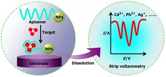
2. Aptasensor Based on Stripping Voltammetry

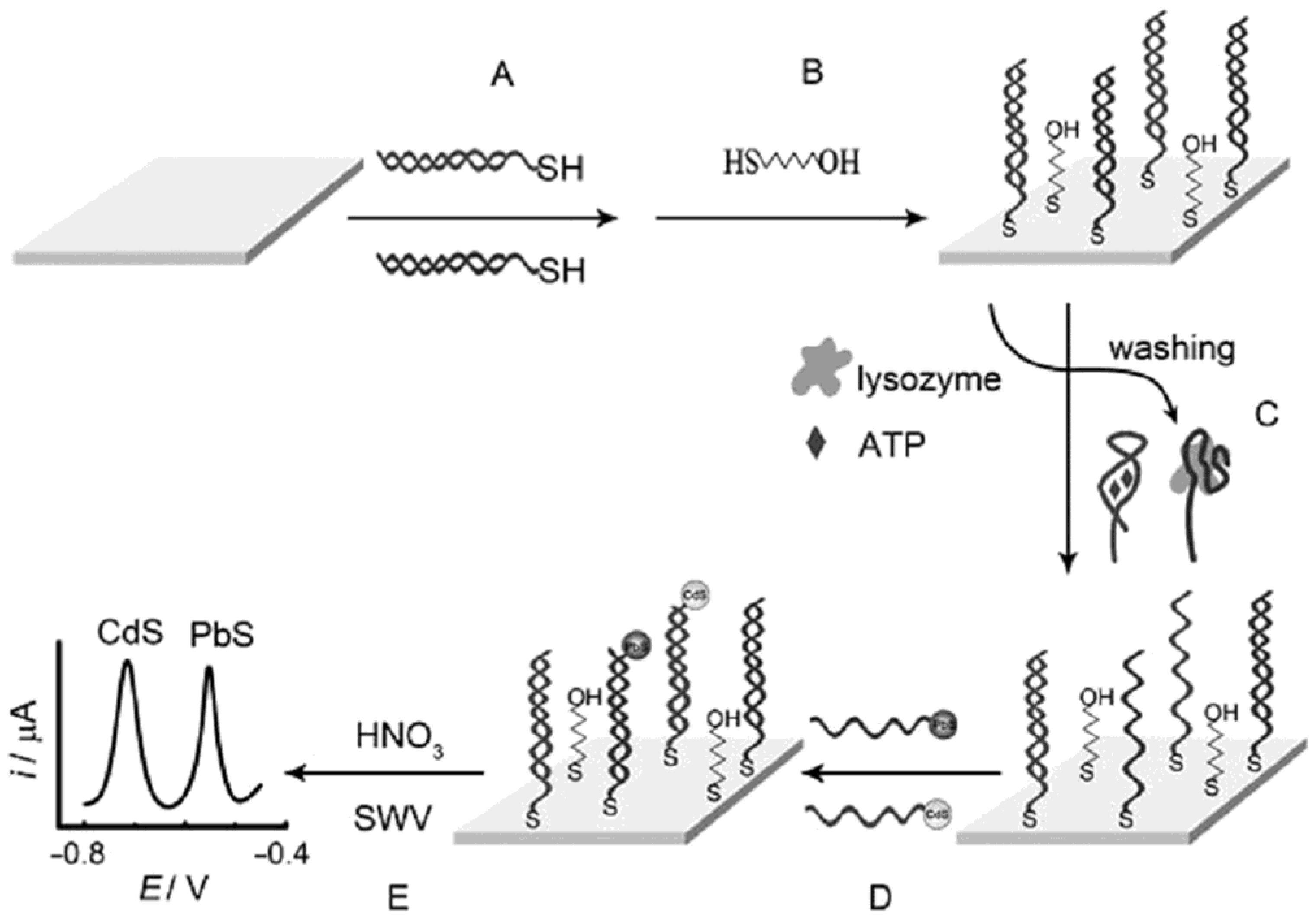
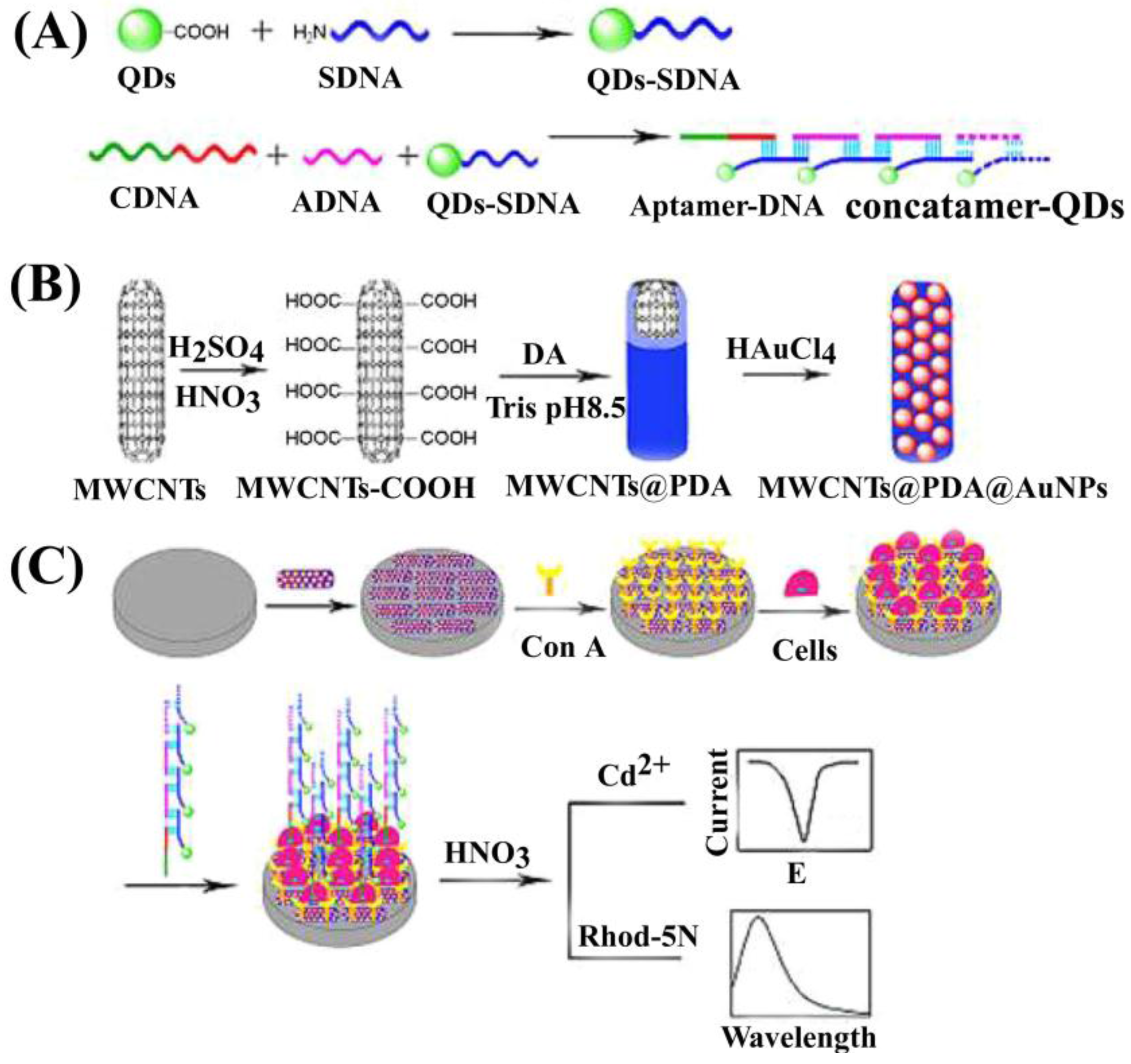
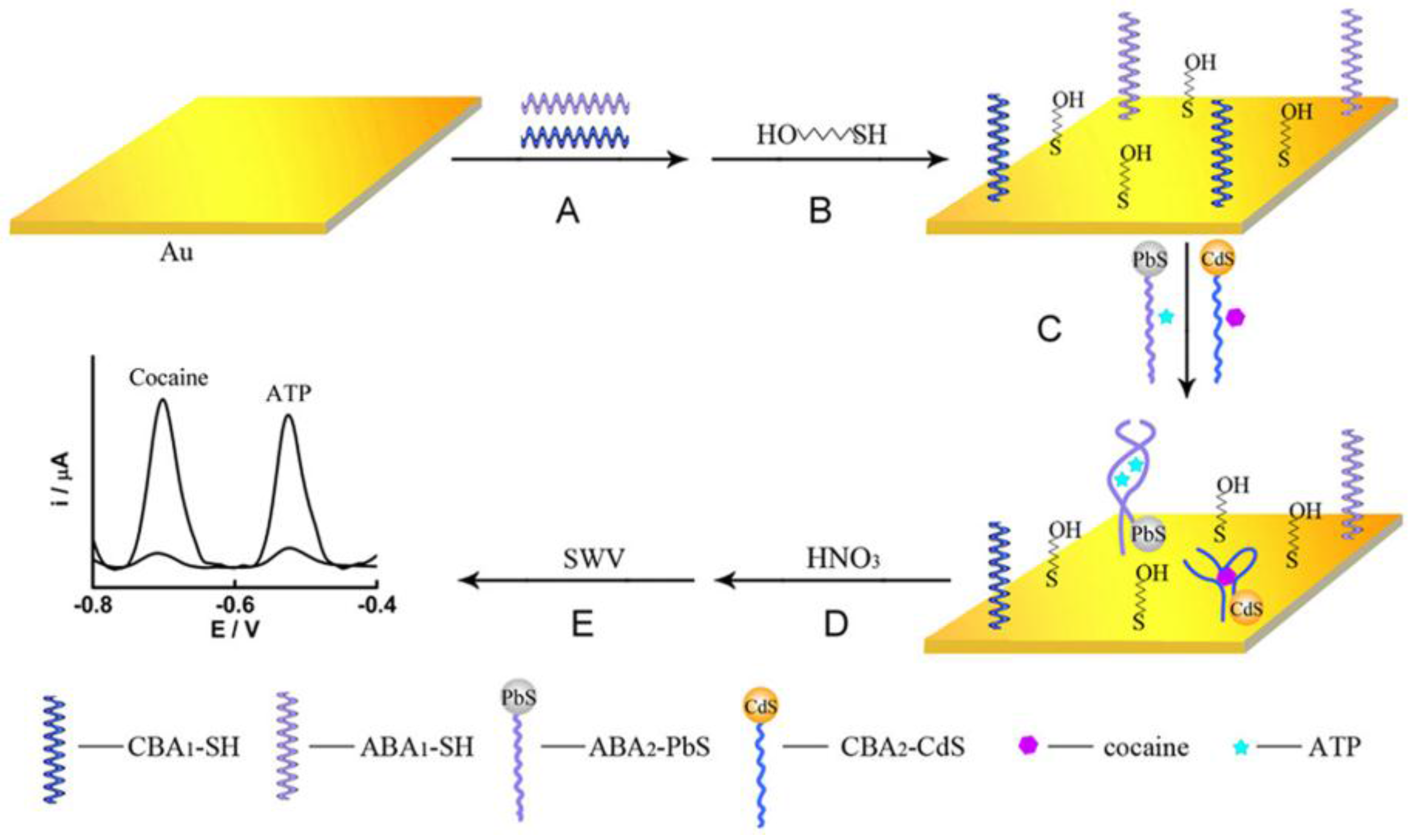
2.1. Aptasensors for Simultaneous Detection of Small Biomolecules

The development of biosensor for sensitive and selective determination of important small biomolecules, such as ATP, cocaine, and adenosine, has been extensively studied for several decades. Quite a number of biosensors can only measure a single analyte. However, the simultaneous detection of several analytes is desirable in many cases (e.g., diagnosis and environmental analysis). Aptasensors based on stripping analysis are a sensitive method for multiplex analysis. For example, Yuan and his group designed an electrochemical strategy for “signal on” and sensitive one-spot simultaneous detection of multiple analytes, such as ATP and cocaine, using two quantum dot (QD) labels [64]. It is a traditional sandwiched strategy, but it involves the self-assembly of two types of thiol-modified primary target-binding aptamers, ATP binding aptamer (ABA1) and cocaine binding aptamer (CBA1) on the gold substrate (Figure 1). These two aptamers contribute to the specific binding of its corresponding target ATP and cocaine, respectively. After simultaneous addition of ATP and cocaine as well as their corresponding QD-conjugated secondary binding aptamers (PbS-ABA2 and CdS-CBA2), ATP and cocaine form a stable sandwiched complex with a QD-conjugated secondary aptamer and primary binding aptamer. This leads to different square wave stripping voltammetric signals via the measurement of released Pb2+ and Cd2+ after an acid dissolution process, enabling simultaneous and sensitive detection of ATP and cocaine due to the inherent amplification feature of the QD labels and the “signal on” detection scheme.
Compared with a single nanoparticle electrochemical label, nanocomposites allow better signal amplification strategies. Zhang and his group utilized QDs-AuNPs nanocomposite labels in simultaneous detection of adenosine and thrombin. The strategy involved a thiol-modified dual-aptamer DNA consisting of both an adenosine aptamer and thrombin aptamer (Figure 2) [22]. The thiol-modified dual-aptamer DNA was immobilized on the Au electrode surface via an Au–S bond. CdS QDs-AuNPs with a reporter DNA complementary to the adenosine aptamer and PbS QDs-AuNPs with a reporter DNA complementary to the thrombin aptamer were used for the detection of adenosine and thrombin by Cd2+ and Pd2+, respectively. In the absence of adenosine and thrombin, CdS QDs-AuNPs were brought to the surface of electrode via DNA hybridization, resulting in a strong Cd stripping peak. When adenosine and thrombin were added, the aptamer could bind tightly and specifically to adenosine and form a tertiary complex. CdS QDs-AuNPs were removed from the surface of electrode because of stronger interaction of adenosine with its aptamer, and PbS QDs-AuNPs were attached to the surface of electrode due to the formation of the sandwich-type structure between thrombin and its two aptamers. As a result, Cd stripping peaks decrease with increasing adenosine concentrations and the Pb stripping peak increases with increasing thrombin concentrations, which allows simultaneous detection of adenosine and thrombin. This strategy of barcode QD tags for simultaneous detection of multiple small molecular analytes supplies an attractive route for screening of small molecules in clinical diagnosis.
These stripping voltammetric strategies mentioned above belong to the direct analysis of small biomolecules. The backfilling strategy belongs to an indirect analysis. Yuan and his group developed a multi-analyte aptasensor through a backfilling strategy for simultaneous detection of ATP and lysozyme using aptamer/nanoparticle bioconjugates as labels [21]. In this backfilling strategy, CdS QDs and PbS QDs are firstly tagged with different DNA sequences, which are complementary with an ATP aptamer and a lysozyme aptamer, respectively (Figure 3). If no ATP and lysozyme exist, duplexes and 6-mercapto-1-hexanol are blocked on the gold substrate. After the addition of ATP and lysozyme, tertiary aptamer/targets are formed and released from the gold substrate. At this time, CdS QDs and PbS QDs tagged with different DNA are added and form backfilling hybridization with thiol-modified DNAs on the gold substrate. This achieves simultaneous detection of ATP and lysozyme by measuring the enhanced square-wave voltammetric stripping signals of the Pb2+ and Cd2+. This aptamer/nanoparticle-based backfilling strategy can improve the sensitivity via the signal-on protocol compared with common target-induced displacement or conformational change signal-off configuration. It opens opportunities for multiplexed clinical diagnosis of different molecules with distinct sizes.

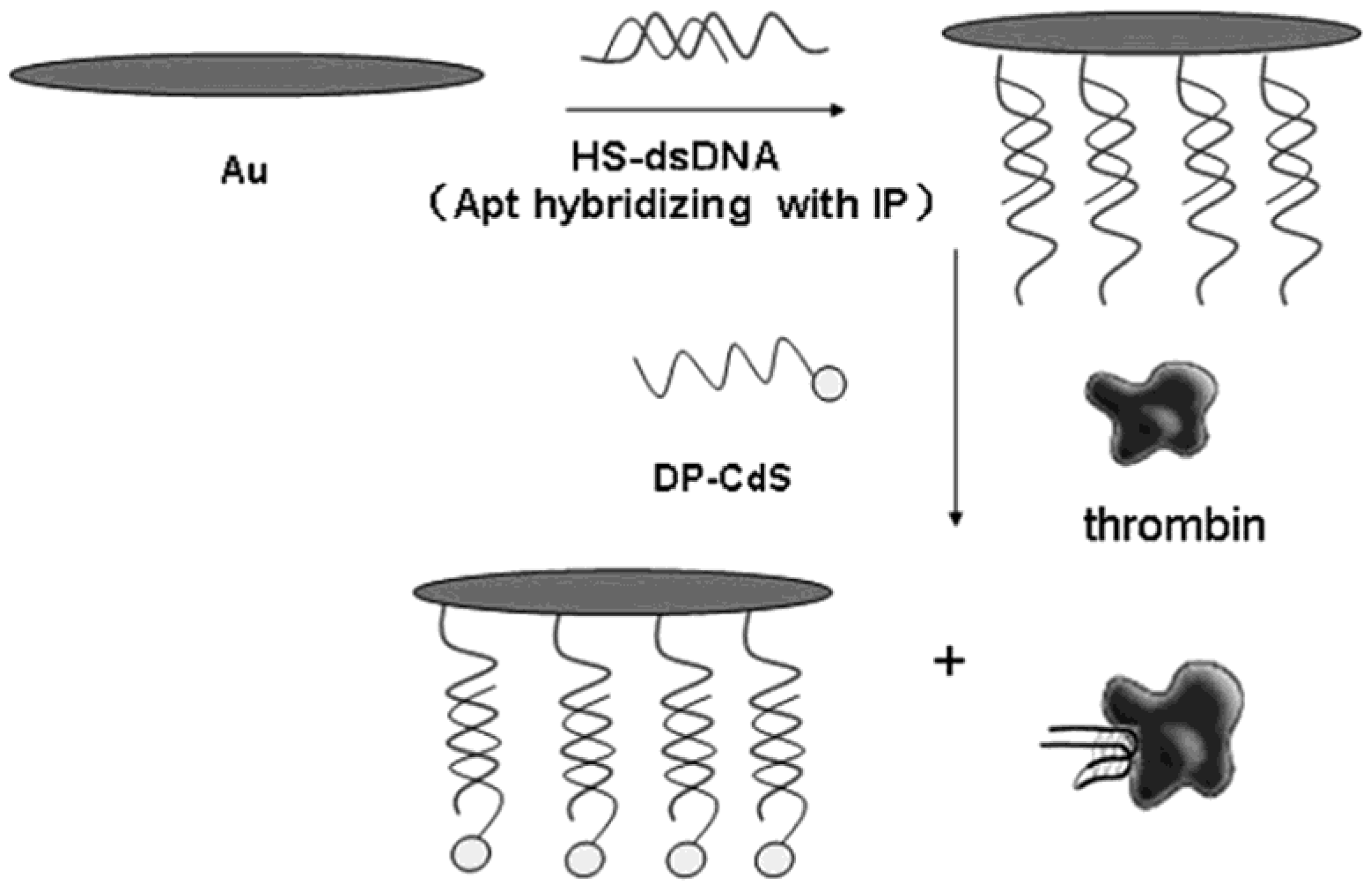
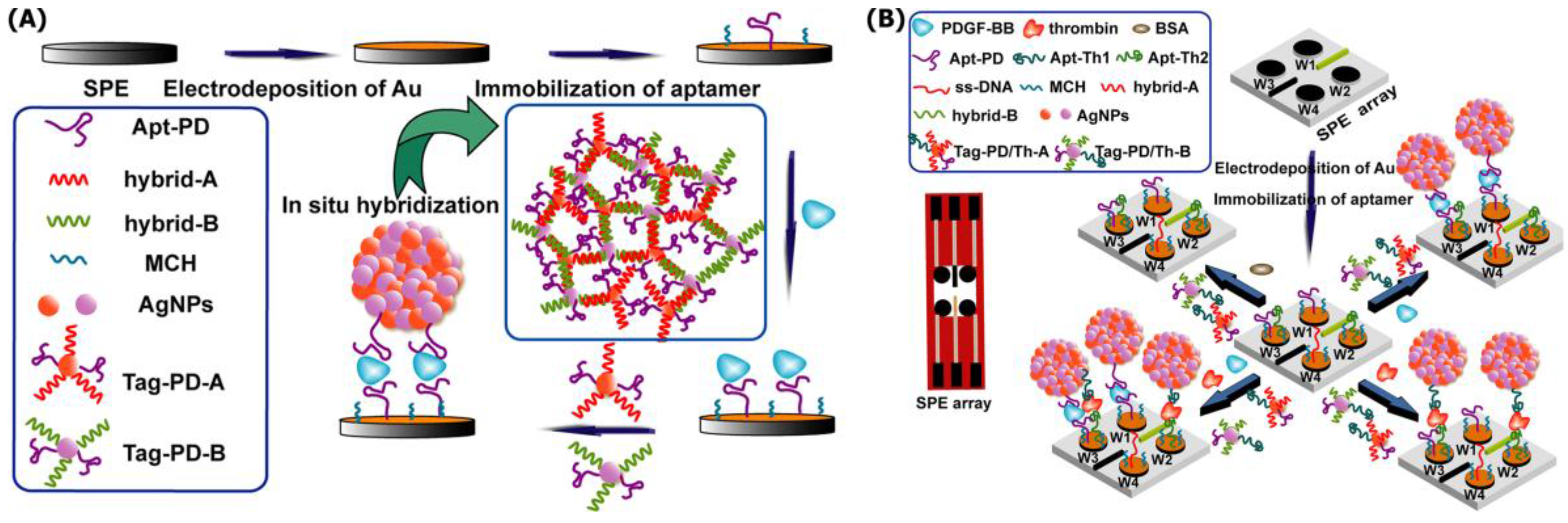
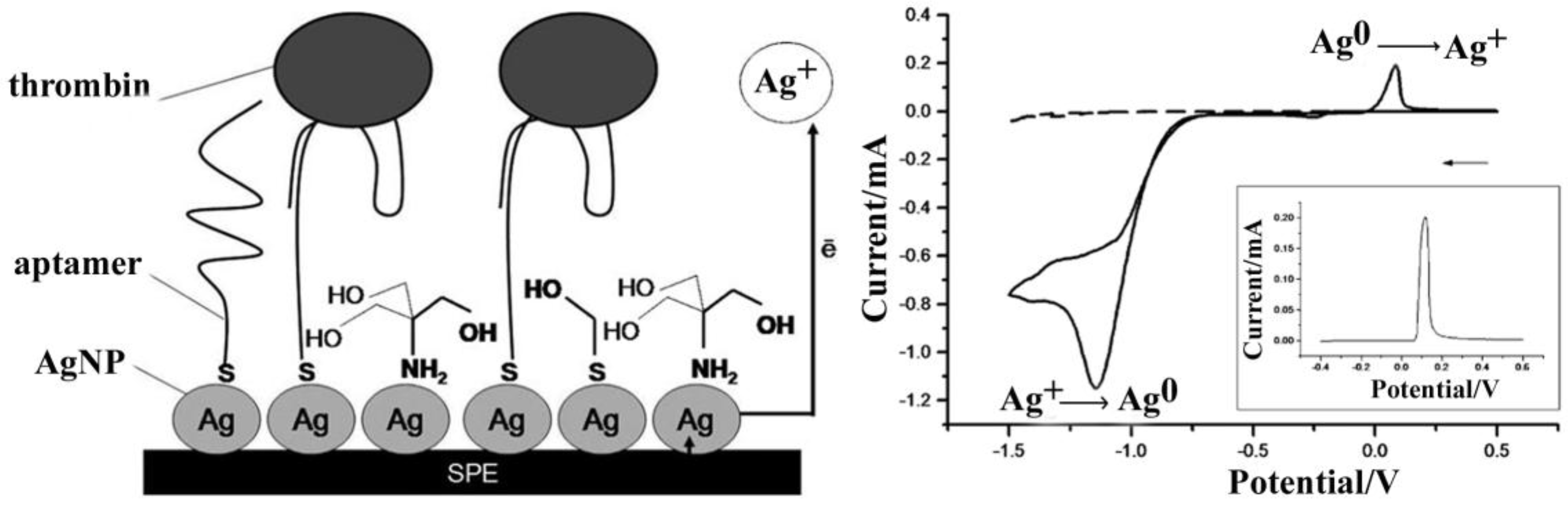
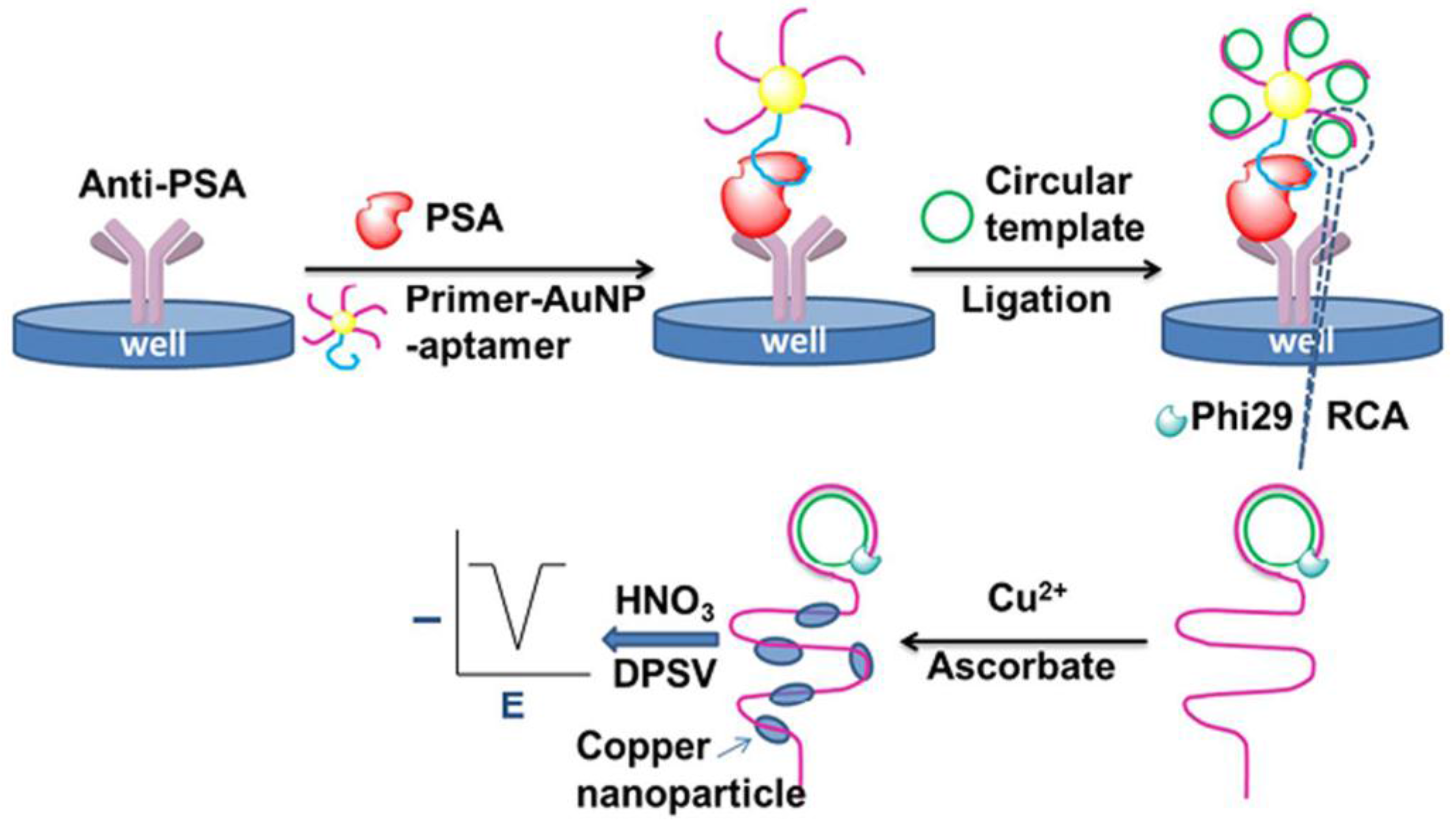
2.2. Aptasensors for Proteins

In bioassays, antibodies have been widely used in the analysis of clinical biofluid specimen, such as urine and blood [50,65,66]. However, antibodies are not stable and may lose activity easily. Aptamers are alternatives to antibodies for bioassays. They can specifically bind to a variety of target molecules, such as proteins [36,39,67,68], drugs [62], small biomolecules [21,22], and cells [28,31,43,69].
In protein aptasensor research area, thrombin is a typical protein model investigated by many researchers. Several works on protein aptasensors based on stripping voltammetry have been reported [26,63,70,71,72,73]. Usually only one type of nanoparticles, such as QDs, are used as labels for stripping voltammetric analysis of thrombin [73] but, compared with a single nanoparticle, nanocomposite materials can obviously achieve signal amplification. Lin and his group reported a novel electrochemical aptasensor for the determination of protein thrombin by anodic stripping voltammetric analysis of Cd2+ using ssDNA-labeled CdS NPs-AuNPs in 2010 [26]. In their strategy, the first step is to obtain detection probe. Linker DNA ssDNA-labeled CdS nanoparticles (NPs) is conjugated with AuNPs through one thrombin-related aptamer. Then, the sandwich type is formed after the addition of target thrombin via another aptamer modified on the surface of gold electrode. In this work, AuNPs and ssDNA-labeled CdS NPs conjugate served not only as a target recognition probe for thrombin, but also as a tool of amplification. Numerous ssDNA-labeled CdS NPs were linked with AuNPs, which enhanced the Cd2+ stripping signals. Therefore, it achieved the determination of thrombin in the linear range of 1.0 × 10⁻¹⁵ to 1.0 × 10⁻¹¹ M with a low detection limit of 0.55 fM. This aptasensor was also confirmed to be able to distinguish the target thrombin from the interferents. Xu and his group also utilized nanocomposite cadmium sulfide nanoparticles (CdS NPs) functionalized with colloidal carbon particles (CPs) in an analysis of thrombin in 2011 [63]. The sandwich-type assay of thrombin was developed by in situ growing of abundant CdS NPs on the surfaces of monodisperse carbon particles (CdS/CPs) via square wave stripping voltammetric signals of the released Cd2+. Both of these two works utilized nanocomposite materials as signal amplification strategy. Additionally, Shumyantseva and his group developed two similar label-free stripping voltammetric analyses of thrombin, respectively using Au+ stripping signals 2008 [71] and using Ag+ stripping signals in 2010 [72]. As shown in Figure 4 [72], a screen-printed electrode (SPE) modified with AgNPs served as the sensing platform and the oxidation of AgNPs (Ag0→Ag+) upon polarization (+100 mV) supplied the detection signals for the proposed aptasensor. Aptamers were immobilized onto the surface of SPE modified with AgNP via an S–Ag bond. In the presence of thrombin, the anodic Ag+ stripping peak was decreased. This direct detection strategy is label-free, simple, and fast, and can be extended to other analytes or biorecognition elements.
Wang and his group took advantage of the signal amplification effect of the labeled CdS NPs with the assistance of target protein-induced strand displacement and developed a sensitive electrochemical aptasensor for thrombin (Figure 5) [68]. In this electrochemical aptasensor for thrombin, a single DNA labeled with CdS nanoparticles was used as a detection probe. In the presence of thrombin, the aptamer in the dsDNA preferred to form a G quarter structure with thrombin releasing one single strand in the dsDNA sequence. In the G quarter structure, multiple guanines are organized around thrombin in a four-stranded structure. After the addition of the CdS NPs labeled probe, the concentration of thrombin is related to the amount of the captured CdS nanoparticles. After dissolving CdS particles, the released Cd2+ is determined by adsorptive stripping voltammeries and this amount is related to the thrombin concentration, allowing reaching a detection limit for the protein of 4.3 × 10⁻¹³ mol/L.
Xu and his group utilized AgNPs/graphene nanocomposite materials for the analysis of the protein human immunoglobulin E (IgE) [42]. IgE plays an important role in allergic reactions and other related diseases. Like other aptasensor of protein, a sandwich-type strategy was used. However, the detection probe was a streptavidin-functionalized AgNPs/graphene hybrid linked with biotinylated anti-human IgE antibody. Meanwhile, thiol-tagged IgE aptamer was used as the capture probe. Owing to specific binding between the aptamer with IgE and the antibody with IgE, a sandwich-type aptasensor for IgE was developed by a square wave anodic stripping voltammetric signal from AgNPs/graphene nanocomposite materials. The high-loading ability of graphene for AgNPs, combined with the unique electrical properties of graphene, contributed to the low detection limit. It brought a dynamic range for IgE detection from 10 to 1000 ng/mL with a low detection limit of 3.6 ng/mL. Furthermore, it is also a portable, simple, and inexpensive electrochemical biosensor for IgE since a disposable screen printed electrode was used as a sensing platform.
Silver staining is commonly used as a signal amplification strategy for detection methods based on nanomaterial labels [41,66]. Xie and his group used gold label/silver staining in detection of immunoglobulin G (IgG) with the low detection limit of 0.2 fg/mL [66]. In their strategy, silver ions can be stained solely on the surface of catalytic AuNPs through chemical reduction of silver cations by hydroquinone. A beforehand “potential control” in air, and then an injection of HNO3 for dissolution of the stained silver, enables rapid cathodic preconcentration of atomic silver onto the electrode surface for anodic stripping voltammetry measurement of silver ions.
SPE is an appropriate electrode for clinical diagnosis because it can satisfy highly sensitive and reproducible determination of target analytes, and meets the requirement for performing rapid in situ analyses. Combining the advantages of SPE and those of electrochemical array technology, SPE array technology shows great advantages in multi-analytes measurements. It has been used in various analytical methods, such as heavy metal ion detection, enzymatic biosensors, immunosensors, and DNA sensors [74,75]. Xu and his group further developed a multiplied protein aptasensor for platelet-derived growth factor (PDGF-BB) and thrombin by constructing a SPE array chip screen-printed electrode array in 2014 [25]. A sandwich-type strategy was utilized and an aptamer was tagged with DNA-functionalized AgNPs aggregate (Figure 6A). It gave an amplified differential pulse stripping voltammetry signal compared to the signal-labeled tag. Different aptamers for PDGF-BB and thrombin were chosen to construct multiplied-protein detection using a SPE array chip (Figure 6B). The novel SPE array chip achieved a wide linear range and low limit of detection in analysis of PDGF-BB and thrombin.

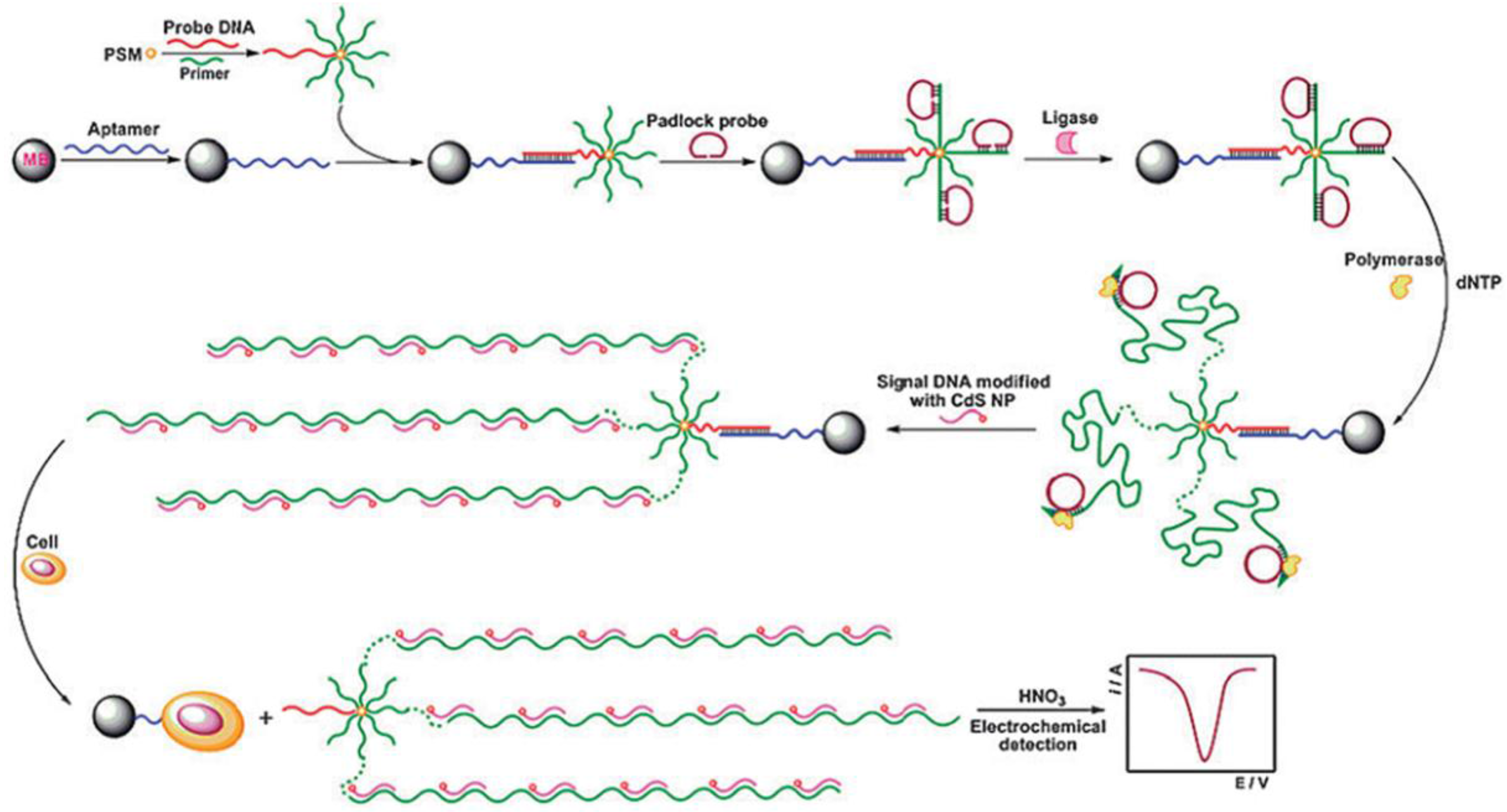
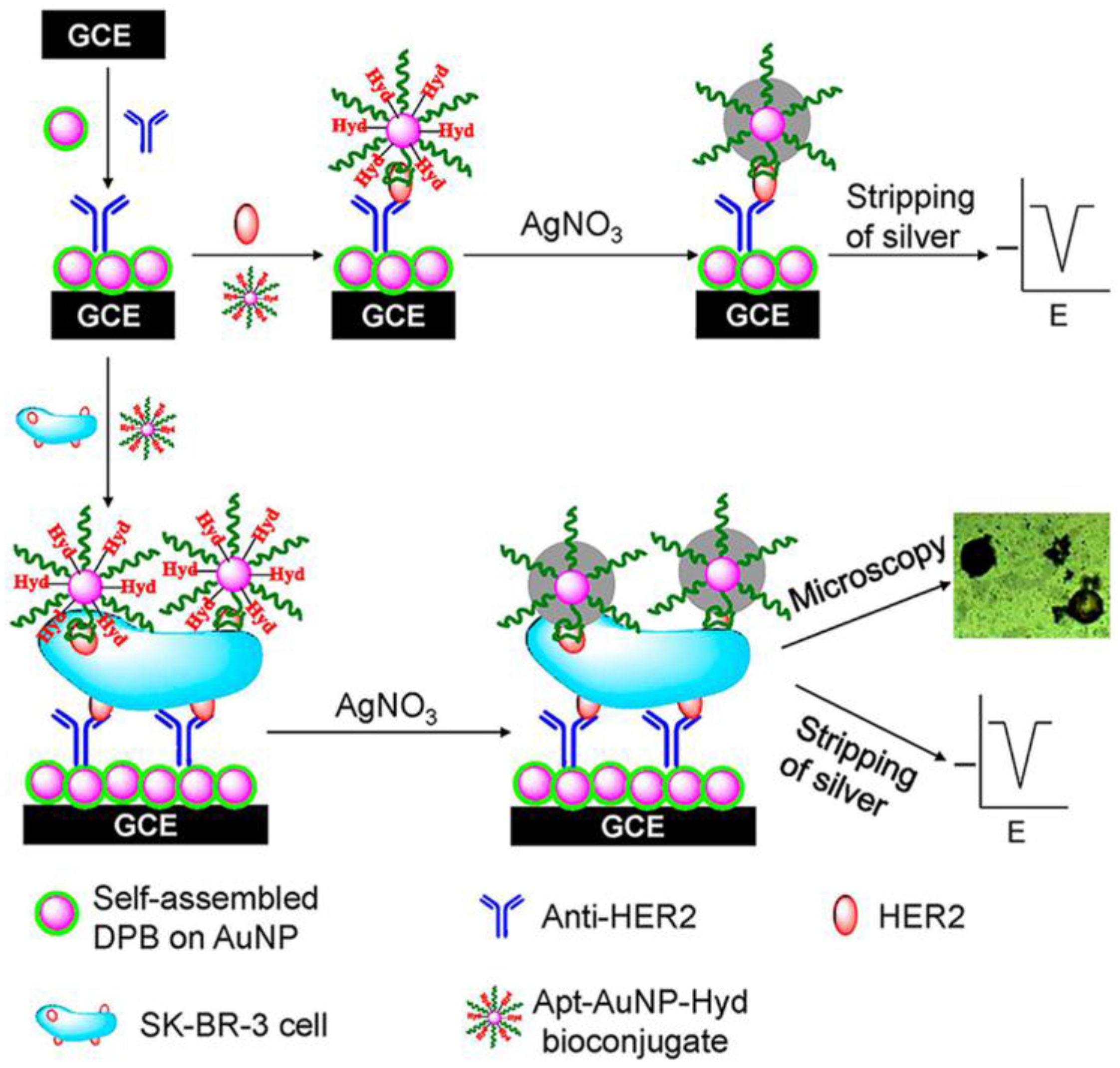
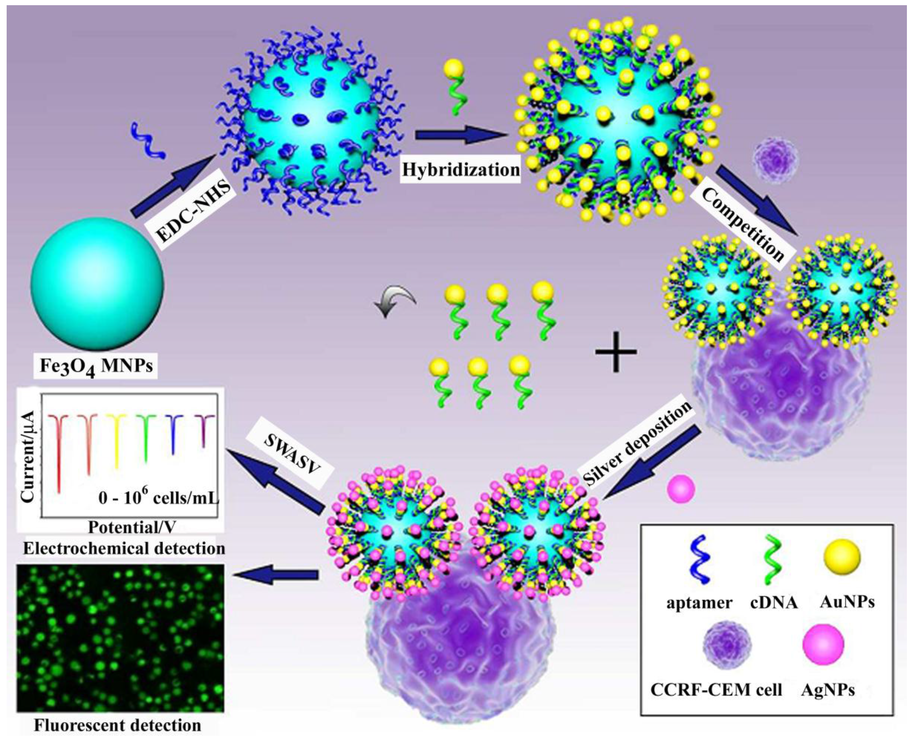
2.3. Aptasensors for Cancer Cells and Diseases

Determining the extent of disease and planning appropriate therapies are essential for cancer treatment at an early stage. A tumor marker is a substance abnormally expressed in response to cancer. Different tumor markers can indicate different types of cancers with altered disease processes. Breast cancer is a kind of cancer mainly occurring in the inner lining of the milk ducts or lobules with different spread, aggressiveness, and genetic makeup. It has become the second most common type of cancer after lung cancer, and the fifth common cause of cancer death [69]. Li and his group developed a sensitive electrochemical aptasensor for the analysis of MCF-7 breast cancer cells by simultaneously detecting two tumor markers, human mucin-1 (MUC1) and carcinoembryonic antigen (CEA), on the surface of MCF-7 breast cancer cells in 2010 [69]. In their strategy, MCF-7 breast cancer cells were firstly recognized by its aptamer immobilized on the surface of a gold electrode. Another tumor marker, CEA, was subsequently captured by CdS nanoparticles (CdS NPs)-labeled anti-CEA. It achieved breast cancer cell analysis by anodic stripping voltammetric signals of Cd2+. It efficiently improved the accuracy of the detection and avoidance of false-positive results due to low specificity. Similarly, B. Shim and his group developed an ultrasensitive and selective electrochemical diagnosis of breast cancer marker (HER2) and HER2-overexpressing breast cancer (SK-BR-3) on the basis of a hydrazine–Au NP–aptamer bioconjugate in 2013 [41]. Human epidermal growth factor receptor 2 (HER2) is a key prognostic marker and an effective therapeutic treatment target for breast cancer because it can be over-expressed in 10%–25% of breast cancers. In this strategy (Figure 7), anti-HER2 immobilized on the surface of an electrode was used to recognize breast cancer (SK-BR-3). 2,5-bis(2-thienyl)-1H-pyrrole-1-(p-benzoic acid) was self-assembled on AuNPs and the hydrazine-AuNP-aptamer bioconjugate was used as a detection probe. During the detection process, the anti-HER2-immobilized probe, HER2, or SK-BR-3 breast cancer cells, and Hyd-AuNP-Apt bioconjugate form a sandwich-type structure. In order to achieve ultrasensitive analysis of breast cancer, the silver-stained signal amplification was used. The silver ion is selectively reduced by hydrazine. The deposited silver is analyzed via square wave stripping voltammetry. This was the first report about the analysis of breast cancer cells utilizing selective silver stain through interaction with the Hyd–AuNP–Apt bioconjugate. Differently, Zhu and his group developed a competitive electrochemical sensor for anodic stripping voltammetric detection of breast cancer by using aptamer-quantum dots conjugates (Apt-QDs) [76]. Amino modified aptamers, which can specifically recognize MUC1 on the surface breast cancer cells, are firstly captured by the thiolated complementary DNA (cDNA) anchored on the gold electrode surface. Then Apt-QDs are formed through the interaction of amino groups with carboxyl groups in QDs. In the presence of breast cancer cells, MUC1 protein could compete with cDNA to conjugate with Apt-QDs conjugates. It leads to the decrease in the number of QDs retained at the electrode. After dissolving the remaining QDs, the concentration of the obtained metal species is detected via anodic stripping and the concentration of breast cancer cells is detected. The competitive electrochemical method is a good way to avoid false positive signals.
In addition to breast cancer, acute leukemia is the most common pediatric malignancy and remains the leading cause of disease-related mortality in children and adolescents. Zhu and his group developed an aptamer-based competition strategy for ultrasensitive electrochemical detection of leukemia cells (Figure 8) [77]. CCRF-CEM acute leukemia cells were chosen as model cells. The strategy was based on a dual signal amplification based on Fe3O4 magnetic nanoparticles (MNPs) carrying AuNPs and AuNP-catalyzed silver, which realized both as an electrosensor and cytosensor for target cells. An aptamer was designed to bind specifically to target cells. A partial cDNA was used to match the aptamer sequence. To fabricate the cytosensor, AuNPs loaded on Fe3O4 MNPs were prepared. Fe3O4 MNPs acted as both the separation tool and the strong nanocarriers for loading AuNPs and AuNP-catalyzed Ag deposition, which achieved signal amplification for the detection. After the addition of target cells, they could compete efficiently with conjugated cDNA to bind specifically with its aptamers. Therefore, the number of Fe3O4 MNPs carrying AuNPs and AgNPs retained at the electrode were decreased. The dissolved Ag+, which was related with the number of target cells, were measured via square wave stripping voltammetry. The combination of the competitive, hybridization-based, and square wave anodic stripping voltammetric sensing platform supplies leukemia detection with high sensitivity, good specificity, desirable reproducibility, and acceptable stability. It exhibits great clinical value in the early diagnosis and prognosis of leukemia.
Zhu and his group developed both electrochemical aptasensor and cytosensor for selective and ultrasensitive detection of cancer cells using aptamer-DNA concatamer quantum dot probes by fabricating an supersandwich structure (Figure 9) in 2013 [78]. DNA concatamers are linear polymeric structures formed by self-association of short DNA fragments through specific interactions. The proposed supersandwich structure combined with numerous CdS QDs achieves significant signal amplifications in aptasensors for cancer cells with the low detection limit of 50 cells/mL.
Multiple amplification strategies include PCR, SDA, HCR, RCA, etc. [43,44,45,79]. RCA is a simple and powerful isothermal enzymatic process. A short DNA or RNA primer is elongated to form a long, single-stranded DNA or RNA, assisted by a circular DNA template and unique DNA or RNA polymerases [80]. Since the RCA product contains thousands of tandem repeats that are complementary to the circular template, it leads to effective signal readout approaches, which are crucial to achieve various analytical purposes. Just owing to the advantages of high speed, efficiency, and specificity, RCA has been widely applied for signal amplification in analysis of DNA, protein, metal ions, and diseased cells. Ding and his group reported a cascade signal amplification strategy for ultrasensitive detection of cancer cell Ramos cells via combining RCA technique in 2012 [31]. In their strategy (Figure 10), NH2-modified probe DNA and NH2-modified primer DNA were immobilized on the surface of carboxyl-coated polystyrene microspheres (PSMs) to form a bio-bar code probe. The probe DNA firstly hybridized partly with the Ramos cell aptamer modified on magnetic beads (MBs) to construct a magnetic biocomplex. Carboxyl-coated MBs acted as the separation tool and the immobilization matrix. When the RCA reaction was started, a large quantity of signal DNA tagged with CdS NPs was added to the mixture. In this way, a long, single-stranded DNA which contains numerous tandem-repeat sequences was synthesized. At the same time, a large number of CdS-DNA probes for enhanced recognition were periodically assembled. But the double helixes between PSMs and MBs conjugate were opened due to much stronger binding of aptamer to Ramos cells than that of ordinary double-stranded DNA. Therefore Ramos cell were separated by magnetic separation procedure and the released Cd2+ form separated CdS-DNA probes was measured through anodic stripping voltammetry. This electrochemical aptasensor for cancer cells exhibited high sensitivity and specificity with the detection limits of 10 Ramos cells/mL.
Jiang and his group combined the RCA reaction with poly(thymine)-templated copper nanoparticles (Cu NPs) for cascade signal amplification in ultrasensitive and highly-selective electrochemical analysis of prostate cancer biomarker PSA in 2016 [32]. DNA-templated metal nanoparticles, such as DNA-templated silver nanoclusters and DNA-templated Cu NPs, as novel-type emerging signal reporters have attracted increasing interest in biosensors owing to their advantages, including low toxicity, good biocompatibility, excellent optical properties, and facile integration with DNA-based recognition and signal amplification strategies. Though DNA-templated Cu NPs have been employed for the detection of small molecules, enzyme activity, nucleic acids, and metal ions, this was the first time of applying poly(thymine) repeats for a poly(thymine)-templated Cu NPs in electrochemical aptasensor of cancer markers. As shown in Figure 11, the RCA product contained thousands of poly(thymine) repeats which were used to synthesize DNA-templated Cu NPs. Once one PSA existed, one primer-AuNP-aptamer would be immobilized on the well and, subsequently, thousands of poly(thymine) sequences would be generated from RCA. It led to the synthesis of numerous DNA-templated Cu NPs and brought enhanced stripping voltammetric signals of released Cu2+. It was a successful attempt at the ultrasensitive detection of PSA with a remarkable detection limit of 0.020 fg/mL for DNA-templated Cu NPs applied in cascade signal amplification.
In addition to cancer, some diseases also attract great attention. Staphylococcus aureus (S. aureus), one of the most important human pathogens, can cause different kinds of illnesses, from minor skin infections to life threatening diseases [28,81]. Abbaspour and his group reported a sensitive and highly-selective sandwich-type electrochemical detection of S. aureus using dual aptamers [28]. The primary aptamer was immobilized on the surface of MBs and the secondary aptamer, conjugated to AgNPs, acted as signal reporters to provide electrochemical stripping voltammetry characteristics. After fast magnetic separation, S. aureus was determined by Ag+ released via differential pulse anodic stripping voltammetric measurement. It is a characteristic aptasensor for disease analysis, combining magnetic separation and AgNPs-based signal amplification technology. Zhang and his group developed a novel method for the determination of sulfate-reducing bacteria with the low detection limit of 1.8 × 10² cfu/mL) by combining a graphene oxide sheet-amplified assay and silver enhancement technology in 2011 [40]. They utilized graphene oxide sheets as promoters for silver reduction into AgNPs by hydroquinone. It is a good example of silver stained-based stripping voltammetry by utilizing the intrinsic catalytic property of graphene oxide sheets.

3. Conclusions

In this article, a mini-review about the most recent developments in the field of aptasensors based on stripping voltammetry is presented. In order to improve the sensitivity of aptasensors nanomaterials, such as metal nanoparticles, semiconductor nanoparticles, and nanocomposite materials, were employed. They can give electrochemical signals by stripping analysis of the dissolved metals, modify the surface of the electrode to construct more sensitive electrochemical transducers, or immobilize capturing probes. Multiple amplifications, such as RCA, are also powerful strategies to improve the sensitivity of aptasensors. Moreover, some alternative electrodes, such as bismuth electrodes, tin electrodes, and even screen-printed electrodes, were employed by researchers. All of these will greatly promote the development of aptasensors on the basis of stripping voltammetry in single-target and multi-target analysis of small biomolecules, proteins, disease markers, and cancer cells.

Acknowledgments

It was supported by the National Natural Science Foundation of China (No. 21505011, 21475123), Chongqing Research Program of Basic Research and Frontier Technology (No. cstc2015jcyjA20019), Scientific and Technological Research Pro-gram of Chongqing Education Committee (No. KI1500306), and the Chinese Academy of Sciences (CAS)–the Academy of Sciences for the Developing World (TWAS) President’s Fellowship Programme.

Conflicts of Interest

The authors declare no conflict of interest.

Abbreviations

The following abbreviations are used in this manuscript:
ABAATP binding aptamer
Apt-QDsaptamer-quantum dots conjugates
ATPadenosine triphosphate
CBAcocaine binding aptamer
cDNAcomplementary DNA
CEAcarcinoembryonic antigen
CPscolloidal carbon particles
CTIPEcyclic target-induced primer extension
HCRhybridization chain reaction
HER2breast cancer marker
IgEhuman immunoglobulin E
IgGimmunoglobulin G
MBsmagnetic beads
MCF-7The number of one type of breast cancer cells
MNPsmagnetic nanoparticles
MUC1human mucin-1
PCRpolymerase chain reaction
PDGF-BBplatelet-derived growth factor
PSMspolystyrene microspheres
PSAprostate-specific antigen
QDquantum dot
RCArolling circle amplification
S. aureusStaphylococcus aureus
SDAstrand-displacement amplification
SELEXsystematic evolution of ligands by exponential enrichment
SK-BR-3HER2-overexpressing breast cancer
SPEscreen-printed electrode

References

  1. Xu, Y.; Cheng, G.; He, P.; Fang, Y. A review: Electrochemical aptasensors with various detection strategies. Electroanalysis 2009, 21, 1251–1259. [Google Scholar] [CrossRef]
  2. Palchetti, I.; Mascini, M. Electrochemical nanomaterial-based nucleic acid aptasensors. Anal. Bioanal. Chem. 2012, 402, 3103–3114. [Google Scholar] [CrossRef] [PubMed]
  3. Thevenot, D.R.; Tóth, K.; Durst, R.A.; Wilson, G.S. Electrochemical biosensors: Recommended definitions and classification. Pure Appl.Chem. 1999, 71, 2333–2348. [Google Scholar] [CrossRef]
  4. Willner, I.; Zayats, M. Electronic aptamer-based sensors. Angew. Chem. Int. Ed. 2007, 46, 6408–6418. [Google Scholar] [CrossRef] [PubMed]
  5. Famulok, M.; Mayer, G. Aptamer modules as sensors and detectors. Acc. Chem. Res. 2011, 44, 1349–1358. [Google Scholar] [CrossRef] [PubMed]
  6. Sarvetnick, N.; Shizuru, J.; Liggitt, D.; Martin, L.; Mclntyre, B.; Gregory, A.; Parslow, T.; Stewart, T. Loss of pancreatic islet tolerance induced by β-cell expression of interferon-γ. Nature 1990, 346, 844–847. [Google Scholar] [CrossRef] [PubMed]
  7. Robertson, D.L.; Joyce, G.F. Selection in vitro of an RNA enzyme that specifically cleaves single-stranded DNA. Nature 1990, 344, 467–468. [Google Scholar] [CrossRef] [PubMed]
  8. Tuerk, C.; Gold, L. Systematic evolution of ligands by exponential enrichment: RNA ligands to bacteriophage T4 DNA polymerase. Science 1990, 249, 505–510. [Google Scholar] [CrossRef] [PubMed]
  9. Lee, J.H.; Yigit, M.V.; Mazumdar, D.; Lu, Y. Molecular diagnostic and drug delivery agents based on aptamer-nanomaterial conjugates. Adv. Drug. Delivery. Rev. 2010, 62, 592–605. [Google Scholar] [CrossRef] [PubMed]
  10. Palchetti, I.; Mascini, M. Electrochemical nanomaterial-based nucleic acid aptasensors. Anal. Bioanal. Chem. 2012, 402, 3103–3114. [Google Scholar] [CrossRef] [PubMed]
  11. Meyer, S.; Maufort, J.P.; Nie, J.; Stewart, R.; McIntosh, B.E.; Conti, L.R.; Ahmad, K.M.; Soh, H.T.; Thomson, J.A. Development of an efficient targeted cell-SELEX procedure for DNA aptamer reagents. PLoS ONE 2013, 8, e71798. [Google Scholar] [CrossRef] [PubMed]
  12. Polonschii, C.; David, S.; Tombelli, S.; Mascini, M.; Gheorghiu, M. A novel low-cost and easy to develop functionalization platform. Case study: Aptamer-based detection of thrombin by surface plasmon resonance. Talanta 2010, 80, 2157–2164. [Google Scholar] [CrossRef] [PubMed]
  13. Yuan, T.; Hu, L.; Liu, Z.; Qi, W.; Zhu, S.; Xu, G. A label-free and signal-on supersandwich fluorescent platform for Hg2+ sensing. Anal. Chim. Acta 2013, 793, 86–89. [Google Scholar] [CrossRef] [PubMed]
  14. Du, J.; Jiang, L.; Shao, Q.; Liu, X.; Marks, R.S.; Ma, J.; Chen, X. Colorimetric detection of mercury ions based on plasmonic nanoparticles. Small 2013, 9, 1467–1481. [Google Scholar] [CrossRef] [PubMed]
  15. Lin, L.; Liu, Y.; Zhao, X.; Li, J. Sensitive and rapid screening of T4 polynucleotide kinase activity and inhibition based on coupled exonuclease reaction and graphene oxide platform. Anal. Chem. 2011, 83, 8396–8402. [Google Scholar] [CrossRef] [PubMed]
  16. Liu, Z.; Qi, W.; Xu, G. Recent advances in electrochemiluminescence. Chem. Soc. Rev. 2015, 44, 3117–3142. [Google Scholar] [CrossRef] [PubMed]
  17. Hu, L.; Xu, G. Applications and trends in electrochemiluminescence. Chem. Soc. Rev. 2010, 39, 3275–3304. [Google Scholar] [CrossRef] [PubMed]
  18. Liu, Z.; Zhang, W.; Qi, W.; Gao, W.; Hanif, S.; Saqib, M.; Xu, G. Label-free signal-on ATP aptasensor based on the remarkable quenching of tris(2,2′-bipyridine)ruthenium(II) electrochemiluminescence by single-walled carbon nanohorn. Chem. Commun. 2015, 51, 4256–4258. [Google Scholar] [CrossRef] [PubMed]
  19. Yu, Y.; Cao, Q.; Zhou, M.; Cui, H. A novel homogeneous label-free aptasensor for 2,4,6-trinitrotoluene detection based on an assembly strategy of electrochemiluminescent graphene oxide with gold nanoparticles and aptamer. Biosens. Bioelectron. 2013, 43, 137–142. [Google Scholar] [CrossRef] [PubMed]
  20. Merkoci, A. Electrochemical biosensing with nanoparticles. FEBS J. 2007, 274, 310–316. [Google Scholar] [CrossRef] [PubMed]
  21. Qian, X.; Xiang, Y.; Zhang, H.; Chen, Y.; Chai, Y.; Yuan, R. Aptamer/nanoparticle-based sensitive, multiplexed electronic coding of proteins and small biomolecules through a backfilling strategy. Chem. Eur. J. 2010, 16, 14261–14265. [Google Scholar] [CrossRef] [PubMed]
  22. Li, X.; Liu, J.; Zhang, S. Electrochemical analysis of two analytes based on a dual-functional aptamer DNA sequence. Chem. Commun. 2010, 46, 595–597. [Google Scholar] [CrossRef] [PubMed]
  23. Shen, B.; Li, J.; Cheng, W.; Yan, Y.; Tang, R.; Li, Y.; Ju, H.; Ding, S. Electrochemical aptasensor for highly sensitive determination of cocaine using a supramolecular aptamer and rolling circle amplification. Microchim. Acta 2015, 182, 361–367. [Google Scholar] [CrossRef]
  24. Zhang, D.-W.; Nie, J.; Zhang, F.-T.; Xu, L.; Zhou, Y.-L.; Zhang, X.-X. Novel homogeneous label-free electrochemical aptasensor based on functional DNA hairpin for target detection. Anal. Chem. 2013, 85, 9378–9382. [Google Scholar] [CrossRef] [PubMed]
  25. Song, W.; Li, H.; Liang, H.; Qiang, W.; Xu, D. Disposable electrochemical aptasensor array by using in situ DNA hybridization inducing silver nanoparticles aggregate for signal amplification. Anal. Chem. 2014, 86, 2775–2783. [Google Scholar] [CrossRef] [PubMed]
  26. Ding, C.; Ge, Y.; Lin, J.M. Aptamer based electrochemical assay for the determination of thrombin by using the amplification of the nanoparticles. Biosens. Bioelectron. 2010, 25, 1290–1294. [Google Scholar] [CrossRef] [PubMed]
  27. Cheng, W.; Ding, S.; Li, Q.; Yu, T.; Yin, Y.; Ju, H.; Ren, G. A simple electrochemical aptasensor for ultrasensitive protein detection using cyclic target-induced primer extension. Biosens. Bioelectron. 2012, 36, 12–17. [Google Scholar] [CrossRef] [PubMed]
  28. Abbaspour, A.; Norouz-Sarvestani, F.; Noori, A.; Soltani, N. Aptamer-conjugated silver nanoparticles for electrochemical dual-aptamer-based sandwich detection of staphylococcus aureus. Biosens. Bioelectron. 2015, 68, 149–155. [Google Scholar] [CrossRef] [PubMed]
  29. Jie, G.; Zhang, J.; Wang, L. A novel quantum dot nanocluster as versatile probe for electrochemiluminescence and electrochemical assays of DNA and cancer cells. Biosens. Bioelectron. 2014, 52, 69–75. [Google Scholar] [CrossRef] [PubMed]
  30. Luo, C.; Lei, Y.; Yan, L.; Yu, T.; Li, Q.; Zhang, D.; Ding, S.; Ju, H. A rapid and sensitive aptamer-based electrochemical biosensor for direct detection of Escherichia coli O111. Electroanalysis 2012, 24, 1186–1191. [Google Scholar] [CrossRef]
  31. Ding, C.; Liu, H.; Wang, N.; Wang, Z. Cascade signal amplification strategy for the detection of cancer cells by rolling circle amplification and nanoparticles tagging. Chem. Commun. 2012, 48, 5019–5021. [Google Scholar] [CrossRef] [PubMed]
  32. Zhu, Y.; Wang, H.; Wang, L.; Zhu, J.; Jiang, W. Cascade signal amplification based on copper nanoparticle-reported rolling circle amplification for ultrasensitive electrochemical detection of the prostate cancer biomarker. ACS Appl. Mater. Interfaces 2016, 8, 2573–2581. [Google Scholar] [CrossRef] [PubMed]
  33. Cui, L.; Wu, J.; Ju, H. Label-free signal-on aptasensor for sensitive electrochemical detection of arsenite. Biosens. Bioelectron. 2016, 79, 861–865. [Google Scholar] [CrossRef] [PubMed]
  34. Li, F.; Feng, Y.; Zhao, C.; Tang, B. Crystal violet as a G-quadruplex-selective probe for sensitive amperometric sensing of lead. Chem. Commun. 2011, 47, 11909–11911. [Google Scholar] [CrossRef] [PubMed]
  35. Cui, L.; Wu, J.; Ju, H. Electrochemical sensing of heavy metal ions with inorganic, organic and bio-materials. Biosens. Bioelectron. 2015, 63, 276–286. [Google Scholar] [CrossRef] [PubMed]
  36. Shumkov, A.A.; Suprun, E.V.; Shatinina, S.Z.; Lisitsa, A.V.; Shumyantseva, V.V.; Archakov, A.I. Gold and silver nanoparticles for electrochemical detection of cardiac troponin I based on stripping voltammetry. BioNanoScience 2013, 3, 216–222. [Google Scholar] [CrossRef]
  37. Kokkinos, C.; Economou, A.; Petrou, P.S.; Kakabakos, S.E. Microfabricated tin-film electrodes for protein and DNA sensing based on stripping voltammetric detection of Cd(II) released from quantum dots labels. Anal. Chem. 2013, 85, 10686–10691. [Google Scholar] [CrossRef] [PubMed]
  38. Liu, B.; Zhang, B.; Chen, G.; Yang, H.; Tang, D. Metal sulfide-functionalized DNA concatamer for ultrasensitive electronic monitoring of ATP using a programmable capillary-based aptasensor. Biosens. Bioelectron. 2014, 53, 390–398. [Google Scholar] [CrossRef] [PubMed]
  39. Szymanski, M.; Noble, J.; Knight, A.; Porter, R.; Worsley, G. Aptamer-mediated detection of thrombin using silver nanoparticle signal enhancement. Anal. Methods 2013, 5, 187–191. [Google Scholar] [CrossRef]
  40. Wan, Y.; Wang, Y.; Wu, J.; Zhang, D. Graphene oxide sheet-mediated silver enhancement for application to electrochemical biosensors. Anal. Chem. 2011, 83, 648–653. [Google Scholar] [CrossRef] [PubMed]
  41. Zhu, Y.; Chandra, P.; Shim, Y.B. Ultrasensitive and selective electrochemical diagnosis of breast cancer based on a hydrazine-Au nanoparticle-aptamer bioconjugate. Anal. Chem. 2013, 85, 1058–1064. [Google Scholar] [CrossRef] [PubMed]
  42. Song, W.; Li, H.; Liu, H.; Wu, Z.; Qiang, W.; Xu, D. Fabrication of streptavidin functionalized silver nanoparticle decorated graphene and its application in disposable electrochemical sensor for immunoglobulin E. Electrochem. Commun. 2013, 31, 16–19. [Google Scholar] [CrossRef]
  43. Wu, L.; Xiong, E.; Zhang, X.; Zhang, X.; Chen, J. Nanomaterials as signal amplification elements in DNA-based electrochemical sensing. Nano Today 2014, 9, 197–211. [Google Scholar] [CrossRef]
  44. Gao, F.; Zhu, Z.; Lei, J.; Geng, Y.; Ju, H. Sub-femtomolar electrochemical detection of DNA using surface circular strand-replacement polymerization and gold nanoparticle catalyzed silver deposition for signal amplification. Biosens. Bioelectron. 2013, 39, 199–203. [Google Scholar] [CrossRef] [PubMed]
  45. Xia, F.; White, R.J.; Zuo, X.; Patterson, A.; Xiao, Y.; Kang, D.; Gong, X.; Plaxco, K.W.; Heeger, A.J. An electrochemical supersandwich assay for sensitive and selective DNA detection in complex matrices. J. Am. Chem. Soc. 2010, 132, 14346–14348. [Google Scholar] [CrossRef] [PubMed]
  46. Rusinek, C.A.; Bange, A.; Warren, M.; Kang, W.; Nahan, K.; Papautsky, I.; Heineman, W.R. Bare and polymer-coated indium tin oxide as working electrodes for manganese cathodic stripping voltammetry. Anal. Chem. 2016, 88, 4221–4228. [Google Scholar] [CrossRef] [PubMed]
  47. Herzog, G.; Beni, V. Stripping voltammetry at micro-interface arrays: A review. Anal. Chim. Acta 2013, 769, 10–21. [Google Scholar] [CrossRef] [PubMed]
  48. Omanović, D.; Garnier, C.; Gibbon–Walsh, K.; Pižeta, I. Electroanalysis in environmental monitoring: Tracking trace metals—A mini review. Electrochem. Commun. 2015, 61, 78–83. [Google Scholar]
  49. Mao, S.; Chang, J.; Zhou, G.; Chen, J. Nanomaterial-enabled rapid detection of water contaminants. Small 2015, 11, 5336–5359. [Google Scholar] [CrossRef] [PubMed]
  50. Wang, J. Nanoparticle-based electrochemical bioassays of proteins. Electroanalysis 2007, 19, 769–776. [Google Scholar] [CrossRef]
  51. Tian, Y.; Hu, L.; Han, S.; Yuan, Y.; Wang, J.; Xu, G. Electrodes with extremely high hydrogen overvoltages as substrate electrodes for stripping analysis based on bismuth-coated electrodes. Anal. Chim. Acta. 2012, 738, 41–44. [Google Scholar] [CrossRef] [PubMed]
  52. Zhan, F.; Gao, F.; Wang, X.; Xie, L.; Gao, F.; Wang, Q. Determination of lead(II) by adsorptive stripping voltammetry using a glassy carbon electrode modified with β-cyclodextrin and chemically reduced graphene oxide composite. Microchimica. Acta 2016, 183, 1169–1176. [Google Scholar] [CrossRef]
  53. Rosolina, S.M.; Chambers, J.Q.; Lee, C.W.; Xue, Z.-L. Direct determination of cadmium and lead in pharmaceutical ingredients using anodic stripping voltammetry in aqueous and DMSO/water solutions. Anal. Chim. Acta 2015, 893, 25–33. [Google Scholar] [CrossRef] [PubMed]
  54. Fang, H.; Zhang, J.; Zhou, S.; Dai, W.; Li, C.; Du, D.; Shen, X. Submonolayer deposition on glassy carbon electrode for anodic stripping voltammetry: An ultra sensitive method for antimony in tap water. Sens. Actuat. B Chem. 2015, 210, 113–119. [Google Scholar] [CrossRef]
  55. Bu, L.; Gu, T.; Ma, Y.; Chen, C.; Tan, Y.; Xie, Q.; Yao, S. Enhanced cathodic preconcentration of As(0) at Au and Pt electrodes for anodic stripping voltammetry analysis of As(III) and As(V). J. Phys. Chem. C 2015, 119, 11400–11409. [Google Scholar] [CrossRef]
  56. Kang, W.; Pei, X.; Bange, A.; Haynes, E.N.; Heineman, W.R.; Papautsky, I. Copper-based electrochemical sensor with palladium electrode for cathodic stripping voltammetry of manganese. Anal. Chem. 2014, 86, 12070–12077. [Google Scholar] [CrossRef] [PubMed]
  57. Laglera, L.M.; Santos-Echeandía, J.; Caprara, S.; Monticelli, D. Quantification of Iron in seawater at the low picomolar range based on optimization of bromate/ammonia/dihydroxynaphtalene system by catalytic adsorptive cathodic stripping voltammetry. Anal. Chem. 2013, 85, 2486–2492. [Google Scholar] [CrossRef] [PubMed] [Green Version]
  58. Mirceski, V.; Hocevar, S.B.; Ogorevc, B.; Gulaboski, R.; Drangov, I. Diagnostics of anodic stripping mechanisms under square-wave voltammetry conditions using bismuth film substrates. Anal. Chem. 2012, 84, 4429–4436. [Google Scholar] [CrossRef] [PubMed]
  59. Roy, E.; Maity, S.K.; Patra, S.; Madhuri, R.; Sharma, P.K. A metronidazole-probe sensor based on imprinted biocompatible nanofilm for rapid and sensitive detection of anaerobic protozoan. RSC Adv. 2014, 4, 32881–32893. [Google Scholar] [CrossRef]
  60. Patra, S.; Roy, E.; Madhuri, R.; Sharma, P.K. Nano-iniferter based imprinted sensor for ultra trace level detection of prostate-specific antigen in both men and women. Biosens. Bioelectron. 2014, 66, 1–10. [Google Scholar] [CrossRef] [PubMed]
  61. Chen, X.; Ge, L.; Guo, B.; Yan, M.; Hao, N.; Xu, L. Homogeneously ultrasensitive electrochemical detection of adenosine triphosphate based on multiple signal amplification strategy. Biosens. Bioelectron. 2014, 58, 48–56. [Google Scholar] [CrossRef] [PubMed]
  62. Yan, J.-L. Determinationation of kanamycin by square-wave cathodic adsorptive stripping voltammetry. Russ. J. Electrochem. 2009, 44, 1334–1338. [Google Scholar] [CrossRef]
  63. Dong, X.-Y.; Mi, X.-N.; Zhao, W.-W.; Xu, J.-J.; Chen, H.-Y. CdS Nanoparticles functionalized colloidal carbon particles: Preparation, characterization and application for electrochemical detection of thrombin. Biosens. Bioelectron. 2011, 26, 3654–3659. [Google Scholar] [CrossRef] [PubMed]
  64. Zhang, H.; Jiang, B.; Xiang, Y.; Zhang, Y.; Chai, Y.; Yuan, R. Aptamer/quantum dot-based simultaneous electrochemical detection of multiple small molecules. Anal. Chim. Acta 2011, 688, 99–103. [Google Scholar] [CrossRef] [PubMed]
  65. Zhou, S.; Wang, Y.; Zhu, J.-J. Simultaneous detection of tumor cell apoptosis regulators Bcl-2 and Bax through a dual-signal-marked electrochemical immunosensor. ACS Appl. Mater. Interfaces 2016, 8, 7674–7682. [Google Scholar] [CrossRef] [PubMed]
  66. Qin, X.; Liu, L.; Xu, A.; Wang, L.; Tan, Y.; Chen, C.; Xie, Q. Ultrasensitive immunoassay of proteins based on gold label/silver staining, galvanic replacement reaction enlargement, and in situ microliter-droplet anodic stripping voltammetry. J. Phys. Chem. C 2016, 120, 2855–2865. [Google Scholar] [CrossRef]
  67. Hansen, J.A.; Wang, J.; Kawde, A.-N.; Xiang, Y.; Gothelf, K.V.; Collins, G. Quantum-dot/aptamer-based ultrasensitive multi-analyte electrochemical biosensor. J. Am. Chem. Soc. 2006, 128, 2228–2229. [Google Scholar] [CrossRef] [PubMed]
  68. Fan, H.; Chang, Z.; Xing, R.; Chen, M.; Wang, Q.; He, P.; Fang, Y. An electrochemical aptasensor for detection of thrombin based on target protein-induced strand displacement. Electroanalysis 2008, 20, 2113–2117. [Google Scholar] [CrossRef]
  69. Li, T.; Fan, Q.; Liu, T.; Zhu, X.; Zhao, J.; Li, G. Detection of breast cancer cells specially and accurately by an electrochemical method. Biosens. Bioelectron. 2010, 25, 2686–2689. [Google Scholar] [CrossRef] [PubMed]
  70. Xiang, Y.; Xie, M.; Bash, R.; Chen, J.J.L.; Wang, J. Ultrasensitive label-free aptamer-based electronic detection. Angew. Chem. Int. Ed. 2007, 46, 9054–9056. [Google Scholar] [CrossRef] [PubMed]
  71. Suprun, E.; Shumyantseva, V.; Bulko, T.; Rachmetova, S.; Rad’ko, S.; Bodoev, N.; Archakov, A. Au-nanoparticles as an electrochemical sensing platform for aptamer–thrombin interaction. Biosens. Bioelectron. 2008, 24, 825–830. [Google Scholar] [CrossRef] [PubMed]
  72. Suprun, E.; Shumyantseva, V.; Rakhmetova, S.; Voronina, S.; Radko, S.; Bodoev, N.; Archakov, A. Label-free electrochemical thrombin aptasensor based on Ag nanoparticles modified electrode. Electroanalysis 2010, 22, 1386–1392. [Google Scholar] [CrossRef]
  73. Yang, H.; Ji, J.; Liu, Y.; Kong, J.; Liu, B. An aptamer-based biosensor for sensitive thrombin detection. Electrochem. Commun. 2009, 11, 38–40. [Google Scholar] [CrossRef]
  74. Du, D.; Wang, L.; Shao, Y.; Wang, J.; Engelhard, M.H.; Lin, Y. Functionalized graphene oxide as a nanocarrier in a multienzyme labeling amplification strategy for ultrasensitive electrochemical immunoassay of phosphorylated p53 (S392). Anal. Chem. 2011, 83, 746–752. [Google Scholar] [CrossRef] [PubMed]
  75. Du, D.; Zou, Z.; Shin, Y.; Wang, J.; Wu, H.; Engelhard, M.H.; Liu, J.; Aksay, I.A.; Lin, Y. Sensitive immunosensor for cancer biomarker based on dual signal amplification strategy of graphene sheets and multienzyme functionalized carbon nanospheres. Anal. Chem. 2010, 82, 2989–2995. [Google Scholar] [CrossRef] [PubMed]
  76. Li, J.; Xu, M.; Huang, H.; Zhou, J.; Abdel-Halimb, E.S.; Zhang, J.-R.; Zhu, J.-J. Aptamer-quantum dots conjugates-based ultrasensitive competitive electrochemical cytosensor for the detection of tumor cell. Talanta 2011, 85, 2113–2120. [Google Scholar] [CrossRef] [PubMed]
  77. Zhang, K.; Tan, T.; Fu, J.-J.; Zheng, T.; Zhu, J.-J. A novel aptamer-based competition strategy for ultrasensitive electrochemical detection of leukemia cells. Analyst 2013, 138, 6323–6330. [Google Scholar] [CrossRef] [PubMed]
  78. Liu, H.; Xu, S.; He, Z.; Deng, A.; Zhu, J.-J. Supersandwich cytosensor for selective and ultrasensitive detection of cancer cells using aptamer-DNA concatamer-quantum dots probes. Anal. Chem. 2013, 85, 3385–3392. [Google Scholar] [CrossRef] [PubMed]
  79. Zhao, Y.; Chen, F.; Li, Q.; Wang, L.; Fan, C. Isothermal amplification of nucleic acids. Chem. Rev. 2015, 115, 12491–12545. [Google Scholar] [CrossRef] [PubMed]
  80. Zhao, W.A.; Cui, C.H.; Bose, S.; Guo, D.G.; Shen, C.; Wong, W.P.; Halvorsen, K.; Farokhzad, O.C.; Teo, G.S.L.; Phillips, J.A.; et al. Bioinspired multivalent DNA network for capture and release of cells. Proc. Natl. Acad. Sci. USA 2012, 109, 19626–19631. [Google Scholar] [CrossRef] [PubMed]
  81. Chang, Y.-C.; Yang, C.-Y.; Sun, R.-L.; Cheng, Y.-F.; Kao, W.-C.; Yang, P.-C. Rapid single cell detection of Staphylococcus aureus by aptamer-conjugated gold nanoparticles. Sci. Rep. 2013, 3, 1863–1869. [Google Scholar] [CrossRef] [PubMed]
Figure 1. Scheme of one-spot simultaneous detection of two small molecules ATP and cocaine on the basis of square wave stripping voltammetry [64].
Chemosensors 04 00012 g001
Figure 2. Scheme of the sensing interface for simultaneous detection of adenosine and thrombin via anodic stripping voltammetry [22].
Chemosensors 04 00012 g002
Figure 3. Scheme of the aptamer/nanoparticle-based backfilling protocol for simultaneous detection of lysozyme and ATP [21].
Chemosensors 04 00012 g003
Figure 4. Scheme of label-free anodic stripping voltammetric detection of thrombin based on the oxidation of silver upon polarization by the determination of AgNPs surface status [72].
Chemosensors 04 00012 g004
Figure 5. Scheme of the electrochemical aptasensor of thrombin based on target protein-induced strand displacement using signal amplification of CdS NPs [68].
Chemosensors 04 00012 g005
Figure 6. Scheme of (A) differential pulse stripping voltammetry detection and (B) multiplexed detection of PDGF-BB and thrombin [25].
Chemosensors 04 00012 g006
Figure 7. Scheme of square wave stripping voltammetric detection of tumor marker (human epidermal growth factor receptor 2, HER2) and HER2-overexpressing breast cancer (SK-BR-3) [41].
Chemosensors 04 00012 g007
Figure 8. Scheme of competition strategy for ultrasensitive electrochemical detection of leukemia cells using dual-signal amplification based on Fe3O4 MNPs carrying AuNPs and AuNP-catalyzed silver [77].
Chemosensors 04 00012 g008
Figure 9. Procedures for (A) the fabrication of aptamer-DNA-concatamer-QDs probes, (B) MWCNTs@polydopamine(PDA)@AuNPs composites. and (C) a supersandwich aptasensor for cancer cells [78].
Chemosensors 04 00012 g009
Figure 10. Scheme of aptasensor for cancer cells via anodic stripping voltammetry [31].
Chemosensors 04 00012 g010
Figure 11. Scheme of rolling circle amplification for stripping voltammetric analysis of prostate cancer biomarker PSA [32].
Chemosensors 04 00012 g011

