Article

Automatable Flow System for Paraoxon Detection with an Embedded Screen-Printed Electrode Tailored with Butyrylcholinesterase and Prussian Blue Nanoparticles

by
Fabiana Arduini
1,2,*,
Daniela Neagu
1,2,
Viviana Scognamiglio
3,
Sabrina Patarino
1,2,
Danila Moscone
1,2 and
Giuseppe Palleschi
1,2
1
Dipartimento di Scienze e Tecnologie Chimiche, Università di Roma Tor Vergata, Via della Ricerca Scientifica, Rome 00133, Italy
2
Consorzio Interuniversitario INBB, Rome 00100, Italy
3
Dipartimento di Scienze Bio-Agroalimentari, Istituto di Cristallografia (IC-CNR), Via Salaria km 29.300, 00015 Monterotondo, Rome 00015, Italy
*
Author to whom correspondence should be addressed.
Chemosensors 2015, 3(2), 129-145; https://doi.org/10.3390/chemosensors3020129
Submission received: 28 February 2015 / Revised: 15 April 2015 / Accepted: 16 April 2015 / Published: 28 April 2015
(This article belongs to the Special Issue Electrochemical Sensors for Environmental and Food Analysis)

Abstract

:
Nowadays extensive volumes of pesticides are employed for agricultural and environmental practices, but they have negative effects on human health. The levels of pesticides are necessarily restricted by international regulatory agencies, thus rapid, cost-effective and in-field analysis of pesticides is an important issue. In the present work, we propose a butyrylcholinesterase (BChE)-based biosensor embedded in a flow system for organophosphorus pesticide detection. The BChE was immobilized by cross-linking on a screen-printed electrode modified with Prussian Blue Nanoparticles. The detection of paraoxon (an organophosphorus pesticide) was carried out evaluating its inhibitory effect on BChE, and quantifying the enzymatic hydrolysis of butyrylthiocholine before and after the exposure of the biosensor to paraoxon, by measuring the thiocholine product at a working voltage of +200 mV. The operating conditions of the flow system were optimized. A flow rate of 0.25 mL/min was exploited for inhibition steps, while a 0.12 mL/min flow rate was used for substrate measurement. A substrate concentration of 5 mM and an incubation time of 10 min allowed a detection limit of 1 ppb of paraoxon (corresponding to 10% inhibition). The stability of the probe in working conditions was investigated for at least eight measurements, and the storage stability was evaluated up to 60 days at room temperature in dry condition. The analytical system was then challenged in drinking, river and lake water samples. Matrix effect was minimized by using a dilution step (1:4 v/v) in flow analysis. This biosensor, embedded in a flow system, showed the possibility to detect paraoxon at ppb level using an automatable and cost-effective bioanalytical system.

Graphical Abstract

1. Introduction

The rapid expansion of the world population engenders a growing exploitation of chemical compounds for agricultural or industrial production, but these substances may pose serious risks on humans, animals and environment. Among the various chemicals of common use, pesticides are considered one of the most dangerous, due to their variable nature and to the highly toxic effects on living organisms and wildlife. Recently, the World Health Organization (WHO) indicated that millions of serious accidental poisoning by pesticides occur each year worldwide, especially among farmers [1]. More than 800 different pesticides have been registered by the Environmental Protection Agency of the United States (EPA) as substances intended to prevent, destroy or mitigate insects, animals, plants or microorganisms undesirable or harmful. About 75% of them are used for agriculture and the remaining 25% for domestic use. Among pesticides, organophosphorus pesticides (OPs) are largely used for agricultural application due to their high effectiveness for insect control, their low volatility and low persistence in the environment. Nevertheless, their residuals in crops are extremely dangerous for human health, having negative effects on nervous system. The toxicity of OPs mainly arises from their capability to inhibit the acetylcholinesterase enzyme (AChE), a crucial enzyme for the central nervous system processes [2,3,4,5]. For these reasons, there is a general concern on pesticide contamination and with their detection in polluted sites.
Traditional methods for the detection of organophosphorus pesticides employ chromatographic techniques, such as gas chromatography (GC) [6,7]. The main advantages of these techniques are the small amount of compound necessary for the analyses, as well as the high accuracy and specificity of the detection. Nevertheless, they require long treatment of the sample, qualified personnel, and sophisticated/expensive instrumentation.
In this context, biosensor technology represents a suitable alternative for rapid, reliable, cost-effective and in-situ analysis, for screening of environmental samples successively investigated with laboratory set-up instrumentation. In particular, several biosensors have been described in literature for organophosphorus pesticides based on their capacity to inhibit cholinesterase (ChE) enzymes [8,9,10,11,12].
In the case of AChE-based biosensors, it is possible to quantify the amount of organophosphorus pesticides present in the sample by measuring the AChE activity before and after the biosensor exposure to environmental samples. The amperometric detection of cholinesterase activity is mainly based on direct or mediated oxidation of thiocholine, the enzymatic product of thiocholine ester.
Similar biosensing systems have been widely described in literature for the detection of paraoxon [13,14], chlorpyrifos [15,16], malaoxon [17,18], and dichlorvos [19,20].
In order to provide on-line sample analysis, several biosensors have been coupled with flow systems, and the analyte detection was carried out during the flow of the samples through the detector. This configuration would be a crucial improvement in the automating of the analysis, if compared with stationary measurements.
The combination of cholinesterase-based biosensors with a flow analysis technique has been widely reported in literature as a suitable method to better control all stages of the measurements as well as to simplify the optimization of the reaction conditions [21,22,23,24,25,26].
In this work, we reported the development of an amperometric biosensor for the determination of paraoxon, based on the enzyme butyrylcholinesterase (BChE) immobilized on screen-printed electrodes (SPEs) modified with Prussian Blue Nanoparticles (PBNPs), and embedded in a flow system.

2. Experimental Section

2.1. Reagents

All chemicals from commercial sources were of analytical grade. Potassium ferricyanide from Carlo Erba (Milano, Italy), potassium chloride, hydrochloric acid, KH2PO4 and DTNB (5,5'-dithiobis-2-nitrobenzoic acid) from Fluka (St. Louis, MO, USA) were used. Butyrylcholinesterase (BChE) from equine serum, bovine serum albumin (BSA), S-butyrylthiocholine chloride and glutaraldehyde solution (Grade I, 25% v/v) were purchased from Sigma Chemical Company (St. Louis, MO, USA). Nafion® (perfluorinated ion-exchange resin, 5% v/v solution in lower alcohols/water) was obtained from Aldrich (Steinheim, Germany). Paraoxon (paraoxon-ethyl) was purchased from Fluka (USA). Cellulose acetate solutions were prepared according previous papers [27,28]. Phosphate buffer 0.05 M containing 0.1 M KCl, pH = 7.4 was used as carrier solution for amperometric measurements.

2.2. Apparatus

Amperometric and cyclic voltammetry measurements were carried out using a portable potentiostat PalmSens (Palm Instruments, Utrecht, The Netherlands). Peristaltic pump Miniplus 3 Gilson, Gilson PVC (Tygon) tubes with internal diameter of 1.02 mm, “3 way valve single key (T)” from Bio Chem Valve Inc Omnifit and a home-made electrochemical cells “Thin Layer” and “batch-type” were used for flow analysis.

2.3. Electrodes

Screen-printed electrodes (SPEs) were produced in our laboratory with a 245 DEK (Weymouth, UK) screen-printing machine. Graphite-based ink (Electrodag 421), silver/silver chloride ink (Electrodag 4038 SS) and insulating ink (Carboflex 25.101.S) were employed. The substrate was a flexible polyester film (Autostat HT5) obtained from Autotype Italia (Milan, Italy). The electrodes were home produced in sheet. The geometric area of the working electrode was 0.07 cm2.

2.4. Preparation of Prussian Blue Nanoparticles (PBNPs) Modified SPEs

Prussian Blue Nanoparticles (PBNPs) modification of SPEs was accomplished by placing a drop (10 μL total volume) of “precursor solution” on the working electrode area. This solution was obtained by mixing 5 μL of 0.1 M potassium ferricyanide in 10 mM HCl with 5 μL of 0.1 M ferric chloride in 10 mM HCl directly on the surface of the working electrode.
The drop was carefully pipetted to be localized exclusively on the working electrode area. The solution was left on the electrode for 10 min and then rinsed with a few millilitres of 10 mM HCl. The electrodes were then left 90 min in the oven at 100 °C to obtain a more stable and active layer of PBNPs [29]. The PBNPs modified electrodes were stored dry at room temperature in dark.
The possibility to use potassium ferricyanide and ferric chloride was previously demonstrated [30], since in the presence of activated carbon particles, an auto-reduction or a catalytic reduction of the highly reactive ferric-ferricyanide complex, which is presumably initially formed, seems to occur. Furthermore, this methodology allows obtaining nanoparticle with dimension of 95 ± 15 nm [29].

2.5. Preparation of BChE Biosensors Based on PBNPs Modified SPEs

To immobilize BChE enzyme on the electrode surface, 2 µL of 0.25% glutaraldehyde was applied with a pipette exclusively on the PBNPs modified working electrode. The solution was left to evaporate; then, 2 µL of a mixture of BSA, enzyme and Nafion was dropped on the working electrode. The mixture was obtained by adding 25 µL of 3% (w/v) BSA, 25 µL of 0.1% (v/v) Nafion® and 25 µL of a stock enzyme solution (40 U/mL). All solutions were prepared in distilled water. In order to improve the working stability of the reference electrode, the Ag/AgCl reference electrode was covered with an acetate cellulose membrane by applying 2 µL on its surface.

2.6. Flow Measurements of Biosensor Enzymatic Activity

Amperometric measurements were performed in a carrier solution consisting of 0.05 M phosphate buffer, 0.1 M KCl pH 7.4 at an applied potential of +200 mV vs. Ag/AgCl. Firstly, the carrier buffer was passed through the electrochemical cell for 5 min to register an intensity current (control). Then, a carrier buffer containing 5 mM butyrylthiocholine was passed through the flow cell where the biosensor was located. The substrate butyrylthiocholine was hydrolysed by BChE immobilized on the SPE-PBNPs producing thiocoline, which is electroactive. The resulted current signals were continuously recorded and the steady state current values were measured. Stabilization of the current in flow conditions was reached in 10 min. The intensity currents were proportional with thiocholine produced, giving information of the enzymatic activity of the immobilized BChE. In order to minimize the reagent consumption, a system of substrate re-utilization was adopted.

2.7. Organophosphate Flow Analysis

The inhibitory effect of organophosphates (i.e., paraoxon) on the BChE biosensor was evaluated by determining the decrease in the current obtained for the oxidation of thiocholine produced by the enzyme. In order to fine-tune an analytical system capable to properly work in waste water samples, without sophisticated sample pre-treatment and interference problems, the “medium exchange method”, we proposed in a previous work, was adopted [15]. This method consists in three steps. In the first step, the enzymatic activity was measured in buffer solution in the presence of the substrate. In the second step, the sample contaminated with paraoxon was passed through the flow cell for a selected time. Thus, the biosensor was exposed to paraoxon, followed by a washing step with distilled water. In the last step, the enzymatic residual activity was finally determined in a flow buffer, in the presence of the enzymatic substrate. In this way, it was possible to avoid electrochemical interferences such as ascorbic acid or phenolic compounds, since the enzymatic activity was always quantified in phosphate buffer in the absence of any electroactive interfering species.
The schematic protocol using the set-up reported in Figure 1 is described as follow:
  • Conditioning step. Applied potential +200 mV; valves V2 and V1 opened in position of BUFFER (B) and valve V3 in position of WASTE (I) for a time of 5 min; flow rate 0.12 mL/min.
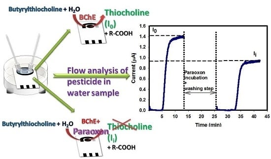
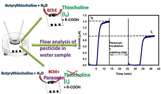
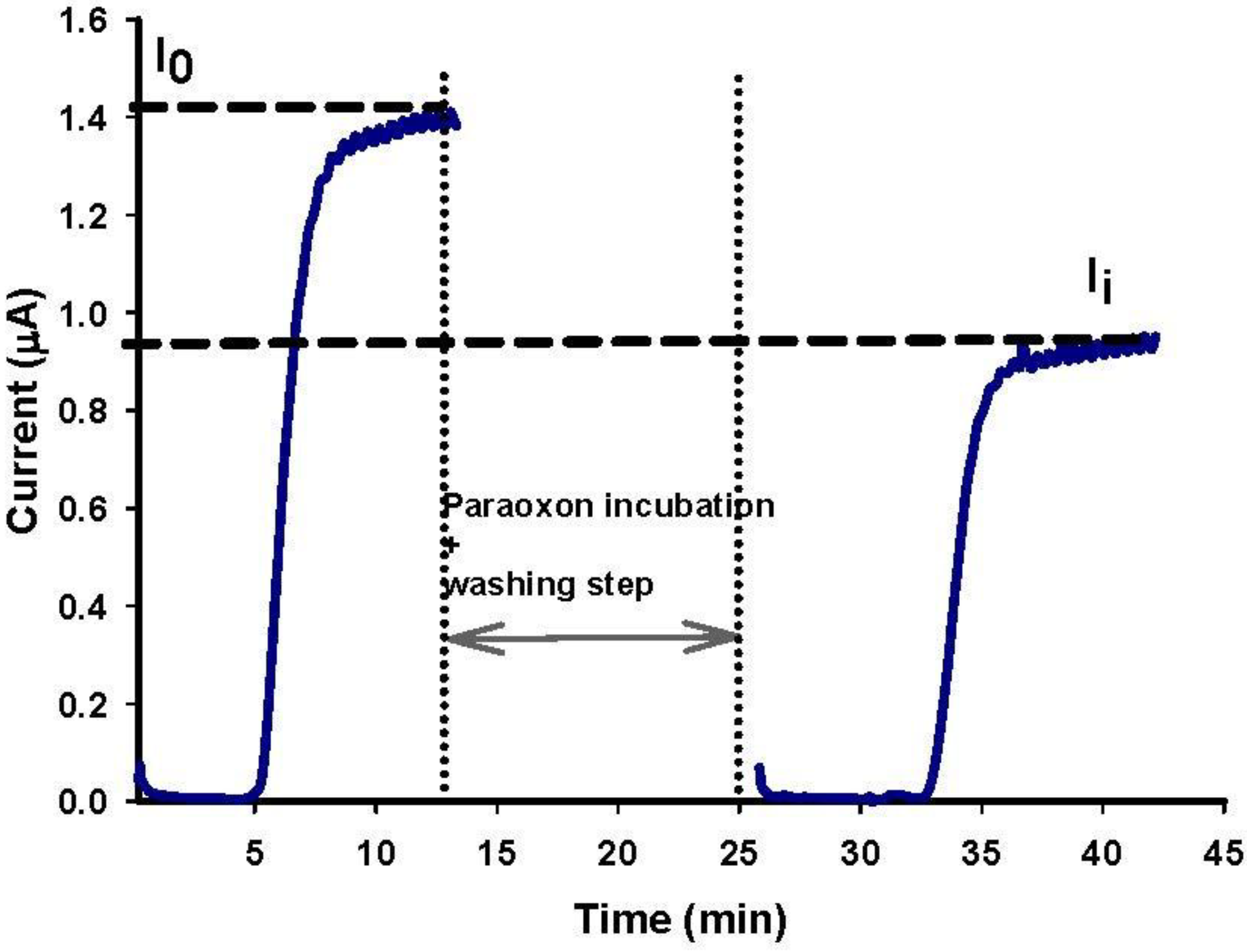
  • Enzymatic activity measurement before enzyme inhibition (I0). Applied potential +200 mV; valve V2 opened in position of SUBSTRATE (A) and valve V3 in position of SUBSTRATE (A) for a time of 10 min; flow rate 0.12 mL/min.
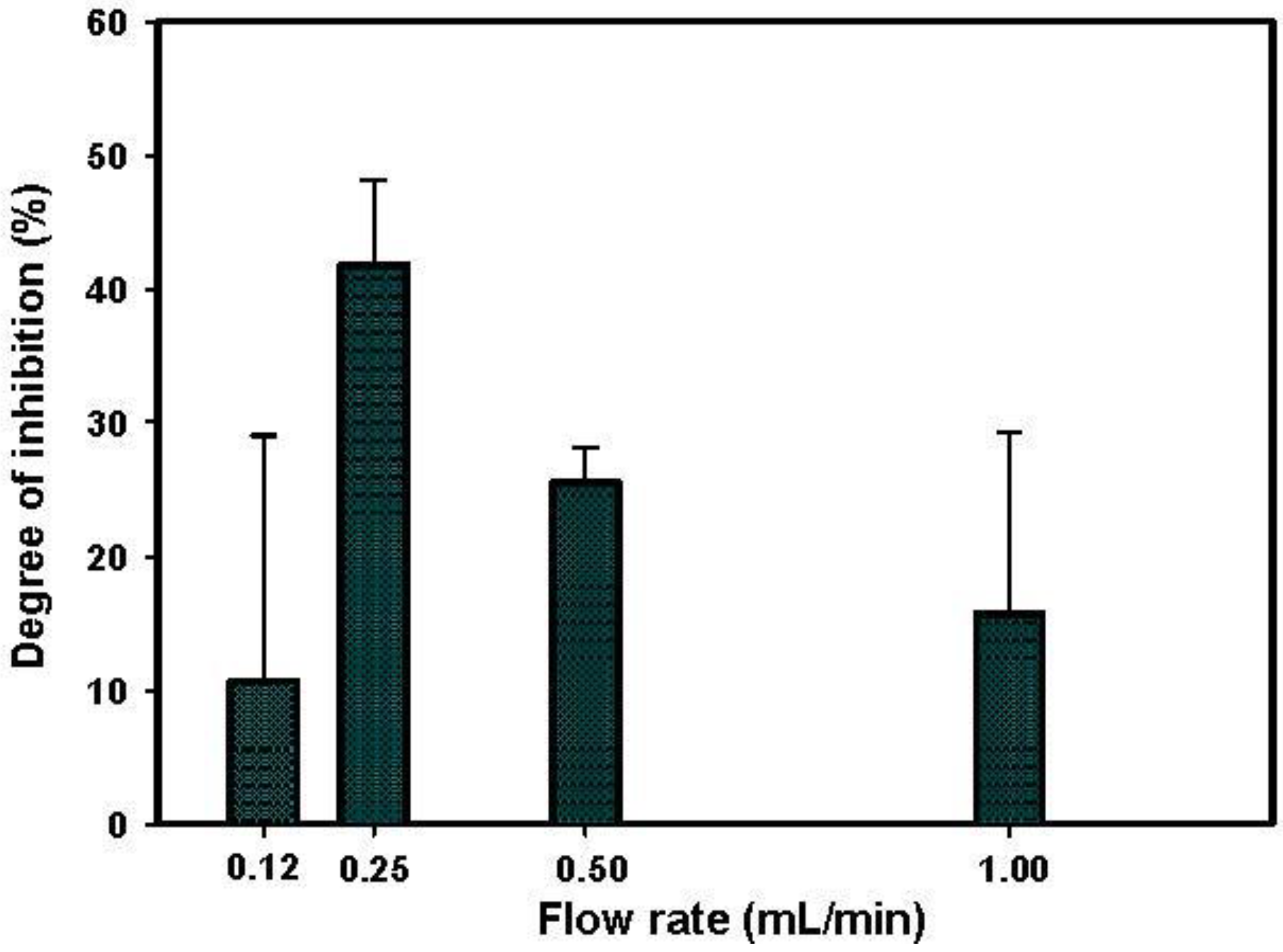
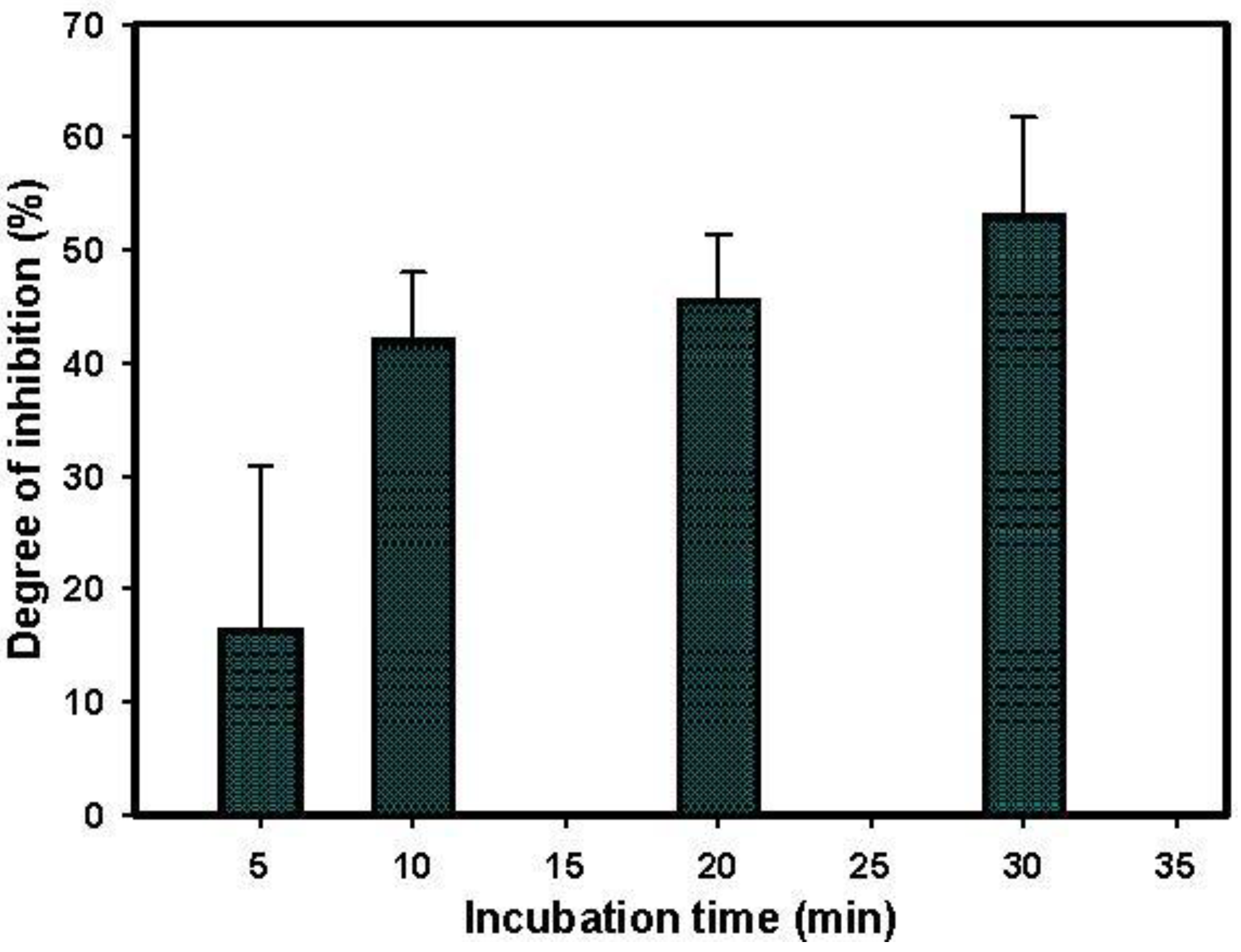
  • Inhibition step. Valves V2 and V1 opened in position of PARAOXON (C) and valve V3 in position of WASTE (I) for a time of 10 min; flow rate 0.25 mL/min. Then, valves V2 and V1 opened in the position of BUFFER (B) and valve V3 in the position of WASTE (I) for a time of 2–3 min; flow rate 0.25 mL/min.
  • Washing step. Valves V2 and V1 opened in position of BUFFER (B) and valve V3 in position of WASTE (I) for a time of 5 min; flow rate 0.12 mL/min.
  • Enzymatic activity measurement after enzyme inhibition (Ii). Applied potential +200 mV; valve V2 opened in position of SUBSTRATE (A) and valve V3 in position of SUBSTRATE (A) for a time of 10 min; flow rate 0.12 mL/min.
Figure 1. Scheme of the flow system.
Chemosensors 03 00129 g001
The resulting currents were measured as described above and the degree of inhibition was calculated as a relative decay of the biosensor response (Equation (1)):
I% = [(I0 − Ii)/I0] × 100
where Io and Ii represent the biosensor responses before and after the incubation procedure, respectively.

2.8. Safety Conditions

The stock solutions of paraoxon were prepared using appropriate safety conditions. In order to avoid contact with the powder, the operators were dressed with lab dresses, gloves, mask and glass. In addition, a dedicated fume hood was used during sample preparation and analysis.

3. Results and Discussion

The automatable amperometric biosensor embedded in a flow system for the detection of paraoxon was based on BChE enzyme immobilized by cross-linking on a SPE modified with PBNPs. In particular, the inhibitory effect of paraoxon on BChE was estimated by determining the decrease in the current obtained for the oxidation of enzymatic product thiocholine, catalyzed by the electrochemical mediator PBNPs. The biosensor was thus embedded in a flow system in order to obtain an automatable biosensing configuration to be applied for continuous monitoring of paraoxon in environmental samples. In addition, in a continuous flow-system biosensor, manual procedures are minimized and analyses can be programmed and remotely delivered, minimizing the operator intervention. In order to optimize the biosensing system, a series of parameters was evaluated as described in the following chapters.

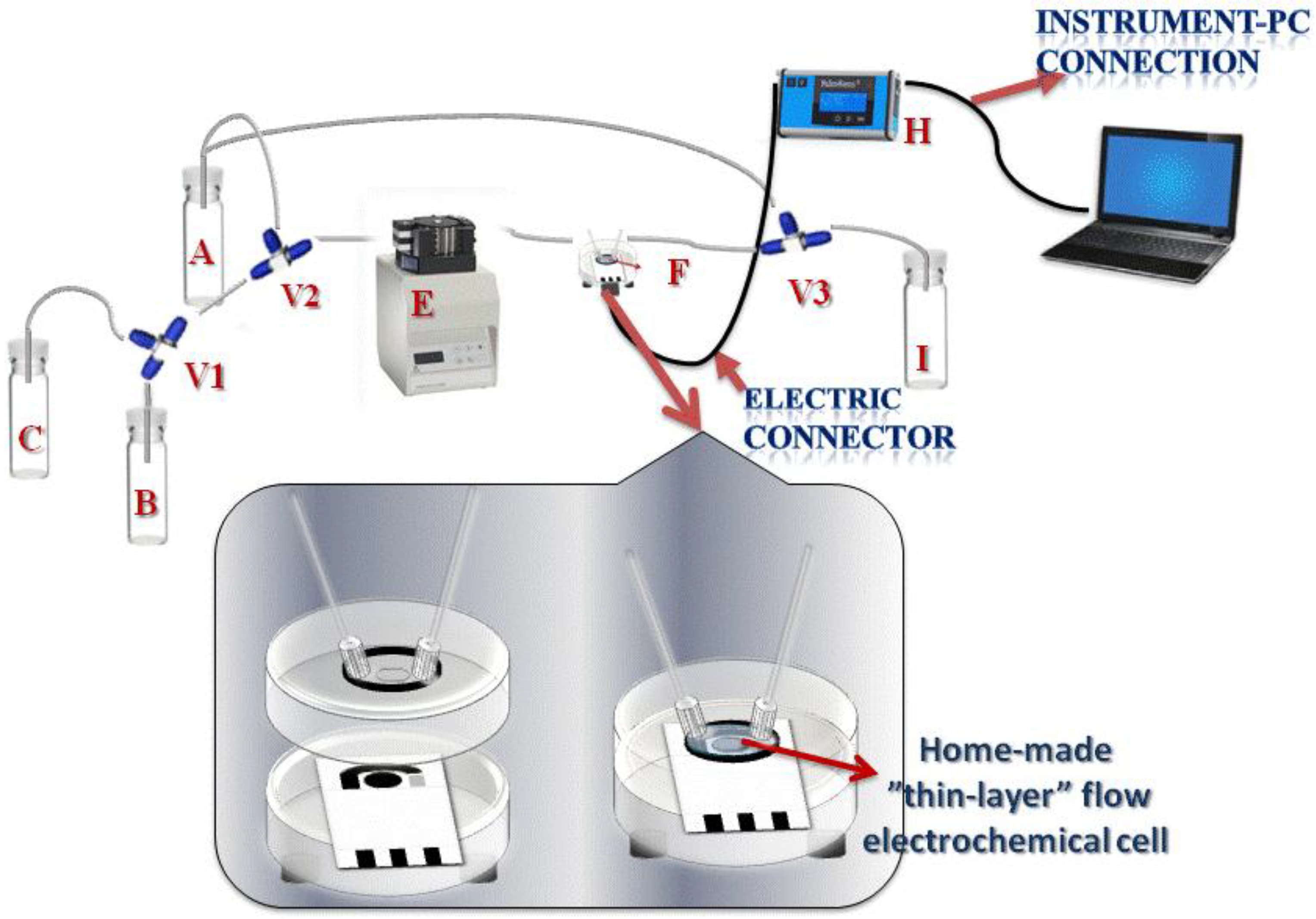
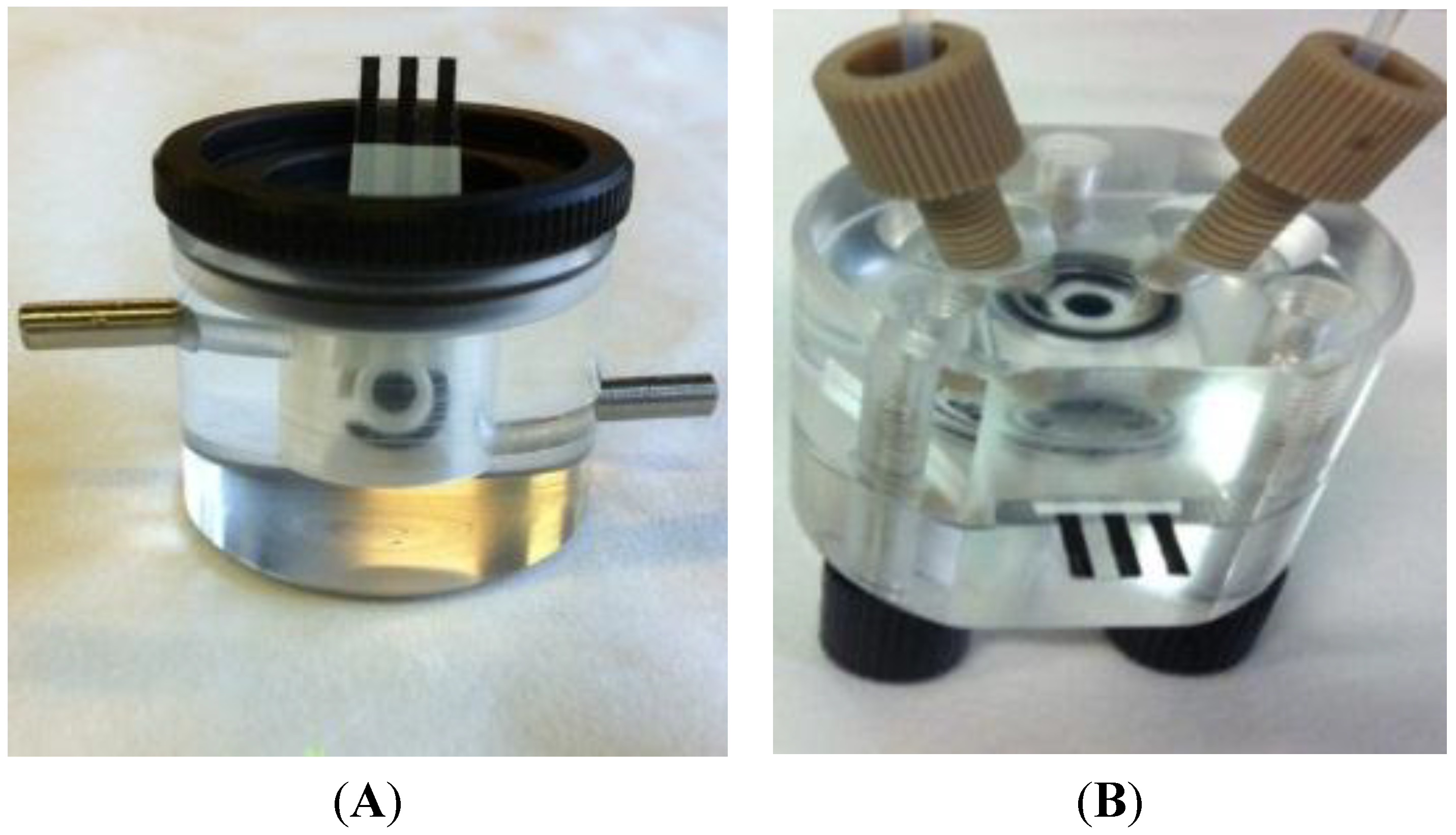
3.1. System Configuration

Amperometric measurements were conducted on PalmSens portable potentiostat, with a constant potential of +200 mV vs. Ag/AgCl, connected with an electrochemical cell. A dedicated software allowed automatic data archiving and measurement conditions set-up. The solutions were passed through the flow electrochemical cell by a peristaltic pump and the sequence of solutions was carried out manually using T valves.
The selection of the flow cells to be employed was accomplished considering a number of parameters, including the substrate consumption and the biosensor performance. First tests were performed on a “Batch-Type” cell, to verify the analysis time and the stabilization of the signal both in buffer and in substrate solutions with a speed of 1 mL/min. The resulted current signals recorded at a concentration of 5 mM butyrylthiocholine tended to decrease when compared with the single measurement of the substrate. This effect depends on the quantity of solution (approximately 800 μL) that the cell may contain, making the exchange of solutions too slow and favoring a substrate dilution during the measurement. In order to avoid this dilution effect and to limit the excessive consumption of the substrate, a different type of electrochemical cell was further considered for the analyses. A “Thin-Layer” cell was thus selected as alternative choice, having the great advantage to control the solution exchange and to avoid dilution of the substrate. In Figure 2, both “Batch-Type” cell and “Thin-Layer” were reported.
Figure 2. (A) “Batch-type” and (B) “Thin Layer” electrochemical cells.
Chemosensors 03 00129 g002

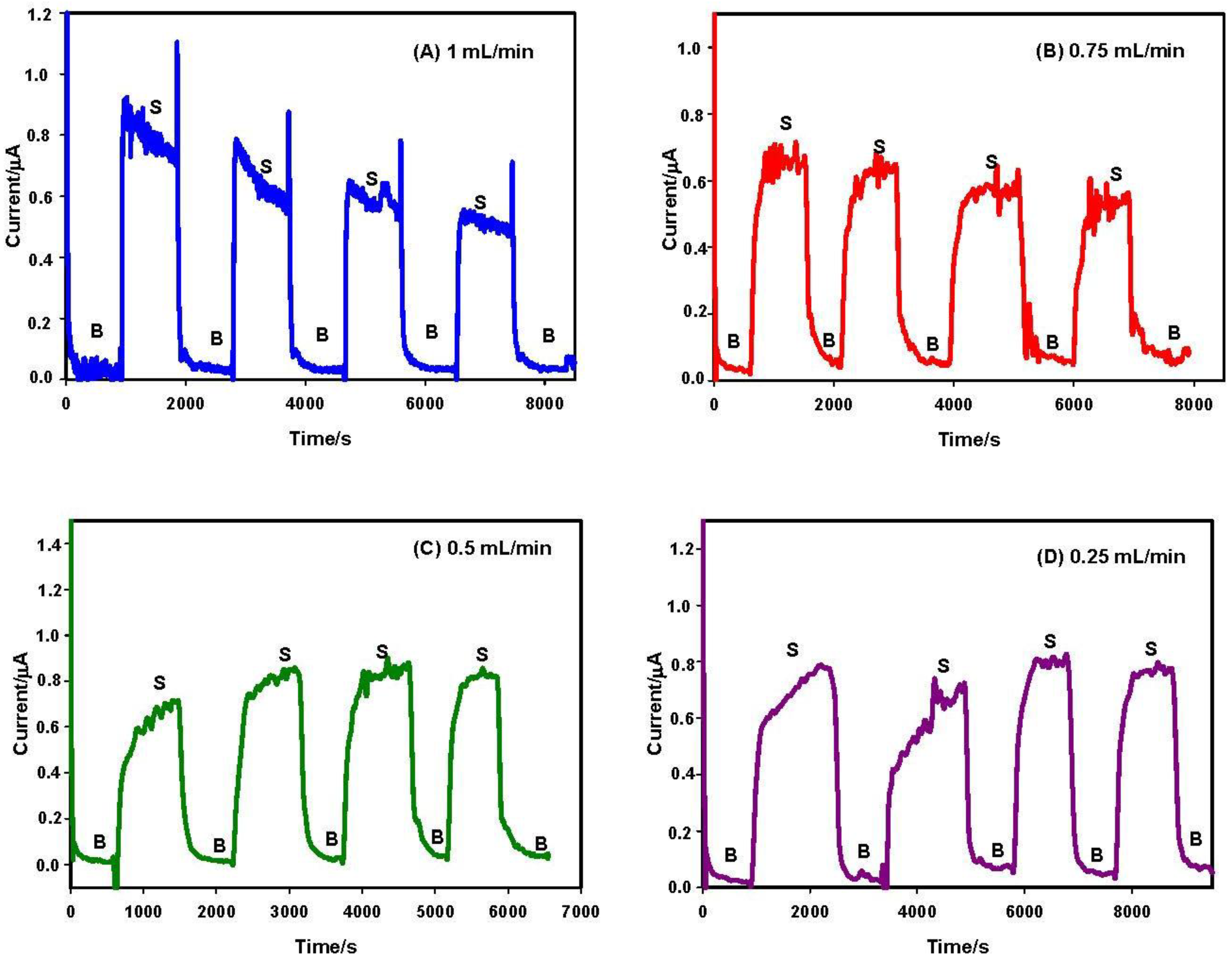
3.2. Flow Rate and Operational Stability Studies

Preliminary studies were conducted to determine the optimal flow rate of the system, by using a flow rate ranging from 1 mL/min to 0.25 mL/min, in order to obtain the optimal sensitivity and repeatability of the measurements. In the amperograms reported in Figure 3, flow rates of 1 mL/min, 0.75 mL/min, 0.5 mL/min and 0.25 mL/min were investigated. Results indicated that the current signals decrease significantly during time when relatively high values of flow rates were employed (1 mL/min and 0.75 mL/min). Moreover, a better signal/noise ratio was observed with a flow rate of 0.25 mL/min.
This behavior could be attributed to two different possible causes:
  • Stability of the enzymatic membrane. During the measurement at high flow rate, the membrane can be partially removed from the surface of the electrode. For this reason, a further test was performed measuring the enzymatic activity “in drop” with a biosensor previously tested in flow at 1 mL/min rate. We have observed (data not shown) a stable current signal, demonstrating that the enzymatic membrane steadily and actively persisted on the sensor surface.
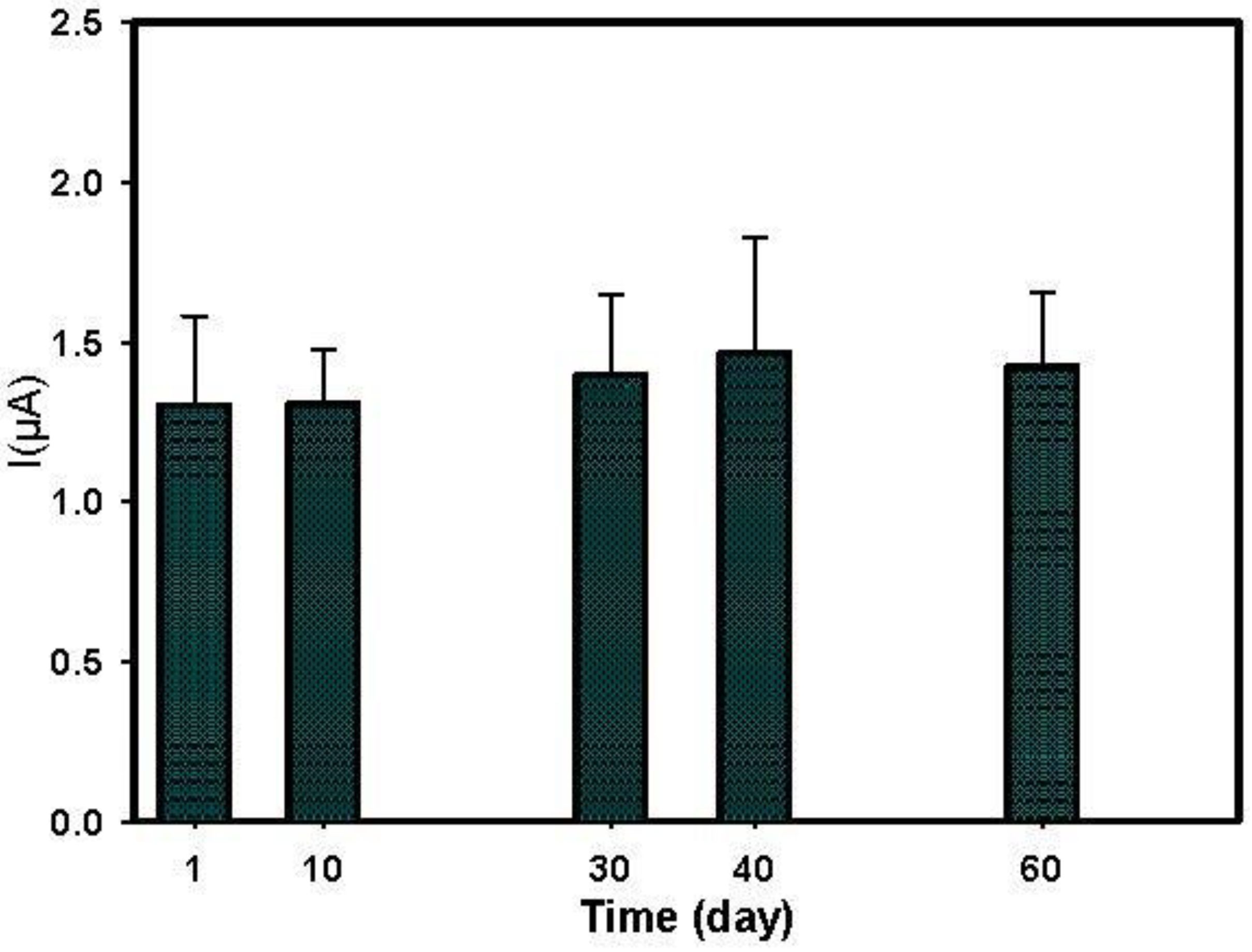
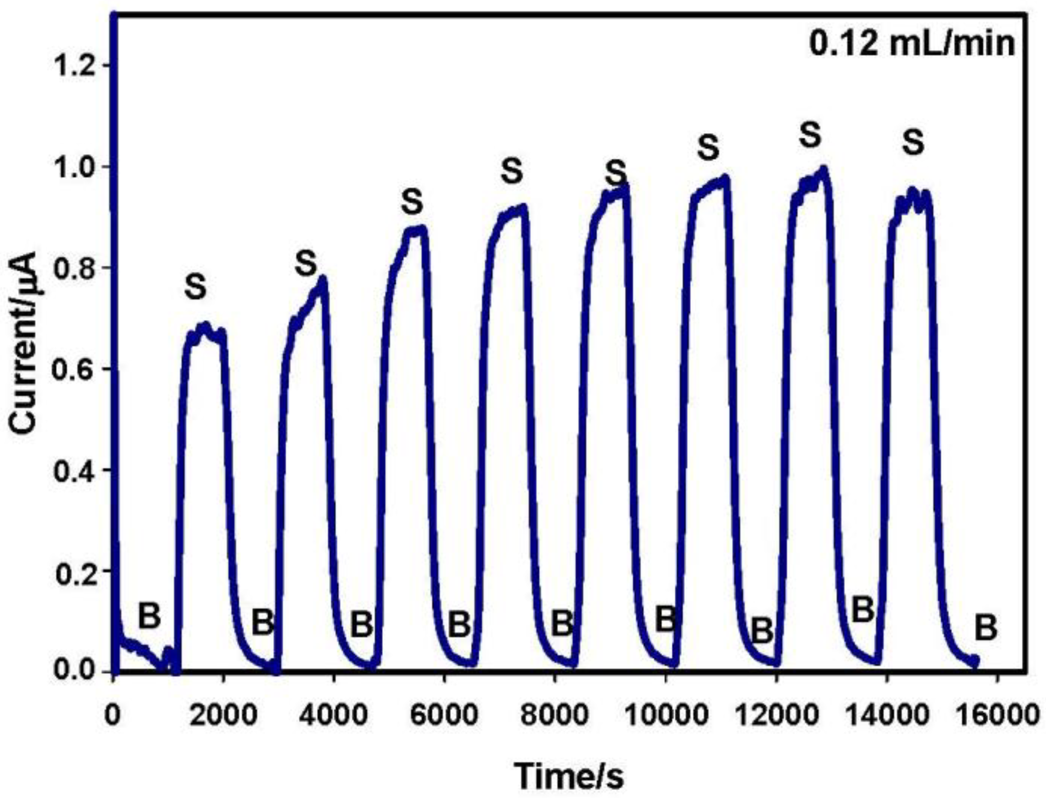
  • Onset of air bubbles. The formation of bubbles within the electrochemical cell occurred more frequently with increased flow rates. For this reason, a flow rate of 0.12 mL/min was finally selected to avoid the formation of air bubbles and to perform measurements for longer times. In Figure 4, current signals with the selected flow rate were reported. These signals were more stable but they increased during the first analyses, probably due to the increase of temperature in the laboratory that can influence the enzyme activity. To overcome this drawback, the use of thermostatic chamber to allocate the flow cell or a temperature correction should be considered. Despite these effects, the stability of the biosensor in working conditions was satisfactory for at least eight substrate measurements.
Figure 3. Flow rate study. Biosensor amperometric responses of buffer (B) and substrate (S). Flow rates: 1 mL/min (A); 0.75 mL/min (B); 0.5 mL/min (C); and 0.25 mL/min (D). Buffer (B): phosphate buffer 0.05 M + KCl 0.1 M, pH = 7.4. Substrate (S): 5 mM butyrylthiocholine in phosphate buffer 0.05 M + KCl 0.1 M, pH 7.4. Applied potential +200 mV vs. Ag/AgCl.
Chemosensors 03 00129 g003
Figure 4. Amperometric measurements with buffer (B) and substrate (S) using 0.12 mL/min as flow rate. Buffer (B): phosphate buffer 0.05 M + KCl 0.1 M, pH 7.4. Substrate (S): 5 mM butyrylthiocholine in phosphate buffer 0.05 M + KCl 0.1 M, pH 7.4. Applied potential +200 mV vs. Ag/AgCl.
Chemosensors 03 00129 g004

3.3. Intra-Electrode Repeatability

To confirm that a flow rate of 0.12 mL/min was the ideal condition, an intra-electrode repeatability study was provided recording the current responses in the presence of a solution of 5 mM butyrylthiocholine. The investigation was carried out with flow rates of 1 mL/min, 0.75 mL/min, 0.5 mL/min and 0.25 mL/min, indicating that a flow rate of 0.12 mL/min can be the best compromise to obtain a higher current intensity and a better intra-electrode repeatability with RDS of 1.2% (Figure 5).
Figure 5. Intra-electrode repeatability study. Response of 5 mM butyrylthiocholine in phosphate buffer 0.05 M + KCl 0.1 M, pH 7.4. Applied potential +200 mV vs. Ag/AgCl (n = 3, error bars expressed as standard deviation).
Chemosensors 03 00129 g005

3.4. Paraoxon Detection

The inhibition measurements were performed according to the protocol discussed in “Materials and Methods” section. The pesticide concentration was calculated in relation to the inhibition percentage according to Equation (1).
For all inhibition measurements, standard solutions of paraoxon at a concentration of 6 ppb were used, and several parameters were optimized including flow rate and incubation time. Incubation time, which means the time of contact between the enzyme and inhibitor, represents an important parameter for an irreversible inhibition to be tuned also according with the flow rate in which the inhibition occurs. Thus, the inhibition percent in current values was analyzed testing flow rates of 1 mL/min, 0.5 mL/min, 0.25 mL/min and 0.12 mL/min, at a fixed incubation time of 10 min (Figure 6). Results indicated that a flow rate of 0.25 mL/min was the best observed for highest percentage of inhibition (6 ppb paraoxon) and greater repeatability.
Figure 6. Study of the effect of flow rate during the incubation time. Paraoxon 6 ppb, butyrylthiocholine 5 mM in phosphate buffer 0.05 M + KCl 0.1 M, pH 7.4, incubation time = 10 min, applied potential +200 mV vs. Ag/AgCl (n = 3, error bars expressed as standard deviation).
Chemosensors 03 00129 g006
Taking into account that the inhibition is irreversible, and thus the inhibition percentage increases with incubation time, the effect of inhibition time was investigated. The study was carried out by varying the incubation time from 5 to 30 min and an incubation time of 10 min was finally chosen as a good compromise between a fast measure and high sensitive analysis (Figure 7).
Figure 7. Study of the effect of incubation time. Paraoxon 6 ppb, butyrylthiocholine 5 mM in phosphate buffer 0.05 M + KCl 0.1 M, pH 7.4, flow rate of 0.25 mL/min, applied potential +200 mV vs. Ag/AgCl (n = 3, error bars expressed as standard deviation).
Chemosensors 03 00129 g007
Once optimized, the above-mentioned parameters, the calibration curve for paraoxon detection in flow system was obtained. Standard solutions of paraoxon were used in a range of concentrations between 2 and 10 ppb. Measurements of each concentration were achieved in triplicate (with three different biosensors). A typical amperogram was reported in Figure 8, showing the current signals of the substrate before and after exposure to 2 ppb paraoxon standard solution. A calibration curve with a linear range between 2 and 10 ppb (y = 5.17·x + 20.7, R2 = 0.952) and a detection limit (LOD) of 1 ppb were obtained. Taking into account the use of medium exchange method, the selectivity of this analytical system relies in the detection of irreversible inhibitors like organophosphorus and carbammic pesticides avoiding the interference of reversible inhibitors (e.g., aflatoxins and heavy metals).
Figure 8. Amperogram of substrate before and after the exposure to 2 ppb paraoxon. Substrate: 5 mM butyrylthiocholine in phosphate buffer 0.05 M + KCl 0.1 M pH 7.4, flow rate 0.12 mL/min, incubation time 10 min with flow rate 0.25 mL/min, applied potential +200 mV vs. Ag/AgCl.
Chemosensors 03 00129 g008
The stability in non-operational conditions (storage stability) was also investigated, since, together with repeatability, undoubtedly it represents a key aspect to consider a biosensor as commercially attractive. The storage stability was evaluated up to 60 days in flow system using 5 mM butyrylthiocholine solution (Figure 9). The biosensor, stored in the dark and at room temperature, gave responses almost constantly after eight weeks of investigation, confirming the robustness of the immobilization procedure.
Figure 9. Study of storage stability. Response of 5 mM butyrylthiocholine in phosphate buffer 0.05 M + KCl 0.1 M pH 7.4, flow rate 0.12 mL/min, applied potential +200 mV vs. Ag/AgCl (n = 3, error bars expressed as standard deviation).
Chemosensors 03 00129 g009

3.5. Paraoxon Detection in Water Sample

The analytical system was challenged in undiluted tap water, Tiber River and Albano lake water samples. A matrix effect was observed in all water samples tested. To overcome this effect, thus avoiding pesticide overestimation, each sample was diluted 1/2 (v/v) and 1/4 (v/v) with the working buffer. Results showed that the matrix effect was negligible with a dilution of 1/4 (v/v) for lake and river waters, and 1/2 (v/v) for tap water. In order to standardize the protocol, a dilution of 1/4 (v/v) was performed for all tested samples.
The accuracy of the method was performed with samples spiked with 25 ppb paraoxon, a level lower than the legal limit of 100 ppb for surface wastewaters [31]. The results were reported in Table 1, demonstrating that this device is suitable as an alarm system to detect polluted samples. Furthermore, when compared with the ones reported in literature (see Table 2), this biosensor embedded in a flow system showed satisfactory sensitivity with an improved storage stability, taking into account its storage in dry conditions at room temperature.
Table 1. Recovery study on real samples from Tap, Albano Lake and Tiber River water samples.
SampleParaoxon Added (ppb)Paraoxon Found ± σ (ppb)Recovery ± σ (%)
Tap water2522.5 ± 1.290 ± 5
Albano Lake2528.8 ± 3.1115 ± 12
Tiber River2529.8 ± 2.3119 ± 9
Table 2. Different electrochemical biosensors embedded in flow system for paraoxon detection.
Biosensor TypeApplied PotentialFlow Analysis TypeAnalyteLinear Range (M)Detection Limit (M)Storage ConditionSamplesReference
Genetically-modified AChE immobilized on CoPc-SPE+100 mVAutomated flow system using a modular syringe pumpParaoxon5 × 107–3 × 1087.5 × 109The biosensor retained full enzymatic activity after storage for up to 1 month in PBS at 4 °CMilk[22]
AChE immobilized on platinum electrode modified with MWCNTs+630 mVFlow-injection systemParaoxon1 × 10−11–1 × 10−89 × 10−13The biosensor retained 80% of enzymatic activity after storage for 30 days in PBS at 4 °C-[21]
AChE immobilized on glassy carbon electrode modified with MWCNTs+150 mVFlow-injection system (stop and flow)Paraoxon1 × 10−12–1 × 10−84 × 10−13The biosensor retained 94% of enzymatic activity after storage for 1 month in PBS at 4 °C-[32]
BChE immobilized on PBNPs-SPE+200 mVFlow system using T valves (continuous flow)Paraoxon7 × 109–4 × 1084 × 109The biosensor retained full enzymatic activity after storage for 60 days dry at RTTap, lake and river water[this work]
AChE = acetylcholinesterase, CoPc-SPE = screen-printed electrode modified with cobalt phthalocyanine, MWCTNs = multi-walled carbon nanotubes, BChE = butyrylcholinesterase PBNPs-SPE = screen-printed electrode modified with Prussian Blue nanoparticles, RT= room temperature.

4. Conclusions

We reported the development of an electrochemical biosensor based on BChE enzyme immobilized on SPEs modified with PBNPs, integrated in an embedded system for continuous flow measurements of organophosphorus pesticides. The use of PBNPs allowed the detection of the enzymatic product, thiocholine, at low applied potential. Several parameters were investigated in order to optimize the flow biosensor system, including different types of electrochemical cells, flow rate during the enzymatic measurement and incubation time. In optimized conditions, we have observed a good working stability for substrate measurements. Storage stability lasted up to 60 days at room temperature in dry conditions, demonstrating an excellent storage stability and making this system highly attractive for commercial use. Furthermore, the analytical system was characterized by satisfactory analytical performances in the detection of organophosphate tested (paraoxon) reaching a linear range of concentration between 2 and 10 ppb with a detection limit (LOD) of 1 ppb in standard solutions. This system was also challenged in drinking, river and lake water samples with satisfactory recovery values using a dilution step of 1:4 v/v. The advantage of this integrated system is the possibility to measure irreversible inhibitors of cholinesterase enzyme (i.e., organophosphorus and carbammic pesticides) in samples without extended pre-treatments and to automate the analyses reducing costs and time. This analytical system can be used as an alarm system for this class of compounds, followed, in the case of alarm, by the HPLC or GC-MS analyses to detect exactly the pesticides present in the sample.

Acknowledgments

This work was supported by National Industria 2015 (MI01 00223) ACQUA-SENSE project.

Author Contributions

All the authors were involved in conceiving the method, designing and performing the experiments, analyzing data and writing the paper.

Conflicts of Interest

The authors declare no conflict of interest.

References

  1. Bajgar, J. Organophosphates/nerve agent poisoning: Mechanism of action, diagnosis, prophylaxis and treatment. In Advances in Clinical Chemistry; Makowsky, G.M., Ed.; Elsevier Academic Press: San Diego, CA, USA, 2004; Volume 38, pp. 151–216. [Google Scholar]
  2. Harel, M.; Sonoda, L.K.; Silman, I.; Sussman, J.L.; Rosenberry, T.L. Crystal structure of thioflavin T bound to the peripheral site of Torpedo californica acetylcholinesterase reveals how thioflavin T acts as a sensitive fluorescent reporter of ligand binding to the acylation site. J. Am. Chem. Soc. 2008, 130, 7856–7861. [Google Scholar] [CrossRef] [PubMed]
  3. Millard, C.B.; Koellner, G.; Ordentlich, A.; Shafferman, A.; Silman, I.; Sussman, J.L. Reaction products of acetylcholinesterase and VX reveal a mobile histidine in the catalytic triad. J. Am. Chem. Soc. 1999, 121, 9883–9884. [Google Scholar] [CrossRef]
  4. Sussman, J.L.; Harel, M.; Frolow, F.; Oefner, C.; Goldman, A.; Toker, L.; Silman, I. Atomic structure of acetylcholinesterase from Torpedo californica: A prototypic acetylcholine-binding protein. Science 1991, 253, 872–879. [Google Scholar] [CrossRef] [PubMed]
  5. Bajgar, J.; Fusek, J.; Kuca, K.; Bartosova, L.; Jun, D. Treatment of organophosphate intoxication using cholinesterase reactivators: Facts and fiction. Mini Rev. Med. Chem. 2007, 7, 461–466. [Google Scholar] [CrossRef] [PubMed]
  6. Bergh, C.; Torgrip, R.; Ostman, C. Simultaneous selective detection of organophosphate and phthalate esters using gas chromatography with positive ion chemical ionization tandem mass spectrometry and its application to indoor air and dust. Rapid Commun. Mass Spectrom. 2010, 24, 2859–2867. [Google Scholar] [CrossRef] [PubMed]
  7. De Freitas Ventura, F.; de Oliveira, J., Jr.; dos Reis Pedreira Filho, W.; Gerardo Ribeiro, M. GC-MS quantification of organophosphorous pesticides extracted from XAD-2 sorbent tube and foam patch matrices. Anal. Methods 2012, 4, 3666–3673. [Google Scholar] [CrossRef]
  8. Arduini, F.; Amine, A.; Moscone, D.; Palleschi, G. Biosensors based on cholinesterase inhibition for insecticides, nerve agents and aflatoxin B1 detection. Microchim. Acta 2010, 170, 193–214. [Google Scholar] [CrossRef] [Green Version]
  9. Andreescu, S.; Marty, J.L. Twenty years research in cholinesterase biosensors: From basic research to practical applications. Biomol. Eng. 2006, 23, 1–15. [Google Scholar] [CrossRef] [PubMed]
  10. Pohanka, M.; Musilek, K.; Kuca, K. Progress of biosensors based on cholinesterase inhibition. Curr. Med. Chem. 2009, 16, 1790–1798. [Google Scholar] [CrossRef] [PubMed]
  11. Periasamy, A.P.; Umasankar, Y.; Chen, S.M. Nanomaterials—Acetylcholinesterase Enzyme Matrices for Organophosphorus Pesticides Electrochemical Sensors: A Review. Sensors 2009, 9, 4034–4055. [Google Scholar] [CrossRef] [PubMed]
  12. Zhang, W.; Asiri, A.M.; Liu, D.; Du, D.; Lin, Y. Nanomaterial-based biosensors for environmental and biological monitoring of organophosphorus pesticides and nerve agents. Trends Anal. Chem. 2014, 54, 1–10. [Google Scholar] [CrossRef]
  13. Arduini, F.; Guidone, S.; Amine, A.; Palleschi, G.; Moscone, D. Acetylcholinesterase biosensor based on self-assembled monolayer-modified gold-screen printed electrodes for organophosphorus insecticide detection. Sens. Actuators B Chem. 2013, 179, 201–208. [Google Scholar] [CrossRef]
  14. Arduini, F.; Forchielli, M.; Amine, A.; Neagu, D.; Cacciotti, I.; Nanni, F.; Moscone, D.; Palleschi, G. Screen-printed biosensor modified with carbon black nanoparticles for the determination of paraoxon based on the inhibition of butyrylcholinesterase. Microchim. Acta 2015, 182, 643–651. [Google Scholar] [CrossRef]
  15. Arduini, F.; Ricci, F.; Tuta, C.S.; Moscone, D.; Amine, A.; Palleschi, G. Detection of carbamic and organophosphorous pesticides in water samples using a cholinesterase biosensor based on Prussian Blue-modified screen-printed electrode. Anal. Chim. Acta 2006, 580, 155–162. [Google Scholar] [CrossRef] [PubMed]
  16. Alonso, G.A.; Istamboulie, G.; Noguer, T.; Marty, J.L.; Muñoz, R. Rapid determination of pesticide mixtures using disposable biosensors based on genetically modified enzymes and artificial neural networks. Sens. Actuators B Chem. 2012, 164, 22–28. [Google Scholar] [CrossRef]
  17. Ivanov, A.N.; Younusov, R.R.; Evtugyn, G.A.; Arduini, F.; Moscone, D.; Palleschi, G. Acetylcholinesterase biosensor based on single-walled carbon nanotubes—Co phtalocyanine for organophosphorus pesticides detection. Talanta 2011, 85, 216–221. [Google Scholar] [CrossRef] [PubMed]
  18. Oujji, N.B.; Bakas, I.; Istamboulié, G.; Ait-Ichou, I.; Ait-Addi, E.; Rouillon, R.; Noguer, T. Sol-gel immobilization of acetylcholinesterase for the determination of organophosphate pesticides in olive oil with biosensors. Food Control 2013, 30, 657–661. [Google Scholar] [CrossRef]
  19. Di Tuoro, D.; Portaccio, M.; Lepore, M.; Arduini, F.; Moscone, D.; Bencivenga, U.; Mita, D.G. An acetylcholinesterase biosensor for determination of low concentrations of Paraoxon and Dichlorvos. New Biotechnol. 2011, 29, 132–138. [Google Scholar] [CrossRef] [Green Version]
  20. Liang, H.; Song, D.; Gong, J. Signal-on electrochemiluminescence of biofunctional CdTe quantum dots for biosensing of organophosphate pesticides. Biosens. Bioelectron. 2014, 53, 363–369. [Google Scholar] [CrossRef] [PubMed]
  21. Ivanov, Y.; Marinov, I.; Portaccio, M.; Lepore, M.; Mita, D.G.; Godjevargova, T. Flow-injection system with site-specific immobilization of acetylcholinesterase biosensor for amperometric detection of organophosphate pesticides. Biotechnol. Biotechnol. Equip. 2012, 26, 3044–3053. [Google Scholar] [CrossRef]
  22. Mishra, R.K.; Dominguez, R.B.; Bhand, S.; Muñoz, R.; Marty, J.L. A novel automated flow-based biosensor for the determination of organophosphate pesticides in milk. Biosens. Bioelectron. 2012, 32, 56–61. [Google Scholar] [CrossRef] [PubMed]
  23. Gong, J.; Guan, Z.; Song, D. Biosensor based on acetylcholinesterase immobilized onto layered double hydroxides for flow injection/amperometric detection of organophosphate pesticides. Biosens. Bioelectron. 2013, 39, 320–323. [Google Scholar] [CrossRef] [PubMed]
  24. Mishra, R.K.; Alonso, G.A.; Istamboulie, G.; Bhand, S.; Marty, J.L. Automated flow based biosensor for quantification of binary organophosphates mixture in milk using artificial neural network. Sens. Actuators B Chem. 2015, 208, 228–237. [Google Scholar] [CrossRef]
  25. Dominguez, R.B.; Alonso, G.A.; Muñoz, R.; Hayat, A.; Marty, J.L. Design of a novel magnetic particles based electrochemical biosensor for organophosphate insecticide detection in flow injection analysis. Sens. Actuators B Chem. 2015, 208, 491–496. [Google Scholar] [CrossRef]
  26. Alonso, G.A.; Dominguez, R.B.; Marty, J.L.; Muñoz, R. An Approach to an Inhibition Electronic Tongue to Detect On-Line Organophosphorus Insecticides Using a Computer Controlled Multi-Commuted Flow System. Sensors 2011, 11, 3791–3802. [Google Scholar] [CrossRef] [PubMed]
  27. Mascini, M.; Mazzei, F. Amperometric sensor for pyruvate with immobilized pyruvate oxidase. Anal. Chim. Acta 1987, 192, 9–16. [Google Scholar] [CrossRef]
  28. Arduini, F.; Neagu, D.; Dall’Oglio, S.; Moscone, D.; Palleschi, G. Towards a Portable Prototype Based on Electrochemical Cholinesterase Biosensor to be Assembled to Soldier Overall of Nerve Agent Detection. Electroanalysis 2012, 24, 581–590. [Google Scholar] [CrossRef]
  29. Cinti, S.; Arduini, F.; Vellucci, G.; Cacciottti, I.; Nanni, F.; Moscone, D. Carbon black assisted tailoring of Prussian Blue nanoparticles to tune sensitivity and detection limit towards H2O2 by using screen-printed electrode. Electrochem. Commun. 2014, 47, 63–66. [Google Scholar] [CrossRef]
  30. Moscone, D.; D’Ottavi, D.; Compagnone, D.; Palleschi, G.; Amine, A. Construction and Analytical Characterization of Prussian Blue-Based Carbon Paste Electrodes and Their Assembly as Oxidase Enzyme Sensors. Anal. Chem. 2001, 73, 2529–2535. [Google Scholar] [CrossRef] [PubMed] [Green Version]
  31. DL 152/2006, 3 April 2006 “Norme in Materia Ambientale”. Available online: http://www.gazzettaufficiale.it/eli/id/2006/04/14/006G0171/sg (accessed on 24 April 2015).
  32. Liu, G.; Lin, Y. Biosensor Based on Self-Assembling Acetylcholinesterase on Carbon Nanotubes for Flow Injection/Amperometric Detection of Organophosphate Pesticides and Nerve Agents. Anal. Chem. 2006, 78, 835–843. [Google Scholar] [CrossRef] [PubMed]

