Article

Fluorescent Assay for Salmonella Detection Based on Triangle Multivalent Aptamer-Initiated Catalytic Hairpin Assembly

1
College of Plant Protection, Nanjing Agricultural University, Nanjing 210090, China
2
Greentown Agricultural Science Testing Technology Co., Ltd., Hangzhou 310050, China
3
College of Food and Engineering, Ningbo University, Ningbo 315800, China
*
Authors to whom correspondence should be addressed.
Chemosensors 2025, 13(9), 334; https://doi.org/10.3390/chemosensors13090334
Submission received: 2 July 2025 / Revised: 20 August 2025 / Accepted: 1 September 2025 / Published: 4 September 2025
(This article belongs to the Special Issue Advanced Material-Based Fluorescent Sensors)

Abstract

Salmonella poses a severe global threat to food safety and public health, necessitating rapid, sensitive, and reliable detection methods. Conventional techniques often suffer from complexity, time consumption, cost, or limited sensitivity. To address this, we developed a novel enzyme-free fluorescence detection platform, termed the MTAI-CHA system, integrating magnetic nanoparticle-based triangle multivalent aptamer-initiators (MTAI) with catalytic hairpin assembly (CHA) signal amplification. The triangular DNA nanostructure contained significantly enhanced binding affinity of multivalent aptamers, increasing the sensitivity compared to monovalent aptamers. The optimized MTAI-CHA system demonstrated exceptional performance: a low detection limit of 10 CFU/mL and excellent specificity against non-target pathogens. This sensitive, specific, and robust strategy, leveraging multivalent aptamer recognition and enzyme-free signal amplification, holds significant potential for rapid pathogen screening in food safety, clinical diagnostics, and environmental monitoring, with adaptability to other targets via aptamer substitution.

1. Introduction

Salmonella, a ubiquitous but invisible foodborne pathogen in the daily food supply chain, poses significant threats to global public health. Its pathogenicity must be considered, as consuming Salmonella-contaminated food without precaution can unfortunately lead to a range of symptoms, including gastroenteritis, sepsis, and typhoid fever [1,2]. Annually, Salmonella causes millions of illnesses worldwide, and in severe cases, it can lead to hospitalization or even death [3]. Compounding the challenge, Salmonella exhibits antimicrobial resistance [4] and is difficult to visually distinguish in food. Consequently, developing efficient and reliable detection methods is critical for ensuring food safety. The conventional methods for identifying Salmonella must go through processes such as enrichment culture, selective isolation, observation of morphological characteristics, physiological and biochemical reactions, and final serological identification, which are complex, time-consuming, and labor-intensive. Alternative immunoassays require costly antibodies [5]. Molecular biology methods such as PCR [6] and LAMP [7], while faster, necessitate DNA extraction and amplification, making them susceptible to aerosol contamination and prone to false-positive or false-negative results. Therefore, there is an urgent need for sensitive, rapid, and simple Salmonella detection methods.
Aptamers represent a viable antibody alternative, widely employed in biomarker discovery due to their specificity, affordability, remarkable stability, ease of synthesis, and chemical versatility [8,9]. Aptamers as single-stranded nucleic acid oligomers often undergo significant structural changes after binding onto the target and enhance aptasensor-switching activity after hybridization to the aptamer’s complementary DNA strand [10]. The aptamer-complementary DNA functions as an aptamer blocker, while the aptamer itself serves as the ligand binder. Upon ligand binding, the aptamer undergoes dehybridization, releasing the free complementary DNA. Then this released DNA is coupled with downstream detection applications, such as fluorescence [11], electrochemistry [12], and signaling cascades [13]. However, aptamers still face challenges in detecting bacteria due to the reduced binding affinity leading to the decreased ligand dependence and fewer free complementary DNA. In recent years, multivalent aptamers have emerged as a focal point of research owing to their ability to engage in multivalent interactions. These interactions, in contrast to monovalent interactions, can significantly enhance the effective concentration of the ligand at the target site, thereby strengthening binding affinity and contributing to improved selectivity in target recognition [14,15]. And to improve avidity owing to increased local aptamer density and facilitating synergistic binding, multivalent aptamers have been modified on DNA nanostructures [16]. Nowadays, multivalent binding has been popularly adopted for enhancing the affinity of aptamers in various fields, such as bacteria detection [17], medical imaging [18], and drug delivery [19]. For example, Qiao et al. [20] successfully constructed a tetrahedron multivalent aptamer DNA nanostructure, resulting in a 3.5-fold increase in binding affinity compared to monovalent aptamers. Thus, DNA nanostructure-based multivalent aptamer-complementary DNA shows great potential in binding affinity improved in downstream applications.
In recent years, signal amplification has emerged as a dominant strategy for addressing sensitivity limitations in detection systems. Catalytic hairpin assembly (CHA) is an enzyme-free, isothermal signal-amplification technique for analyte detection based on DNA circuits. DNA circuits utilize the negative free energy change inherent in base pairing to generate amplified duplex DNA from two complementary nucleic acid hairpins [21,22]. Owing to attributes like simple design, high turnover rates and low background signal, CHA finds broad application in detecting trace levels of diverse analytes, particularly nucleic acids [23] and proteins [24]. However, CHA systems operating independently often achieve insufficient signal amplification and sensitivity for low-abundance targets. To enhance performance, cascade amplification approaches that combine CHA with complementary methods, such as DNA nanostructure scaffolds, have emerged, facilitating the creation of highly sensitive detection platforms [25,26].
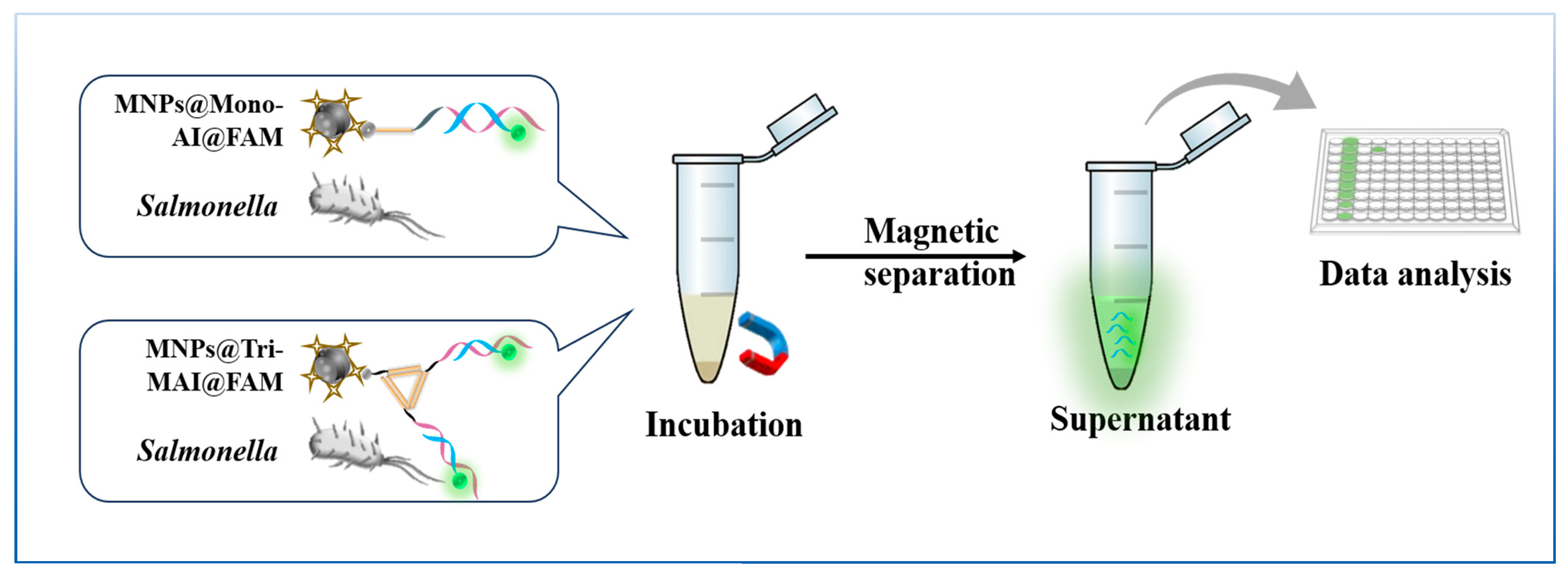
Herein, a sensitive method based on magnetic nanoparticles-triangle multivalent aptamer-initiator (MTAI) and CHA amplification for Salmonella detection was developed, where the detection platform is termed as a MTAI-CHA system. The principle of the detection strategy is shown in Scheme 1. We synthesized a triangular multivalent aptamer-initiator (Tri-MAI), which has two sides modifying the Salmonella aptamer [27] (Scheme 1. pink DNA) partially blocked with an initiator (Scheme 1. blue DNA) and the other side modifying a link DNA (Scheme 1. yellow DNA). The linker DNA embedded Tri-MAI binds to Streptavidin-coated magnetic nanoparticles (SA-MNPs) to produce MTAI via biotin-streptavidin interaction. Upon introduction of Salmonella, the bacteria and the initiator competed for aptamer binding sites. Due to the superior affinity of Salmonella for the aptamer, the initiator was displaced. Subsequent magnetic separation then isolated the released initiator within the supernatant. The free initiators in supernatant were used to trigger the CHA circuit. Two hairpins H1 (labeled with a FAM fluorophore and BHQ1 quencher) and H2 were involved in the CHA circuit. Initiators can hybridize with H1, leading to the successive hybridization reaction between H1 and H2. Then the FAM fluorophore on H1 was recovered and then released the initiator to trigger the next CHA circuit to generate numerous H1-H2 duplexes. The H1-H2 duplexes achieved intense fluorescence signal owing to the CHA amplification. Therefore, by coupling MTAI with CHA, a convenient, and sensitive method for the detection of Salmonella was achieved.

2. Materials and Methods

2.1. Materials and Apparatus

Phosphate-buffered saline (PBS), TAE buffer, agarose, and 4S GelRed, were sourced from Shanghai Sangon Biotechnology Co., Ltd. (Shanghai, China). Luria Broth (LB) was acquired from Hangzhou Microbial Reagent Co., Ltd. (Hangzhou, China). Brain Heart Infusion Broth (BHI) was sourced from Qingdao Hope Bio-Technology Co., Ltd. (Qingdao, China). 1×TMS buffer was composed of 10 mM Tris, 80 mM MgCl2. The pH of the 1×TMS buffer was adjusted to 7.5 using glacial acetic acid. Fluorescence was measured under monochromatic blue light excitation (460–485 nm). The UV-Vis and fluorescence data were acquired using a SpectraMax i3 multimode microplate reader (Molecular Devices, San Jose, CA, USA).

2.2. Bacterial Culture

The Salmonella typhimurium (ATCC 14028) as target bacteria was cultivated in BHI liquid culture medium, while the Salmonella enteritidis (CICC 10982), E. coli O157:H7 (ATCC 43889), Vibrio parahaemolyticus (ATCC 17802), S. aureus (ATCC 25923), Listeria monocytogenes (CICC 21540), Vibrio parahaemolyticus (CICC 21617), Vibrio alginolyticus (ATCC 17749), and Vibrio vulnificus (ATCC 27562) as non-target bacteria were cultured with LB liquid culture medium at 37 °C for 20 h in a shaking incubator. The bacterial cultures were washed thrice by centrifugation at 6000 rpm for 8 min, followed by serial dilution in sterile PBS to achieve concentrations ranging from 10 to 107 CFU/mL. For Salmonella quantification, 200 mL of selected dilutions were spread-plated on Xylose-Lysine-Desoxycholate (XLD) agar plates. And then, they were incubated at 37 °C for 24 h. Colonies on the plates were recorded to calculate viable cell concentrations in terms of colony forming units per milliliter (CFU/mL). Finally, the obtained bacteria were subsequently inactivated by boiling for 5 min and refrigerated at 4 °C for storage.

2.3. Preparation of Hairpin Probes

In our design, the initiator not only participates in the CHA reaction but also is complementary to partial sequences of the aptamer. NUPACK software (NUPACK 4.0.) was used to develop and test all DNA oligonucleotides, and Sangon Co., Ltd. (China) provided HPLC-purified DNA oligonucleotides. The initiator and two hairpin probes (H1, H2. The 5′ end of hairpin H1 was marked with FAM, and the 3′ end was marked with BHQ1.) dissolved in PBS were denatured at 95 °C for 10 min and then gradually cooled to room temperature over a 4 h period. All annealed probes were stored at 4 °C for further use.

2.4. Gel Electrophoresis and Fluorescence Analysis

The result of CHA signal amplification was demonstrated with agarose gel electrophoresis under optimized experimental conditions. The trigger H1 and H2 were mixed according to experimental needs. The CHA reactions were performed at room temperature for 30 min. 1 μL loading buffer was added into 5 μL of initiator (0.5 μM), 5 μL of H1 (1 μM), 5 μL of H2 (1.5 μM), 5 μL H1/H2, 5 μL H1/H2/initiator, and DNA marker, respectively. The mixture was loaded onto agarose gel and ran in 1× tris-acetate-EDTA (TAE) buffer at 130 V for 30 min. The results of gel electrophoresis were developed under Bio-Rad Gel Doc XR+ system (Bio-Rad, Richmond, CA, USA) instrument.

2.5. Modification and Detection of Monovalent Aptamer-Initiator (Mono-AI) on the Surface of Streptavidin Magnetic Beads (SA-MNPs)

First, 10 μM Salmonella aptamers and 15 μM initiator were mixed and prehybridized at 95 °C for 5 min and gradually cooled down to form AI. Second, 4 μL of the 10 mg/mL SA- MNPs w washed with 50 μL of PBS and then resuspended in 25 μL Mono-AI mixture and incubated for 0.5 h at room temperature. Then the supernatant with excess Mono-AI was discarded through magnetic separation and washed three times with 200 μL of PBS. After that, added 25 μL of the pre-treated Salmonella solution 106 to the Mon-AI modified magnetic beads and incubate at room temperature for 1 h. Finally, the supernatant containing initiator were separated. 1 μM H1 and 1.5 μM H2 were added into the supernatant incubated at room temperature for 1 h to perform the CHA reaction. The generated fluorescent signals were recorded from 510 nm to 550 nm.

2.6. Modification and Detection of Triangle Multivalent Aptamer-Initiator (Tri-MAI) on the Surface of Streptavidin Magnetic Beads (SA-MNPs)

First, we synthesized a triangular planar multivalent aptamer-initiator, and all the DNA sequences were dissolved in 1×TMS buffer to a final concentration of 50 μM before operation. Four equal amounts of DNA sequences with a final concentration of 1 μM and an initiator with a final concentration of 4.5 μM were added in 1×TMS buffer. The solution (25 μL) was heated to 95 °C for 4 min and rapidly cooled to 4 °C, and the solution was kept at 4 °C for 1 min to fabricate Tri-MAI. The free strands were removed by ultrafiltration to obtain the pure Tri-MAI. Then, the way of the Tri-MAI binds to the magnetic beads is consistent with the above monovalent, and the method for subsequent detection of bacteria is also consistent. All the Nucleic acid sequences used in MTAI-CHA system were shown in Table 1.

2.7. Preparation of Spiked Samples

Locally sourced milk, eggs, and chicken were selected as food samples. Milk was centrifuged (6000 rpm, 10 min) to remove some impurities, and the resulting supernatant was spiked with Salmonella. Eggshells were cleaned with 70% (v/v) ethanol and broken with sterilized forks. The egg white was then collected by centrifuging at 1000 rpm for 5 min. 25 g of chicken meat were added to 225 mL of PBS and mixed evenly for about 2 min in homogenizer. The suspension was then centrifuged at 2000 rpm for 10 min, and the supernatant was collected as the chicken meat matrix. Different concentrations (101–107 CFU/mL) of Salmonella were added to the milk, eggs, and chicken meat through dilution. The samples with added Salmonella were tested using the proposed detection method. In addition, a traditional plate counting method was used to verify the bacterial concentrations in the samples with added Salmonella.

3. Results and Discussion

3.1. Construction and Characterization of the MTAI

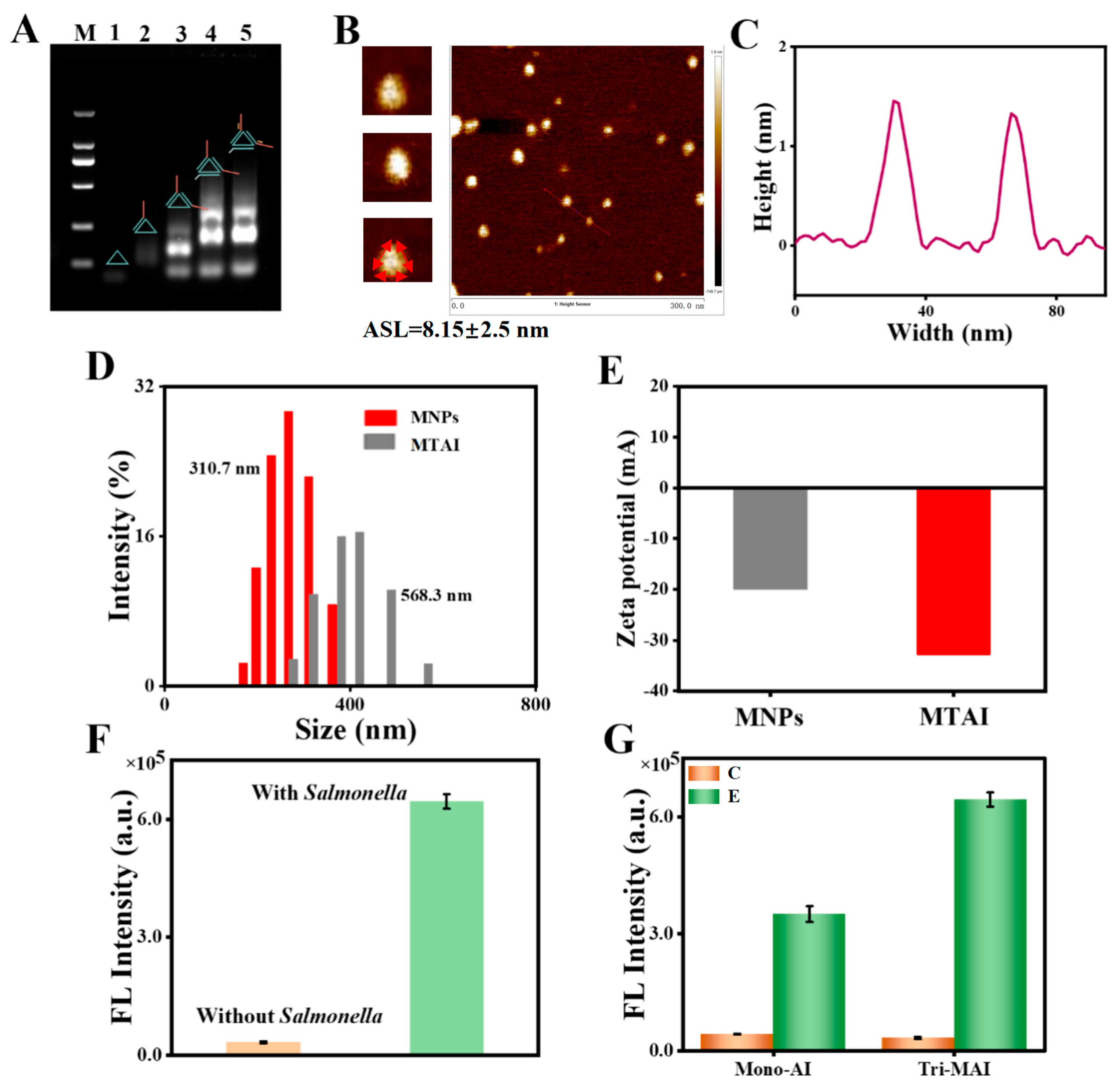
As shown in Figure 1A, agarose gel electrophoretic method was performed to demonstrate the Tri-MAI structure self-assembly. Following incremental addition of strands from lane 1 to lane 3, a marked decline in electrophoretic mobility could be observed, resulting from the increased valence of the aptamer. The triangle multivalent aptamer hybridized with the linker DNA (lane 4) exhibited reduced electrophoretic mobility compared to the three single-stranded sequences (lane 1, lane 2 and lane 3). By comparing lane 5 (the DNA hybridization between aptamer and initiator) with lane 4, the slower band could be observed in lane 5, demonstrating that the Tri-MAI had been successfully formed. The geometric structure and size of the Tri-MAI were characterized by atomic force microscopy (AFM) measurement as shown in Figure 1B. The triangular geometry is roughly observed, and the average side length (ASL) of Tri-MAI is 8.15 nm, which is little higher than the theoretical value (7.14 nm) because of the well-known tip-broadening effect [28]. The cross-sectional profile of the triangle taken along the red line in Figure 1B showed the height of the Tri-MAI is about 1.6 nm (Figure 1C), which is consistent with the theoretical value of dsDNA. Next, the hydrated diameter of the MTAI was characterized by dynamic light scattering (DLS). The average hydrated diameter of streptavidin MNPs was 310.7 nm and it was enlarged to 568.3 nm, indicating the successful preparation of MTAI (Figure 1D). For the evolution of zeta potentials of MNPs, MTAI were also conducted, and the results were determined as—20.01 mV and—32.85 mV in Figure 1E, demonstrating the negative charged Tri-MAI had been successfully attached on the surface of MNPs to increase the zeta potential. Then the feasibility of triggered initiator release based on aptamer recognition owing to the competition between Salmonella and initiator for the binding site of the aptamer was validated in Figure 1F.
In the presence of Salmonella, the fluorescence intensity was enhanced significantly, showing a large amount of FAM labeled initiator entered the supernatant and an almost inconspicuous fluorescence signal in the absence of Salmonella. The result has revealed the competition between Salmonella and initiator for aptamer successfully. Moreover, comparing the monovalent aptamers with multivalent aptamer, as shown in Scheme 2, the Tri-MAI structure shows about twice the sensitivity to Salmonella than to Mono-AI (Figure 1G), indicating the superior multivalence interactions of Tri-MAI.

3.2. Validation of the CHA Performance

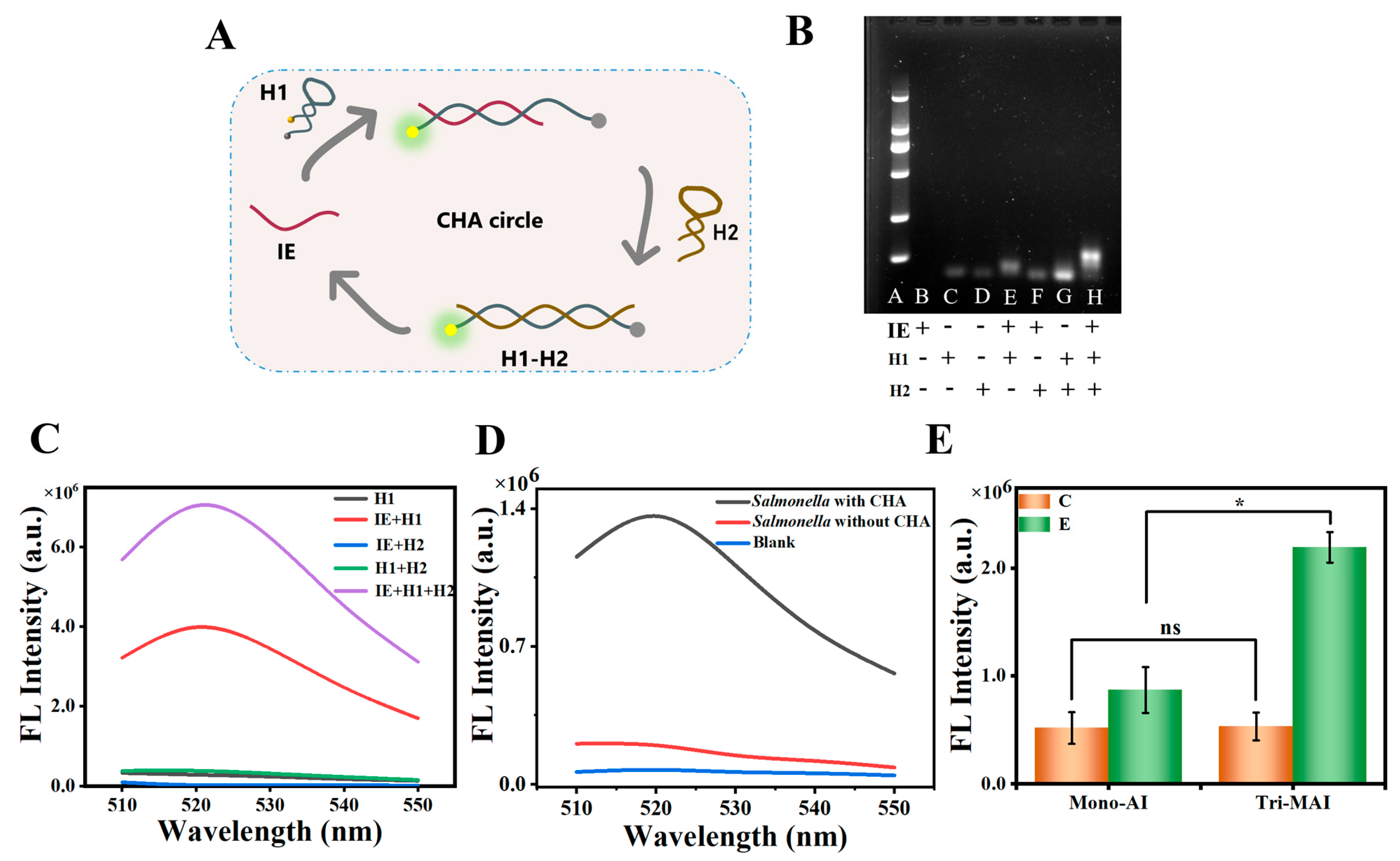
The practicality of the proposed CHA performance was confirmed through dual mode verification: DNA gel electrophoresis and fluorescence detection. As Figure 2A demonstrated, through DNA hybridization and unwinding, initiator-mediated unfolding of the H1 hairpin structure led to the cessation of FRET [29] between FAM and BHQ1, thereby causing a significant increase in fluorescence. Then H2 complements with H1, generating the H1-H2 duplex, by which the initiator could be released to catalyze a repeated CHA cycle, generating stronger fluorescence. In the CHA cycle, the electrophoresis image in Figure 2B shows that initiator (lane B), H1 (lane C) and H2 (lane D) were consistent with our design. In the presence of the initiator, the complexes generated by the initiator unfolding the H1 hairpin were observed in lane E, indicating that the formation of complexes led to increased size and slower motion rate. While the mixture of initiator and H2 (lane F) as well as the mixture of H1 and H2 (lane G) showed no change in motion rate, it indicated that H1 and H2 do not react in the absence of the initiator, ensuring a low background sign. The slowest motion rate was observed (lane H) when initiator, H1, and H2 were present, confirming successful complex formation of H1-H2. Additionally, the results of the fluorescence experiment demonstrated that the initiator activated the CHA reaction with an obvious FI increase, and that the H1 + H2 + initiator group was 181.5-fold to the H1 + H2 group (Figure 2C), thereby performing effective signal amplification. To confirm the high signal amplification efficiency of CHA, the whole Salmonella detection procedure using Mono-AI was explored in Figure 2D, and it was found that Salmonella with CHA generated a strong fluorescence signal (black line) and achieved 6.85-fold fluorescence signal higher than Salmonella without CHA in 0.5 h (red line). The background fluorescence signal in Salmonella detection was virtually negligible based on the very weak signal observed in the blank control (without Salmonella; blue line). At the same time, the signal amplification effect of Tri-MAI and Mono-AI was also compared in the detection of bacteria with the participation of CHA. As shown in Figure 2E, the fluorescence intensity of Tri-MAI showed a 1.5-fold of Mono-AI, indicating the higher binding affinity of Tri-MAI over the monovalent. All results demonstrated the high signal amplification efficiency of CHA as well as the good feasibility of MTAI-CHA for Salmonella detection.

3.3. Optimization of Experimental Parameters

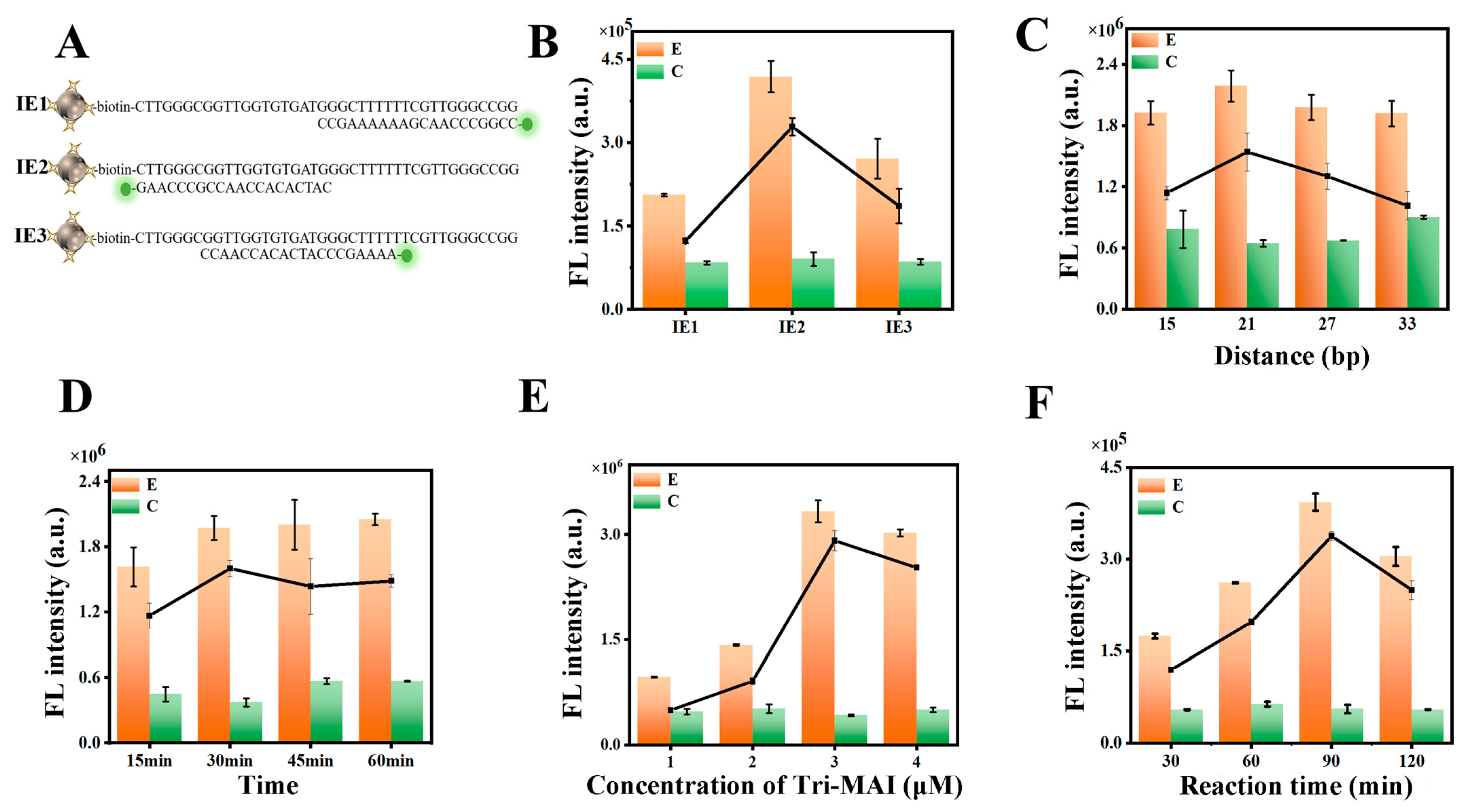
To identify the Salmonella-binding region of the aptamer, which could be influenced by initiator blocking potentially affecting final fluorescence intensity, three FAM-labeled probe strands, 20 nucleotides in length, complementary to distinct aptamer regions, were designed (Figure 3A). The selection of the most suitable regions was optimized by changing the complementary pairing position of the initiator and adapter as a variable. The other experimental conditions were the same as the previous experiment of capturing Salmonella with the aptamers. Salmonella was added to the aptamer–initiator complex-modified MNPs and the supernatant was collected for fluorescence detection after incubation. It can be observed in Figure 3B that the fluorescence intensity was higher when the initiator paired with the aptamers (IE2). That was probably owing to the affinity of IE2 and aptamers being weaker, so the IE2 was easier to be released. Then, the distance between two adjacent aptamers in the MAI is considered to be a crucial factor influencing binding affinity [30]. We therefore optimized this distance by precisely controlling the length of the DNA (15 bp, 21 bp, 27 bp and 33 bp). From the results in Figure 3C, optimal fluorescence emission is observed in Salmonella samples with 21 bp length of DNA, whereas considerably weaker signal intensities are recorded for both shorter (15 bp) and longer (27 bp, 33 bp) length of DNA. Consequently, the 21 bp length of DNA was selected for subsequent studies. In addition, the time of CHA reaction also affected the formation yield of H1-H2 duplexes. Figure 3D indicates that fluorescence signal increased with more incubation time and reached a plateau at 30min, suggesting 30min was the best reaction time for CHA amplification. The concentration of Tri-MAI modified to the MNPs also plays an important role in this approach. As we can see in Figure 3E, the fluorescent signal increased gradually until the 3 μM, leading to the sufficient capture of Salmonella. The time of Salmonella incubation with aptamers-modified MNPs affected the detection as well, and the sample signal did not grow higher after 90 min incubation in Figure 3F. Hence, we chose 90 min as the optimal incubation time.

3.4. Sensitivity of the Assay

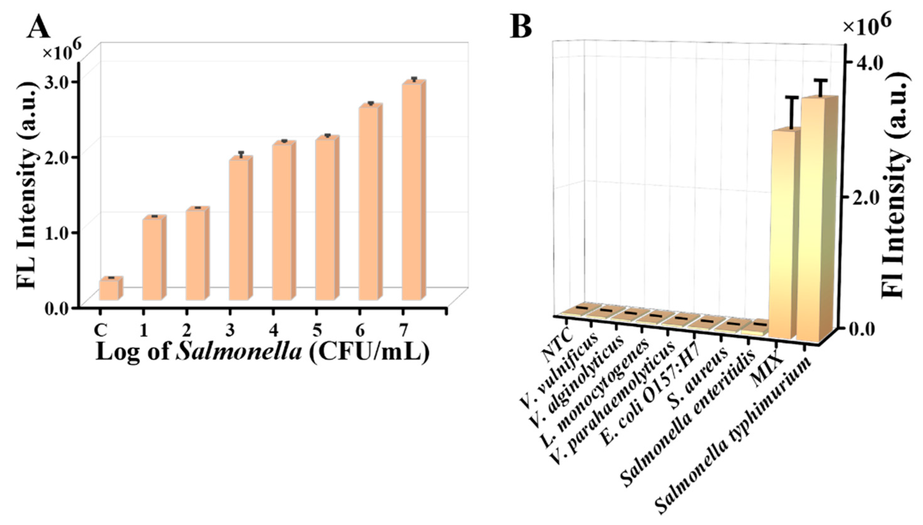
Under optimized experimental conditions, fluorescence signal intensity was measured at varying Salmonella concentrations to evaluate the quantitative detection capability of the assay. The fluorescence profiles with Salmonella concentrations of 0 ~ 1 × 10⁷ CFU/mL are presented in Figure 4A, and the detectable limit of Salmonella is 10 CFU/mL. We ascribe the improvement of good sensitivity in this detection to the higher apparent affinity of multivalent aptamer binding on the Salmonella surface and the participation of CHA signal amplification. Therefore, it could be reasoned that this proposed detection method had a good sensitivity for the detection of Salmonella and one should consider applying this method to the detection of other pathogenic bacteria to contribute to food safety testing.

3.5. Specificity of the Assay

To confirm whether the increase in the fluorescence signal was specifically induced by Salmonella, other nontarget pathogens including Salmonella enteritidis, E. coli O157:H7, S. aureus, Listeria monocytogenes, Vibrio parahaemolyticus, Vibrio alginolyticus, and Vibrio vulnificus were selected as the target analytes to react with Tri-MAI. As depicted in Figure 4B, only the presence of target Salmonella or the mixture containing all species caused the sensing system to produce a very strong fluorescence signal, while all nontarget analytes only yielded fluorescence signals indistinguishable from the background signal. This marked contrast demonstrated the high specificity of this assay for Salmonella detection, attributed to the use of Salmonella aptamers as selective recognition elements.

3.6. Analysis of Real Samples

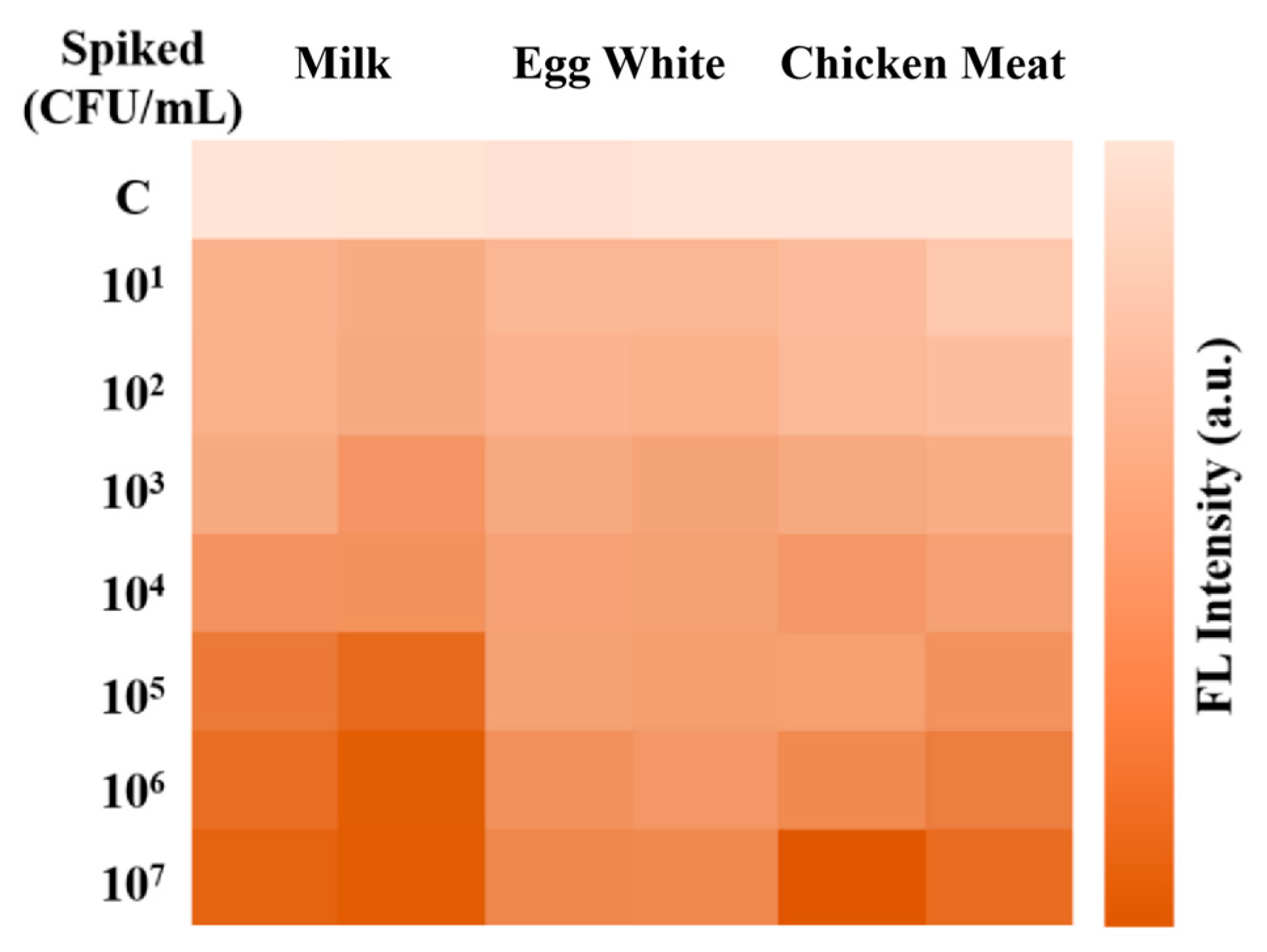
To further investigate the practical application of the assay, different concentrations of Salmonella were spiked into milk, egg whites, and chicken meat as simulated practical complex samples. As depicted in Figure 5, the fluorescence intensity increased as the concentration of Salmonella increased. And the detection yielded positive signals when the spiked concentrations of Salmonella was 10 CFU/mL, indicating the suitability of the proposal for Salmonella detection in real samples.

4. Conclusions

In summary, a simple, robust and enzyme-free fluorescence assay for Salmonella detection was developed by combining triangle multivalent aptamer with high binding affinity and CHA signal amplification for the first time. Benefiting from the released initiators that are bound to aptamers and CHA signal amplification, the proposed method exhibits high sensitivity for Salmonella detection with low limit of detection of 10 CFU/mL, suggesting the potential for directly detecting Salmonella from samples. Furthermore, this wide detection range confirms the suitability of this assay for direct Salmonella detection, eliminating the need for sample dilution or enrichment. Consequently, the platform holds significant potential for universal pathogen detection. By simply substituting the aptamer sequence with those specific to different targets, it can be readily adapted to identify diverse pathogens, offering substantial utility in food safety surveillance, environmental monitoring, and clinical diagnostics.

Author Contributions

Conceptualization, Z.Q.; Methodology, S.C.; Investigation, C.W., X.H., Z.W., W.L. and X.P.; Writing—original draft preparation. Z.W., W.L., C.W.,S.C. and X.P.; Formal analysis, Z.W., C.W. and X.H. Writing—review and editing, X.H. and Z.Q. All authors have read and agreed to the published version of the manuscript.

Funding

This work was supported by the National Natural Science Foundation of China (Grant No. 32001773).

Data Availability Statement

No new data were created or analyzed in this study.

Acknowledgments

The authors extend their appreciation to all collaborators of College of Food and Pharmaceutical Sciences, Ningbo University, for their technical support and unwavering assistance.

Conflicts of Interest

Author Shu Chen, Zhen Wang, Wen Lu, Xingxing Peng and Chuanpi Wang were employed by the company Greentown Agricultural Science Testing Technology Co., Ltd. The remaining authors declare that the research was conducted in the absence of any commercial or financial relationships that could be construed as a potential conflict of interest. The authors declare no conflicts of interest.

References

  1. Teklemariam, A.D.; Al-Hindi, R.R.; Albiheyri, R.S.; Alharbi, M.G.; Alghamdi, M.A.; Filimban, A.A.R.; Al Mutiri, A.S.; Al-Alyani, A.M.; Alseghayer, M.S.; Almaneea, A.M.; et al. Human Salmonellosis: A Continuous Global Threat in the Farm-to-Fork Food Safety Continuum. Foods 2023, 12, 1756. [Google Scholar] [CrossRef]
  2. Majowicz, S.E.; Musto, J.; Scallan, E.; Angulo, F.J.; Kirk, M.; O’Brien, S.J.; Jones, T.F.; Fazil, A.; Hoekstra, R.M.; International Collaboration on Enteric Disease “Burden of Illness” Studies. The global burden of nontyphoidal Salmonella gastroenteritis. Clin. Infect. Dis. 2010, 50, 882–889. [Google Scholar] [CrossRef]
  3. Salmonella (Non-Typhoidal). Available online: https://www.who.int/news-room/fact-sheets/detail/Salmonella-(non-typhoidal) (accessed on 4 October 2024).
  4. Rinn, N.; Müller, A.; Braun, A.S.; Greif, G.; Stiefel, D.; Kehrenberg, C. Food products confiscated from air passengers travelling from third countries into the European Union: Microbiological analyses and genomic characterization of zoonotic and multiresistant bacteria. Food Microbiol. 2025, 131, 104783. [Google Scholar] [CrossRef] [PubMed]
  5. Silva, T.C.; Isaksson, M.; Nilsson, B.; Eppink, M.; Ottens, M. Optimization of multi-column chromatography for capture and polishing at high protein load. Biotechnol. Prog. 2025, e70047. [Google Scholar] [CrossRef]
  6. Zhou, C.; Li, W.; Zhao, Y.; Gu, K.; Liao, Z.; Guo, B.; Huang, Z.; Yang, M.; Wei, H.; Ma, P.; et al. Sensitive detection of viable salmonella bacteria based on tertiary cascade signal amplification via splintR ligase ligation-PCR amplification-CRISPR/Cas12a cleavage. Anal. Chim. Acta 2023, 1248, 340885. [Google Scholar] [CrossRef] [PubMed]
  7. Sun, Y.; Zhang, S.; Qi, L.; Zhang, X.; Yang, M.; Guo, Z.; Wang, Z.; Du, Y. Advancing Multiple Detection in RT-LAMP with a Specific Probe Assembled from Plural Three-Way-Junction Structures. Anal. Chem. 2023, 95, 17808–17817. [Google Scholar] [CrossRef] [PubMed]
  8. Zhao, F.; Yan, H.; Zheng, Y.; Zu, Y.; Yang, S.; Hu, H.; Shi, S.; Liang, H.; Niu, X. Joint concanavalin A-aptamer enabled dual recognition for anti-interference visual detection of Salmonella typhimurium in complex food matrices. Food Chem. 2023, 426, 136581. [Google Scholar] [CrossRef]
  9. Wang, L.; Wang, R.; Chen, F.; Jiang, T.; Wang, H.; Slavik, M.; Wei, H.; Li, Y. QCM-based aptamer selection and detection of Salmonella typhimurium. Food Chem. 2017, 221, 776–782. [Google Scholar] [CrossRef]
  10. Lu, Y.; Xie, Q.; Chen, J.; Chu, Z.; Zhang, F.; Wang, Q. Aptamer-mediated double strand displacement amplification with microchip electrophoresis for ultrasensitive detection of Salmonella typhimurium. Talanta 2024, 273, 125875. [Google Scholar] [CrossRef]
  11. Li, H.; Xu, H.; Shi, X.; Zhao, C.; Li, J.; Wang, J. Colorimetry/fluorescence dual-mode detection of Salmonella typhimurium based on a “three-in-one” nanohybrid with high oxidase-like activity for AIEgen. Food Chem. 2024, 449, 139220. [Google Scholar] [CrossRef]
  12. Wang, L.; Huo, X.; Qi, W.; Xia, Z.; Li, Y.; Lin, J. Rapid and sensitive detection of Salmonella Typhimurium using nickel nanowire bridge for electrochemical impedance amplification. Talanta 2020, 211, 120715. [Google Scholar] [CrossRef]
  13. Zhang, P.; Song, M.; Dou, L.; Xiao, Y.; Li, K.; Shen, G.; Ying, B.; Geng, J.; Yang, D.; Wu, Z. Development of a fluorescent DNA nanomachine for ultrasensitive detection of Salmonella enteritidis without labeling and enzymes. Mikrochim. Acta 2020, 187, 376. [Google Scholar] [CrossRef]
  14. Ooi, H.W.; Kocken, J.M.M.; Morgan, F.L.C.; Malheiro, A.; Zoetebier, B.; Karperien, M.; Wieringa, P.A.; Dijkstra, P.J.; Moroni, L.; Baker, M.B. Multivalency Enables Dynamic Supramolecular Host–Guest Hydrogel Formation. Biomacromolecules 2020, 21, 2208–2217. [Google Scholar] [CrossRef]
  15. Dong, N.; Liu, Z.; He, H.; Lu, Y.; Qi, J.; Wu, W. “Hook&Loop” multivalent interactions based on disk-shaped nanoparticles strengthen active targeting. J. Contr. Release 2023, 354, 279–293. [Google Scholar]
  16. Guo, L.; Zhang, Y.; Wang, Y.; Xie, M.; Dai, J.; Qu, Z.; Zhou, M.; Cao, S.; Shi, J.; Wang, L.; et al. Directing Multivalent Aptamer-Receptor Binding on the Cell Surface with Programmable Atom-Like Nanoparticles. Angew. Chem. Int. Ed. Engl. 2022, 61, e202117168. [Google Scholar] [CrossRef] [PubMed]
  17. Zhang, X.; Peng, Y.; Yao, L.; Shang, H.; Zheng, Z.; Chen, W.; Xu, J. Self-Assembly of Multivalent Aptamer-Tethered DNA Monolayers Dedicated to a Fluorescence Polarization-Responsive Circular Isothermal Strand Displacement Amplification for Salmonella Assay. Anal. Chem. 2023, 95, 2570–2578. [Google Scholar] [CrossRef]
  18. Xu, Z.; Shi, T.; Mo, F.; Yu, W.; Shen, Y.; Jiang, Q.; Wang, F.; Liu, X. Programmable Assembly of Multivalent DNA-Protein Superstructures for Tumor Imaging and Targeted Therapy. Angew. Chem. Int. Ed. Engl. 2022, 61, e202211505. [Google Scholar] [CrossRef]
  19. Li, H.; Cheng, S.; Zhang, Q.; Zhou, T.; Zhang, T.; Liu, S.; Peng, Y.; Yu, J.; Xu, J.; Wang, Q.; et al. Dual-Multivalent Aptamer-Based Drug Delivery Platform for Targeted SRC Silencing to Enhance Doxorubicin Sensitivity in Endometrial Cancer. Int. J. Biol. Sci. 2024, 20, 5812–5830. [Google Scholar] [CrossRef]
  20. Qiao, Z.; Xue, L.; Sun, M.; Ma, N.; Shi, H.; Yang, W.; Cheong, L.Z.; Huang, X.; Xiong, Y. Dual-Functional Tetrahedron Multivalent Aptamer Assisted Amplification-Free CRISPR/Cas12a Assay for Sensitive Detection of Salmonella. J. Agric. Food Chem. 2024, 72, 857–864. [Google Scholar] [CrossRef] [PubMed]
  21. Jiang, Y.S.; Bhadra, S.; Li, B.; Ellington, A.D. Mismatches improve the performance of strand-displacement nucleic Acid circuits. Angew. Chem. Int. Ed. Engl. 2014, 53, 1845–1848. [Google Scholar] [CrossRef] [PubMed]
  22. Bhadra, S.; Ellington, A.D. Design and application of cotranscriptional non-enzymatic RNA circuits and signal transducers. Nucleic Acids Res. 2014, 42, e58. [Google Scholar] [CrossRef]
  23. Zhang, P.; Wu, X.; Yuan, R.; Chai, Y. An “off-on” electrochemiluminescent biosensor based on DNAzyme-assisted target recycling and rolling circle amplifications for ultrasensitive detection of microRNA. Anal. Chem. 2015, 87, 3202–3207. [Google Scholar] [CrossRef]
  24. Tang, Y.; Lin, Y.; Yang, X.; Wang, Z.; Le, X.C.; Li, F. Universal strategy to engineer catalytic DNA hairpin assemblies for protein analysis. Anal. Chem. 2015, 87, 8063–8066. [Google Scholar] [CrossRef]
  25. Qing, Z.; Hu, J.; Xu, J.; Zou, Z.; Lei, Y.; Qing, T.; Yang, R. An intramolecular catalytic hairpin assembly on a DNA tetrahedron for mRNA imaging in living cells: Improving reaction kinetics and signal stability. Chem. Sci. 2019, 11, 1985–1990. [Google Scholar] [CrossRef] [PubMed]
  26. Li, C.; Lv, W.; Yang, F.; Zhen, S.; Huang, C. Simultaneous Imaging of Dual microRNAs in Cancer Cells through Catalytic Hairpin Assembly on a DNA Tetrahedron. ACS Appl. Mater. Interfaces 2022, 14, 12059–12067. [Google Scholar] [CrossRef] [PubMed]
  27. Kolovskaya, O.S.; Savitskaya, A.G.; Zamay, T.N.; Reshetneva, I.T.; Zamay, G.S.; Erkaev, E.N.; Wang, X.; Wehbe, M.; Salmina, A.B.; Perianova, O.V.; et al. Development of bacteriostatic DNA aptamers for salmonella. J. Med. Chem. 2013, 56, 1564–1572. [Google Scholar] [CrossRef] [PubMed]
  28. Wang, R.; Kurouski, D. Elucidation of Tip-Broadening Effect in Tip-Enhanced Raman Spectroscopy (TERS): A Cause of Artifacts or Potential for 3D TERS. J. Phys. Chem. C 2018, 122, 24334–24340. [Google Scholar] [CrossRef]
  29. Johansson, M.K.; Fidder, H.; Dick, D.; Cook, R.M. Intramolecular dimers: A new strategy to fluorescence quenching in dual-labeled oligonucleotide probes. J. Am. Chem. Soc. 2022, 124, 6856–6950. [Google Scholar] [CrossRef]
  30. Jia, W.; Xie, D.; Li, F.; Wu, X.; Wang, R.; Yang, L.; Liu, L.; Yin, W.; Chang, S. Evaluation the effect of nanoparticles on the structure of aptamers by analyzing the recognition dynamics of aptamer functionalized nanoparticles. Anal. Chim. Acta 2021, 1183, 338976. [Google Scholar] [CrossRef]
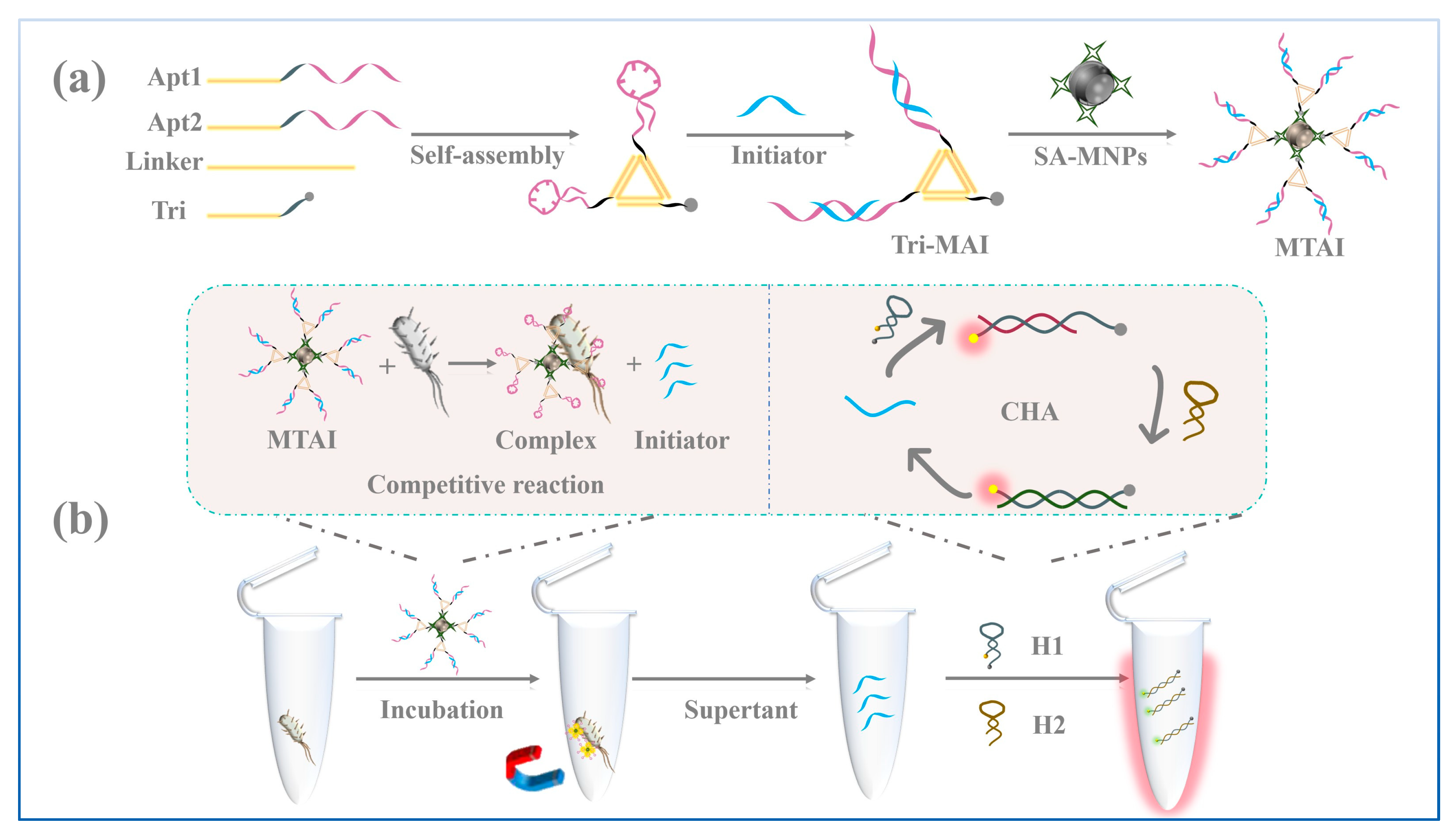
Scheme 1. Schematic overview depicting the MTAI-CHA system for detecting Salmonella.
Chemosensors 13 00334 sch001
Figure 1. (A) Agarose gel electrophoresis results of the assembly of Tri-MAI: Lane M: marker, Lanes 1–5: L, L + Apt1, L + Apt1 + Apt2, L + Apt1 + Apt2 + linker, L + Apt1 + Apt2 + linker + initiator (CHA). (B) AFM image of the Tri-MAI accompanied by the enlarged images. ASL notes the average side length. (C) The height profile along the red line of Figure 1B. (D) hydrated size of streptavidin MNPs and MTAI measured by DLS. (E) Zeta potential of streptavidin MNPs and MTAI. (F) The fluorescent intensity of MTAI detection with and without Salmonella. (G) Comparison of the binding ability of Tri-MAI and Mono-AI without CHA amplification.
Chemosensors 13 00334 g001
Scheme 2. Schematic overview depicting the Mono-AI and Tri-MAI system for detecting Salmonella.
Chemosensors 13 00334 sch002
Figure 2. Verification of the feasibility of our proposed trigger-initiated CHA system. (A) Scheme of CHA systems. (B) Gel image of CHA products: Marker (lane A), initiator (IE, lane B), H1 (lane C), H2 (lane D), IE + H1 (lane E), IE + H2 (lane F), H1 + H2 (lane G), and IE + H1 + H2 (lane H). (C) Fluorescence spectra of CHA amplification (IE2: 0.5 μM, H1: 1μM, H2: 1.5 μM. IE2 represents the initiator based on aptamer recognition. H1 and H2 represent the hairpin probes designed for aptamer recognition system.). (D) The fluorescence intensity of Mono-AI in Salmonella detection with and without the participation of CHA amplification. (E) Comparison of the fluorescence intensity of Tri-MAI and Mono-AI in Salmonella detection with the participation of CHA amplification. “ns”: the experimental results (two sets of data C) are not statistically significant. “*”: there are significant differences between the two sets of data E.
Chemosensors 13 00334 g002
Figure 3. Optimization of the experimental conditions. (A) Illustration of different initiators complemented to three different regions of the aptamer sequence. (B) The fluorescence intensity of different groups about the aptamers and initiators with and without Salmonella. Optimization of the distance between the adjacent aptamers of Tri-MAI (C), the time of CHA amplification (D), the concentration of Tri-MAI (E) and capture time for Salmonella (F) in detection system.
Chemosensors 13 00334 g003
Figure 4. (A) Fluorescent signals of the MTAI-CHA system with varied concentrations of Salmonella from low to high: 0, 1.0 × 101, 1.0 × 10², 1.0 × 10³, 1.0 × 10⁴, 1.0 × 10⁵, 1.0 × 10⁶, and 1.0 × 10⁷. (B) The specificity of the MTAI-CHA system at a concentration of 1.0 × 10⁹ CFU/mL.
Chemosensors 13 00334 g004
Figure 5. A heat map of the fluorescent intensity of food samples based on the MTAI-CHA system.
Chemosensors 13 00334 g005
Table 1. Nucleic acid sequences used in MTAI-CHA system.
NameSequence (5′–3′)
Apt1(linker1-apt).CTATCATGGCAAGGTCCTACGCAAAACTCCTCTGACTGTAACCACGGTGGTTTGATCACTATTGGGCCTTTGTGATGTCGGTAGT
Apt2(linker2-apt)CTACGTCAGCGTTAGACTGAACAAAACTCCTCTGACTGTAACCACGGTGGTTTGATCACTATTGGGCCTTTGTGATGTCGGTAGT
TriCGAGTCATCTGTCTACTGAGCCAAAAAAAAA-biotin
LinkerGATGACTCGTTTCAGTCTAACGCTGACGTAGTCGTAGGACCTTGCCATGATAGTGCTCAGTAGACA
H1FAM-GGTTACAGTTATGTGTACCACTGTAACCACGGTG-BHQ1
H2TATGTGTACCTGGTTACAGTGGTACACATAACTGTAACC
InitiatorCACCGTGGTTACAGT
Disclaimer/Publisher’s Note: The statements, opinions and data contained in all publications are solely those of the individual author(s) and contributor(s) and not of MDPI and/or the editor(s). MDPI and/or the editor(s) disclaim responsibility for any injury to people or property resulting from any ideas, methods, instructions or products referred to in the content.