Article

Development and Validation of an HPLC-MS/MS Method for Quantifying Deoxynivalenol and Zearalenone Biomarkers in Dried Porcine Blood Spots

by
Isadora Fabris Laber
1,2,
Cristina Tonial Simões
1,2,*,
Cristiane Rosa da Silva
1,2,
Luara Medianeira de Lima Schlösser
1,2,
Janine Alves Sarturi
1,2,
Luriane Medianeira Carossi Leal
1,2,
Renê Valmor Theobald
1,2 and
Carlos Augusto Mallmann
1,2
1
Graduate Program in Veterinary Medicine, Federal University of Santa Maria, Santa Maria 97105-900, RS, Brazil
2
Laboratory of Mycotoxicological Analyses—LAMIC, Federal University of Santa Maria, Santa Maria 97105-900, RS, Brazil
*
Author to whom correspondence should be addressed.
Chemosensors 2025, 13(8), 296; https://doi.org/10.3390/chemosensors13080296 (registering DOI)
Submission received: 26 June 2025 / Revised: 1 August 2025 / Accepted: 7 August 2025 / Published: 9 August 2025

Abstract

Deoxynivalenol (DON) and zearalenone (ZEN) are common mycotoxins in animal feeds, and their metabolites can be detected in exposed animals. Traditional methods focus on mycotoxin detection in feed, whereas biomarker-based approaches are used for evaluating individual exposure. This study aimed to develop and validate a multi-analyte method for the detection of biomarkers of ZEN, DON, and their metabolites α-zearalanol (α-ZAL), zearalanone (ZAN), deepoxy-DON (DOM-1), and 3-acetyl-DON (3-ADON) in swine using dried blood spots (DBSs) on qualitative filter paper. Analysis was performed using high-performance liquid chromatography–tandem mass spectrometry. Blank blood samples from three male pigs were fortified with 20, 40, and 60 μg/L of each analyte. Aliquots of 40 μL were spotted onto filter paper and then extracted and analyzed. Method validation included evaluating limits of detection and quantification, linearity, matrix effects, recovery, repeatability, intermediate precision, and selectivity. All analytes were detectable in DBS. Also, ZEN, ZAN, DON, and DOM-1 met all validation criteria, with recovery values of 89.10%, 79.79%, 101.50%, and 79.50%, respectively. Both α-ZAL and 3-ADON showed lower recoveries (74.66% and 58.66%). The method was successfully validated for simultaneous analysis of ZEN, ZAN, DON, and DOM-1 in swine DBS, offering a practical and minimally invasive tool for biomonitoring mycotoxin exposure.

1. Introduction

Brazil is the fourth largest producer and exporter of pork worldwide, with a production volume of 5.31 million tons in 2024, of which 1.35 million tons were exported [1]. Due to their predominantly cereal-based diet, pigs are continuously exposed to mycotoxins- secondary metabolites produced by fungi [2]. Two of the most harmful mycotoxins affecting swine health and performance are zearalenone (ZEN) and deoxynivalenol (DON), both produced by fungi of the genus Fusarium. Globally, these mycotoxins have been detected in up to 80% and 40% of animal feed samples, respectively [3].
In pigs, ZEN induces reproductive disorders such as pelvic organ prolapse, anestrus, and pseudopregnancy. These clinical signs are primarily caused by disrupted sex hormone regulation due to the strong affinity of ZEN metabolites for porcine estrogen receptors [4,5]. Swine are also particularly susceptible to the adverse effects of DON, including reduced feed intake, impaired growth performance, decreased nutrient utilization, and gastrointestinal disorders when fed DON-contaminated diets [6,7].
The quantification of mycotoxin levels in feed is widely employed to estimate animal exposure to these substances. However, this approach does not account for individual animal exposure or the heterogeneous distribution of toxins within feed batches. Even when using reference methods such as high-performance liquid chromatography coupled with tandem mass spectrometry (HPLC-MS/MS) for feed analysis, the results may not accurately reflect the real exposure of each animal. This is due to both the heterogeneous distribution of mycotoxins in feed and the varying susceptibility among animals—factors that often manifest in the field as performance variability and lack of uniformity within animal groups. Therefore, a more comprehensive evaluation of animal exposure can be achieved by combining the analysis of mycotoxin levels in feed with the monitoring of corresponding biomarkers in biological matrices such as blood, urine, and feces [8].
The biotransformation of ZEN occurs primarily through the action of hepatic enzymes, as well as enzymes from the intestinal mucosa and the microbiota, leading to the formation of several phase I metabolites, including zearalanone (ZAN), α-zearalenol (α-ZEL), β-zearalenol (β-ZEL), α-zearalanol (α-ZAL), and β-zearalanol (β-ZAL) [9]. Among these, α-ZAL is one of the reduced metabolites of ZEN and is commonly monitored as an indicator of ZEN exposure. Regarding DON, its major derivatives include acetylated forms produced by fungi, such as 3-acetyl-DON (3-ADON) and 15-acetyl-DON (15-ADON) [10]. Additionally, microbial metabolism, particularly intestinal bacteria, produces several DON phase I metabolites, including deepoxy-DON (DOM-1), 3-epi-DON, and 3-keto-DON.
A biomarker is defined as the original substance; its metabolites or biological parameters may change due to interactions with biomolecules or physiological compartments within the organism [11]. Biomarker analysis can serve as a valuable alternative for monitoring the risk of mycotoxin intoxication in animals as well as for evaluating the efficacy of anti-mycotoxin additives (AMAs) used in feed to mitigate the effects of mycotoxins [12,13]. Biomarkers of ZEN, α-ZAL, DON, and DOM-1 in urine have previously been used to estimate mycotoxin intake and its corresponding levels in the feed consumed by the monitored pigs [14]. The HPLC–MS/MS techniques have also been validated for the detection of mycotoxin biomarkers in biological matrices such as blood and serum in both humans [15] and animals [16,17]. Chen et al. (2022) developed a simple and sensitive HPLC–MS/MS method to assess the toxicokinetics of aflatoxins in vivo in rats [16]. Additionally, Panisson et al. (2023) demonstrated a correlation between DON intake and its concentrations in both urine and blood serum, indicating that the analysis of DON and its metabolites in biological samples can serve as a reliable indicator of exposure in pigs [17].
The dried blood spots (DBSs) sampling technique became widespread in the 1960s and was initially used to screen newborns for phenylketonuria [18]. This method preserves analyte stability over extended periods without requiring refrigeration during storage and transport, thereby reducing the risk of sample contamination [19,20,21]. Recent research has demonstrated the applicability of this technique for determining mycotoxins and their metabolites in animal studies using HPLC-MS/MS, showing good correlation between DBS and plasma data and highlighting its potential for future toxicokinetic and exposure assessment studies [22]. Despite the relevance of this complementary diagnostic method, Brazil currently lacks laboratories capable of analyzing mycotoxin biomarkers in DBS. Therefore, this study aimed to develop and validate a method for analyzing ZEN, DON, and their metabolites α-ZAL, ZAN, DOM-1, and 3-ADON in swine using DBS on qualitative filter paper as well as HPLC-MS/MS.

2. Materials and Methods

2.1. Reagents and Materials

Tubes containing K3EDTA (CRAL®, São Paulo, Brazil) were used to collect blood from the animals.
Circles with a 20 mm diameter were pre-marked on qualitative filter paper (Qualy®, JProlab, São José dos Pinhais, Brazil), with a basis weight of 80 g/m2 and a thickness of 205 µm. The paper featured an ash content of 0.5%, a pore size of 14 µm, and an air permeability of 14 L/m2/s.
Methanol (MeOH), acetonitrile (ACN) (HPLC grade, 98–99.9% purity; J.T. Baker, Phillipsburg, NJ, USA), and acetone (PA and ACS grade, 99.5% purity; Neon, São Paulo, Brazil) were used as organic solvents, while formic acid (FA) and ammonium acetate (AmAc) (HPLC grade; J.T. Baker, Phillipsburg, NJ, USA) were used as reagents. The ultrapure water was purified using the Milli-Q® Direct 8 system (Merck KGaA, Darmstadt, Germany). A 1.0 M AmAc solution was prepared by dissolving 7.708 g of ammonium acetate (CH3COONH4) into 100 mL of ultrapure water.
The analytical standard of ZEN and its phase I metabolites, α-ZAL and ZAN, each at a concentration of 10,000 μg/L in ACN, were obtained from Biopure™ (Romer Labs®, Tulln, Austria) and stored at 2 to 8 °C. The analytical standard of DON, at a concentration of 100,000 μg/L in ACN, and its derivatives DOM-1 and 3-ADON, at concentrations of 50.1 and 10,000 μg/L in ACN, respectively, were purchased from Biopure™. The certified analytical standards were dissolved in ACN to prepare a standard solution (SS) containing 1000 μg/L of each analyte. The SS was stored at −20 °C and used for sample fortification.

2.2. Sample Collection

Blood samples (~3 mL each) were collected from three 70-day-old male Landrace pigs, slaughtered for human consumption at a pork processing plant (Frigorífico Sabor Gaúcho, Santa Maria, RS, Brazil) in accordance with the sanitary protocols established by Ordinance No. 1304 [23]. Blood was collected into K3EDTA tubes during the bleeding process and immediately transported to the Laboratory of Mycotoxicological Analyses (LAMIC) at the Federal University of Santa Maria. Afterwards, samples were analyzed using HPLC-MS/MS to confirm the absence of target toxins (blank samples). The present study did not involve experiments with live animals; therefore, no submission to the Animal Ethics Committee was required.

2.3. Dried Blood Spots Preparation and Extraction Procedure

For method validation, the blood was divided into three different tubes and fortified with the SS to achieve three different concentrations of each metabolite: 20, 40, and 60 μg/L. After fortification, the samples were incubated overnight at room temperature. Subsequently, 40 μL of each fortified blood concentration was pipetted in septuplicate onto the paper circles and allowed to dry completely.

2.4. Extracting Dried Blood Spots

For the extraction of DBS on filter paper, a solution of water/ACN/acetone (30/35/35, v/v/v) was used, following protocols previously established for DBS extraction in humans [24,25]. The extraction procedure, adapted from Lauwers et al. (2019), involved detaching and cutting out the blood-impregnated paper circles, which were then placed in test tubes containing 2 mL of the extraction solution [22]. Subsequently, the samples underwent ultrasonication for 60 min (Eco-sonics, Indaiatuba, Brazil). After extraction, the filter paper was discarded, and the resulting solution was concentrated under a gentle stream of nitrogen (N2) at 50 ± 5 °C until complete solvent evaporation. The concentrated material was reconstituted in 100 µL of water/MeOH/FA (60/40/0.1, v/v/v), ultrasonicated for 5 min, vortexed using a FISATOM mixer (São Paulo, Brazil) at 2500 rpm for 60 s, and transferred to vials for HPLC-MS/MS quantification.

2.5. HPLC-MS/MS Performance Parameters

Bloodstain analyses were performed using HPLC-MS/MS at the LAMIC laboratory. An Infinity 1200 series HPLC system (Agilent, Santa Clara, CA, USA) coupled to an API 5000 triple quadrupole mass spectrometer system, equipped with the Ion Turbo V source and QJet ion guide technology (Sciex, Framingham, MA, USA), was used.
Chromatographic separation was achieved with a Zorbax XDB-C18 column (4.6 × 150 mm, 5 μm particle size, Agilent, Santa Clara, CA, USA). Samples were analyzed with a mobile phase comprising water with 0.1% of the AmAc solution (solvent A) and MeOH containing 0.1% of the AmAc solution (solvent B). The gradient program started with a composition of 90% A and 10% B for 2 min. Subsequently, both phases A and B were held at 50% for 5 min. Then, phase A was decreased to 10% while phase B was increased to 90%. For the subsequent minute, phases A and B were maintained at 20% and 80%, respectively. The gradient was then adjusted to achieve 100% B over 5 min. In the last 4 min, the gradient program returned to the initial conditions of 90% A and 10% B to re-equilibrate the column, resulting in a total run time of 17 min. The column temperature was set at 40 °C, with a flow rate of 1 mL/min and an injection volume of 20 µL. The Analyst software, version 1.6.2 (Sciex, Framingham, MA, USA), was used for instrument control and data analysis.
The optimization of compound-specific mass spectrometry parameters, including declustering potential (DP), collision energy (CE), and collision cell exit potential (CXP), was carried out using a dwell time of 150 milliseconds to improve signal intensity and detection reliability. For each analyte, the most intense and selective precursor-to-product ion transitions were carefully selected to maximize sensitivity and specificity. The instrument was operated in the multiple reaction monitoring (MRM) mode with negative electrospray ionization (ESI-), monitoring two transitions per compound—one for quantification and another for confirmation. The optimized MS/MS parameters, including retention times and transition ions, are detailed in Table 1.

2.6. Method Validation

Method validation was performed in accordance with the guidelines for the validation of analytical methods established by Commission Regulation No. 401 (EC 401/2006) [26]. To achieve this, the procedures described in Section 2.3 and Section 2.4 were replicated over three consecutive days by three different analysts. The following parameters were evaluated: limit of detection (LOD), limit of quantification (LOQ), linearity, calibration curve, matrix effect, recovery, selectivity, repeatability, and intermediate precision.

2.6.1. Limit of Detection and Quantification

The limits of detection (LOD) and quantification (LOQ) were estimated based on the analysis of standards with ascending concentrations (0.5–5 µg/L). The signal-to-noise (S/N) ratio approach was used, considering a minimum S/N ratio of 3:1 for LOD and 10:1 for LOQ, as recommended by EURACHEM (2014) [27]. Analyses were performed accordingly, and noise was evaluated directly in baseline regions of the chromatogram where no peaks were present. The LOQ was defined as the lowest concentration at which acceptable precision (S/N ratio ≥ 10) could be achieved. The LOD was defined as the lowest concentration of the analyte that could be clearly distinguished from zero (S/N ratio ≥ 3).

2.6.2. Analytical Curve and Linearity

The SS at 1000 μg/L and a diluent composed of solvent A/solvent B (50:50 v/v) were used to create a calibration curve within the concentrations ranging from 10 to 70 μg/L. Calibration points of 2.5, 5, 7.5, 10, 12.5, 15, and 17.5 μg/L were used. These corresponded, respectively, to real concentrations of 10, 20, 30, 40, 50, 60, and 70 μg/L for each analyte. Linearity was assessed using the linear regression equation and correlation coefficient (r), as calculated by the Analyst software (version 1.6.2).

2.6.3. Matrix Effect and Selectivity

To assess the impact of the matrix effect, calibration curves were generated in triplicate, including one curve without the addition of a matrix (filter paper impregnated with porcine blank blood) and another with the addition of a matrix. Data were analyzed using the F-test to compare the variances between the replicates with and without a matrix to determine whether the variances were statistically different. The calculation involved dividing the larger variance by the smaller one, and the resulting value was compared to the F-value from the statistical table, considering degrees of freedom (n − 1) and a 95% confidence level.
Selectivity was assessed by comparing the chromatogram of a blank sample (showing no peaks corresponding to the six analytes) to those of samples spiked with 20 μg/L of ZEN, DON, and their metabolites.

2.6.4. Recovery, Repeatability, and Intermediate Precision

The mean recovery (Re) was estimated from the analysis of samples fortified with 20, 40, and 60 μg/L of each analyte. According to European Commission Regulation (EC) No. 401/2006 [26], acceptable recovery rates for ZEN are 60–120% for concentrations below 50 μg/L and 70–120% for concentrations above 50 μg/L. For DON, recovery criteria apply to concentrations above 100 μg/L, with recovery limits also ranging from 60% to 120%. For method validation purposes, recovery limits between 60% and 120% were considered for fortification levels of 20 and 40 μg/L, and between 70% and 120% for 60 μg/L, in accordance with the EC recommendations for ZEN.
Repeatability was assessed by extracting and quantifying fortified samples in septuplicate at three concentration levels (20, 40, and 60 μg/L). The results were evaluated based on the relative standard deviation (RSDr) calculated at each fortification level. Intermediate precision was assessed by calculating the RSDR from the extraction and analysis of fortified samples performed on three separate days by three different analysts. According to EC No. 401/2006 [26], values of RSDr and RSDR below 40% were considered acceptable.

2.7. Real Samples Analysis

The validated method was also applied to the determination of target analytes in 20 real blood samples (~0.5 mL each) individually collected from 70-day-old male Landrace pigs. Sample collection and preparation were conducted as previously described.

3. Results

The LOD achieved for the six analytes evaluated in this study was 1 µg/L, and the LOQ was 2.5 µg/L. As the EC 401/2006 [26] does not establish acceptance criteria for limits of detection (LOD) and quantification (LOQ), the requirements established by EURACHEM (2014) [27] were adopted for this purpose. The present values are consistent with the acceptance criteria outlined for method validation.
The obtained r values for the calibration curves with and without a matrix were 0.9972 and 0.9993 for ZEN, 0.9955 and 0.9941 for α-ZAL, 0.9937 and 0.9961 for ZAN, 0.9958 and 0.9976 for DON, 0.9949 and 0.9932 for DOM-1, and 0.9939 and 0.9948 for 3-ADON.
Regarding the matrix effect, the calculated F values were 0.895, 1.225, 0.966, 1.077, 1.010, and 1.010 for ZEN, α-ZAL, ZAN, DON, DOM-1, and 3-ADON, respectively. All calculated F values were below the tabulated F value (2.484), indicating no statistically significant difference between the variances between the groups with and without a matrix. Therefore, no significant matrix effect was observed, and the calibration curve prepared in solvent was considered suitable for method validation.
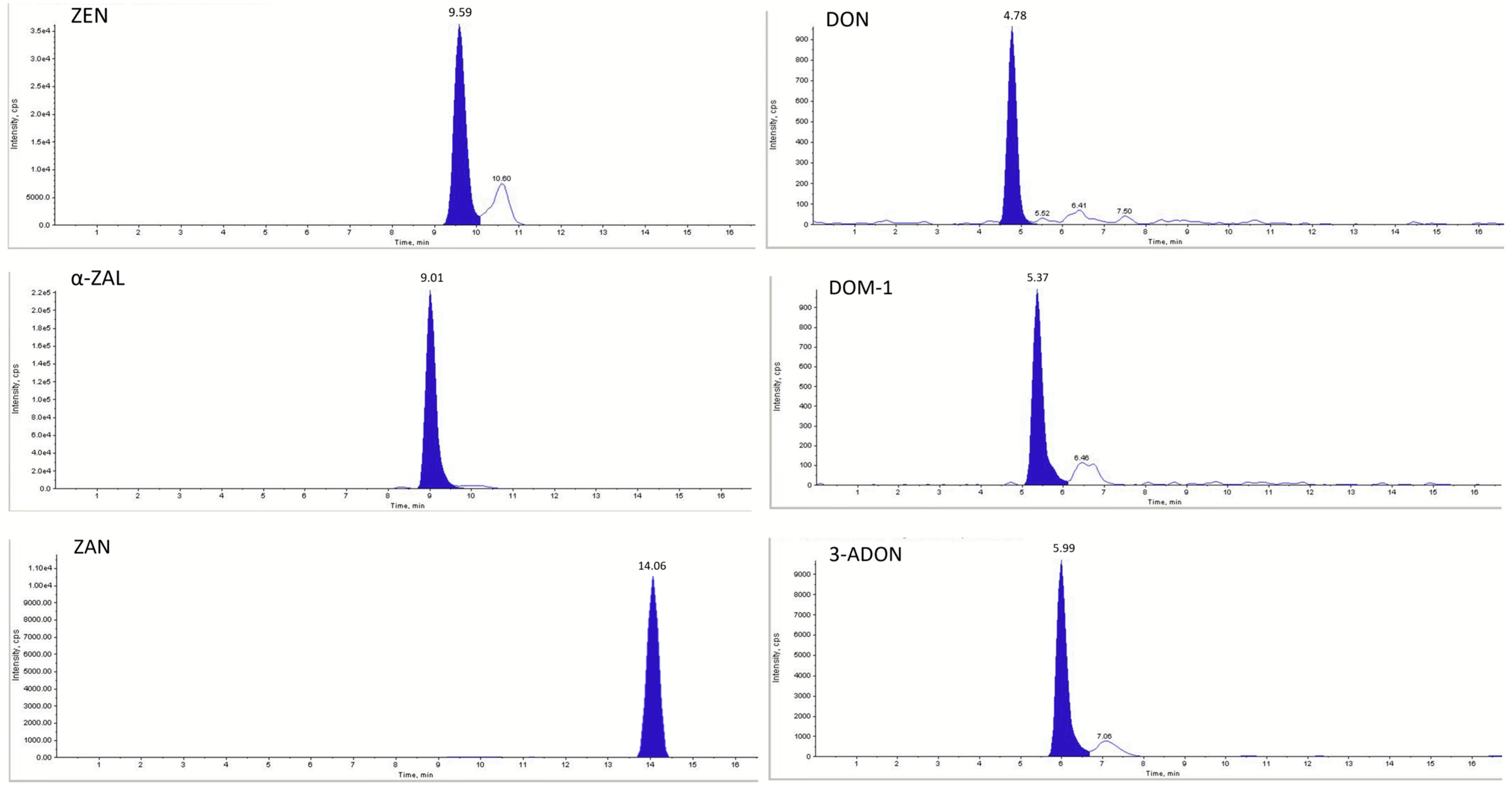
In the current study, unfortified blood samples (blank samples) were pipetted on the filter paper in triplicate, dried, and then extracted according to the previously described protocol. A comparison of the chromatogram of a blank sample (Figure 1) with those of fortified samples (Figure 2) showed no peaks at the retention times corresponding to the target metabolites.
The repeatability, intermediate precision, and mean recoveries for ZEN, α-ZAL, ZAN, DON, DOM-1, and 3-ADON are summarized in Table 2. The developed HPLC-MS/MS method was able to detect and quantify all six analytes across three concentration levels (20, 40, and 60 μg/L).
For intra-day performance (repeatability), relative standard deviation (RSDr) values ranged from 6.57% (DON at 60 μg/L) to 25.61% (DOM-1 at 20 μg/L), indicating variable precision depending on the analytes and concentration levels. The highest recovery during intra-day analyses was observed for DON at 20 μg/L (112.43%), while the lowest recovery was observed for 3-ADON at 60 μg/L (55.60%). ZEN, α-ZAL, and ZAN generally exhibited acceptable repeatability and recovery values at all concentration levels, although α-ZAL presented notably lower recoveries at 40 and 60 μg/L (62.11% and 66.43%, respectively).
Regarding inter-day performance (intermediate precision), RSDR values ranged from 7.64% (ZEN at 40 μg/L) to 33.59% (DOM-1 at 20 μg/L). Again, DON had the most consistent and accurate performance across days, with inter-day recoveries ranging from 94.23% to 106.94% and RSDR values below 20% at all concentration levels. In contrast, both α-ZAL and 3-ADON showed unsatisfactory mean values for Re at multiple concentration levels. For α-ZAL, Re values were below the regulatory acceptance range at 40 μg/L (57.86%) and 60 μg/L (67.25%). Similarly, 3-ADON showed inadequate recovery, especially at 40 μg/L (62.26%) and 60 μg/L (59.05%). As a result, both analytes failed to meet the acceptance criteria for Re established by EC 401/2006 [26].
Overall, while the method met the acceptance criteria set by EC 401/2006 [26] for most analytes and concentration levels, α-ZAL and 3-ADON did not fully comply with the regulatory limits for recovery and precision, particularly at higher concentrations for α-ZAL, and across all concentrations for 3-ADON.

4. Discussion

DON and ZEN, along with other metabolites assessed in this study, have been previously detected in the blood serum of swine [8,14,17,28]. However, differences persist across studies, particularly regarding the toxicokinetics of ZEN, its modified forms, and phase I metabolites. These metabolites were found at concentrations below the LOQ in swine plasma, likely due to the extensive first-pass biotransformation of ZEN [29,30]. Results from previous studies emphasize the need for reliable methodologies to advance toxicokinetic and biomarker research. Moreover, limited progress has been made in applying the DBS technique for detecting biomarkers in swine whole blood. Therefore, ZEN, α-ZAL, ZAN, DON, DOM-1, and 3-ADON were selected for the present study with the primary objective of developing a novel methodology to support future toxicokinetic investigations. This technique also represents a sustainable and innovative alternative for evaluating the efficacy of AMAs in vivo and is in line with the guidelines established by the EFSA [31], which recognizes the detection of specific biomarkers as valid endpoints for evaluating the efficacy of AMAs.
The 40 µL blood volume applied to the filter paper in the present research was determined according to Lauwers et al. (2019), who found no significant difference in detecting ZEN, α-ZAL, ZAN, DON, DOM-1, and 3-ADON among porcine blood volumes of 40, 50, 60, 70, and 80 µL on Whatman® 903 protein saver cards, which is specialized paper for biomaterial collection [22]. Whatman® 903 was also used to investigate the presence of ochratoxin A (OTA) in human blood, with a volume of 75 µL for the bloodstains [32]; it was also used to analyze 27 mycotoxins, including DON, ZEN, and ZAN, using 100 µL for bloodstains [25]. A similar volume was used by Cramer et al. (2015), who tested stains of 75, 100, and 125 μL and found no significant difference in OTA and 2’R-ochratoxin A (2’R-OTA) recoveries [24]. Despite the promising results, Whatman® 903 cards are relatively expensive, which may limit the widespread use of DBS methods for routine analysis in livestock. Therefore, the utilization of more accessible and affordable materials should be prioritized when developing new diagnostic methodologies, especially in developing countries where access to expensive supplies is limited. To the best of the authors’ knowledge, this is the first study to evaluate qualitative filter paper for the assessment of DBS containing mycotoxin biomarkers.
Regarding the organic extraction solvent, the present study supported prior research in both human and animal studies, favoring a water/ACN/acetone (30:35:35 v/v/v) solution for the extraction of DBS [22,25,32]. The influence of the sonication period for this type of extraction varied in the studies conducted from 30 min [22,25] to 1 h [24,32]. Considering the method developed herein, the optimal results were achieved with a 1 h sonication period. Consistent with the findings of Lauwers et al. (2019), the reconstitution solution based on water/MeOH/FA (60/40/0.1 v/v/v) yielded favorable results and was, therefore, selected for method validation [22].
In the current method, successful results were obtained using a water/AmAc solution (995:5, v/v) (solution A) and MeOH/water/AmAc solution (900:95:5, v/v/v) (solution B) for gradient mobile phases, as suggested by Mallmann et al. (2021) [33]. Regarding mobile phases, clear differences and similarities were observed among studies. Cramer et al. (2015) and Osteresch et al. (2016) used MeOH as solvent A and water as solvent B, each with 0.1% FA, demonstrating consistency in the choice of acidifying agent to improve ionization in mass spectrometric detection [24,32]. Lauwers et al. (2019) used water (solvent A) and ACN (solvent B) with 1% AA, indicating a divergence in the organic solvent and acid modifier selected [22]. Additionally, Osteresch et al. (2017) used ACN with 2% AA as eluent A and water with 0.1% AA as eluent B, further varying the concentration and type of acid used [25]. In contrast to previous research, the use of acids in the mobile phases did not yield good chromatographic results in the present method.
The linearity results obtained in the present method corroborate with previous studies [16,22,34,35] that also evaluated mycotoxins in complex biological matrices—such as blood and intestinal explants from rats, poultry, and swine—and reported correlation coefficients above 0.99. The high r values consistently observed across the studies reflect the linearity and robustness of the LC-MS/MS analytical methods applied for mycotoxin quantification, even when dealing with challenging matrices that may introduce matrix effects and analytical variability.
No significant matrix (DBS) effects were observed in the present study. Warth et al. (2013) identified matrix effects as one of the main challenges in the development of published multi-biomarker methods, which is an important limitation, especially when using HPLC-MS/MS [36]. In agreement with the present findings, Osteresch et al. (2017) did not find matrix effects for DON, ZEN, and ZAN in DBS [25].
According to Rosa da Silva et al. (2022) [37], method selectivity refers to the ability to accurately measure target analytes within a complex sample, even in the presence of potential interfering substances. In the present study, it was possible to observe that the blank chromatograms did not display any peaks at the retention times of DON, ZEN, and their metabolites, thereby confirming the selectivity of the method.
The present analytical results are similar to those reported by Osteresch et al. (2017) for DON, ZAN, and ZEN, with recoveries ranging from 78% to 114%, 100% to 113%, and 101% to 114% for the respective analytes [25]. In general, the recovery results for all analytes were, in some cases, higher than those found by Lauwers et al. (2019) [22], who identified recoveries lower than 60% for some analytes. To address this issue, the authors used internal standards, which were not used in the present study.
The EC (2006) [26] established validation criteria for analytical methods targeting the detection of DON and ZEN, excluding their metabolites. The performance requirements for DON are defined as concentration levels exceeding 100 µg/kg, which is considerably higher than the fortifications adopted in the present study (20, 40, and 60 µg/L). In contrast, the performance criteria for ZEN apply to concentrations either below or above 50 µg/kg, which are more consistent with the fortification levels employed in this study. Accordingly, the performance criteria for ZEN were considered for the validation of the method for ZEN, DON, and their metabolites. The results of ZEN, ZAN, DON, and DOM-1 met all the requirements established by the EC (2006) [26]. These four analytes did not exhibit an RSD greater than 25% at any concentration for repeatability (RSDr) and 40% for intermediate precision (RSDR), and their recoveries were within the established range for the concentrations of 20, 40, and 60 μg/L. In contrast, 3-ADON showed the lowest recovery at 58.66%, falling below acceptable levels. Inter-day recoveries for α-ZAL at 40 μg/L and 60 μg/L were also below the recommended limit, with a recovery of 57.86% and 67.25%, respectively.
The method demonstrated suitable performance parameters for most analytes; however, limitations were observed for 3-ADON and α-ZAL. The low overall recovery of 3-ADON (58.66%) indicates potential analytical challenges for this compound in the DBS matrix, possibly due to instability or low extraction efficiency. Although α-ZAL achieved a mean recovery of 74.66%, it failed to meet the minimum inter-day acceptable recoveries of 60% at 40 μg/L and 70% at 60 μg/L. These findings highlight the difficulties involved in the quantification of modified mycotoxins such as 3-ADON and α-ZAL in complex biological matrices, possibly due to matrix effects or limitations in extraction efficiency. Further method optimization could help improve the performance of these metabolites in future applications.
Following method validation, a total of 20 real blood samples were individually collected from pigs intended for human consumption at a local pork processing plant. No contamination with the validated analytes—ZEN, ZAN, DON, and DOM-1—was detected. Notably, this monitoring was conducted with no prior knowledge of the animals’ dietary history or the potential presence of mycotoxins in their feed. To obtain positive samples and assess the method’s sensitivity under known exposure conditions, a controlled in vivo trial using feed contaminated with known levels of DON and ZEN would be necessary. Nevertheless, the method described herein demonstrates potential for application in both experimental studies and field investigations—particularly in cases where DON or ZEN intoxication is suspected. In such scenarios, the DBS results could be correlated with feed contamination data to support diagnosis and risk assessment.
Muñoz-Solano et al. (2024) emphasized the need for the development of innovative methodologies capable of simultaneously detecting multiple compounds, with sufficient sensitivity to detect and quantify the low levels typically present in biological matrices [13]. In recent years, the DBS technique has emerged as a valuable tool for both qualitative and quantitative biological analyses [21]. In addition, previous authors have also highlighted that DBS techniques, due to their non-contagious nature and enhanced analyte stability within the dried matrix, present fewer logistical challenges and lower risks during storage and transport [22,38]. In this context, the methodology developed in the current study represents the first in a series of research that will be conducted with the aim of providing the livestock industry with a reliable tool for diagnosing mycotoxicosis from a small sample of blood impregnated on qualitative filter paper, which is an approach that is both affordable and suitable for long-distance transport.

5. Conclusions

The present study demonstrates the successful development and validation of a multi-analyte method for detecting ZEN, α-ZAL, ZAN, DON, DOM-1, and 3-ADON in swine using dried blood spots (DBSs). This method met regulatory acceptance criteria for the quantification of ZEN, ZAN, DON, and DOM-1, demonstrating its analytical performance for future in vivo studies. These findings reinforce the suitability of DBS as a minimally invasive and logistically practical sampling method. The utilization of qualitative filter paper, a novel and cost-effective approach, maintains analytical performance while enhancing the feasibility of mycotoxin biomonitoring in diverse and resource-limited settings. Future research may benefit from this validated approach to investigate the toxicokinetic profiles of DON and ZEN in swine under various exposure scenarios and to further explore its application as an innovative and sustainable tool for evaluating the efficacy of anti-mycotoxin additives in vivo.

Author Contributions

I.F.L.: Conceptualization, methodology, investigation, and writing—original draft; C.T.S.: formal analysis, data curation, and writing—review and editing; C.R.d.S.: methodology, validation, and formal analysis; L.M.d.L.S.: investigation and writing—review and editing; J.A.S.: methodology and writing—review and editing; L.M.C.L.: investigation and writing—review and editing; R.V.T.: investigation and writing—review and editing; C.A.M.: supervision, project administration, and writing—review and editing. All authors have read and agreed to the published version of the manuscript.

Funding

This study was financed in part by the Coordenação de Aperfeiçoamento de Pessoal de Nível Superior—Brasil (CAPES)—Finance Code 001.

Institutional Review Board Statement

Ethical review and approval were waived for this study due to the fact that the present study did not involve experiments with live animals.

Informed Consent Statement

Not applicable.

Data Availability Statement

The data presented in this study are available on request from the corresponding author.

Acknowledgments

The authors would like to thank the Coordenação de Aperfeiçoamento de Pessoal de Nível Superior (CAPES, Brazil) for providing financial support for this study.

Conflicts of Interest

The authors declare no conflicts of interest.

Abbreviations

The following abbreviations are used in this manuscript:
DONDeoxynivalenol
ZENZearalenone
α-ZALα-zearalanol
β-ZALβ-zearalanol
α-ZELα-zearalenol
β-ZELβ-zearalenol
ZANZearalanone
DOM-1Deepoxy-DON
3-ADON3-acetyl-DON
15-ADON15-acetyl-DON
HPLC-MS/MSHigh-performance liquid chromatography coupled with tandem mass spectrometry
AMAsAnti-mycotoxin additives
DBSsDried blood spots
MeOHMethanol
ACNAcetonitrile
FAFormic acid
AmAcAmmonium acetate
SSStandard solution
LODLimit of detection
LOQLimit of quantification
RSDRelative standard deviation
ReMean recovery

References

  1. ABPA—Associação Brasileira de Proteína Animal. Annual Report 2025; ABPA: São Paulo, Brazil, 2025; Available online: https://abpa-br.org/abpa-relatorio-anual/ (accessed on 19 May 2025).
  2. Pierron, A.; Alassane-Kpembi, I.; Oswald, I.P. Impact of Mycotoxin on Immune Response and Consequences for Pig Health. Anim. Nutr. 2016, 2, 63–68. [Google Scholar] [CrossRef]
  3. Eskola, M.; Kos, G.; Elliott, C.T.; Hajšlová, J.; Mayar, S.; Krska, R. Worldwide Contamination of Food-Crops with Mycotoxins: Validity of the Widely Cited ‘FAO Estimate’ of 25%. Crit. Rev. Food Sci. Nutr. 2020, 60, 2773–2789. [Google Scholar] [CrossRef] [PubMed]
  4. Zheng, W.; Feng, N.; Wang, Y.; Noll, L.; Xu, S.; Liu, X.; Lu, N.; Zou, H.; Gu, J.; Yuan, Y.; et al. Effects of Zearalenone and Its Derivatives on the Synthesis and Secretion of Mammalian Sex Steroid Hormones: A Review. Food Chem. Toxicol. 2019, 126, 262–276. [Google Scholar] [CrossRef] [PubMed]
  5. Pack, E.; Stewart, J.; Rhoads, M.; Knight, J.; De Vita, R.; Clark-Deener, S.; Schmale, D.G. Quantification of Zearalenone and α-Zearalenol in Swine Liver and Reproductive Tissues Using GC-MS. Toxicon X 2020, 8, 100058. [Google Scholar] [CrossRef] [PubMed]
  6. Serviento, A.M.; Brossard, L.; Renaudeau, D. An Acute Challenge with a Deoxynivalenol-Contaminated Diet Has Short- and Long-Term Effects on Performance and Feeding Behavior in Finishing Pigs. J. Anim. Sci. 2018, 96, 5209–5221. [Google Scholar] [CrossRef]
  7. Wellington, M.O.; Bosompem, M.A.; Rodrigues, L.A.; Columbus, D.A. Effect of Long-Term Feeding of Graded Levels of Deoxynivalenol on Performance, Nutrient Utilization, and Organ Health of Grower-Finisher Pigs (35 to 120 kg). J. Anim. Sci. 2021, 99, skab109. [Google Scholar] [CrossRef]
  8. Tkaczyk, A.; Jedziniak, P.; Zielonka, Ł.; Dąbrowski, M.; Ochodzki, P.; Rudawska, A. Biomarkers of Deoxynivalenol, Citrinin, Ochratoxin A and Zearalenone in Pigs after Exposure to Naturally Contaminated Feed Close to Guidance Values. Toxins 2021, 13, 750. [Google Scholar] [CrossRef] [PubMed]
  9. Al-Jaal, B.A.; Jaganjac, M.; Barcaru, A.; Horvatovich, P.; Zomer, P.; Horvatovich, M. Aflatoxin, Fumonisin, Ochratoxin, Zearalenone and Deoxynivalenol Biomarkers in Human Biological Fluids: A Systematic Literature Review, 2001–2018. Food Chem. Toxicol. 2019, 129, 211–228. [Google Scholar] [CrossRef]
  10. Payros, D.; Alassane-Kpembi, I.; Pierron, A.; Loiseau, N.; Pinton, P.; Oswald, I.P. Toxicology of Deoxynivalenol and Its Acetylated and Modified Forms. Arch. Toxicol. 2016, 90, 2931–2957. [Google Scholar] [CrossRef]
  11. Llorens, P.; Herrera, M.; Juan-García, A.; Payá, J.J.; Moltó, J.C.; Ariño, A.; Juan, C. Biomarkers of Exposure to Zearalenone in In Vivo and In Vitro Studies. Toxins 2022, 14, 291. [Google Scholar] [CrossRef]
  12. Tonini, C.; Oliveira, M.S.; Parmeggiani, E.B.; Sturza, D.A.F.; Mallmann, A.O.; Rubin, M.I.B.; Mallmann, C.A. Serological Biomarkers of Zearalenone Exposure in Beef Heifers Receiving Anti-Mycotoxin Additive. World Mycotoxin J. 2021, 14, 357–365. [Google Scholar] [CrossRef]
  13. Muñoz-Solano, B.; Lizarraga Pérez, E.; González-Peñas, E. Monitoring Mycotoxin Exposure in Food-Producing Animals (Cattle, Pig, Poultry, and Sheep). Toxins 2024, 16, 218. [Google Scholar] [CrossRef] [PubMed]
  14. Gambacorta, L.; Olsen, M.; Solfrizzo, M. Pig Urinary Concentration of Mycotoxins and Metabolites Reflects Regional Differences, Mycotoxin Intake and Feed Contaminations. Toxins 2019, 11, 378. [Google Scholar] [CrossRef] [PubMed]
  15. Delgado-Povedano, M.d.M.; Maris, E.; Kellner, N.; Mulisa, G.; Gámiz-Gracia, L.; García-Campaña, A.M.; De Boevre, M.; De Saeger, S.; Pero-Gascon, R. Liquid Chromatography–Tandem Mass Spectrometry for the Determination of Multiple Mycotoxins in Serum through Suspect Screening and Targeted Approaches: Advancing Human Mycotoxin Biomonitoring. Microchem. J. 2025, 208, 112562. [Google Scholar] [CrossRef]
  16. Chen, M.; Liu, X.; Yang, S.; Chen, Z.; Di, B.; Liu, W.; Yan, H. HPLC–MS/MS Method for the Simultaneous Determination of Aflatoxins in Blood: Toxicokinetics of Aflatoxin B1 and Aflatoxin M1 in Rats. J. Anal. Sci. Technol. 2022, 13, 27. [Google Scholar] [CrossRef]
  17. Panisson, J.C.; Wellington, M.O.; Bosompem, M.A.; Nagl, V.; Schwartz-Zimmermann, H.E.; Columbus, D.A. Urinary and Serum Concentration of Deoxynivalenol (DON) and DON Metabolites as an Indicator of DON Contamination in Swine Diets. Toxins 2023, 15, 120. [Google Scholar] [CrossRef]
  18. Guthrie, R.; Susi, A. A simple phenylalanine method for detecting phenylketonuria in large populations of newborn infants. Pediatrics 1963, 32, 338–343. [Google Scholar] [CrossRef]
  19. Moretti, M.; Freni, F.; Tomaciello, I.; Vignali, C.; Groppi, A.; Visonà, S.D.; Tajana, L.; Osculati, A.M.M.; Morini, L. Determination of Benzodiazepines in Blood and in Dried Blood Spots Collected from Post-Mortem Samples and Evaluation of the Stability over a Three-Month Period. Drug Test. Anal. 2019, 11, 1403–1411. [Google Scholar] [CrossRef]
  20. Gupta, K.; Mahajan, R. Applications and Diagnostic Potential of Dried Blood Spots. Int. J. Appl. Basic. Med. Res. 2018, 8, 1–2. [Google Scholar] [CrossRef] [PubMed]
  21. Malsagova, K.; Kopylov, A.; Stepanov, A.; Butkova, T.; Izotov, A.; Kaysheva, A. Dried Blood Spot in Laboratory: Directions and Prospects. Diagnostics 2020, 10, 248. [Google Scholar] [CrossRef] [PubMed]
  22. Lauwers, M.; Croubels, S.; De Baere, S.; Sevastiyanova, M.; Romera Sierra, E.M.; Letor, B.; Gougoulias, C.; Devreese, M. Assessment of Dried Blood Spots for Multi-Mycotoxin Biomarker Analysis in Pigs and Broiler Chickens. Toxins 2019, 11, 541. [Google Scholar] [CrossRef] [PubMed]
  23. Ministério da Agricultura, Pecuária e Abastecimento. Portaria n° 1.304, de 7 de agosto de 2018. In Normas técnicas de instalações e equipamentos para abate e industrialização de suínos; Ministério da Agricultura, Pecuária e Abastecimento: Brasília, DF, Brasil, 2018. [Google Scholar]
  24. Cramer, B.; Osteresch, B.; Muñoz, K.A.; Hillmann, H.; Sibrowski, W.; Humpf, H.-U. Biomonitoring Using Dried Blood Spots: Detection of Ochratoxin A and Its Degradation Product 2’R-Ochratoxin A in Blood from Coffee Drinkers*. Mol. Nutr. Food Res. 2015, 59, 1837–1843. [Google Scholar] [CrossRef]
  25. Osteresch, B.; Viegas, S.; Cramer, B.; Humpf, H.-U. Multi-Mycotoxin Analysis Using Dried Blood Spots and Dried Serum Spots. Anal. Bioanal. Chem. 2017, 409, 3369–3382. [Google Scholar] [CrossRef]
  26. Commission Regulation (EC). No. 401/2006 of 23 February 2006 Laying down the Methods of Sampling and Analysis for the Official Control of the Levels of Mycotoxins in Foodstuffs (Text with EEA Relevance). Off. J. Eur. Union. 2006, L70, 12–34. [Google Scholar]
  27. Eurachem. The Fitness for Purpose of Analytical Methods: A Laboratory Guide to Method Validation and Related Topics, 2nd ed.; Eurachem: Teddington, UK, 2014; ISBN 978-91-87461-59-0. [Google Scholar]
  28. Brezina, U.; Rempe, I.; Kersten, S.; Valenta, H.; Humpf, H.-U.; Dänicke, S. Diagnosis of Intoxications of Piglets Fed with Fusarium Toxin-Contaminated Maize by the Analysis of Mycotoxin Residues in Serum, Liquor and Urine with LC-MS/MS. Arch. Anim. Nutr. 2014, 68, 425–447. [Google Scholar] [CrossRef] [PubMed]
  29. Catteuw, A.; Broekaert, N.; De Baere, S.; Lauwers, M.; Gasthuys, E.; Huybrechts, B.; Callebaut, A.; Ivanova, L.; Uhlig, S.; De Boevre, M.; et al. Insights into In Vivo Absolute Oral Bioavailability, Biotransformation, and Toxicokinetics of Zearalenone, α-Zearalenol, β-Zearalenol, Zearalenone-14-Glucoside, and Zearalenone-14-Sulfate in Pigs. J. Agric. Food Chem. 2019, 67, 3448–3458. [Google Scholar] [CrossRef]
  30. Fleck, S.C.; Churchwell, M.I.; Doerge, D.R. Metabolism and Pharmacokinetics of Zearalenone Following Oral and Intravenous Administration in Juvenile Female Pigs. Food Chem. Toxicol. 2017, 106, 193–201. [Google Scholar] [CrossRef] [PubMed]
  31. EFSA Panel on Additives and Products or Substances used in Animal Feed (FEEDAP); Bampidis, V.; Azimonti, G.; Bastos, M.d.L.; Christensen, H.; Durjava, M.; Dusemund, B.; Kouba, M.; López-Alonso, M.; López Puente, S.; et al. Guidance on the Assessment of the Efficacy of Feed Additives. EFSA J. 2024, 22, e8856. [Google Scholar] [CrossRef]
  32. Osteresch, B.; Cramer, B.; Humpf, H.-U. Analysis of Ochratoxin A in Dried Blood Spots—Correlation between Venous and Finger-prick Blood, the Influence of Hematocrit and Spotted Volume. J. Chromatogr. B 2016, 1020, 158–164. [Google Scholar] [CrossRef] [PubMed]
  33. Mallmann, C.A.; Tonial Simões, C.; Kobs Vidal, J.; Rosa da Silva, C.; de Lima Schlösser, L.M.; Araújo de Almeida, C.A. Occurrence and Concentration of Mycotoxins in Maize Dried Distillers’ Grains Produced in Brazil. World Mycotoxin J. 2021, 14, 259–268. [Google Scholar] [CrossRef]
  34. Duarte, V.; Mallmann, A.O.; Tonini, C.; Liberalesso, D.; da Silva, C.R.; Simões, C.T.; Gressler, L.T.; Bracarense, A.P.F.R.L.; Mallmann, C.A. Ex Vivo and in Vitro Poultry Intestinal Models to Evaluate Antimycotoxins Additives. Cienc. Rural. 2021, 52, e20210277. [Google Scholar] [CrossRef]
  35. Alves Sarturi, J.; Tonial Simões, C.; Rosa da Silva, C.; Fabris Laber, I.; de Lima Schlösser, L.M.; Sturza, D.F.; Mallmann, C.A. An Investigation of Different Antimycotoxin Additives in Swine Intestinal Explants Challenged with Aflatoxin and Fumonisin: Ex Vivo and In Vitro Models. World Mycotoxin J. 2023, 16, 337–348. [Google Scholar] [CrossRef]
  36. Warth, B.; Sulyok, M.; Krska, R. LC-MS/MS-Based Multibiomarker Approaches for the Assessment of Human Exposure to Mycotoxins. Anal. Bioanal. Chem. 2013, 405, 5687–5695. [Google Scholar] [CrossRef] [PubMed]
  37. Rosa da Silva, C.; Tonial Simões, C.; Kobs Vidal, J.; Reghelin, M.A.; Araújo de Almeida, C.A.; Mallmann, C.A. Development and Validation of an Extraction Method Using Liquid Chromatography-Tandem Mass Spectrometry to Determine Patulin in Apple Juice. Food Chem. 2022, 366, 130654. [Google Scholar] [CrossRef] [PubMed]
  38. Velghe, S.; De Troyer, R.; Stove, C. Dried Blood Spots in Therapeutic Drug Monitoring and Toxicology. Expert. Opin. Drug Metab. Toxicol. 2018, 14, 1–3. [Google Scholar] [CrossRef] [PubMed]
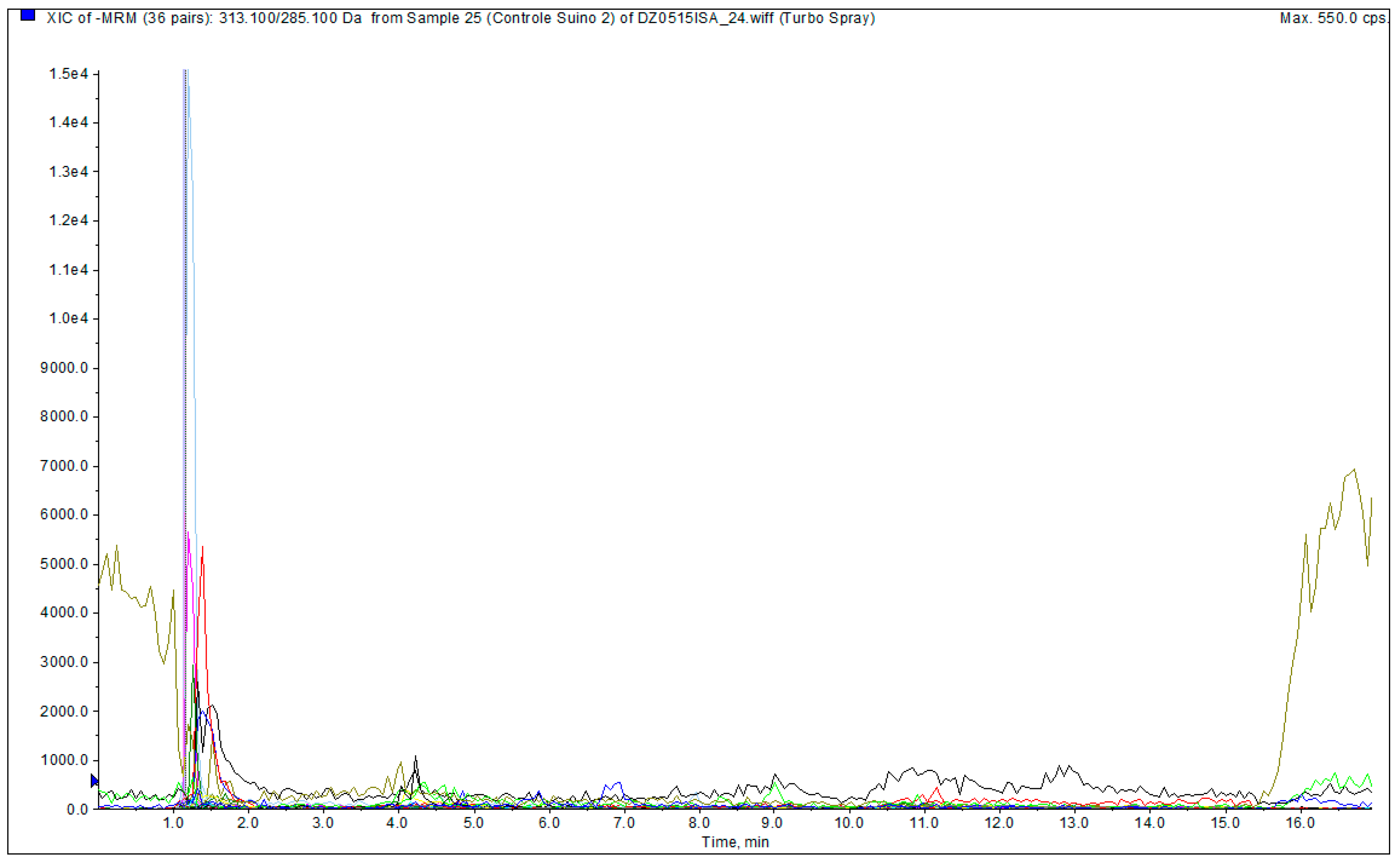
Figure 1. The chromatogram from an uncontaminated (blank) porcine DBS sample.
Chemosensors 13 00296 g001
Figure 2. Chromatograms of a porcine DBS sample fortified with 20 μg/L of ZEN, DON, and their metabolites.
Chemosensors 13 00296 g002
Table 1. Optimized HPLC-MS/MS parameters for the analysis of ZEN, α-ZAL, ZAN, DON, DOM-1, and 3-ADON in porcine dried blood spots (DBSs) using the multiple reaction monitoring (MRM) mode with negative electrospray ionization (ESI).
Analyte 1Precursor Ion (m/z)Product Ions (m/z)Retention Time (min)Declustering Potential (V)Collision Energy (V)Collision Cell Exit Potential (V)
QuantificationConfirmation
ZEN317.1175.0130.09.59−175−35−17
α-ZAL321.1277.0258.09.01−95−34−11
ZAN321.1277.0258.014.06−175−45−17
DON295.0265.0137.84.78−60−16−7
DOM-1278.9260.9248.85.37−45−14−13
3-ADON397.1384.2307.15.99−40−18−14
1 ZEN = zearalenone; α-ZAL = α-zearalanol; ZAN = zearalanone; DON = deoxynivalenol; DOM-1 = deepoxy-DON; and 3-ADON = 3-acetyl-DON.
Table 2. Validation results for intra- and inter-day precision and recovery of deoxynivalenol and zearalenone biomarkers in porcine blood impregnated on qualitative filter paper.
Analyte 1Repeatability and Intra-Day Recovery
(n = 7)
Intermediate Precision and Inter-Day Recovery
(n = 21)
20 μg/L40 μg/L60 μg/L20 μg/L40 μg/L60 μg/L
RSDr a (%)Re b (%)RSDr a (%)Re b (%)RSDr a (%)Re b (%)RSDR c (%)Re d (%)RSDR c (%)Re d (%)RSDR c (%)Re d
(%)
ZEN13.8191.217.2091.2110.5678.7419.1890.427.6487.4815.0789.40
α-ZAL14.81106.6410.1162.1110.8866.4325.4487.6516.6857.8615.1667.25
ZAN16.6379.5711.0578.939.8781.0515.4080.0010.3078.429.9980.97
DON9.35112.439.9095.186.57110.2919.98103.3316.9694.238.76106.94
DOM-125.6192.438.4786.647.2199.1733.5983.4820.8984.2426.2392.22
3-ADON13.22101.0012.8358.9611.1355.6019.7893.1817.9462.2628.0459.05
1 ZEN = zearalenone; α-ZAL = α-zearalanol; ZAN = zearalenone; DON = deoxynivalenol; DOM-1 = deepoxy-DON; and 3-ADON = 3-acetyl-DON. a Relative standard deviation of repeatability. b Mean recovery of the samples analyzed in septuplicate on the same day. c Relative standard deviation of the intermediate precision. d Mean recovery of the 21 replicates analyzed on three separate days.
Disclaimer/Publisher’s Note: The statements, opinions and data contained in all publications are solely those of the individual author(s) and contributor(s) and not of MDPI and/or the editor(s). MDPI and/or the editor(s) disclaim responsibility for any injury to people or property resulting from any ideas, methods, instructions or products referred to in the content.