Article

Electrochemical Pretreatment and Functionalization of Pencil Graphite Electrodes for Enhanced Transducer Performance in Biosensing

by
Rafael Mendes Coelho
1,
Alexandre Rafael Moraes da Silva
1,
Geycson Figueiredo Dias
1,
Danilo Bretas de Oliveira
2,
Arnaldo César Pereira
3,
Diego Leoni Franco
4 and
Lucas Franco Ferreira
1,*
1
Institute of Science and Technology, Federal University of the Jequitinhonha and Mucuri Valleys (UFVJM), Diamantina 39100-000, Minas Gerais, Brazil
2
Faculty of Medicine, Federal University of the Jequitinhonha and Mucuri Valleys (UFVJM), Diamantina 39100-000, Minas Gerais, Brazil
3
Department of Natural Sciences, Federal University of São João del-Rei (UFSJ), São João del-Rei 36307-352, Minas Gerais, Brazil
4
Chemistry Institute, Federal University of Uberlândia (UFU), Patos de Minas 38701-002, Minas Gerais, Brazil
*
Author to whom correspondence should be addressed.
Chemosensors 2025, 13(3), 84; https://doi.org/10.3390/chemosensors13030084
Submission received: 4 January 2025 / Revised: 13 February 2025 / Accepted: 25 February 2025 / Published: 2 March 2025
(This article belongs to the Special Issue Advanced Biosensors for Diagnostic Applications)

Abstract

:
This study aimed to optimize the electrochemical pretreatment and functionalization of pencil graphite electrodes (PGEs) for the performance evaluation of a transducer applied in initial studies in the development of an immunosensor for vaccinia virus (VACV) detection. The effects of the applied potential, duration, and supporting electrolyte type and concentration on PGE activation were investigated. Functionalization using a polymeric film derived from 2-hydroxybenzamide (2-HXB) was optimized by varying the applied potential, deposition time, and monomer concentration. Optimal activation conditions were found to be +0.90 V in 0.02 M of H2SO4 for 300 s, promoting the formation of hydrogenated groups and increasing electrode wettability. For electropolymerization, +1.20 V for 300 s with a 2-HXB concentration of 2.50 mM provided the best results, ensuring proper film formation and adhesion. Scanning electron microscopy revealed a rough, sheet-like surface on the polished PGE, while energy dispersive spectroscopy confirmed poly(2-HXB) adsorption through increased oxygen and nitrogen content on the functionalized electrode. The optimized pretreatment and functionalization conditions significantly influenced the response of the transducer used for VACV detection, demonstrating its crucial role in device development. These findings contribute to the advancement of inexpensive and effective electrochemical transducers and highlight the importance of pretreatment and modification of PGEs in biosensing applications.

1. Introduction

Historically, diseases have posed a significant risk to global public health [1,2,3]. A majority, exceeding 70%, are zoonotic, indicating their transmission between animals and humans owing to their genetic similarities [4,5,6,7,8]. Prominent examples of such zoonotic diseases include the Black Plague, influenza, dengue, chikungunya, and Zika viruses, along with more recent outbreaks, such as COVID-19 and monkeypox [9,10]. Gaining insights into zoonotic diseases is vital for preempting the rise in new pathogens that may pose future global health threats. Among these, the vaccinia virus (VACV) is a significant zoonotic agent that has been extensively investigated. Diseases caused by VACV and its variants have been identified in nearly 100 countries worldwide. Although global data on its prevalence are limited, outbreaks are more common in regions with extensive agricultural practices, especially in those with limited access to quality health services. It occurs more frequently in places known for dairy farming on American and Asian continents [11,12,13,14]. Despite extensive studies on VACV, its origin and natural reservoirs remain elusive [15,16,17].
Vaccinia is a zoonotic disease that initially causes cutaneous infections and vesicular lesions on the arms, hands, and face. The later stages involve symptoms such as fever, prostration, myalgia, lymphadenopathy, and increased susceptibility to secondary bacterial infections. Transmission can occur between humans, particularly within family groups [18,19,20,21]. The disease caused by the VACV of the Poxviridae family, which includes buffalopox and monkeypox viruses, resembles smallpox but is more aggressive and virulent in terms of contagion and symptoms [22,23,24,25,26].
VACV is found not only in low-income regions but also in African, Asian, and Latin American villages with low Human Development Index scores [11,15,19,27]. In Brazil, the virus is common among dairy animals and rural workers, particularly in Rio de Janeiro, Minas Gerais, and the Amazon region [19,20,28,29]. In a recent study conducted in Serro, Minas Gerais, approximately 240 individual samples were collected from both urban and rural areas, of which 74 individuals had immunity against the vaccinia virus, indicating a past infection, and 8 were currently symptomatic. This demonstrates the presence of the virus even in urban areas of interior cities, primarily because of its transmission through animal products, such as milk and cheese, which are essential to the local economy [30,31,32].
Early detection of the vaccinia virus is crucial to prevent complications, especially in immunosuppressed individuals. Current detection methods are costly, slow, and focus on the presence of anti-VACV antibodies, leading to delayed treatment and potential disease progression [22,33,34,35,36]. The early detection of pathogens is essential for outbreak control and disease prevention. However, traditional methods, such as an enzyme-linked immunosorbent assay (ELISA) and polymerase chain reaction (PCR), are expensive and require sophisticated laboratory infrastructure, limiting their use in remote areas. This highlights the need for new research into alternative detection methods, particularly in the early stages of infection when the virus is present but asymptomatic [37,38,39].
Electrochemical biosensors have emerged as promising alternatives, offering advantages such as low cost, operational simplicity, and miniaturization potential. Electrochemical methods, which are known for their sensitivity and wide applicability in clinical, environmental, and food industry analyses, are a promising approach [40,41,42,43]. Pencil graphite electrodes (PGEs) are accessible, low-cost, easy-to-modify, and highly versatile materials for analytical applications [44,45]. The possibility of functionalization with polymeric films can further increase their selectivity and sensitivity, making them suitable for detecting biomolecules and pathogens [37,46,47]. Furthermore, electrochemical pretreatment is often essential for these electrodes, making them even more specific and improving their surface properties, such as increasing their active area, functionalization, and adsorption capacity, which are crucial characteristics of biosensors. It should also be noted that the use of electrochemical techniques for VACV detection is rare in the literature. The few studies that exist in the area do not combine the processes of pre-treatment and electropolymerization on the working electrode [48,49]. However, electrochemical impedance spectroscopy (EIS) has been applied to this type of process.
VACV is typically associated with skin infections and their dissemination in rural areas, particularly in regions with intensive agricultural practices. Despite their importance in epidemiology, current detection techniques have limitations for field use, emphasizing the need for new approaches that combine accuracy, speed, and cost-effectiveness. Therefore, this study aimed to address some of these gaps by investigating PGE pretreatment and applying it in initial studies to develop an immunosensor for VACV. In addition, electrode modification was evaluated by electropolymerization with a polymeric film (PF) derived from 2-hydroxybenzamide (2-HXB), which was previously studied by the research group and proved to be effective for this purpose [37]. The study specifically focused on evaluating the impacts of the electrochemical pretreatment effects of PGEs, such as the applied potential and duration, as well as the type and concentration of the supporting electrolyte used. Moreover, it optimized the parameters of electrode functionalization with PF, including the applied potential, deposition time, and monomer concentration. This investigation may lead to new possibilities for portable and automated applications for VACV detection and applications of these transducers in biosensing.

2. Materials and Methods

2.1. Chemicals

All solutions used in this study were prepared with ultrapure water with a resistivity of 18.2 MΩ cm and conductivity of 0.054 μS cm−1, obtained using the Master System MS 2000 from Gehaka (São Paulo, Brazil). The main reagents used in the optimization experiments for PGE activation and functionalization steps included potassium chloride (KCl) with a purity of 99.0% from Dinâmica (Indaiatuba, Brazil), potassium ferrocyanide (K4[Fe(CN)6]·3H2O) with a purity greater than 99.5% from Sigma-Aldrich (São Paulo, Brazil), potassium ferricyanide (K3[Fe(CN)6]) with a purity greater than 99.0% (Sigma-Aldrich, São Paulo, Brazil), 2-hydroxybenzamide (HOC6H4CONH2) with a purity of 98.0% from Sigma-Aldrich (São Paulo, Brazil), sulfuric acid (H2SO4, 98.0%) from Dinâmica (Indaiatuba, Brazil), monopotassium phosphate (KH2PO4, 98.0%) from Vetec (Duque de Caxias, Brazil), dipotassium phosphate (K2HPO4) with a purity greater than 98.0% from Alphatec (São Bernardo do Campo, Brazil), and sodium hydroxide (NaOH) with a purity of 99.0% from Vetec (Duque de Caxias, Brazil). All solutions, except for the biological samples, were deoxygenated with N2 gas prior to analysis.

2.2. Virus and Antibodies Samples

Samples containing VACV (from the Western Reserve strain) were provided by the Laboratory of Infectious and Parasitic Diseases at the Federal University of Jequitinhonha and Mucuri Valleys (Brazil). The viruses’ mother solution (5.0 × 10⁵ virus mL−1) was diluted in phosphate-buffer saline (PBS) to a ten times lower concentration (5 × 10⁴ virus mL−1). Anti-VACV antibodies were obtained from Thermo Fisher Scientific Inc. (Waltham, USA) and diluted to 100 ng mL−1 before use.
Positive and negative controls were fetal bovine serum samples from Sigma Aldrich (São Paulo, Brazil), containing or without the VACV.

2.3. Electrochemical Measurements

Electrochemical measurements were performed using a Metrohm Autolab Potentiostat (PGSTAT 128N, St. Gallen, Switzerland) equipped with a FRA32M module and controlled using Nova 2.1.7 software. EIS experiments were conducted in a custom-designed electrochemical cell with a capacity of 300 µL with PGEs (HB, 0.90 mm, Hi-polymer Pentel) with a surface area of 0.64 mm2 as the working electrode, a graphite auxiliary electrode, and a Ag/AgCl (3.0 M of KCl) reference electrode. A 15.0 mL electrochemical cell was employed for the activation and electropolymerization steps, maintaining the same electrode configuration.

2.4. PGE Activation

Prior to activation, the PGE surface was isolated using a commercial nail polish to avoid any interference from graphite in the pencil. The activation procedure was performed following previous works [37]. Briefly, the electrodes were immersed in aqueous solutions of KCl and H2SO4, where a potential of +1.10 and −1.10 V for 300 s were applied. Different concentrations of the best supporting electrolyte (H2SO4) were tested between +0.7 and +1.70 V at different activation times. In all cases, the PGE was pre-polished, and electrodeposition was performed by applying +1.30 V for 300 s in an acidic solution containing 2.50 mM of the 2-HXB monomer. The charge transfer resistance (Rct) obtained by EIS of the immunosensor was compared with that of the positive and negative controls to evaluate each variable.

2.5. Electrochemical Analysis

An electrochemical analysis of the PGE behavior was conducted in 5.0 mM of potassium ferro/ferricyanide prepared in 0.10 M of KCl, at a scan rate of 50 mV s−1, within the potential range of −0.10 to +0.60 V. Electropolymerization was conducted in a 2.50 mM 2-HXB solution containing 0.50 M of sulfuric acid, in the potential range between +0.20 and +1.20 V. EIS analyses were also performed in 5.0 mM of K4[Fe(CN)6]/K3[Fe(CN)6] prepared in 0.10 M of KCl within the frequency range of 100 kHz to 10 mHz with a sinusoidal excitation amplitude of 10 mV.
The experimental data were fitted with a specific equivalent circuit using nova 2.1.7 software described by [Rs(Qdl[RctW])(R2Q2)] for the PGE-modified and [Rs(Qdl[RctW]) for the non-modified (before the formation of PF), where Rs is the ohmic resistance of the solution, Qdl is the electric double-layer capacitance, Rct is the charge-transfer resistance, W is the Warburg impedance, R2 is the polymer resistance, and Q2 is the polymer capacitance.

2.6. Electrodeposition of the Polymeric Film

For the electrodeposition of poly(2-HXB) on PGEs, three experimental parameters were investigated: potential, time, and monomer concentration. First, the potentials above the monomer oxidation peak (approximately +1.07 V) were studied, ranging from +1.10 to +1.50 V. The optimal potential was then used to vary the electropolymerization time between 100 s and 500 s. Lastly, the concentration of 2-HXB was varied from 25.0 µM to 25.0 mM. All experiments were conducted at room temperature under constant agitation using a magnetic stirrer at medium stirring speed.

2.7. Morphological Analysis

The electrode morphology was analyzed using scanning electron microscopy (SEM) with a Tescan bench microscope (Veja 3 LMH) equipped with an energy-dispersive X-ray spectroscopy (EDS) module. The surfaces of the electrode samples were covered with a thin layer of gold, and images were captured at 30 kV with magnifications of 200× and 5000×. The chemical compositions of the four types of electrodes were evaluated by EDS before immunosensor construction: (i) untreated PGE, (ii) PGE polished with 600-grit sandpaper, (iii) activated PGE, and (iv) PGE with electrodeposited poly(2-HXB).

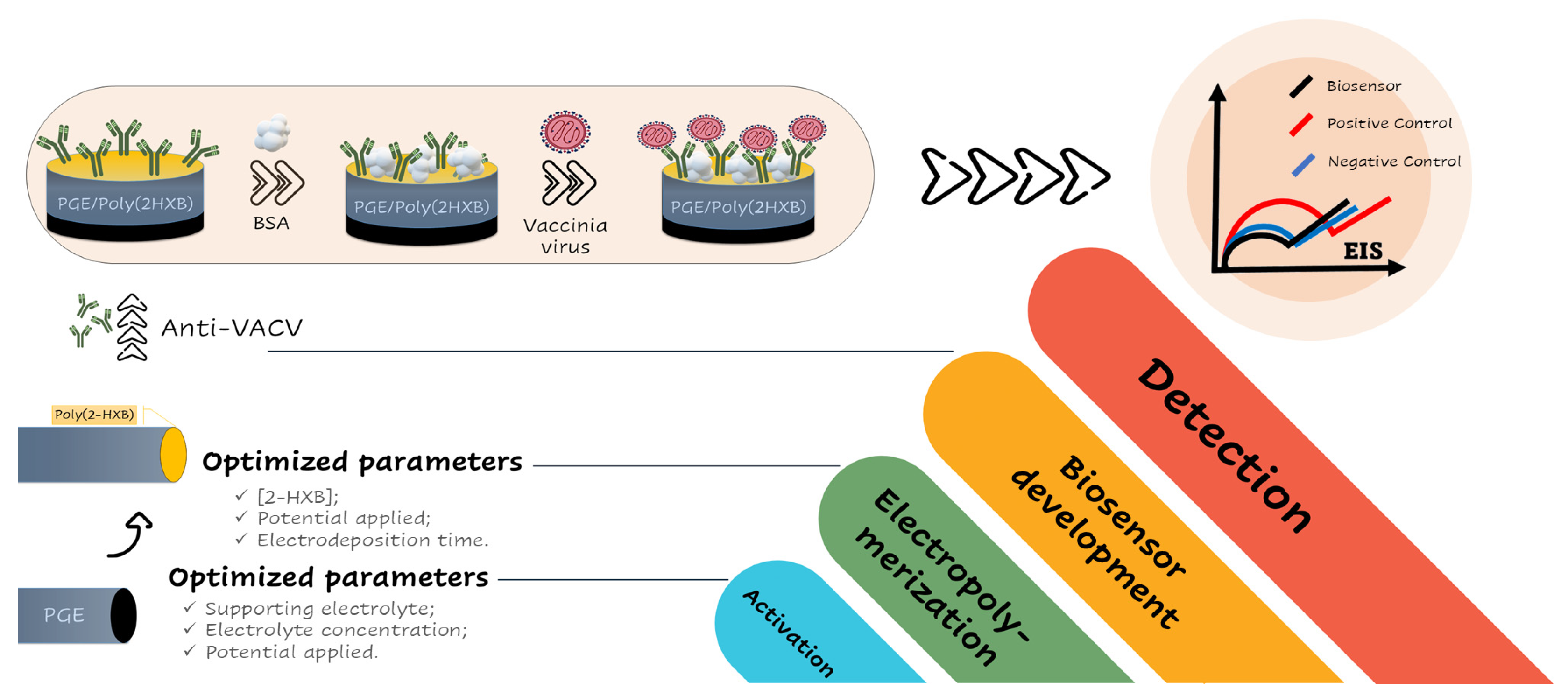
2.8. Immunosensors Fabrication Protocol

The immunosensor was developed by adding 15 µL of purified antibody solution (100 ng mL−1, 30 min) to the surface of PGE/poli(2-HXB) through physical adsorption. Then, 15 µL of 0.01% w/v bovine serum albumin (BSA) was added to the surface of the immunosensors for 10 min to block nonspecific interactions, and EIS analysis was used to monitor antigen–antibody interactions on the immunosensor surface. After each stage, the electrodes were washed with PBS (pH 7.20).
The interaction was monitored after exposing the immunosensor to bovine serum samples containing VACV (positive control, PC) or healthy bovine serum samples (negative control, NC). In this step, 15 µL of the PC or NC solution was placed on the immunosensor surface, and EIS analysis was performed. Figure 1 illustrates both the optimization steps (activation and functionalization) and the immunosensor development.

3. Results and Discussion

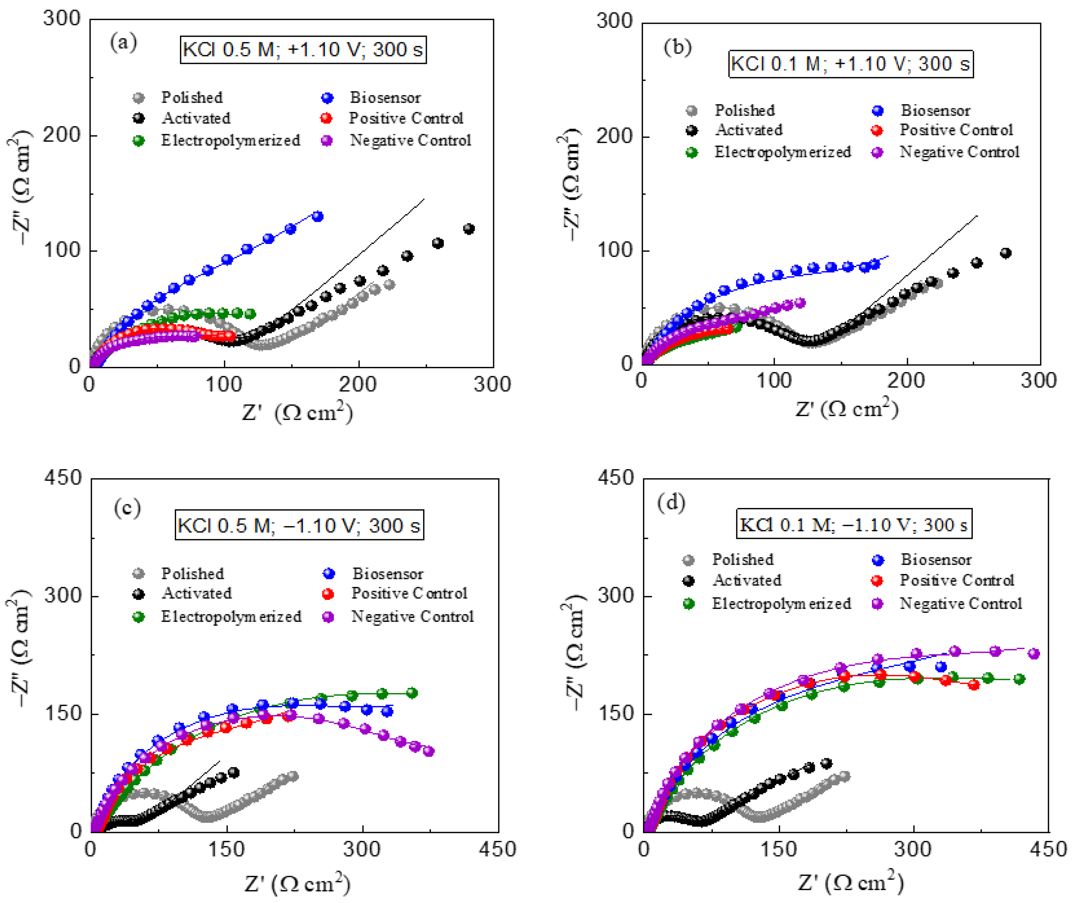
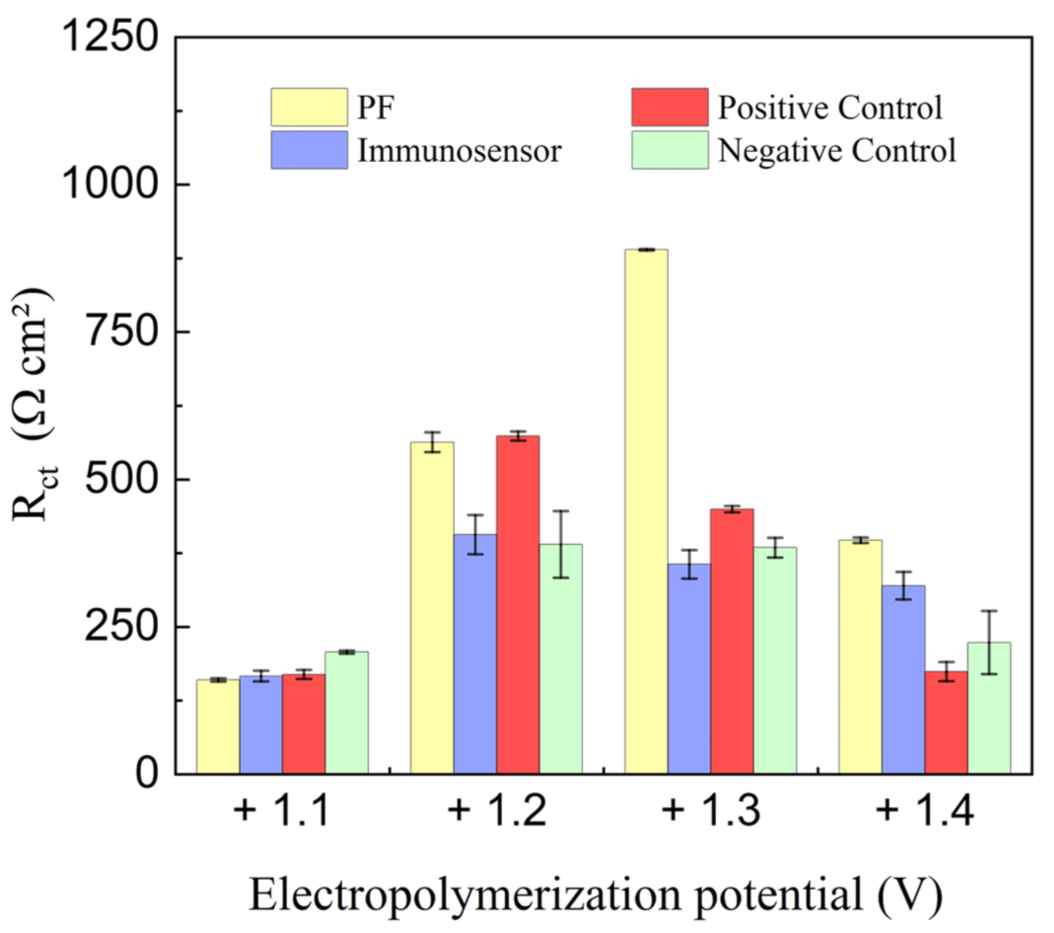
3.1. Investigation of Electrochemical Pretreatment of PGEs on Imunosensor Response

The initial analysis focused on the supporting electrolyte used during the activation step, employing KCl and H2SO4 at concentrations of 0.10 M and 0.50 M. Additionally, two potentials were applied for activation, a negative (−1.10 V) and a positive (+1.10 V). To develop an immunosensor for VACV detection, Nyquist diagrams were obtained for each condition, as shown in Figure 2, and the charge transfer resistances were analyzed.
The anodic or cathodic activations using KCl as the supporting electrolyte did not yield satisfactory results for virus detection, showing a negligible distinction between the Rct of the immunosensor and the positive and negative virus samples, as illustrated in the Nyquist diagrams of Figure 2, and the charge transfer resistances found in each are best described in Table 1. These results are in line with the findings in the literature [50,51,52,53,54,55,56], which predominantly recommend the use of acidic or buffered solutions as electrolytes for PGE activation, particularly when applying a positive potential or using cyclic voltammetry for this purpose.
For the activation performed with H2SO4, the Nyquist diagrams indicate an improved signal compared to KCl, especially with anodic activation at lower concentrations (0.10 M), as shown in Figure 3.
However, cathodic treatment using sulfuric acid, whether at 0.10 M or 0.50 M, did not show significant differences between the immunosensor response and the controls. Negative potential activation is not optimal, as some studies have reported that cathodic polarization does not generate functional groups on electrode surfaces [57,58]. Similar to the KCl results, there were no significant Rct variations in the positive or negative controls, except for the treatment with 0.10 M of H2SO4 at +1.10 V. Interestingly, the signal of the immunosensor with the positive control decreased by approximately 50%, as can also be seen in Table 2, contrary to expectations of an increase due to antigen–antibody interactions. This unexpected decrease in the signal might be due to weak antibody interactions with PGE/poli(2-HBX), influenced by electrochemical treatment.
When the immunosensor contacts the positive control, the immobilized antibodies may be removed, decreasing charge-transfer resistance. Anodic polarization, as described in the literature, is suitable for activating carbon-based electrodes by generating functional groups on the electrode surface, thereby increasing the response of the device. This treatment also increases the hydrophilicity of the material, thereby expanding the electrochemically active area in contact with the solution [57,59].
The concentration of the supporting electrolyte during the activation process is crucial for this type of electrode, as it is directly linked to the ionic strength of the medium [60,61]. However, higher concentrations of sulfuric acid did not yield satisfactory results. Therefore, more diluted H2SO4 concentrations were analyzed, maintaining the applied potential at +1.10 V. To test this hypothesis, activation was performed by using different dilutions of sulfuric acid. The Rct values for each stage of immunosensor construction are listed in Table 3.
The best reproducibility of impedimetric results for positive and negative VACV samples was achieved using a 0.02 M sulfuric acid concentration during activation, with the most significant difference in charge transfer resistance between the immunosensor and the positive control occurring under this condition, as shown in Figure 4. This suggests that using more dilute acidic electrolytes can facilitate the formation of graphite oxide on the electrode surface and hydrate the platform, enhancing conductivity [58]. However, concentrations that were too diluted, such as 0.01 M, do not produce this effect due to the low conductivity of the medium.
The optimal activation potential for the developed method was evaluated, spanning the range from +0.70 to +1.70 V in H2SO4 0.02 M. Additionally, the development of the immunosensor was analyzed under two conditions: without any prior electrochemical treatment; and without activation but with the presence of the polymer. As shown in Figure 5, the immunosensors constructed from PGEs that did not undergo the activation process or the electropolymerization of poly(2-HXB) did not satisfactorily distinguish between the positive samples and immunosensor, with the least satisfactory results observed for PGEs without activation and the polymer film. This was anticipated because activation ensures better electropolymerization of the polymer film through interactions between the functional groups generated during functionalization and the functional groups of the polymer film, increasing its adsorption on the transducer platform [57,59,62].
Activation potentials above +1.00 V did not yield promising results, probably because of the degradation of clay on the electrode surface and discrepancies in the electrochemically active area among the electrodes, making the surface excessively conductive and impeding the detection of increased resistance during the film electropolymerization or biological sample immobilization [63]. Moderate potentials, such as +0.70 V and +0.90 V, are preferable for activating this material type, ensuring adequate PGE activation without clay detachment from the surface and excessive gas generation from water electrolysis. Among these potentials, +0.90 V was chosen for subsequent tests because of the reproducibility of impedimetric results obtained for the immunosensor, positive control, and negative control.
Furthermore, using a potential of +0.90 V during VACV detection resulted in increased charge transfer resistance, contrasting with the reduction observed at higher potentials, and the response obtained for the negative control is very close to the immunosensor, which indicates another positive point when applying the potential of +0.90 V in the activation step.

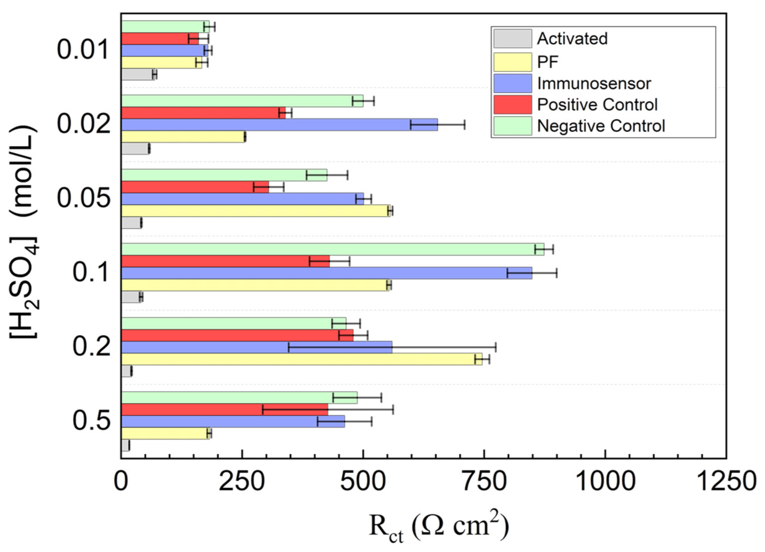
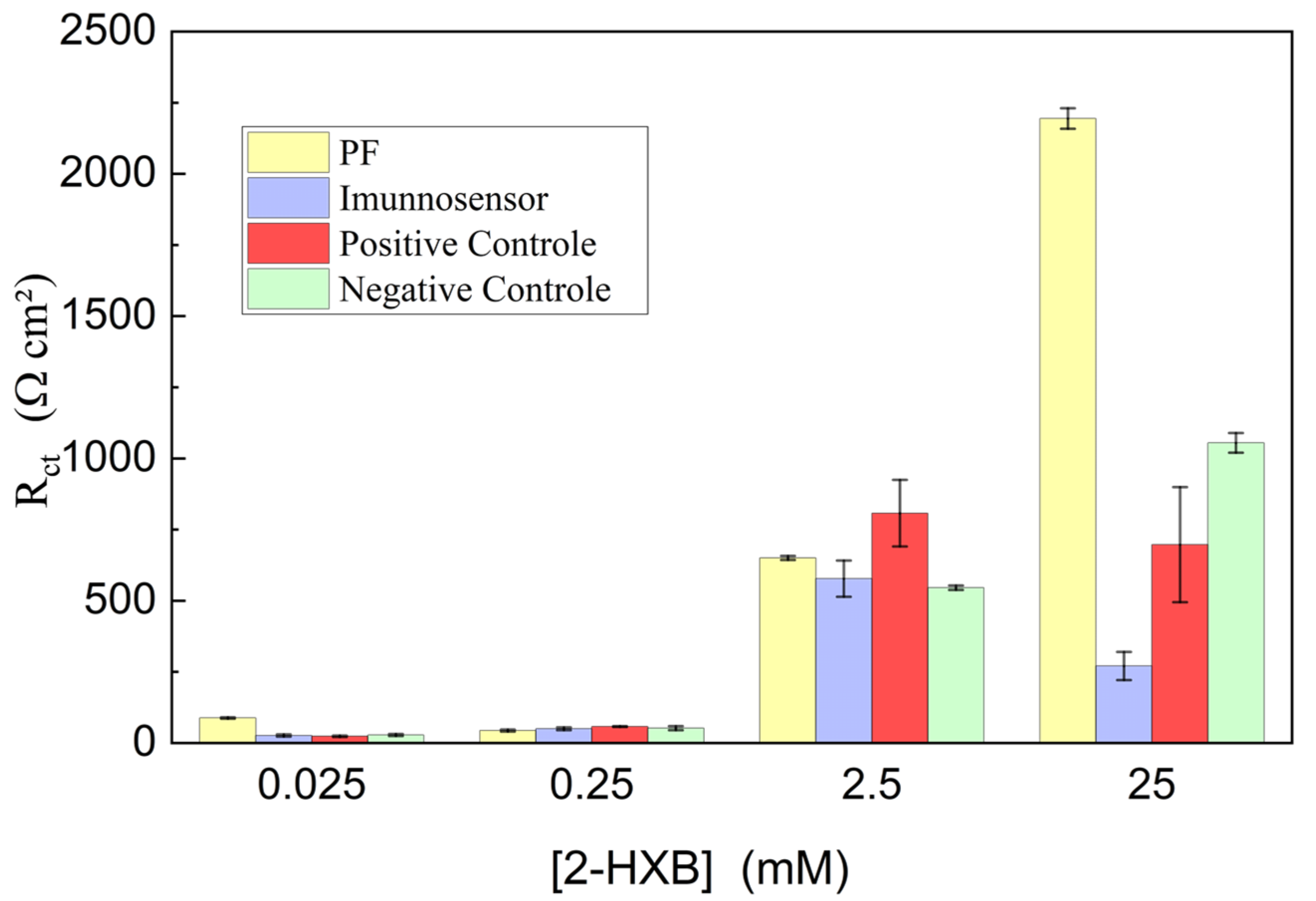
3.2. Investigation of PGE Functionalization Conditions in the Immunosensor Response

Electropolymerization potentials ranging from +1.10 to +1.40 V were tested, and the results are illustrated in Figure 6. The most suitable potentials for PF formation were identified as +1.20 V and +1.30 V, primarily due to the increased charge transfer resistance observed during the impedimetric detection of VACV in positive controls. However, the potential of +1.10 V was not ideal. Electrochemically, this potential is close to the oxidation potential of the monomer, leading to fewer radicals being available for polymer film formation. This results in low resistance associated with the formed film, indicating insufficient PF formation, which hinders efficient immobilization during immunosensor construction.
Conversely, applying higher potentials (+1.40 V) might result in excessive PF formation, potentially causing the film to detach from the platform, particularly due to agitation. This could introduce errors in subsequent viral detection, as evidenced by the lower Rct values compared to +1.20 V and +1.30 V potentials. Supporting the selection of the most appropriate potential for poly(2-HXB) on PGE, the potential of +1.20 V provided the best results. The higher ΔRct between the positive and negative controls, relative to the immunosensor, indicated better response for VACV detection. Ideally, a positive sample should exhibit an increase in ΔRct, while a negative sample should show a decrease or values close to zero, reflecting the absence of antigen–antibody interactions, just like when +1.20 V is used as electropolymerization potential.
Electropolymerization time is a crucial factor in PF formation. For electropolymerization times shorter than 300 s, the system failed to properly distinguish between positive and negative controls. Specifically, at 200 s, high standard deviations were observed for negative samples, indicating inconsistent detection. At 100 s, Rct decreased for VACV, but the impedimetric response was still too close to that of the immunosensor, which is undesirable, as shown in Table 4. At deposition times exceeding 300 s, an anomalous decrease in the resistance during VACV detection was observed. This decrease was likely due to detachment of the PF/anti-VACV/VACV interaction from the PGE surface during electrode washing. This detachment results from an excessive amount of deposited film, which can peel off the electrode, leaving the active area exposed and reducing the impedimetric response. A 300 s electropolymerization time resulted in an approximately 39% increase in VACV detection compared to the immunosensor and a 5.4% reduction in resistance for negative samples.
The final parameter evaluated for the functionalization of PGE was the concentration of the monomer (2-HXB). These studies revealed that monomer concentration significantly influenced the response of the immunosensor. Very dilute concentrations resulted in negligible changes, likely because of insufficient polymer film incorporation into the electrode. On the other hand, at saturated monomer concentrations, the polymer film response was considerable, indicating high adsorption of PF, which may have subsequently detached from the PGE surface, abruptly reducing Rct, as shown in Figure 7. Among the concentrations investigated, 2.50 mM provided the best response. Lower concentrations showed minimal variation in the Rct values, regardless of the presence of PC or NC. At 25 mM of 2-HXB, the NC response overshadowed the PC response.
An optimal monomer concentration of 2.50 mM was found to strike a balance between sufficient polymer film incorporation and stable adsorption on the PGE surface. This concentration allowed the formation of a robust and responsive polymer layer without oversaturation or detachment issues. The electrochemical behavior observed at this concentration demonstrated clear distinctions between the positive and negative controls, enabling the reliable detection of the target analyte.
Further investigation into the kinetics of polymer formation and the mechanisms of analyte interaction with the functionalized surface could provide valuable insights for optimizing the immunosensor performance. Additionally, exploring the effects of environmental factors such as pH, temperature, and ionic strength on the polymer film stability and sensor response could enhance the overall robustness and applicability of immunosensors in various analytical settings.

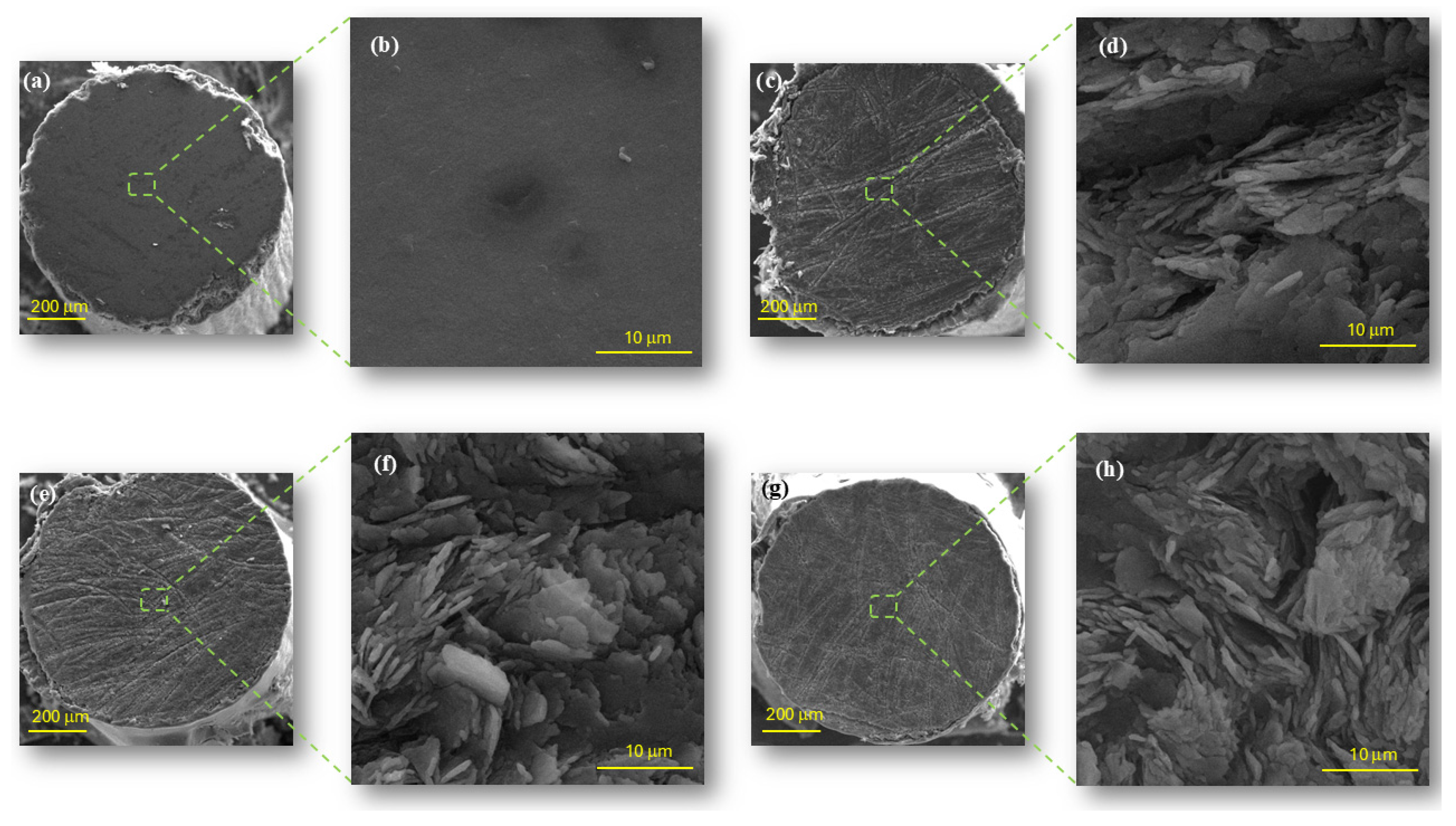
3.3. Morphological Analysis by Scanning Electron Microscopy (SEM) and Energy Dispersive Spectrometry (EDS)

Considering the optimized activation and electropolymerization conditions, the surface morphology of the PGE was analyzed at each stage of transducer preparation, including untreated, activated, and functionalized PGE. This analysis was conducted primarily to verify the effects of polishing and electrode functionalization. The scanning electron microscopy (SEM) images of each electrode are shown in Figure 8. By analyzing the images, it was observed that the electrodes subjected to polishing, activation, and electropolymerization did not exhibit drastic changes in their surface morphology. This result is plausible because the changes occurred at the molecular level and did not significantly alter the structure of the material. The rough, scaly surfaces observed in Figure 8d,f,h have been reported in the literature as a characteristic of this type of material when polished with sandpaper. Depending on the type of sandpaper used, these scales can vary in size and shape [64,65,66].
Comparing these electrodes, the most significant morphological difference was observed between the untreated PGE (in natura, Figure 8a) and polished PGE (Figure 8b). There are two main differences: (i) the transition from a smooth, undeformed surface to a rough structure resembling small plates due to polishing, and (ii) the lateral coating with enamel, which appears uniform but is affected by polishing at its edges. This effect is mainly visible in the left portion of Figure 8b.
Constitutional analysis of the electrodes was performed using energy-dispersive spectroscopy (EDS). The elemental percentages on the surfaces of each PGE are listed in Table 5. Throughout the PGE fabrication process, there were no constitutional changes owing to material polishing. Upon activation, minimal changes occurred on the electrode surface, with an almost imperceptible increase in the amount of oxygen on the platform.
A significant compositional change occurred, owing to electropolymerization on the PGE surface. A substantial increase in oxygen and nitrogen was observed, strongly indicating the deposition of poly(2-HXB), where these elements are present in the precursor monomer structure. Although carbon exists in both materials (electrode and polymeric film), its percentage decreases because of the presence of atoms such as O and N, which make up the polymeric film electrodeposited on the surface, reducing the amount of surface carbon in the solid. Another important aspect to highlight is the presence of silicon in the form of clay, which constitutes all analyzed electrodes. Optimization of the activation and functionalization steps of PGE plays a crucial role in constructing an effective immunosensor for VACV detection. It is evident that these parameters significantly influence the response of the device. Ongoing studies will be carried out for the development of immunosensors using such procedures, aiming to evaluate the sensitivity and selectivity of the device, thus ensuring reliable results in subsequent analyses.

4. Conclusions

The optimized results of the PGE pretreatment and electropolymerization steps were fundamental for improving the response of the electrochemical transducer applied in the impedimetric detection of VACV. The activation of the PGE was crucial, with the most favorable conditions being a potential of +0.90 V in 0.02 M of H2SO4 for 300 s. This promoted the formation of hydrogenated groups on the electrode surface and increased its wettability, which was essential for the purpose of this study. Regarding electropolymerization, excessively high potentials above the oxidation potential of the precursor monomer were unsuitable because of the removal of clay material from the PGE surface. The most suitable potential was +1.20 V, with an electropolymerization time of 300 s to ensure the proper formation of the polymeric film. Additionally, the monomer concentration was critical, with a concentration of 2.50 mM being the most effective for PGE functionalization, avoiding excesses that could compromise the adhesion of the film to the electrode.
SEM analyses revealed that the surface of the material polished with 600-grit sandpaper was rough, with overlapping sheets. An EDS constitutional analysis of the electrodes indicated no significant differences in the composition among the untreated, polished, and activated PGE. However, when analyzing the functionalized electrode, there was a clear increase in the oxygen percentage and the appearance of nitrogen on the PGE surface, indicating the electropolymerization and adsorption of poly(2-HXB).

Author Contributions

R.M.C.: conceptualization, methodology, investigation, writing—original draft. A.R.M.d.S.: methodology, investigation, writing—original draft. G.F.D.: methodology, investigation. D.B.d.O.: resources, conceptualization, writing—review and editing. A.C.P. and D.L.F.: writing—review and editing, supervision, project administration. L.F.F.: conceptualization, supervision, project administration, funding acquisition, writing—review and editing. All authors have read and agreed to the published version of the manuscript.

Funding

The authors gratefully acknowledge financial support from Fundação de Amparo à Pesquisa do Estado de Minas Gerais (FAPEMIG) grant numbers APQ-00207-21, RED-00032-22, and RED-00196-23. Conselho Nacional de Desenvolvimento Científico e Tecnológico (CNPq) grant number 404210/2021-0 and Program for National Institutes of Science and Technology-Poxviruses (INCT-Pox) by CNPq grant number 406441/2022-7. This study was financed in part by the Coordenação de Aperfeiçoamento de Pessoal de Nível Superior, Brazil (CAPES) (Finance Code 001).

Institutional Review Board Statement

Not applicable.

Informed Consent Statement

Not applicable.

Data Availability Statement

Data supporting the results of this study are included in this article.

Conflicts of Interest

The authors declare no conflicts of interest.

References

  1. Bragazzi, N.L.; Lehr, T. Big Epidemiology: The Birth, Life, Death, and Resurgence of Diseases on a Global Timescale. Epidemiologia 2024, 5, 669–691. [Google Scholar] [CrossRef] [PubMed]
  2. Barouki, R.; Kogevinas, M.; Audouze, K.; Belesova, K.; Bergman, A.; Birnbaum, L.; Boekhold, S.; Denys, S.; Desseille, C.; Drakvik, E.; et al. The COVID-19 Pandemic and Global Environmental Change: Emerging Research Needs. Environ. Int. 2021, 146, 106272. [Google Scholar] [CrossRef]
  3. Leik, N.K.O.; Ahmedy, F.; Mac Guad, R.; Baharuddin, D.M.P. COVID-19 Vaccine and Its Consequences in Pregnancy: Brief Review. Ann. Med. Surg. 2021, 72, 103103. [Google Scholar] [CrossRef] [PubMed]
  4. Jones, K.E.; Patel, N.G.; Levy, M.A.; Storeygard, A.; Balk, D.; Gittleman, J.L.; Daszak, P. Global Trends in Emerging Infectious Diseases. Nature 2008, 451, 990–993. [Google Scholar] [CrossRef] [PubMed]
  5. Souza, M.J. One Health: Zoonoses in the Exotic Animal Practice. Vet. Clin. N. Am. Exot. Anim. Pract. 2011, 14, 421–426. [Google Scholar] [CrossRef]
  6. Rahman, M.T.; Sobur, M.A.; Islam, M.S.; Ievy, S.; Hossain, M.J.; Zowalaty, M.E.E.; Rahman, A.M.M.T.; Ashour, H.M. Zoonotic Diseases: Etiology, Impact, and Control. Microorganisms 2020, 8, 1405. [Google Scholar] [CrossRef]
  7. Chomel, B.B. Zoonoses. In Encyclopedia of Microbiology; Elsevier: Amsterdam, The Netherlands, 2009; p. 820. [Google Scholar] [CrossRef]
  8. Borremans, B.; Faust, C.; Manlove, K.R.; Sokolow, S.H.; Lloyd-Smith, J.O. Cross-Species Pathogen Spillover across Ecosystem Boundaries: Mechanisms and Theory. Philos. Trans. R. Soc. B 2019, 374, 20180344. [Google Scholar] [CrossRef]
  9. Morens, D.M.; Fauci, A.S. Emerging Pandemic Diseases: How We Got to COVID-19. Cell 2020, 182, 1077–1092. [Google Scholar] [CrossRef]
  10. Bunge, E.M.; Hoet, B.; Chen, L.; Lienert, F.; Weidenthaler, H.; Baer, L.R.; Steffen, R. The Changing Epidemiology of Human Monkeypox—A Potential Threat? A Systematic Review. PLoS Negl. Trop. Dis. 2022, 16, e0010141. [Google Scholar] [CrossRef]
  11. Luques, M.N.; Oliveira, R.L.; Hir, S.; Nunes, D.d.S.; Higa, L.M.; Mendonça, A.F.; Pereira, L.A.; Sousa, F.; Castiñeiras, T.M.P.P.; Tanuri, A.; et al. Co-Circulation of Vaccinia and Monkeypox Viruses in Rural Areas of Brazil: Importance of Differential Molecular Diagnosis. Travel Med. Infect. Dis. 2023, 53, 102578. [Google Scholar] [CrossRef]
  12. Alcamí, A. Pathogenesis of the Circulating Mpox Virus and Its Adaptation to Humans. Proc. Natl. Acad. Sci. USA 2023, 120, e2301662120. [Google Scholar] [CrossRef] [PubMed]
  13. Roberts, K.L.; Smith, G.L. Vaccinia Virus Morphogenesis and Dissemination. Trends Microbiol. 2008, 16, 472–479. [Google Scholar] [CrossRef] [PubMed]
  14. Xu, T.; Pan, Z.; Li, X.; Zhao, M.; Li, Z.; Zhang, L. Narciclasine Inhibits Vaccinia Virus Infection by Activating the RhoA Signaling Pathway. Biosaf. Health 2024, 6, 341–349. [Google Scholar] [CrossRef]
  15. Trindade, G.S.; Emerson, G.L.; Carroll, D.S.; Kroon, E.G.; Damon, I.K. Brazilian Vaccinia Viruses and Their Origins. Emerg. Infect. Dis. 2007, 13, 965–972. [Google Scholar] [CrossRef]
  16. Drumond, B.P.; Leite, J.A.; da Fonseca, F.G.; Bonjardim, C.A.; Ferreira, P.C.P.; Kroon, E.G. Brazilian Vaccinia Virus Strains Are Genetically Divergent and Differ from the Lister Vaccine Strain. Microbes Infect. 2008, 10, 185–197. [Google Scholar] [CrossRef]
  17. Gubser, C.; Hué, S.; Kellam, P.; Smith, G.L. Poxvirus Genomes: A Phylogenetic Analysis. J. Gen. Virol. 2004, 85, 105–117. [Google Scholar] [CrossRef]
  18. Dunlop, L.R.; Oehlberg, K.A.; Reid, J.J.; Avci, D.; Rosengard, A.M. Variola Virus Immune Evasion Proteins. Microbes Infect. 2003, 5, 1049–1056. [Google Scholar] [CrossRef]
  19. Diaz, J.H. The Disease Ecology, Epidemiology, Clinical Manifestations, Management, Prevention, and Control of Increasing Human Infections with Animal Orthopoxviruses. Wilderness Environ. Med. 2021, 32, 528–536. [Google Scholar] [CrossRef]
  20. Fiocruz, F.O.C. Pesquisadores Explicam a Atual Importância Do Vírus Vaccinia. Available online: https://agencia.fiocruz.br/pesquisadores-explicam-a-atual-importância-do-vírus-vaccinia (accessed on 24 February 2022).
  21. Pasternak, N. Varíola é a Única Doença Erradicada No Mundo−Saúde−Estadão. Available online: https://saude.estadao.com.br/noticias/geral,variola-e-a-unica-doenca-erradicada-no-mundo,70003870535 (accessed on 26 February 2022).
  22. Tegnell, A.; Wahren, B.; Elgh, F. Smallpox—Eradicated, but a Growing Terror Threat. Clin. Microbiol. Infect. 2002, 8, 504–509. [Google Scholar] [CrossRef]
  23. Xiang, Y.; Lane, R.K. Vaccinia Virus (Poxviridae). Encycl. Virol. 2021, 1–5, 854–859. [Google Scholar] [CrossRef]
  24. Fonseca, F.G.; Kroon, E.G.; Nogueira, M.L.; de Souza Trindade, G. Zoonotic Vaccinia Virus Outbreaks in Brazil. Futur. Virol. 2011, 6, 697–707. [Google Scholar] [CrossRef]
  25. Kleo, K.; Kapp, A.; Ascher, L.; Lisdat, F. Detection of Vaccinia Virus DNA by Quartz Crystal Microbalance. Anal. Biochem. 2011, 418, 260–266. [Google Scholar] [CrossRef]
  26. Donaldson, K.A.; Kramer, M.F.; Lim, D.V. A Rapid Detection Method for Vaccinia Virus, the Surrogate for Smallpox Virus. Biosens. Bioelectron. 2004, 20, 322–327. [Google Scholar] [CrossRef]
  27. Trindade, G.D.S.; Emerson, G.L.; Sammons, S.; Frace, M.; Govil, D.; Mota, B.E.F.; Abrahão, J.S.; de Assis, F.L.; Olsen-Rasmussen, M.; Goldsmith, C.S.; et al. Serro 2 Virus Highlights the Fundamental Genomic and Biological Features of a Natural Vaccinia Virus Infecting Humans. Viruses 2016, 8, 328. [Google Scholar] [CrossRef] [PubMed]
  28. Quixabeira-Santos, J.C.; Medaglia, M.L.G.; Pescador, C.A.; Damaso, C.R. Animal Movement and Establishment of Vaccinia Virus Cantagalo Strain in Amazon Biome, Brazil. Emerg. Infect. Dis. 2011, 17, 726–729. [Google Scholar] [CrossRef] [PubMed]
  29. Sasso, E.; D’Alise, A.M.; Zambrano, N.; Scarselli, E.; Folgori, A.; Nicosia, A. New Viral Vectors for Infectious Diseases and Cancer. Semin. Immunol. 2020, 50, 101430. [Google Scholar] [CrossRef] [PubMed]
  30. Costa, G.B.; Borges, I.A.; Alves, P.A.; Miranda, J.B.; Luiz, A.P.M.F.; Ferreira, P.C.P.; Abrahão, J.S.; Moreno, E.C.; Kroon, E.G.; Trindade, G.d.S. Alternative Routes of Zoonotic Vaccinia Virus Transmission, Brazil. Emerg. Infect. Dis. 2015, 21, 2244–2246. [Google Scholar] [CrossRef]
  31. Costa, G.B.; Moreno, E.C.; Trindade, G.d.S. Neutralizing Antibodies Associated with Exposure Factors to Orthopoxvirus in Laboratory Workers. Vaccine 2013, 31, 4706–4709. [Google Scholar] [CrossRef]
  32. Costa, G.B.; Augusto, L.T.S.; Leite, J.A.; Ferreira, P.C.P.; Bonjardim, C.A.; Abrahão, J.S.; Kroon, E.G.; Moreno, E.C.; Trindade, G.d.S. Seroprevalence of Orthopoxvirus in Rural Brazil: Insights into Anti-OPV Immunity Status and Its Implications for Emergent Zoonotic OPV. Virol. J. 2016, 13, 1–8. [Google Scholar] [CrossRef]
  33. Fedorko, D.P.; Preuss, J.C.; Fahle, G.A.; Li, L.; Fischer, S.H.; Hohman, P.; Cohen, J.I. Comparison of Methods for Detection of Vaccinia Virus in Patient Specimens. J. Clin. Microbiol. 2005, 43, 4602–4606. [Google Scholar] [CrossRef]
  34. Generalov, V.M.; Naumova, O.V.; P’yankov, S.A.; Kolosova, I.V.; Safatov, A.S.; Zaitsev, B.N.; Zaitseva, E.G.; Buryak, G.A.; Cheremiskina, A.A.; Filatova, N.A.; et al. Identifying the Vaccinia Virus with the Use of a Nanowire Silicon-on-Insulator Biosensor. Optoelectron. Instrum. Data Process. 2021, 57, 37–43. [Google Scholar] [CrossRef]
  35. Rakhimbekova, A.; Kudaibergenov, B.; Seitkamal, K.; Bellone, A.; Dauletova, A.; Sypabekova, M.; Olivero, M.; Perrone, G.; Radaelli, A.; Zanotto, C.; et al. Rapid detection of vaccinia virus using biofunctionalized fiber-optic ball-tip biosensors. Sci. Rep. 2023, 13, 17470. [Google Scholar] [CrossRef]
  36. Li, Z.; Liu, R. Construction and Isolation of Recombinant Vaccinia Virus by Homologous Recombination Using Fluorescent Protein Markers. In Vaccinia, Mpox, and Other Poxviruses: Methods and Protocols, 1st ed.; Springer Protocols; Yang, Z., Satheshkumar, P.S., Eds.; Humana Press: New York, NY, USA, 2025; Volume 2860, pp. 83–95. [Google Scholar] [CrossRef]
  37. Coelho, R.M.; de Oliveira Almeida, A.; Soares, P.I.; Rocha, K.L.S.; de Oliveira, D.B.; Pereira, A.C.; Franco, D.L.; Ferreira, L.F. A Low-Cost Electrochemical Biosensor for Vaccinia Virus Using Pencil Graphite Electrodes Modified with Poly(Hydroxybenzamide). Chem. Pap. 2023, 77, 7563–7575. [Google Scholar] [CrossRef]
  38. Cohen, J.I.; Hohman, P.; Preuss, J.C.; Li, L.; Fischer, S.H.; Fedorko, D.P. Detection of Vaccinia Virus DNA, but Not Infectious Virus, in the Blood of Smallpox Vaccine Recipients. Vaccine 2007, 25, 4571–4574. [Google Scholar] [CrossRef] [PubMed]
  39. Que, H.; Zhang, D.; Guo, B.; Wang, T.; Wu, H.; Han, D.; Yan, Y. Label-Free and Ultrasensitive Electrochemical Biosensor for the Detection of EBV-Related DNA Based on AgDNCs@DNA/AgNCs Nanocomposites and Lambda Exonuclease-Assisted Target Recycling. Biosens. Bioelectron. 2019, 143, 111610. [Google Scholar] [CrossRef] [PubMed]
  40. Bhalla, N.; Jolly, P.; Formisano, N.; Estrela, P. Introduction to Biosensors. Essays Biochem. 2016, 60, 1–8. [Google Scholar] [CrossRef]
  41. Kaya, H.O.; Cetin, A.E.; Azimzadeh, M.; Topkaya, S.N. Pathogen Detection with Electrochemical Biosensors: Advantages, Challenges and Future Perspectives. J. Electroanal. Chem. 2021, 882, 114989. [Google Scholar] [CrossRef]
  42. Evtugyn, G. Biosensors: Essentials; Springer: Berlin/Heidelberg, Germany; New York, NY, USA; Kazan, Rússia, 2014; Volume 1, ISBN 9783642402418. [Google Scholar]
  43. Blum, L.J.; Coulet, P.R. Biosensor Principles and Applications; Biotechnology and Bioprocessing; Pierre, R., Coulet, L.J.B., Eds.; CRC: Boca Raton, FL, USA, 1991; Volume 15, ISBN 9780824785468. [Google Scholar]
  44. David, I.G.; Popa, D.-E.; Buleandra, M. Pencil Graphite Electrodes: A Versatile Tool in Electroanalysis. J. Anal. Methods Chem. 2017, 2017, 1–22. [Google Scholar] [CrossRef]
  45. Torrinha, Á.; Amorim, C.G.; Montenegro, M.C.B.S.M.; Araújo, A.N. Biosensing Based on Pencil Graphite Electrodes. Talanta 2018, 190, 235–247. [Google Scholar] [CrossRef]
  46. de Oliveira Almeida, A.; Coelho, R.M.; Machado, Â.R.; Martins, H.R.; Pereira, A.C.; Santos, F.L.N.; Celedon, P.A.F.; Ferreira, L.F. Low-Cost Immunosensing Approach for Chagas Disease: Exploiting Modified Pencil Graphite Electrodes with Polymer Films. J. Solid State Electrochem. 2024, 29, 935–949. [Google Scholar] [CrossRef]
  47. e Silva, T.S.; e Freitas, G.R.O.; Ferreira, L.F.; Franco, D.L. Development of a Label-Free Impedimetric Immunosensor for the Detection of Respiratory Syncytial Virus. J. Solid State Electrochem. 2024, 28, 4015–4027. [Google Scholar] [CrossRef]
  48. Labib, M.; Zamay, A.S.; Muharemagic, D.; Chechik, A.V.; Bell, J.C.; Berezovski, M.V. Aptamer-Based Viability Impedimetric Sensor for Viruses. Anal. Chem. 2012, 84, 1813–1816. [Google Scholar] [CrossRef]
  49. Madiyar, F.R.; Haller, S.L.; Farooq, O.; Rothenburg, S.; Culbertson, C.; Li, J. AC Dielectrophoretic Manipulation and Electroporation of Vaccinia Virus Using Carbon Nanoelectrode Arrays. Electrophoresis 2017, 38, 1515–1525. [Google Scholar] [CrossRef]
  50. Buleandra, M.; Rabinca, A.A.; Badea, I.A.; Balan, A.; Stamatin, I.; Mihailciuc, C.; Ciucu, A.A. Voltammetric Determination of Dihydroxybenzene Isomers Using a Disposable Pencil Graphite Electrode Modified with Cobalt-Phthalocyanine. Microchim. Acta 2017, 184, 1481–1488. [Google Scholar] [CrossRef]
  51. Buleandră, M.; Popa, D.E.; David, I.G.; Ciucu, A.A. A Simple and Efficient Cyclic Square Wave Voltammetric Method for Simultaneous Determination of Epinephrine and Norepinephrine Using an Activated Pencil Graphite Electrode. Microchem. J. 2021, 160, 105621. [Google Scholar] [CrossRef]
  52. Kamyabi, M.; Hajari, N. Low Potential and Non-Enzymatic Hydrogen Peroxide Sensor Based on Copper Oxide Nanoparticle on Activated Pencil Graphite Electrode. J. Braz. Chem. Soc. 2016, 28, 808–818. [Google Scholar] [CrossRef]
  53. Kamyabi, M.A.; Hajari, N. Easy Activation of Pencil Graphite Electrode as Sensing Platform for Determination of Bisphenol A. J. Anal. Chem. 2019, 74, 286–295. [Google Scholar] [CrossRef]
  54. Patrascu, D.; David, I.; David, V.; Mihailciuc, C.; Stamatin, I.; Ciurea, J.; Nagy, L.; Nagy, G.; Ciucu, A.A. Selective Voltammetric Determination of Electroactive Neuromodulating Species in Biological Samples Using Iron(II) Phthalocyanine Modified Multi-Wall Carbon Nanotubes Paste Electrode. Sens. Actuators B Chem. 2011, 156, 731–736. [Google Scholar] [CrossRef]
  55. Sajeevan, B.; MG, G.; Sreelekshmi; Wilson, A.; Narayan Pillai, A.; Saraswathyamma, B. Poly-5-Amino-1-Naphthol Modified Pencil Graphite Electrode for the Simultaneous Electrochemical Determination of Pyridoxine and Riboflavin. Mater. Today Proc. 2023, 80, 570–576. [Google Scholar] [CrossRef]
  56. Srinivas, S.; Senthil Kumar, A. Surface-Activated Pencil Graphite Electrode for Dopamine Sensor Applications: A Critical Review. Biosensors 2023, 13, 353. [Google Scholar] [CrossRef]
  57. Huang, Y.; Liu, X.; Zhang, Q.; Xu, Y.; Kunte, H.J.; De Marco, R. Hydrogen Release from Carbon Steel in Chloride Solution under Anodic Polarization. Int. J. Hydrogen Energy 2020, 45, 3307–3315. [Google Scholar] [CrossRef]
  58. Jürgen, D.; Steckhan, E. Influence of the Supporting Electrolyte and the PH on the Electrooxidative Activation of Glassy Carbon Electrodes. J. Electroanal. Chem. 1992, 333, 177–193. [Google Scholar] [CrossRef]
  59. Yagi, I.; Notsu, H.; Kondo, T.; Tryk, D.A.; Fujishima, A. Electrochemical Selectivity for Redox Systems at Oxygen-Terminated Diamond Electrodes. J. Electroanal. Chem. 1999, 473, 173–178. [Google Scholar] [CrossRef]
  60. Skoog, D.A.; Holler, F.J.; Crouch, S.R. Principles of Instrumental Analysis; Cengage Learning: Boston, MA, USA, 2002; ISBN 9781305577213. [Google Scholar]
  61. Annu; Sharma, S.; Jain, R.; Raja, A.N. Review—Pencil Graphite Electrode: An Emerging Sensing Material. J. Electrochem. Soc. 2020, 167, 037501. [Google Scholar] [CrossRef]
  62. Santos, C.D.C.; Pimenta, T.C.; Thomasini, R.L.; Verly, R.M.; Franco, D.L.; Ferreira, L.F. Electropolymerization of Phenol and Aniline Derivatives: Synthesis, Characterization and Application as Electrochemical Transducers. J. Electroanal. Chem. 2019, 846, 113163. [Google Scholar] [CrossRef]
  63. Navratil, R.; Kotzianova, A.; Halouzka, V.; Opletal, T.; Triskova, I.; Trnkova, L.; Hrbac, J. Polymer Lead Pencil Graphite as Electrode Material: Voltammetric, XPS and Raman Study. J. Electroanal. Chem. 2016, 783, 152–160. [Google Scholar] [CrossRef]
  64. Sajeevan, B.; Gopika, M.G.; Murali, A.S.; Saraswathyamma, B. An Electrochemical Sensor Based on a Pencil Graphite Electrode Modified with Poly-Riboflavin for 4-Nitrophenol Quantification. Mater. Chem. Phys. 2023, 301, 127568. [Google Scholar] [CrossRef]
  65. Li, X.; Čechal, J.; Spanhel, L.; Toscani, S.; Martinik, J.; Oborilova, R.; Trnkova, L. Intriguing Properties of Graphite/Polysiloxane Composite-Based Pencil Electrodes. Electrochim. Acta 2024, 475, 143615. [Google Scholar] [CrossRef]
  66. Sahana, H.R.; Arthoba Nayaka, Y.; Pradeepa, E. Development of Simple and Effective Poly Methyl Orange Modified Pencil Graphite Electrode for the Voltammentric Investigation of Aminotriazole in Presence of 2,4-Dichlorophenoxyacetic Acid. Microchem. J. 2024, 199, 109959. [Google Scholar] [CrossRef]
Figure 1. Illustrative scheme of PGE optimization steps for activation, functionalization with poly(2-HXB), and immunosensor fabrication protocol.
Chemosensors 13 00084 g001
Figure 2. Nyquist diagrams are obtained for each stage in the development of the immunosensor, including the interaction with the controls (PC and NC) by varying the concentration of KCl and the applied potential. Anodic treatment at +1.10 V for 300 s in (a) 0.50 M of KCl and (b) 0.10 M of KCl. Cathodic treatment at −1.10 V for 300 s in (c) 0.50 M of KCl and (d) 0.10 M of KCl. The solid lines represent the fit to the experimental data (symbols) obtained using the proposed equivalent circuit.
Chemosensors 13 00084 g002
Figure 3. Nyquist diagrams are obtained for each stage in the development of the immunosensor, including the interaction with the controls (PC and NC) by varying the concentration of H2SO4 and the applied potential. Anodic treatment at +1.10 V for 300 s in (a) 0.50 M of H2SO4 and (b) 0.10 M of H2SO4. Cathodic treatment at −1.10 V for 300 s in (c) 0.50 M of H2SO4 and (d) 0.10 M of H2SO4. The solid lines represent the fit to the experimental data (symbols) obtained using the proposed equivalent circuit.
Chemosensors 13 00084 g003
Figure 4. Relation of the Rct values obtained for each stage in the development of the immunosensor, including the interaction with the controls (positive and negative) as a function of the concentration of H2SO4, keeping the applied potential at +1.10 V (300 s).
Chemosensors 13 00084 g004
Figure 5. Relation of the Rct values obtained for each stage in the development of the immunosensor, including the interaction with the controls, by varying the potential applied during PGE activation.
Chemosensors 13 00084 g005
Figure 6. Variation in the Rct values obtained for the positive and negative controls in relation to the immunosensor as a function of the potential applied during PGE functionalization.
Chemosensors 13 00084 g006
Figure 7. Resistances to charge transfer were obtained for the immunosensor, including interactions with the controls (positive and negative) as a function of the concentration of 2-HXB.
Chemosensors 13 00084 g007
Figure 8. SEM images obtained for the PGE at different magnifications (200× and 5000×), where (a,b) PGE; (c,d) PGE polished with 600 grit water sandpaper; (e,f) PGE after the electrochemical activation step; and (g,h) PGE functionalized with poly(2-HXB).
Chemosensors 13 00084 g008
Table 1. Rct values obtained for each stage in the development of the immunosensor as a function of KCl concentration and potential applied during PGE activation.
Activation Method R c t (Ω cm2)
PolishedActivatedPFIMIM + PCIM + NC
KCl 0.50 M (+1.10 V)104.399.9140.6134.275.772.3
KCl 0.50 M (−1.10 V)54.1500.5258.9178.1205.4
KCl 0.10 M (+1.10 V)123.466.8160.987.690.5
KCl 0.10 M (−1.10 V)51.5444.0376.5340.3356.2
PF: polymeric film; IM: immunosensor; IM + PC: immunosensor after interaction with the positive control; and IM + NC: immunosensor after interaction with the negative control.
Table 2. Rct values were obtained for each stage in the development of the immunosensor, as a function of H2SO4 concentration and the potential applied during PGE activation.
Activation Method R c t (Ω cm2)
PolishedActivatedPFIMIM + PCIM + NC
H2SO4 0.50 M (+1.10 V)104.317.2182.5461.7427.9431.2
H2SO4 0.50 M (−1.10 V)80.7238.5550.8505.0501.4
H2SO4 0.10 M (+1.10 V)41.3553.3849.1430.9843.5
H2SO4 0.10 M (−1.10 V)48.9367.0398.7382.2374.1
PF: polymeric film; IM: immunosensor; IM + PC: immunosensor after interaction with the positive control; and IM + NC: immunosensor after interaction with the negative control.
Table 3. Rct values obtained for each stage in the development of the immunosensor, including interactions with the controls as a function of the H2SO4 concentration.
[H2SO4] (mol L−1) R c t (Ω cm2)
PolishedActivatedPFIMIM + PCIM + NC
0.50104.317.2182.5461.7427.9431.2
0.2021.6745.8559.9479.6464.8
0.1041.3553.3849.1430.9843.5
0.0541.9556.0500.9305.0425.5
0.0258.2256.1653.8339.3500.2
0.0169.3166.8179.7160.1182.4
PF: polymeric film; IM: immunosensor; IM + PC: immunosensor after interaction with the positive control; and IM + NC: immunosensor after interaction with the negative control.
Table 4. Rct values obtained for the immunosensor, including interaction with the controls (positive and negative), as a function of the time of application of the potential (+1.20 V) during electropolymerization.
Electropolymerization
Time (s)
R c t (Ω cm2)
PolishedActivatedPFIMIM + PCIM + NC
100104.356.8355.9420.1177.8388.3
200559.3380.0387.7562.5
300650.8577.8807.7546.0
400949.61402.31065.31497.8
500903.91081.21186.11033.5
PF: polymeric film; IM: immunosensor; IM + PC: immunosensor after interaction with the positive control; and IM + NC: immunosensor after interaction with the negative control.
Table 5. Surface chemical composition of PGE-09 HB electrodes in natura, sanded with 600 grit sandpaper, activated, and electropolymerized obtained by EDS.
ElementIn naturaPolishedActivatedPF
Atomic %
Carbon95959492
Oxygen4458
Silicon<1<1<1<1
Nitrogen≅0≅0≅0<1
Disclaimer/Publisher’s Note: The statements, opinions and data contained in all publications are solely those of the individual author(s) and contributor(s) and not of MDPI and/or the editor(s). MDPI and/or the editor(s) disclaim responsibility for any injury to people or property resulting from any ideas, methods, instructions or products referred to in the content.

