Article

Assay of Two Antibacterial/Anticoccidial Drugs in Combination with Vitamin K3 for Oral Solutions: Stability Studies and Method Development Using HPLC-DAD: Appraisal of the Method’s Eco-Friendliness and Functionality

by
Lateefa A. Al-Khateeb
1,
Mohamed Ahmed Elsayed
2,
Rehab Moussa Tony
3 and
Mohammed Gamal
4,*
1
Department of Chemistry, Faculty of Science, King Abdulaziz University, P.O. Box 80203, Jeddah 21589, Saudi Arabia
2
Pharmaceutical Analytical Chemistry Department, Faculty of Pharmacy, Fayoum University, Fayoum 63514, Egypt
3
Pharmaceutical Analytical Chemistry Department, Faculty of Pharmacy, Modern University for Technology and Information, Mokatam 5th District, Cairo 11571, Egypt
4
Pharmaceutical Analytical Chemistry Department, Faculty of Pharmacy, Beni-Suef University, Alshaheed Shehata Ahmed Hegazy St., Beni-Suef 62574, Egypt
*
Author to whom correspondence should be addressed.
Chemosensors 2025, 13(12), 406; https://doi.org/10.3390/chemosensors13120406
Submission received: 1 November 2025 / Revised: 17 November 2025 / Accepted: 19 November 2025 / Published: 24 November 2025
(This article belongs to the Section Analytical Methods, Instrumentation and Miniaturization)

Abstract

A novel, green, stability-illustrating HPLC-DAD method was validated for the simultaneous analysis of menadione (MND), dimetridazole (DMT), and sulfadimethoxine sodium (SLF) in a veterinary powder for the first time. These compounds are commonly combined in veterinary premixes and powders to enhance animal growth, prevent bacterial infections, and improve feed efficiency. Separation was achieved isocratically on a C18 column using a mobile phase of 0.05M KH2PO4: acetonitrile (80:20, v/v) at a flow rate of 2.0 mL/min, with detection at 260 nm. The represented HPLC-DAD method was rapid, yielding retention times under 5.2 min, and exhibited excellent linearity over the tested ranges (10.0–30.0, 20.0–60.0, and 20.0–60.0 µg/mL for MND, DMT, and SLF, respectively). Forced degradation studies, conducted according to the International Council for Harmonisation (ICH) guidelines, confirmed the method’s specificity in distinguishing the active pharmaceutical ingredients from their degradation products. The highest degradation was observed for MND (photolytic, 26.52%), DMT (alkaline, 21.12%), and SLF (oxidative, 27.16%). The method’s environmental sustainability was evaluated using the Analytical GREEnness (AGREE) metric (score: 0.75) and the Green Analytical Procedure Index (GAPI), while its practicality was supported by a high Blue Applicability Grade Index (BAGI) score of 80.0. This stability-indicating method represents the first robust, green, and reliable analytical approach for this triple veterinary formulation.

1. Introduction

An efficient stability technique must be developed to accurately quantify drugs without interference from impurities or degradation products [1,2]. Preserving Earth’s resources and human health also relies on employing sustainable and environmentally friendly analytical techniques [3,4,5]. Green analytical chemistry and sustainability have become key areas of focus for chemists. Chromatographic methods based on sustainable principles are widely applied to monitor pollutants in soil, water, and air [6,7]. Reusing extraction solvents prior to chromatographic analysis represents an additional step toward sustainability [8,9]. Implementing sustainability principles in chromatographic processes is strongly encouraged to protect the environment and ensure operator safety. All such eco-friendly guidelines should be followed consistently [5]. Pharmaceutical quality control now requires the regular application of green chemistry principles [10,11,12].
Vitamin K3, also known as menadione (MND) [2-methyl-1,4-naphthoquinone], is a fat-soluble vitamin [13]. Similarly to other compounds in the vitamin K group, MND functions as an essential cofactor in blood clotting and bone metabolism [14]. MND has the molecular formula C11H8O2 and a molecular weight of 172.18 g.mol−1, as illustrated in Figure S1 (Supplementary Data). Dimetridazole (DMT) is a nitroimidazole antibiotic and antiprotozoal agent with the chemical name 1,2-dimethyl-5-nitroimidazole [15]. When administered in high doses, DMT may leave residues of its metabolites in food products, potentially posing health risks such as carcinogenicity and mutagenicity [16]. DMT has the molecular formula C5H7N3O2, and a molecular mass of 141.13 g.mol−1, as illustrated in Figure S1 (Supplementary Data). Sulfadimethoxine (SLF) is a commonly used antimicrobial agent for preventing and treating bacterial infections in poultry [17]. SLF residues have been detected in food and environmental water samples, suggesting persistence and the potential to contribute to continuous environmental contamination [18]. These residues may also facilitate the development of antimicrobial resistance, raising public health concerns associated with prolonged exposure. As illustrated in Figure S1 (Supplementary Data), SLF has the molecular formula C12H14N4O4S and a molecular mass of 310.33 g.mol−1. The selection of MND, DMT, and SLF was based on their coexistence in veterinary pharmaceutical formulations that combine vitamin supplementation and antimicrobial therapy. These mixtures are commonly used as feed additives and veterinary powders to enhance animal growth, prevent infections, and support metabolic activity. It should be noted that the aforementioned ternary mixture is intended only for veterinary use. Therefore, developing a reliable, simultaneous analytical method is crucial for routine quality control, stability assessment, and regulatory compliance of such combination products. Moreover, given the persistence and residue formation of these compounds in food and the environment, accurate and environmentally friendly determination is essential for both pharmaceutical and ecological safety. Furthermore, the potential carcinogenicity of DMT and the noted environmental persistence of SLF highlight the critical need for precise analytical control over their formulated products. Ensuring accurate dosage and batch uniformity is paramount for safety. Consequently, developing a robust, sensitive, and efficient analytical method for the simultaneous quality control of DMT and SLF in their pharmaceutical dosage form is of significant importance.
The literature reports various analytical techniques for MND determination, including spectrophotometric methods [13,14,19], spectrofluorometric methods [20,21,22], voltammetric methods [23], chemiluminescence [23,24,25], potentiometric methods [26,27], LC [28,29,30,31,32], LC–MS/MS [33], and gas chromatography [34,35]. Several analytical procedures have also been described for DMT quantification in real samples, such as capillary electrophoresis [15,16], LC [36,37], adsorptive stripping voltammetry [38], LC–MS [39,40,41,42,43], gas chromatography [44,45,46], immunoassays [47], and electrochemical methods [48]. A polarographic method has also been applied to DMT determination in tablets [49]. For SLF, numerous techniques have been developed to detect trace levels in complex matrices, including electrochemical analysis [17], the microbiological inhibition method [18], and HPLC [50,51,52,53,54]. Additional methods for SLF quantification in pharmaceutical formulations include spectrophotometric [55] and RP-HPLC techniques [56]. However, routine LC–MS analysis of MND, DMT, and SLF is impractical for pharmaceutical manufacturers due to the instrument’s high cost and energy consumption [57]. Despite the availability of individual analytical techniques for each compound, no stability-indicating HPLC method has been previously established for their simultaneous determination in a single run. This study presents the first direct and green HPLC-DAD approach capable of separating these three analytes and their degradation products without derivatization. The developed method also incorporates comprehensive greenness and blueness assessment tools (AGREE, GAPI, and BAGI), offering a practical and sustainable solution for pharmaceutical quality control.
Additionally, results from the stability-indicating method were used to propose optimal storage conditions for the MND, DMT, and SLF admixture in veterinary pharmacies.

2. Materials and Methods

2.1. Apparatuses

The Agilent 1200 HPLC system (Agilent Technologies, Waldbronn, Germany) was equipped with a quaternary pump (Model G1311A), a micro vacuum degasser (Model G1379B), a thermostatted column compartment (Model G1316B), and an autosampler thermostat (Model G1330B). Detection and quantification of the analytes were performed using a UV detector (Model G1315B) controlled by Agilent ChemStation Rev. B.04.03 software (Agilent Technologies, Waldbronn, Germany). Sonication was carried out in an ultrasonic bath (Memmert GmbH & Co. KG, Schwabach, Germany) to ensure complete dissolution and degassing of the solutions. The HPLC-DAD conditions were optimized through multiple trials. The stationary phase, detection wavelength, mobile phase composition, and flow rate were individually adjusted to obtain the best possible chromatogram, characterized by optimal peak resolution for MND, DMT, and SLF, minimal baseline noise, and maximum peak area.

2.2. Pure Standard Samples

Pure standard samples of menadione (MND), dimetridazole (DMT), and sulfadimethoxine sodium (SLF), each with a purity exceeding 97.99%, were kindly provided by the Egyptian Drug Authority (Abu Al-Hawl Tourist Street, Company Bridge, Mansouriya, El-Giza, Egypt).

2.3. Pharmaceutical Formulation

The pharmaceutical formulation used was a Powder for Oral Solution (Livisto Co., Senden-Bösensell, Germany) containing MND, DMT, and SLF. Each kilogram of powder consists of 100 g of DMT, 100 g of SLF, 50 g of MND, 1 g of colloidal silicon dioxide, and 749 g of lactose monohydrate.

2.4. Solvents and Reagents

Analytical-grade acetonitrile (≥99.9%) used in the chromatographic analysis was obtained from Sigma-Aldrich Chemie GmbH (Steinheim, Germany). Potassium dihydrogen phosphate (KH2PO4) was supplied by El-Nasr Pharmaceutical Company (Abu Zabal, Cairo, Egypt). Deionized water was prepared in-house using an Arium® Mini Ultrapure Water Purification System (Sartorius, Goettingen, Germany). All liquid solutions were filtered and degassed before chromatographic analysis using 47.0 mm Tisch® nylon membrane filters.

2.5. Stock and Working Solutions

Individual stock solutions of menadione (MND), dimetridazole (DMT), and sulfadimethoxine sodium (SLF) were prepared separately by accurately weighing 100 mg of each pure compound into three 50 mL volumetric flasks. Approximately 30 mL of the mobile phase (0.05 M KH2PO4:acetonitrile, 80:20, v/v) was added to each flask, and the mixtures were sonicated for 5 min to ensure complete dissolution. The flasks were then filled to volume with the same mobile phase, producing stock solutions of 2000 µg/mL for each analyte. Working standard solutions were freshly prepared from these stock solutions by appropriate serial dilutions with the mobile phase to obtain the desired concentration ranges for calibration and validation studies (10.0–30.0 µg/mL for MND, 20.0–60.0 µg/mL for DMT, and 20.0–60.0 µg/mL for SLF).

2.6. Pharmaceutical Formulation Solutions

A 50 mL volumetric flask was accurately filled with 1 g of the tested powder for oral solution, containing 100 mg each of DMT and SLF and 50 mg of MND, to prepare the stock solution for the veterinary formulation. Appropriate dilutions were then made to achieve specific concentrations within the linear calibration ranges described in Section 2.5.

2.7. Analytical Procedures

2.7.1. HPLC Technique Development and Optimization

When selecting the stationary phase, the more polar C8 and cyano columns were excluded. The C8 column produced broader peaks compared to the C18 column. The Supelcosil C18 column [5 µm, 25 cm × 4.6 mm I.D.] provided optimal resolution; therefore, C18 was chosen for its superior peak symmetry and effective separation of degradation products. Water was initially preferred for the mobile phase as the most environmentally friendly solvent. Phosphate salts were used as buffering agents in the aqueous phase. The addition of phosphate buffer (pH 3.0) to the mobile phase was essential to minimize peak tailing. Although other buffers, such as formate and acetate, were tested, phosphate buffer produced the best results. Various organic modifiers, including methanol, ethanol, and acetonitrile, were also evaluated, and acetonitrile was selected due to its improved peak symmetry and uniformity. The optimized mobile phase consisted of phosphate buffer (pH 3.0) and acetonitrile (80:20, v/v), yielding the best chromatographic performance. The phosphate buffer (pH 3.0 ± 0.05) was prepared by dissolving 6.80 g of KH2PO4 in one liter of distilled water, followed by pH adjustment with orthophosphoric acid. For the DAD detector, a wavelength of 260 nm was selected as it provided maximum sensitivity for the simultaneous detection of MND, DMT, and SLF, as illustrated in Figure S2. Multiple trials were conducted to optimize the mobile phase flow rate for the best resolution in the shortest time, with the optimal conditions achieved at a flow rate of 2.0 mL/min. The column was operated at ambient temperature.

2.7.2. Evaluation of the Validation Standards of the HPLC-DAD Method

The validation methodology followed the criteria established by the International Council for Harmonisation (ICH), which considers several key parameters, including linearity, accuracy, specificity, precision, ruggedness, robustness, and the limits of detection and quantitation [58,59].

2.7.3. Guidelines for Achieving Stability Studies for MND, DMT, and SLF

The detailed procedures of the novel RP-HPLC method were applied to assay MND, DMT, and SLF in the described powder under various stress conditions in accordance with ICH guidelines. All degradation conditions were thoroughly described. A blank solution was also analyzed for comparison; it was prepared by mixing all the inactive ingredients in the same proportions as in the formulation, excluding MND, DMT, and SLF. For light-induced degradation, the prepared solutions were filtered using syringe filters after exposure to either direct sunlight for two days or UV radiation for 12 h. For heat-induced degradation, the pharmaceutical solution was placed in a temperature-controlled water bath at 80 °C for eight hours to achieve adequate thermal degradation within a reasonable timeframe, ensuring observable changes without excessive decomposition that might hinder peak resolution. After cooling, the solution was immediately filtered through sterile syringe filters.
For acid and alkaline stability studies, 1 g of the pharmaceutical powder (containing 100 mg of DMT, 100 mg of SLF, and 50 mg of MND) was dissolved in 5 mL of either 1 N HCl or 1 N NaOH in a 50 mL volumetric flask. The flask was then heated at 80 °C for 60 min in a thermostatically controlled water bath. The pH was subsequently adjusted to 7.0 ± 0.1 using small volumes of 1 N HCl or NaOH as required. The mobile phase (30 mL) was then added, and the solution was sonicated for five minutes to reach the desired final volume. For oxidative degradation, 1 g of the powder was dissolved in 5 mL of 0.5% H2O2 in a 50 mL volumetric flask. The flask was then placed in a temperature-controlled water bath at 80 °C for 60 min. Afterward, 30 mL of the mobile phase was added, and the pH was adjusted to 7.0 ± 0.1 using 1 N NaOH. The solution was then sonicated for 5 min before being diluted to volume with the mobile phase.

2.8. Evaluation of the Environmental Advantages of the HPLC Procedures Using AGREE and GAPI Tools

The greenness of the developed method was evaluated using the computerized AGREE [60] and GAPI [61] tools to identify potential hazards to analysts and the environment. Method-specific pictograms were generated, where green segments represent fully safe analytical practices. These assessment tools have been shown to be reliable and widely applicable in numerous studies [62].

3. Results and Discussions

Considering cost and energy efficiency, the HPLC technique remains the preferred instrument for drug analysis in pure forms, even though few publications have reported LC–MS methodologies for the pharmacokinetic evaluation of MND, DMT, and SLF. Moreover, the reliability and practicality of the HPLC method for routine drug testing are significantly enhanced by its rapidity and environmental compatibility [63]. Developing an environmentally friendly, direct, stability-indicating chromatographic method for the combined analysis of MND, DMT, and SLF is therefore essential for the daily quality control operations of pharmaceutical manufacturers.

3.1. Assessing the Improvement of the HPLC Technique

When selecting the stationary phase, the more polar C8 and cyano columns were excluded. The Supelcosil C18 column [5 µm, 25 cm × 4.6 mm I.D.], as shown in Figure 1, provided satisfactory resolution. The cyano column was disregarded because MND, DMT, and SLF exhibit variable acidic and basic characteristics (pKa = 7.20, 2.81, and 5.94 at 25 °C, respectively). Furthermore, the carbonyl groups in MND, the nitro and heterocyclic structure of DMT, and the presence of primary, secondary, and sulfonamide groups in SLF can influence their chromatographic behavior and stability. Interactions or hydrogen bonding between these functional groups and the cyano stationary phase could affect retention and separation efficiency. In comparison, the C8 column produced broader peaks than the C18 column. Therefore, the C18 column was selected for its superior peak symmetry and effective resolution of the primary drug peaks from their degradation products.
Regarding greenness, water was the primary choice for the mobile phase. Phosphate salts were used as buffering agents in the aqueous component. The addition of phosphate buffer (pH 3.0) was essential due to the moderately acidic nature of MND, DMT, and SLF, as well as to minimize peak tailing. Phosphate buffers were recommended, while other buffers were also tested to confirm the optimal mobile phase composition. An organic solvent such as acetonitrile was necessary to enhance peak symmetry and uniformity. Among the non-aqueous solvents examined, methanol and ethanol were considered the most environmentally friendly alternatives to acetonitrile; however, the chromatograms obtained with these solvents were unsatisfactory. Ultimately, the optimal chromatographic performance was achieved using a phosphate buffer (pH 3.0) and acetonitrile (80: 20, v/v) as the mobile phase. A detection wavelength of 260 nm was selected for the DAD detector, providing maximum sensitivity for the simultaneous detection of MND, DMT, and SLF. Although the individual spectra of these analytes do not share identical λmax values, 260 nm was chosen as a common detection wavelength, representing an optimal point where all three compounds exhibit sufficient and comparable absorbance intensities.
Multiple trials were conducted to optimize the mobile phase flow rate and achieve the best resolution within the shortest possible time. At a flow rate of 0.5 mL/min, the retention times for MND, DMT, and SLF were relatively long, though no interference from degradation products was observed. However, at 1.0 mL/min, the resolution was insufficient. The optimal conditions were achieved at a flow rate of 2.0 mL/min, which provided excellent resolution and minimal retention times. The column was operated at ambient temperature. All chromatographic parameters and materials used are summarized in Table 1.
Compared with previously reported HPLC and LC–MS methods for the individual determination of MND, DMT, and SLF [28,29,30,31,32,33,34,35,36,37,50,51,52,53,54], the developed method offers several advantages. Earlier LC–MS/MS procedures, although highly sensitive, required gradient elution, costly instrumentation, and longer analysis times (typically exceeding 10 min per sample). Other published HPLC methods used more hazardous solvents, such as methanol or mobile phases containing 50–70% acetonitrile, and were not designed to be stability-indicating or environmentally friendly. In contrast, the present isocratic HPLC–DAD method achieves complete baseline separation of the three compounds within 5.2 min using only 20% acetonitrile and a phosphate buffer, thereby minimizing solvent waste and energy consumption.

3.2. Validation

The International Council for Harmonisation (ICH) guidelines were followed to ensure the validity of the developed HPLC method [59]. All validation parameters yielded excellent results, as summarized in Table 2.
The linearity of the developed RP-HPLC method was evaluated in accordance with the ICH Q2(R1) guideline. Five concentration levels of the authentic standards of MND, DMT, and SLF were prepared and analyzed (Table 2). Calibration curves were constructed by plotting the peak areas against the corresponding concentrations for each analyte. Excellent linearity was achieved over the concentration ranges of 10.00–30.00 µg mL−1 for MND and 20.00–60.00 µg mL−1 for both DMT and SLF. The regression equations were Y = 0.0753X + 12.487 (r = 0.99936) for MND, Y = 0.5965X + 2.7112 (r = 0.99998) for DMT, and Y = 2.063X + 12.757 (r = 0.99999) for SLF. The correlation coefficients (r) being close to unity indicate excellent linear relationships between analyte concentrations and peak areas, confirming the method’s linearity within the tested ranges. The raw regression data and calibration curves are presented in Supplementary Figure S3.
The formulas and numerical values used to calculate the limits of detection (LOD) and quantitation (LOQ) are provided in Table 2. The LOD and LOQ values for MND were 1.07 and 3.26 µg/mL, respectively; for DMT, 0.40 and 1.22 µg/mL; and for SLF, 0.20 and 0.61 µg/mL. In the commercial veterinary powder formulation, each gram contains 50 mg of MND and 100 mg each of DMT and SLF. These concentrations were easily quantifiable using appropriate serial dilutions with the mobile phase, as detailed in Table S1.
The accuracy of the developed RP-HPLC method was assessed in accordance with the ICH Q2(R1) guideline through recovery studies using the standard addition technique. Known quantities of MND, DMT, and SLF standards were spiked into a placebo matrix containing all formulation excipients. Each analyte was analyzed in triplicate at three concentration levels corresponding to 50%, 100%, and 150% of the nominal concentrations (20.00 µg/mL for MND and 40.00 µg/mL for both DMT and SLF).
The mean percentage recoveries (±SD) were 100.37 ± 0.25%, 98.36 ± 0.31%, and 100.03 ± 0.28% for MND; 100.35 ± 0.19%, 99.97 ± 0.22%, and 99.89 ± 0.30% for DMT; and 98.83 ± 0.42%, 99.68 ± 0.26%, and 100.05 ± 0.34% for SLF, as shown in Table 2. All recovery values fell within the acceptable range of 98–102%, confirming the method’s high accuracy across the tested linear range (Table S2). The precision of the proposed method was assessed through repeatability studies at nominal concentration levels of 20.00 µg.mL−1 for MND and 40.00 µg.mL−1 for both DMT and SLF. Six replicate analyses were performed for each compound, and the results were expressed as relative standard deviation (RSD %). The obtained RSD values were 0.19%, 0.71%, and 0.47% for MND, DMT, and SLF, respectively. These low RSD values (<2%) indicate excellent repeatability, confirming the precision and reliability of the developed HPLC method (Table S3).
The retention times were highly consistent across multiple runs and different HPLC instruments during the ruggedness evaluation. The mean retention times (±SD, n = 6) were 2.50 ± 0.04 min for MND, 3.71 ± 0.03 min for DMT, and 5.17 ± 0.03 min for SLF, demonstrating excellent reproducibility. These results, presented in Table S4, provide quantitative evidence of the method’s consistency and robustness across runs and instruments.
The specificity of the developed RP-HPLC method was confirmed by comparing the chromatographic profiles of MND, DMT, and SLF standards with those of the sample (oral solution) and placebo. The obtained chromatograms showed no interference from excipients at the retention times of the analytes, confirming the method’s capability to unequivocally quantify each compound in the presence of formulation components. Comparable retention times and peak area values, as summarized in Table S5, further verify the selectivity of the method. Representative chromatograms of the standards and placebo are shown in Figure 1.
Furthermore, forced degradation studies were performed under various stress conditions, including acidic, basic, oxidative, thermal, and photolytic environments. Well-resolved peaks were obtained for MND, DMT, and SLF without interference from degradation products, confirming the excellent specificity and stability-indicating capability of the developed RP-HPLC method. Supporting evidence is presented in Figure 2 and further discussed in Section 3.3.
Method ruggedness was evaluated by analyzing three replicate samples on different days and by different analysts using the same nominal concentrations (20.00 µg.mL−1 for MND and 40.00 µg.mL−1 for DMT and SLF). The pooled RSD values were 2.41% and 0.66% for MND, 0.54% and 0.20% for DMT, and 1.92% and 0.64% for SLF, as shown in Tables S6 and S7. All values were within the acceptable limit (<3%), confirming the ruggedness and reproducibility of the developed method under varying analytical conditions.
The robustness of the HPLC method was assessed by intentionally introducing minor variations in key chromatographic parameters, including the mobile phase composition (slightly altered water/acetonitrile ratios) and flow rate (±0.1 mL/min, i.e., 1.9 and 2.1 mL/min). Three replicate analyses were performed at concentrations of 20.00 µg mL−1 for MND and 40.00 µg mL−1 for DMT and SLF under each condition. The method remained unaffected by these minor modifications, with pooled RSD values of 0.64% for MND, 0.06% for DMT, and 0.39% for SLF, as presented in Table 3. Additionally, system suitability parameters such as resolution (>2.0), tailing factor (0.98–1.12), and theoretical plate count were all within acceptable limits. These findings confirm the robustness, reproducibility, and reliability of the developed HPLC method under slight variations in chromatographic conditions.
Its suitability was further evaluated to confirm that the developed HPLC method was suitable for the intended analytical purpose [61]. Parameters such as tailing factor, resolution factor, and selectivity factor were calculated, all of which yielded satisfactory results (Table 4).

3.3. Outcomes of the Detailed Degradation Studies

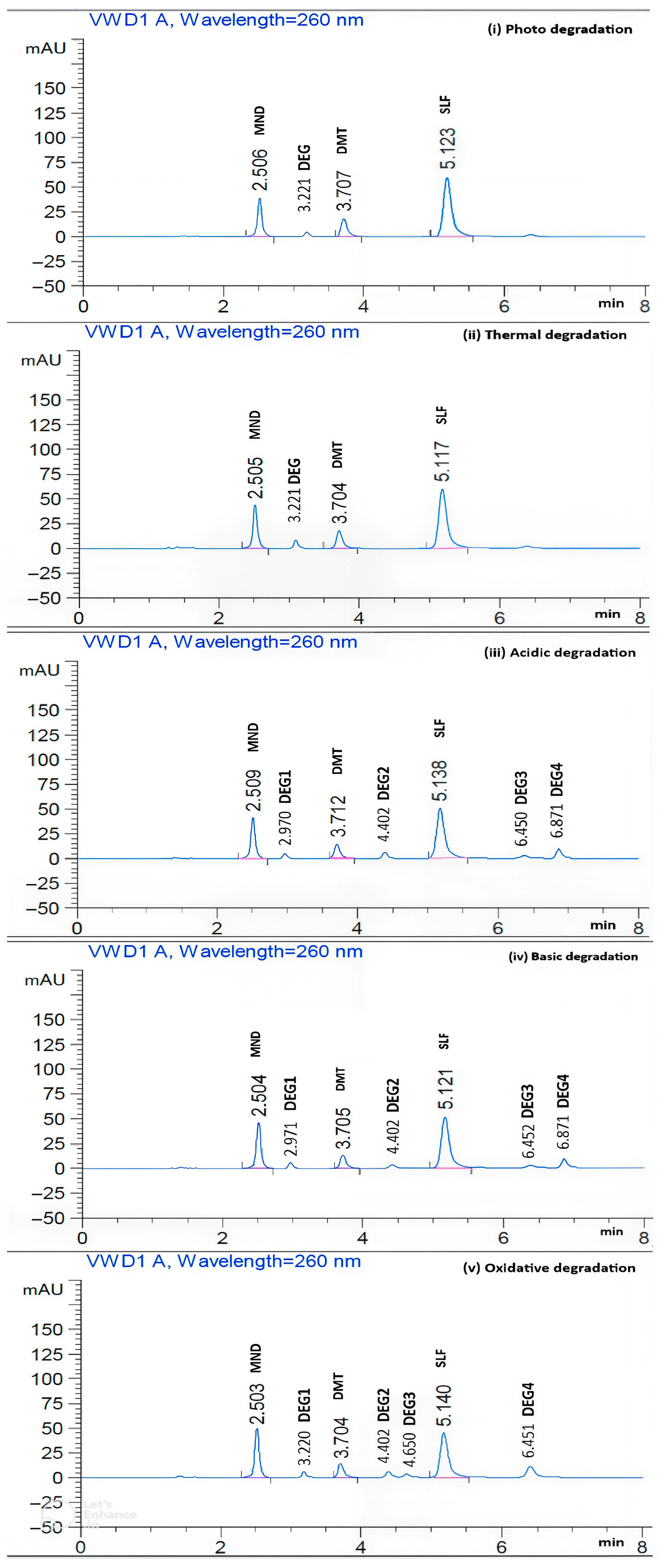
Chromatograms of MND, DMT, and SLF combinations under various severe degradation conditions are presented in Figure 2, demonstrating the method’s effectiveness in separating the three compounds from their potential degradation products.
In summary, the signals of MND, DMT, and SLF were clearly distinguished from those of other degradates. The main observable degradation products for each of the stressful circumstances were listed in Table 1. Furthermore, MND, DMT, and SLF peaks exhibited retention times (Rts) of 2.5, 3.7, and 5.1 min, whereas degradates peaks showed Rts of 2.97, 3.22, 4.31, 4.40, 4.65, 6.45, and 6.87 min. The proportions of breaking down are presented by Table 5.
The degradation percentages were determined by comparing the peak areas of the intact drugs after degradation with those of the non-degraded standards. In terms of degradation behavior, MND exhibited the highest degradation under photolytic conditions (26.52%), while oxidative conditions resulted in the lowest degradation (9.58%). DMT showed maximum degradation in an alkaline medium (21.12%) and no degradation under photolytic exposure (0.00%), indicating its relative stability to light. SLF demonstrated the highest degradation under oxidative conditions (27.16%), with minimal degradation observed under thermal stress (0.23%).
Chromatographic evaluation of the stressed samples revealed distinct degradation peaks, confirming that the developed method effectively separated degradation products from the parent analytes. Specifically, additional peaks appeared at 3.22 min (heat/light stress); 2.97, 4.40, 6.45, and 6.87 min (basic and oxidative stress); and 3.22, 4.40, 4.65, and 6.45 min (oxidative stress only). The main analyte peaks were consistently observed at 2.56, 3.75, and 5.16 min for MND, DMT, and SLF, respectively, throughout the 8 min chromatographic run, confirming the stability and reproducibility of the retention times.

3.4. Evaluation of the Method’s Greenness and Comparisons with Previously Reported Chromatographic Approaches

The HPLC method’s environmental sustainability was evaluated using two computational greenness assessment tools, AGREE [60] and GAPI [61]. The GAPI pictogram (Figure 3) uses five pentagrams to demonstrate the environmental impact of the developed technique, with stages having a color code of green, yellow, and red representing low, medium, and high environmental impact, respectively. Application of this approach to the developed HPLC method revealed that the developed method is green since it had 9 parts colored green, 4 yellow, and only 2 red, as shown in Figure 3. It was certain that the developed method had a slight harmful impact on the environment. The two red segments, corresponding to parameters 1 and 15, indicating the use of an offline analytical technique and the absence of waste handling for the mobile phase. The remaining 13 sections validated the greenness of the HPLC stability method, with nine rated green and four rated yellow.
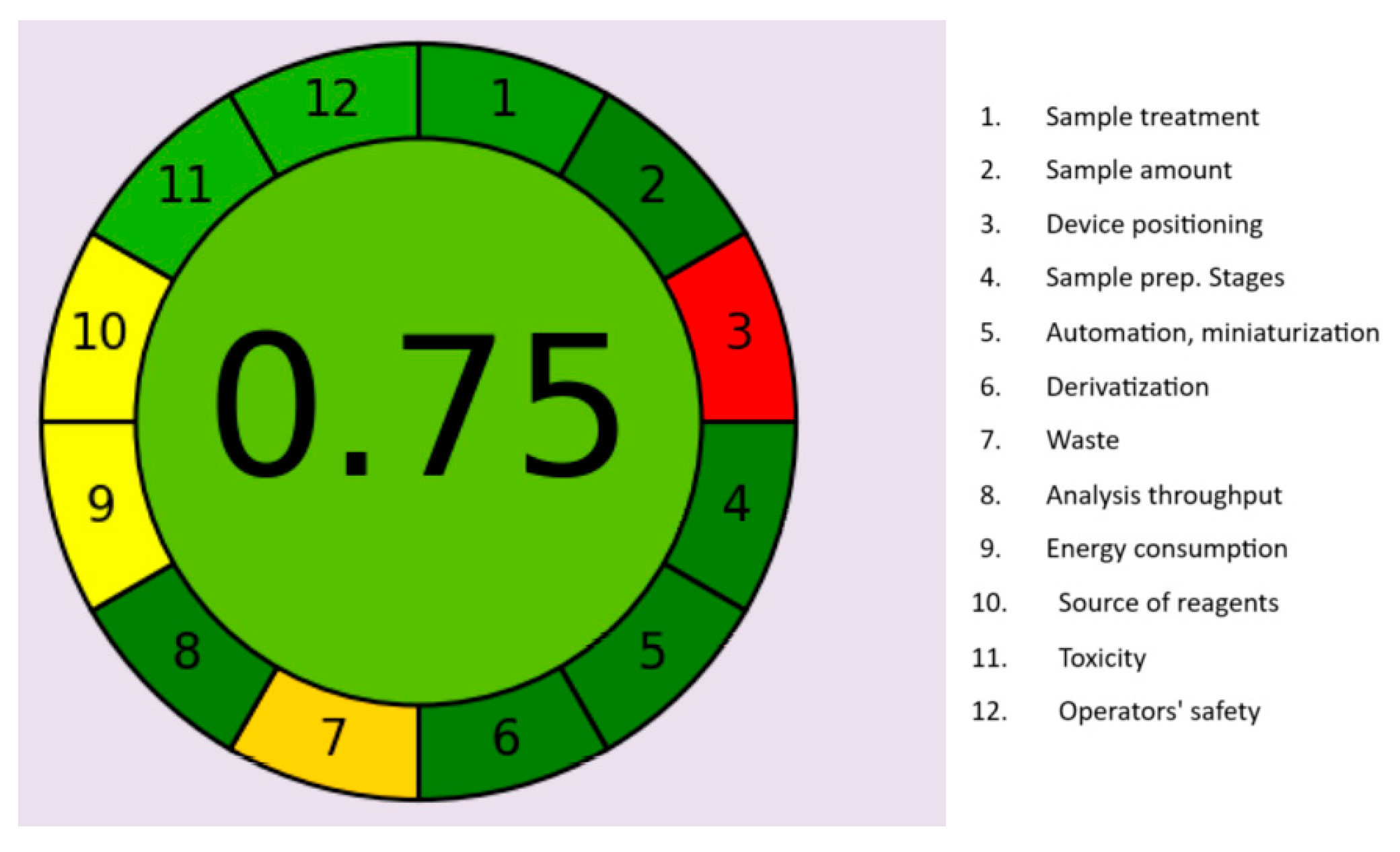
Similarly, the AGREE pictogram (Figure 4) supported the environmentally friendly nature of the proposed stability-indicating method. Only parameter 3 appeared in red, attributed to the use of an offline analytical device. Subdivision 7 was orange due to the moderate mobile phase waste volume of 8 mL per run. The remaining ten sections, ranging from dark to light green, demonstrated the method’s strong environmental performance, yielding an overall AGREE score of 0.75.
During each chromatographic run, the total waste generated was approximately 8 mL of mobile phase. According to the AGREE tool, this corresponds to a moderate rating for Principle 7 (minimization of waste), represented by a dull orange color in the AGREE diagram. The overall AGREE score of 0.75 was automatically generated using AGREE software (version 0.5) with all 12 principles equally weighted under default settings. This score indicates good environmental compatibility and strong alignment with the principles of Green Analytical Chemistry. Although methanol and water are greener solvents, a limited amount of acetonitrile was required to achieve acceptable chromatographic resolution. It is worth noting that while acetonitrile is not a per- and polyfluoroalkyl substance (PFAS), it remains a hazardous solvent and was therefore used in minimal proportion. An important indicator of environmental sustainability is the carbon footprint (kg CO2 equivalent). As described by Ballester-Caudet et al. [64], it can be calculated using the HEXAGON approach. The computation formula and detailed results are provided in Supplementary Data S1. The total carbon dioxide equivalent generated was 0.00659 kg. Since this value is below 0.10, and the ideal benchmark is 0 out of 5 for the overall carbon footprint, the developed method can be considered environmentally friendly [12].
Compared with previously published LC–MS bioanalytical procedures, the newly developed HPLC stability method offers clear advantages in terms of energy efficiency, cost-effectiveness, environmental friendliness, and operational simplicity. Its application to the analysis of powder for oral solutions is further presented in Table 6.
The newly developed stability-indicating HPLC-DAD method demonstrates significant advantages over previously reported techniques. It offers a substantially greener analytical approach for the simultaneous determination of MND, DMT, and SLF in both pure powders and oral powder formulations. Unlike earlier RP-HPLC and polarographic methods [49,56] that required expensive extraction steps or employed toxic reagents such as mercury electrodes, the present method is practical, eco-friendly, and extraction-free. Furthermore, the total analysis time is reduced to only 8 min, compared with 14 and 15 min for earlier spectrophotometric and HPLC methods [55,56], thereby improving analytical throughput, cost efficiency, and environmental sustainability.

3.5. The Advantages of the Novel HPLC Technique for the Industrial Pharmacy Field

For the determination of MND, DMT, and SLF, the newly developed HPLC method serves as a cost-effective alternative, as HPLC instrumentation is considerably more economical than LC–MS systems. Moreover, the HPLC method is preferred over LC–MS for its operational simplicity and suitability for routine quality control analysis to verify drug safety and efficacy once the compounds have been separated from their degradation products. Compared with LC–MS–grade solvents, HPLC–grade solvents are generally more accessible, affordable, and readily available. LC–MS–grade solvents undergo additional purification and filtration to remove impurities that could interfere with mass spectrometric detection, increasing cost and preparation time. Furthermore, HPLC instruments consume approximately ten times less energy than LC–MS systems, making the developed method a more sustainable analytical option.

3.6. Advice Concerning the Pharmaceutical Handling of MND, DMT, and SLF

Based on the degradation ratios presented in Table 5, drugs with alkaline, acidic, or oxidizing properties should not be co-administered with MND, DMT, or SLF in combination formulations. It is also recommended to store MND, DMT, and SLF away from heat and direct sunlight to maintain their stability.

3.7. The Blue Applicability Grade Index (BAGI) Tool Is Used to Consider the Method’s Usefulness and Usability

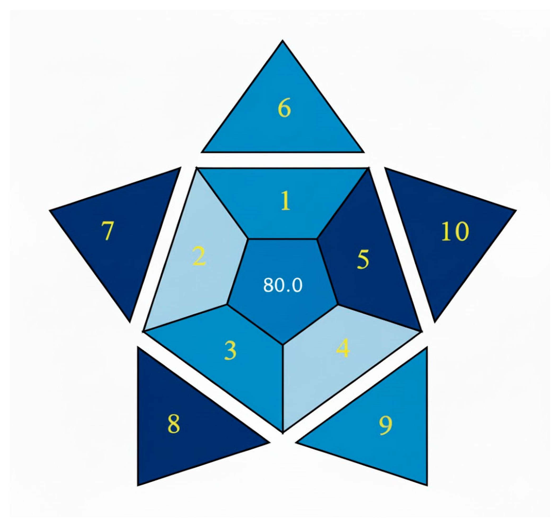
The BAGI approach, introduced in 2023 by Manousi and colleagues [65], evaluates analytical methods based on ten criteria: (1) analysis type, (2) number of compounds analyzed per run, (3) number of samples, (4) number of samples prepared simultaneously, (5) sample preparation steps, (6) number of samples analyzed per hour, (7) availability of reagents, (8) preconcentration procedures, (9) automation level of the analytical instrument, and (10) amount of pharmaceutical samples evaluated. Each parameter is rated on a scale of 0 to 10, with higher scores indicating better performance, and the total score (out of 100) representing the overall applicability of the method. For the developed RP-HPLC method, deep blue ratings for parameters 5, 7, 8, and 10 increased the total BAGI score to 80.0. According to BAGI criteria, a total score above 60 denotes a practical and efficient analytical method, as illustrated in Figure 5.

3.8. The Estimated Constraints and Prospective Plans

The RP-HPLC method was not evaluated in animal fluids or tissues, as the primary objective of this study was to analyze MND, DMT, and SLF in powder formulations for oral solutions using a conventional HPLC system. However, LC–MS techniques would be more suitable for assessing MND, DMT, and SLF in biological matrices, as they enable detection at the nanogram level. The facility did not have access to GC–MS, LC–MS, or LC–MS/MS instrumentation, which are the recommended techniques for determining the molecular masses of degradation products during such studies. Furthermore, the current sensitivity of the method is primarily suited for pharmaceutical quality control and formulation analysis rather than trace-level assay in complex matrices like food or environmental water.

4. Conclusions

This study successfully developed and validated a novel, rapid, and robust HPLC method for the simultaneous determination of menadione (MND), dimetridazole (DMT), and sulfadimethoxine sodium (SLF) in pure and oral powder formulations. The method exhibited excellent selectivity, achieving complete baseline separation of the analytes from their degradation products within eight minutes. Stability studies showed that MND was most susceptible to photodegradation (26.52%), DMT underwent the highest degradation under alkaline conditions (21.12%), and SLF was most sensitive to oxidative stress (27.16%). These results highlight the importance of proper storage and handling of these compounds, particularly avoiding exposure to high temperatures, direct sunlight, and incompatible drug combinations. Moreover, the developed HPLC method demonstrated strong environmental compatibility, as confirmed by AGREE and GAPI evaluations. This eco-friendly and reliable approach makes it a suitable choice for routine quality control (QC) analysis of MND, DMT, and SLF in pharmaceutical formulations.

Supplementary Materials

The following supporting information can be downloaded at: https://www.mdpi.com/article/10.3390/chemosensors13120406/s1, Figure S1. Molecular structures, formulas, and molecular masses of menadione (172.18 g/mol), dimetridazole (141.13 g.mol−1), and sulfadimethoxine (310.33 g.mol−1). Figure S2. Ultraviolet spectra of menadione (20.00 µg.mL−1), dimetridazole, and sulfadimethoxine (40.00 µg.mL−1) dissolved in methanol. Figure S3. Raw regression data and calibration curves (r2 values, slopes, and intercepts) for menadione, dimetridazole, and sulfadimethoxine. Supplementary Data S1: Consideration of the environmental impact (ecosystem influence). Table S1. Estimated linear ranges for MND, DMT, and SLF. Table S2. Accuracy of the developed stability-indicating HPLC method for MND, DMT, and SLF at different concentrations 50, 100, and 150% of the intermediate concentration; (20.00 µg.mL−1 for MND and 40.00 µg.mL−1 for DMT and SLF). Table S3. Precision of the developed HPLC method, determined using RSD calculations for six replicate measurements of MND (20.00 µg.mL−1), DMT, and SLF (40.00 µg.mL−1), the precision of the projected stability indicating HPLC approach was ascertained. Table S4. RSD calculations and retention times for six replicate measurements of MND, DMT, and SLF, demonstrating excellent reproducibility and confirming method consistency and robustness across runs and instruments. Table S5. Evidence supporting the specificity of the developed HPLC method for MND, DMT, and SLF in pure standards, powder for oral solution, and placebo. Table S6. Detailed ruggedness evaluation of the HPLC method for MND, DMT, and SLF (different days) for concentrations of MND (20.00 µg.mL−1), DMT, and SLF (40.00 µg.mL−1). Table S7. Detailed ruggedness evaluation of the HPLC method for MND, DMT, and SLF (different analysts) for concentrations of MND (20.00 µg.mL−1), DMT, and SLF (40.00 µg.mL−1).

Author Contributions

L.A.A.-K.: Resources, methodology, data curation, project administration, funding acquisition, and review and editing. R.M.T.: Methodology, investigation, review, and editing. M.A.E.: Data curation, original draft writing, investigation, resources, review, and editing. M.G.: Supervision, project administration, data curation, investigation, review, and editing. All authors have read and agreed to the published version of the manuscript.

Funding

This project was funded by the Deanship of Scientific Research (DSR) at King Abdulaziz University, Jeddah, under grant no (IPP: 1045-247-2025). The authors, therefore, acknowledge with thanks DSR for the technical and financial support.

Institutional Review Board Statement

Not applicable.

Informed Consent Statement

Not applicable.

Data Availability Statement

The data presented in this study are available on request from the corresponding author.

Acknowledgments

This project was funded by the Deanship of Scientific Research (DSR) at King Abdulaziz University, Jeddah, under grant no (IPP: 1045-247-2025). The authors, therefore, acknowledge with thanks DSR for the technical and financial support.

Conflicts of Interest

The authors declare no conflicts of interest related to this research project.

References

  1. Sehrawat, R.; Maithani, M.; Singh, R. Regulatory Aspects in Development of Stability-Indicating Methods: A Review. Chromatographia 2010, 72, 1–6. [Google Scholar] [CrossRef]
  2. Chew, Y.L.; Khor, M.A.; Lim, Y.Y. Choices of Chromatographic Methods as Stability Indicating Assays for Pharmaceutical Products: A Review. Heliyon 2021, 7, e06553. [Google Scholar] [CrossRef]
  3. Hegazy, A.M.; Batubara, A.S.; Abdelgawad, M.A.; El-Sherbiny, M.; Ghoneim, M.M.; Ahmed, A.M.; Gamal, M. Recommended and Verified Stability Indicating GC–MS Procedures for Green Separation of Quaternary Mixture of Naphazoline, Ephedrine, Methylparaben, and Naphazoline Impurity. Microchem. J. 2022, 183, 108058. [Google Scholar] [CrossRef]
  4. El-Maraghy, C.M. Implementation of Green Chemistry to Develop HPLC/UV and HPTLC Methods for the Quality Control of Fluconazole in Presence of Two Official Impurities in Drug Substance and Pharmaceutical Formulations. Sustain. Chem. Pharm. 2023, 33, 101124. [Google Scholar] [CrossRef]
  5. Bhukya, V.N.; Beda, D.P. Implementation of Green Analytical Principles to Develop and Validate the HPLC Method for the Separation and Identification of Degradation Products of Panobinostat, and Its Characterization by Using LC-QTOF-MS/MS and Its In-Silico Toxicity Prediction Using ADMET Software. Green Anal. Chem. 2024, 8, 100090. [Google Scholar] [CrossRef]
  6. Darji, H.; Dedania, Z. Simultaneous Estimation of Azelnidipine and Metoprolol Succinate with Greenness Assessment Using HPLC and UV-Spectrophotometric Methods. Green Anal. Chem. 2023, 7, 100079. [Google Scholar] [CrossRef]
  7. Kokilambigai, K.S.; Lakshmi, K.S. Analytical Quality by Design Assisted RP-HPLC Method for Quantifying Atorvastatin with Green Analytical Chemistry Perspective. J. Chromatogr. Open 2022, 2, 100052. [Google Scholar] [CrossRef]
  8. Mohamed, D.; Fouad, M.M. Application of NEMI, Analytical Eco-Scale and GAPI Tools for Greenness Assessment of Three Developed Chromatographic Methods for Quantification of Sulfadiazine and Trimethoprim in Bovine Meat and Chicken Muscles: Comparison to Greenness Profile of Reported HPLC Methods. Microchem. J. 2020, 157, 104873. [Google Scholar] [CrossRef]
  9. Elmansi, H.; Belal, F. Development of an Eco-Friendly HPLC Method for the Simultaneous Determination of Three Benzodiazepines Using Green Mobile Phase. Microchem. J. 2019, 145, 330–336. [Google Scholar] [CrossRef]
  10. Nugrahani, I.; Manosa, E.Y.; Chintya, L. FTIR-Derivative as a Green Method for Simultaneous Content Determination of Caffeine, Paracetamol, and Acetosal in a Tablet Compared to HPLC. Vibr. Spectrosc. 2019, 104, 102941. [Google Scholar] [CrossRef]
  11. Arabi, M.; Ostovan, A.; Bagheri, A.R.; Guo, X.; Li, J.; Ma, J.; Chen, L. Hydrophilic Molecularly Imprinted Nanospheres for the Extraction of Rhodamine B Followed by HPLC Analysis: A Green Approach and Hazardous Waste Elimination. Talanta 2020, 215, 120933. [Google Scholar] [CrossRef]
  12. De Marco, B.A.; Rechelo, B.S.; Tótoli, E.G.; Kogawa, A.C.; Salgado, H.R.N. Evolution of Green Chemistry and Its Multidimensional Impacts: A Review. Saudi Pharm. J. 2019, 27, 1–8. [Google Scholar] [CrossRef]
  13. Suresh, S.; Raghu, D.; Karunagaran, D. Menadione (Vitamin K3) induces apoptosis of human oral cancer cells and reduces their metastatic potential by modulating the expression of epithelial to mesenchymal transition markers and inhibiting migration. Asian Pac. J. Cancer Prev. 2013, 14, 5461–5465. [Google Scholar] [CrossRef]
  14. Zoughaib, M.; Pashirova, T.N.; Nikolaeva, V.; Kamalov, M.; Nakhmetova, F.; Salakhieva, D.V.; Abdullin, T.I. Anticancer and chemosensitizing effects of menadione-containing peptide-targeted solid lipid nanoparticles. J. Pharm. Sci. 2024, 113, 2258–2267. [Google Scholar] [CrossRef]
  15. Food Safety Commission of Japan. Risk Assessment Report: Dimetridazole (1,2-dimethyl-5-nitroimidazole) in Food-Producing Animals; Food Safety Commission Japan (FSCJ) Report; Food Safety Commission Japan: Tokyo, Japan, 2015. [Google Scholar]
  16. Mital, A. Synthetic Nitroimidazoles: Biological Activities and Mutagenicity Relationships. Sci. Pharm. 2009, 77, 497–520. [Google Scholar] [CrossRef]
  17. The public health issue of antibiotic residues in food and feed. Front. Microbiol. 2022, 13, 9047141.
  18. Meradji, S.; Basher, N.S.; Sassi, A.; Ibrahim, N.A.; Idres, T.; Touati, A. The Role of Water as a Reservoir for Antibiotic-Resistant Bacteria. Antibiotics 2025, 14, 763. [Google Scholar] [CrossRef]
  19. Sastry, C.S.P.; Rajendraprasad Singh, N.; Narayana Reddy, M.; Sankar, D.G. Spectrophotometric determination of menadione and menadione sodium bisulfite in pharmaceutical preparations. Int. J. Pharm. 1987, 39, 137–140. [Google Scholar] [CrossRef]
  20. Ruiz, T.P.; Lozano, C.M.; Tomas, V.; Martin, J. Flow-injection fluorimetric determination of menadione using on-line photo-reduction in micellar media. Anal. Chim. Acta 2004, 514, 259–264. [Google Scholar] [CrossRef]
  21. Gil Torró, I.; García Mateo, J.V.; Martínez Calatayud, J. Spectrofluorimetric determination of vitamin K3 by a solid-phase zinc reactor immobilized in a flow injection assembly. Analyst 1997, 122, 139–142. [Google Scholar] [CrossRef]
  22. Nevado, J.J.; Pulgarin, J.A.; Laguna, M.A. Spectrofluorimetric study of the b-cyclodextrin: Vitamin K3 complex and determination of vitamin K3. Talanta 2001, 53, 951–959. [Google Scholar] [CrossRef]
  23. Vire, J.C.; El Maali, N.A.; Patriarche, G.J. Square-wave adsorptive stripping voltammetry of menadione (vitamin K(3)). Talanta 1988, 35, 997–1000. [Google Scholar] [CrossRef]
  24. Huang, Y.; Zhang, C.; Zhang, X.; Zhang, Z. Chemiluminescence analysis of menadione sodium bisülfite and analgin in pharmaceutical preparations and biological fluids. J. Pharm. Biomed. Anal. 1999, 21, 817–825. [Google Scholar] [CrossRef]
  25. Pérez-Ruíz, T.; Martínez-Lozano, C.; Tomás, V.; Martín, J. Flow injection determination of vitamin K3 by a photoinduced chemiluminescent reaction. Analyst 1999, 124, 197–201. [Google Scholar] [CrossRef] [PubMed]
  26. Rizk, N.M.H. Potentiometric determination of Menadione (Vitamin K3). Microchim. Acta 2002, 138, 53–58. [Google Scholar] [CrossRef]
  27. Gaikwad, M.N.; Gaikwad, S.T.; Rajbhoj, A.S. Potentiometric study of vitamin K3 complexes with transition metals in methanol–water and acetonitrile–water medium. Int. J. ChemTech Res. 2012, 4, 1392–1395. [Google Scholar]
  28. Laffi, R.; Marchetti, S.; Marchetti, M. Normal-phase liquid chromatographic determination of menadione in animal feeds. J. Assoc. Off. Anal. Chem. 1988, 71, 826–828. [Google Scholar] [CrossRef]
  29. Pérez-Ruiz, T.; Martínez-Lozano, C.; García, M.A.; Martín, J. High-performance liquid chromatography: Photochemical reduction in aerobic conditions for determination of K vitamins using fluorescence detection. J. Chromatogr. A 2007, 1141, 67–72. [Google Scholar] [CrossRef]
  30. Liu, Z.; Li, T.; Li, J.; Wang, E. Detection of menadione sodium bisulfite (vitamin K3) by reversed-phase high performance liquid chromatography with series dual-electrode amperometric detector. Anal. Chim. Acta 1997, 338, 57–62. [Google Scholar] [CrossRef]
  31. Speek, A.J.; Schrijver, J.; Schreurs, W.H. Fluorimetric determination of menadione sodium bisulphite (vitamin K3) in animal feed and premixes by high-performance liquid chromatography with post-column derivatization. J. Chromatogr. 1984, 301, 441–447. [Google Scholar] [CrossRef]
  32. Saleh, E.M.; Chikako, S.; Naoya, K.; Kaname, O.; Mitsuhiro, W.; Naotaka, K. Development and Validation of the First Assay Method Coupling Liquid Chromatography with Chemiluminescence for the Simultaneous Determination of Menadione and Its Thioether Conjugates in Rat Plasma. Chem. Res. Toxicol. 2013, 26, 1409–1417. [Google Scholar] [CrossRef]
  33. Maya, K.; Yoshihisa, H.; Yoshitomo, S.; Naoko, T.; Kimie, N.; Toshio, O.; Hiroshi, H. Determination of Menadione by Liquid Chromatography–Tandem Mass Spectrometry Using Pseudo Multiple Reaction Monitoring. Anal. Sci. 2017, 33, 863–867. [Google Scholar]
  34. Castello, G.; Bruschi, E.; Ghelli, G. Gas chromatographic determination of the purity of vitamin K3 (menadione). J. Chromatogr. 1977, 139, 195–202. [Google Scholar] [CrossRef]
  35. Demirkaya-Miloglu, F.; Kadioglu, Y.; Senol, O. GC-FID and HPLC-DAD Methods for the Determination of Menadione Sodium Bisulphite Directly and by Converting Menadione Sodium Bisulphite to Menadione in Pharmaceutical Preparation. Iran J. Pharm. Res. 2014, 13, 353–364. [Google Scholar] [PubMed]
  36. Sams, M.J.; Strutt, P.R.; Barnes, K.A.; Damant, A.P.; Rose, M.D. Determination of dimetridazole, ronidazole and their common metabolite in poultry muscle and eggs by high performance liquid chromatography with UV detection and confirmatory analysis by atmospheric pressure chemical ionisation mass spectrometry. Analyst 1998, 123, 2545–2549. [Google Scholar] [CrossRef]
  37. Buizer, F.G.; Severijnen, M. Determination of dimetridazole in feedstuffs and pre-mixes by high-speed liquid chromatography. Analyst 1975, 100, 854–856. [Google Scholar] [CrossRef] [PubMed]
  38. Wang, Z.-H.; Liu, J.-Y.; Zhou, S.-P. Study on the determination of dimetrionidazole using a carbon paste electrode. Fenxi Shiyanshi 1998, 17, 40–43. [Google Scholar]
  39. Stubbings, G.; Bigwood, T. The development and validation of a multiclass liquid chromatography tandem mass spectrometry (LC-MS/MS) procedure for the determination of veterinary drug residues in animal tissue using a Que Chers (Quick, Easy, Cheap, Effective, Rugged and Safe) approach. Anal. Chim. Acta 2009, 637, 68–78. [Google Scholar]
  40. Gentili, A.; Perret, D.; Marchese, S. Liquid chromatography-tandem mass spectrometry for performing confirmatory analysis of veterinary drugs in animal-food products. TrAC Trends Anal. Chem. 2005, 24, 704–733. [Google Scholar] [CrossRef]
  41. Cannavan, A.; Kennedy, D.G. Determination of dimetridazole in poultry tissues and eggs using liquid chromatography-thermospray mass spectrometry. Analyst 1997, 122, 963–966. [Google Scholar] [CrossRef]
  42. Miho, S.; Kazue, T.; Takeo, S.; Tomoko, K.; Hiroshi, H.; Setsuko, K.; Maki, K.; Toshihiro, N. Determination of Dimetridazole, Metronidazole and Ronidazole in Salmon and Honey by Liquid Chromatography Coupled with Tandem Mass Spectrometry. Food Hygiene Saf. Sci. 2011, 52, 51–58. [Google Scholar]
  43. Petrovic, M.; Skrbic, B.; Zivancev, J.; Ferrando-Climent, L.; Barcelo, D. Determination of 81 pharmaceutical drugs by high performance liquid chromatography coupled to mass spectrometry with hybrid triple quadrupole linear ion trap in different types of water in Serbia. Sci. Total Environ. 2014, 468–469, 415–428. [Google Scholar] [CrossRef] [PubMed]
  44. Ho, C.; Sin, D.W.M.; Wong, K.M.; Tang, H.P.O. Determination of dimetridazole and metronidazole in poultry and porcine tissues by gas chromatography electron capture negative ionization mass spectrometry. Anal. Chim. Acta 2005, 530, 23–31. [Google Scholar] [CrossRef]
  45. Wang, J.H. Determination of three nitroimidazole residues in poultry meat by gas chromatography with nitrogen phosphorus detection. J. Chromatogr. A 2001, 918, 435–438. [Google Scholar] [CrossRef] [PubMed]
  46. Hughes, D.D. Determination of Dimetridazole and Ipronidazole in Feeds at Cross-Contamination Levels. J. Assoc. Off. Anal. Chem. 1988, 71, 474–477. [Google Scholar] [CrossRef]
  47. Thompson, C.S.; Traynor, I.M.; Fodey, T.L.; Crooks, S.R. Improved screening method for the detection of a range of nitroimidazoles in various matrices by optical biosensor. Anal. Chim. Acta 2009, 637, 259–264. [Google Scholar] [CrossRef]
  48. Jallal, Z.; Yassine, E.; Chaimae, R.; Nadia, B.; Abderrahim, I.; Idriss, B.; Ali, A. Electrocatalytic detection of dimetridazole using an electrochemical sensor Ag. Anal. Appl. Milk, tomato juice and human urine. Sens. Int. 2021, 2, 100105. [Google Scholar]
  49. Rami Reddy, Y.V.; Rajendra Kumar Reddy, P.; Suresh Reddy, C.; Jayarama Reddy, S. Polarographic determination of nitro group containing drugs in tablets. Indian J. Pharm. Sci. 1996, 58, 96–99. [Google Scholar]
  50. Zhang, J.; Chen, Z.P.; Tang, S.; Luo, X.G.; Xi, J.B.; He, Z.L.; Yu, J.X.; Wu, F.S. Fabrication of porphyrin-based magnetic covalent organic framework for effective extraction and enrichment of sulfonamides. Anal. Chim. Acta 2019, 1089, 66–77. [Google Scholar] [CrossRef]
  51. Guo, Y.; Wei, W.; Zhang, Y.; Dai, Y.; Wang, W.; Wang, A. Determination of sulfadimethoxine in milk with aptamer-functionalized Fe3O4/graphene oxide as magnetic solid-phase extraction adsorbent prior to HPLC. J. Sep. Sci. 2020, 43, 3499–3508. [Google Scholar] [CrossRef]
  52. Mengelers, M.J.; Oorsprong, M.B.; Kuiper, H.A.; Aerts, M.M.; van Gogh, E.R.; van Miert, A.S. Determination of sulfadimethoxine, sulfamethoxazole, trimethoprim and their main metabolites in porcine plasma by column switching HPLC. J. Pharm. Biomed. Anal. 1989, 7, 1765–1776. [Google Scholar] [CrossRef] [PubMed]
  53. Boison, J.O.; Keng, L.J. Determination of sulfadimethoxine and sulfamethazine residues in animal tissues by liquid chromatography and thermospray mass spectrometry. J. AOAC Int. 1995, 78, 651–658. [Google Scholar] [CrossRef] [PubMed]
  54. Primus, T.M.; Jojola, S.M.; Robinson, S.J.; Johnston, J.J. Determination of Sulfadimethoxine Residues in Skunk Serum by HPLC. J. Liq. Chromatogr. Relat. Technol. 2007, 30, 2095–2102. [Google Scholar] [CrossRef]
  55. Dos Santos, L.F.; de Andrade, L.R.; Cardoso, T.F.; Caminha, K.B.; Pires, D.S.; Machado, E.P.; Severino, P.; do Amaral, M.S.; Souto, E.B.; Kassab, N.M. Innovative UV derivative spectrophotometric method for simultaneous quantification of sulfadimethoxine and metronidazole. J. Indian Chem. Soc. 2025, 102, 101655. [Google Scholar] [CrossRef]
  56. Ghanem, M.M.; Abu-Lafi, S.A. Validation of a stability-indicating RP-HPLC method for the simultaneous determination of trimethoprim and sulfadimethoxine sodium in oral liquid dosage form. Sci. Pharm. 2013, 81, 459. [Google Scholar] [CrossRef]
  57. Al-Sanea, M.M.; Gamal, M. Critical analytical review: Rare and recent applications of refractive index detector in HPLC chromatographic drug analysis. Microchem. J. 2022, 178, 107339. [Google Scholar] [CrossRef]
  58. Ali, H.M.; Gamal, M.; Ghoneim, M.M.; Abd Elhalim, L.M. Quantitative Analysis of Abamectin, Albendazole, Levamisole HCl and Closantel in Q-DRENCH Oral Suspension Using a Stability-Indicating HPLC-DAD Method. Molecules 2022, 27, 764. [Google Scholar] [CrossRef] [PubMed]
  59. International Council for Harmonisation (ICH). ICH Harmonised Guideline Q2(R2): Validation of Analytical Procedures; ICH: Geneva, Switzerland, 2023. [Google Scholar]
  60. Pena-Pereira, F.; Wojnowski, W.; Tobiszewski, M. AGREE-Analytical GREEnness Metric Approach and Software. Anal. Chem. 2020, 92, 10076–10082. [Google Scholar] [CrossRef]
  61. Gamal, M.; Naguib, I.A.; Panda, D.S.; Abdallah, F.F. Comparative study of four greenness assessment tools for selection of greenest analytical method for assay of hyoscine: N -butyl bromide. Anal. Methods 2021, 13, 369–380. [Google Scholar] [CrossRef]
  62. Abdelrahman, M.M. Green Analytical Chemistry Metrics and Life-Cycle Assessment Approach to Analytical Method Development. In Green Chemical Analysis and Sample Preparations; Springer: Cham, Switzerland, 2022; pp. 29–99. [Google Scholar] [CrossRef]
  63. Wells, M.; Dantus, M. Validation of chromatographic methods. In Ewing’s Analytical Instrumentation Handbook, 3rd ed.; Taylor and Fransis: Abingdon, UK, 2004; pp. 1015–1033. [Google Scholar] [CrossRef]
  64. Ballester-Caudet, A.; Campíns-Falcó, P.; Pérez, B.; Sancho, R.; Lorente, M.; Sastre, G.; González, C. A new tool for evaluating and/or selecting analytical methods: Summarizing the information in a hexagon. TrAC Trends Anal. Chem. 2019, 118, 538–547. [Google Scholar] [CrossRef]
  65. Manousi, N.; Wojnowski, W.; Płotka-Wasylka, J.; Samanidou, V. Blue applicability grade index (BAGI) and software: A new tool for the evaluation of method practicality. Green Chem. 2023, 25, 7598–7604. [Google Scholar] [CrossRef]
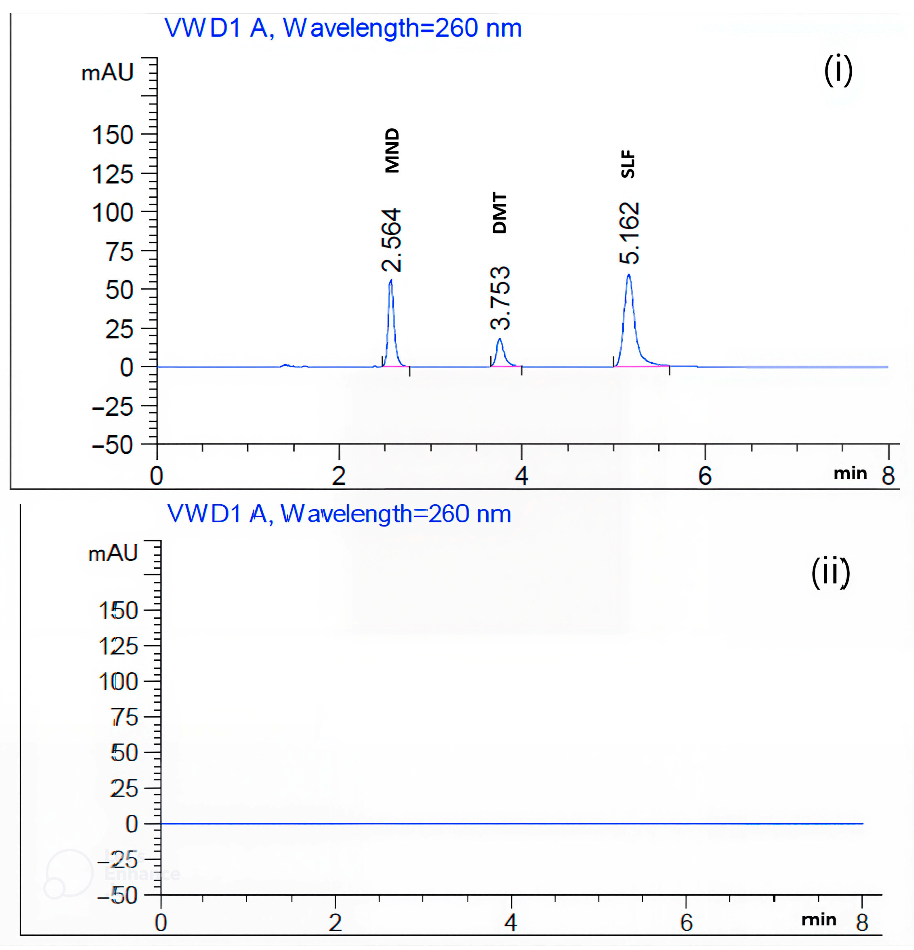
Figure 1. (i) Representative HPLC chromatogram of MND (20.00 µg mL−1), DMT (40.00 µg mL−1), and SLF (40.00 µg mL−1) obtained using a C18 column. The mobile phase consisted of 0.05 M KH2PO4 and acetonitrile (80:20, v/v), delivered at a flow rate of 2.0 mL min−1, with UV detection at 260 nm. (ii) Corresponding blank chromatogram of the mobile phase under the same chromatographic conditions.
Chemosensors 13 00406 g001
Figure 2. Representative HPLC chromatograms illustrating the stability profiles of MND (20.00 µg mL−1), DMT (40.00 µg mL−1), and SLF (40.00 µg mL−1) after exposure to various stress degradation conditions: (i) light, (ii) heat, (iii) acidic, (iv) basic, and (v) oxidative degradation. Chromatographic separation was achieved on a C18 column using a mobile phase composed of 0.05 M KH2PO4 and acetonitrile (80:20, v/v) at a flow rate of 2.0 mL min−1, with UV detection at 260 nm.
Chemosensors 13 00406 g002
Figure 3. GAPI (Green Analytical Procedure Index) pictogram illustrating the anticipated sustainability and environmental impact of the developed RP-HPLC method for the assay of MND, DMT, and SLF.
Chemosensors 13 00406 g003
Figure 4. AGREE (Analytical GREEnness) pictogram representing the predicted sustainability and greenness of the developed RP-HPLC method for the assay of MND, DMT, and SLF.
Chemosensors 13 00406 g004
Figure 5. Evaluation of the blueness of the developed HPLC method using the Blue Assessment of Greenness Index (BAGI) approach.
Chemosensors 13 00406 g005
Table 1. Chromatographic parameters of the developed RP-HPLC method for the simultaneous analysis of MND, DMT, and SLF.
Stationary phaseSupelcosil C18 (I.D 4.6 mm—length 25 cm—particle size 5 µ)
Mobile phase0.05 M KH2PO4: Acetonitrile (80:20 v/v)
DetectorAt 260 nm—UV
Pumping sytemIsocratic
TemperatureAmbient
The volume of Injection10 µL
Flow rate2.0 mL.min−1
Full run time8 min
Retention timesMND: 2.56 min DMT: 3.75 minSLF: 5.16 min
Acidic and basic degradation products (2.97, 4.40, 6.45, and 6.87 min)
Oxidative degradation products (3.22, 4.40, 4.65, and 6.45 min)
Photo and thermal degradation products (3.22 min)
Table 2. Validation results of the developed stability-indicating RP-HPLC method for the simultaneous determination of MND, DMT, and SLF, including linearity range, robustness, ruggedness, limits of detection (LOD) and quantification (LOQ), precision, accuracy, and specificity.
Validation CriteriaValues MeasuredApproved Criteria That Follow the Guidelines of the ICH [59]
MNDDMTSLF
Linearity and Range (Five concentration levels)10.0 to 30.0 µg.mL−1
r = 0.99936
20.0 to 60.0 µg.mL−1
r = 0.99998
20.0 to 60.0 µg.mL−1
r = 0.99999
r ≥ 0.99
Precision
(for 6 replicates)
0.190.710.47RSD ≤ 2%
Accuracy99.59%
±1.076
100.07%
±0.246
99.52%
±0.626
100 ± 2%
Specificity/SelectivityDistinct peak for MND at a finer resolution than the other peaks for degradation productsDistinct peak for DMT at a finer resolution than the other peaks for degradation productsDistinct peak for SLF at a finer resolution than the other peaks for degradation productsThere was no interference detected
Limit of Detection1.07 µg.mL−10.40 µg.mL−10.20 µg.mL−1Using the formula: 3.3 × SD/a (where SD is the response standard deviation and a is the calibration curve’s slope)
Limit of Quantitation3.26 µg.mL−11.22 µg.mL−10.61 µg.mL−1Using the formula: 10 × SD/a
Ruggedness2.41 (distinguished days)
0.66 (distinguished analysts)
0.54 (distinguished days)
0.20 (distinguished analysts)
1.92 (distinguished days)
0.64 (distinguished analysts)
Every alteration should have a pooled RSD of less than 3%
Robustness
(modifies a few aspects of the mobile phase)
0.640.060.39Every alteration should have a pooled RSD of less than 3%
Table 3. Robustness evaluation of the developed RP-HPLC method for the determination of MND (20.00 µg mL−1), DMT (40.00 µg mL−1), and SLF (40.00 µg mL−1) under slight variations in the mobile phase composition.
ReplicateMNDDMTSLFStandards for Approval
Case One *Case Two **Case One *Case two **Case One *Case Two **
One241.47239.09107.69107.79512.68515.01
Two240.47237.94107.62107.71512.80516.32
Three241.04238.05107.63107.74513.03517.40
Pooled average239.71107.70514.51
Pooled SD1.510.102.01
Pooled RSD0.640.060.39Equal or less than 3%
* 0.05 M KH2PO4: Acetonitrile (82:18 v/v). ** 0.05 M KH2PO4: Acetonitrile (78:22 v/v).
Table 4. System suitability parameters of the developed RP-HPLC method for the simultaneous assay of MND, DMT, and SLF, including tailing factor, resolution, and selectivity factor.
Parameters of System SuitabilityInformation Obtained from the Anticipated TechniqueReference Values [59]
MNDMTSLF
Retention Time (Rt) ± SD2.56 ± 0.043.75 ± 0.025.16 ± 0.03More than 1
Capacity Factor (K)1.8443.1664.7331–10 Good
Theoretical Plate No. (N)812610,54810,401As its worth rises, so does its efficacy
HETP = height equivalent to theoretical plate (cm per plate)0.00310.00240.0024As the HETP score falls, column effectiveness rises
Tailing Factor (T)0.770.690.71Less than or equal 2
Resolution factor (Rs)9.138.08More than or equal 1.5
Selectivity factor (α)1.461.38More than 1
Table 5. The percentage of degradation of the parent drugs (Mean ± SD, n = 3) of MND (20.00 µg mL−1), DMT (40.00 µg mL−1), and SLF (40.00 µg mL−1) after exposure to different stress degradation conditions: (i) light, (ii) heat, (iii) acidic, (iv) basic, and (v) oxidative degradation. Chromatographic separation was performed on a C18 column using a mobile phase consisting of 0.05 M KH2PO4 and acetonitrile (80:20, v/v) at a flow rate of 2.0 mL min−1 with UV detection at 260 nm.
Method of Degradations A Description of the CircumstancesAreaDegradation %
(Mean ± SD, n = 3)
MNDDMTSLFMNDDMTSLF
Light *Light (48 h)/
UV (12 h)
184.02108.39502.8126.52 ± 1.040.00 ± 0.961.36 ± 1.08
Heat80 °C (8 h)220.42106.97508.5611.99 ± 1.640.79 ± 1.370.23 ± 1.18
Acid1 N HCl/80 °C (1 h)213.6091.07441.5214.71 ± 0.9915.54 ± 1.1013.39 ± 1.21
Base1 N NaOH/80 °C (1 h)222.5185.06441.6311.15 ± 0.8921.12 ± 1.0513.36 ± 0.94
Oxidation0.50% H2O2/80 °C (1 h)226.4685.63371.339.58 ± 0.9920.58 ± 1.1227.16 ± 1.06
* Light degradation includes 48 h exposure to light followed by 12 h of UV radiation. The UV exposure was carried out using a 254 nm UV lamp, with the samples placed at a fixed distance from the light source. All experiments were performed in a controlled chamber under ambient temperature.
Table 6. Comparative assessment of previously reported HPLC, LC–MS, LC–MS/MS, polarographic, and spectrophotometric methods for MND, DMT, and SLF relative to the developed HPLC method, highlighting sample matrices, analytical performance, total analysis time, cost-effectiveness, operational simplicity, and quantitation ranges.
ApproachesThe Matrix and MeritsQuantification ConstraintsEasy to Use and Reasonably PricedRun TimeReferences
HPLCMenadione assay in animal feed1.00 µg.g−1Pricey procedures for extraction are employed; assay MND only in the absence of DMT and SLF11 min(Speek et al., 1984)
[31]
HPLCMenadione and Its Thioether Conjugates assay in Rat Plasma10.00 nMolarPricey procedures for extraction are employed; assay MND only in the absence of DMT and SLF9 min(Saleh et al., 2013)
[32]
LC-MSMenadione assay using pseudo multiple reaction monitoring1.70 ng.mL−1Pricey procedures for extraction are employed; assay MND only in the absence of DMT and SLF14 min(Maya et al., 2017)
[33]
GC and HPLCMenadione assay in Pharmaceutical Preparation0.50 µg.mL−1Pricey procedures for extraction are employed; assay MND only in the absence of DMT and SLF14 min for HPLC and GC(Demirkaya et al., 2014)
[35]
HPLCDimetridazole, ronidazole and their common metabolite assay in poultry muscle and eggs1.50 µg.kg−1Pricey procedures for extraction are employed; assay DMT only in the absence of MND and SLF12 min(Sams et al., 1998)
[36]
HPLCDimetridazole assay in feedstuffs150 mg.kg−1Pricey procedures for extraction are employed; assay DMT only in the absence of MND and SLF15 min(Buizer et al., 1975)
[37]
LC-MSDimetridazole assay in poultry tissues and eggs using5.00 ng.g−1Pricey procedures for extraction are employed; assay DMT only in the absence of MND and SLF15 min(Cannavan et al., 1997)
[41]
LC-MSDimetridazole, metronidazole and ronidazole assay in salmon and honey1.50 µg.kg−1Pricey procedures for extraction are employed; assay DMT only in the absence of MND and SLF20 min(Miho et al., 2011)
[42]
PolarographyDimetridazole and nimesulide assay in tablets25.00 µg.mL−1Dropping mercury electrode is used which is poisonous, Surface area of a drop of mercury is never constant and applied voltage produces changes in surface tension and hence change in drop size.5 min(RAMI REDDY et al., 1996)
[49]
HPLCSulfadimethoxine assay in milk using Fe3O4/graphene oxide as adsorbent5.00 µg.L−1Pricey procedures for extraction are employed; assay SLF only in the absence of MND and DMT5 min(Yinan et al., 2020)
[51]
HPLCSulfadimethoxine, sulfamethoxazole, and trimethoprim assay in porcine plasma25.00 ng.mL−1Pricey procedures for extraction are employed; assay SLF only in the absence of MND and DMT15 min(Mengelers et al., 1989)
[52]
HPLCSulfadimethoxine and sulfamethazine residues assay in animal tissues6.00 ng.g−1Pricey procedures for extraction are employed; assay SLF only in the absence of MND and DMT10 min(Boison et al., 1995)
[53]
HPLCSulfadimethoxine residues assay in skunk serum0.10 µg.mL−1Pricey procedures for extraction are employed; assay SLF only in the absence of MND and DMT14 min(Primus et al., 2007)
[54]
Spectrophotometricsulfadimethoxine and metronidazole in commercial veterinary tablets 0.51 µg.L−1assay SLF only in the absence of MND and DMT15 min(dos Santos et al., 2025)
[55]
RP- HPLCTrimethoprim and Sulfadimethoxine Sodium in Oral Liquid Dosage Form3.3 μg/mLPricey procedures for extraction are employed; assay SLF only in the absence of MND and DMT14 min(Ghanem et al., 2013)
[56]
HPLC-DADConsiderably green technique for both genuine powder and powder oral solutions analysis and stability illustration 10.00 µg.mL−1 for MND
20.00 µg.mL−1 for DMT and SLF
Affordable and does not need any special extraction techniques8 minCurrent methodology
Disclaimer/Publisher’s Note: The statements, opinions and data contained in all publications are solely those of the individual author(s) and contributor(s) and not of MDPI and/or the editor(s). MDPI and/or the editor(s) disclaim responsibility for any injury to people or property resulting from any ideas, methods, instructions or products referred to in the content.