Communication

A Novel Pyrene-Based Fluorescent Probe for the Detection of Cu2+

1
Jilin Provincial International Joint Research Center of PhotoFunctional Materials and Chemistry, School of Chemistry & Environmental Engineering, Changchun University of Science and Technology, Changchun 130022, China
2
Tongliao Substation, Inner Mongolia Environmental Monitoring Center, Tongliao 028000, China
3
Institute of Natural Sciences, Buryat State University, Buryat 671207, Russia
*
Authors to whom correspondence should be addressed.
Chemosensors 2025, 13(11), 403; https://doi.org/10.3390/chemosensors13110403
Submission received: 23 October 2025 / Revised: 7 November 2025 / Accepted: 18 November 2025 / Published: 20 November 2025
(This article belongs to the Special Issue Advanced Material-Based Fluorescent Sensors)

Abstract

A novel fluorescent probe (PYB) for selective and sensitive detection of Cu2+ ions was rationally designed and synthesized via a multi-step organic reaction using pyrene as the fluorophore and salicylaldehyde-diethylenetriamine Schiff base as the recognition moiety. The structural characterization of PYB was confirmed by 1H NMR, 13C NMR, and high-resolution mass spectrometry (HRMS). Photophysical properties investigation revealed that the probe exhibited strong fluorescence emission at 362 nm in DMF/HEPES-NaOH buffer solution (v:v = 1:1, pH 7.4), which underwent a significant fluorescence quenching response (quenching efficiency up to 77%) upon the addition of Cu2+, attributed to the formation of a 1:1 PYB-Cu2+ complex (binding constant K = 799.65 M−1). The probe showed excellent selectivity for Cu2+ over other common metal ions (Ba2+, Na+, Mg2+, Zn2+, Cd2+, Ca2+, Mn2+, Pb2+, Hg2+, Fe3+, Co2+), with a low detection limit of 8.35 × 10⁻⁷ M, which is well below the maximum allowable concentration of Cu2+ in drinking water specified by the World Health Organization (WHO). Furthermore, a portable fluorescent test strip based on PYB was successfully fabricated, enabling rapid and visual detection of Cu2+ under UV light. Fluorescence imaging experiments in living HepG2 cells demonstrated that PYB could penetrate cell membranes efficiently and realize the intracellular detection of exogenous Cu2+. These results collectively indicate that PYB holds great potential as a practical tool for Cu2+ detection in environmental monitoring, food safety, and biological systems.

1. Introduction

Copper ions (Cu2+) are essential trace elements in living organisms and exert crucial regulatory effects on various physiological and metabolic processes through multiple mechanisms [1,2,3]. As key cofactors for numerous enzymes, including cytochrome oxidase, ceruloplasmin, and copper-zinc superoxide dismutase (Cu/Zn-SOD), Cu2+ participates in core biological events such as energy metabolism, redox balance, and material synthesis [4,5,6]. Specifically, cytochrome oxidase mediates electron transfer in the mitochondrial respiratory chain, which is indispensable for oxidative phosphorylation and ATP production [4]; ceruloplasmin catalyzes the oxidation of ferrous ions (Fe2+) to ferric ions (Fe3+) in serum, thereby promoting iron binding to transferrin and regulating systemic iron homeostasis [5]; and Cu/Zn-SOD scavenges intracellular superoxide anions (O2·) through dismutation, mitigating oxidative stress and protecting cells from oxidative damage [6]. Beyond enzymatic catalysis, Cu2+ modulates diverse metabolic pathways; it influences glucose metabolism by regulating the balance of insulin and glucagon [7], participates in lipid synthesis and decomposition, and indirectly affects hemoglobin and protein biosynthesis via regulating iron absorption and transport [8]. Deficiency of Cu2+ may lead to hypercholesterolemia, cardiovascular diseases, and bone mineralization disorders, while excessive Cu2+ accumulation can induce hepatotoxicity, neurotoxicity, and even life-threatening conditions [9,10]. Additionally, Cu2+ plays a pivotal role in immune function regulation by modulating the activity of T lymphocytes and B lymphocytes and the secretion of pro-inflammatory cytokines (e.g., TNF-α and IL-6) [11,12,13], and is involved in the biosynthesis of neurotransmitters such as dopamine and catecholamine in the central nervous system, safeguarding nerve cell integrity [14,15,16]. The maintenance of Cu2+ homeostasis relies on precise absorption (primarily in small intestinal epithelial cells) and excretion (predominantly via bile, with minor elimination through feces and urine) [17,18,19,20]. Given the dual role of Cu2+ as an essential nutrient and a potential toxin, the development of efficient, selective, and sensitive methods for Cu2+ detection is of great significance for environmental monitoring, food safety, and biomedical research.
Fluorescent probes have emerged as a powerful tool for metal ion detection due to their advantages of high sensitivity, rapid response, non-invasiveness, and real-time visualization [21,22,23]. Among various fluorophores, pyrene and its derivatives have garnered extensive attention in the design of fluorescent probes, attributed to their unique photophysical properties: strong fluorescence quantum yield, long fluorescence lifetime, characteristic monomer/excimer emission peaks (typically around 375–395 nm for monomers and 470 nm for excimers), and excellent photostability [14]. These features enable pyrene-based probes to achieve signal transduction through mechanisms such as photoinduced electron transfer (PET), intramolecular charge transfer (ICT), excimer formation/dissociation, and fluorescence resonance energy transfer (FRET), making them suitable for the detection of various analytes, including metal ions, anions, and biomolecules [24].
In recent years, numerous pyrene-derived fluorescent probes for Cu2+ detection have been reported. For example, pyrene Schiff base derivatives have been designed to recognize Cu2+ via PET quenching, exhibiting moderate sensitivity with detection limits in the range of 0.5–5 μmol·L−1 [24]. Pyrene-based macrocyclic probes (e.g., crown ethers and cryptands) have shown improved selectivity but suffer from complex synthesis routes and poor water solubility [25]. Some pyrene-amide conjugates have achieved Cu2+ detection in aqueous media, but their fluorescence quenching efficiency is relatively low and their application in biological systems is limited due to inadequate cell membrane permeability [26]. Despite these advances, existing pyrene-derived Cu2+ probes still face challenges such as insufficient water solubility, low quenching efficiency, poor biocompatibility, or complicated synthesis processes, which hinder their practical application in environmental and biological samples [27].
To address these limitations, herein we designed and synthesized a novel pyrene-based fluorescent probe (PYB) by integrating pyrene as the fluorophore and salicylaldehyde-diethylenetriamine Schiff base as the Cu2+ recognition moiety. The Schiff base moiety was selected for its strong chelating ability with Cu2+, while the pyrene fluorophore ensured excellent photophysical properties. Compared with previously reported pyrene-derived Cu2+ probes, PYB exhibits high fluorescence quenching efficiency, and a low detection limit. Furthermore, the probe demonstrates negligible cytotoxicity and efficient cell membrane penetration, enabling intracellular Cu2+ imaging. Additionally, a portable fluorescent test strip based on PYB was fabricated for rapid and visual Cu2+ detection. Collectively, this work provides a promising tool for Cu2+ detection in environmental and biological systems, expanding the application scope of pyrene-based fluorescent probes.

2. Materials and Methods

2.1. Materials and Instruments

The chemicals and reagents involved in the study were purchased from commercial suppliers (Aladdin Reagent, Shanghai, China)and used without further purification. The solvents for spectra detection were HPLC (High Performance Liquid Chromatography Grad) reagents without fluorescent impurity. Solutions of different ions (Cu2+, Ca2+, Ba2+, Hg2+, Fe3+, Mn2+, Na+, Mg2+, Co2+, Cd2+, Zn2+, Pb2+) in titration experiments were from CuCl2, CrCl3·6H2O, CaCl2, FeCl2, Hg(NO3)2, FeCl3, Mn(NO3)2·6H2O, NaNO3, MgCl2, CoCl2, CdCl2, ZnCl2, Pb(NO3)2,(Aladdin Reagent, Shanghai, China) and dissolved in HEPES (N-2-Hydroxyethylpiperazine-N-2-Ethane Sulfonic Acid)-NaOH buffer solution at pH 7.4.
Experimental intermediate and Probe PYB were characterized by 1H NMR (Nuclear Magnetic Resonance), 13C NMR (Varian mercury-300 spectrometer, Palo Alto, CA, USA) and HRMS (High Resolution Mass Spectrometry) analyses (Agilent 1290-micro TOF QII, Santa Clara, CA, USA). Relevant data are provided in the Supporting Information (Figures S1–S3). UV-vis spectra were obtained with a Shimadzu UV-2600 (Kyoto, Japan), using a quartz cuvette (d = 1 cm) in the wavelength range of 275–375 nm. The fluorescence spectra were obtained with a Hitachi F-4500 spectrofluorimeter (Tokyo, Japan), using quartz cell (d = 1 cm) in the wavelength range of 325–450 nm, the excitation and emission slits are both 5 nm. The test instrument for pH is Mettler–Toledo Instrument DELTE 320 pH (Parramatta, NSW, Australia). The fluorescent cell image experimental device consisted of an Olympus IX-70 fluorescence microscope and an Olympus c-5050 digital camera (Tokyo, Japan).

2.2. Synthesis of Probe PYB

Synthesis of Intermediate 1: Salicylaldehyde (2.6 g, 21 mmol, 98%, Aladdin Reagent, Shanghai, China) was dissolved in 100 mL of anhydrous ethanol (≥99.5%, Sinopharm Chemical Reagent Co., Ltd., Shanghai, China) under ambient temperature. Diethylenetriamine (1.0 g, 10 mmol, 99%) was added dropwise to the above homogeneous solution with continuous stirring over a period of 15 min, and the resulting mixture was stirred at room temperature (25 °C) for 2 h. Subsequently, the reaction system was heated to 75 °C in an oil bath and maintained at this temperature for an additional 2 h with magnetic stirring. After completion of the reaction (monitored by thin-layer chromatography, TLC), the solvent was removed under reduced pressure using a rotary evaporator to afford the crude product as a brownish viscous liquid. The crude product was purified by silica gel column chromatography (SiO2, 200–300 mesh) using dichloromethane/ethanol (v/v = 10:1) as the eluent. Intermediate 1 was obtained as a pale yellow oil (2.34 g) with a yield of 75.0%. 1H NMR (300 MHz DMSO-d6, 25 °C, TMS) δ: 11.11 (s, 2H, -OH). 8.50 (s, 2H, -CH=), 7.77–7.76 (d, 2H, Ar-H), 7.34–7.32 (m, 2H, Ar-H), 7.07–6.96 (m, 2H, Ar-H), 6.77–6.76 (d, 2H, Ar-H), 3.76 (d, 4H, -CH2-), 2.87 (d, 4H, -CH2-), 1.50 (s, H, -NH-). 13C NMR (75 MHz DMSO-d6, 25 °C, TMS) δ: 163.30, 158.01, 132.22, 127.58, 122.26, 116.54, 54.91, 49.95. (Figures S1 and S2).
Synthesis of Intermediate 2: 1-Pyrenemethanol (0.232 g, 1 mmol, 97%, Aladdin Reagent, Shanghai, China), 4-bromobutyric acid (0.184 g, 1 mmol, 98%, Aladdin Reagent, Shanghai, China), 1-(3-dimethylaminopropyl)-3-ethylcarbodiimide hydrochloride (EDC·HCl, 0.335 g, 1.75 mmol, 99%, Aladdin Reagent, Shanghai, China) and 4-dimethylaminopyridine (DMAP, 0.012 g, 0.1 mmol, 99%, Aladdin Reagent, Shanghai, China) were sequentially added to a 50 mL round-bottomed flask equipped with a magnetic stir bar. Anhydrous dichloromethane (20 mL, ≥99.8%) was added to dissolve the reactants, and the flask was sealed with a rubber stopper under atmospheric conditions. The reaction mixture was stirred magnetically at room temperature (25 °C) for 15 h. After TLC confirmation of complete conversion, the solvent was evaporated under reduced pressure to yield the crude product. Purification was performed by silica gel column chromatography (SiO2, 200–300 mesh) using petroleum ether/ethyl acetate (v/v = 2:1) as the eluent, affording Intermediate 2 as a bright yellow oil (0.25 g) with a yield of 65.4%. 1H NMR (300 MHz DMSO-d6, 25°C, TMS) δ: 8.37–7.66 (m, 9H, Ar-H), 5.50 (s, 2H, -CH2-), 3.51 (d, 2H, -CH2-), 2.32 (d, 2H, -CH2-), 2.16 (d,2H,-CH2-). 13C NMR (75 MHz DMSO-d6, 25 °C, TMS) δ: 173.36, 139.60, 133.69, 132.23, 126.11, 125.65, 125.23, 124.45, 123.69, 123.29, 122.55, 122.27, 121.82, 65.98, 33.29, 32.55, 27.53. (Figures S3 and S4).
Potassium carbonate (K2CO3, 1.40 g, anhydrous, 99%), potassium iodide (KI, 0.07 g, 99%, used as a catalyst), Intermediate 1 (0.23 g, 0.75 mmol) and Intermediate 2 (0.19 g, 0.50 mmol) were placed in a 100 mL three-necked flask fitted with a reflux condenser and a nitrogen inlet. Anhydrous acetonitrile (50 mL, ≥99.5%) was added as the solvent, and the reaction system was purged with high-purity nitrogen for 30 min to remove oxygen. The mixture was heated to reflux temperature (82 °C) under nitrogen atmosphere and stirred magnetically for 6 h. After the reaction was completed (monitored by TLC), the solvent was removed under reduced pressure to obtain crude product. The crude product was purified by silica gel column chromatography (SiO2, 200–300 mesh) using dichloromethane/ethanol (v/v = 10:1) as the eluent. The target Probe PYB, pyren-1-ylmethyl 4-(bis(2-(((E)-2-hydroxybenzylidene)amino)ethyl)amino) butanoate, was obtained as a yellow viscous solid (0.34 g) with a yield of 55.5%. The detailed synthetic route is illustrated in Scheme 1. 1H NMR (300 MHz DMSO-d6, 25 °C, TMS) δ: 11.13 (s, 2H, -OH).8.49 (d, 2H, -CH=), 7.86–7.71 (m, 6H, Ar-H), 7.59–7.41 (m, 5H, Ar-H), 7.25–7.12 (m, 4H, Ar-H), 6.78–6.77 (m, 2H, Ar-H), 5.30 (m, 2H, -CH2-), 3.81–3.80 (d, 4H,-CH2-), 2.94–2.90 (d, 4H, -CH2-), 2.52 (s, 2H, -CH2-), 2.48 (s, 2H, -CH2-), 1.76–1.72 (d,2H,-CH2-). 13C NMR (75 MHz DMSO-d6, 25 °C, TMS) δ: 176.79, 161.79, 158.76, 139.53, 133.07, 132.59, 131.82, 126.29, 124.53, 124.11, 123.29, 122.82, 121.86, 121.38, 119.72, 119.38, 114.07, 113.18, 63.18, 61.07, 59.38, 51.98, 32.00, 21.82. ESI-MS m/z [M]+ calc. 611.3, obs. 611.7. (Figures S5–S7).

3. Results

3.1. Absorption Spectral Response

The UV-vis absorption spectra of Probe PYB in different concentrations of Cu2+ (1 × 10⁻⁴ mol/L–5 × 10−4 mol/L) are illustrated in Figure 1. Probe PYB was dissolved in a DMF(N,N-Dimethylformamide)/HEPES-NaOH buffer solution (5 × 10⁻⁴ mol/L, v:v = 1:1, pH 7.4) solution. The maximum absorption peak of blank Probe PYB appeared at 320 nm. Under the same testing environment, different concentrations of Cu2+ were added to the solution of probe PYB. The absorbance at 320 nm gradually enhanced with the increase of Cu2+ concentration. Therefore, in the subsequent determination of fluorescence emission spectra, 320 nm was used as the maximum excitation wavelength.

3.2. Fluorescence Spectral Response

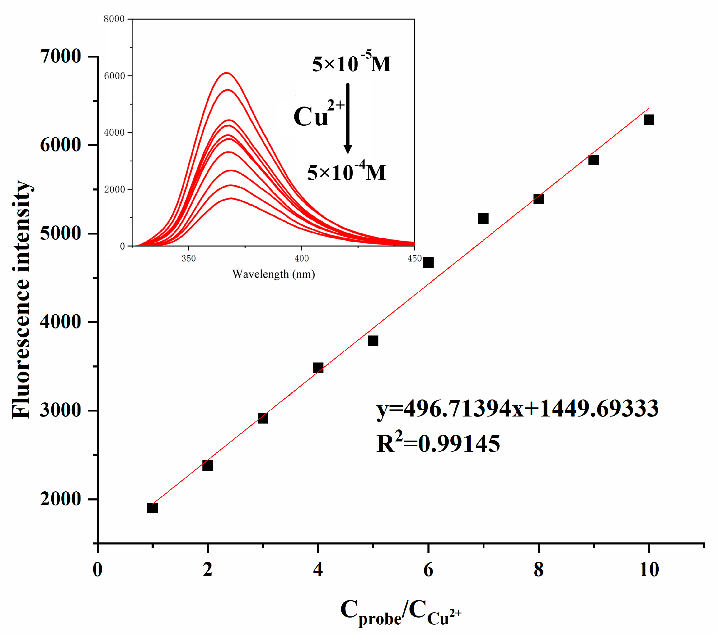
To clarify the sensitivity of Probe PYB to Cu2+, the fluorescence spectra of Probe PYB in different concentrations of Cu2+ (5 × 10⁻⁵ mol/L–5 × 10−4 mol/L) were tested (VDMF:VHEPES-NaOH buffer solution = 1:1, pH 7.4, λex = 320, λem = 362). As illustrated in Figure 2, it could be clearly observed that the fluorescence intensity of Probe PYB gradually decreased with the increase of Cu2+ concentration. When the molar ratio of the Probe PYB to Cu2+ reached 1:1, the fluorescence quenching rate was approximately 71%. By measuring the relationship between the maximum fluorescence emission intensities (362 nm) and Cu2+ concentrations, it was determined that this fluorescence quenching phenomenon conforms to a linear relationship (y = 496.71394x + 1449.69333, R2 = 0.99145), indicating that Probe PYB can perform quantitative analysis on Cu2+ and verifying its good sensitivity to Cu2+. And based on the detection limit formula (DL = 3 s/m, where s is the standard deviation of replication measurements, and m is the slope of the calibration curve), the detection limit of Probe PYB is calculated to be 8.35 × 10⁻⁷ M [21].
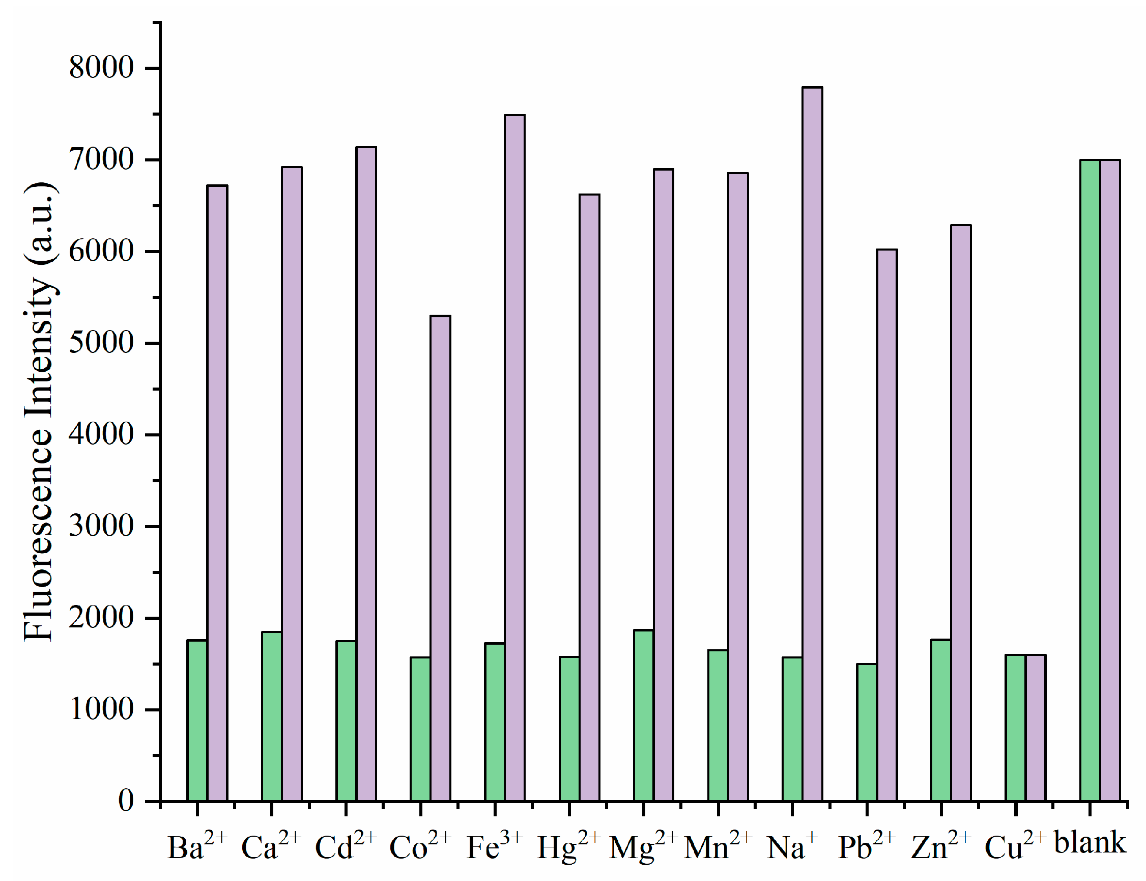
Selectivity is an important indicator for measuring the quality of fluorescent probes. In order to verify the selectivity of Probe PYB for different metal ions, several common metal ions (Ba2+, Na+, Mg2+, Zn2+, Cd2+, Ca2+, Mn2+, Pb2+, Cu2+, Hg2+, Fe3+, Co2+, 5 × 10⁻⁴ mol/L) were added to the Probe PYB (5 × 10⁻⁴ mol/L) solution under the same measurement conditions (VDMF:Vbuffer = 1:1, pH 7.4) and their fluorescence intensities were tested separately. As illustrated in Figure 3, the purple bar represented the maximum fluorescence emission intensities of Probe PYB in different metal ions (λex = 320 nm, λem = 362 nm). It could be obviously determined that only Cu2+ caused a significant decrease in the fluorescence intensity of Probe PYB, while other metal ions would not have a significant impact. To further verify the detection ability of Probe PYB for Cu2+, in complex environments, we also introduced competitive experiments as a reference. After inducing quenching of Probe PYB (5 × 10⁻⁴ mol/L) with Cu2+ (5 × 10⁻⁴ mol/L), 10 times the concentration of other metal ions (Ba2+, Na+, Mg2+, Zn2+, Cd2+, Ca2+, Mn2+, Pb2+, Hg2+, Fe3+, Co2+, 5 × 10⁻³ mol/L) were added and the fluorescence intensity of the system was measured (VDMF:VHEPES-NaOH buffer solution = 1:1, pH 7.4). The maximum fluorescence intensities at 362 nm were selected as experimental data. As shown in Figure 3, the green bar represents the competitive experiments. It could be found that the introduction of 10 equivalents of other metal ions did not significantly affect Cu2+ recognition based on fluorescence quenching. This meant that Probe PYB could still effectively recognize Cu2+ in the presence of high concentrations of interfering ions, and also verifies Probe PYB’s good qualitative analysis ability for Cu2+.
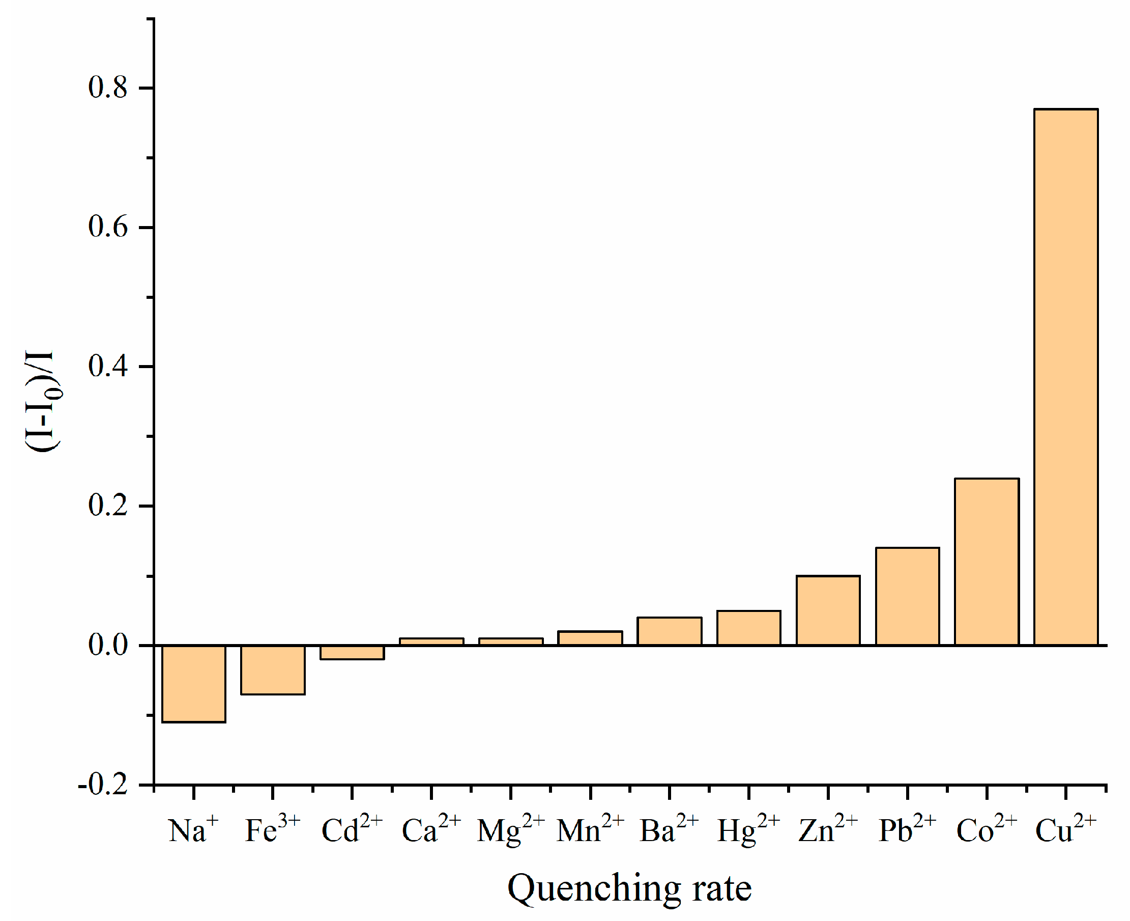
Moreover, the quenching rates of Probe PYB in above-mentioned ions were also measured. As illustrated in Figure 4, the quenching rate of Probe PYB induced by Cu2+ is very prominent, reaching 77%, which is significantly better than other metal ion.
lg ( I 0 I I ) = lg K S V + nlg ( Q )
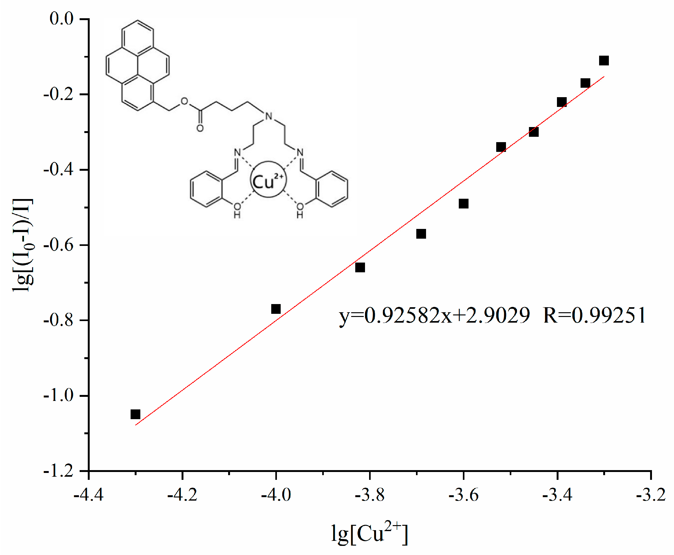
The Stern–Volmer equation (Equation (1)) was introduced to calculate the quenching constant and binding ratio of Probe PYB to Cu2+ [23]. Wherein, I0 is the fluorescence intensity of Probe PYB, I is the fluorescence intensity of Probe PYB induced by Cu2+, KSV is the Stern Volmer constant, n is the number of binding sites, and Q is the concentration of Cu2+. Based on the fluorescence intensity of Probe PYB under the condition of Cu2+ concentration from 5 × 10⁻⁵ M to 5 × 10⁻⁴ M, the equation is calculated to be y = 0.92582x + 2.9029. By calculating the slope of the equation, n is equal to 0.92, approximately equal to 1, indicating that the coordination ratio between Cu2+ and Probe PYB is 1:1. And according to the intercept of the equation, the quenching constant can be calculated to be 799.65 M−1. Based on the coordination atoms present in the probe molecule and relevant literature reports, we have plotted the coordination model between Probe PYB and Cu2+ in Figure 5.

3.3. Test Strip Experiment

In addition, filter paper was selected as the carrier for Probe PYB for convenient and rapid detection of Cu2+. The test strip for detecting copper ions was prepared by uniformly dropping Probe PYB solution onto neutral filter paper and drying it. Subsequently, solutions of the above-mentioned metal ions were dropped onto the surface of the test paper and placed under the irradiation of ultraviolet light, as shown in Figure 6, only Cu2+ induced fluorescence quenching of the test strip, while other metal ions did not significantly affect the fluorescence signal of the test strip.

3.4. Biological Investigation

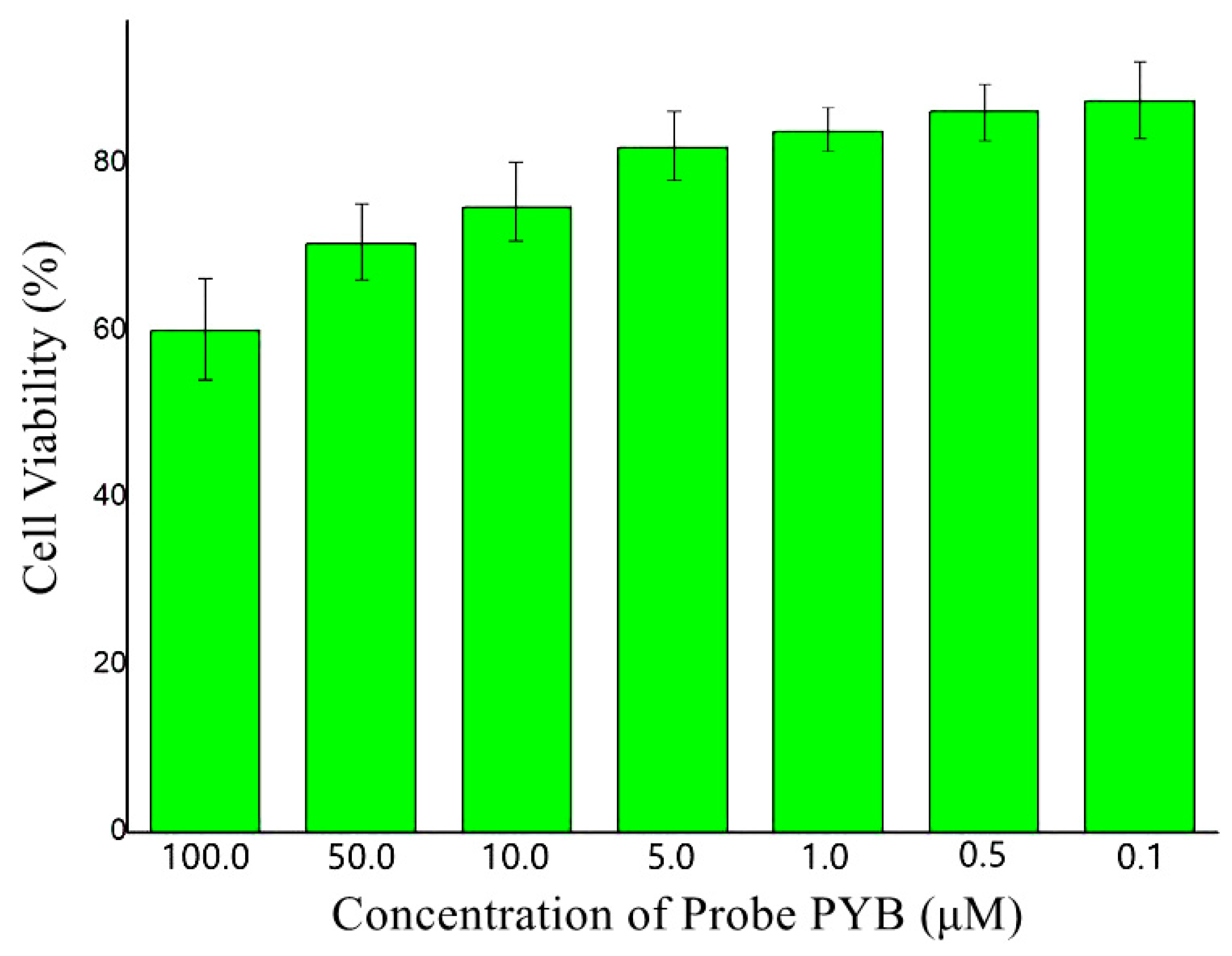
In order to expand the application of Probe PYB in the field of biology, the biocompatibility of Sensor PYB was assessed in HepG2 cells. The MTT assay was performed to confirm the cytotoxicity in different concentrations of Sensor PYB (0.1–10 µM). As illustrated in Figure 7, the cell viability had no dramatic differences within the concentration range of 0.1–5 µM, when the concentration of Sensor PYB reached 10 µM or above, cell viability began to decrease. Even so, at the highest concentration of 100 µM, the cells maintained fine viability (59%), and the IC50 value was 171.5 µM. So the cytotoxicity of Sensor PYB was relatively low under the experimental conditions.
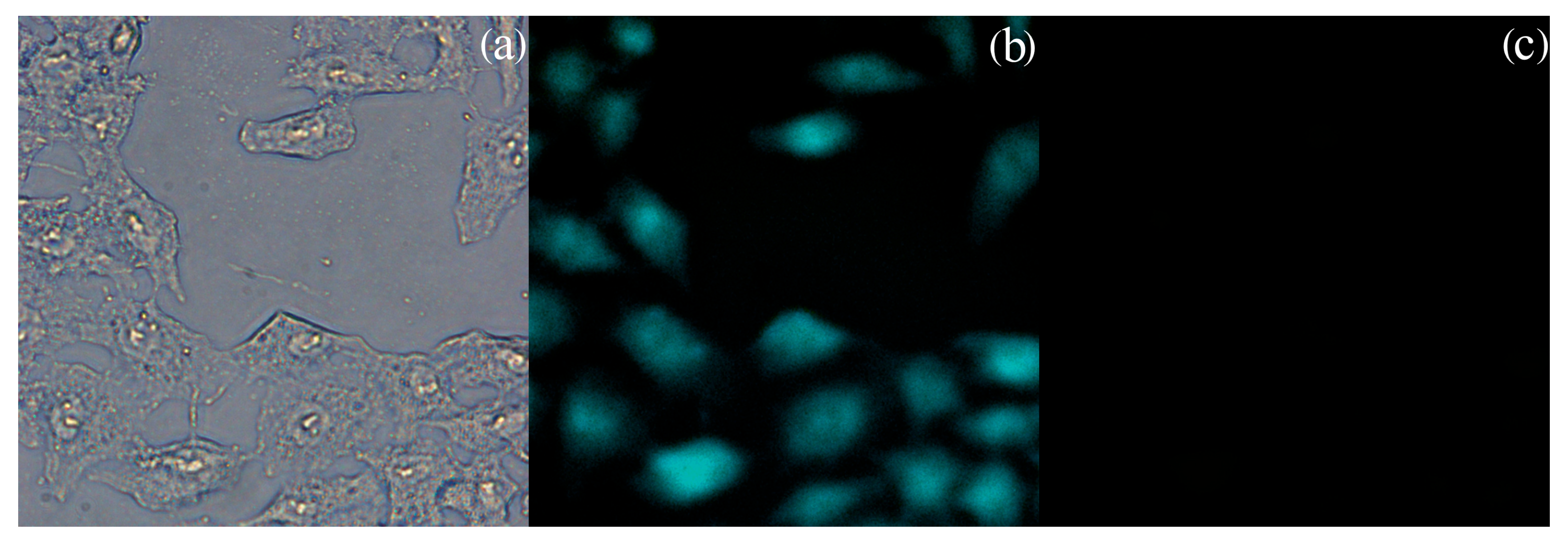
Furthermore, we conducted fluorescence cell imaging experiments on HepG2 cells using Probe PYB. The pre-cultured HepG2 cells were removed from the incubator, the cell culture medium was aspirated using a micropipette and washed 3 times with phosphate-buffer solution (pH = 7.4) to ensure removal of the medium. Then added Probe PYB solution (1 × 10⁻⁵ M in DMSO) to the cell culture plate, and cultivated for 30 min at room temperature to ensure Probe PYB completely entered the cells. Afterwards, washed the cells 3 times with buffer solution and transferred to a fluorescence microscope for observation. As shown in Figure 8a, the cell outline was clear and complete, indicating that Probe PYB did not cause damage to the cells. From Figure 8b, it could be observed that the cells treated with Probe PYB exhibited significant fluorescence emission, indicating that the probe could penetrate the cell membrane and be adsorbed by the cells. Subsequently, 1 × 10⁻⁵ M Cu2+ buffer solution (pH 7.4) was added to the cells and cultured for 30 min. The corresponding fluorescence imaging is shown in Figure 8c. After interacting with Cu2+, the fluorescence of cells treated with Probe PYB significantly disappeared, indicating that Probe PYB coordinated with Cu2+ inside the cell and demonstrating its good biocompatibility.
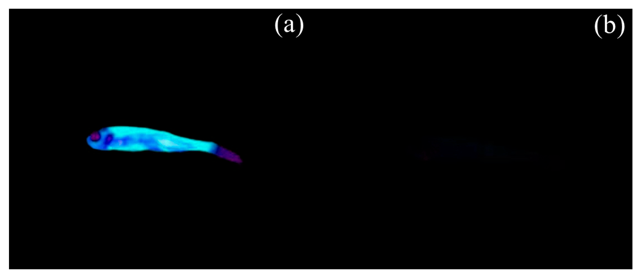
To further validate the fluorescence imaging ability of Probe PYB, zebrafish was selected as the test sample for the experiment. Zebrafish is a highly dynamic tropical fish with a body length of about 3 cm and a genetic similarity of up to 87% to humans. First, adult zebrafish was cultivated in distilled water containing 20% Probe PYB (10 µM) in DMSO solution for 1 hour. Then transferred the zebrafish to distilled water and washed it three times with buffer solution (pH 7.4) before conducting fluorescence imaging experiments. As illustrated in Figure 9a, the zebrafish cultured in Probe PYB solution showed fluorescence under UV light irradiation, with clear contours, indicating that Probe PYB had been successfully adsorbed. Afterward, transferred the zebrafish to 1 × 10⁻⁵ M Cu2+ buffer solution (pH 7.4) for 1 hour of cultivation, washed it three times with deionized water, and then performed fluorescence imaging experiments. As shown in Figure 9b, under UV light irradiation, the fluorescence on the surface of zebrafish was quenched after treatment with Cu2+ solution. This phenomenon once again demonstrated the biocompatibility of Probe PYB and proved its practical value in the field of biology.

4. Conclusions

In this study, a novel Cu2+ fluorescent probe was successfully designed and synthesized based on a pyrene derivative. Through titration experiments, Probe PYB exhibits good selectivity and sensitivity toward Cu2+, and it still retains the ability to recognize Cu2+ in the presence of high concentrations of interfering ions. The detection limit of Probe PYB is calculated to be 8.35 × 10⁻⁷ M based on the detection limit formula, and the complexation constant between Probe PYB and Cu2+ is calculated to be 799.65 M−1 in a 1:1 binding mode based on the Stern–Volmer equation. The ability of Probe PYB to quickly and conveniently detect Cu2+ is verified by preparing fluorescent test strips soaked with probe solution. Moreover, Probe PYB has been successfully applied in live-cell fluorescence imaging and biological imaging, thus demonstrating its value in the field of fluorescence tracing.

Supplementary Materials

The following supporting information can be downloaded at: https://www.mdpi.com/article/10.3390/chemosensors13110403/s1, Figure S1: 1H NMR spectrum of Probe PYB; Figure S2: 13C NMR spectrum of Probe PYB; Figure S3: LC-MS of Probe PYB. Figure S4: 13C NMR spectrum of Intermediate 2; Figure S5: 1H NMR spectrum of Probe PYB; Figure S6: 13C NMR spectrum of Probe PYB; Figure S7. LC-MS of Probe PYB.

Author Contributions

Conceptualization, C.Z. (Chen Zhou); methodology, H.W., N.X. and Y.W.; software, E.K. and H.W.; validation, E.K. and Y.W.; formal analysis, C.Z. (Chen Zhou), M.L. and N.X.; investigation, N.X., C.Z. (Chenyang Zou) and J.S.; resources, C.Z. (Chenyang Zou) and J.S.; data curation, M.L. and N.X.; writing—original draft preparation, C.Z. (Chen Zhou) and E.K.; writing—original draft preparation, H.W. and Y.W.; visualization, H.W. and C.Z. (Chenyang Zou); supervision, E.K. and N.X.; project administration, C.Z. (Chen Zhou) and E.K.; funding acquisition, C.Z. (Chen Zhou) and J.S. All authors have read and agreed to the published version of the manuscript.

Funding

This research was funded by the Free Exploration Project of Jilin Provincial Department of Science and Technology, YDZJ202301ZYTS308.

Institutional Review Board Statement

All animal procedures were performed in accordance with the Guidelines for Care and Use of Laboratory Animals of Changchun University of Science and Technology, and experiments were approved by the Animal Ethics Committee of Changchun University of Science and Technology.

Informed Consent Statement

Not applicable.

Data Availability Statement

The original contributions presented in this study are included in the article/Supplement Materials. Further inquiries can be directed to the corresponding author.

Conflicts of Interest

The authors declare no conflicts of interest.

References

  1. Tan, S.; Yuan, X.; Song, Z.; Lin, Z.; Zhao, F.; Wang, L.; Gao, J. A sequential treatment strategy by copper ion-doped nanofiber dressing for highly efficient biofilm combating and rapid wound healing. Chem. Eng. J. 2024, 500, 156974. [Google Scholar] [CrossRef]
  2. Nakajima, H.; Fujimoto, N.; Yamamoto, Y.; Amemiya, T.; Itoh, K. Effect of Cu on the fluorescence of the Cu-hyperaccumulator lichen stereocaulon sorediiferum. Environ. Sci. Pollut. Res. 2019, 26, 36754–36763. [Google Scholar] [CrossRef] [PubMed]
  3. Burghardt, I.; Lüthen, F.; Prinz, C.; Kreikemeyer, B.; Zietz, C.; Neumann, H.; Rychly, J. A dual function of copper in designing regenerative implants. Biomaterials 2015, 44, 36–44. [Google Scholar] [CrossRef]
  4. He, T.; Tang, Q.; Ren, Q.; Liu, Y.; He, G.; Pan, Y.; Wang, Z.; Huang, P.; Lin, J. Different Valence States of Copper Ion Delivery against Triple-Negative Breast Cancer. ACS Nano 2024, 18, 5434–5445. [Google Scholar] [CrossRef]
  5. Huang, Y.; Zeng, G.; Zhang, H.; Jiang, S.; Jiang, L.; Li, X.; Tang, K.; Fu, X.; Su, Z.; Jin, L.; et al. Copper ion-chelated chitosan-coated urinary catheters: Synergistic antibacterial and anti-inflammatory effects in clinical applications. Mater. Des. 2025, 258, 114664. [Google Scholar] [CrossRef]
  6. Gu, G.; Erişen, D.; Yang, K.; Zhang, B.; Shen, M.; Zou, J.; Qi, X.; Chen, S.; Xu, X. Antibacterial and anti-inflammatory activities of chitosan/copper complex coating on medical catheters: In vitro and in vivo. J. Biomed. Mater. Res. Part B Appl. Biomater. 2022, 110, 1899–1910. [Google Scholar] [CrossRef] [PubMed]
  7. Kielhorn, J.; Melber, C.; Keller, D.; Mangelsdorf, I. Palladium-A review of exposure and effects to human health. Int. J. Hyg. Environ. Health 2002, 205, 417–432. [Google Scholar] [CrossRef]
  8. Kolemen, S.; Akkaya, E.U. Reaction-based BODIPY probes for selective bio-imaging. Coord. Chem. Rev. 2018, 354, 121–134. [Google Scholar] [CrossRef]
  9. Tigreros, A.; Portilla, J. Recent progress in chemosensors based on pyrazole derivatives. RSC Adv. 2020, 10, 19693–19712. [Google Scholar] [CrossRef]
  10. Zhang, R.; Zhao, J.; Han, G.; Liu, Z.; Liu, C.; Zhang, C.; Liu, B.; Jiang, C.; Liu, R.; Zhao, T.; et al. Real-time discrimination and versatile profiling of spontaneous reactive oxygen species in living organisms with a single fluorescent probe. J. Am. Chem. Soc. 2016, 138, 3769–3778. [Google Scholar] [CrossRef]
  11. Yang, X.; He, L.; Xu, K.; Yang, Y.; Lin, W. A turn-on fluorescent formaldehyde probe regulated by combinational PET and ICT mechanisms for bioimaging applications. Anal. Methods 2018, 10, 2963–2967. [Google Scholar] [CrossRef]
  12. Nootem, J.; Daengngern, R.; Sattayanon, C.; Wattanathana, W.; Wannapaiboon, S. The synergy of CHEF and ICT toward fluorescence ‘turn-on’ probes based on push-pull benzothiazoles for selective detection of Cu2+ in acetonitrile/water mixture. J. Photochem. Photobiol. A Chem. 2021, 415, 113318. [Google Scholar] [CrossRef]
  13. Sedgwick, A.C.; Wu, L.; Han, H.; Bull, S.; He, X.; James, T.; Sessler, J.; Tang, B.; Tian, H.; Yoon, J. Excited-state intramolecular proton-transfer (ESIPT) based fluorescence sensors and imaging agents. Chem. Soc. Rev. 2018, 47, 8842–8880. [Google Scholar] [CrossRef]
  14. Jiang, D.; Zheng, M.; Yan, X.; Huang, H.; Huang, H.; Gong, T.; Liu, K.; Liu, J. A “turn-on” ESIPT fluorescence probe of 2-(aminocarbonyl)phenylboronic acid for the selective detection of Cu(ii). RSC Adv. 2022, 12, 31186–31191. [Google Scholar] [CrossRef] [PubMed]
  15. Chen, G.; Song, F.; Xiong, X.; Peng, X. Fluorescent nanosensors based on fluorescence resonance energy transfer (FRET). Ind. Eng. Chem. Res. 2013, 52, 11228–11245. [Google Scholar] [CrossRef]
  16. He, C.; Zhou, H.; Yang, N.; Niu, N.; Hussain, E.; Li, Y.; Yu, C. A turn-on fluorescent BOPHY probe for Cu2+ ion detection. New J. Chem. 2018, 42, 2520–2525. [Google Scholar] [CrossRef]
  17. Qi, J.; Li, B.; Wang, X.; Zhang, Z.; Wang, Z.; Han, J.; Chen, L. Three-dimensional paper-based microfluidic chip device for multiplexed fluorescence detection of Cu2+ and Hg2+ ions based on ion imprinting technology. Sens. Actuators B-Chem. 2017, 251, 224–233. [Google Scholar] [CrossRef]
  18. Carter, K.P.; Young, A.M.; Palmer, A. Fluorescent sensors for measuring metal ions in living systems. Chem. Rev. 2014, 114, 4564–4601. [Google Scholar] [CrossRef]
  19. Ma, X.; Qian, K.; Kandawa-Schulz, M.; Miao, W.; Wang, Y. Direct determination of Cu2+ based on the electrochemical catalytic reaction of Fe3+/Cu2+. Electroanalysis 2020, 32, 620–625. [Google Scholar] [CrossRef]
  20. Gao, W.; W, H.; Pu, S. A highly selective fluorescent probe for Cu2+ based on a diarylethene with a benzo [1,2,5] oxadiazol-4-ylamine Schiff base unit. J. Photochem. Photobiol. A Chem. 2018, 364, 208–218. [Google Scholar] [CrossRef]
  21. Dayanidhi, K.; Vadivel, P.; Jothi, S.; Eusuff, N. White Eggshells: A potential biowaste material for synergetic adsorption and naked-eye colorimetric detection of heavy metal ions from aqueous solution. ACS Appl. Mater. Interfaces 2020, 12, 1746–1756. [Google Scholar] [CrossRef]
  22. Li, T.; Xiao, X.; Zhou, C.; Luo, M. Design and application of Cu2+ fluorescent sensor based on carbazole derivatives. J. Fluoresc. 2025, 35, 2993–3001. [Google Scholar] [CrossRef]
  23. Li, F.; Zhou, C.; Hou Zhang, H.; Hou, Y.; Pan, Q.; Sun, J.; Li, X. A novel colorimetric and ‘‘turn-on” fluorescent sensor for selective detection of Cu2+. Arab. J. Chem. 2022, 15, 104176–104183. [Google Scholar] [CrossRef]
  24. Yin, J.; Wang, Z.; Zhao, F.; Yang, H.; Li, M.; Yang, Y. A novel dual functional pyrene-based turn-on fluorescent probe for hypochlorite and copper (II) ion detection and bioimaging applications. Spectrochim. Acta Part A Mol. Biomol. Spectrosc. 2020, 239, 118470. [Google Scholar] [CrossRef]
  25. Wu, C.; Yu, Y.; You, J.; Chen, S.; Wu, W.; Liang, Y. Synthesis and application of pyrene-based fluorescent probe for the continuous detection of Cu2+ and picric acid. Luminescence 2025, 40, 70300. [Google Scholar] [CrossRef] [PubMed]
  26. Yu, C.; Yang, M.; Zhang, J. A pyrene-based multifunctional fluorescent probe for the detection of Cu2+ and Al3+. Microchem. J. 2025, 208, 112531. [Google Scholar] [CrossRef]
  27. Wang, Y.; Di, H. A pyrene-based fluorescent probe for the fluorescent recognition of copper ions. J. Phys. Conf. Ser. 2025, 3021, 012034. [Google Scholar] [CrossRef]
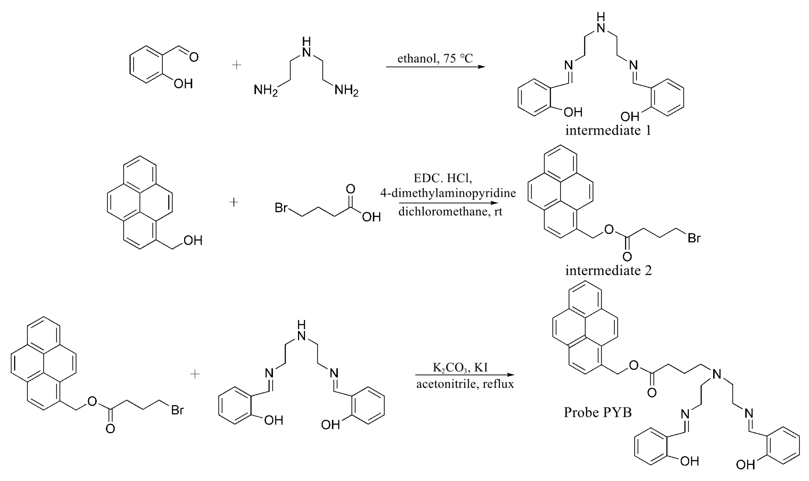
Scheme 1. Synthesis of Probe PYB.
Chemosensors 13 00403 sch001
Figure 1. UV-vis spectra of Probe PYB (5 × 10⁻⁴ mol/L) in the presence of different concentrations of Cu2+ (1 × 10⁻⁴–5 × 10⁻⁴ mol/L) in DMF/HEPES-NaOH buffer solution (v:v = 1:1, pH 7.4).
Chemosensors 13 00403 g001
Figure 2. Fluorescence spectra of Probe PYB (5 × 10⁻⁴ mol/L) in the presence of different concentrations of Cu2+ (1 × 10⁻⁴–5 × 10⁻⁴ mol/L) in DMF/HEPES-NaOH buffer solution (v:v = 1:1, pH 7.4, λex = 320 nm, λem = 362 nm).
Chemosensors 13 00403 g002
Figure 3. Selectivity experiments (purple bar) and Competition experiments (green bar) of Probe PYB for detecting Cu2+ in DMF/HEPES-NaOH buffer solution (v:v = 1:1, pH 7.4, λex = 320 nm, λem = 362 nm).
Chemosensors 13 00403 g003
Figure 4. Fluorescence quenching rate of Probe PYB in different metal ion solutions in DMF/HEPES-NaOH buffer solution (v:v = 1:1, pH 7.4, λex = 320 nm, λem = 362 nm).
Chemosensors 13 00403 g004
Figure 5. The linear fitting of the fluorescence titration curve of Probe PYB (5 × 10⁻⁴ mol/L) in different concentrations of Cu2+ (1 × 10⁻⁴–5 × 10⁻⁴ mol/L) in DMF/HEPES-NaOH buffer solution (v:v = 1:1, pH 7.4, λex = 320 nm, λem = 362 nm) and the combined model diagram between Probe PYB and Cu2+.
Chemosensors 13 00403 g005
Figure 6. Fluorescence changes of filter paper containing Probe PYB treated with various metal ions under UV lamp.
Chemosensors 13 00403 g006
Figure 7. Concentration-dependent cell viability assay.
Chemosensors 13 00403 g007
Figure 8. (a) Bright-field transmission image of live cells processed with Probe PYB; (b) Fluorescence imaging of live cells processed by Probe PYB; (c) Fluorescence imaging of live cells processed by Probe PYB–Cu2+.
Chemosensors 13 00403 g008
Figure 9. Image of adult zebrafish under UV light at 365 nm: (a) Zebrafish cultivated with Probe PYB; (b) Zebrafish cultivated with Probe PYB and Cu2+.
Chemosensors 13 00403 g009
Disclaimer/Publisher’s Note: The statements, opinions and data contained in all publications are solely those of the individual author(s) and contributor(s) and not of MDPI and/or the editor(s). MDPI and/or the editor(s) disclaim responsibility for any injury to people or property resulting from any ideas, methods, instructions or products referred to in the content.