Review

Advancing Biosensing and Imaging with DNA-Templated Metal Nanoclusters: Synthesis, Applications, and Future Challenges—A Review

School of Biomedical Engineering, Shenzhen Key Laboratory for Nano-Biosensing Technology, Guangdong Key Laboratory of Biomedical Measurements and Ultrasound Imaging, Marshall Laboratory of Biomedical Engineering, Shenzhen University Medical School, Shenzhen University, Shenzhen 518060, China
*
Author to whom correspondence should be addressed.
Chemosensors 2024, 12(12), 271; https://doi.org/10.3390/chemosensors12120271
Submission received: 15 November 2024 / Revised: 7 December 2024 / Accepted: 16 December 2024 / Published: 19 December 2024

Abstract

:
Metal nanoclusters (MNCs) are emerging as a novel class of luminescent nanomaterials with unique properties, bridging the gap between individual atoms and nanoparticles. Among these, DNA-templated MNCs have gained significant attention due to the synergistic combination of MNCs’ properties (such as exceptional resistance to photostability, size-tunable emission, and excellent optical characteristics) with the inherent advantages of DNA, including programmability, functional modification, molecular recognition, biocompatibility, and water solubility. The programmability and biocompatibility of DNA offer precise control over the size, shape, and composition of MNCs, leading to tunable optical, electrical, and magnetic properties. This review delves into the complex relationship between DNA sequence, structure, and the resulting MNC properties. By adjusting parameters such as DNA sequence, length, and conformation, the size, morphology, and composition of the corresponding MNCs can be fine-tuned, enabling insights into how DNA structure influences the optical, electrical, and magnetic properties of MNCs. Finally, this review highlights the remarkable versatility and latest advancements of DNA-templated MNCs, particularly in biosensing and imaging, and explores their future potential to revolutionize biomedical applications.

1. Introduction

Metal nanoclusters (MNCs) are a novel class of nanomaterials composed of several to hundreds of metal atoms, typically stabilized by surrounding ligands [1,2,3]. These ultra-small ( < 3 nm) clusters exhibit an intermediate state between metal atoms and nanoparticles, leading to unique physical and chemical properties arising from their size, which is close to the Fermi wavelength of electrons [4,5]. Their properties include excellent water solubility, ultra-small core sizes, easy availability, strong photoluminescence, large Stokes shifts, and better biocompatibility, making them adaptable for various applications. MNCs mainly consist of a metal core surrounded by ligands, which stabilize the nanoclusters and influence their physicochemical characteristics. Different ligands such as biomolecules (such as peptides, amino acids, proteins, and DNA) play a crucial role in MNCs and found widespread applications in energy conversion water solubility and biocompatibility, nanomedicine, catalysis, biosensing, and imaging [6,7,8,9,10,11,12].
Among various ligands, DNA stands out as a superior ligand for MNCs due to its unique structural and functional properties [13]. DNA provides several advantages as a ligand: (1) Programmability of DNA sequences: specific DNA sequences can be designed and synthesized to control the formation of MNCs with desired metal compositions (e.g., AgNCs, CuNCs, AuNCs), allowing controlled MNC formation; for instance, DNA templates rich in cytosine (C), thymine (T), and adenine (A) favor the formation of Ag nanoclusters (AgNCs), Cu nanoclusters (CuNCs, or fluorescent Cu nanoparticles), and Au nanoclusters (AuNCs), respectively [13]. (2) Biocompatibility: as a naturally occurring biomolecule, DNA offers excellent biocompatibility and biodegradability, making DNA-templated MNCs suitable for applications in biomedical and biological systems [14,15]. (3) Multifunctionality: DNA molecules can be modified with various functional groups or ligands (e.g., chemical linking groups, including -OH, -SH, -NH2, -COOH, etc.) and molecules’ complementary sequences (e.g., aptamers, DNases, or fluorescent groups). This high versatility allows DNA-templated MNCs to be tailored for applications involving metal ions, small molecules, proteins, living cells, and even in vivo imaging [16,17]. (4) Ease of preparation: DNA-MNC synthesis is simple, requiring only DNA–metal ion complexes and mild reducing agents at room temperature. Synthesis typically involves reacting a prepared DNA–metal ion complex with an appropriate reducing agent at room temperature or lower, completing the process within minutes to a few hours.
Among the DNA-templated metal nanoclusters (DNA-MNCs), DNA-templated AgNCs (DNA-AgNCs) have emerged as a captivating research area due to their unique optical properties. The seminal work by Braun et al. in 1993 demonstrated DNA’s ability to bridge two gold electrodes, acting as a template for silver nanowire fabrication, which set a foundational precedent for DNA-templated synthesis of nanomaterials [18]. In 2004, the Dickson group pioneered the synthesis of water-soluble DNA-AgNCs in aqueous solutions, illustrating how DNA sequence and structure could be modified to control AgNC optical properties [19]. By 2008, Dickson et al. investigated DNA base pairs’ influence on AgNC formation, demonstrating that varying the composition of DNA sequences can result in AgNCs with distinct emission colors, ranging from blue to near-infrared (NIR) [20]. For instance, a series of 12 single-stranded, cytosine-rich (C-rich) DNA chains composed of cytosine, thymine, and adenine (excluding guanine to avoid self-aggregation) were employed to synthesize AgNCs with distinct optical properties. The resulting AgNCs exhibited five unique color emissions: blue, green, yellow, red, and near-infrared. This selective application of DNA sequences enables the controlled fabrication of numerous AgNCs with tailored emission colors and photophysical characteristics, a key attribute for potential applications in bioimaging and sensing. Over the past decade, significant advancements have been made in understanding the structural and optical properties of DNA-AgNCs, expanding their potential in nanotechnology and materials science. Huard et al. reported the structure of a green-emitting (DNA)2Ag8NC, and Cerretani et al. characterized a NIR-emitting Ag16NC and its seven mutants [21,22]. In the published crystal structure of this NIR emitter, the observed electron density suggested the presence of two additional silver atoms with low occupancy (≈0.3). However, this finding lacked corroborating mass spectrometry data, which typically support structural interpretations by providing precise information about the molecular composition. A recent study by Gonzàlez-Rosell et al. combined mass spectrometry with X-ray analysis to reveal chloride ligands in the DNA-Ag16NC structure, refining prior interpretations of its electron density [23]. The electron density of chloride ions aligns with about one-third of that of silver ions, and the experimental mass data confirm the theoretical isotopic distribution of (DNA)2[Ag16Cl2]8+. This alignment supports the updated structural model, where two chloride ligands are indeed present in the DNA-Ag16NC structure. Combining mass spectrometry and crystallographic findings has gained significant insights into the structural intricacies and unique properties of DNA-AgNCs, particularly in understanding how ligands influence their stability and emission characteristics. Despite these advances, unresolved questions persist, highlighting the complexity and ongoing challenges in fully elucidating the structural complications of DNA-AgNCs. Therefore, further research is needed to describe the precise mechanisms underlying the formation of DNA-AgNCs, to explore the full range of their potential applications, and to develop scalable and reproducible synthetic methods.
Compared to silver, copper is more chemically active, leading to the formed CuNCs being more prone to oxidation and thus the quenching of their fluorescence. Selecting an appropriate template, which can tightly protect CuNCs, is a crucial factor in the synthesis of CuNCs. In 2010, the Mokhir group first used double-stranded DNA (dsDNA) to synthesize fluorescent CuNCs [24]. The size and fluorescence intensity of CuNCs are positively linked to the length of the dsDNA. Subsequently, both the Wu [25] and Yang [26] groups almost simultaneously found that single-stranded poly thymine DNA (poly T) could serve as an effective template for the formation of CuNCs. However, the poor photostability of DNA-CuNCs has hindered their application development. In contrast to unstable AgNCs and CuNCs, AuNCs should be ideal substitutes, due to their higher chemical inertness. AuNCs with exotic structure and intriguing properties have been well developed with versatile stabilizing ligands, including thiol molecules, dendrimers and polymers, peptides, and proteins [27,28]. However, DNA-templated AuNCs are still in their infancy because of sequence-irrelevant emission and low quantum yields (QYs). The first study on DNA-templated AuNCs was reported by the Shao group, using a 23-mer ssDNA as the template and Dimethylamine borane (DMAB) as the reducing agent [29]. Liu and colleagues, through their study of different reducing agents, inferred that the emission of AuNCs does not depend on the sequence of the DNA template but rather on the type of reducing agent [30]. Li et al. prepared AuNCs using the citrate reduction method; however, the highest QY was only 2% [31]. Therefore, it is still a considerable challenge to construct synthetic methods for DNA-templated AuNCs with sequence-specific and highly emissive fluorescence. Overall, compared to DNA-AgNCs, the development of DNA-CuNCs and DNA-AuNCs is relatively deficient, and much effort should be devoted in their reliable synthesis methodology to enhance their emission and stability.
DNA-templated MNCs offer superior properties, including high quantum yields, tunable emissions, large Stokes shifts, and two-photon absorption, setting them apart from conventional organic dyes and enhancing their value in biosensing and imaging applications. Understanding the emission mechanisms of DNA-MNCs is crucial for designing and optimizing their ideal optical properties. However, there is currently a lack of comprehensive reviews comparing the structural origins, i.e., DNA sequences and structures, of these nanoclusters. This review aims to bridge this gap between DNA structure and applications of corresponding MNCs. We primarily categorized and studied the synthesis, structural design, and emission mechanisms of DNA-MNCs (AgNCs, AuNCs, and CuNCs) based on different DNA sequences and structures, thereby elucidating the fundamental principles behind their superior properties. We then classify DNA-MNCs according to their structural templates, including single-stranded DNA (ssDNA), double-stranded DNA (dsDNA), triple-stranded DNA (tsDNA), and DNA nanostructures, to highlight their versatility and utility in biosensing and imaging applications. This classification aids in understanding how the DNA structure influences the formation, functionality, and usage of MNCs. Beyond merely presenting recent advancements and applications in biosensing and imaging, this review will also delve into the current challenges, including methods to enhance the optical characteristics of MNCs and optimize their synthesis, and explore potential future research directions, such as their utility in medical and disease therapeutic fields.

2. MNCs with Different DNA Sequences and Structural Variations

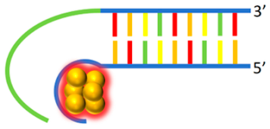
DNA, with its diverse structural possibilities, serves as an ideal template and ligand for the synthesis of fluorescent MNCs and advancing biosensors (Figure 1). Commonly used DNA structures include single-stranded DNA (ssDNA) with primary and secondary structures, double-stranded DNA (dsDNA), triple-stranded DNA (tsDNA), and more complicated DNA nanostructures. MNCs synthesized with these different DNA templates display distinct structural and functional properties. This section reviews representative studies on the synthesis and properties of MNCs, organized by DNA structural type, highlighting how each DNA template influences the resulting nanocluster characteristics.

2.1. ssDNA Templates

ssDNA oligomer can be classified into primary and secondary structures. To avoid confusion, the term primary structure is defined by the specific sequence of nucleobases within an oligonucleotide, analogous to protein primary sequences. In contrast, the secondary structure originates from stable hydrogen bonding interactions between units within the primary structure [32]. This review will specifically focus on key types of secondary structures, including i-motifs, G-quadruplexes, and hairpin loops, which play a critical role in templating metal nanoclusters (MNCs). These structures, formed through hydrogen bonding and base pairing, provide unique conformational features that influence the synthesis and properties of MNCs, especially AgNCs and AuNCs. Their stability and forming well-defined 3D shapes make them valuable in designing MNCs with distinct optical and photophysical characteristics. Common lengths for single-stranded DNA (ssDNA) templates typically range from 12 to 30 bases [13]. This length range allows for sufficient structural flexibility and stability, enabling effective templating of metal nanoclusters (MNCs) while maintaining control over their size and emission properties. Shorter ssDNA sequences may limit structural diversity, while longer sequences provide greater flexibility for forming complex secondary structures, thus influencing the properties of the resulting nanoclusters. The flexibility and programmability of ssDNA complicate the properties of MNCs to predict. Recent advancements in high-throughput experiments combined with machine learning have provided new insights for exploring correlations among theory, application of DNA templates, and AgNC fluorescence colors [20,33]. However, the link between DNA sequences and nanocluster size or shape remains elusive, illustrating a gap in understanding that impedes further control over MNC design. ssDNA-templated MNCs are summarized in Table 1.
Table 1. Reported ssDNA used to form MNCs.
Template TypeDNA Sequence (5′-3′)λex/λem (nm)QY (%)MetalRef.
ssDNA
Chemosensors 12 00271 i001
AG2TCGC2GC3560/638-Ag[19]
C12340/485, 440/525, 580/655-Ag[34]
C12650/70017Ag[35]
C3T3A2C4
C3TCT2A2C3
C3T2A2TC4
C2TC2T2C2TC2
C3TA2CTC4
-/485
-/520
-/572
-/620
-/705


-


Ag


[20]

T15
T20
T30

340/615
-
3.1 (T20)
6.8 (T30)

Cu

[25,26]
GAG2CGCTGC5AC2ATGAGC467-Au[29]
Hairpin Loop
Chemosensors 12 00271 i002
CGCGC12CGCG560/61542Ag[36]
TATCCGTCnACGGATA (n = 5–9)420/636-Au[37]
TGCCTATT5ACGGATA340/620-Cu[25]
i-motif
Chemosensors 12 00271 i003
(TA2C4)4


(C4A2)3C4
460/560
560/652
500/570
560/625
-

-

Ag


[38]

G-Quadruplexes
Chemosensors 12 00271 i004
(GGT)4TG(TGG)4510/680-Ag[39]
-: Unavailable.

2.1.1. Primary Structure ssDNA

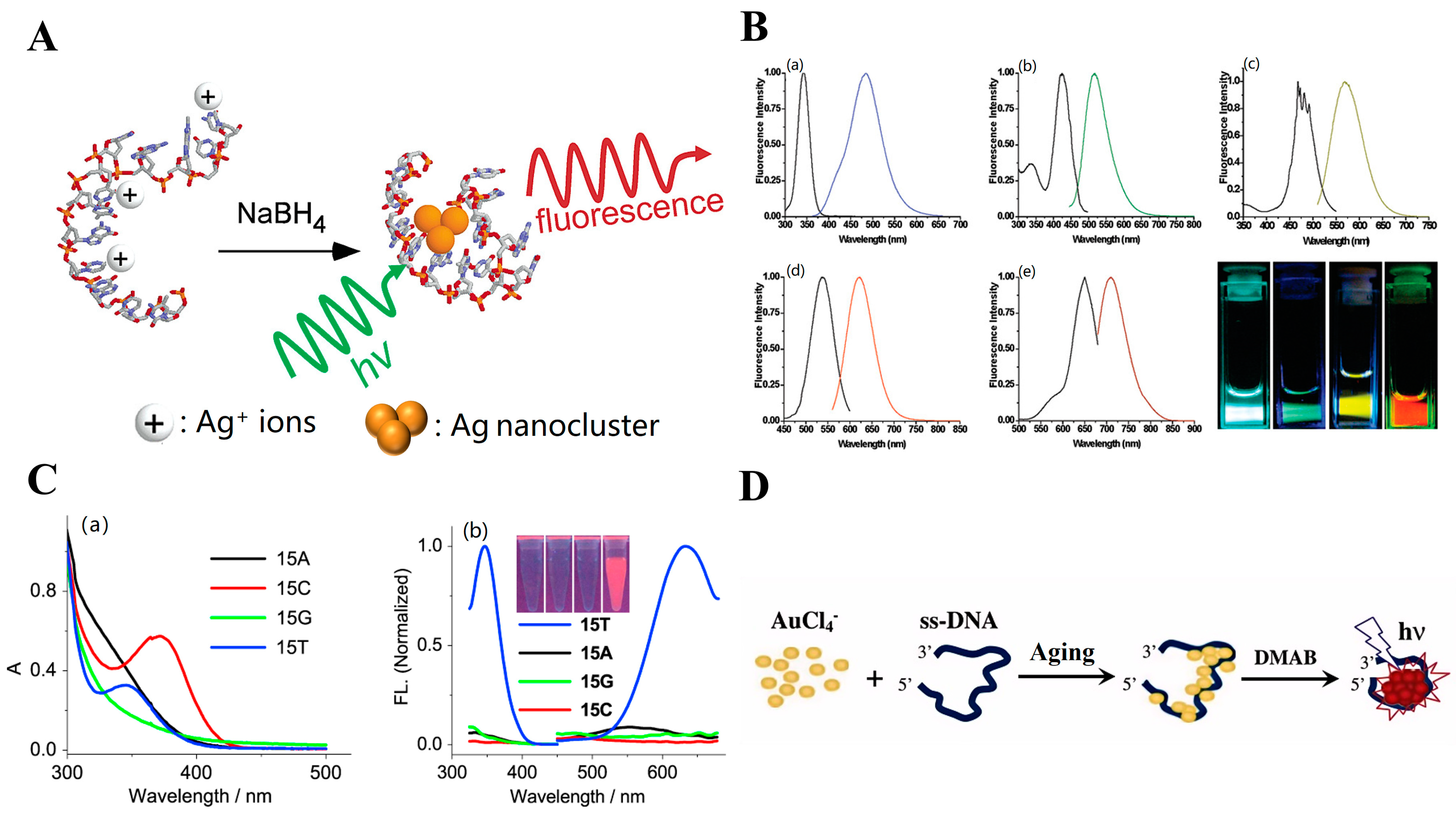
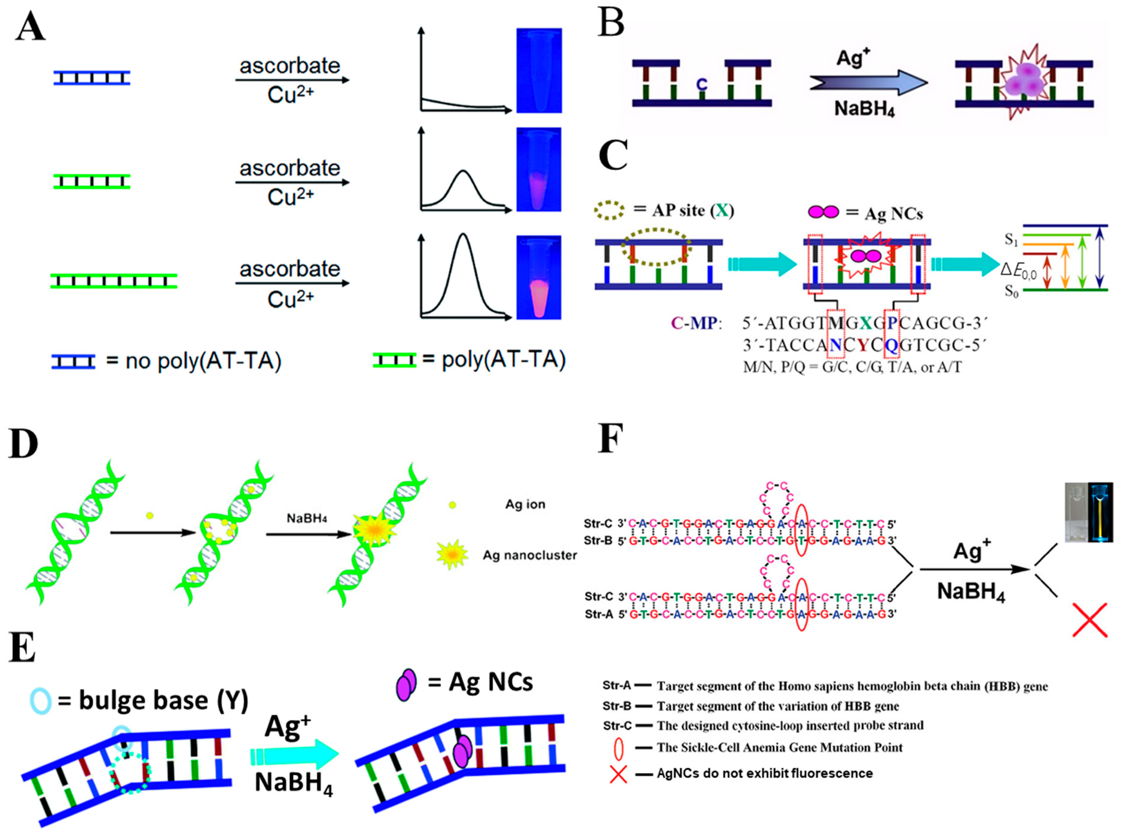
Single-stranded DNA (ssDNA) is frequently chosen as an oligomer for MNC synthesis due to its conformational flexibility, which facilitates effective binding to metal ions [19,25,29,30,31,34,35,40,41,42,43]. The Dickson group pioneered the synthesis of water-soluble fluorescent AgNCs using ssDNA as a template [19]. Specifically, they utilized a DNA sequence DNA (5′-AGGTCGCCGCCC-3′) to bind Ag+ ions, which were subsequently reduced by the addition of NaBH4, along with vigorous shaking, to produce fluorescent AgNCs. This process yielded AgNCs in a water-soluble form and efficiently prevented the aggregation into larger silver nanoparticles, as illustrated in Figure 2A. These synthesized AgNCs exhibited a broad range of emission wavelengths, indicating multiple AgNC types in the product, with maximum emission at 638 nm and excitation at 560 nm. Further studies by Dickson and co-workers in 2008 examined the specific DNA base pairs’ effect on AgNC fluorescence characteristics [20]. They identified five distinct AgNC emitters: a blue emitter (λem = 485 nm, sequence: 5′-CCCTTTAACCCC-3′), a green emitter (λem = 520 nm, sequence: 5′-CCCTCTTAACCC-3′), a yellow emitter (λem = 572 nm, sequence: 5′-CCCTTAATCCCC-3′), a red emitter (λem = 620 nm, sequence: 5′-CCTCCTTCCTCC-3′), and an NIR emitter (λem = 705 nm, sequence: 5′-CCCTAACTCCCC-3′), with emission controlled by DNA sequence variations (Figure 2B). The single-stranded DNA poly, such as thymine (poly T), could also serve uniquely as a template for CuNC (poly T-CuNCs; Figure 2C) formation, almost simultaneously reported by both Wang’s [26] and Shao’s groups [25]. However, other types of ssDNA, such as random ssDNA, polyadenine (poly A), polycytosine (poly C), and poly guanine (poly G), do not facilitate the formation of fluorescent CuNCs. This restriction suggests that specific DNA sequences, like poly T, are uniquely suited for stabilizing and templating CuNCs, likely due to their distinct structural and binding properties with metal ions. The efforts using ssDNA-templated as a template for synthesizing AuNCs has been devoted by Shao’s group [29]. They used 23 bases as ssDNA sequence (designated as 23-C, with the sequence: 5′-GAGGCGCTGCCCCCACCATGAGC-3′) as a scaffold. This DNA template was incubated with chloroauric acid, followed by the addition of DMAB as a reducing agent under mildly acidic conditions. This process synthesized water-soluble AuNCs with a red-emission profile, featuring an excitation wavelength of 467 nm and an emission wavelength of 725 nm (illustrated in Figure 2D).
Although DNA and other biomolecule-stabilized nanoclusters remain less understood than those stabilized by traditional organic molecules [27,44], in recent years, rapid advancements have emerged in the study of DNA-AgNCs, leading to a deeper understanding of their structure and fluorescent properties. High-throughput experiments combined with innovative methods like data mining and machine learning have significantly deepened the understanding of the relationship between ssDNA sequences and AgNC fluorescence properties [33,45]. In 2014, Copp et al. successfully applied machine learning to design DNA-AgNCs for the first time [46]. Their experimental results confirmed that C- and G-rich DNA base sequences are particularly effective in stabilizing brightly emissive DNA-AgNCs, while T-rich sequences tend to disfavor the formation of emissive DNA-AgNCs. However, due to the experimental limitations, their support vector machines (SVMs) could only predict overall brightness and not specific emission wavelengths. This finding spurred the development of advanced machine learning (ML) models aimed at accurately predicting and distinguishing the emission colors of DNA-AgNCs based on DNA sequence information (Figure 3A). To address this, Copp and colleagues later advanced the ML approach to predict emission colors of DNA-AgNCs. They developed an ensemble of SVMs capable of classifying 10-base DNA sequences by different emission wavelengths [47]. These training sequences were classified according to their emission wavelengths into four categories: green, red, very red, and dark (Figure 3B). Moreover, this approach significantly enhanced the selectivity for DNA-AgNCs with targeted emission wavelengths. Specifically, it increased the selectivity for long-wavelength DNA-AgNCs near the visible and NIR spectra boundary by 330% and for short-wavelength “green” DNA-AgNCs by 70%. The model was further generalized to accommodate DNA sequences of various lengths [48]. Utilizing the trained model, new sequences were designed as templates for DNA-AgNCs with N = 8, 10, 12, and 16 bases to target peak fluorescence within the “red” spectral range of 600–660 nm (Figure 3C). Experimental validation confirmed that these sequences consistently exhibited color selectivity emission for DNA-AgNCs within the specified wavelength range of 600–660 nm, indicating the model’s adaptability to different DNA lengths. In 2022, Copp et al. further sophisticated this approach by integrating high-throughput experimental data with machine learning and fundamental information about the crystal structure of DNA-AgNCs. They extracted significant DNA sequence features that determine the color emission across the known DNA-AgNCs spectrum [49]. They demonstrated that the emission color of these nanoclusters could be accurately predicted using a defined set of concise, short-chain nucleobase features. By representing DNA sequences according to these primitive elements, the ML model increased the design success rate of NIR emitting DNA-AgNCs by 12.3 times compared to training data, nearly doubling the number of documented emitters beyond 800 nm. These results indicate that incorporating known sequence–structure–property relationships into ML models substantially boosts material design, even when training data are sparse and imbalanced (Figure 3D). Through machine learning research, the development of ssDNA-AgNCs has been significantly advanced in two key ways: (1) Enhanced understanding of the relationship between DNA sequences and emission mechanisms: By integrating high-throughput experimental data with machine learning, researchers have deepened their comprehension of how DNA sequences influence the emission properties of AgNCs. For example, experimental results have shown that ssDNA sequences rich in G and C bases enhance the photostability of AgNCs, while sequences rich in T bases are less conducive to their formation. (2) Rapid screening and prediction of specific emission wavelengths: Machine learning facilitates the quick screening and prediction of AgNCs with specific emission wavelengths. Currently, machine learning models have significantly increased the success rate of designing NIR-emitting ssDNA-AgNCs and found nearly double the number of emitters above 800 nm, providing promises for their applications in in vivo imaging and therapy.
ssDNA is commonly used to synthesize MNCs due to its ability to effectively bind metal ions. Specific DNA sequences are crucial for stabilizing various MNCs, as shown by research from the Dickson [19] and Shao groups [25]. Additionally, Copp et al. have used machine learning and ssDNA sequence analysis to design AgNCs with targeted emission properties, greatly enhancing the design process, especially for NIR-emitting DNA-AgNCs with bioimaging potentials [46,47,48,49,50].

2.1.2. Hairpin Loop

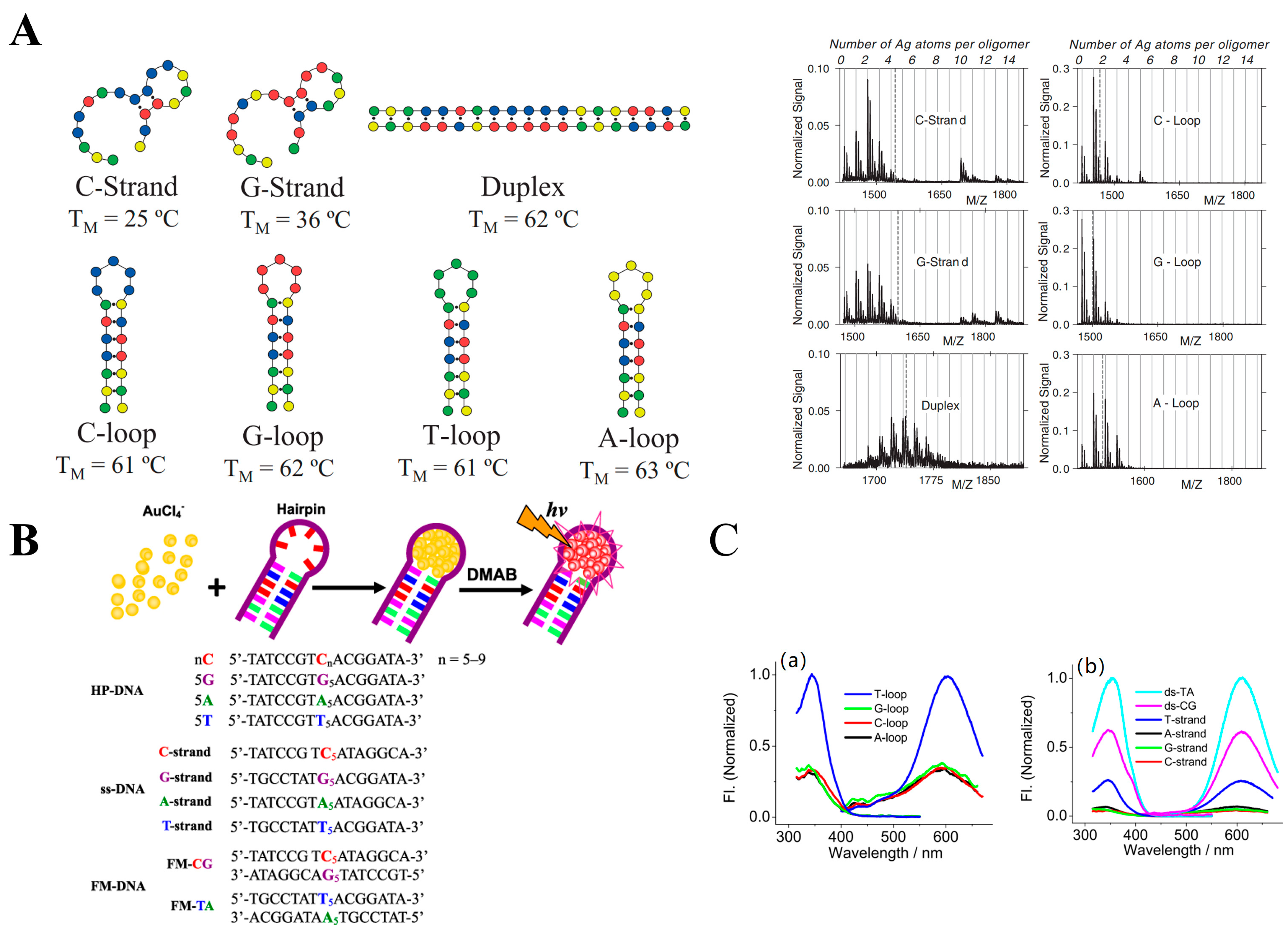
The hairpin structure, a prevalent secondary structure found in ssDNA, has become essential in the synthesis and application of DNA-MNCs due to its unique geometry. A hairpin loop forms when the ends of an ssDNA strand pair, leaving a segment of unpaired bases in a U-shaped loop. Gwinn and colleagues first employed hairpin loop DNA as a template for their studies, paving the way for subsequent research in this field [51]. To elucidate the relationship between DNA sequences and the fluorescence properties of AgNCs, researchers designed a series of hairpin DNAs with different bases in the loop region, following the design 5′-TATCCGTX5ACGGATA-3′. Here, X represents different loop bases such as C, G, A, or T to create C-, G-, A-, and T-loops, respectively. By synthesizing AgNCs using these tailored hairpin structures, they aimed to systematically investigate the impact of loop base composition on the resulting fluorescence characteristics of the AgNCs. The findings revealed that both C-loops and G-loops served as effective templates for the formation of AgNCs, though the emission intensity from cytosine/guanine loop samples was relatively lower than those from the corresponding ssDNA, possibly due to geometric constraints that hinder Ag+/Ag0 incorporation. In contrast, A-loop produced only weak fluorescence, and the T-loop was ineffective for AgNC synthesis. It has been hypothesized that when Ag+ binds to a cytosine- or guanine-loop, it may induce structural deformation and charge repulsion, impeding the effective interaction between Ag and the duplex stem (Figure 4A). However, Dickson and coworkers reported a notable exception: a “C-loop” DNA with 12 cytosine bases in the loop (5′-CGCGC12CGCG-3′) produced AgNCs with an exceptionally high fluorescence quantum yield of 42%, approximately twice that of dC12-AgNCs synthesized from linear sequences. This finding explores the complex interplay between DNA sequence, structure, and the resulting optical properties of AgNCs [36]. Subsequently, in 2012, Liu et al. studied the influence of hairpin structures (DNA sequences) on AuNC to form DNA-AuNCs [37]. They used hairpin DNA templates with various loops (A, T, G, and C) and maintained synthesis conditions and primary sequence templates constant with previously described methods [29]. The results indicated that all hairpin DNAs could yield fluorescent AuNCs (Figure 4B), with the 5C DNA template displaying a higher fluorescence intensity, outperforming the 5G, 5A, and 5T DNA templates. However, when using linear ssDNA, only the C-strand ssDNA (5′-TATCCGTC5ATAGGCA-3′) effectively produced highly fluorescent AuNCs, whereas the poly C failed. For DNA-CuNCs, the hairpin DNAs of sequence 5′-TATCCGTY5ACGGATA-3′ (Y-loop, Y = T, C, A, and G) were employed to analyze sequence dependency in Cu NP synthesis. Each template included seven base-pair stems and five identical base loops [25]. The loop segments are ssDNA structures in nature. As shown in Figure 4C, A-loop, C-loop, and G-loop templates produced similar fluorescence, suggesting that the fluorescence mainly results from the dsDNA stem segments, and the ssDNA segments composed of the identical A, C, and G in these loops have a minor role in the formation of CuNCs. However, T-loop is notably more efficient in producing fluorescent CuNCs than A-loop, C-loop, and G-loop, suggesting a unique interaction between thymine-rich loops and Cu ions in the nanocluster formation process. These findings highlight the importance of secondary structures like hairpins in tuning the optical properties of DNA-MNCs and suggest that loop base composition significantly affects MNC formation and fluorescence intensity.
The loop structure plays a crucial role in the synthesis of fluorescent harpin DNA-protected MNCs. Researchers have utilized hairpin DNA templates with varying loop bases to study their impact on AgNCs, AuNCs, and CuNCs. Findings indicate that the loop base composition significantly affects MNC formation and fluorescence intensity, with cytosine- and guanine-rich loops generally being more effective for AgNCs and AuNCs, while thymine-rich loops are particularly efficient for CuNCs.

2.1.3. i-Motifs and G-Quadruplexes as Template
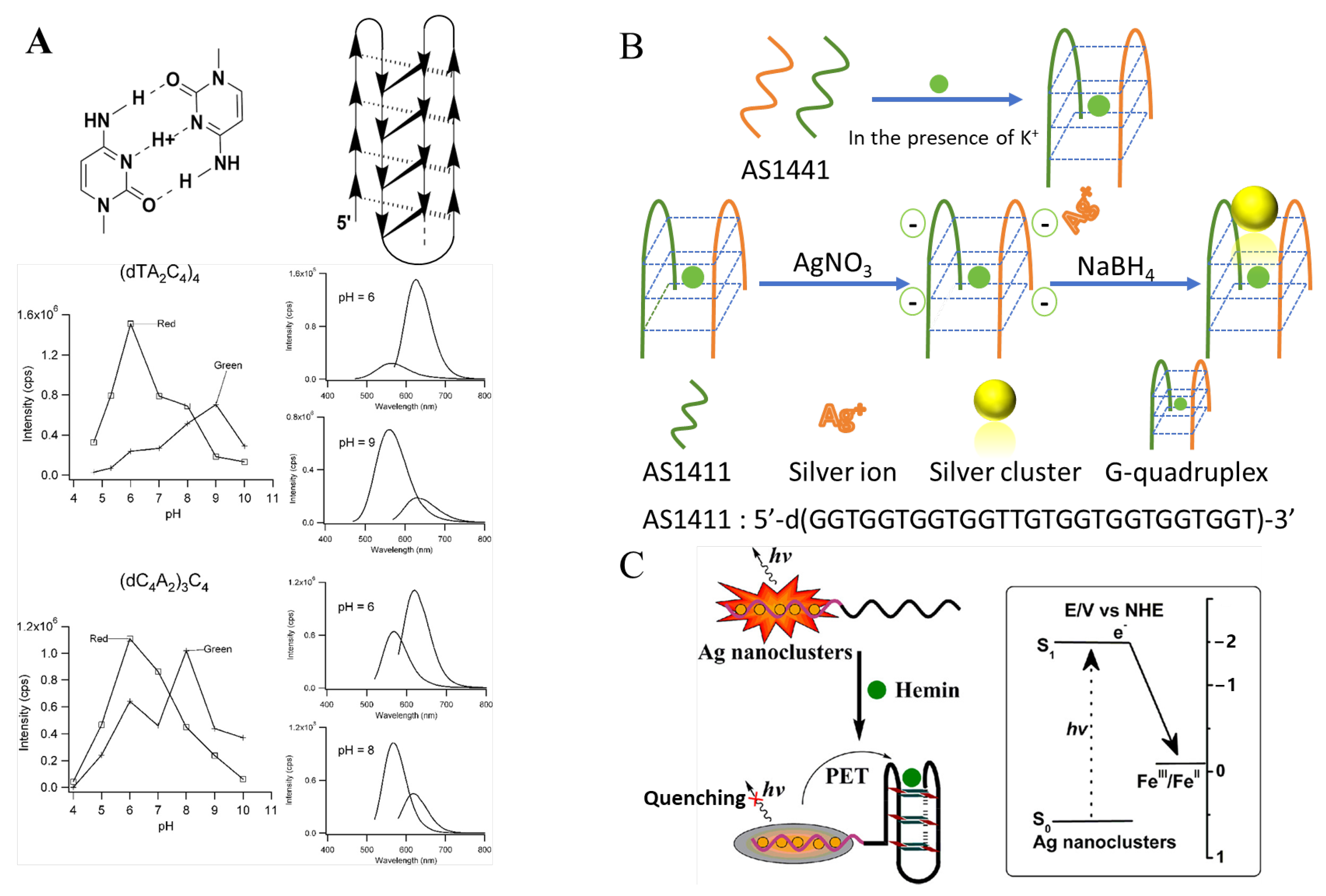
In this section, we investigate two important secondary structures of single-stranded DNA (ssDNA) that play significant roles in the synthesis of fluorescent nanoclusters: i-motifs and G-quadruplexes. The i-motif is an intercalating topology favored by cytosine-rich DNA sequences, particularly those with repeating four stretches of cytosine (C4) that tend to form intramolecular i-motif folds. This structure stabilizes at slightly acidic to neutral pH levels, where the cytosine bases become hemi protonated, facilitating the formation of the tetrameric structure [52,53]. Studies, such as those by Li et al., investigated the effect of polymorphic DNA structures on the formation of AgNCs by comparing the binding affinity of Ag ions to DNA (Ag–DNA) in different structural forms. They found that the secondary structure of DNA controls the binding affinity in the following order: coiled cytosine-rich strand > i-motif > duplex > G-quadruplex [54]. This trend matches well with the previous studies, affirming the importance of the structural flexibility of DNA in facilitating the nucleation and growth of AgNCs. Specifically, the more flexible conformations, such as coiled cytosine-rich strands and i-motifs, were more effective at binding Ag+ ions and promoting the synthesis of fluorescent AgNCs. Petty et al. investigated the formation of i-motif structures using two cytosine-rich oligonucleotides, (dTA2C4)4 and (dC4A2)3C4 [38]. They found that at low pH, ssDNA primarily adopts the i-motif structures, leading to the synthesis of predominantly red fluorescent AgNCs. However, at higher pH, the ssDNA unfolds and mainly forms green-fluorescent nanoclusters (Figure 5A). These results demonstrate that polymorphic DNA structures, such as i-motifs, can serve as reactive templates for the synthesis of novel fluorescent nanomaterials, and the pH-dependent transition between structural forms significantly influences the fluorescence properties of the resulting nanoclusters. This highlights the potential of using DNA’s structural flexibility to tune the properties of synthesized nanomaterials.
On the other hand, G-quadruplexes are formed by guanine-rich sequences that fold into square planar arrays of four Hoogsteen-paired guanines (known as tetrads), stacked on top of each other through π π interactions. These structures are typically stabilized by monovalent cations, such as K+, Na+, or NH4+, which reside in the center of the tetrads [55]. Similar to i-motifs, G-quadruplexes provide an excellent scaffold for protecting AgNCs from aggregation due to their stable, ordered structure. However, the N7 site of guanine, which plays a key role in base pairing within the tetrad, may reduce the binding affinity of the G-quadruplex for Ag+ [54]. This reduced affinity may limit the ability of G-quadruplex structures to nucleate and stabilize AgNCs, as the interaction between the guanine bases and Ag+ is less favorable compared to other structural forms, such as i-motifs. For instance, Wang and coworkers synthesized AgNCs using AS1411, a G-quadruplex that binds to nucleolin, which is overexpressed in cancer cells [39]. By mixing G-quadruplex templates with silver ions and a reducing agent, novel AgNCs were synthesized. It was found that after the formation of AgNCs, AS1411 retains its structure and remains capable of binding to nucleolin overexpressed in cancer cells (Figure 5B). Additionally, this binding behavior not only preserves the functional capacity of the G-quadruplex but also significantly enhances the fluorescence intensity of the AgNCs, a property that can be directly utilized for bioimaging in HeLa cells. Wang et al., for the first time, observed photoinduced electron transfer (PET) between DNA/Ag fluorescent nanoclusters (NCs) and G-quadruplex/hemin complexes, accompanied by a decrease in the fluorescence intensity of DNA-AgNCs (Figure 5C). In this PET process, the formed DNA-AgNCs showed strong fluorescence, while the G-rich DNA sequence could fold into a G-quadruplex structure. When hemin was introduced, the G-quadruplex/hemin complex was formed and acted as an electron acceptor. PET from DNA-AgNCs to the G-quadruplex/hemin complex then led to the fluorescence quenching of DNA-AgNCs. This novel PET system enables the detection of target biomolecules, such as DNA and ATP, with high sensitivity, specificity, and versatility according to the selection of different target sequences [56]. Overall, secondary structures of ssDNA (i-motifs and G-quadruplexes) also affect fluorescent NCs, through using them as templates for MNC synthesis or as adjacent structures/motifs to tune the emission of MNCs.
Figure 5. i-motifs and G-quadruplexes as template. (A) The i-motif DNA-templated preparation of fluorescent AgNCs. Fluorescence intensities of red (open square) and green (crosses) emitting clusters as a function of pH for (dTA2C4)4 (top) and (dC4A2)3C4 (bottom), with their emission spectra at intensity maxima shown on the right. At a low pH, ssDNA primarily adopts the i-motif structures, leading to the synthesis of predominantly red fluorescent AgNCs. At a higher pH, the ssDNA unfolds and mainly forms green-fluorescent nanoclusters [38]. (B) The G-quadruplex DNA-templated preparation of fluorescent AgNCs [39]. (C) Hemin induces the formation of G-quadruplex, which leads to the decrease in DNA-AgNC fluorescence via photoinduced electron transfer (PET) [56].
Chemosensors 12 00271 g005

2.2. dsDNA Strand

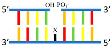
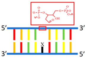
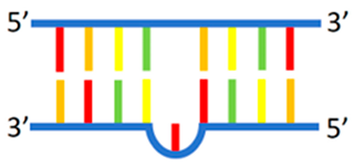
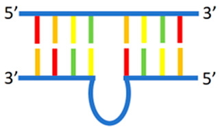
In addition to ssDNA, dsDNA can also serve as a template for synthesizing MNCs, particularly for AgNCs and CuNCs. However, complementary DNA pairs rich in cytosine and guanine tend to produce MNCs with considerably weaker fluorescence when dsDNA forms compared to ssDNA. This reduction in fluorescence occurs because, in fully complementary dsDNA, the favorable binding sites for metal ions (i.e., the heterocyclic bases) are inaccessible due to Watson–Crick (WC) base pairing [57]. The limited accessibility of metal ions in dsDNA can be annihilated by various structural derivatives, including gap [58], abasic [59,60], mismatched site [57,61], bulge [62,63], loop [64,65], and nanocluster beacons [66,67,68]. All these elements feature imperfect duplexes, where the ‘imperfect site’ facilitates the nucleation of metal ions. dsDNA-templated MNCs are summarized in Table 2.
Table 2. Reported dsDNA used to form MNCs.
Template TypeDNA Sequenceλex/λem (nm)QY (%)MetalRef.
Duplex
Chemosensors 12 00271 i005
5′-ATATATATATATATATATATAT-3′
3′-TATATATATATATATATATATA-5′
340/590-Cu[69]
Gap
Chemosensors 12 00271 i006
5′-GCTCATG2TG2 CGCAGCGCCTC-3′
3′-CGAGTAC2AC2YGCGTCGCGGAG-5′
Y = C, A, G, or T
560/643 (Y = C)47.2Ag[58]
5′-C2ACG2ATCTGA G3TGA3TAT2CTC-3′
3′-G2TGC2TAGACTYC3ACT3ATA2GAG-5′
Y = C, A, G, or T
585/665 (Y = C)-Ag[58]
AP site
Chemosensors 12 00271 i007
5′-ATGT2GGYGGTCATTYGGT2ATG-3′
3′-TACA2CCCCCAGTCCCCCA2TAC-5′
588/670-Ag[60]
5′-ATG2TGGYGGCAGCG-3′
3′-TAC2ACCXCCGTCGC-5′
588/670 (X = C)-Ag[59]
Mismatched site
Chemosensors 12 00271 i008
5′-C3TA2C3TA2C3TA2C3T-3′
3′-G3AT2G3XT2G3AT2G3A-5′
X = A, T, G, or C
520/570 (X = T)8.1Ag[57]
5′-GCATGTAC2CnG2A2GATCG-3′
3′-CGTACATG2GnC2T2CTAGC-5′
n = 3, 4, 5
563/654 (n = 4)-Ag[61]
Bulge
Chemosensors 12 00271 i009
5′-ATGGTGG GGCAGCG-3′
3′-TACCACCYCCGTCGC-5′
Y = bulge base (C, A, G, or T)
589/652 (Y = C)-Ag[62]
5′-CGCTGCGYGCACCAT-3′
3′-GCGACGCXCGTGGAT-5′
Y = T, C, A, or G, X = AP site
565/624 (Y = T)-Ag[63]
Loop
Chemosensors 12 00271 i010
5′-GTGCAC2TGACTC2TGTG2AGA2G-3′
3′-CACGTG2ACTGAG2CnCAC2TCT2C-5′
n = 4, 6, 8
520/572 (n = 6)-Ag[64]
5′-CT2CTC2AC6CAG2AGTCAG2TGCAC-3′
3′-GA2GAG2AGTC2TCAGTC2ACGTGACT2GAT2GT-5′
575/635-Ag[65]
Nanocluster beacons
Chemosensors 12 00271 i011
5′-CCCTTAATCCCCTGTAGCTAGACCAAA- ATCACCTAT-3′
3′-CCCACTCCATCGAGATTTCAC GGGTGGGGTGGGGTGGGG-5′
580/636-Ag[66]
5′-CCCTTAATCCCCTATTTCAAGCCGGAA ATAGCAATAAGAC-3′
3′-GGGTCATCAAGATACAGCAAGAAG- ATAGGGTGGGGTGGGGTGGGG-5′
580/636-Ag[66]
-: Unavailable.

2.2.1. dsDNA Containing Duplex, Gap, Abasic, Mismatched Site, Bulge, and Loop as Template

To investigate the influence of DNA sequences on the structure and properties of MNCs, studies initially experimented with fully complementary double-stranded DNA (dsDNA) as a template. In 2008, Gwinn and colleagues synthesized AgNCs using a fully complementary dsDNA template, but the fluorescence output was minimal [51]. This limited success motivated further investigations, and in 2010, Mokhir and colleagues achieved the synthesis of CuNCs using a DNA template in the presence of ascorbic acid as a reducing agent, marking the first synthesis of MNCs on a dsDNA template [24]; however, the used dsDNA was not fully complementary. Following these efforts, Wang and colleagues [69] later synthesized fluorescent CuNCs using poly(AT-TA) fully complementary sequences, as shown in Figure 6A. Due to the weak fluorescence signals observed from AgNCs synthesized using a fully complementary dsDNA template, research was shifted to focus on various derivative structures based on dsDNA to enhance metal binding and fluorescence [45]. One such modification is the formation of a gap site. This structure is created by hybridizing a long ssDNA strand with two shorter complementary ssDNA strands, omitting one or more nucleotides to leave an imperfect site within the duplex, which facilitates metal ion nucleation.
As shown in Figure 6B, Gui and co-workers utilized the gap sites in DNA duplexes as an innovative scaffold for AgNC synthesis [58]. Their results demonstrate that gap sites in DNA duplexes enable the rapid formation of fluorescent AgNCs, providing both bright emission and excellent stability, comparable to traditional DNA-AgNC synthesis methods. An alternative structural modification is the use of an abasic (AP) site, which contains one or more missing DNA bases with a phosphodiester backbone remaining intact. Hence, this configuration prolongs fluorescence evolution by repulsion forces from the phosphate backbone. Studies have explored that Ag2 dimers form within the confined space of AP (abasic) sites [60], such as when two consecutive AP sites cause a blue shift in λem, indicating the formation of larger Ag4 clusters, as displayed in Figure 6C. These findings recommend that the electronic properties of DNA bases in different stacking environments profoundly affect the emission characteristics of AgNCs. Another method for constructing an imperfect duplex involves introducing mismatched pairs, i.e., non-Watson–Crick (non-WC) pairs (base pairs other than guanine–cytosine and thymine–adenine). Such mismatches create unique structural environments that further influence the nucleation and emission properties of AgNCs within the DNA template. In this structure, Ag+ ions bind at or near these mismatched sites, where they are subsequently reduced and stabilized by the dsDNA scaffold. The fluorescence intensity varies depending on the mismatch type, decreasing in the order of thymine–cytosine > thymine–guanine > thymine–thymine mismatches, reflecting the affinity of the nucleobases for Ag+. Qu and co-workers successfully fabricated fluorescent AgNCs through mismatched dsDNA templates in a simple and efficient manner (Figure 6D) [57]. Another structural variant, known as a bulge site or insertion mutation in genomic DNA, is formed when two complementary DNA strands hybridize, with one strand containing extra inserted bases. Liu and colleagues found that the bulge site in the DNA duplex can be used to selectively grow fluorescent silver nanoclusters [62], as illustrated in Figure 6E. Additionally, dsDNA-derived loop structures, resembling the hairpin loops in ssDNA secondary structures, also serve as effective templates. Unlike bulges, these loops have a stem created by two separate DNA strands and contain more unpaired bases that form circular loop structures. Cytosine-rich loops in dsDNA are particularly conducive to AgNC formation. Similarly, dsDNA with cytosine-rich loops is particularly interesting because it facilitates the formation of AgNCs. Wang and co-workers [64] reported a yellow-emitting species formed by complementary double-strands with inserted cytosine-rich loops (Figure 6F). Notably, when a single-base mismatch is located fewer than three bases away from the loop, the fluorescence is quenched, likely due to conformational changes in the loop that affect the stability of the encapsulated AgNCs. This observation suggests that precise positioning of mismatches relative to loops can influence AgNC stability and fluorescence, providing a versatile tool for tuning AgNC properties through dsDNA templates. In short, the introduction of derivative structure into dsDNA, such as gap-sites, abasic sites, mismatched pairs, bulge sites, and loop structures, facilities metal ion binding with nucleobases and thus the formation of fluorescent MNCs.

2.2.2. NanoCluster Beacons

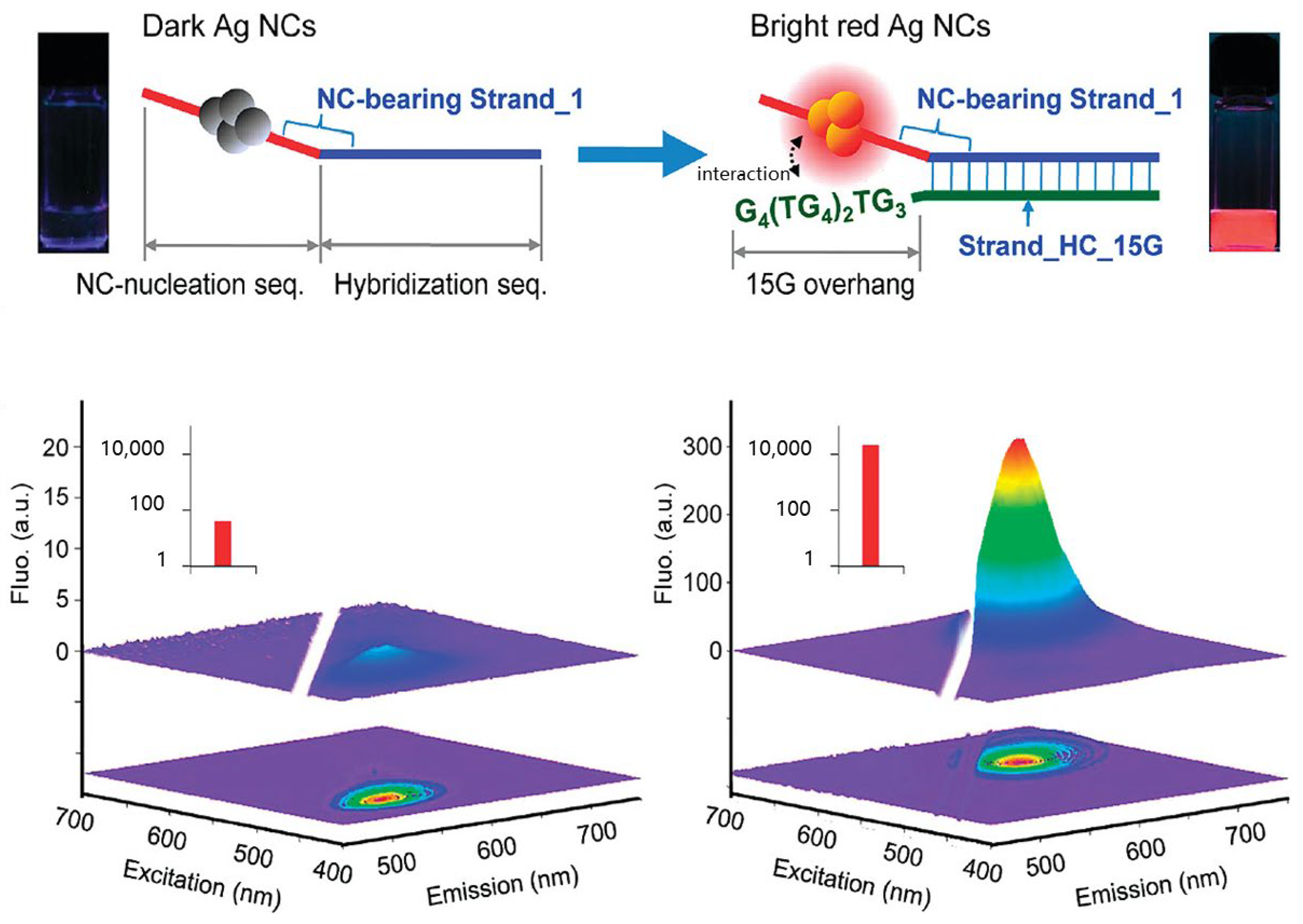
Molecular beacons are stem-loop oligomers with fluorophores and quenchers labeled at their ends, commonly applied in sensing applications. In their closed state, the fluorescence is internally quenched due to the close proximity of the fluorophore and quencher. Upon binding to a target sequence complementary to the loop region, the stem hybrid is forced to open, thus restoring the fluorescence signal [70]. Leveraging this design, Yeh and colleagues discovered that DNA/AgNCs exhibit a remarkable 500-fold increase in red fluorescence when placed in proximity to guanine-rich DNA sequences (Figure 7). This finding led to the design of a DNA detection probe known as the NanoCluster Beacon (NCB). This innovative probe “lights up” upon binding the target sequence, providing a highly sensitive, fluorescence-based method for DNA detection [66].

2.3. tsDNA and DNA Nanostructures

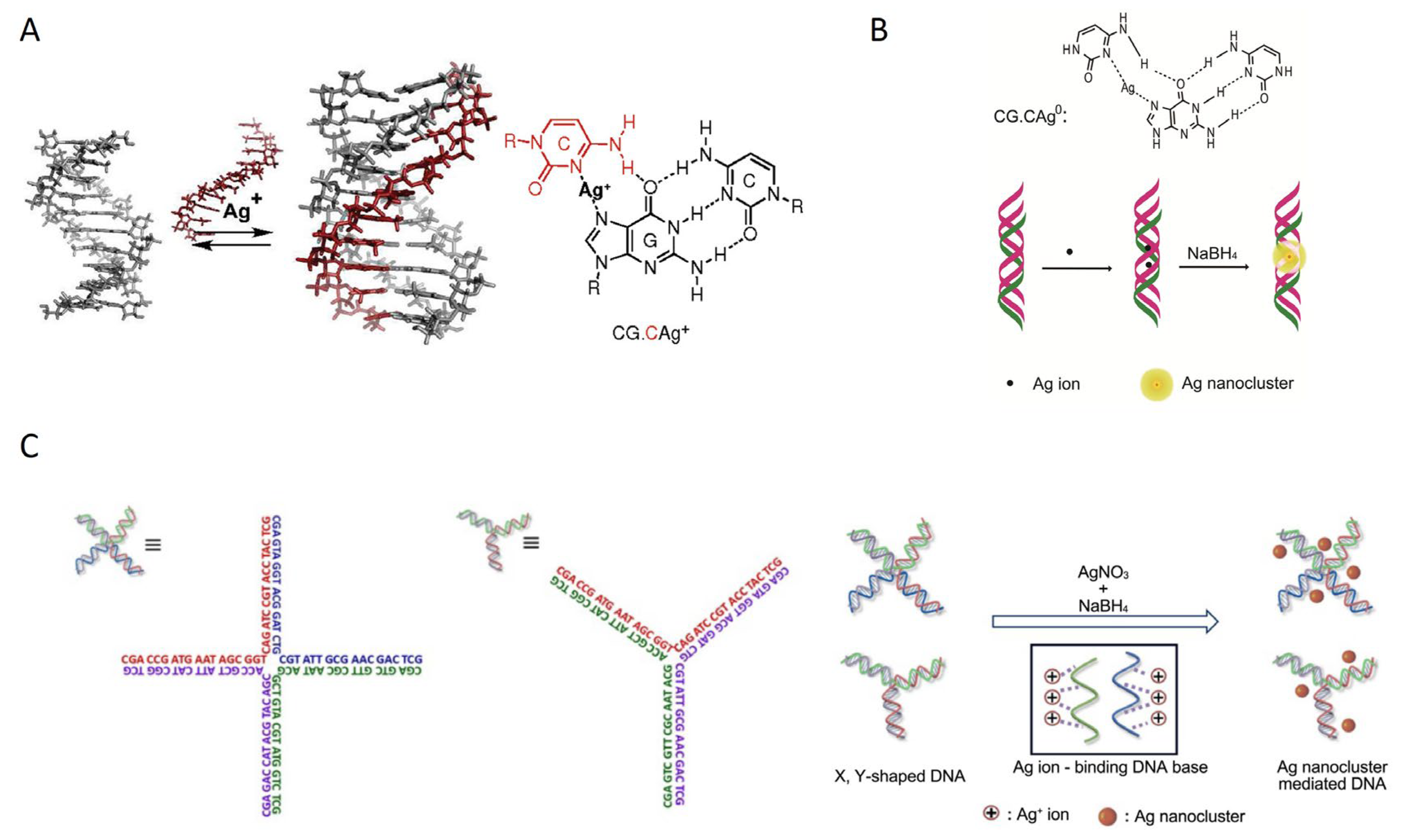
Beyond the simpler ssDNA and dsDNA templates, more complex DNA scaffolds like triplex DNA (tsDNA) and multi-stranded (DNA) nanostructures can also be used as templates for synthesizing MNCs. Although research on these more complex DNA templates, particularly for DNA-AgNCs, is less developed, the initial findings are promising. tsDNA is formed when a third strand binds to the major groove of a B-form double helix. A common base triad in this structure is CG.C+, which relies on protonated cytosine (C+) to form a Hoogsteen-based parallel triplex. As shown in Figure 8A, the Ag+ significantly stabilizes the structure of a parallel-motif DNA triplex structure [71]. Several ligands that specifically bind to DNA triplexes have also been developed; for example, Feng and colleagues designed a strategy to synthesize highly stable, site-specific, homogeneous, and bright AgNCs using tsDNA as a template [72]. Specifically, at the CG.C+ site, by careful DNA sequence design, a homogeneous Ag2 cluster was obtained in tsDNA (Figure 8B). Furthermore, DNA nanostructures, assembled from multiple ssDNA components, such as X-shaped DNA (X-DNA) and Y-shaped DNA (Y-DNA), which contain four and three arms, respectively, can provide branching and junctions that act as scaffolds for AgNC encapsulation. Park and colleagues pioneered a method to use DNA nanostructures as templates for AgNC synthesis [73], with both X-DNA and Y-DNA templates enhancing AgNC stability and fluorescence (Figure 6C). Interestingly, the addition of Cu ions to the AgNCs prepared with these DNA nanostructures quenched fluorescence, contrasting with the brightening effect seen in single-stranded DNA-stabilized AgNCs. This distinctive behavior highlights the potential for tuning fluorescence properties based on DNA scaffold complexity and the presence of other metal ions.

3. Applications of Structurally Diverse DNA-MNCs for Biosensing and Imaging

DNA-MNCs offer outstanding fluorescence properties, superior water solubility, biocompatibility, flexible probe design, and simple modification capabilities. These advantages make DNA-MNCs exceptionally suitable for biosensing applications as fluorescent nanomaterials. This section reviews recent advancements in the application of DNA-templated MNCs with various DNA structures for bioanalysis and bioimaging. In bioanalytical applications, DNA-MNCs have been extensively utilized for detecting nucleic acid–ions [74,75,76,77,78], protein detection [79,80,81,82,83,84,85,86,87], small molecule detection [84,88,89,90,91,92,93,94], and ion detection [95,96,97,98,99].

3.1. ssDNA-Templated MNCs

ssDNA is the most commonly used template for synthesizing MNCs due to its unique conformational flexibility of ssDNA, which allows efficient binding with various metal ions to form stable nanostructures. This flexible binding capability ensures that ssDNA can maintain its functional properties under different environmental conditions, positioning it as an ideal scaffold for biosensing and bioimaging applications. In biosensing, ssDNA can specifically recognize and bind to target molecules, such as specific proteins or nucleic acid fragments, enabling high sensitivity and high selectivity in detection. For bioimaging, ssDNA-MNC complexes serve as optimal imaging probes due to their exceptional fluorescence performance and stability. In addition, it can be used for high-selective imaging in live cells and tissues, making them valuable tools in both research and clinical applications [13].
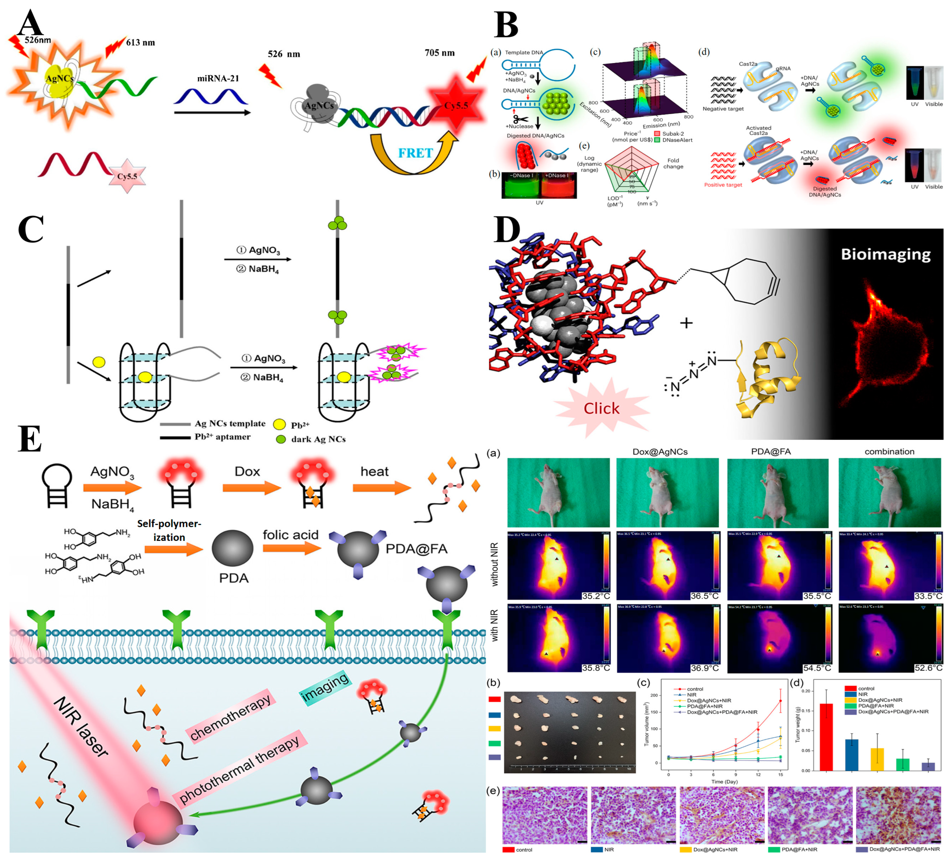
Abnormal expression levels of DNA, mRNA, or miRNA are linked to various diseases, making their high-sensitivity and high-selectivity detection of nucleic acids crucial for medical diagnostics. Sequence-specific DNA detection is crucial for diagnosing genetic and pathogenic diseases, assessing disease risk, and even guiding subsequent treatment and drug selection. For instance, Kashanian et al. designed a specific cytosine-rich ssDNA template to synthesize ssDNA-AgNCs for targeted miRNA detection. This ssDNA template formed the basis for a fluorescence resonance energy transfer (FRET) sensing platform aimed at miRNA-21 detection. As shown in Figure 9A, red-emitting DNA-AgNCs were employed to miRNA-21 in the presence of an NIR as the FRET donor, while a Cy5.5-modified probe, which emits NIR region, served as the FRET acceptor. This interaction forms a double-stranded miRNA-21/DNA probe structure, which facilitates as a bridge to efficiently transfer energy from the excited DNA-AgNCs to the Cy5.5 in its ground state. This hybrid structure results in a linear enhancement of Cy5.5 fluorescence intensity proportional to the concentration of miRNA-21. The FRET-based technique possesses high selectivity, a low detection limit (4.0 × 10⁻³ nm), and a wide dynamic range (0.02–100.00 nm), establishing it as a promising approach for developing miRNA-based specific clinical diagnostics [76].
Nucleases are essential enzymes in nucleic acid biochemistry, catalyzing cleavage reactions by hydrolyzing nucleic acids into single or oligonucleotide fragments, which are integral to various biological processes, including DNA replication, transformation, recombination, repair, genotyping, mapping, and molecular cloning [100]. Fluorescence resonance energy transfer (FRET) reporters are widely used in the final stages of nucleic acid amplification tests to indicate the presence of a nucleic acid target, where fluorescence activation occurs upon nuclease cleavage of the FRET probe. However, the manufacturing process requires dual labeling and purification, making FRET reporters costly. A cost-effective alternative, shown in Figure 9B, is a silver nanocluster reporter that eliminates the need for FRET as an on/off switching mechanism. Instead, it operates via a fluorescence color change triggered by nuclease-induced cluster transformation. Notably, a 90 nm redshift in emission occurs after nuclease digestion, an effect unattainable with conventional FRET systems. Electrospray ionization mass spectrometry (ESI-MS) results reveal that this color shift likely results from the transformations of an Ag13 (in the intact DNA host) to an Ag10 (in the cleaved fragment) cluster in the cleaved product. This mechanism provides a unique approach for nuclease detection and expands understanding of metal-DNA interactions, positioning DNA-templated AgNCs as valuable probes in studying nuclease activity and metal-DNA nanomaterials [80].
Industrial and urban development has led to significant wastewater contamination with heavy metal ions, which accumulate in natural water sources and, subsequently, enter the human body via the food chain, posing severe health risks. Heavy metals, unlike organic pollutants, do not degrade in the environment and can lead to conditions such as kidney, liver, and neurological diseases, hypertension in adults, and developmental delays in infants and children. Pb2+, in particular, is a highly toxic ion known to cause multiple adverse health effects [101]. Therefore, monitoring heavy metals in environmental and biological systems is of great importance. To address the urgent need for heavy metal detection, Wei et al. developed a novel turn-on fluorescent biosensor using C-PS2.M-DNA-templated AgNCs, with an average diameter of about 1 nm, featuring minimal toxicity and high sensitivity and selectivity for Pb2+ detection. As shown in Figure 9C, the DNA template is strategically designed with two segments: one segment at the ends serves as the AgNCs nucleation site, while the other segment in the middle is the Pb2+ specific aptamer region. Pb2+ induces the aptamer to form a G-quadruplex, bringing the two dark DNA-AgNCs at the 3′ and 5′ termini into close proximity, producing fluorescence emission. This biosensor can detect Pb2+ with a low detection limit of 3.0 nm and demonstrates a linear response within the range of 5 to 50 nm (R = 0.9862). Furthermore, its efficacy was confirmed by its successful application in detecting Pb2+ in real water samples, highlighting its potential for environmental monitoring of heavy metal contamination [95].
Live-cell imaging is essential for disease prevention, clinical diagnosis, and the study of disease mechanisms and treatments. Detecting biomolecules within live cells or cancer cells with high selectivity is critical for early cancer diagnosis and monitoring its metastasis [102,103]. As previously mentioned, MNCs, especially DNA-encoded MNCs, are advantageous as fluorescent labels for these applications, largely due to their excellent optical properties, ease of functionalization, and biocompatibility. DNA-encoded silver nanoclusters (AgNCs) were first applied in cellular imaging by Dickson et al., pioneering the use of DNA as a scaffold to direct the formation of AgNCs with specific optical features, making them ideal for biological labeling and imaging [104]. As shown in Figure 9D, Rück et al. introduced a copper-free click chemistry method for conjugating well-defined DNA-AgNCs with target molecules. This method allowed for the precise attachment of AgNCs to three different peptides and a small protein, human insulin. The binding to the target compounds was verified through MS (mass spectrometry), HPLC (High-Performance Liquid Chromatography), and time-resolved anisotropy measurements. This innovative approach highlights DNA-AgNCs as powerful tools for bioimaging and bio-labeling in live-cell environments, with significant implications for advancing cancer diagnostics and therapeutics. Hence, further research confirmed that the conjugation reaction did not alter the spectral characteristics of DNA-AgNCs, preserving their unique optical properties. Specifically, DNA-AgNCs conjugated with human insulin were tested in fluorescence imaging on Chinese hamster ovary (CHO) cells overexpressing human insulin receptor B (hIR-B). These findings demonstrated the modification of ssDNA-AgNCs with well-defined structures without altering their properties, thereby enabling the utilization of their unique characteristics for various applications. The versatile conjugation strategy is expected to inspire further exploration of DNA-AgNCs as functional probes in biological research [105]. In a complementary study, Wang et al. encapsulated DNA-AgNCs into liposomes and utilized two-photon fluorescence correlation spectroscopy to measure cerebral blood flow in living mice with high spatiotemporal resolution [106]. This innovative approach highlights the capability of NIR-emitting DNA-AgNCs for advanced biomedical imaging applications. Together, these studies underscore the broad potential of DNA-AgNCs in the development of high-selectivity imaging tools for diagnostics and therapeutic monitoring.
In addition to their utility in bioanalysis and cellular imaging, MNCs can be integrated with photodynamic or photothermal agents or other nanomaterials. As fluorescent labels, MNCs are also employed in the development of theranostic nanoprobes for both in vitro and in vivo cancer therapy [107,108,109,110]. For example, Wang and colleagues’ study effectively leveraged the unique microenvironment of cancer through the in situ self-assembly of fluorescent DNA-AuNCs complexes to facilitate safe and targeted cancer theranostics [111]. Both in vitro and in vivo tumor models indicated that such methods could enable precise bioimaging and inhibit cancer growth merely by injection of DNA and gold precursors, showing the potentials as an effective non-invasive technique for accurate cancer bioimaging and treatment and providing a promising and safe platform for cancer theranostics. For the case of DNA-AgNCs, Wu et al. pioneered a new theranostic platform for label-free imaging of cell surface glycans, accompanied by fluorescence-guided photothermal therapy (PTT) [112]. They employed Dibenzocyclooctyne (DBCO)-functionalized DNA to attach to the cell surface, facilitating the self-assembly of DNA-AgNCs. The inherent fluorescence of the AgNCs was harnessed to visualize the glycans on the cell surface. The DBCO-DNA-AgNCs were also able to absorb light and convert it into localized heat, making them highly effective for PTT. When mice bearing tumors were treated with glycoprotein labeling reagent (Ac4ManNAz)-modified DNA-AgNCs and subsequently exposed to 808 nm laser irradiation, significant tumor regression was observed, demonstrating the potential of DNA-MNCs for theranostic nanomedicine. DNA-MNCs can be utilized as a key component for constructing multifunctional theragnostic platforms. For example, Miao et al. synthesized temperature-sensitive AgNCs on a hairpin DNA template, which also served as a carrier for the anticancer drug doxorubicin (Dox) and attached them on the folic acid (FA, for cancer cell targeting)-modified polydopamine (PDA) to improve their NIR absorption and heat conversion properties [113]. After accumulating in cancer cells, PDA generated cytotoxic heat upon NIR light excitation for photothermal therapy. Simultaneously, the increased temperature disrupted the DNA template, resulting in fluorescence changes and the controlled release of Dox for chemotherapy. This integrated nanoplatform shows superior capabilities in fluorescence tracking, NIR photothermal conversion, and drug delivery for chemotherapy, which holds great promise for future clinical applications (Figure 9E).
Overall, ssDNA has emerged as a versatile template for the synthesis of MNCs due to its conformational flexibility, enabling efficient binding with metal ions to form stable nanostructures. This adaptability of ssDNA endows ssDNA-MNCs an ideal scaffold for applications in biosensing and bioimaging, where it can selectively detect nucleic acids, enzymes, and heavy metals with high sensitivity. Thus, ssDNA-MNCs are valuable in live-cell imaging for disease diagnostics and monitoring, showcasing their potentials in clinical applications.
Figure 9. Applications of ssDNA-AgNCs. (A) The developed DNA/AgNCs nano-bioprobe based on FRET for the determination of miRNA-21 [76]. (B) A schematic of Subak reporters and their fragmentation-induced color-switching property [80]. (C) The sensing method for the detection of Pb2+ by using ssDNA-scaffolded AgNCs combined with PS2.M aptamer [95]. (D) ssDNA-AgNCs for cell imaging [105]. (a) IR thermal images of tumor-bearing mice before and after 808 nm laser irradiation. (b) Photographs of harvested subcutaneous tumors (15 days). (c) Time-dependent tumor volume growth curves. (d) The weights of tumors harvested from mice after treatments (15 days). (e) Immunohistochemical staining of the apoptotic marker cleaved caspase-3, from different groups. Scale bars are 20 μm. (E) Synergistic chemotherapy and hyperthermia of tumors by hairpin DNA-templated silver nanoclusters and polydopamine nanoparticles [113].
Chemosensors 12 00271 g009

3.2. dsDNA-Templated MNCs

Compared to ssDNA, dsDNA offers greater stability and is less susceptible to degradation by nucleases. However, its rigid double-stranded structure can hinder the formation of multiple emitting species, which may restrict its use in certain applications. Therefore, instead of fully double-stranded, the introduction of derivative structures, including gaps, abasic sites, mismatched sites, bulges, loops, and NCBs, can provide more flexible design options while also displaying the growth of more structures and properties of MNCs, which have been widely applied in the field of biosensing.
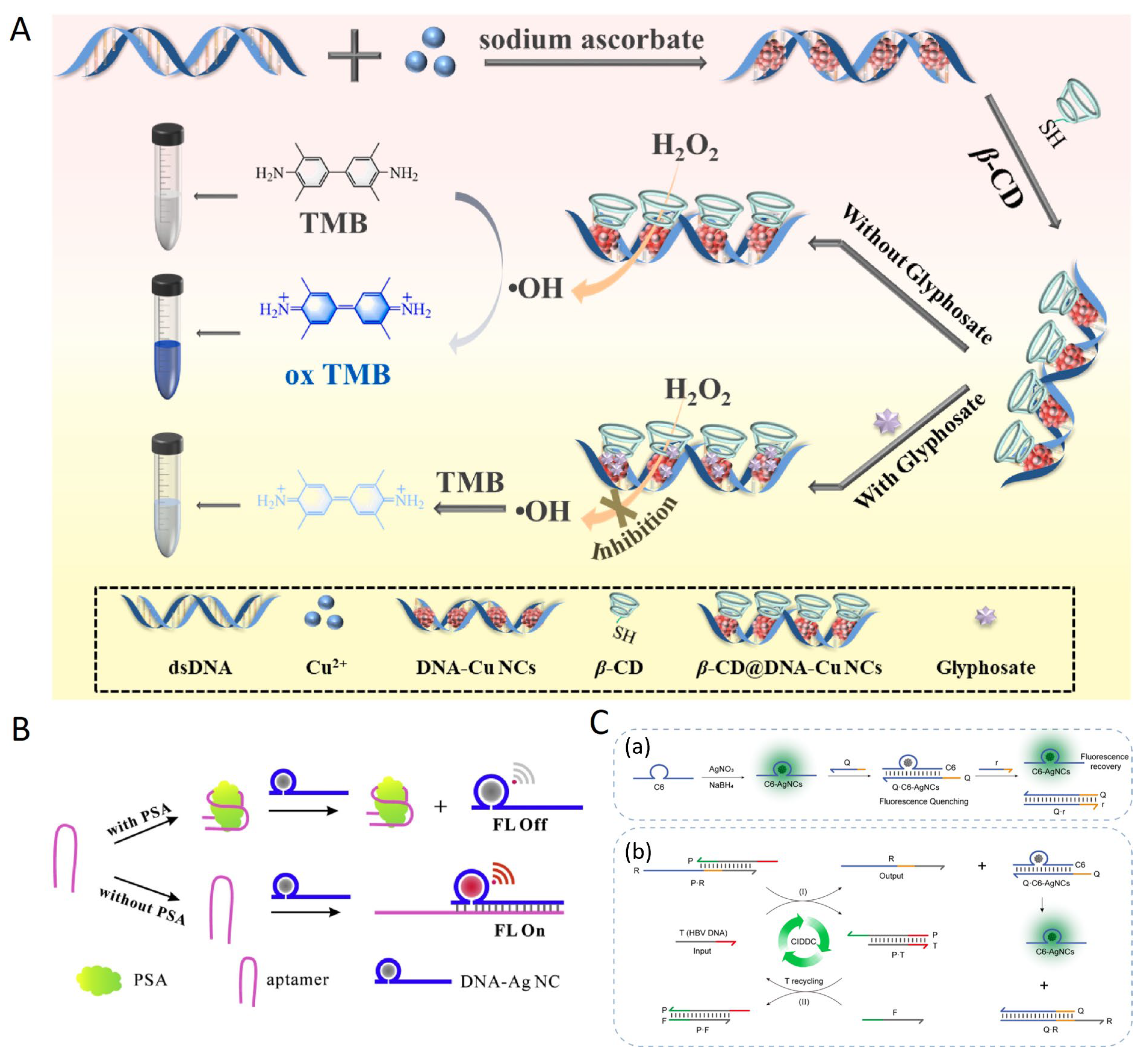
Glyphosate (Glyp), a widely used efficient organophosphate herbicide, can be effectively detected using DNA-templated MNCs. In a study by Tai et al., dsDNA as a template was utilized to synthesize DNA-CuNCs for a colorimetric detection method for Glyp (Figure 10A) [114]. The presence of Glyp disrupts the Cu2+/Cu+ redox couple on the surface of CuNCs, reducing their peroxidase-like activity and altering the absorbance signal at 652 nm. The linear range for Glyp detection was determined to be 0.02–2 μg/mL, with a detection limit of 0.85 ng/mL, providing a rapid and sensitive detection approach of Glyp in lake water and food samples with excellent selectivity. The approach demonstrates significant potential for monitoring Glyp residues in food due to its high sensitivity, selectivity, and simplicity. In another study, Zhao et al. present a label-free silver nanocluster (AgNC)-based fluorescent probe for detecting tumor marker prostate-specific antigen (PSA) [79]. As shown in Figure 10B, this approach exploited the fluorescence differences between AgNCs synthesized using dsDNA and ssDNA templates. In the experiments, DNA sequences containing complementary regions to different parts of a PSA analogue were used to synthesize DNA-AgNCs. Owing to the specific interaction between PSA and its aptamer, which blocks the aptamer’s ability to enhance the fluorescence of DNA-AgNCs, a probe for the targeted detection of PSA was created utilizing a fluorescence quenching approach. This probe can effectively detect PSA concentrations ranging from 2 to 150 ng mL−1, with a detection limit of 1.14 ng mL−1, showcasing its sensitivity and utility in targeted PSA analysis. Zhu and colleagues, based on a similar principle, developed a DNA-AgNC nanoprobe, Q·C6-AgNCs, combined with a DNA operational amplifier driven by concentration imbalance, for the detection of hepatitis B virus (HBV). By incorporating the DNA circuit, not only was the sensitivity improved, reducing the detection limit to 0.11 nm, but it also demonstrated high specificity for single-base mismatch detection of HBV DNA [115]. In short, various derivative structures of dsDNA templates are conducive to not only the formation of dsDNA-templated MNCs but also stimuli-responsive optical properties for applications in biosensing and clinical diagnostics.

3.3. tsDNA and Nanostructure-Templated MNCs

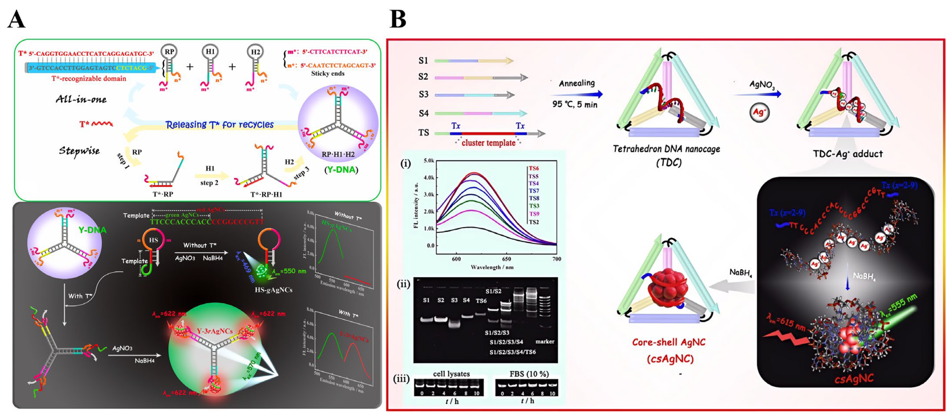
tsDNA (templated single-stranded DNA) and DNA nanostructures have been used as stabilizers for MNCs and applied in biosensing, though their study is less extensive compared to ssDNA and dsDNA. Xu et al. investigated the bioresponsive fluorescent properties of emissive AgNCs embedded in DNA branched scaffolds. As shown in Figure 11A, to detect target nucleic acids (T*), a custom-designed three-way junction DNA construct (TWJDC) was assembled through a competitive hybridization cascade involving three stem-loop hairpins with specific base sequences. The repetitive cycling of T* results in exponential amplification of the rigid TWJDC output. The stable hybridization products exhibit high T*-stimulus responsiveness and construction directionality. Among the three branch arms, the unpaired sticky ends provide isotropic binding sites for the signal hairpin, which is templated by two C-rich green and red AgNCs. The identical ligation of the signal probe to the three arms of the TWJDC releases its locked stems, allowing the red clusters in each of the three branches to grow independently. As aforementioned, three red AgNCs exhibit superior self-enhanced fluorescence performance, good biocompatibility, and low cytotoxicity compared to one or two clusters. Utilizing bicolored AgNCs as dual emitters with inversely changing emission intensities, they developed an innovative ratiometric strategy that shows sensitive linear dose dependency for variable T* concentrations as low as 1.9 p.m. This approach shows promise for biosensing, bioanalysis, and clinical applications [116]. DNA framework structures serve as natural, stable encapsulating shells for sequence-templated silver nanocluster cores (csAgNCs), presenting both intriguing and challenging applications in fluorescent biosensing. These DNA frameworks provide a controlled microenvironment for the csAgNCs, enhancing their stability and emission properties. One critical aspect of this approach is guiding the nucleation of csAgNCs through carefully designed cluster scaffolds within DNA cages. By precisely positioning template sequences within these nanostructures, researchers can optimize the conditions for in situ synthesis of the nanoclusters, resulting in improved fluorescence stability, enhanced spectral characteristics, and potentially higher sensitivity for biosensing applications.
Additionally, Xu et al. reported the first design of a symmetric tetrahedral DNA nanocage (TDC) for guiding the nucleation of csAgNCs (sequence-templated AgNCs) within DNA cages. As shown in Figure 11B, the TDC framework uses C-rich csAgNC template strands and self-assembles from four single strands in a one-pot reaction. Within the constructed soft TDC framework, the template sequences logically bridge from one side to the opposite side rather than on the same face, thus guiding the in situ synthesis of emissive csAgNCs. Due to the strong electrostatic repulsion of the negatively charged TDC, the formed csAgNCs exhibit significantly improved fluorescence stability and excellent spectral behavior. By integrating a recognizable target microRNA (miRNA) module at one vertex, an updated TDC (uTDC) biosensing platform was established. This platform utilizes the photoinduced electron transfer effect between the csAgNCs and the hemin/G-quadruplex (hG4) complex. The “on-off-on” fluorescence signal response occurs due to the spatial proximity and separation of the csAgNCs and hG4 caused by the target-induced disruption of the csAgNCs in three states. This simpler and more cost-effective strategy, without the need for complex chemical modifications, can provide precise cellular imaging of miRNAs and further indicates potential therapeutic applications, with a wide linear range and a detection limit as low as the picomolar level for miRNA detection [117].
Therefore, MNCs stabilized by tsDNA and DNA nanostructures are shown to be promising functional nanomaterials for biosensing applications. Framework nucleic acids and DNA origami, such as three-way junction DNA constructs and symmetric tetrahedral DNA nanocages, have been utilized to guide the nucleation and enhance the fluorescence properties of sequence-templated AgNCs. These structures not only improve the stability of MNCs but also enable ratiometric detection strategies and multiplexing biosensing platforms for nucleic acids and miRNAs, indicating their potent potentials in diagnostics and therapeutic monitoring.
Generally speaking, this review highlights recent advancements in DNA-templated MNCs as promising platforms for biosensing and biomedical applications. DNA templates provide precise control over the size, stability, and optical properties of MNCs, enabling versatile applications such as detecting heavy metals, nucleic acids, proteins, and small molecules. Innovations in structural design, including the use of dsDNA, branched DNA scaffolds, and DNA frameworks, have expanded the capabilities of MNCs, enhancing their sensitivity, selectivity, and biocompatibility. Novel strategies, such as cluster transformation, hybridization-responsive DNA circuits, and DNA-based encapsulation, have further improved MNC functionality in imaging, environmental monitoring, and clinical diagnostics. These findings underscore the potential of DNA-templated MNCs in biosensing, with opportunities for future research in precise disease diagnostics and real-time cellular imaging.

4. Conclusions and Outlook

This review summarizes key advancements in the synthesis, performance, and applications of DNA-templated MNCs for biosensing and imaging. DNA structures provide a compatible template that imparts MNCs with excellent fluorescence properties, water solubility, biocompatibility, and modifiability, which are crucial for bioanalysis and bioimaging. These characteristics, alongside the flexibility in probe design, underscore the potential of DNA-templated MNCs in biomedical applications. However, despite their many advantages, DNA-templated MNCs still face several challenges and opportunities.
  • Enhancing stability and quantum yield: Explore new DNA sequences and metal ion combinations to improve the stability and QYs of DNA-MNCs. This might involve designing DNA scaffolds to better stabilize and facilitate the formation of highly emissive NCs. The development of DNA-AuNCs should be an effective pathway to acquire stable DNA-MNCs due the chemical inertness of Au. To enhance the QYs of DNA-MNCs, the introduction of aggregation-induced emission (AIE) effect should be a desirable approach. Possible ligand molecule designs with restricted inter/inner molecular motion could significantly improve the fluorescence of DNA-MNCs.
  • Development of NIR-II DNA-MNC emitters: Emitters in the second NIR window (NIR-II) are exceptionally rare, hindering their application in biosensing and therapy. Utilize machine learning and deep learning models to guide the synthesis of DNA-AgNCs with emissions in the NIR-II window. This would involve creating large databases of DNA sequences and their corresponding emission properties for model training. Developing predictive models could generate DNA sequences likely to produce NIR-II emitters, reducing the need for extensive experimental screening.
  • Crystal growth innovation: The current scarcity of known crystal structures for DNA-MNCs poses significant challenges for their atomical-precision study. Thus, rational designs for functionalizing them with proper targeted labeling are difficult, due to the lack of exact structural information. Explore new techniques for growing DNA-MNCs suitable for X-ray crystallography, including the use of novel crystallization agents, temperature control, or alternative growth environments. Also, explore the use of microcrystal electron diffraction (microED) and other electron microscopy techniques, such as cryo-electron microscopy, to overcome the limitations of traditional X-ray diffraction, facilitating the structural determination of DNA-MNCs.
  • Biocompatibility and toxicity studies: While in vitro cytotoxicity experiments of DNA-MNCs have shown good biocompatibility, their in vivo toxicity and organ metabolism are far more than understanding. Thus, it is encouraged to conduct comprehensive in vivo studies to evaluate the long-term toxicity and metabolism of DNA-MNCs. Pharmacokinetic and biodistribution studies would help us to understand the biological destination of DNA-MNCs and their behavior in biological systems.
Overall, DNA-MNCs have shown great potential and prospects in the fields of biomedical analysis and bioimaging. We anticipate that these DNA-MNCs will become increasingly pivotal in various biomedical applications, including in vivo imaging, tumor diagnosis, and targeted therapies.

Author Contributions

Conceptualization, J.L. and T.S.; literature search and review, J.L. and S.P.; writing—original draft preparation, J.L.; writing—review and editing, S.P. and T.S.; visualization, J.L. and S.P.; supervision, T.S.; project administration, T.S.; funding acquisition, T.S. All authors have read and agreed to the published version of the manuscript.

Funding

The present study was funded by the National Natural Science Foundation of China (grant No. 22274100), the Guangdong Basic and Applied Basic Research Foundation (2024A1515012054), and the Shenzhen Science and Technology Program (KQTD20221101093605019).

Institutional Review Board Statement

Not applicable.

Informed Consent Statement

Not applicable.

Data Availability Statement

The data presented in this study are available on request from the corresponding author.

Conflicts of Interest

The authors have no conflicts of interest to declare.

References

  1. Yau, S.H.; Varnavski, O.; Goodson, T., III. An ultrafast look at Au nanoclusters. Acc. Chem. Res. 2013, 46, 1506–1516. [Google Scholar] [CrossRef]
  2. Zhang, L.; Wang, E. Metal nanoclusters: New fluorescent probes for sensors and bioimaging. Nano Today 2014, 9, 132–157. [Google Scholar] [CrossRef]
  3. Tao, Y.; Li, M.; Ren, J.; Qu, X. Metal nanoclusters: Novel probes for diagnostic and therapeutic applications. Chem. Soc. Rev. 2015, 44, 8636–8663. [Google Scholar] [CrossRef]
  4. Lu, Y.; Chen, W. Sub-nanometre sized metal clusters: From synthetic challenges to the unique property discoveries. Chem. Soc. Rev. 2012, 41, 3594–3623. [Google Scholar] [CrossRef] [PubMed]
  5. Qian, H.; Zhu, M.; Wu, Z.; Jin, R. Quantum sized gold nanoclusters with atomic precision. Acc. Chem. Res. 2012, 45, 1470–1479. [Google Scholar] [CrossRef] [PubMed]
  6. Xie, J.; Zheng, Y.; Ying, J.Y. Protein-directed synthesis of highly fluorescent gold nanoclusters. J. Am. Chem. Soc. 2009, 131, 888–889. [Google Scholar] [CrossRef] [PubMed]
  7. Shang, L.; Dong, S.; Nienhaus, G.U. Ultra-small fluorescent metal nanoclusters: Synthesis and biological applications. Nano Today 2011, 6, 401–418. [Google Scholar] [CrossRef]
  8. Li, J.; Zhu, J.-J.; Xu, K. Fluorescent metal nanoclusters: From synthesis to applications. TrAC Trends Anal. Chem. 2014, 58, 90–98. [Google Scholar] [CrossRef]
  9. Chen, L.-Y.; Wang, C.-W.; Yuan, Z.; Chang, H.-T. Fluorescent gold nanoclusters: Recent advances in sensing and imaging. Anal. Chem. 2015, 87, 216–229. [Google Scholar] [CrossRef]
  10. Higaki, T.; Li, Y.; Zhao, S.; Li, Q.; Li, S.; Du, X.S.; Yang, S.; Chai, J.; Jin, R. Atomically tailored gold nanoclusters for catalytic application. Angew. Chem. 2019, 131, 8377–8388. [Google Scholar] [CrossRef]
  11. Xu, J.; Zhu, X.; Zhou, X.; Khusbu, F.Y.; Ma, C. Recent advances in the bioanalytical and biomedical applications of DNA-templated silver nanoclusters. TrAC Trends Anal. Chem. 2020, 124, 115786. [Google Scholar] [CrossRef]
  12. Zheng, Y.; Wu, J.; Jiang, H.; Wang, X. Gold nanoclusters for theranostic applications. Coord. Chem. Rev. 2021, 431, 213689. [Google Scholar] [CrossRef]
  13. Xu, F.; Qing, T.; Qing, Z. DNA-coded metal nano-fluorophores: Preparation, properties and applications in biosensing and bioimaging. Nano Today 2021, 36, 101021. [Google Scholar] [CrossRef]
  14. Yang, M.; Xie, Y.; Zhu, L.; Li, X.; Xu, W. Functional Nucleic Acid Enzymes: Nucleic Acid-Based Catalytic Factories. ACS Catal. 2024, 14, 16392–16422. [Google Scholar] [CrossRef]
  15. Zhou, B.; Khan, I.M.; Ding, X.; Niazi, S.; Zhang, Y.; Wang, Z. Fluorescent DNA-Silver nanoclusters in food safety detection: From synthesis to application. Talanta 2024, 273, 125834. [Google Scholar] [CrossRef]
  16. Niazi, S.; Khan, I.M.; Akhtar, W.; ul Haq, F.; Pasha, I.; Khan, M.K.I.; Mohsin, A.; Ahmad, S.; Zhang, Y.; Wang, Z. Aptamer functionalized gold nanoclusters as an emerging nanoprobe in biosensing, diagnostic, catalysis and bioimaging. Talanta 2023, 268, 125270. [Google Scholar] [CrossRef] [PubMed]
  17. Tang, X.; Lu, M.; Wang, J.; Man, S.; Peng, W.; Ma, L. Recent advances of DNA-templated metal nanoclusters for food safety detection: From synthesis, applications, challenges, and beyond. J. Agric. Food Chem. 2024, 72, 5542–5554. [Google Scholar] [CrossRef] [PubMed]
  18. Duguid, J.; Bloomfield, V.A.; Benevides, J.; Thomas, G. Raman spectroscopy of DNA-metal complexes. I. Interactions and conformational effects of the divalent cations: Mg, Ca, Sr, Ba, Mn, Co, Ni, Cu, Pd, and Cd. Biophys. J. 1993, 65, 1916–1928. [Google Scholar] [CrossRef] [PubMed]
  19. Petty, J.T.; Zheng, J.; Hud, N.V.; Dickson, R.M. DNA-templated Ag nanocluster formation. J. Am. Chem. Soc. 2004, 126, 5207–5212. [Google Scholar] [CrossRef] [PubMed]
  20. Richards, C.I.; Choi, S.; Hsiang, J.-C.; Antoku, Y.; Vosch, T.; Bongiorno, A.; Tzeng, Y.-L.; Dickson, R.M. Oligonucleotide-stabilized Ag nanocluster fluorophores. J. Am. Chem. Soc. 2008, 130, 5038–5039. [Google Scholar] [CrossRef]
  21. Huard, D.J.; Demissie, A.; Kim, D.; Lewis, D.; Dickson, R.M.; Petty, J.T.; Lieberman, R.L. Atomic structure of a fluorescent Ag8 cluster templated by a multistranded DNA scaffold. J. Am. Chem. Soc. 2018, 141, 11465–11470. [Google Scholar] [CrossRef] [PubMed]
  22. Cerretani, C.; Kanazawa, H.; Vosch, T.; Kondo, J. Crystal structure of a NIR-emitting DNA-stabilized Ag16 nanocluster. Angew. Chem. Int. Ed. 2019, 58, 17153–17157. [Google Scholar] [CrossRef] [PubMed]
  23. Gonzàlez-Rosell, A.; Malola, S.; Guha, R.; Arevalos, N.R.; Matus, M.F.; Goulet, M.E.; Haapaniemi, E.; Katz, B.B.; Vosch, T.; Kondo, J. Chloride ligands on DNA-stabilized silver nanoclusters. J. Am. Chem. Soc. 2023, 145, 10721–10729. [Google Scholar] [CrossRef]
  24. Rotaru, A.; Dutta, S.; Jentzsch, E.; Gothelf, K.; Mokhir, A. Selective dsDNA-templated formation of copper nanoparticles in solution. Angew. Chem. Int. Ed. 2010, 49, 5665–5667. [Google Scholar] [CrossRef]
  25. Liu, G.; Shao, Y.; Peng, J.; Dai, W.; Liu, L.; Xu, S.; Wu, F.; Wu, X. Highly thymine-dependent formation of fluorescent copper nanoparticles templated by ss-DNA. Nanotechnology 2013, 24, 345502. [Google Scholar] [CrossRef] [PubMed]
  26. Qing, Z.; He, X.; He, D.; Wang, K.; Xu, F.; Qing, T.; Yang, X. Poly (thymine)-templated selective formation of fluorescent copper nanoparticles. Angew. Chem. Int. Ed. 2013, 52, 9719–9722. [Google Scholar] [CrossRef] [PubMed]
  27. Jin, R.; Zeng, C.; Zhou, M.; Chen, Y. Atomically precise colloidal metal nanoclusters and nanoparticles: Fundamentals and opportunities. Chem. Rev. 2016, 116, 10346–10413. [Google Scholar] [CrossRef] [PubMed]
  28. Yao, Q.; Zhu, M.; Yang, Z.; Song, X.; Yuan, X.; Zhang, Z.; Hu, W.; Xie, J. Molecule-like synthesis of ligand-protected metal nanoclusters. Nat. Rev. Mater. 2024. [Google Scholar] [CrossRef]
  29. Liu, G.; Shao, Y.; Ma, K.; Cui, Q.; Wu, F.; Xu, S. Synthesis of DNA-templated fluorescent gold nanoclusters. Gold Bull. 2012, 45, 69–74. [Google Scholar] [CrossRef]
  30. Lopez, A.; Liu, J. DNA-templated fluorescent gold nanoclusters reduced by Good’s buffer: From blue-emitting seeds to red and near infrared emitters. Can. J. Chem. 2015, 93, 615–620. [Google Scholar] [CrossRef]
  31. Chen, Y.; Tao, G.; Lin, R.; Pei, X.; Liu, F.; Li, N. Pre-Incubation of Auric Acid with DNA Is Unnecessary for the Formation of DNA-Templated Gold Nanoclusters. Chem.–Asian J. 2016, 11, 1677–1681. [Google Scholar] [CrossRef] [PubMed]
  32. Blackburn, G.M. Nucleic Acids in Chemistry and Biology; Royal Society of Chemistry: London, UK, 2006. [Google Scholar]
  33. Mastracco, P.; Copp, S.M. Beyond nature’s base pairs: Machine learning-enabled design of DNA-stabilized silver nanoclusters. Chem. Commun. 2023, 59, 10360–10375. [Google Scholar] [CrossRef] [PubMed]
  34. Ritchie, C.M.; Johnsen, K.R.; Kiser, J.R.; Antoku, Y.; Dickson, R.M.; Petty, J.T. Ag nanocluster formation using a cytosine oligonucleotide template. J. Phys. Chem. C 2007, 111, 175–181. [Google Scholar] [CrossRef] [PubMed]
  35. Vosch, T.; Antoku, Y.; Hsiang, J.-C.; Richards, C.I.; Gonzalez, J.I.; Dickson, R.M. Strongly emissive individual DNA-encapsulated Ag nanoclusters as single-molecule fluorophores. Proc. Natl. Acad. Sci. USA 2007, 104, 12616–12621. [Google Scholar] [CrossRef] [PubMed]
  36. Choi, S.; Yu, J.; Patel, S.A.; Tzeng, Y.-L.; Dickson, R.M. Tailoring silver nanodots for intracellular staining. Photochem. Photobiol. Sci. 2011, 10, 109–115. [Google Scholar] [CrossRef] [PubMed]
  37. Liu, G.; Shao, Y.; Wu, F.; Xu, S.; Peng, J.; Liu, L. DNA-hosted fluorescent gold nanoclusters: Sequence-dependent formation. Nanotechnology 2012, 24, 015503. [Google Scholar] [CrossRef] [PubMed]
  38. Sengupta, B.; Springer, K.; Buckman, J.G.; Story, S.P.; Abe, O.H.; Hasan, Z.W.; Prudowsky, Z.D.; Rudisill, S.E.; Degtyareva, N.N.; Petty, J.T. DNA templates for fluorescent silver clusters and i-motif folding. J. Phys. Chem. C 2009, 113, 19518–19524. [Google Scholar] [CrossRef]
  39. Ai, J.; Guo, W.; Li, B.; Li, T.; Li, D.; Wang, E. DNA G-quadruplex-templated formation of the fluorescent silver nanocluster and its application to bioimaging. Talanta 2012, 88, 450–455. [Google Scholar] [CrossRef]
  40. Sengupta, B.; Ritchie, C.M.; Buckman, J.G.; Johnsen, K.R.; Goodwin, P.M.; Petty, J.T. Base-directed formation of fluorescent silver clusters. J. Phys. Chem. C 2008, 112, 18776–18782. [Google Scholar] [CrossRef]
  41. Zhang, X.; Qian, Y.; Ma, X.; Xia, M.; Li, S.; Zhang, Y. Thiolated DNA-templated silver nanoclusters with strong fluorescence emission and a long shelf-life. Nanoscale 2018, 10, 76–81. [Google Scholar] [CrossRef] [PubMed]
  42. Chakraborty, S.; Babanova, S.; Rocha, R.C.; Desireddy, A.; Artyushkova, K.; Boncella, A.E.; Atanassov, P.; Martinez, J.S. A Hybrid DNA-Templated Gold Nanocluster for Enhanced Enzymatic Reduction of Oxygen. J. Am. Chem. Soc. 2015, 137, 11678–11687. [Google Scholar] [CrossRef] [PubMed]
  43. Li, Z.-Y.; Wu, Y.-T.; Tseng, W.-L. UV-light-induced improvement of fluorescence quantum yield of DNA-templated gold nanoclusters: Application to ratiometric fluorescent sensing of nucleic acids. ACS Appl. Mater. Interfaces 2015, 7, 23708–23716. [Google Scholar] [CrossRef] [PubMed]
  44. Matus, M.F.; Häkkinen, H. Understanding ligand-protected noble metal nanoclusters at work. Nat. Rev. Mater. 2023, 8, 372–389. [Google Scholar] [CrossRef]
  45. New, S.; Lee, S.; Su, X. DNA-templated silver nanoclusters: Structural correlation and fluorescence modulation. Nanoscale 2016, 8, 17729–17746. [Google Scholar] [CrossRef] [PubMed]
  46. Copp, S.M.; Bogdanov, P.; Debord, M.; Singh, A.; Gwinn, E. Base motif recognition and design of DNA templates for fluorescent silver clusters by machine learning. Adv. Mater. 2014, 26, 5839–5845. [Google Scholar] [CrossRef]
  47. Copp, S.M.; Gorovits, A.; Swasey, S.M.; Gudibandi, S.; Bogdanov, P.; Gwinn, E.G. Fluorescence color by data-driven design of genomic silver clusters. ACS Nano 2018, 12, 8240–8247. [Google Scholar] [CrossRef] [PubMed]
  48. Copp, S.M.; Swasey, S.M.; Gorovits, A.; Bogdanov, P.; Gwinn, E.G. General approach for machine learning-aided design of DNA-stabilized silver clusters. Chem. Mater. 2019, 32, 430–437. [Google Scholar] [CrossRef]
  49. Mastracco, P.; Gonzàlez-Rosell, A.; Evans, J.; Bogdanov, P.; Copp, S.M. Chemistry-informed machine learning enables discovery of DNA-stabilized silver nanoclusters with near-infrared fluorescence. ACS Nano 2022, 16, 16322–16331. [Google Scholar] [CrossRef] [PubMed]
  50. Copp, S.M.; Schultz, D.; Swasey, S.; Pavlovich, J.; Debord, M.; Chiu, A.; Olsson, K.; Gwinn, E. Magic numbers in DNA-stabilized fluorescent silver clusters lead to magic colors. J. Phys. Chem. Lett. 2014, 5, 959–963. [Google Scholar] [CrossRef]
  51. Gwinn, E.G.; O’Neill, P.; Guerrero, A.J.; Bouwmeester, D.; Fygenson, D.K. Sequence-Dependent fluorescence of DNA-Hosted silver nanoclusters. Adv. Mater. 2008, 20, 279–283. [Google Scholar] [CrossRef]
  52. Guéron, M.; Leroy, J.-L. The i-motif in nucleic acids. Curr. Opin. Struct. Biol. 2000, 10, 326–331. [Google Scholar] [CrossRef] [PubMed]
  53. Day, H.A.; Huguin, C.; Waller, Z.A. Silver cations fold i-motif at neutral pH. Chem. Commun. 2013, 49, 7696–7698. [Google Scholar] [CrossRef] [PubMed]
  54. Li, W.; Liu, L.; Fu, Y.; Sun, Y.; Zhang, J.; Zhang, R. Effects of polymorphic DNA on the fluorescent properties of silver nanoclusters. Photochem. Photobiol. Sci. 2013, 12, 1864–1872. [Google Scholar] [CrossRef]
  55. Huppert, J.L. Four-stranded nucleic acids: Structure, function and targeting of G-quadruplexes. Chem. Soc. Rev. 2008, 37, 1375–1384. [Google Scholar] [CrossRef]
  56. Zhang, L.; Zhu, J.; Guo, S.; Li, T.; Li, J.; Wang, E. Photoinduced electron transfer of DNA/Ag nanoclusters modulated by G-quadruplex/hemin complex for the construction of versatile biosensors. J. Am. Chem. Soc. 2013, 135, 2403–2406. [Google Scholar] [CrossRef] [PubMed]
  57. Huang, Z.; Pu, F.; Hu, D.; Wang, C.; Ren, J.; Qu, X. Site-specific DNA-programmed growth of fluorescent and functional silver nanoclusters. Chem.–Eur. J. 2011, 17, 3774–3780. [Google Scholar] [CrossRef]
  58. Cui, Q.; Ma, K.; Shao, Y.; Xu, S.; Wu, F.; Liu, G.; Teramae, N.; Bao, H. Gap site-specific rapid formation of fluorescent silver nanoclusters for label-free DNA nucleobase recognition. Anal. Chim. Acta 2012, 724, 86–91. [Google Scholar] [CrossRef]
  59. Ma, K.; Cui, Q.; Liu, G.; Wu, F.; Xu, S.; Shao, Y. DNA abasic site-directed formation of fluorescent silver nanoclusters for selective nucleobase recognition. Nanotechnology 2011, 22, 305502. [Google Scholar] [CrossRef] [PubMed]
  60. Ma, K.; Shao, Y.; Cui, Q.; Wu, F.; Xu, S.; Liu, G. Base-stacking-determined fluorescence emission of DNA abasic site-templated silver nanoclusters. Langmuir 2012, 28, 15313–15322. [Google Scholar] [CrossRef] [PubMed]
  61. Peng, J.; Shao, Y.; Liu, L.; Zhang, L.; Fu, W.; Liu, H. Role of anion polarizability in fluorescence sensitization of DNA-templated silver nanoclusters. Nanotechnology 2014, 25, 235501. [Google Scholar] [CrossRef]
  62. Peng, J.; Shao, Y.; Liu, L.; Zhang, L.; Liu, H. Specific recognition of DNA bulge sites by in situ grown fluorescent Ag nanoclusters with high selectivity. Dalton Trans. 2014, 43, 1534–1541. [Google Scholar] [CrossRef]
  63. Peng, J.; Shao, Y.; Liu, L.; Zhang, L.; Liu, H.; Wang, Y. Ag nanoclusters as probes for turn-on fluorescence recognition of TpG dinucleotide with a high selectivity. Anal. Chim. Acta 2014, 850, 78–84. [Google Scholar] [CrossRef] [PubMed]
  64. Guo, W.; Yuan, J.; Dong, Q.; Wang, E. Highly sequence-dependent formation of fluorescent silver nanoclusters in hybridized DNA duplexes for single nucleotide mutation identification. J. Am. Chem. Soc. 2010, 132, 932–934. [Google Scholar] [CrossRef]
  65. Guo, W.; Yuan, J.; Wang, E. Strand exchange reaction modulated fluorescence “off–on” switching of hybridized DNA duplex stabilized silver nanoclusters. Chem. Commun. 2011, 47, 10930–10932. [Google Scholar] [CrossRef]
  66. Yeh, H.-C.; Sharma, J.; Han, J.J.; Martinez, J.S.; Werner, J.H. A DNA−silver nanocluster probe that fluoresces upon hybridization. Nano Lett. 2010, 10, 3106–3110. [Google Scholar] [CrossRef] [PubMed]
  67. Yeh, H.-C.; Sharma, J.; Han, J.J.; Martinez, J.S.; Werner, J.H. A beacon of light. IEEE Nanotechnol. Mag. 2011, 5, 28–33. [Google Scholar] [CrossRef]
  68. Obliosca, J.M.; Babin, M.C.; Liu, C.; Liu, Y.-L.; Chen, Y.-A.; Batson, R.A.; Ganguly, M.; Petty, J.T.; Yeh, H.-C. A complementary palette of NanoCluster Beacons. ACS Nano 2014, 8, 10150–10160. [Google Scholar] [CrossRef] [PubMed]
  69. Qing, T.; Qing, Z.; Mao, Z.; He, X.; Xu, F.; Wen, L.; He, D.; Shi, H.; Wang, K. dsDNA-templated fluorescent copper nanoparticles: Poly (AT-TA)-dependent formation. RSC Adv. 2014, 4, 61092–61095. [Google Scholar] [CrossRef]
  70. Tyagi, S.; Marras, S.A.; Kramer, F.R. Wavelength-shifting molecular beacons. Nat. Biotechnol. 2000, 18, 1191–1196. [Google Scholar] [CrossRef]
  71. Ihara, T.; Ishii, T.; Araki, N.; Wilson, A.W.; Jyo, A. Silver ion unusually stabilizes the structure of a parallel-motif DNA triplex. J. Am. Chem. Soc. 2009, 131, 3826–3827. [Google Scholar] [CrossRef]
  72. Feng, L.; Huang, Z.; Ren, J.; Qu, X. Toward site-specific, homogeneous and highly stable fluorescent silver nanoclusters fabrication on triplex DNA scaffolds. Nucleic Acids Res. 2012, 40, e122. [Google Scholar] [CrossRef]
  73. Park, J.; Song, J.; Park, J.; Park, N.; Kim, S. Branched DNA-based synthesis of fluorescent silver nanocluster. Bull. Korean Chem. Soc. 2014, 35, 1105–1109. [Google Scholar] [CrossRef]
  74. Peng, M.; Fang, Z.; Na, N.; Ouyang, J. A versatile single-molecule counting-based platform by generation of fluorescent silver nanoclusters for sensitive detection of multiple nucleic acids. Nanoscale 2019, 11, 16606–16613. [Google Scholar] [CrossRef] [PubMed]
  75. Zou, R.; Zhang, F.; Chen, C.; Cai, C. DNA-programming multicolor silver nanoclusters for sensitively simultaneous detection of two HIV DNAs. Sens. Actuators B Chem. 2019, 296, 126608. [Google Scholar] [CrossRef]
  76. Nasirian, V.; Shamsipur, M.; Molaabasi, F.; Mansouri, K.; Sarparast, M.; Salim, V.; Barati, A.; Kashanian, S. miRNA-21 rapid diagnosis by one-pot synthesis of highly luminescent red emissive silver nanoclusters/DNA. Sens. Actuators B Chem. 2020, 308, 127673. [Google Scholar] [CrossRef]
  77. Ma, L.; Wang, J.; Li, Y.; Liao, D.; Zhang, W.; Han, X.; Man, S. A ratiometric fluorescent biosensing platform for ultrasensitive detection of Salmonella typhimurium via CRISPR/Cas12a and silver nanoclusters. J. Hazard. Mater. 2023, 443, 130234. [Google Scholar] [CrossRef] [PubMed]
  78. Liu, X.; Wang, F.; Aizen, R.; Yehezkeli, O.; Willner, I. Graphene oxide/nucleic-acid-stabilized silver nanoclusters: Functional hybrid materials for optical aptamer sensing and multiplexed analysis of pathogenic DNAs. J. Am. Chem. Soc. 2013, 135, 11832–11839. [Google Scholar] [CrossRef]
  79. Fang, B.-Y.; An, J.; Liu, B.; Zhao, Y.-D. Hybridization induced fluorescence enhanced DNA-Ag nanocluster/aptamer probe for detection of prostate-specific antigen. Colloids Surf. B Biointerfaces 2019, 175, 358–364. [Google Scholar] [CrossRef]
  80. Hong, S.; Walker, J.N.; Luong, A.T.; Mathews, J.; Shields, S.W.; Kuo, Y.-A.; Chen, Y.-I.; Nguyen, T.D.; He, Y.; Nguyen, A.-T. A non-FRET DNA reporter that changes fluorescence colour upon nuclease digestion. Nat. Nanotechnol. 2024, 19, 810–817. [Google Scholar] [CrossRef] [PubMed]
  81. Hu, R.; Liu, Y.-R.; Kong, R.-M.; Donovan, M.J.; Zhang, X.-B.; Tan, W.; Shen, G.-L.; Yu, R.-Q. Double-strand DNA-templated formation of copper nanoparticles as fluorescent probe for label free nuclease enzymedetection. Biosens. Bioelectron. 2013, 42, 31–35. [Google Scholar] [CrossRef]
  82. Ge, J.; Dong, Z.-Z.; Bai, D.-M.; Zhang, L.; Hu, Y.-L.; Ji, D.-Y.; Li, Z.-H. A novel label-free fluorescent molecular beacon for the detection of 3′–5′ exonuclease enzymatic activity using DNA-templated copper nanoclusters. New J. Chem. 2017, 41, 9718–9723. [Google Scholar] [CrossRef]
  83. Lee, C.Y.; Park, K.S.; Jung, Y.K.; Park, H.G. A label-free fluorescent assay for deoxyribonuclease I activity based on DNA-templated silver nanocluster/graphene oxide nanocomposite. Biosens. Bioelectron. 2017, 93, 293–297. [Google Scholar] [CrossRef] [PubMed]
  84. Li, R.; Liu, Q.; Jin, Y.; Li, B. Fluorescent enzyme-linked immunoassay strategy based on enzyme-triggered in-situ synthesis of fluorescent copper nanoclusters. Sens. Actuators B Chem. 2019, 281, 28–33. [Google Scholar] [CrossRef]
  85. Peng, M.; Na, N.; Ouyang, J. A fluorescence light-up silver nanocluster beacon modulated by metal ions and its application in telomerase-activity detection. Chem.–Eur. J. 2019, 25, 3598–3605. [Google Scholar] [CrossRef] [PubMed]
  86. Wang, X.; Long, C.; Xu, X.; Qing, T.; Zhang, P.; Feng, B. High specific MNase assay for rapid identification of Staphylococcus aureus using AT-rich dsDNA substrate. Talanta 2019, 204, 693–699. [Google Scholar] [CrossRef] [PubMed]
  87. Zhang, K.; Wang, X.; Long, C.; Xu, J.; Jiang, Z.; Feng, B.; Zhang, P.; Fei, J.; Qing, T. DNA/RNA chimera-templated copper nanoclusters for label-free detection of reverse transcription-associated ribonuclease H. Sens. Actuators B Chem. 2020, 316, 128072. [Google Scholar] [CrossRef]
  88. Huang, L.; Li, P.; Lin, C.; Wu, Y.; Chen, Z.; Fu, F. DNA-templated fluorescent silver nanoclusters on-off switch for specific and sensitive determination of organic mercury in seafood. Biosens. Bioelectron. 2021, 183, 113217. [Google Scholar] [CrossRef]
  89. Rong, Y.; Hassan, M.M.; Ouyang, Q.; Zhang, Y.; Wang, L.; Chen, Q. An upconversion biosensor based on DNA hybridization and DNA-templated silver nanoclusters for the determination of acrylamide. Biosens. Bioelectron. 2022, 215, 114581. [Google Scholar] [CrossRef] [PubMed]
  90. Zhang, Y.; Cai, Y.; Qi, Z.; Lu, L.; Qian, Y. DNA-templated silver nanoclusters for fluorescence turn-on assay of acetylcholinesterase activity. Anal. Chem. 2013, 85, 8455–8461. [Google Scholar] [CrossRef]
  91. Wang, H.-B.; Zhang, H.-D.; Chen, Y.; Huang, K.-J.; Liu, Y.-M. A label-free and ultrasensitive fluorescent sensor for dopamine detection based on double-stranded DNA templated copper nanoparticles. Sens. Actuators B Chem. 2015, 220, 146–153. [Google Scholar] [CrossRef]
  92. Wang, H.-B.; Zhang, H.-D.; Chen, Y.; Liu, Y.-M. Inhibition of double-stranded DNA templated copper nanoparticles as label-free fluorescent sensors for L-histidine detection. New J. Chem. 2015, 39, 8896–8900. [Google Scholar] [CrossRef]
  93. Peng, J.; Ling, J.; Tan, Y.-Y.; Jing, C.-J.; Li, X.-J.; Chen, L.Q.; Cao, Q.-E. Poly (thymine)-templated copper nanoparticles as a fluorescence probe for highly selective and rapid detection of cysteine. Spectrosc. Lett. 2017, 50, 137–142. [Google Scholar] [CrossRef]
  94. Zhang, Z.; Liu, T.; Wang, S.; Ma, J.; Zhou, T.; Wang, F.; Wang, X.; Zhang, G. DNA-templated gold nanocluster as a novel fluorometric sensor for glutathione determination. J. Photochem. Photobiol. A Chem. 2019, 370, 89–93. [Google Scholar] [CrossRef]
  95. Zhang, B.; Wei, C. Highly sensitive and selective detection of Pb2+ using a turn-on fluorescent aptamer DNA silver nanoclusters sensor. Talanta 2018, 182, 125–130. [Google Scholar] [CrossRef]
  96. Wang, J.; Zhang, Z.; Gao, X.; Lin, X.; Liu, Y.; Wang, S. A single fluorophore ratiometric nanosensor based on dual-emission DNA-templated silver nanoclusters for ultrasensitive and selective Pb2+ detection. Sens. Actuators B Chem. 2019, 282, 712–718. [Google Scholar] [CrossRef]
  97. Huang, X.-F.; Ren, B.-X.; Peng, C.-F.; Wei, X.-L.; Xie, Z.-J. Fluorescent sensing of mercury (II) and copper (II) ions based on DNA-templated Cu/Ag nanoclusters. Microchem. J. 2020, 158, 105214. [Google Scholar] [CrossRef]
  98. Wang, J.; Zhang, Q.; Liu, Z.; Yang, C.; Li, Y.; Ma, X.; Lv, T.; Sun, C. Label-free fluorescence sensing strategy based on functional nucleic acids via energy transfer between DNA-templated silver nanoclusters and gold nanorods. J. Agric. Food Chem. 2022, 70, 12220–12231. [Google Scholar] [CrossRef]
  99. Deng, L.; Zhou, Z.; Li, J.; Li, T.; Dong, S. Fluorescent silver nanoclusters in hybridized DNA duplexes for the turn-on detection of Hg 2+ ions. Chem. Commun. 2011, 47, 11065–11067. [Google Scholar] [CrossRef] [PubMed]
  100. Yang, W. Nucleases: Diversity of structure, function and mechanism. Q. Rev. Biophys. 2011, 44, 1–93. [Google Scholar] [CrossRef]
  101. Awual, M.R.; Hasan, M.M.; Shahat, A. Functionalized novel mesoporous adsorbent for selective lead (II) ions monitoring and removal from wastewater. Sens. Actuators B Chem. 2014, 203, 854–863. [Google Scholar] [CrossRef]
  102. Qing, Z.; Xu, J.; Hu, J.; Zheng, J.; He, L.; Zou, Z.; Yang, S.; Tan, W.; Yang, R. In situ amplification-based imaging of RNA in living cells. Angew. Chem. 2019, 131, 11698–11709. [Google Scholar] [CrossRef]
  103. Qing, Z.; Hu, J.; Xu, J.; Zou, Z.; Lei, Y.; Qing, T.; Yang, R. An intramolecular catalytic hairpin assembly on a DNA tetrahedron for mRNA imaging in living cells: Improving reaction kinetics and signal stability. Chem. Sci. 2020, 11, 1985–1990. [Google Scholar] [CrossRef] [PubMed]
  104. Yu, J.; Choi, S.; Richards, C.I.; Antoku, Y.; Dickson, R.M. Live cell surface labeling with fluorescent Ag nanocluster conjugates. Photochem. Photobiol. 2008, 84, 1435–1439. [Google Scholar] [CrossRef]
  105. Rück, V.; Mishra, N.K.; Sørensen, K.K.; Liisberg, M.B.; Sloth, A.B.; Cerretani, C.; Mollerup, C.B.; Kjaer, A.; Lou, C.; Jensen, K.J. Bioconjugation of a near-infrared DNA-stabilized silver nanocluster to peptides and human insulin by copper-free click chemistry. J. Am. Chem. Soc. 2023, 145, 16771–16777. [Google Scholar] [CrossRef] [PubMed]
  106. Wang, X.; Liisberg, M.B.; Nolt, G.L.; Fu, X.; Cerretani, C.; Li, L.; Johnson, L.A.; Vosch, T.; Richards, C.I. DNA-AgNC Loaded Liposomes for Measuring Cerebral Blood Flow Using Two-Photon Fluorescence Correlation Spectroscopy. ACS Nano 2023, 17, 12862–12874. [Google Scholar] [CrossRef] [PubMed]
  107. Vankayala, R.; Kuo, C.L.; Nuthalapati, K.; Chiang, C.S.; Hwang, K.C. Nucleus-targeting gold nanoclusters for simultaneous in vivo fluorescence imaging, gene delivery, and NIR-light activated photodynamic therapy. Adv. Funct. Mater. 2015, 25, 5934–5945. [Google Scholar] [CrossRef]
  108. Chen, Y.; Montana, D.M.; Wei, H.; Cordero, J.M.; Schneider, M.; Le Guével, X.; Chen, O.; Bruns, O.T.; Bawendi, M.G. Shortwave infrared in vivo imaging with gold nanoclusters. Nano Lett. 2017, 17, 6330–6334. [Google Scholar] [CrossRef] [PubMed]
  109. Li, D.; Qiao, Z.; Yu, Y.; Tang, J.; He, X.; Shi, H.; Ye, X.; Lei, Y.; Wang, K. In situ fluorescence activation of DNA–silver nanoclusters as a label-free and general strategy for cell nucleus imaging. Chem. Commun. 2018, 54, 1089–1092. [Google Scholar] [CrossRef] [PubMed]
  110. Heo, G.S.; Zhao, Y.; Sultan, D.; Zhang, X.; Detering, L.; Luehmann, H.P.; Zhang, X.; Li, R.; Choksi, A.; Sharp, S. Assessment of copper nanoclusters for accurate in vivo tumor imaging and potential for translation. ACS Appl. Mater. Interfaces 2019, 11, 19669–19678. [Google Scholar] [CrossRef]
  111. Wang, M.; Chen, Y.; Cai, W.; Feng, H.; Du, T.; Liu, W.; Jiang, H.; Pasquarelli, A.; Weizmann, Y.; Wang, X. In situ self-assembling Au-DNA complexes for targeted cancer bioimaging and inhibition. Proc. Natl. Acad. Sci. USA 2020, 117, 308–316. [Google Scholar] [CrossRef]
  112. Wu, J.; Li, N.; Yao, Y.; Tang, D.; Yang, D.; Ong’achwa Machuki, J.; Li, J.; Yu, Y.; Gao, F. DNA-stabilized silver nanoclusters for label-free fluorescence imaging of cell surface glycans and fluorescence guided photothermal therapy. Anal. Chem. 2018, 90, 14368–14375. [Google Scholar] [CrossRef]
  113. Jiang, Y.; Sun, M.; Ouyang, N.; Tang, Y.; Miao, P. Synergistic chemo-thermal therapy of cancer by DNA-templated silver nanoclusters and polydopamine nanoparticles. ACS Appl. Mater. Interfaces 2021, 13, 21653–21660. [Google Scholar] [CrossRef]
  114. Tai, S.; Qian, Z.; Ren, H.; Barimah, A.O.; Peng, C.; Wei, X. Highly selective and sensitive colorimetric detection for glyphosate based on β-CD@ DNA-CuNCs enzyme mimics. Anal. Chim. Acta 2022, 1222, 339992. [Google Scholar] [CrossRef] [PubMed]
  115. Lv, S.; Yao, Q.; Yi, J.; Si, J.; Gao, Y.; Su, S.; Zhu, C. Leveraging Concentration Imbalance-Driven DNA Circuit as an Operational Amplifier to Enhance the Sensitivity of Hepatitis B Virus DNA Detection with Hybridization-Responsive DNA-Templated Silver Nanoclusters. JACS Au 2024, 4, 2323–2334. [Google Scholar] [CrossRef]
  116. Yang, C.; Deng, H.; He, J.; Zhang, X.; Gao, J.; Shang, X.; Zuo, S.; Yuan, R.; Xu, W. Amplifiable ratiometric fluorescence biosensing of nanosilver multiclusters populated in three-way-junction DNA branches. Biosens. Bioelectron. 2022, 199, 113871. [Google Scholar] [CrossRef] [PubMed]
  117. He, J.; Luo, S.; Deng, H.; Yang, C.; Zhang, Y.; Li, M.; Yuan, R.; Xu, W. Fluorescent features and applicable biosensing of a core–shell Ag nanocluster shielded by a DNA tetrahedral nanocage. Anal. Chem. 2023, 95, 14805–14815. [Google Scholar] [CrossRef]
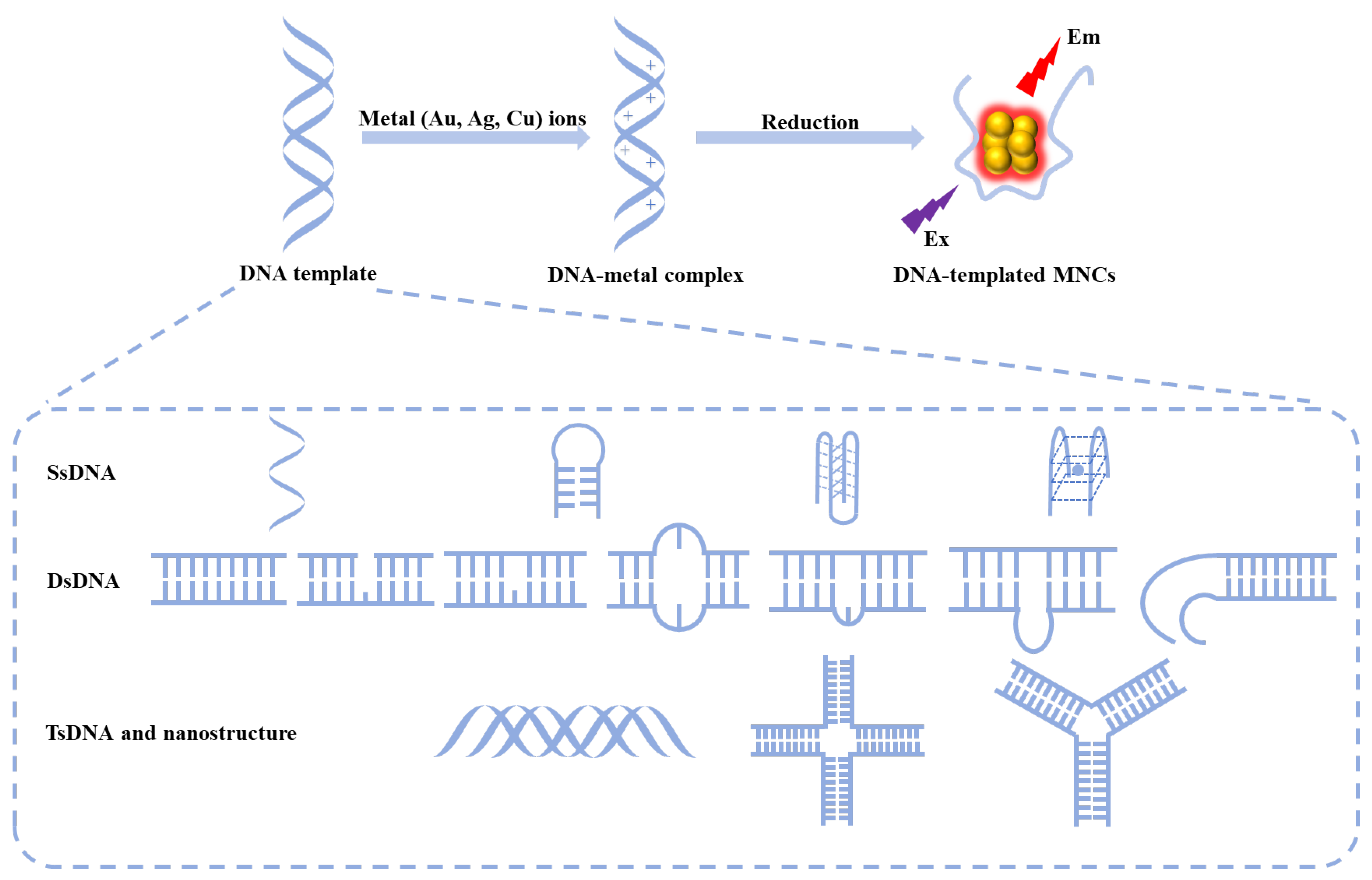
Figure 1. Schematic representation of the synthesis process for MNCs templated by DNA.
Chemosensors 12 00271 g001
Figure 2. Synthesis of ssDNA regulated MNCs. (A) Synthesis of ssDNA-AgNCs [19]. (B) The fluorescence properties of five distinct ssDNA-encapsulated AgNCs. (a) Blue emitters created in 5′-CCCTTTAACCCC-3′, (b) green emitters created in 5′-CCCTCTTAACCC-3′, (c) yellow emitters created in 5′-CCCTTAATCCCC-3′, (d) red emitters created in 5′-CCTCCTTCCTCC-3′, and (e) near-IR emitters created in 5′CCCTAACTCCCC-3′ [20]. (C) Poly T has been found to effectively serve as a template for the synthesis of CuNCs [25]. Absorption (a) and fluorescence (b) spectra of 20 mM PBS (pH 8.4) containing 0.1 M NaCl and 1 mM Mg2+ in the presence of 5 µM DNAs, 400 µM Cu(NO3)2, and 2000 µM ascorbate. (D) The synthesis of ssDNA-templated AuNCs [29].
Chemosensors 12 00271 g002
Figure 3. Using machine learning to study the relationship between ssDNA sequences and the properties of AgNCs. (A) Base motif recognition and design of DNA templates for fluorescent silver clusters by machine learning [46]. (B) Sequence design method for DNA templates that stabilize DNA-AgNCs within specific color bands [47]. (C) Color distributions for designed DNA-AgNC templates of varying lengths [48]. Histograms of all brightly fluorescent peak wavelengths identified in training data (black, right axes) as compared to sequences designed for the Red class with varying lengths (left axes): (a) 8 bases (purple), (b) 10 bases (blue), (c) 12 bases (green), and (d) 16 bases (red). Orange brackets above the graphs represent the 600−660 nm wavelength range defined for the Red class. (D) Workflow and training data for DNA-AgNC color prediction [49]. (a) Schematic of the workflow for ML-enabled DNA-AgNCs discovery. (b) A histogram of DNA-AgNCs peak wavelength (λp) values in the training data, with colors indicating the boundaries of Green (green), Red (red), Far Red (dark red), and NIR (blue) classes. Purple bars represent λp values from sequences excluded from the training data, as the magic numbers of AgN-DNAs in this range are unknown. (c) Class sizes for the five DNA-AgNCs color classes.
Chemosensors 12 00271 g003
Figure 4. Hairpin loop as template. (A) The hairpin loop DNA-templated preparation of fluorescent AgNCs. Blue = cytosine (C), green = thymine (T), red = guanine (G), and yellow = adenine (A) [51]. (B) The hairpin loop DNA-templated preparation of fluorescent AuNCs [37]. (C) The hairpin loop DNA-templated preparation of fluorescent CuNCs [25]. Fluorescence spectra (a) and (b) of 20 mM PBS (pH 8.4) containing 0.1 M NaCl and 1 mM Mg2+ in the presence of 5 µM DNAs, 750 µM Cu(NO3)2 and 3.75 mM ascorbate.
Chemosensors 12 00271 g004
Figure 6. DsDNA as template. (A) The effects of different dsDNA on the formation of fluorescent CuNCs [69]. (B) The gap site-directed formation of fluorescent AgNCs [58]. (C) The AP site-constrained formation of fluorescent AgNCs [60]. (D) The mismatched site-directed formation of fluorescent AgNCs [57]. (E) The bulge site-constrained formation of fluorescent AgNCs [62]. (F) Use of two different DNA duplexes with inserted cytosine loops working as synthetic scaffolds to generate fluorescent silver clusters [64].
Chemosensors 12 00271 g006
Figure 7. When G-rich DNA was in proximity, the integrated red fluorescence emission of DNA-AgNCs was enhanced by about 500 times (In the three-dimensional graph, the colors from purple to red represent gradual increasing in fluorescence intensity) [66].
Chemosensors 12 00271 g007
Figure 8. TsDNA and DNA nanostructures as templates. (A) The Ag+-mediated base triplet CG.CAg+ [71]. (B) The triplex DNA-templated preparation of fluorescent AgNCs [72]. (C) The synthetic procedure for AgNC/branched DNAs [73].
Chemosensors 12 00271 g008
Figure 10. Applications of dsDNA-AgNCs. (A) The schematic diagram for the synthesis of β-CD@DNA-CuNCs and the colorimetric detect of Glyp [114]. (B) Schematic of the PSA detection based on inhibiting the fluorescence enhancement of DNA-AgNC induced by PSA aptamer [79]. (C) Design strategy for the DNA-AgNC probe and principle of HBV DNA detection. (a) Schematic diagram showing the design of a DNA-AgNCs-based nanoprobe. (b) Schematic diagram illustrating the working mechanism of the concentration imbalance-driven DNA circuit (CIDDC) and the sensing principle of the CIDDC-based assay for HBV DNA detection [115].
Chemosensors 12 00271 g010
Figure 11. Applications of DNA nanostructures. (A) Schematic illustration of creating TWJDC and T*-responsive ratiometric fluorescence of red-emitting trimeric AgNCs (3rAgNC) located on the arms of TWJDC, relative to green-emitting gAgNC [116]. (B) Schematic illustration of the TDC assembly and the formation of TDC-Ag+ adducts and core-shell AgNC (csAgNC) [117]. (i) Fluorescence spectra of csAgNC in different TDC structures including TSx (x = 2-9); (ii) PAGE image of S1, S2, S3, S4, TS6, S1 + S2, S1 + S2 + S3, S1 + S2 + S3 + S4, S1 + S2 + S3 + S4 + TS6, and the DNA marker with 25, 50, 75, 100, 200, 300, 400, and 500 bp; (iii) TDC containing TS6 after incubation with cell lysates and FBS (10%) for 0–10 h.
Chemosensors 12 00271 g011
Disclaimer/Publisher’s Note: The statements, opinions and data contained in all publications are solely those of the individual author(s) and contributor(s) and not of MDPI and/or the editor(s). MDPI and/or the editor(s) disclaim responsibility for any injury to people or property resulting from any ideas, methods, instructions or products referred to in the content.