Article

Carboxy Bodipy-Based Fast Trigger Fluorescent Probe for Imaging Endogenous Hypochlorous Acid

1
School of Basic Medical Sciences, Youjiang Medical University for Nationalities, Baise 533000, China
2
School of Ophthalmology & Optometry, School of Biomedical Engineering, Wenzhou Medical University, Wenzhou 325000, China
3
Wenzhou Institute, University of Chinese Academy of Sciences, Wenzhou 325000, China
*
Authors to whom correspondence should be addressed.
Chemosensors 2023, 11(1), 26; https://doi.org/10.3390/chemosensors11010026
Submission received: 11 November 2022 / Revised: 21 December 2022 / Accepted: 25 December 2022 / Published: 28 December 2022
(This article belongs to the Special Issue Fluorescent Sensors for Disease Diagnosis and Therapy)

Abstract

:
Although hypochlorous acid (HClO/ClO) is regarded as a harmful reactive oxygen species (ROS) in cells, it plays an essential role in many physiological and pathological processes, such as an innate immunity and metabolic balance. In this paper, we developed a new carboxy Bodipy-based probe for rapid, sensitive, and specific monitoring of ClO. Bp-S produces bright fluorescent Bp-COOH based on the selective recognition of ClO to thiocarbamate groups. Bp-S exhibits high selectivity, high sensitivity, and high resistance to photobleaching in the recognition of ClO. Fluorescence imaging of this probe in Hela cells and RAW264.7 cells also successfully detected changes in exogenous/endogenous ClO, respectively, suggesting that Bp-S has high potential for future disease diagnosis and research.

Graphical Abstract

1. Introduction

ClO, H2O2, O2·−, and .OH, named reactive oxygen species (ROS), is an internally generated signal molecule and/or pressure agent that plays an important role in many physiological and pathological processes [1,2,3,4]. Because of its important antibacterial characteristics, ClO has garnered the most attention among them [5]. Myeloperoxidase (MPO), a double-edged biological system enzyme, catalyzes the oxidation process of H2O2 and chloride ions in organisms, which results in the production of ClO [6]. On the plus side, ClO can safeguard human health by eliminating invasive bacteria and pathogens in the body’s immune system [7]. On the other hand, mounting evidence suggests that an excess of ClO may cause the oxidation of lipids, proteins, and biomolecules, including nucleic acids, which can cause tissue damage, inflammation, and a number of illnesses such as cancer and liver ischemia–reperfusion injury [8]. For these reasons, it is essential to investigate a highly specialized and highly sensitive ClO and its deeper biological activities.
The small molecule-based fluorescence imaging method is very desirable for the real-time detection of active species in biological systems due to its high spatial and temporal resolution, high sensitivity, and ease of visualization. The ClO level of biological systems is typically elusive, and others have quickly disturbed ROS competitors because of their short lifecycle and high ClO activity [9]. The characteristics of high sensitivity, high selectivity, and quick reaction of ClO should therefore at least be met by fluorescent probe ClO imaging [10]. A number of ClO detectors based on various fluorophores, including cyanine, rhodamine, fluorescein, coumarin, naphthalimides, and boron–dipyrromethene (Bodipy), have been reported in recent years to conjugate various trigger groups to produce particular ClO reactions [3,11,12,13,14,15,16,17,18,19,20,21,22,23,24,25,26,27,28,29,30,31,32,33,34,35]. The emission wavelengths of these probes cover the visible to near-infrared light range, and many also have nM-level sensitivity [11]. However, most of the fluorophores are weakly resistant to oxidation and light and are easily bleached in a high concentration oxide environment or after light excitation [36,37,38].
In this paper, we created a new Bodipy-based fluorescent probe called Bp-S for the detection of ClO using the readily available Bodipy fluorophore Bp-COOH (Scheme 1). Bp-S is changed into Bp-COOH after reacting with ClO, which produces a sizable amount of green fluorescence. This offers a straightforward and practical method for the fluorescence detection of ClO. Importantly, Bp-S is unaffected by other ROS molecules and benefits from excellent ClO selectivity, quick response times (within few minutes), strong resistance to photobleaching, and high sensitivity levels (LOD = 6.3 nM). Additionally, we demonstrated that Bp-S may be utilized to monitor both endogenous and exogenous ClO in living cells, showing that Bp-S has a wide range of potential applications.

2. Experimental Section

2.1. Materials and Methods

4-Hydroxybenzyl alcohol, N, N-dimethyldithioaminomethane, lithium iodide, and phosphorus tribromide were purchased from Yanfeng Tech. Ltd. (Panjin, China). 2,4-Dimethyl-lH-pyrrole and methyl oxalyl chloride were purchased from Heowns. Tech. Ltd. (Tianjin, China). The solvents used in the reaction were all dried over 4 angstrom molecular sieves.

2.2. Equipment

UV-Vis spectra were measured by a LiSpecRed UVIR-1700 spectrophotometer (LiSen Optics, Vero Beach, FL, USA). Photoluminescence spectra were detected by a F-7400 fluorescence spectrophotometer (HITACHI, Tokyo, Japan). The hydrogen spectra carbon spectra of the compounds were measured by a SCM1000 NMR spectrometer (Thermo Scientific, Waltham, MA, USA). The molecular weights of the compounds of interest were measured by electrospray ionization high-resolution mass spectrometry (ESI-HRMS) on a NexION 2000 spectrometer (Perkielmer, Waltham, MA, USA). Cell fluorescence imaging was obtained by NCF950 laser confocal (Nexcope, Shanghai, China), green fluorescence channel, using a 40X objective).

2.3. Synthesis Methods

Bp-COOH was synthesized according to previous literature with slight modifications [39,40]. Detailed synthetic protocols and the characterization of all intermediates and products are given in the supporting information (Scheme S1, Figures S1–S4).

2.4. DFT Calculations

The ORCA computational package 5.03 was used to perform the DFT calculations [41]. The structure was first optimized for minimum energy using the BP/DEF2-TZVP method, and then the single-point energy was calculated using B3LYP/DEF2-TZVP. To determine the highest occupied molecular orbital (HOMO) and lowest unoccupied molecular orbital (LUMO) of the optimized structure, Avogadro 1.2.0 was used [42].

2.5. Spectroscopic Methods

All spectral measurements, unless otherwise mentioned, were made in 20 mM phosphate-buffered saline (PBS: EtOH V: V = 4:1, pH 7.4). Aqueous solutions of NaCl, NaClO, H2O2, KO2, t-BuOOH, Na2S, Na2SO3, NaHSO3, and Na2SO4 at a concentration of 40 μM were employed for the analytes’ competition research. The reaction between 40 μM Fe2+ and 400 μM H2O2 created the hydroxyl radical (.OH) solution, which was then employed immediately. To make a solution of peroxynitrite (ONOO), NaNO2 (40 μM) and H2O2 (70 μM) were combined, and the mixture was then acidified with HCl (60 μM) before being made alkaline with KOH (150 μM).
Limit of Detection (LOD) = 3 × (S.D.)/Slope
S. D.: The standard deviation of the blank sample. Slope: Slope value of linear curve of concentration and fluorescence intensity.

2.6. Cell Culture and Cytotoxicity

The cell culture and toxicity assays were performed with minor modifications based on the methods of an earlier study [40]. RAW 246.7 cells were grown in Dulbecco’s modified Eagle medium (DMEM) supplemented with 10% fetal bovine serum (Xinyu Biotechnology, Shanghai, China). Ninety-six-well plates with 104 RAW 246.7 cells planted in each well were incubated for 24 h. The vitality of RAW 246.7 cells was ultimately determined by a CCK8 test following incubation with various doses of Bp-S for 24 h.

2.7. Confocal Microscopy Imaging

RAW 246.7 cells were inoculated onto confocal plates with 105 cells per well and incubated for one day. RAW 246.7 were incubated with LPS for 30 min and washed twice with PBS before being incubated with/without ABAH for 30 min and washed twice with PBS. Finally, they were incubated with Bp-S for 20 min, washed twice with PBS, and fluorescence imaged using confocal microscopy.
HeLa cells were inoculated on confocal culture dishes with 105 cells per well and incubated for one day. Bp-S was then incubated with the HeLa cells for 20 min and washed twice with PBS. Then, it was incubated with different concentrations of ClO for 30 min and washed twice with PBS. Finally, fluorescence imaging was performed using confocal microscopy.

3. Results and Discussion

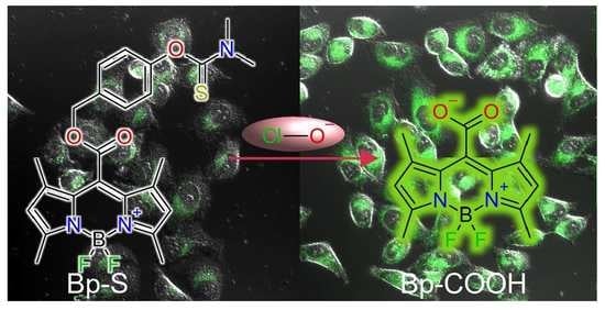
3.1. Rational Design and Synthesis of Bp-S

Considering that the existing probes have a weak antioxidant ability and are easy to be photobleached, we rationally designed a ClO probe to avoid these defects. The boron–dipyrromethene (Bodipy) platform is well known for its high absorption coefficient, good fluorescence quantum yield, excellent structural stability, and narrow emission band [43]. In our previous report, we used a Bodipy-COOH fluorescent probe to detect hydroxyl radicals [40]. This probe showed excellent sensitivity and selectivity. Therefore, we chose Bodipy as an antioxidant and photobleaching-resistant fluorophore to design a new ClO probe. The N, N-dimethylthiobamyl group was chosen as the recognition group for hypochlorous acid because of its higher expansibility and selectivity compared with other recognition groups such as thiourea, thioester, thioether, etc. Therefore, we designed a new ClO fluorescent probe (Bp-S) by linking the recognition group (N, N-dimethylthiocarbamate) and the fluorophore (Bp-COOH) through a self-eliminating linker (p-hydroxybenzyl alcohol). The designed structure is shown in Scheme 1. When Bp-S interacts with ClO, the non-fluorescent state of Bp-S is oxidized to an unstable intermediate product, which is then hydrolyzed by the self-eliminating group to form Bp-COOH, emitting intense green fluorescence. The photophysical properties of Bp-COOH and Bp-S were fully studied in the buffer solution (Table S1). The synthetic route of Bp-S is shown in Figure S1. Bp-COOH was prepared in a low yield of 14% from 2,4-dimethylpyrrole by modifying the existing synthetic method [40]. The recognition part of Bp-S, the dimethyl carbamodithioate, was synthesized from commercially available N,N-Dimethyl carbamodithioic in a multi-step procedure following a previously published procedure with an overall yield of 39% [23]. For the synthesis of Bp-S, Bp-COONa was reacted with compound 3 in dimethylformamide. After purification by silica column, Bp-S was obtained in a moderate yield of 52%, and the chemical structure of Bp-S was well verified by 1H NMR, 13C NMR, and ESI-HRMS.

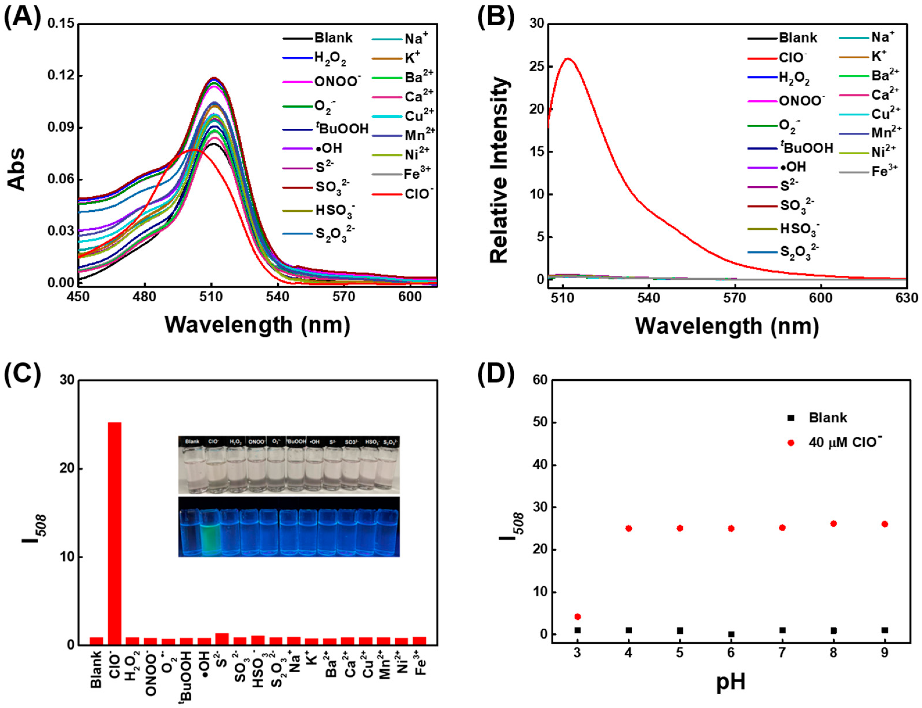
3.2. Fluorescence Response of Bp-S toward ClO

The spectral properties of Bp-S for ClO were first briefly investigated. As shown in Figure 1, Bp-S has a distinct absorption peak at 513 nm, with almost no obvious emission peak. In the presence of NaClO, the blue shift to 495 nm and the fluorescence intensity at 508 nm (I508) increased significantly. Moreover, the I508 increased more than 70-fold after the contact of Bp-S with ClO, which is very suitable for the highly sensitive determination of ClO. In addition, the absorption and fluorescence spectra of the reaction system became consistent with the spectral characteristics of the fluorophore Bp-COOH (Figure S5), which implies that Bp-COOH was retrieved from the reaction system. To further the formation of Bp-COOH after the interaction of Bp-S with hypochlorous acid, the reaction products were analyzed in detail using electrospray ionization mass spectrometry and HPLC. As shown in Figure S6, the m/z = 273.1205 [M-F]+ mass peak unique to Bp-COOH was found during the molecular weight detection of the reaction solution, and a new HPLC peak was generated at 3.51 min, which was consistent with the retention peak time of Bp-COOH (Figure S7).

3.3. Competition Estimation and pH Influence

To investigate the selectivity of Bp-S for ClO, first, the spectral response of Bp-S to different ROS was investigated (Figure 2A–C). When other ROS and reactive sulfur species (RSS), including H2O2, ONOO, O2·−, t-BuOOH, .OH, S2−, SO32−, HSO3, and S2O32−, were added to the fresh buffer of Bp-S, the absorbance peak at 513 nm and the fluorescence intensity at 508 nm showed few changes, indicating its high specificity for ClO. Considering that there are many kinds of metal ions in the physiological environment, we also measured the effect of common metal ions on the Bp-S emission spectra, including Na+, K+, Ba2+, Ca2+, Cu2+, Mn2+, Ni2+, and Fe3+, and found no fluorescence change for these metal ions, again demonstrating the high specificity of Bp-S (Figure 2A–C). Bp-S showed exclusive selectivity for ClO, which was attributed to the specific oxidation of ClO to thiocarbamate groups and the excellent chemical stability of the Bodipy structure. In addition, the selectivity of Bp-S for ClO was tested over a wide pH (3–9) range, and ClO was well detected in PBS buffer solutions at pH 3–9 (Figure 2D). The above results not only confirm the amazing selectivity of the sensor Bp-S for ClO, but also the adequate response to HClO/ClO when the pH value is changed from 4 to 9, a range that covers the whole physiological pH value.

3.4. Sensing Response of Bp-S toward ClO

After demonstrating the outstanding selectivity of Bp-S for ClO, we next investigated in detail the spectral behavior of the probe Bp-S interacting with different concentrations of ClO. First, a spectroscopic titration test was performed. As more and more ClO was added to the solution of Bp-S, the absorption peak of Bp-S at 513 nm progressively shifted toward 498 nm (Figure 3A). Meanwhile, a fresh absorption peak at 508 nm emerged on the fluorescence spectrum, and its intensity increased with an increasing hypochlorite concentration (Figure 3B). Meanwhile, the fluorescence intensity of Bp-S reached a maximum at 508 nm and remained stable when more than 20 equivalents of ClO were added (Figure 3C). There was a good linear relationship between I508 of Bp-S and the ClO concentration in the range of 0-2 μM (Figure 3D). The detection limit of the probe for ClO was determined to be 6.3 nM (n = 5) when calculated from the titration curve. Bp-S has a lower detection limit than some recently published ClO probes (Table S2).
The kinetic study of the reaction between the probe Bp-S and ClO was carried out with time-dependent fluorescence intensity (I508) measurements. The intensity of Bp-S at I508 was very weak before it interacted with ClO; however, it increased sharply and reached stability after a few minutes of co-incubation with different concentrations ClO, which confirmed that ClO could be removed quickly and in real time with Bp-S (Figure S8). The photostability of the probe affects the confidence of the bioimaging results. After continuous irradiation of the Bp-S (-/+ ClO) solution with a 490 nm xenon lamp for 1 h, respectively, the solution showed almost no change in emission intensity at 508 nm (Figure S9), which demonstrated the excellent resistance of the probe to photobleaching.

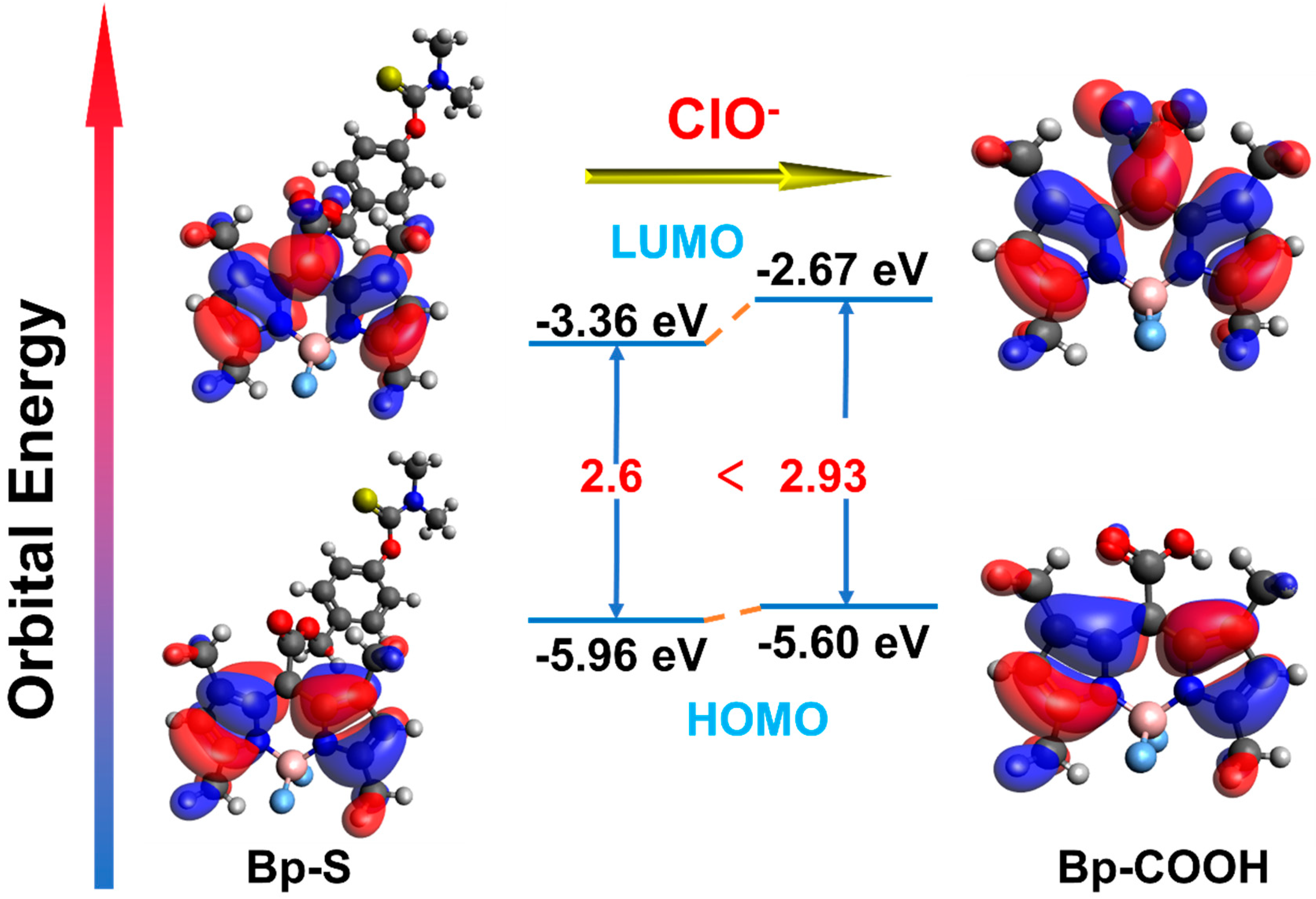
3.5. Density Functional Theory (DFT) Calculations

To gain a deeper understanding of the mechanism of the changes in the absorption spectra of the probes Bp-S and Bp-COOH, we used the B3LYP/DEF2-TZVP method to calculate the electronic orbitals of the probes Bp-COOH and Bp-S. All calculations were carried out using ORCA 5.3 [41]. The calculations show that the HOMO–LUMO (occupied molecular orbital–unoccupied molecular orbital) energy gaps of Bp-COOH and Bp-S are different, being 2.93 eV and 2.6 eV, respectively (Figure 4). The lower energy orbital gap is responsible for the longer absorption wavelengths possessed by Bp-S than Bp-COOH.

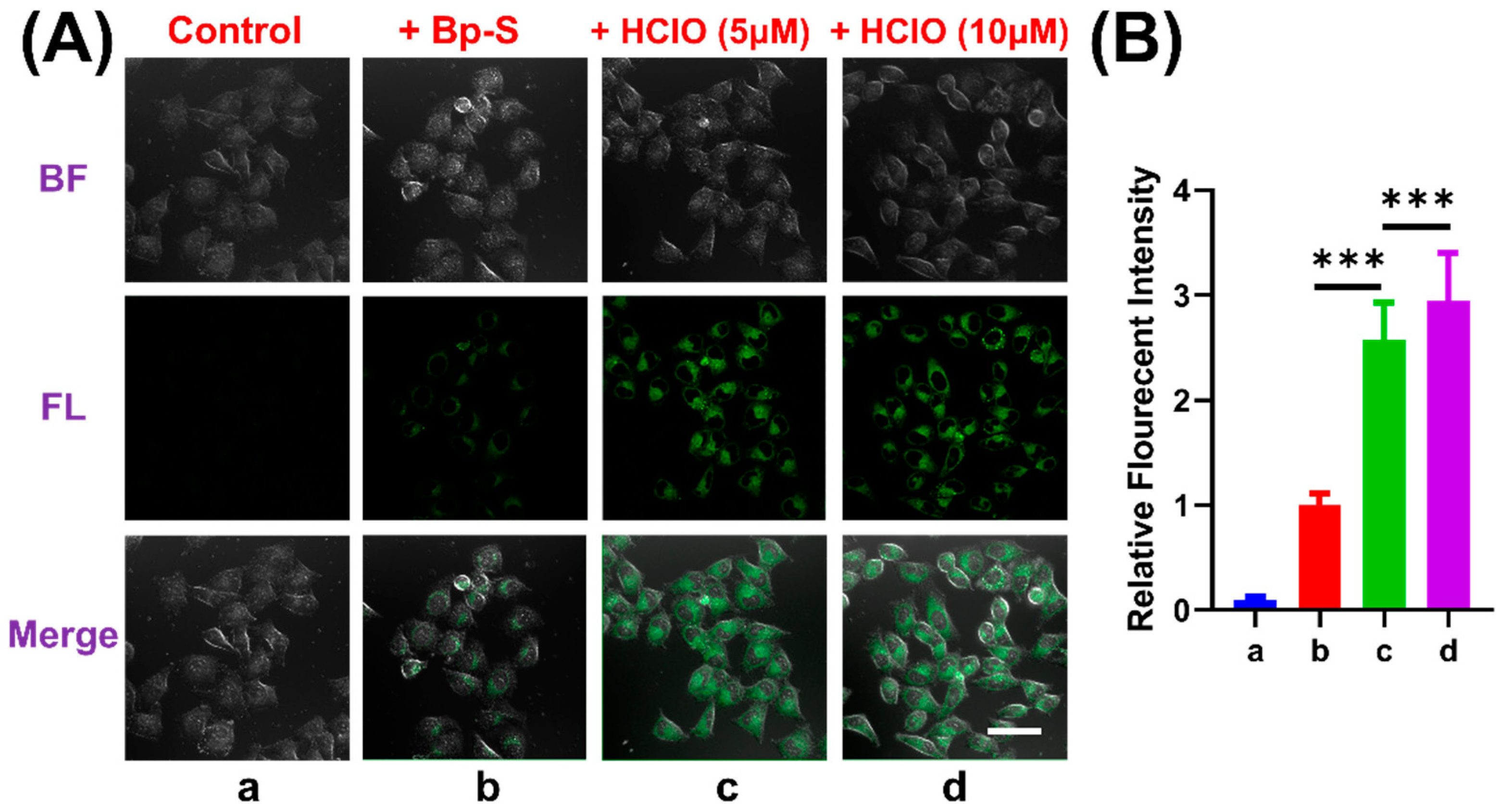
3.6. Confocal Fluorescence Imaging Experiments

The probe Bp-S showed excellent photophysical properties in aqueous solution. To confirm whether Bp-S can be used to detect intracellular, we first examined the toxicity of the probe Bp-S to RAW 264.7 cells using the classical CCK-8 assay. As shown in Figure S10, after treating cells with different concentrations (0 to 100 μM) of Bp-S, cell survival remained greater than 85% even at very high concentrations, indicating that Bp-S was minimally cytotoxic.
Next, we evaluated the ability of the probe Bp-S to assay endogenous ClO within living cells. Lipopolysaccharide (LPS) is a major constituent of the external cell structure of Gram-negative bacteria that has been extensively used to elicit an inflammatory reaction in immune cells. As shown in Figure 5, when only probe Bp-S was present, there was weak fluorescence in RAW 264.7 cells; however, after incubation with 1 μg/mL LPS for 30 min, brighter green fluorescence could be seen in the cells, showing that the probe Bp-S can smoothly penetrate the cell surface and reflect the changes of ClO in the irritated cells. To further demonstrate the intracellular ClO-selective ability of Bp-S, 4-aminobenzoic acid hydrazide (ABAH), a widely used MPO suppressant, was coincubated with RAW 264.7 cells for 30 min. The fluorescence brightness of pretreated ABAH cells was significantly lower than that of cells treated with LPS only (Figure 5Ad), indicating that the suppression of MPO activity decreased intracellular ClO production. Thus, the fluorescence-enhancing behavior of Bp-S in macrophages was basically attributed to the increase in hypochlorite concentration triggered by MPO activity.
After the above experiments confirmed that the probe Bp-S could detect endogenous ClO in living cells, we then further explored whether the probe could respond to changes in exogenous ClO concentration at the cellular level (Figure 6). Taking the HeLa cell line as an example, after treatment with probe Bp-S, the fluorescence was weak without further stimulation. After the exogenous addition of 5 μM and 10 μM ClO, the fluorescence intensity was significantly enhanced, which further proved that the probe Bp-S could well recognize ClO.

4. Conclusions

In summary, we prepared and comprehensively evaluated the novel probe Bp-S, an enhanced fluorescent probe with high selectivity for ClO. Bp-S has a thiocarbamate recognition group that is selectively cleaved by ClO to generate phenolic hydroxyl groups, and the nascent phenolic hydroxyl groups sever carbonate bonds via 1,6-elimination reactions while releasing Bp-COOH fluorophores that emit intense fluorescence. Photophysical properties and spectroscopic characterization showed that Bp-S reacts rapidly (within minutes), is highly selective and sensitive to ClO (detection limit of 6.3 nM), insensitive to ambient pH (pH 3–9), and extremely resistant to photobleaching. Finally, Bp-S was successfully applied to track endogenous ClO in RAW264.7 cells. In view of its excellent sensing properties mentioned above, it is expected that this new probe will be a useful tool to deeply probe the physiological function of ClO and to rapidly monitor the redox progression of cells.

Supplementary Materials

The following supporting information can be downloaded at: https://www.mdpi.com/article/10.3390/chemosensors11010026/s1, Scheme S1: Synthetic procedures of Bp-S; Figure S1: ESI-HRMS of Compound 2; Figure S2: ESI-MS of Bp-S; Figure S3: 1H NMR of Compound Bp-S; Figure S4: 13C NMR of Bp-S; Figure S5: (A) Absorption and (B) fluorescence emission spectra of Bp-COOH (2 μM) in pH7.4 PBS; Figure S6: ESI-MS of the reaction solution of Bp-S (2 μM) with NaClO (100 μM); Figure S7: Chromatograms of different reaction systems; Figure S8: Fluorescence emission of Bp-S (2 μM) for different concentrations of ClO with time; Figure S9: Dynamic emission intensity of Bp-S (2 μM) with or without ClO (40 μM); Figure S10: Cytotoxic effect of Bp-S; Table S1: Photophysical properties of Bp-COOH and Bp-S; Table S2. Some recently published ClO- fluorescent probes.

Author Contributions

H.Z. and Q.F contributed equally to this work. Conceptualization, methodology and investigation, H.Z.; DFT calculations, Z.L.; writing—original draft preparation, H.Z. and Q.F.; writing—review and editing, J.-T.H. and J.S. All authors have read and agreed to the published version of the manuscript.

Funding

This work was funded by the National Natural Science Foundation of China (No. 31800833, 21977081), Zhejiang Provincial Natural Science of Foundation of China (No. LZ19H180001, Q21H190019), Wenzhou Science & Technology Bureau Project (No. Y20190054, Y20180078), Wenzhou Medical University (No. KYYW201901, QTJ20022), University of Chinese Academy of Science (No. WIBEZD2017001-03, WIUCASYJ2020001).

Institutional Review Board Statement

Not applicable.

Informed Consent Statement

Not applicable.

Data Availability Statement

Not applicable.

Conflicts of Interest

The authors declare no conflict of interest.

References

  1. Yang, B.; Chen, Y.; Shi, J. Reactive Oxygen Species (ROS)-Based Nanomedicine. Chem. Rev. 2019, 119, 4881–4985. [Google Scholar] [CrossRef] [PubMed]
  2. Dickinson, B.C.; Chang, C.J. Chemistry and Biology of Reactive Oxygen Species in Signaling or Stress Responses. Nat. Chem. Biol. 2011, 7, 504–511. [Google Scholar] [CrossRef] [Green Version]
  3. Deng, Y.; Feng, S.; Xia, Q.; Gong, S.; Feng, G. A Novel Reaction-Based Fluorescence Probe for Rapid Imaging of HClO in Live Cells, Animals, and Injured Liver Tissues. Talanta 2020, 215, 120901. [Google Scholar] [CrossRef] [PubMed]
  4. Chen, X.; Wang, F.; Hyun, J.Y.; Wei, T.; Qiang, J.; Ren, X.; Shin, I.; Yoon, J. Recent Progress in the Development of Fluorescent, Luminescent and Colorimetric Probes for Detection of Reactive Oxygen and Nitrogen Species. Chem. Soc. Rev. 2016, 45, 2976–3016. [Google Scholar] [CrossRef] [PubMed]
  5. Hampton, M.B.; Kettle, A.J.; Winterbourn, C.C. Inside the Neutrophil Phagosome: Oxidants, Myeloperoxidase, and Bacterial Killing. Blood 1998, 92, 3007–3017. [Google Scholar] [CrossRef] [PubMed]
  6. Li, H.; Cao, Z.; Moore, D.R.; Jackson, P.L.; Barnes, S.; Lambeth, J.D.; Thannickal, V.J.; Cheng, G. Microbicidal Activity of Vascular Peroxidase 1 in Human Plasma via Generation of Hypochlorous Acid. Infect. Immun. 2012, 80, 2528–2537. [Google Scholar] [CrossRef] [PubMed] [Green Version]
  7. Prokopowicz, Z.M.; Arce, F.; Biedron, R.; Chiang, C.L.-L.; Ciszek, M.; Katz, D.R.; Nowakowska, M.; Zapotoczny, S.; Marcinkiewicz, J.; Chain, B.M. Hypochlorous Acid: A Natural Adjuvant That Facilitates Antigen Processing, Cross-Priming, and the Induction of Adaptive Immunity. J. Immunol. 2010, 184, 824–835. [Google Scholar] [CrossRef] [Green Version]
  8. Wu, L.; Sedgwick, A.C.; Sun, X.; Bull, S.D.; He, X.-P.; James, T.D. Reaction-Based Fluorescent Probes for the Detection and Imaging of Reactive Oxygen, Nitrogen, and Sulfur Species. Acc. Chem. Res. 2019, 52, 2582–2597. [Google Scholar] [CrossRef] [Green Version]
  9. Xu, Q.; Lee, K.-A.; Lee, S.; Lee, K.M.; Lee, W.-J.; Yoon, J. A Highly Specific Fluorescent Probe for Hypochlorous Acid and Its Application in Imaging Microbe-Induced HOCl Production. J. Am. Chem. Soc. 2013, 135, 9944–9949. [Google Scholar] [CrossRef] [PubMed]
  10. Wu, D.; Chen, L.; Xu, Q.; Chen, X.; Yoon, J. Design Principles, Sensing Mechanisms, and Applications of Highly Specific Fluorescent Probes for HOCl/OCl. Acc. Chem. Res. 2019, 52, 2158–2168. [Google Scholar] [CrossRef]
  11. Hou, J.-T.; Kwon, N.; Wang, S.; Wang, B.; He, X.; Yoon, J.; Shen, J. Sulfur-Based Fluorescent Probes for HOCl: Mechanisms, Design, and Applications. Coord. Chem. Rev. 2022, 450, 214232. [Google Scholar] [CrossRef]
  12. Kim, T.-I.; Park, S.; Choi, Y.; Kim, Y. A BODIPY-Based Probe for the Selective Detection of Hypochlorous Acid in Living Cells. Chem. Asian J. 2011, 6, 1358–1361. [Google Scholar] [CrossRef] [PubMed]
  13. Shu, W.; Jia, P.; Chen, X.; Li, X.; Huo, Y.; Liu, F.; Wang, Z.; Liu, C.; Zhu, B.; Yan, L.; et al. A Highly Selective Ratiometric Fluorescent Probe for the Sensitive Detection of Hypochlorous Acid and Its Bioimaging Applications. RSC Adv. 2016, 6, 64315–64322. [Google Scholar] [CrossRef]
  14. Zhang, H.; Yin, X.; Hong, J.; Deng, Y.; Feng, G. A NIR Fluorescence Probe Having Significant Fluorescence Turn-on Signal at 700 Nm and Large Stokes Shift for Rapid Detection of HOCl in vivo. Talanta 2021, 223, 121768. [Google Scholar] [CrossRef] [PubMed]
  15. Zhang, W.; Wang, H.; Li, F.; Chen, Y.; Kwok, R.T.K.; Huang, Y.; Zhang, J.; Hou, J.; Tang, B.Z. A Ratiometric Fluorescent Probe Based on AIEgen for Detecting HClO in Living Cells. Chem. Commun. Camb. Engl. 2020, 56, 14613–14616. [Google Scholar] [CrossRef] [PubMed]
  16. Wu, X.; Li, Z.; Yang, L.; Han, J.; Han, S. A Self-Referenced Nanodosimeter for Reaction Based Ratiometric Imaging of Hypochlorous Acid in Living Cells. Chem. Sci. 2012, 4, 460–467. [Google Scholar] [CrossRef]
  17. Xia, Q.; Wang, X.; Liu, Y.; Shen, Z.; Ge, Z.; Huang, H.; Li, X.; Wang, Y. An Endoplasmic Reticulum-Targeted Two-Photon Fluorescent Probe for Bioimaging of HClO Generated during Sleep Deprivation. Spectrochim. Acta. A Mol. Biomol. Spectrosc. 2020, 229, 117992. [Google Scholar] [CrossRef]
  18. Kenmoku, S.; Urano, Y.; Kojima, H.; Nagano, T. Development of a Highly Specific Rhodamine-Based Fluorescence Probe for Hypochlorous Acid and Its Application to Real-Time Imaging of Phagocytosis. J. Am. Chem. Soc. 2007, 129, 7313–7318. [Google Scholar] [CrossRef]
  19. Wang, J.; Cheng, D.; Zhu, L.; Wang, P.; Liu, H.-W.; Chen, M.; Yuan, L.; Zhang, X.-B. Engineering Dithiobenzoic Acid Lactone-Decorated Si-Rhodamine as a Highly Selective near-Infrared HOCl Fluorescent Probe for Imaging Drug-Induced Acute Nephrotoxicity. Chem. Commun. Camb. Engl. 2019, 55, 10916–10919. [Google Scholar] [CrossRef]
  20. Zhu, Y.; Ma, Y.; Liu, Y.; Liu, Z.; Ma, S.; Xing, M.; Cao, D.; Lin, W. Fluorescence Response of a Fluorescein Derivative for Hypochlorite Ion and Its Application for Biological Imaging in Wounded Zebrafish and Living Mice. Sens. Actuators B Chem. 2021, 327, 128848. [Google Scholar] [CrossRef]
  21. Yuan, L.; Lin, W.; Xie, Y.; Chen, B.; Song, J. Fluorescent Detection of Hypochlorous Acid from Turn-on to FRET-Based Ratiometry by a HOCl-Mediated Cyclization Reaction. Chem. Weinh. Bergstr. Ger. 2012, 18, 2700–2706. [Google Scholar] [CrossRef] [PubMed]
  22. Zhu, B.; Li, P.; Shu, W.; Wang, X.; Liu, C.; Wang, Y.; Wang, Z.; Wang, Y.; Tang, B. Highly Specific and Ultrasensitive Two-Photon Fluorescence Imaging of Native HOCl in Lysosomes and Tissues Based on Thiocarbamate Derivatives. Anal. Chem. 2016, 88, 12532–12538. [Google Scholar] [CrossRef] [PubMed]
  23. Ji, D.; Li, G.; Zhang, S.; Zhu, M.; Li, C.; Qiao, R. Mitochondria-Targeted Fluorescence Probe for Endogenous Hypochlorite Imaging in Living Cells and Zebrafishs. Sens. Actuators B Chem. 2018, 259, 816–824. [Google Scholar] [CrossRef]
  24. Qian, X.; Yu, H.; Zhu, W.; Yao, X.; Liu, W.; Yang, S.; Zhou, F.; Liu, Y. Near Infrared Fluorescent Probe for in Vivo Bioimaging of Endogenous Hypochlorous Acid. Dyes Pigments 2021, 188, 109218. [Google Scholar] [CrossRef]
  25. Vedamalai, M.; Kedaria, D.; Vasita, R.; Gupta, I. Oxidation of Phenothiazine Based Fluorescent Probe for Hypochlorite and Its Application to Live Cell Imaging. Sens. Actuators B Chem. 2018, 263, 137–142. [Google Scholar] [CrossRef]
  26. Shen, Y.; Zheng, W.; Yao, Y.; Wang, D.; Lv, G.; Li, C. Phenoxazine-based Near-infrared Fluorescent Probes for the Specific Detection of Copper (II) Ions in Living Cells. Chem.—Asian J. 2020, 15, 2864–2867. [Google Scholar] [CrossRef]
  27. Zhang, Y.-R.; Liu, Y.; Feng, X.; Zhao, B.-X. Recent Progress in the Development of Fluorescent Probes for the Detection of Hypochlorous Acid. Sens. Actuators B Chem. 2017, 240, 18–36. [Google Scholar] [CrossRef]
  28. Zhu, H.; Fan, J.; Wang, J.; Mu, H.; Peng, X. An “Enhanced PET”-Based Fluorescent Probe with Ultrasensitivity for Imaging Basal and Elesclomol-Induced HClO in Cancer Cells. J. Am. Chem. Soc. 2014, 136, 12820–12823. [Google Scholar] [CrossRef]
  29. Xu, Q.; Heo, C.H.; Kim, G.; Lee, H.W.; Kim, H.M.; Yoon, J. Development of Imidazoline-2-Thiones Based Two-Photon Fluorescence Probes for Imaging Hypochlorite Generation in a Co-Culture System. Angew. Chem. Int. Ed. 2015, 54, 4890–4894. [Google Scholar] [CrossRef]
  30. Mao, G.-J.; Liang, Z.-Z.; Bi, J.; Zhang, H.; Meng, H.-M.; Su, L.; Gong, Y.-J.; Feng, S.; Zhang, G. A Near-Infrared Fluorescent Probe Based on Photostable Si-Rhodamine for Imaging Hypochlorous Acid during Lysosome-Involved Inflammatory Response. Anal. Chim. Acta 2019, 1048, 143–153. [Google Scholar] [CrossRef]
  31. Duan, Q.; Jia, P.; Zhuang, Z.; Liu, C.; Zhang, X.; Wang, Z.; Sheng, W.; Li, Z.; Zhu, H.; Zhu, B.; et al. Rational Design of a Hepatoma-Specific Fluorescent Probe for HOCl and Its Bioimaging Applications in Living HepG2 Cells. Anal. Chem. 2019, 91, 2163–2168. [Google Scholar] [CrossRef] [PubMed]
  32. Xie, X.; Wu, T.; Wang, X.; Li, Y.; Wang, K.; Zhao, Z.; Jiao, X.; Tang, B. A Two-Photon Fluorescent Probe for Ratiometric Visualization of Hypochlorous Acid in Live Cells and Animals Based on a Selenide Oxidation/Elimination Tandem Reaction. Chem. Commun. 2018, 54, 11965–11968. [Google Scholar] [CrossRef] [PubMed]
  33. Wang, S.; Chen, L.; Jangili, P.; Sharma, A.; Li, W.; Hou, J.-T.; Qin, C.; Yoon, J.; Kim, J.S. Design and Applications of Fluorescent Detectors for Peroxynitrite. Coord. Chem. Rev. 2018, 374, 36–54. [Google Scholar] [CrossRef]
  34. Hou, J.-T.; Kim, H.S.; Duan, C.; Ji, M.S.; Wang, S.; Zeng, L.; Ren, W.X.; Kim, J.S. A Ratiometric Fluorescent Probe for Detecting Hypochlorite in the Endoplasmic Reticulum. Chem. Commun. 2019, 55, 2533–2536. [Google Scholar] [CrossRef] [PubMed]
  35. Wang, B.; Yuan, F.; Wang, S.; Duan, R.; Ren, W.X.; Hou, J.-T. Detection of Atherosclerosis-Associated HOCl Using a Mitochondria-Targeted Fluorescent Probe. Sens. Actuators B Chem. 2021, 348, 130695. [Google Scholar] [CrossRef]
  36. Lin, Q.; Yang, L.; Wang, Z.; Hua, Y.; Zhang, D.; Bao, B.; Bao, C.; Gong, X.; Zhu, L. Coumarin Photocaging Groups Modified with an Electron-Rich Styryl Moiety at the 3-Position: Long-Wavelength Excitation, Rapid Photolysis, and Photobleaching. Angew. Chem. Int. Ed. Engl. 2018, 57, 3722–3726. [Google Scholar] [CrossRef]
  37. Platkov, M.; Tirosh, R.; Kaufman, M.; Zurgil, N.; Deutsch, M. Photobleaching of Fluorescein as a Probe for Oxidative Stress in Single Cells. J. Photochem. Photobiol. B 2014, 140, 306–314. [Google Scholar] [CrossRef]
  38. Lange, N.; Szlasa, W.; Saczko, J.; Chwiłkowska, A. Potential of Cyanine Derived Dyes in Photodynamic Therapy. Pharmaceutics 2021, 13, 818. [Google Scholar] [CrossRef]
  39. Kim, S.; Kim, H.; Choi, Y.; Kim, Y. A New Strategy for Fluorogenic Esterase Probes Displaying Low Levels of Non-Specific Hydrolysis. Chem.-Eur. J. 2015, 21, 9645–9649. [Google Scholar] [CrossRef]
  40. Li, Z.; Li, S.; Lv, H.; Shen, J.; He, X.; Peng, B. BODIPY-Based Rapid Response Fluorescence Probe for Sensing and Bioimaging Endogenous Superoxide Anion in Living Cells. Spectrochim. Acta. A Mol. Biomol. Spectrosc. 2021, 269, 120766. [Google Scholar] [CrossRef]
  41. Neese, F.; Wennmohs, F.; Becker, U.; Riplinger, C. The ORCA Quantum Chemistry Program Package. J. Chem. Phys. 2020, 152, 224108. [Google Scholar] [CrossRef] [PubMed]
  42. Hanwell, M.D.; Curtis, D.E.; Lonie, D.C.; Vandermeersch, T.; Zurek, E.; Hutchison, G.R. Avogadro: An Advanced Semantic Chemical Editor, Visualization, and Analysis Platform. J. Cheminformatics 2012, 4, 17. [Google Scholar] [CrossRef] [PubMed] [Green Version]
  43. Ulrich, G.; Ziessel, R.; Harriman, A. The Chemistry of Fluorescent Bodipy Dyes: Versatility Unsurpassed. Angew. Chem. Int. Ed. 2008, 47, 1184–1201. [Google Scholar] [CrossRef] [PubMed]
Scheme 1. The suggested recognition mechanism for Bp-S to Bp-COOH conversion by ClO.
Chemosensors 11 00026 sch001
Figure 1. The optical reactions of Bp-S (2 μM) for ClO in a PBS buffer (10 mM, pH 7.4, 20% EtOH, v/v) at 25 °C: (A) Changes in the Uv-Vis spectrum of Bp-S (2 μM) for ClO (20 μM). (B) Changes in the fluorescence spectrum of Bp-S (2 μM) for ClO (40 μM).
Chemosensors 11 00026 g001
Figure 2. (A) UV-vis absorption spectra of Bp-S (2 μM) in 20% EtOH solution before and after the addition of different reactive species (40 μM) and metal ions (40 μM) (10 mM, pH 7.4, PBS). (B) Emission spectra of Bp-S (2 μM) in a 20% EtOH solution before and after the addition of different reactive species (40 μM) and metal ions (40 μM) (10 mM, pH 7.4, PBS). (C) The intensity changes of Bp-S before and after the addition of various reactive species (40 μM) and metal ions (40 μM); λex = 495 nm, slit width: 1.5 mm/1.5 mm. (D) Fluorescence emission intensity of Bp-S (2 μM) in B-R buffer solutions of various pH values with or without ClO (40 μM). λex = 495 nm, slit width: 1.5 mm/1.5 mm.
Chemosensors 11 00026 g002
Figure 3. (A) UV-visible drops of Bp-S (2 μM) in 20% EtOH solution for different concentrations of ClO- (PBS, pH 7.4, 10 mM); (B) Bp-S (2 μM) fluorescence titration against different concentrations of ClO in 20% EtOH solution (10 mM, pH 7.4, PBS). (C) Fluorescence titration plot of Bp-S (2 μM) toward ClO; λex = 495 nm, slit width: 1.5 mm/1.5 mm. (D) Linear relationship between fluorescence intensity of Bp-S (2 μM) and various low concentrations of ClO. λex = 495 nm, slit width: 1.5 mm/1.5 mm.
Chemosensors 11 00026 g003
Figure 4. Calculations were based on ground state geometry by DFT at the DEF2-TZVP level for Bp-S and Bp-COOH orbitals.
Chemosensors 11 00026 g004
Figure 5. (A) Confocal fluorescence images of RAW 264.7 cells treated with different methods. (a) RAW 264.7 cells that have not been treated; (b) RAW 264.7 cells treated with Bp-S (2 μM) at 37 °C for 20 min; (c) RAW 264.7 cells pretreated with LPS (1 μg/mL) for 30 min and then incubated with Bp-S (2 μM) for 20 min; (d) Pretreatment of RAW 264.7 cells with LPS (1 μg/mL) for 30 min, then treatment with Tiron (50 μM) for 30 min, and then incubated with Bp-S (2 μM) for 20 min. Green channel: 515–580 nm (excited wavelength 488 nm). Bar = 40 μm. (B) The right figure shows the quantitative analysis of the average fluorescence in a–d from four groups of cells. *** p < 0.001.
Chemosensors 11 00026 g005
Figure 6. (A) Confocal imaging of ClO in living HeLa cells using Bp-S (2 μM). (a) HeLa cells that have not been treated; (b) HeLa cells treated with Bp-S (2 μM) for 20 min at 37 °C; (c, d) Cells were pre-incubated with the probe for 30 min and then with 5 and 10 μM of ClO for 10 min, respectively. Green channel: 515–580 nm (excited wavelenght 488 nm). Bar = 40 μm. (B) The right figure shows the quantitative analysis of the average fluorescence in a–d from four groups of cells. *** p < 0.001.
Chemosensors 11 00026 g006
Disclaimer/Publisher’s Note: The statements, opinions and data contained in all publications are solely those of the individual author(s) and contributor(s) and not of MDPI and/or the editor(s). MDPI and/or the editor(s) disclaim responsibility for any injury to people or property resulting from any ideas, methods, instructions or products referred to in the content.

