Review

Recent Advances in Molecular Fluorescent Probes for CYP450 Sensing and Imaging

State Key Laboratory of Chemo/Biosensing and Chemometrics, College of Chemistry and Chemical Engineering, Hunan University, Changsha 410082, China
*
Authors to whom correspondence should be addressed.
Chemosensors 2022, 10(8), 304; https://doi.org/10.3390/chemosensors10080304
Submission received: 1 July 2022 / Revised: 26 July 2022 / Accepted: 29 July 2022 / Published: 2 August 2022
(This article belongs to the Collection Advances of Chemical and Biosensors in China)

Abstract

:
Cytochrome P450 (CYP450) is a major drug-metabolizing enzyme system mainly distributed in liver microsomes and involved in the metabolism of many endogenous substances (such as fatty acids and arachidonic acids), and exogenous compounds (such as drugs, toxicants, carcinogens, and procarcinogens). Due to the similarity in structures and catalytic functions between CYP450 isoforms, the lack of effective selective detection tools greatly limits the understanding and the research of their respective physiological roles in living organisms. Until now, several small-molecular fluorescent probes have been employed for selective detection and monitoring of CYP450s (Cytochrome P450 enzymes) in vitro or in vivo owing to the tailored properties, biodegradability, and high temporal and spatial resolution imaging in situ. In this review, we summarize the recent advances in fluorescent probes for CYP450s (including CYP1, CYP2, and CYP3 families), and we discuss and focus on their identification mechanisms, general probe design strategies, and bioimaging applications. We also highlight the potential challenges and prospects of designing new generations of fluorescent probes in CYP450 studies, which will further enhance the diversity, practicality, and clinical feasibility of research into CYP450.

1. Introduction

The cytochrome P450 enzyme system (CYP450) is a class of heme-containing monooxygenases that are located on the endoplasmic reticulum or mitochondrial membranes [1]. For CYP450s, which are engaged in drug metabolism and in vivo biotransformation, one of the most crucial functions is to degrade xenobiotics, such as converting drugs to water-soluble molecules to speed up their excretion from the body [2]. Research on the role of CYP450 has been carried out mainly from the following two aspects (Figure 1) [3,4]: One is the function of CYP450s in the biosynthesis and metabolism of endogenous compounds, which can catalyze fatty acids, steroids, and other endogenous substances; and the other theme is the significance of CYP450s in the metabolism of xenobiotics (exogenous compounds present in the body, including compounds not produced by the body, such as drugs, toxicants, environmental substances, or pollutants), predominantly in detoxication [5]. Previous reports have shown that CYP450s are associated with the metabolism of procarcinogens to carcinogens and tumor development and progression, owing to their crucial role in the catalysis of endogenous and exogenous compounds [6].
It has been acknowledged that the level and the function of CYP450s are important factors leading to individual differences in drug disposal and treatment results. The CYP450 enzyme family is complex in composition, and it can be influenced by genetic diversity, which is referred to as the CYP450 gene superfamily [7]. Many CYP450s are involved in genetic polymorphisms and, in combination with environmental and other factors, together lead to inter-individual and inter-ethnic differences in the ability to metabolize the same substrate [8,9]. Treatment failure, toxicity, side effects, and drug–drug interactions (DDI) are the adverse effects that may result from significant changes in both the level and the function of CYP450s [6]. Due to the diversity, catalytic types, and broad spectrum of CYP450 species, as well as their important roles in many physiological and pathological processes [3], the development of effective tools, or innovative methods to detect CYP450s activity has attracted widespread attention.
Reassuringly, over the past two decades, CYP450 inhibition assays have evolved from determining substrate metabolism in individual reactions by high-performance liquid chromatography (HPLC) to detecting changes in the optical signal indicative of substrate metabolism without sample separation or purification. Using fluorescent probes to selectively track human CYP450s and to explore their roles in the drug metabolism and pathologic process has important theoretical and practical significance for the safe and effective use of drugs and the implementation of individualized dosing regimens. To date, although reviews of the catalytic metabolism of CYP450s and their relevance to toxicity and drug interactions have been reported [10,11,12,13,14], there are only a few reviews of fluorescent assays for CYP450 [15], which could have important guiding significance in the development of satisfactory probes for the visualization and the real-time monitoring of CYP450s activity. The purpose of this review is to summarize the research progress of fluorescent probes used to study CYP450 activities and their contribution to drug-metabolizing. The reduction reaction catalyzed by CYP450s reductase occurs in the absence of oxygen, and fluorescent probes for imaging and monitoring CYP450s reductase in hypoxia have also been reported [16,17,18], which is beyond the scope of this review.

2. Brief Introduction of CYP450

2.1. Relevance to Diseases and Drug Interactions

CYP450 is a kind of catalytically diverse biocatalyst. In recent years, significant progress has been made in the study of the structure and the function of CYP450s, especially their roles in drug metabolism, which play pivotal roles in the body’s protective detoxification and metabolism of most clinical drugs, such as the antidepressant drug clomipramine [19].
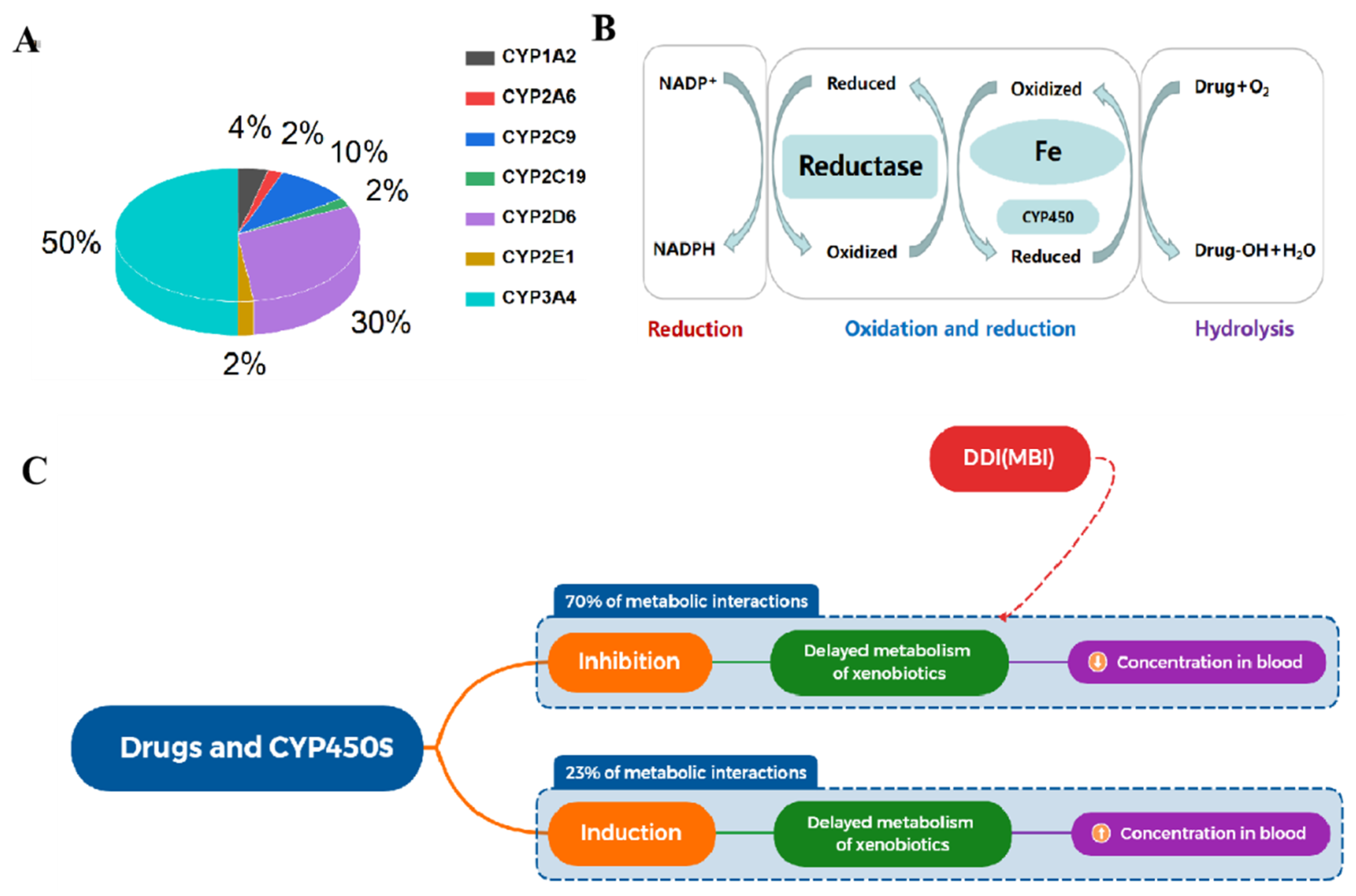
CYP450 is a superfamily consisting of many isozymes. According to the percentage amino acid sequence homology, it can be easily divided into families and subfamilies (Figure 2), mainly including CYP1, CYP2, and CYP3 families (Table 1) [20]. CYP450s have more than 50 isoforms among which CYP1A2, 2A6, 2C9, 2C19, 2E1, 2D6, and 3A4 are mainly expressed in the liver, small intestine, lung, placenta, and kidney, and they are involved in the metabolism of drugs, accounting for 80% [21] (Figure 3A). As the main drug-metabolizing enzyme family, CYP450s mainly play an important role in the phase I metabolism of drugs through oxidative metabolism by coenzyme NADPH and molecular oxygen (Figure 3B) [22]. Meanwhile, due to the important roles of CYP450s in the metabolism of most anticancer drugs, CYP450s are related to inflammation and cancer development, including rectal cancer, breast cancer, lung cancer, and other diseases. Accordingly, accumulating evidence suggests that CYP450s are involved in cancer formation, chemoprevention, metastasis, and chemotherapy [6].
CYP450s have been reported to react with many drugs in a variety of ways, and the catalytic activity of CYP450s can be affected by drug inhibition or drug induction (Figure 3C) [23]. Among them, the inhibitory effect of CYP450s owing to DDI will have a considerable negative impact on health. Therefore, understanding the interaction between CYP450s and drugs is of great significance for rational and safe treatment.
In the course of clinical treatment, the combined use of multiple drugs cannot be avoided. However, unknown drug combinations may lead to many adverse reactions, and even increase the risk of fatal adverse drug events. Inhibition of the CYP450 enzyme function is the most common mechanism for understanding DDI. There are two mechanisms of CYP450 inhibition: reversible and irreversible/quasi-irreversible [24]. Among them, CYP450s irreversible inhibition (competitive or non-competitive) is related to some important pharmacokinetic drug interactions and predisposes to cause unexpected DDI. Mechanism-Based Inhibition (MBI, Figure 3C), an irreversible means of inhibition, is usually covalently linked to CYP450s, resulting in their inactivation [25]. A mechanism-based enzyme inactivator, an unreactive compound whose structure is similar to the substrate of the target enzyme, is transformed into an active and electrophilic metabolite that tightly binds to the enzyme’s active site, leading to a long-lasting inactivation [26]. Irreversible inhibition induced by covalent modification of biological macromolecules predisposes to adverse, unfavorable DDI. Therefore, the discovery and evaluation of the key metabolic enzyme MBI have important implications for drug development, safe use, and public health, implying an urgent need to develop effective tools that can visualize the effects of MBI.

2.2. CYP450-Catalyzed Reactions

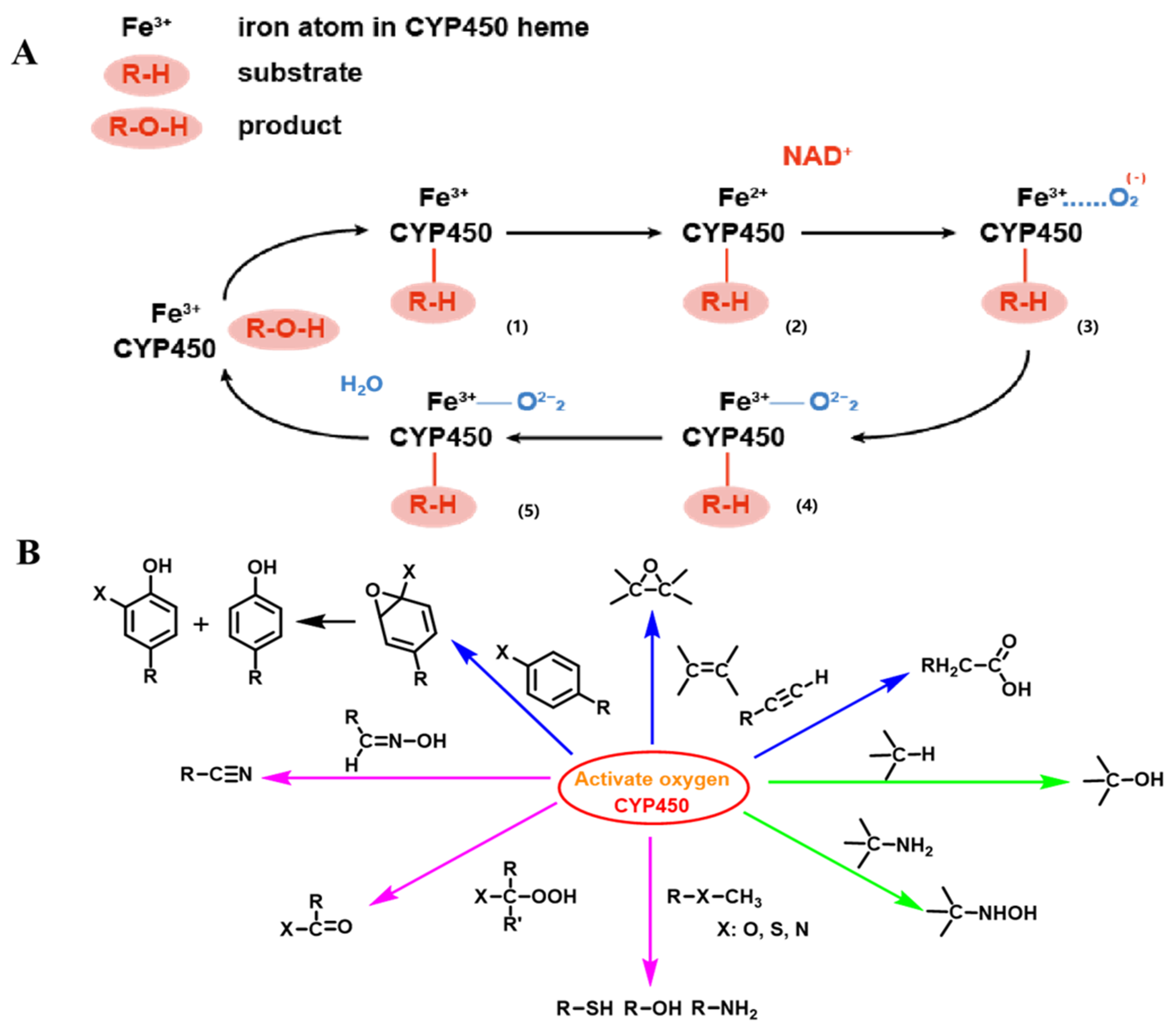
CP450, a kind of heme-containing monooxygenases whose function requires the joint participation of coenzyme NADPH and O2, is primarily involved in oxidation reactions in the progress of drug biotransformation, including electron loss, dehydrogenation reactions, and oxidation reactions. Generally, CYP450 enzyme adds oxygen to drugs by activating molecular oxygen, connecting one of the oxygen atoms to an organic molecule, while reducing the other oxygen atom to water. The CYP450s’ generalized catalytic cycle is depicted in Figure 4A [27,28]: In CYP450s, iron ions exist in the form of Fe3+, and the drug (R-H) first binds to the enzymes; then, the complex of drug and enzyme is reduced by NADPH, and Fe3+ is reduced to Fe2+. In the third step, oxygen molecules oxidize Fe2+ to Fe3+. The fourth step is similar to the second step, except that the electrons are received by oxygen atoms rather than Fe3+. In the fifth step, two protons react with the complex, two electrons on oxygen are transferred to two protons, and water is produced. Finally, oxygen atoms are transferred to the drug, the enzyme releases the metabolite ROH, and CYP450s return to their initial state.
The classical catalytic reaction of CYP450s is to insert one oxygen atom of an oxygen molecule into the inactivated hydrocarbon bond of a substrate molecule to produce the corresponding alcohol and to reduce the other oxygen atom to water [29]. In addition, CYP450s catalyze many different types of reactions (Figure 4B), including hydroxylation of carbon atoms, dealkylation of N-, O-, and S atoms, sulfur oxidation, epoxidation, deamidation, desulfurization, dehalogenation, peroxidation, N-oxide reduction, C-C bond breaking, etc. [30,31,32]. Based on the catalytic reactions of substrates and the structural characteristics of CYP450, researchers have developed many small molecule tools with specific detection functions using fluorescent probes as substrates. Most frequently, hydroxylation and dealkylation metabolic processes are used in probe design.

2.3. Assays for CYP450

Normally, CYP450s catalyze the oxidative metabolism of endogenous and exogenous chemicals mainly through dealkylation, hydroxylation, and epoxidation reactions. Developing effective, convenient, and sensitive assays for the determination of CYP450 enzyme activity is of great importance for visualizing CYP450 enzyme inhibition and screening potential co-substrates and/or inhibitors of key CYP450 enzymes, which is highly conducive to investigating their physiological functions. Traditional methods for studying CYP450 activity are mainly chromatography-based analytical methods, such as HPLC and mass spectrometry (MS), which relies on labor- and equipment-intensive sample extraction. Another major reason for the limited studying of CYP450 activity is the large assay volumes and the high enzyme concentrations required to reach the limit of detection. As a result, high-throughput detection of enzyme activities has gained considerable attention.
High-throughput methods currently available for the study of CYP450 activity include luminescence-based assay, MS-based assay, and radioactivity-based assay [33,34]. However, most of them are not compatible with living cells or in vivo applications. Compared to these methods, the fluorescence-based assay stands out for its effectiveness, noninvasiveness, high sensitivity, and selectivity, and it has been widely used as a powerful tool for monitoring target enzymes in living systems [35]. Among them, small-molecule fluorescent probes as detection tools for the fluorescence-based assay are more suitable for assessing complex biological effects mediated by target proteins, such as CYP450s than other fluorescent probes (nanoprobes, etc.), owing to their outstanding advantages, such as high sensitivity, tailored properties, hypo-toxicity, biodegradability, and low cost [36,37]. It is worth noting that small-molecule fluorescent probes have numerous applications in a variety of disciplines, including clinical diagnosis, chemical biology, and drug development [38].
Over the past 20 years, a few fluorescent probes have been made to measure CYP450s inhibition by monitoring CYP450s activity, such as DFB (3,4-difluorobenzyl)-4-(4-methylsulfonylphenyl)-5,5-dimethyl-(5H)furan-2-one) (CYP3A) [39]. However, the common reason that limits their application is their inability to specifically create a visual image of CYP450s in living systems. It is reassuring to note that many specific real-time fluorescent probes for key CYP450 isoenzymes have been developed through improvements in the performance of fluorescent dyes and local modifications of recognition sites. The recognition mechanism and design strategy of CYP450-specific fluorescent probes, and its biological applications are the main topics of this review. In addition, we summarized that in the developed probe general design strategy of an ideal probe substrate targeting CYP450 isoforms, the following rules should at least be obeyed (Figure 5): (1) Use fluorophores whose fluorescence intensity can be altered based on the protection-deprotection of hydroxyl groups [40,41,42]; (2) Choose appropriate recognition processes, such as dealkylation and hydroxylation, based on the substrate preference of the active cavity of CYP450 isoenzymes, which can be used to design specific turn-on or ratiometric fluorescent probes; (3) Employ self-eliminating linkers or introduce moieties that offer hydrogen bonding sites and boost water solubility, which can be used to better adapt probes to the cavity structural characteristics of different CYP450 isoenzymes. It is gratifying that the significant advancements made recently in the development of the CYP450-specific probes, such as the development of the probe’s excitation wave near-infrared and the successful evolution of the probe’s application from cellular research to in vivo imaging. These point to the substantial potential for the use of fluorescence-based assays in the investigation of CYP450-related drug-metabolizing, as well as associated malignancies and other disorders.

3. Probes for CYP1 Family

3.1. Probe(s) for CYP1A

CYP450 1A, also known as CYP1A, is a highly expressed endoplasmic reticulum membrane-bound protease that is split into two primary isoforms, CYP1A1 and CYP1A2 [43]. The most significant member of the CYP1A subfamily, CYP1A1, is broadly expressed in a variety of human tissues, including extra- and hepatic tissues, whereas CYP1A2 is exclusively highly expressed in the liver [44,45]. Aflatoxin and heterocyclic olefin in vivo activation, which results in organ damage and cancer, are two examples of xenobiotics tightly linked to CYP1A [46]. In addition, lung disorders caused by xenobiotic toxicity are increasing due to severe environmental pollution and frequent “haze”. The creation and use of specialized probes for the membrane-bound protease CYP1A are dependent on several variables, including the probes’ selectivity and resistance to biological interference. 7-ER (7-ethoxyresorufin) has been a popular commercial fluorescent probe for CYP1A probe substrate research [47], however, due to its low selectivity for CYP1A (response to CYP1A1, CYP1A2, CYP1B1), it can only be utilized for in vitro screening tests for recombinant CYP450s inhibitors. Thus, the specific probes for CYP1A isoforms are crucial tools in both academic and clinical applications to study the interactions between the CYP1A isoforms and the hazards and diseases connected to xenobiotic activation.
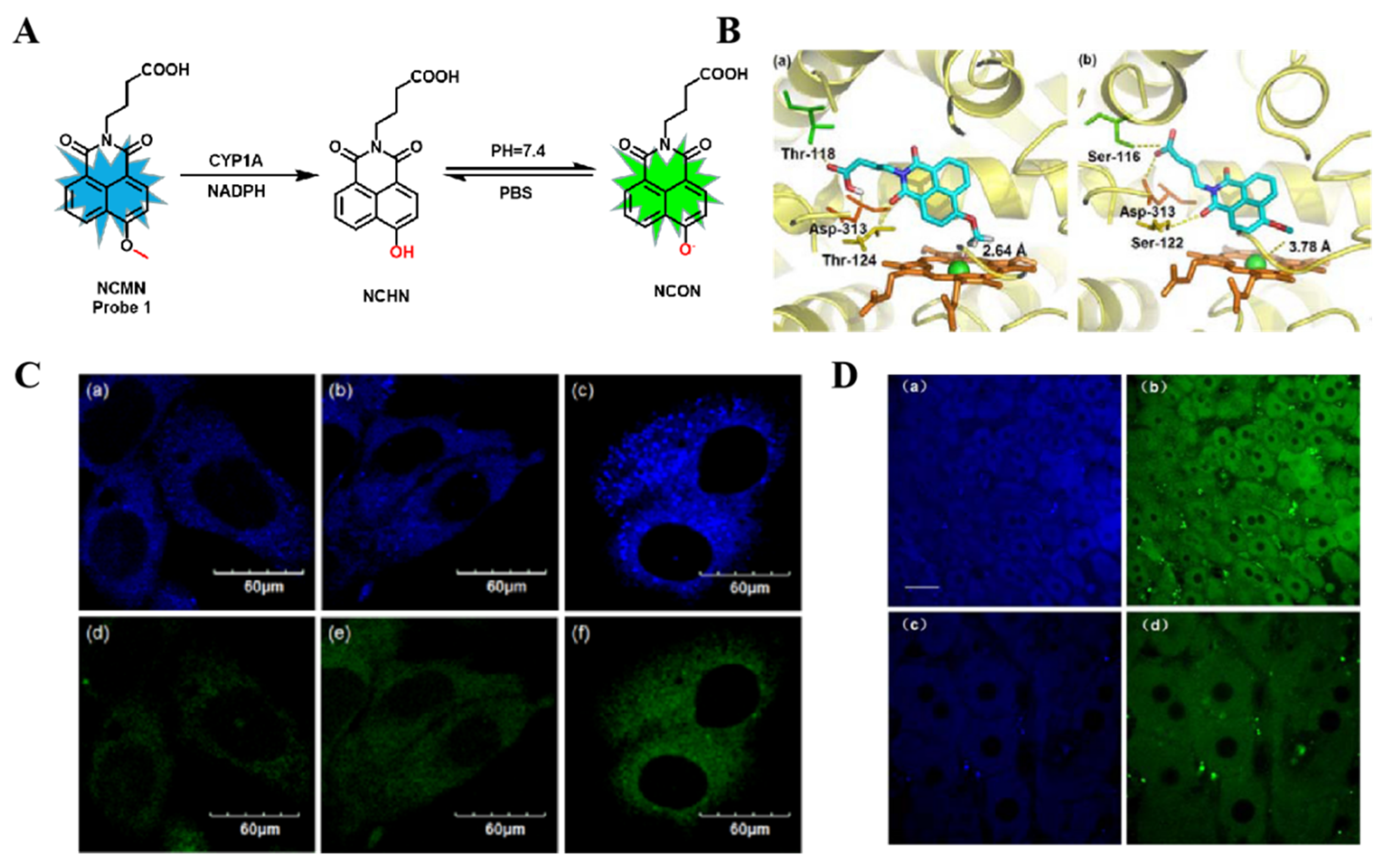
The 4-methoxy-1,8-naphthalimide (HN) derivatives are effective CYP1A substrates, according to earlier research by Yang Ling’s team [48]. In 2015, based on the 3D structure information of the CYP1A enzymes, Yang’s team reported a highly selective CYP1A probe with high sensitivity and an anti-biological matrix interference probe 1 (NCMN) (Figure 6A). Molecular docking studies showed that probe 1 could be well docked into the active cavity of CYP1A2 and CYP1A1 with strong hydrogen-bond interactions (Figure 6B). The authors successfully combined ratiometric fluorescence detection and two-photon fluorescence imaging analysis of probe 1 to accurately detect CYP1A enzyme activity at the living cells and tissue level for the first time [49] (Figure 6C,D).
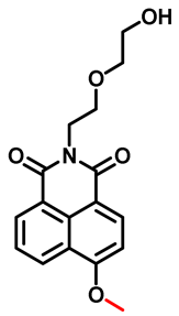
Since the active site of CYP1A is packed with hydroxyl amino acids, hydrogen-bond and π−π interactions may play an important role in the catalysis of CYP1A. Qi’s team obtained a novel ratiometric fluorescent probe N-((2-hydroxyl ethoxy) ethyl)-4-methoxy-1,8-naphthalimide (NEMN, probe 2, Table 2) for monitoring CYP1A. This probe is also based on the O-demethylation reaction of 1,8-naphthylamide but differs from NCMN in N-substituted by (2-hydroxyethoxy)ethyl, providing both a hydrogen bond formation site and good water solubility [50]. As expected, the ratiometric signal (I552/I458) emerged with a good linear relationship to both the reaction time and the enzyme concentration in a certain range. The design strategy for CYP1A-specific probes provides a good reference value for the subsequent discovery of more specific fluorescent probes for CYP1A isoforms.
Unfortunately, the probes above cannot achieve good specificity in detecting the two enzyme isoforms, owing to the amino acid homology and substrate spectra overlap between CYP1A1 and CYP1A2 [51]. Moreover, both CYP1A1 and CYP1A2 can dealkylate substrates in the catalytic process. Taking advantage of the small difference in the catalytic active cavity between human CYP1A1 and CYP1A2 (compared to CYP1A2, CYP1A1 exhibits a relatively planar and small active cavity) [52], Yang’s team synthesized and developed the first CYP1A1-specific two-photon fluorescent probe 3 (NBCeN, Table 2), using a proximal local modification strategy close to the catalytic site of the enzyme, with the aid of computer-assisted screening and metabolic phenotype detection. The probe is highly specific for the target enzymes, which can distinguish CYP1A1 from CYP1A2 and other similar enzymes [53]. This strategy for designing CYP450-specific probes can also be utilized to direct the creation of additional substrates for metabolic enzyme-specific probes.
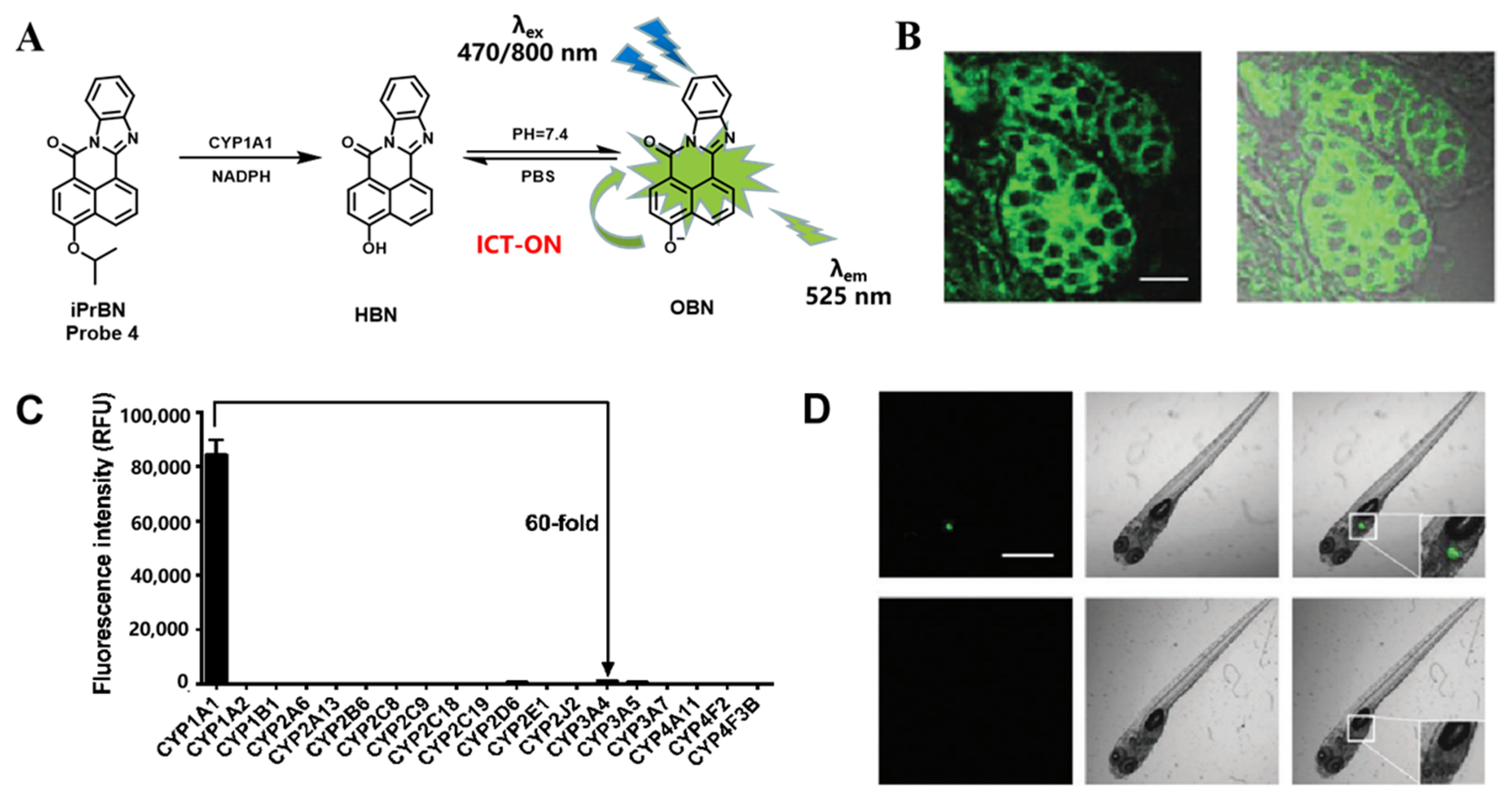
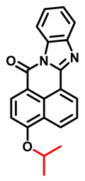
Probes from previous studies have achieved high specificity of CYP1As. However, the extremely low expression of CYP1A1 in extrahepatic tissues was ignored [54,55]. The detection limit value (LOD = 2.5 nM) of the previously mentioned NBCeN did not reach the value we expect, leading to the limits of real-time monitoring or in situ imaging of CYP1A1. To achieve better sensitivity of the probe, Ma’s team designed and synthesized a series of CYP1A1 fluorescent probes based on 4-hydroxy-7H-benzo[de]benzo [4,5]imidazo [2,1-a]isoquinolin-7-one (HBN) with a polycyclic aromatic skeleton and desirable photophysical properties, including high photostability, an excellent fluorescence quantum yield, and two-photon absorbability [56]. Among others, 4-isopropyl-7H-benzo[de]benzo [4,5]imidazo-[2,1-a]isoquinolin-7-one (probe 4, iPrBN), which was designed based on the substrate preference of CYP1A1 and the intramolecular charge transfer (ICT) principle (Figure 7A), displayed a high sensitivity and selectivity (Figure 7C) toward CYP1A1 compared with the other CYP450 isoforms. In this work, the authors screened a series of o-alkylated derivatives of HBN in vitro and finally found that only the isopropyl-modified probe iPrBN exhibited rapid responsiveness, high sensitivity, and excellent selectivity (LOD = 0.036 nM), and significant fluorescence enhancement in the presence of CYP1A1. The imaging results showed that this two-photon fluorescent probe is a powerful tool for sensing CYP1A1 in complex biological systems, including human tissue preparations, living cells, and even zebrafish (Figure 7B,D).
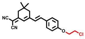
As we mentioned earlier, CYP1A1 has a larger catalytic cavity than CYP1A2, so CYP1A1 can catalyze fluorescent substrates with a larger recognition group. Taking advantage of the deeper penetration depth advantage of near-infrared (NIR) imaging to minimize the effects of tissue background fluorescence and light scattering, Qi’s team proposed in 2019 that a highly specific NIR probe 5 (DPCl, Table 2) for CYP1A1 can be obtained by tuning the generation of fluorescence through DPCl-O-dechloroethylation on the NIR fluorophore dicyanoisophorone derivative (673 nm emission) [57]. Different from the dealkylation catalytic reaction of the previous probes, the design strategy of O-dechloroethylation in this study greatly improved the sensitivity (LOD = 0.026 nM) and selectivity of the probe, as the size of O-chloroethyl fit well into the catalytic cavity of CYP1A1 and the lipophilicity and polarity of O-chloroethyl. Probe 5 successfully achieves real-time detection of CYP1A1 activity in complex biological systems (cell and zebrafish), and it is a highly CYP1A1-specific detection tool with high affinity and a significant stokes shift, which could provide support for the study of the biological function of CYP1A1 and the related screening of inhibitors.
In 2019, Qi’s team modified the non-reactive site of 1,8-naphthylamide with (2-hydroxyethoxy)ethyl substitution, which can provide a hydrogen bond formation site and good water solubility, then introduced different alkyl groups for local modification at the probe recognition site, and screened the probe with higher sensitivity and selectivity through in vitro experiments. The experimental results demonstrated that CYP1A1 can catalyze the de-population of probe 6 (NEiPN, Table 2) to recover the fluorescence of the probe, which provides visible signal changes for high-throughput screening of CYP1A1 modulators in complex biological systems [58].
Instead of using the traditional 1,8-naphthylamide as the base fluorophore, Ge’s team developed a series of O-alkylated resorufin derivatives as potential fluorescent substrates for CYP1A. In vitro screening experiments showed that a chloroethyl derivative (7-(2-chloroethoxy)-3H-phenoxazin-3-one, (CHPO, probe 7, Table 2), has good specificity and is isoform-specific for CYP1A1, which also has been successfully applied to the bioimaging of CYP1A1 in cells and tissues [59].
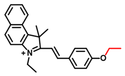
CYP1A1 expression is abnormal in some cancers, for example, CYP1A1 is overexpressed in breast cancer. Although several fluorescent sensors have been developed for the selective and sensitive detection of endogenous CYP1A1 enzyme, however, few reports have been reported for the isoform-specific CYP1A1 activities in tumor-bearing mice. In 2021, Chen’s team developed the CYP1A1-specific detection fluorescent probe 8 (BCy-CYP, Table 2) for the first time, using benzoindocyanine dye (BCy) as the fluorophore and introducing ethoxy modifications as the quencher at the recognition site [60]. The authors not only visualized the association between CYP1A1 and breast cancer using probe 8 but also demonstrated that the chemotherapy drug cisplatin, in combination with inhibition of CYP1A1 by carnosol, had a more significant therapeutic effect on breast cancer than cisplatin alone, which plays a crucial role in studying the vital function of CYP1A1 in breast cancer, and it is of great significance for the early diagnosis of breast cancer. Meanwhile, probe 8 is the first to enable us to monitor not only the changes in CYP1A1 level in the breast’s normal and cancer cells but also in tumor-bearing mice.
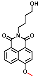
Reports have indicated that 2,3,7,8-tetrachlorodibenzo-p-dioxin (TCDD) is one of the major carcinogens affecting human life and health as an environmental pollutant [61,62]. In vitro bioassays on the hazardous carcinogen, TCDD (2,3,7,8-tetrachlorodibenzo-p-dioxin), have mostly been carried out utilizing the probe (7-ethoxyresorufin) 7-ER to identify TCDD-induced aberrant expression of CYP1A1 in murine cells [63]. In animal and human cells, TCDD increases several forms of CYP1A expression, including CYP1A1 in murine hepatocytes and CYP1A2 in human hepatocytes, according to recent toxicity studies [64]. As a result, the bioanalysis of TCDD in human cells is constrained by the 7-ER’s substandard response to CYP1A2. However, bioanalysis of TCDD in human cells is restricted because of 7-ER’s subpar sensitivity to CYP1A2. Liu’s team designed a fluorescent probe 9 (N-(3-hydroxybutyl)-4-methoxy-1,8-naphthalimide, HBMN, Table 2) based O-demethylation reaction in 2018 [65], which has a significantly higher affinity to CYP1A2 than CYP1A1, to enable the bioanalysis of TCDD in human cells. This means that researchers can now track changes in CYP1A2 expression levels and distribution in human cells for the first time using visible optical signal changes. Even though CYP1A1 causes a less intense fluorescent response than CYP1A2 (CYP1A2 was nearly 25 times higher than CYP1A1), it still influences the specificity of the probe.

3.2. Probe(s) for CYP1B1

In 1991, Colin R. Jefcoate, a professor at the University of Wisconsin, initially discovered that CYP1B1 is a novel member of the CYP450 family of drug-metabolizing enzymes. Unlike CYP1A1 and CYP1A2, CYP1B1 is expressed in both extrahepatic tissues and the liver. Numerous endogenous metabolites in the body, such as steroid hormones, fatty acids, melatonin, and vitamins, as well as many exogenous compounds, such as polycyclic aromatic hydrocarbons, are mainly metabolized by CYP1B1 [66].
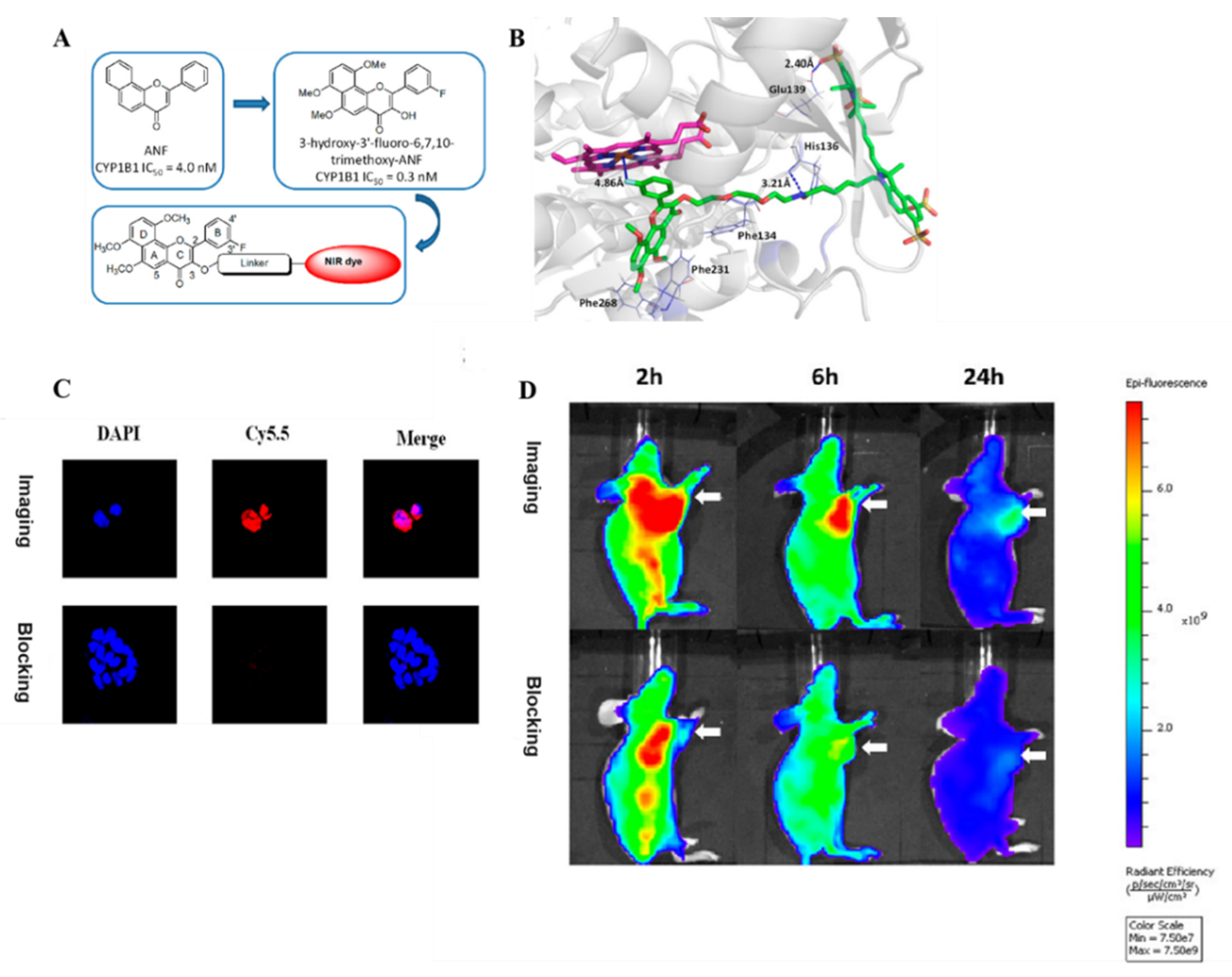
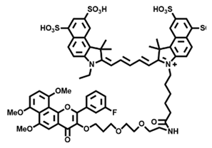
Li’s team created the first NIR fluorescent probe 10 (6b), which introduced a linker group to a known CYP1B1 selective inhibitor (ANF derivative) and a NIR fluorophore, for specifically binding to CYP1B1 and imaging tumors [67] (Figure 8A). The experiments in this work demonstrated that the introduction of the linker did not negatively affect the binding affinity and the inhibitory effect of the inhibitor, which was also verified by competition experiments. Additionally, the observed nonorange binding affinity of probe 10 to CYP1B1 during the cell-based assay was explained by the binding modes (Figure 8B), and the introduction of the cyanine group through the polymethylene linker contributed to the binding of the probes to the CYP1B1 protein rather than interfering with it. By using imaging tests both in vivo and in vitro, the authors were able to confirm the rapid and selective accumulation of CYP1B1 in HCT-15 xenograft tumors (Figure 8C,D). CYP1B1-targeted imaging may be a promising tool for the early detection of CYP1B1-associated tumors, and it provides new ideas for the subsequent development of CYP1B1-specific fluorescent probes.

4. Probes for CYP2 Family

4.1. Probe(s) for CYP2J2

CYP2J2, an important member of the CYP2 family, is mainly distributed in the heart, liver, and other organs, which is involved in the metabolism of a variety of endogenous substances. Recent studies have revealed that CYP2J2 expression is significantly upregulated in a variety of tumor tissues. The high expression of CYP2J2 is not only closely associated with tumorigenesis but has also been shown to promote the proliferation and the migration of tumor cells. In addition, since CYP2J2 can catalyze the generation of epoxyeicosatrienoic acids (EETs) from endogenous polyunsaturated fatty acids in humans [68], CYP2J2 plays an important role in tumor angiogenesis and tumor invasion. Given that, CYP2J2 can be regarded as an important biomarker of tumor diagnosis. Therefore, the development of highly selective probes for CYP2J2 has important clinical applications. Although liquid mass spectrometry methods for detecting CYP2J2 activity have been reported [69], fluorescence assays are more promising due to their outstanding sensitivity, convenience, in situ visualization, and high spatial and temporal resolution. In the three-dimensional spatial structure, CYP2J2 has the typical feature of narrow substrate channels near the catalytic active center, which restricts the metabolism of small molecule ligands (especially rigid substrates) to a certain extent. The challenge in the design and the development of CYP2J2 fluorescent probes lies in how to modify the fluorescent probe structure for the catalytic characteristics of the CYP2J2 enzyme to improve reaction efficiency and to achieve specific detection. Thus, rational modification of the fluorescent substrate structure to adapt to the narrow region near the catalytic activity center of CYP2J2 is an effective way to solve this problem.
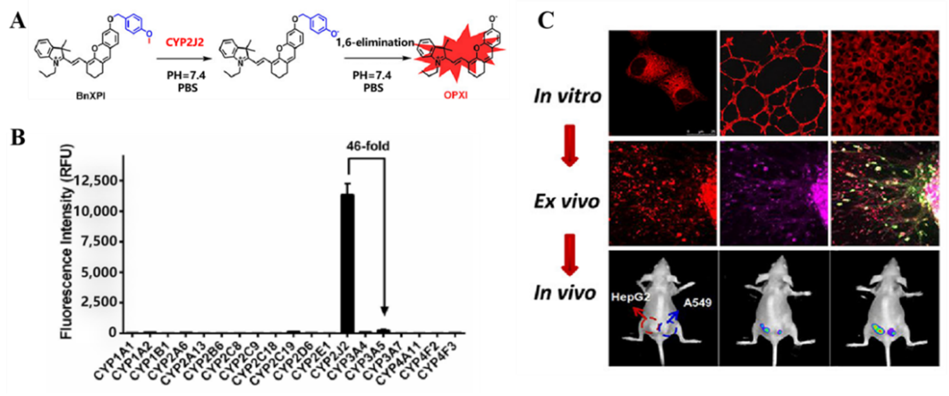
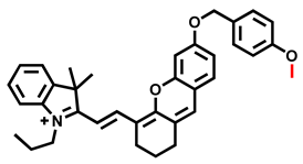
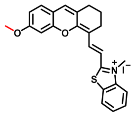
Inspired by the key structural domain (benzyl) of the CYP2J2 non-fluorescent probe astemizole, Ma’s team introduced a benzyl linker arm between the fluorophore HXPI and the dealkylation site to shorten the distance between the CYP2J2 catalytic center and the metabolic recognition site, to enhance the sensitivity and the isoform selectivity of the probe [70]. The authors employed (E)-2-(2-(6-hydroxy-2,3-dihydro-1H-xanthen-4-yl)vinyl)-3,3-dimethyl-1-propyl-3H-indol-1-iumiodide (HXPI) as the fluorophore, a relatively long and flexible aromatic backbone, which can fit into the narrow catalytic pocket at the bottom of CYP2J2. In addition, this class of fluorescent molecules has ideal photophysical properties, excellent tissue penetration, and minimal biological damage. Based on the strategy of introducing suitable linker arms to adapt the fluorescent probe to the cavity structure and catalytic characteristics of CYP2J2 catalytic activity, Ma’s team has successfully developed a highly specific NIR fluorescent probe 11 (BnXPI) for detecting CYP2J2 activity (Figure 9A,B), which can be used for rapid detection of CYP2J2 levels in human tissue samples and cells. However, HXPI derivatives without a linker are difficult to be catalyzed by CYP2J2 and also show a relatively weak fluorescence response and low isotype specificity. In addition, probe 11 has been used to detect the functional level of CYP2J2 in the blood leukocytes of leukemia patients, which can be combined with clinical immunophenotypic analysis for the early diagnosis of leukemia. With the help of probe 11, this team has also successfully visualized CYP2J2 in subcutaneous tumors of tumor-bearing mice (Figure 9C), further highlighting the potential application of probe 11 as a tool molecule for visualizing the tumor-associated metabolic enzyme CYP2J2 in fluorescence-mediated tumor resection.
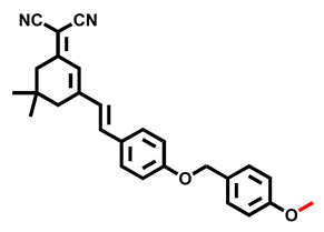
To address the problem that the substrate channel of CYP2J2 narrows in the cylinder near the heme prosthetic group and therefore limits further oxidation of active heme iron by the substrate, Qi’s team introduced a linker in the design of probe 12 (DPBM, Table 3) to shorten the spatial distance between the metabolic site of the substrate and the catalytic center of CYP2J2 [71]. The probe design differs from Ma’s team in that they applied 2-(3-(4-hydroxystyryl)-5,5-dimethylcyclohex-2-enylidene)malononitrile (DPOH) as the fluorophore, which has the advantages of ideal optical properties, fantastic tissue permeability, and minimal damage to the organism and its long aromatic skeleton. This study’s primary design concept was to create potential fluorescent substrates with various O-alkylations based on the widely utilized three-molecule fragmentation technique, and in the presence of CYP2J2, probe 12 showed a rapid and a sensitive fluorescent signal response. To validate probe 12 as a molecular tool for real-time fluorescent bioimaging of CYP2J2 enzyme activity in vivo, which is valuable for clinical diagnosis of related diseases. The authors completed in vivo and ex vivo assays of CYP2J2 in different biological systems, including HLMs, living cells, isolated organs, and in situ of tumor-bearing BALB/c nude mice.
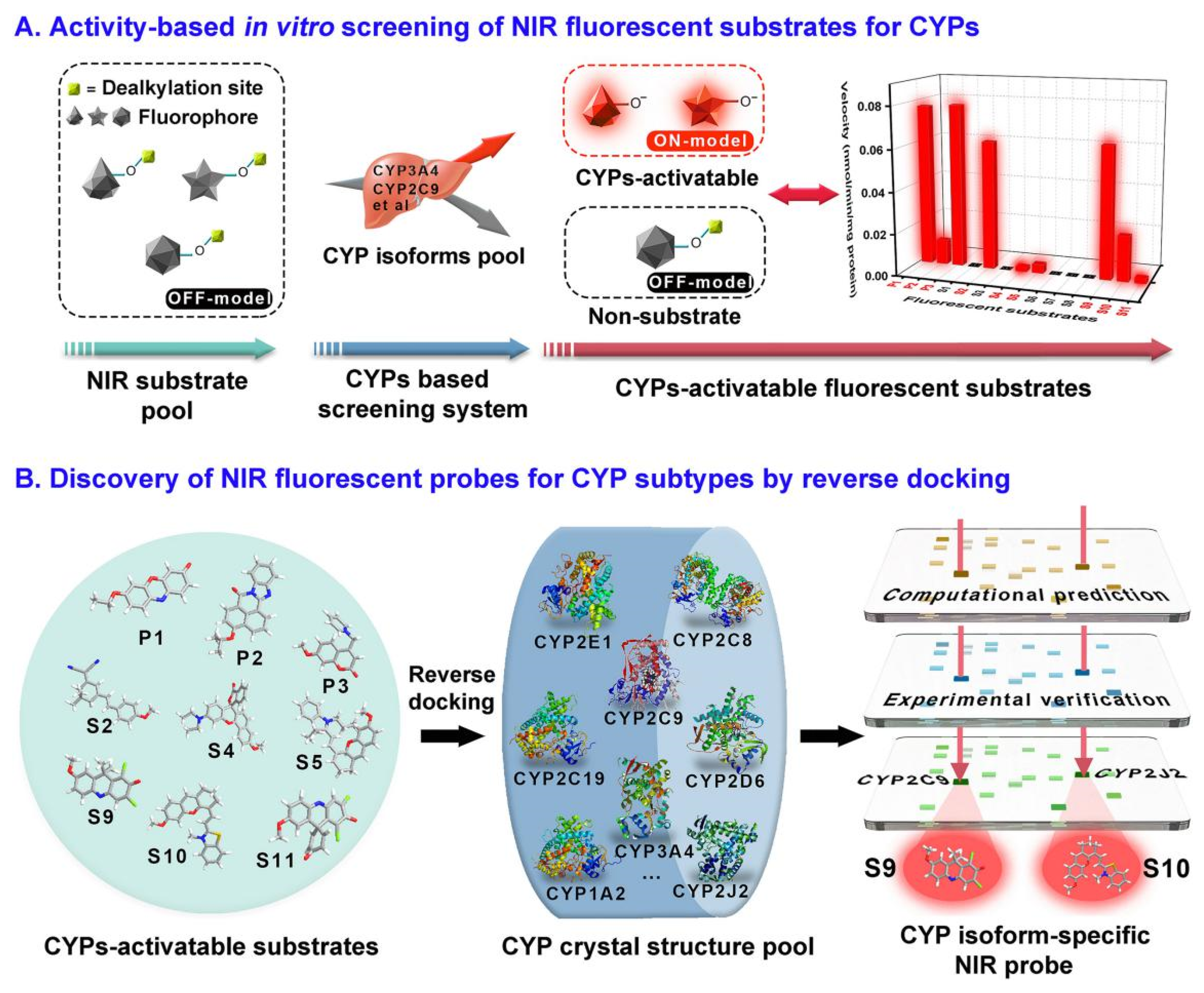
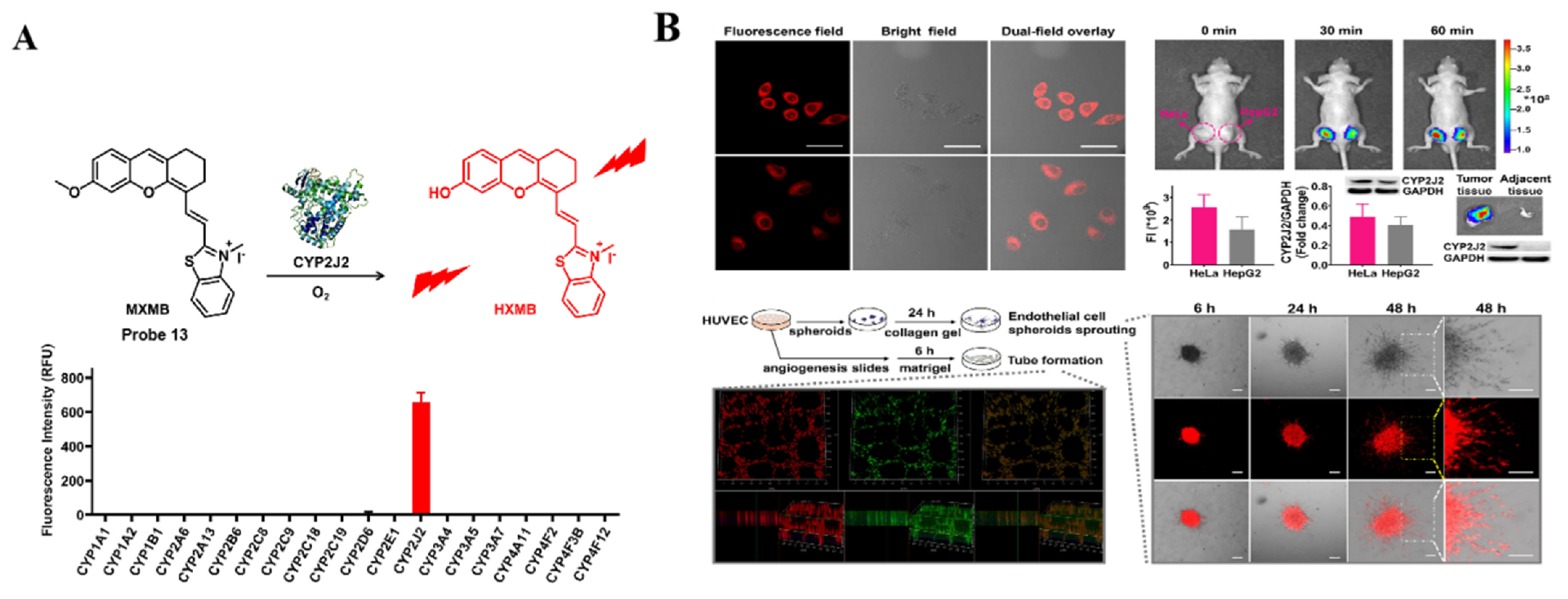
In 2021, Ma’s team made a new breakthrough in detecting CYP2J2-specific fluorescent probes after probe 11. The authors developed a new double-filtering strategy for the development of CYP450 isozyme-specific near-infrared fluorescent probes based on the structural characteristics of human CYP450 [72] (Figure 10). In the first stage of screening, the authors used CYP450-catalyzed dealkylation reactions on a series of fluorophores with NIR emission by introducing methoxy to quench fluorescence, followed by screening using CYP450s in vitro screening system (human-derived CYP450s extract) to obtain some candidate compounds. In the second stage, the NIR fluorescent substrate candidates were screened using the docking of fluorescent substrates with the reverse protein ligands of CYP450, which is able to identify potential CYP450 isozyme-specific probes. The specific process is to obtain the three-dimensional structure of each isoform docking and to analyze the theoretical distance from the catalytic site to the reaction site, which is considered as not catalytically active when the theoretical distance of the catalytic reaction is exceeded. In addition, they further investigated the match between the probe and CYP450 characteristic isoforms by molecular dynamics simulations and free energy calculations. Among those screened probes, probe 13 (MXMB) can be used for the selective activation process of CYP2J2 in various tumor models, thus facilitating real-time, multidimensional tracking of CYP2J2 activity in various biological systems (Figure 11). More importantly, this developed method is universal and practical, and the combination of “real” and “virtual” screening can further provide new ideas for the development of other fluorescent probes in enzyme activation and isoform specificity.

4.2. Probe(s) for CYP2C9

It is well known that DDIs play a vital role in drug efficacy and safety. CYP2C9 is thought to be associated with the DDI of an anticoagulant drug, warfarin, which causes elevated plasma concentrations leading to internal bleeding [73,74]. However, fluorescent probes that specifically respond to CYP2C9 have never been reported. To better understand the biological and the physiological roles of CYP2C9, there is an urgent need to develop CYP2C9-specific probes for highly selective and sensitive detection of the real activities of CYP2C9 in complex biological systems to fill the blank.
In 2021, based on the developed double-filtering strategy, Ma’s team not only developed a CYP2C9-specific probe 14 (DDAM) but also identified the compound DDAM as the most suitable substrate for CYP2C9 [72]. Probe 14 can be used to detect CYP2C9 activity and high-throughput screening for CYP2C9 modulators (Figure 12), which may provide a useful guide for assessing the risk of CYP2C9-mediated clinical DDI.

4.3. Probe(s) for CYP2D6

Drug metabolizing enzymes catalyze the metabolism of drugs, in most cases inactivating them and allowing their easy excretion. The CYP450 enzyme family is the most important phase I metabolizing enzyme, and its member CYP2D6 plays an important role in the metabolism of antipsychotics. Although CYP2D6 metabolizes 25% of clinical drugs with a high affinity for substrates [75], due to its low and saturated metabolic capacity, multiple doses are required and so differences in metabolic capacity have a significant impact on patient plasma concentrations. CYP2D6 has significant polymorphisms and can be classified according to metabolic capacity: ultrafast metabolizer (UM), extensive metabolizer (EM), intermediate metabolizer (IM), and poor metabolizer (PM) [76]. CYP2D6 genetic polymorphisms are closely related to blood concentrations, adverse effects, and drug interactions of related antipsychotic drugs [77]. The effects of the drug are not only the result of the drug’s concentration but also of the drug’s interactions. As mentioned earlier, fluorescent probes for the detection of CYP1A1, CYP1A2, CYP2J2, and CYP3A have been widely developed, while there is a gap in the specific detection of fluorescent probes for CYP2D6.
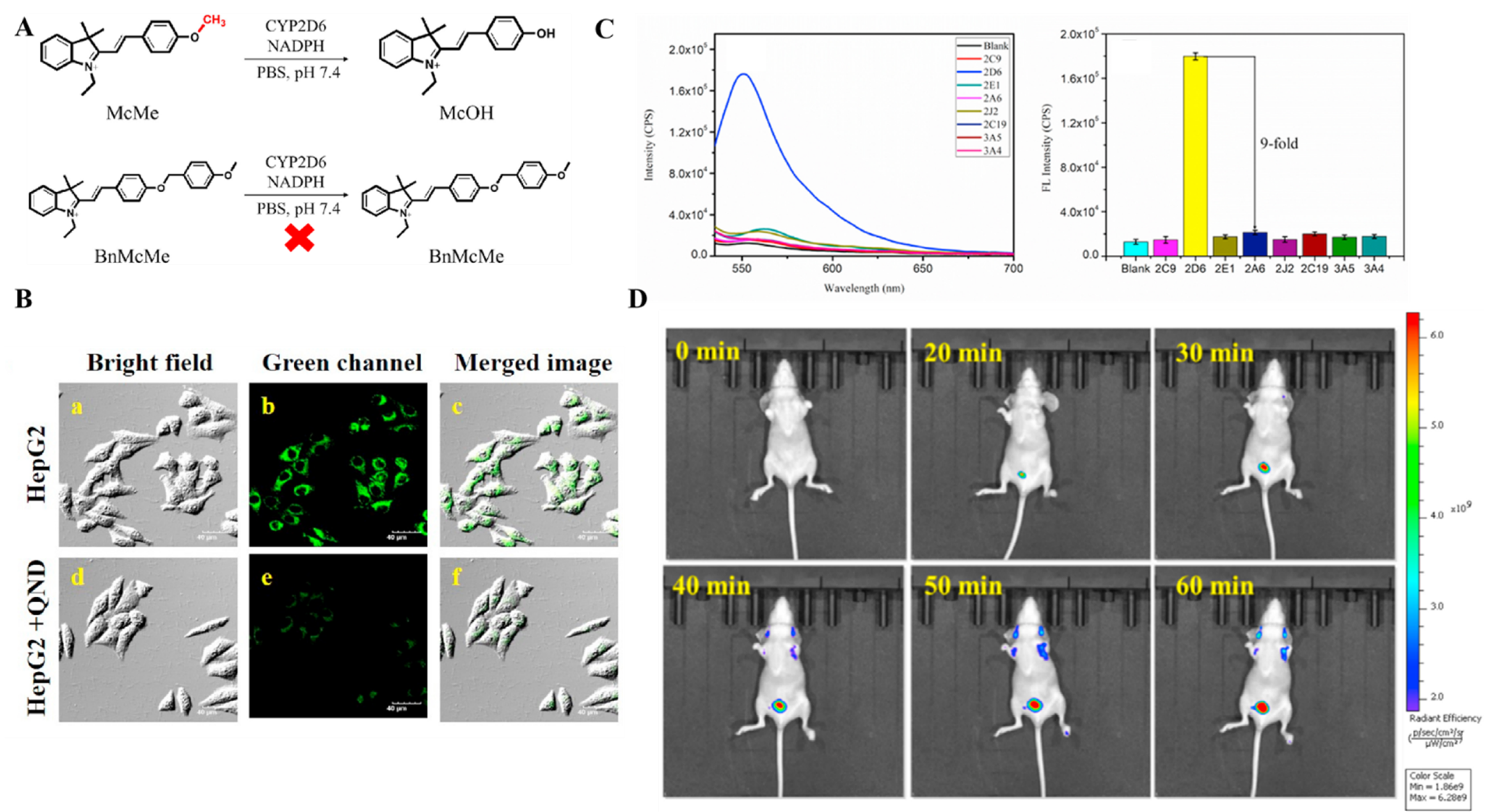
The active cavity structure of CYP2D6 is very similar to CYP2J2, whereas CYP2D6 can only catalyze substrates containing basic nitrogen (protonated) and planar aromatic rings. Taking a variety of advantages of merocyanine dye, a typical intramolecular charge transfer (ICT) effect fluorophore, which possesses nitrogen positive ion and rigid structure, introduces methoxy and 4-methoxybenzyl alcohol as the metabolic recognition moiety. In 2022, Gao’s team proposed two CYP2D6-specific probes (McMe and BnMcMe) (Figure 13A) [78]. O-demethylation of probe 15 (McMe) brought a remarkable fluorescence enhancement at 553 nm after the addition of CYP2D6 in the presence of other isoforms (Figure 13B). The fluorescence intensity of the probe BnMcMe, however, did not significantly change when CYP2D6 and other CYP450 isoforms were co-incubated with it. This suggests that probes without linkers have a better response to CYP2D6. Here is the response mechanism: the CYP2D6 recognition group is cleaved while releasing the dissociation product McOH, which can form a keto-form (pH 7.4) in a weak alkaline environment and restore the ICT effect of the fluorophore, causing significant fluorescence enhancement. As the first CYP2D6-specific molecule tool that successfully achieved real-time detection of CYP2J2 activity in complex biological systems (Figure 13C,D), probe 15 offers a broad prospect for studying the biological functions of CYP2D6 and an important guide for rational clinical drug use.

5. Probes for CYP3 Family

CYP3A is the most abundant CYP450 subfamily involved in the metabolism of various antibiotics, drugs, and carcinogens, mainly composed of CYP3A4, CYP3A5, and CYP3A7 [79,80]. CYP3A continues to receive the attention of biochemists and pharmacologists because of its pivotal role in drug metabolism. Reports have shown abnormality in CYP3A levels may influence the circulating levels of estrogens and the risk of breast cancer [81]. The high degree of genetic polymorphism of CYP3As and the different responses to environmental, hormonal, or nutritional influences make levels of CYP3As and their functions varied in different individuals [82,83], which indicates the urgent need to develop a CYP3A isoform-specific probe for understanding of the precise role of CYP3As.
CYP3A4 is predominant in CYP3A-mediated drug catalysis, and it is considered to be the major component of CYP3A isoform enzymes [84]. Common fluorescent substrates for CYP3A4 in fluorescence-based assays include typical steroids (testosterone) [85], bufalin [86]. Unfortunately, most of them lack isoform selectivity and application in living systems of CYP3A4. To further characterize the real functions of CYP3A4 in biological samples and to evaluate the effect of CYP3A4 in drug metabolism and DDI, it is necessary to develop effective tools for the real-time specific monitoring of CYP3A4.
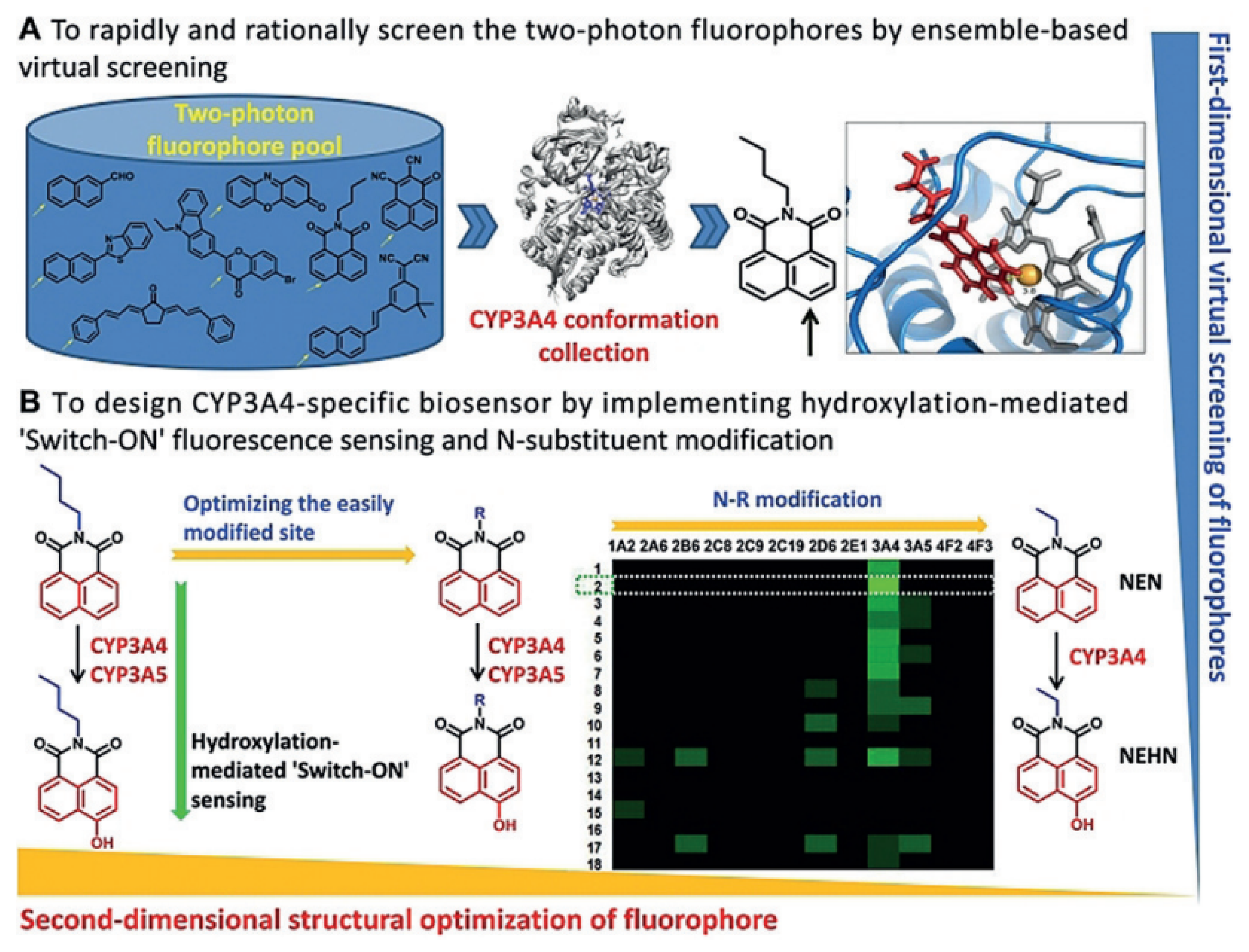
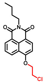
To address this issue, based on the catalytic reaction of CYP450 hydroxylation, Ma’s team designed and synthesized probe 16 (NEN, Table 4) which is activated by site-specific hydroxylation of the recognition site [87]. To take full advantage of the high sensitivity of imaging, real-time high spatial resolution imaging, and deep tissue bioimaging of two-photon (TP) fluorescence microscopy, Ma’s team employed N-butyl and N-phenyl derivatives of naphthalimide (NN) as a fluorophore, which has typical two-photon characteristics. Most impressively, they proposed a two-dimensional design strategy to virtually screen putative two-photon fluorescent substrates (Figure 14): in the first dimension, with the help of a docking program to virtually screen TP fluorophores, which can greatly improve efficiency and save cost; and, in the second design dimension, the screened fluorophore NN was modified at non-reactive sites to adjust the reaction rate and isoform selectivity. Finally, probe 16 was developed to monitor CYP3A4 activity. Through inhibitor assays, the authors validated the selectivity of probe 16 for CYP3A4 isoform. Probe 16 has successfully been further utilized for real-time imaging of CYP3A4 in various living systems.
Among the CYP3A isoforms, CYP3A5 is an important class of bioactive substances not only involved in the metabolism of some anticancer drugs (such as tamoxifen, tacrolimus, cyclosporine, and vincristine) but also including a characteristic identifier of some tumors (such as colorectal cancer [88] and ovarian cancer [89]). Compared to CYP3A4, the shape of the CYP3A5 active site is longer and narrower than that of CYP3A4, making the development of CYP3A5 probes much more difficult.
In 2017, Ling Yang’s team discovered and reported the first naturally occurring probe [90], a dibenzocyclooctadiene derivative of lignan (schisantherin E, 12), which can well orient the molecule of 12 into the active site of CYP3A5 by docking simulations and site-directed mutagenesis. However, the characterization of probe response still depends on LC-MS and HPLC instead of the “naked-eye” optical signal changes.
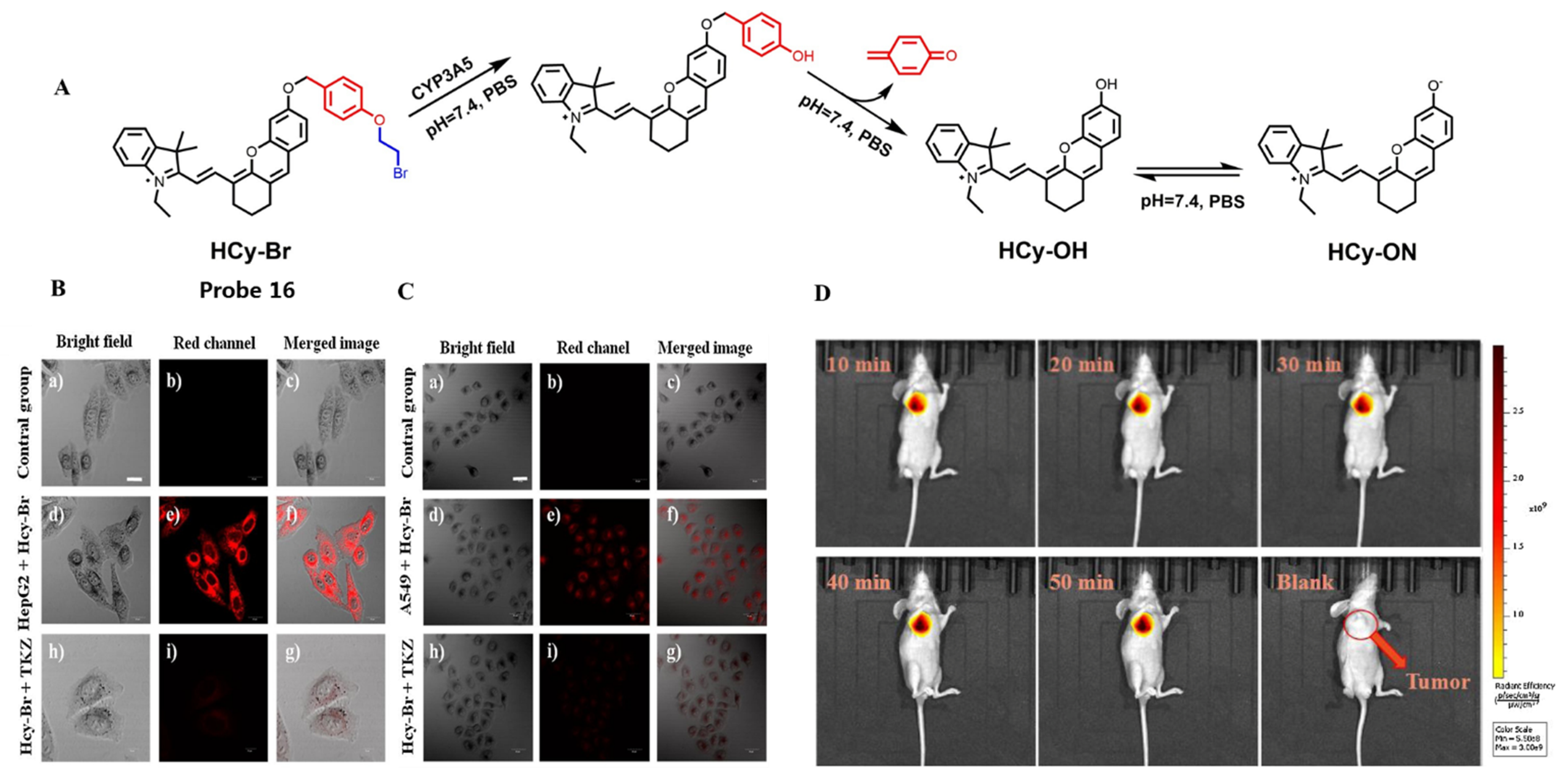
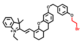
To further the real-time detection of CYP3A5, Zhu’s team synthesized two NIR ratiometric fluorescent probes (HCy-Br and HCy-Cl) for the specific monitoring of CYP3A5 in vivo and then used probe 17 (HCy-Br, Table 4) as the primary probe due to its low cytotoxicity [91]. In this work, the hemicyanine dye was determined as the basic fluorophore, due to its high fluorescence quantum yield and good stability. Inspired by the target–response strategy, bromoethoxy was chosen as the recognition site. Considering that CYP3A5 has a narrow and a long cavity, a self-immolation linker (p-hydroxybenzoyl) was introduced into the fluorophore and the recognition group to shorten the distance between the probe and the cavity of the CYP3A5 enzyme, which can greatly facilitate the reaction efficiency. The introduction of N-ethylbenzoindolium not only improves the water solubility of the molecule but also increases the molecule’s targeting of mitochondria, which is a two-in-one design. After the addition of CYP3A5, the bromo-ethoxy group was broken and the 1,6-elimination reaction occurred to release the fluorophore to generate the NIR fluorescence signal (Figure 15A). Probe 17 was used not only to detect CYP3A5 activity under physiological conditions but also to quantify changes in CYP3A5 activity within living cells and tumor-bearing mice (Figure 15B–D).
Since CYP3A has a broad substrate spectrum for drugs, CYP3A enzyme activity is most affected by MBI. The dangers of MBI re-emphasize the importance of monitoring CYP3A activity to identify and to assess MBI-related biological effects. Covalent modification of biomolecules by CYP3A-mediated MBI ultimately results in terrible toxicity, such as the toxicity of aflatoxin, which is caused by highly toxic epoxide metabolites catalyzed by CYP3A [92]. Although CYP3A4 and CYP3A5 have a high amino acid sequence homology (>84%), and their substrates often overlap, it is worth noting that the substrate spectra of these two isoenzymes are not completely overlapping, and individual differences in CYP3A4 and CYP3A5 expression are also significant, such as expression level, specificity, and relationship to disease [93,94]. Therefore, monitoring CYP3A4 or CYP3A5 alone is not a valid basis for assessing MBI.
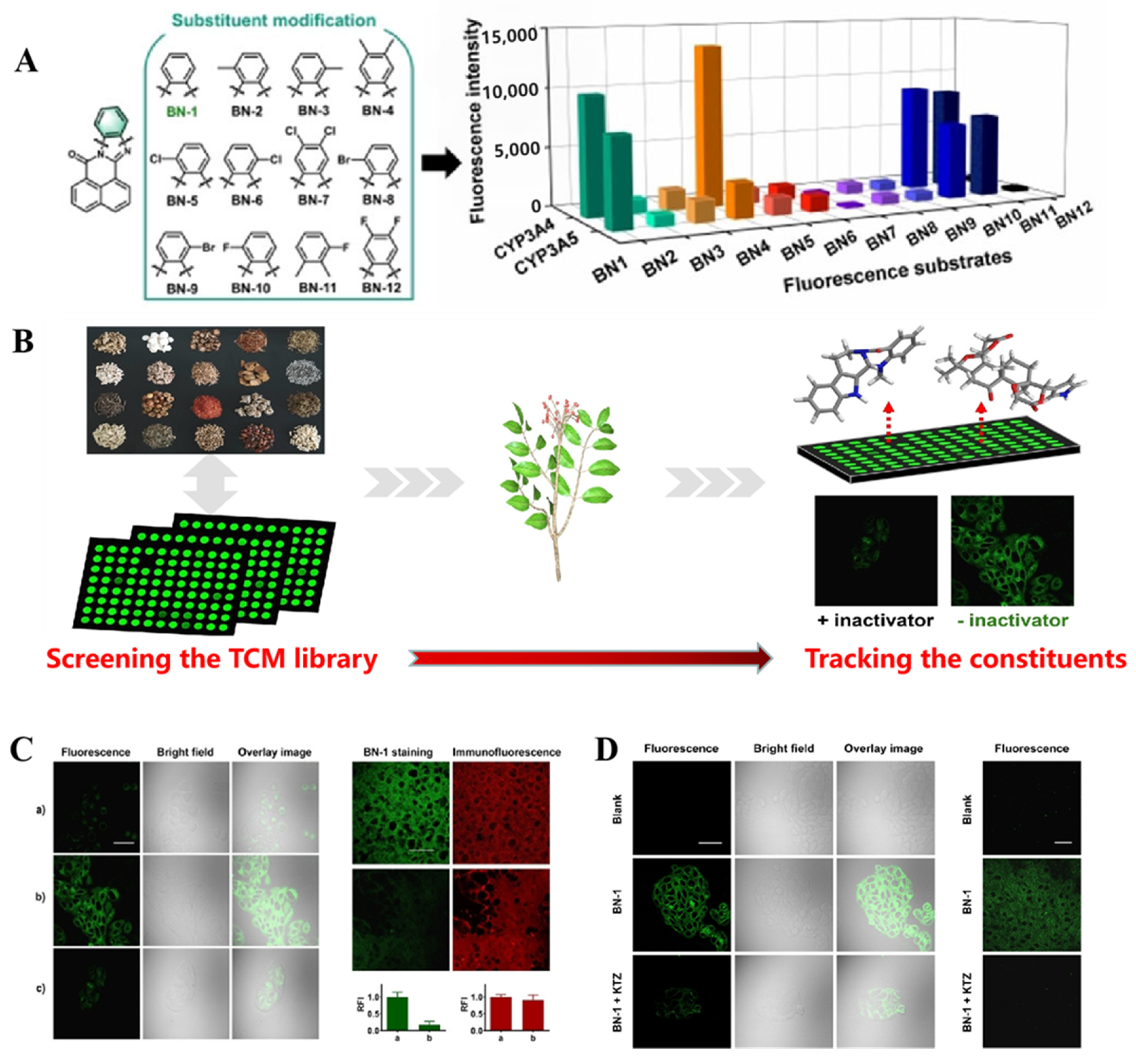
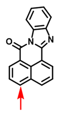
To address the problem, a fluorescent probe for sensitively monitoring CYP3A was developed by Ma’s team in 2021. During enzyme catalysis, the stable binding of the substrate in the enzyme lumen had a great influence on enzyme catalysis [96]. Ligand binding stability can be significantly improved by modifying the fluorophore structure of key interaction domains. Therefore, the authors designed a series of CYP3A fluorescent probes using 7H-benzo[de]benzo [4,5]imidazo [2,1-a]isoquinolin-7-one (BN) as a fluorophore, which has two-photon absorption and a high fluorescence quantum yield [95]. The docking of BN with CYP3A protein revealed that the benzimidazole ring on the probe structure plays an important role in the binding of the probe to the active cavity of CYP3A. To achieve a higher specificity, the authors further introduced multiple halogen and alkyl groups on the benzene ring to modulate the interaction of the probe with CYP3A (Figure 16A). Among others, only probe 18 (BN-1, Table 4) showed as highly selective for CYP3A4/3A5. In contrast, other CYP450 isoenzymes did not cause significant fluorescence intensity changes. They found that probe 18 undergoes 4-hydroxylation in the presence of CYP3A, which introduces a strong electron donor hydroxyl group in its D-π-A structure, resulting in CYP3A-mediated substrate fluorescence. Data from inhibitor experiments in liver microsomes also showed outstanding selectivity of the probe. With the help of probe 18, researchers have not only efficiently visualized and evaluated the MBI behaviors of dozens of commonly used herbal medicines, but also successfully traced the main MBI components of herbal medicines (Figure 16B). They further used probe 18 for semi-quantitative detection and imaging of endogenous CYP3A in different biological systems, which is not only a powerful molecular tool for the discovery and the characterization of CYP3A-mediated MBI in biological systems, it also provides a high-throughput screening system capable of assessing MBI-related CYP3A hepatotoxicity (Figure 16C,D).

6. Summary and Prospects

Abnormalities in the expression and function of the CYP450 enzyme are often closely related to the various physiological and pathological processes of the organism. Small-fluorescent probes, one of the most potent techniques, have produced promising results over the past two decades in monitoring the localization and the activities of CYP450 isoforms in vitro and in complex living systems, revealing the mechanism of organ toxicity of xenobiotic activation with high sensitivity and visualization. Considering that negative DDI may lead to the termination of drug development programs, the withdrawal of drugs from the market, or the limitation of their therapeutic use, CYP450s inhibition studies are increasingly becoming an important part of the drug discovery process. The capability of CYP450-specific probes to screen and to discover relevant inhibitors and inducers as well as to provide an efficient molecular tool for achieving real-time imaging of intracellular CYP450s is also noteworthy, which can serve as a crucial foundation for the clinical combination of medications and the assessment of new drug development.
A promising fluorescent probe should offer in vivo detection capability, rapid response to target molecules, and significant fluorescence intensity change before and after the reaction. There are still several problems that merit further research and solutions, even if these probes have shown encouraging improvements in biological studies of CYP450s compared to traditional HPLC.
  • Development of highly selective probes for imaging of other important CYP450 isoforms. The paucity of fluorescent probes for other important CYP450 isoforms (such as CYP2A6, CYP2E1, and CYP1A2) is primarily due to the similar catalytic activities and structures among the isoforms. It is challenging to design isoform-specific probe substrates because many isoforms of the CYP450 family often have similar structures and largely overlapping substrate profiles. Other selective probe design strategies may be helpful to improve the specificity of discrimination CYP450 isomerase isoforms. For example, Ma’s team designed a highly selective monoamine oxidase A (MAO-A) probe based on a targeting moiety of utilizing MAO-A specific inhibitor to realize differentiate detection of MAO-A from MAO-B in living cells [97].
  • Investigate and identify more adjustable NIR fluorophores. 1,8-Naphthylamide has evolved into the “star” dye for CYP450-specific probe design because of its exceptional photophysical characteristics. However, many probes based on 1,8-naphthylamide derivatives were insufficient for practical applications intended for in vivo environments. NIR fluorophore-based probes may well address this problem because of their capability to improve deep tissue penetration and reduce photon scattering, such as probe 17, which is able to achieve high-fidelity and high-resolution detection of CYP3A5 activity in tumor-bearing nude mice.
  • Development of fluorescent probes with diverse recognition reactions. CYP450 is a multifunctional catalytic metabolic enzyme that can catalyze a variety of reactions in Figure 4. Nevertheless, the fluorescent probes developed now mainly rely on O-dealkylation and hydroxylation, so we may be able to develop more efficient fluorescent probes based on other CYP450-catalyzed reactions (Figure 4B), such as dealkylation of N- and S- atoms. Furthermore, numerous probe design strategies suggest that molecular docking and inhibitor biomimicry is beneficial to the development of a novel recognition site for highly selective CYP450 isoform probes.
  • Discovery of probes that explore the relationship between CYP450 isoforms and their relevant carcinogens. CYP450s are involved in the metabolism of carcinogens in different types of cancer (including lung cancer, oral cancer, breast cancer, prostate cancer, hepatocellular and colorectal cancer, etc.), and the application of the above probes in this area is currently limited by the detection of expression levels in hepatocellular and breast cancers. However, other CYP450s-related diseases are seriously neglected, such as drug-induced liver injury, which is the common and the main reason for the current drug withdrawal from the market [98]. In addition, the current biological applications of CYP450 fluorescent probes are relatively simple and are limited to endogenous detection of cell tissues in mice. However, its related disease pathogenesis and its pathological role are still unclear, needing further exploration and discovery. Despite the detection and the diagnosis of many tumor mice models, there is still a long way to go before the real clinical application of CYP450 fluorescent probes.
Accurate characterization of CYP450 expression and function in complex biological systems is of great significance for applied research in pathophysiology and in biomedical basic research, disease diagnosis, and translational medicine. CYP450-specific probes can specifically reflect the activity of target CYP450 isoforms enzymes under physiological conditions in real-time, which may be a common tool for evaluating the catalytic function of CYP450 metabolic enzymes in the biomedical field. However, practical strategies for designing highly specific fluorescent probes for CYP450s remain challenging. Therefore, the design and the development of isoform-specific probe substrates have been a long-standing challenge in the biomedical field.

Author Contributions

Conceptualization, L.Y.; investigation and writing—original draft preparation, H.L., W.L. and Y.S.; validation and formal analysis, W.L. and Y.S.; data curation, writing—review and editing, project administration, supervision, and funding acquisition, W.L. and L.Y. All authors have read and agreed to the published version of the manuscript.

Funding

This work was financially supported by the National Natural Science Foundation of China (21877029, 22074036), the Science and Technology Project of Hunan Province (2021RC4021), and the China Postdoctoral Science Foundation (2022M711105).

Institutional Review Board Statement

Not applicable.

Informed Consent Statement

Not applicable.

Data Availability Statement

Not applicable.

Acknowledgments

We gratefully acknowledge the support of this work by the National Natural Science Foundation of China (21877029, 22074036), the Science and Technology Project of Hunan Province (2021RC4021), and the China Postdoctoral Science Foundation (2022M711105).

Conflicts of Interest

The authors declare no conflict of interest.

References

  1. Korobkova, E.A. Effect of Natural Polyphenols on CYP Metabolism: Implications for Diseases. Chem. Res. Toxicol. 2015, 28, 1359–1390. [Google Scholar] [CrossRef] [PubMed]
  2. Munro, A.W.; Girvan, H.M.; Mason, A.E.; Dunford, A.J.; McLean, K.J. What makes a P450 tick? Trends Biochem. Sci. 2013, 38, 140–150. [Google Scholar] [CrossRef]
  3. Sangar, M.C.; Bansal, S.; Avadhani, N.C. Bimodal targeting of microsomal cytochrome P450s to mitochondria: Implications in drug metabolism and toxicity. Expert Opin. Drug Metab. Toxicol. 2010, 6, 1231–1251. [Google Scholar] [CrossRef] [PubMed] [Green Version]
  4. Mast, N.; Norcross, R.; Andersson, U.; Shou, M.G.; Nakayama, K.; Bjorkhem, I.; Pikuleva, I.A. Broad substrate specificity of human cytochrome P450 46A1 which initiates cholesterol degradation in the brain. Biochemistry 2003, 42, 14284–14292. [Google Scholar] [CrossRef] [PubMed]
  5. Guengerich, F.P. Intersection of the Roles of Cytochrome P450 Enzymes with Xenobiotic and Endogenous Substrates: Relevance to Toxicity and Drug Interactions. Chem. Res. Toxicol. 2017, 30, 2–12. [Google Scholar] [CrossRef]
  6. Stipp, M.C.; Acco, A. Involvement of cytochrome P450 enzymes in inflammation and cancer: A review. Cancer Chemother. Pharmacol. 2021, 87, 295–309. [Google Scholar] [CrossRef]
  7. Nebert, D.W.; Russell, D.W. Clinical importance of the cytochromes P450. Lancet 2002, 360, 1155–1162. [Google Scholar] [CrossRef]
  8. Court, M.H. Interindividual variability in hepatic drug glucuronidation: Studies into the role of age, sex, enzyme inducers, and genetic polymorphism using the human liver bank as a model system. Drug Metab. Rev. 2010, 42, 209–224. [Google Scholar] [CrossRef]
  9. Gong, L.; Zhang, C.-M.; Lv, J.-F.; Zhou, H.-H.; Fan, L. Polymorphisms in cytochrome P450 oxidoreductase and its effect on drug metabolism and efficacy. Pharmacogenet. Genom. 2017, 27, 337–346. [Google Scholar] [CrossRef]
  10. Ragia, G.; Giannakopoulou, E.; Karaglani, M.; Karantza, I.-M.; Tavridou, A.; Manolopoulos, V.G. Frequency of CYP450 enzyme gene polymorphisms in the Greek population: Review of the literature, original findings and clinical significance. Drug Metab. Drug Interact. 2014, 29, 235–248. [Google Scholar] [CrossRef]
  11. Gharavi, N.; El-Kadi, A.O.S. Expression of cytochrome P450 in lung tumor. Curr. Drug Metab. 2004, 5, 203–210. [Google Scholar] [CrossRef] [PubMed]
  12. Hou, J.; Sun, E.; Song, J.; Yang, L.; Zhang, Z.-H.; Ning, Q.; Jia, X.-B. [Relationship between hepatic drug-metabolizing enzymes CYP450 and traditional Chinese medicine-induced hepatotoxicity]. China J. Chin. Mater. Med. 2016, 41, 2774–2780. [Google Scholar]
  13. Lu, T.-l.; Su, L.-l.; Ji, D.; Gu, W.; Mao, C.-q. Interaction between CYP450 enzymes and metabolism of traditional Chinese medicine as well as enzyme activity assay. China J. Chin. Mater. Med. 2015, 40, 3524–3529. [Google Scholar]
  14. Wu, J.; Guan, X.; Dai, Z.; He, R.; Ding, X.; Yang, L.; Ge, G. Molecular probes for human cytochrome P450 enzymes: Recent progress and future perspectives. Coord. Chem. Rev. 2021, 427, 213600. [Google Scholar] [CrossRef]
  15. Feng, L.; Ning, J.; Tian, X.; Wang, C.; Yu, Z.; Huo, X.; Xie, T.; Zhang, B.; James, T.D.; Ma, X. Fluorescent probes for the detection and imaging of Cytochrome P450. Coord. Chem. Rev. 2021, 437, 213740. [Google Scholar] [CrossRef]
  16. Chen, J.; Fang, Y.; Sun, L.; Zeng, F.; Wu, S. An activatable probe for detecting alcoholic liver injury via multispectral optoacoustic tomography and fluorescence imaging. Chem. Comm. 2020, 56, 11102–11105. [Google Scholar] [CrossRef]
  17. Gao, Y.; Lin, Y.; Liu, T.; Chen, H.; Yang, X.; Tian, C.; Du, L.; Li, M. Bioluminescent Probe for Tumor Hypoxia Detection via CYP450 Reductase in Living Animals. Anal. Chem. 2017, 89, 12488–12493. [Google Scholar] [CrossRef]
  18. O’Connor, L.J.; Mistry, I.N.; Collins, S.L.; Folkes, L.K.; Brown, G.; Conway, S.J.; Hammond, E.M. CYP450 Enzymes Effect Oxygen-Dependent Reduction of Azide-Based Fluorogenic Dyes. ACS Cent. Sci. 2017, 3, 20–30. [Google Scholar] [CrossRef]
  19. Yin, F.C.; Zhang, X.; Wang, L.; Wang, Y.Q.; Zhu, Y.J.; Li, Z.Y.; Tao, T.T.; Chen, W.W.; Yu, H.; Qin, J.H. HiPSC-derived multi-organoids-on-chip system for safety assessment of antidepressant drugs. Lab Chip 2021, 21, 571–581. [Google Scholar] [CrossRef]
  20. Nelson, D.R. A world of cytochrome P450s. Philos. Trans. R. Soc. B 2013, 368, 20120430. [Google Scholar] [CrossRef]
  21. Wei, P.; Zhang, J.; Egan-Hafley, M.; Liang, S.; Moore, D.D. The nuclear receptor CAR mediates specific xenobiotic induction of drug metabolism. Nature 2000, 407, 920–923. [Google Scholar] [CrossRef] [PubMed]
  22. Mittal, B.; Tulsyan, S.; Kumar, S.; Mittal, R.D.; Agarwal, G. Cytochrome P450 in Cancer Susceptibility and Treatment. Adv. Clin. Chem. 2015, 71, 77–139. [Google Scholar]
  23. Lynch, T.; Price, A. The effect of cytochrome P450 metabolism on drug response, interactions, and adverse effects. Am. Fam. Physician 2007, 76, 391–396. [Google Scholar] [PubMed]
  24. Deodhar, M.; Al Rihani, S.B.; Arwood, M.J.; Darakjian, L.; Dow, P.; Turgeon, J.; Michaud, V. Mechanisms of CYP450 Inhibition: Understanding Drug-Drug Interactions Due to Mechanism-Based Inhibition in Clinical Practice. Pharmaceutics 2020, 12, 846. [Google Scholar] [CrossRef] [PubMed]
  25. Fontana, E.; Dansette, P.M.; Poli, S.M. Cytochrome p450 enzymes mechanism based inhibitors: Common sub-structures and reactivity. Curr. Drug Metab. 2005, 6, 413–454. [Google Scholar] [CrossRef] [PubMed]
  26. Silverman, R.B. Design and Mechanism of GABA Aminotransferase Inactivators. Treatments for Epilepsies and Addictions. Chem. Rev. 2018, 118, 4037–4070. [Google Scholar] [CrossRef] [PubMed]
  27. Munro, A.W.; Girvan, H.M.; McLean, K.J. Variations on a (t)heme--novel mechanisms, redox partners and catalytic functions in the cytochrome P450 superfamily. Nat. Prod. Rep. 2007, 24, 585–609. [Google Scholar] [CrossRef]
  28. Schlichting, I.; Berendzen, J.; Chu, K.; Stock, A.M.; Maves, S.A.; Benson, D.E.; Sweet, B.M.; Ringe, D.; Petsko, G.A.; Sligar, S.G. The catalytic pathway of cytochrome P450cam at atomic resolution. Science 2000, 287, 1615–1622. [Google Scholar] [CrossRef]
  29. Guengerich, F.P. Common and Uncommon Cytochrome P450 Reactions Related to Metabolism and Chemical Toxicity. Chem. Res. Toxicol. 2001, 14, 611–650. [Google Scholar] [CrossRef] [PubMed]
  30. Isin, E.M.; Guengerich, F.P. Complex reactions catalyzed by cytochrome P450 enzymes. Biochim. Biophys. Acta 2007, 1770, 314–329. [Google Scholar] [CrossRef]
  31. Bernhardt, R. Cytochromes P450 as versatile biocatalysts. J. Biotechnol. 2006, 124, 128–145. [Google Scholar] [CrossRef] [PubMed]
  32. Shaik, S.; Kumar, D.; de Visser, S.P.; Altun, A.; Thiel, W. Theoretical Perspective on the Structure and Mechanism of Cytochrome P450 Enzymes. Chem. Rev. 2005, 105, 2279–2328. [Google Scholar] [CrossRef] [PubMed]
  33. Ung, Y.T.; Ong, C.E.; Pan, Y. Current High-Throughput Approaches of Screening Modulatory Effects of Xenobiotics on Cytochrome P450 (CYP) Enzymes. High-Throughput 2018, 7, 29. [Google Scholar] [CrossRef] [PubMed] [Green Version]
  34. Zlokarnik, G.; Grootenhuis, P.D.; Watson, J.B. High throughput P450 inhibition screens in early drug discovery. Drug Discov. Today 2005, 10, 1443–1450. [Google Scholar] [CrossRef]
  35. Zhang, H.; Fan, J.; Wang, J.; Dou, B.; Zhou, F.; Cao, J.; Qu, J.; Cao, Z.; Zhao, W.; Peng, X. Fluorescence discrimination of cancer from inflammation by molecular response to COX-2 enzymes. J. Am. Chem. Soc. 2013, 135, 17469–17475. [Google Scholar] [CrossRef] [PubMed]
  36. Chen, X.; Tian, X.; Shin, I.; Yoon, J. Fluorescent and luminescent probes for detection of reactive oxygen and nitrogen species. Chem. Soc. Rev. 2011, 40, 4783–4804. [Google Scholar] [CrossRef] [PubMed]
  37. Cheng, D.; Peng, J.; Lv, Y.; Su, D.; Liu, D.; Chen, M.; Yuan, L.; Zhang, X. De Novo Design of Chemical Stability Near-Infrared Molecular Probes for High-Fidelity Hepatotoxicity Evaluation In Vivo. J. Am. Chem. Soc. 2019, 141, 6352–6361. [Google Scholar] [CrossRef] [PubMed]
  38. Xu, W.; Zeng, Z.; Jiang, J.H.; Chang, Y.T.; Yuan, L. Discerning the Chemistry in Individual Organelles with Small-Molecule Fluorescent Probes. Angew. Chem. Int. Ed. 2016, 55, 13658–13699. [Google Scholar] [CrossRef]
  39. Chauret, N.; Tremblay, N.; Lackman, R.L.; Gauthier, J.-Y.; Silva, J.M.; Marois, J.; Yergey, J.A.; Nicoll-Griffith, D.A. Description of a 96-Well Plate Assay to Measure Cytochrome P4503A Inhibition in Human Liver Microsomes Using a Selective Fluorescent Probe. Anal. Biochem. 1999, 276, 215–226. [Google Scholar] [CrossRef] [PubMed]
  40. Lin, Z.; Li, M.; Lv, S.; Zhang, K.; Lu, M.; Tang, D. In situ synthesis of fluorescent polydopamine nanoparticles coupled with enzyme-controlled dissolution of MnO2 nanoflakes for a sensitive immunoassay of cancer biomarkers. J. Mater. Chem. 2017, 5, 8506–8513. [Google Scholar] [CrossRef]
  41. Lv, S.; Tang, Y.; Zhang, K.; Tang, D. Wet NH3-Triggered NH2-MIL-125(Ti) Structural Switch for Visible Fluorescence Immunoassay Impregnated on Paper. Anal. Chem. 2018, 90, 14121–14125. [Google Scholar] [CrossRef] [PubMed] [Green Version]
  42. Yin, Z.; Zhu, L.; Lv, Z.; Li, M.; Tang, D. Persistent luminescence nanorods-based autofluorescence-free biosensor for prostate-specific antigen detection. Talanta 2021, 233, 122563. [Google Scholar] [CrossRef] [PubMed]
  43. Hasler, J.A.; Estabrook, R.; Murray, M.; Pikuleva, I.; Waterman, M.; Capdevila, J.; Holla, V.; Helvig, C.; Falck, J.R.; Farrell, G.; et al. Human cytochromes P450. Mol. Aspects Med. 1999, 20, 1–137. [Google Scholar] [CrossRef]
  44. Bulus, H.; Oguztuzun, S.; Simsek, G.G.; Kilic, M.; Ada, A.O.; Gol, S.; Kocdogan, A.K.; Kaygin, P.; Bozer, B.; Iscan, M. Expression of CYP and GST in human normal and colon tumor tissues. Biotech. Histochem. 2019, 94, 1–9. [Google Scholar] [CrossRef]
  45. Lang, D.; Radtke, M.; Bairlein, M. Highly Variable Expression of CYP1A1 in Human Liver and Impact on Pharmacokinetics of Riociguat and Granisetron in Humans. Chem. Res. Toxicol. 2019, 32, 1115–1122. [Google Scholar] [CrossRef] [PubMed]
  46. Shimada, T.; Fujii-Kuriyama, Y. Metabolic activation of polycyclic aromatic hydrocarbons to carcinogens by cytochromes P450 1A1 and1B1. Cancer Sci. 2004, 95, 1–6. [Google Scholar] [CrossRef]
  47. Roberts, E.A.; Xie, Z.W.; Yang, S.; Lipa, J. Inducibility of enzyme activities associated with the cytochrome P-450 1A family, ethoxyresorufin O-deethylase, and methoxyresorufin O-demethylase in human hepatocyte lines derived from normal liver tissue. Drug Metab. Dispos.. 1993, 21, 56–61. [Google Scholar]
  48. Zhang, C.; Ren, A.M.; Guo, J.F.; Wang, D.; Yu, L.Y. Theoretical design and investigation of 1,8-naphthalimide-based two-photon fluorescent probes for detecting cytochrome P450 1A with separated fluorescence signal. Phys. Chem. Chem. Phys. 2018, 20, 13290–13305. [Google Scholar] [CrossRef]
  49. Dai, Z.R.; Ge, G.B.; Feng, L.; Ning, J.; Hu, L.H.; Jin, Q.; Wang, D.D.; Lv, X.; Dou, T.Y.; Cui, J.N.; et al. A Highly Selective Ratiometric Two-Photon Fluorescent Probe for Human Cytochrome P450 1A. J. Am. Chem. Soc. 2015, 137, 14488–14495. [Google Scholar] [CrossRef] [PubMed]
  50. Zhang, X.; Zhou, Y.; Gu, X.; Cheng, Y.; Hong, M.; Yan, L.; Ma, F.; Qi, Z. Synthesis of a selective ratiometric fluorescent probe based on Naphthalimide and its application in human cytochrome P450 1A. Talanta 2018, 186, 413–420. [Google Scholar] [CrossRef]
  51. Hammons, G.J.; Milton, D.; Stepps, K.; Guengerich, F.P.; Tukey, R.H.; Kadlubar, F.F. Metabolism of carcinogenic heterocyclic and aromatic amines by recombinant human cytochrome P450 enzymes. Carcinogenesis 1997, 18, 851–854. [Google Scholar] [CrossRef] [PubMed] [Green Version]
  52. Dong, D.; Wu, B.; Chow, D.; Hu, M. Substrate selectivity of drug-metabolizing cytochrome P450s predicted from crystal structures and in silico modeling. Drug Metab. Rev. 2012, 44, 192–208. [Google Scholar] [CrossRef] [PubMed]
  53. Dai, Z.R.; Feng, L.; Jin, Q.; Cheng, H.L.; Li, Y.; Ning, J.; Yu, Y.; Ge, G.B.; Cui, J.N.; Yang, L. A practical strategy to design and develop an isoform-specific fluorescent probe for a target enzyme: CYP1A1 as a case study. Chem. Sci. 2017, 8, 2795–2803. [Google Scholar] [CrossRef] [PubMed] [Green Version]
  54. Ma, Q. Induction of CYP1A1. The AhR/DRE paradigm: Transcription, receptor regulation, and expanding biological roles. Curr. Drug Metab. 2001, 2, 149–164. [Google Scholar] [CrossRef] [PubMed]
  55. Wohak, L.E.; Krais, A.M.; Kucab, J.E.; Stertmann, J.; Ovrebo, S.; Seidel, A.; Phillips, D.H.; Arlt, V.M. Carcinogenic polycyclic aromatic hydrocarbons induce CYP1A1 in human cells via a p53-dependent mechanism. Arch. Toxicol. 2016, 90, 291–304. [Google Scholar] [CrossRef] [Green Version]
  56. Ning, J.; Tian, Z.H.; Wang, B.; Ge, G.B.; An, Y.; Hou, J.; Wang, C.; Zhao, X.Y.; Li, Y.N.; Tian, X.G.; et al. A highly sensitive and selective two-photon fluorescent probe for real-time sensing of cytochrome P450 1A1 in living systems. Mater. Chem. Front. 2018, 2, 2013–2020. [Google Scholar] [CrossRef]
  57. Xue, T.Z.; Dai, Y.P.; Zhang, X.X.; Cheng, Y.; Gu, X.F.; Ji, H.F.; Misal, S.; Qi, Z.J. Ultrasensitive near-infrared fluorescent probe with large stokes shift for real-time tracing of CYP1A1 in living cells and zebrafish model. Sens. Actuators B 2019, 293, 265–272. [Google Scholar] [CrossRef]
  58. Ji, H.F.; Zhang, X.X.; Dai, Y.P.; Xue, T.Z.; Misal, S.; Qi, Z.J. A highly selective ratiometric fluorescent probe based on naphthalimide for detection and imaging of CYP1A1 in living cells and zebrafish. Analyst 2019, 144, 7390–7397. [Google Scholar] [CrossRef]
  59. Jin, Q.; Ma, H.Y.; Feng, L.; Wang, P.; He, R.J.; Ning, J.; Yang, L.; Ge, G.B. Sensing cytochrome P450 1A1 activity by a resorufin-based isoform-specific fluorescent probe. Chin. Chem. Lett. 2020, 31, 2945–2949. [Google Scholar] [CrossRef]
  60. Huang, Y.; Yu, L.; Lu, P.P.; Wei, Y.H.; Wang, X.Y.; Chen, L.X. Evaluate the inhibition of cytochrome P450 1A1 for enhancing breast cancer chemotherapy with a turn-on fluorescent probe. Sens. Actuators B 2021, 344, 130233. [Google Scholar] [CrossRef]
  61. Ozeki, J.; Uno, S.; Ogura, M.; Choi, M.; Maeda, T.; Sakurai, K.; Matsuo, S.; Amano, S.; Nebert, D.W.; Makishima, M. Aryl hydrocarbon receptor ligand 2,3,7,8-tetrachlorodibenzo-p-dioxin enhances liver damage in bile duct-ligated mice. Toxicology 2011, 280, 10–17. [Google Scholar] [CrossRef] [PubMed] [Green Version]
  62. Van den Berg, M.; Birnbaum, L.S.; Denison, M.; De Vito, M.; Farland, W.; Feeley, M.; Fiedler, H.; Hakansson, H.; Hanberg, A.; Haws, L.; et al. The 2005 World Health Organization Reevaluation of Human and Mammalian Toxic Equivalency Factors for Dioxins and Dioxin-Like Compounds. Toxicol. Sci. 2006, 93, 223–241. [Google Scholar] [CrossRef] [PubMed] [Green Version]
  63. Lewis, B.C.; Mackenzie, P.I.; Miners, J.O. Comparative homology modeling of human cytochrome P4501A1 (CYP1A1) and confirmation of residues involved in 7-ethoxyresorufin O-deethylation by site-directed mutagenesis and enzyme kinetic analysis. Arch. Biochem. Biophys. 2007, 468, 58–69. [Google Scholar] [CrossRef] [PubMed]
  64. Xu, L.; Li, A.P.; Kaminski, D.L.; Ruh, M.F. 2,3,7,8 Tetrachlorodibenzo-p-dioxin induction of cytochrome P4501A in cultured rat and human hepatocytes. Chem.-Biol. Interact. 2000, 124, 173–189. [Google Scholar] [CrossRef]
  65. Shangguan, L.; Wei, Y.; Wang, K.; Zhang, Y.; Liu, S. Highly sensitive fluorescent bioassay of 2,3,7,8-tetrachloro-dibenzo-p-dioxin based on abnormal expression of cytochrome P450 1A2 in human cells. Anal. Chim. Acta 2019, 1046, 179–184. [Google Scholar] [CrossRef]
  66. Li, F.; Zhu, W.F.; Gonzalez, F.J. Potential role of CYP1B1 in the development and treatment of metabolic diseases. Pharmacol. Ther. 2017, 178, 18–30. [Google Scholar] [CrossRef]
  67. Meng, Q.Q.; Wang, Z.T.; Cui, J.H.; Cui, Q.; Dong, J.Y.; Zhang, Q.J.; Li, S.S. Design, Synthesis, and Biological Evaluation of Cytochrome P450 1B1 Targeted Molecular Imaging Probes for Colorectal Tumor Detection. J. Med. Chem. 2018, 61, 10901–10909. [Google Scholar] [CrossRef]
  68. Pozzi, A.; Macias-Perez, I.; Abair, T.; Wei, S.Z.; Su, Y.; Zent, R.; Falck, J.R.; Capdevila, J.H. Characterization of 5,6-and 8,9-epoxyeicosatrienoic acids (5,6-and 8,9-EET) as potent in vivo angiogenic lipids. J. Biol. Chem. 2005, 280, 27138–27146. [Google Scholar] [CrossRef] [Green Version]
  69. Matsumoto, S.; Yamazoe, Y. Involvement of multiple human cytochromes P450 in the liver microsomal metabolism of astemizole and a comparison with terfenadine. Br. J. Clin. Pharmacol. 2001, 51, 133–142. [Google Scholar]
  70. Ning, J.; Liu, T.; Dong, P.P.; Wang, W.; Ge, G.B.; Wang, B.; Yu, Z.L.; Shi, L.; Tian, X.G.; Huo, X.K.; et al. Molecular Design Strategy to Construct the Near-Infrared Fluorescent Probe for Selectively Sensing Human Cytochrome P450 2J2. J. Am. Chem. Soc. 2019, 141, 1126–1134. [Google Scholar] [CrossRef] [Green Version]
  71. Dai, Y.; Xue, T.; Ji, H.; Zhang, P.; Zhang, D.; Qi, Z. In situ target enzyme-activated near-infrared fluorescent probe: A case study of CYP2J2 using three-fragmentary molecular assembly engineering. Sens. Actuators B 2021, 328, 129034. [Google Scholar] [CrossRef]
  72. Feng, L.; Tian, X.G.; Yao, D.H.; Yu, Z.L.; Huo, X.K.; Tian, Z.H.; Ning, J.; Cui, J.N.; James, T.D.; Ma, X.C. A practical strategy to develop isoform-selective near-infrared fluorescent probes for human cytochrome P450 enzymes. Acta Pharm. Sin. B 2022, 12, 1976–1986. [Google Scholar] [CrossRef] [PubMed]
  73. Ohlsson, S.; Holm, L.; Myrberg, O.; Sundstrom, A.; Yue, Q.Y. Noscapine may increase the effect of warfarin. Br. J. Clin. Pharmacol. 2008, 65, 277–278. [Google Scholar] [CrossRef] [PubMed] [Green Version]
  74. Rosenborg, S.; Stenberg, M.; Otto, S.; Ostervall, J.; Masquelier, M.; Yue, Q.Y.; Bertilsson, L.; Eliasson, E. Clinically significant CYP2C inhibition by noscapine but not by glucosamine. Clin. Pharmacol. Ther. 2010, 88, 343–346. [Google Scholar] [CrossRef] [PubMed] [Green Version]
  75. Wang, B.; Yang, L.P.; Zhang, X.Z.; Huang, S.Q.; Bartlam, M.; Zhou, S.F. New insights into the structural characteristics and functional relevance of the human cytochrome P450 2D6 enzyme. Drug Metab. Rev. 2009, 41, 573–643. [Google Scholar] [CrossRef]
  76. Zanger, U.M.; Schwab, M. Cytochrome P450 enzymes in drug metabolism: Regulation of gene expression, enzyme activities, and impact of genetic variation. Pharmacol. Ther. 2013, 138, 103–141. [Google Scholar] [CrossRef]
  77. Li, Q.; Wang, R.; Guo, Y.; Wen, S.; Xu, L.; Wang, S. Relationship of CYP2D6 genetic polymorphisms and the pharmacokinetics of tramadol in Chinese volunteers. J. Clin. Pharm. Ther. 2010, 35, 239–247. [Google Scholar] [CrossRef]
  78. Xu, S.Q.; Xing, X.Y.; Liu, Y.Y.; Gao, H. A hemicyanine-based fluorescence probe for selective detection of CYP2D6 in living cells and tumor-bearing mice. Dyes Pigm. 2022, 198, 109959. [Google Scholar] [CrossRef]
  79. Shimada, T. Special issue on human cytochromes P450 (human CYPs): Human cytochrome P450 enzymes, a status report summarizing their reactions, substrates, inducers, and inhibitors - 1st update - Concluding remarks. Drug Metab. Rev. 2002, 34, 449–450. [Google Scholar] [CrossRef]
  80. Mao, J.L.; Mohutsky, M.A.; Harrelson, J.P.; Wrighton, S.A.; Hall, S.D. Prediction of CYP3A-Mediated Drug-Drug Interactions Using Human Hepatocytes Suspended in Human Plasma. Drug Metab. Dispos.. 2011, 39, 591–602. [Google Scholar] [CrossRef] [Green Version]
  81. Huang, Z.; Guengerich, F.P.; Kaminsky, L.S. 16Alpha-hydroxylation of estrone by human cytochrome P4503A4/5. Carcinogenesis 1998, 19, 867–872. [Google Scholar] [CrossRef] [PubMed] [Green Version]
  82. Roy, J.N.; Lajoie, J.; Zijenah, L.S.; Barama, A.; Poirier, C.; Ward, B.J.; Roger, M. Cyp3A5 genetic polymorphisms in different ethnic populations. Drug Metab. Dispos.. 2005, 33, 884–887. [Google Scholar] [CrossRef] [PubMed]
  83. Kuehl, P.; Zhang, J.; Lin, Y.; Lamba, J.; Assem, M.; Schuetz, J.; Watkins, P.B.; Daly, A.; Wrighton, S.A.; Hall, S.D.; et al. Sequence diversity in CYP3A promoters and characterization of the genetic basis of polymorphic CYP3A5 expression. Nat. Genet. 2001, 27, 383–391. [Google Scholar] [CrossRef] [Green Version]
  84. Guengerich, F.P. Cytochrome P-450 3A4: Regulation and role in drug metabolism. Annu. Rev. Pharmacol. Toxicol. 1999, 39, 1–17. [Google Scholar] [CrossRef]
  85. Chougnet, A.; Stoessel, C.; Woggon, W.D. Design and synthesis of a new fluorescent probe for cytochrome P450 3A4 (CYP 3A4). Bioorg. Med. Chem. Lett. 2003, 13, 3643–3645. [Google Scholar] [CrossRef] [PubMed]
  86. Ge, G.B.; Ning, J.; Hu, L.H.; Dai, Z.R.; Hou, J.; Cao, Y.F.; Yu, Z.W.; Ai, C.Z.; Gu, J.K.; Ma, X.C.; et al. A highly selective probe for human cytochrome P450 3A4: Isoform selectivity, kinetic characterization and its applications. Chem. Commun. 2013, 49, 9779–9781. [Google Scholar] [CrossRef] [PubMed]
  87. Ning, J.; Wang, W.; Ge, G.B.; Chu, P.; Long, F.D.; Yang, Y.L.; Peng, Y.L.; Feng, L.; Ma, X.C.; James, T.D. Target Enzyme-Activated Two-Photon Fluorescent Probes: A Case Study of CYP3A4 Using a Two-Dimensional Design Strategy. Angew. Chem. Int. Ed. 2019, 58, 9959–9963. [Google Scholar] [CrossRef] [PubMed]
  88. Downie, D.; McFadyen, M.C.; Rooney, P.H.; Cruickshank, M.E.; Parkin, D.E.; Miller, I.D.; Telfer, C.; Melvin, W.T.; Murray, G.I. Profiling cytochrome P450 expression in ovarian cancer: Identification of prognostic markers. Clin. Cancer Res. 2005, 11, 7369–7375. [Google Scholar] [CrossRef] [Green Version]
  89. Kumarakulasingham, M.; Rooney, P.H.; Dundas, S.R.; Telfer, C.; Melvin, W.T.; Curran, S.; Murray, G.I. Cytochrome p450 profile of colorectal cancer: Identification of markers of prognosis. Clin. Cancer Res. 2005, 11, 3758–3765. [Google Scholar] [CrossRef] [PubMed] [Green Version]
  90. Wu, J.J.; Cao, Y.F.; Feng, L.; He, Y.Q.; Hong, J.Y.; Dou, T.Y.; Wang, P.; Hao, D.C.; Ge, G.B.; Yang, L. A Naturally Occurring Isoform-Specific Probe for Highly Selective and Sensitive Detection of Human Cytochrome P450 3A5. J. Med. Chem. 2017, 60, 3804–3813. [Google Scholar] [CrossRef]
  91. Ji, H.F.; Ma, F.L.; Dai, Y.P.; Zhao, X.X.; Xue, K.; Misal, S.; Zhang, P.; Qi, Z.J.; Zhu, H.Y. A near-infrared ratiometric fluorescent probe for selectively tracking human cytochrome P450 3A5 in living cells and tumor-bearing mice. Sens. Actuators B 2021, 331, 129372. [Google Scholar] [CrossRef]
  92. Dohnal, V.; Wu, Q.H.; Kuca, K. Metabolism of aflatoxins: Key enzymes and interindividual as well as interspecies differences. Arch. Toxicol. 2014, 88, 1635–1644. [Google Scholar] [CrossRef] [PubMed]
  93. Bissada, J.E.; Truong, V.; Abouda, A.A.; Wines, K.J.; Crouch, R.D.; Jackson, K.D. Interindividual Variation in CYP3A Activity Influences Lapatinib Bioactivation. Drug Metab. Dispos. 2019, 47, 1257–1269. [Google Scholar] [CrossRef]
  94. Shirasaka, Y.; Chang, S.Y.; Grubb, M.F.; Peng, C.C.; Thummel, K.E.; Isoherranen, N.; Rodrigues, A.D. Effect of CYP3A5 Expression on the Inhibition of CYP3A-Catalyzed Drug Metabolism: Impact on Modeling CYP3A-Mediated Drug-Drug Interactions. Drug Metab. Dispos. 2013, 41, 1566–1574. [Google Scholar] [CrossRef] [PubMed] [Green Version]
  95. Ning, J.; Tian, Z.H.; Wang, J.Y.; Wang, B.; Tian, X.G.; Yu, Z.L.; Huo, X.K.; Feng, L.; Cui, J.N.; James, T.D.; et al. Rational Design of a Two-Photon Fluorescent Probe for Human Cytochrome P450 3A and the Visualization of Mechanism-Based Inactivation. Angew. Chem. Int. Ed. 2022, 61, e202113191. [Google Scholar] [CrossRef] [PubMed]
  96. Garcia-Viloca, M.; Gao, J.; Karplus, M.; Truhlar, D.G. How enzymes work: Analysis by modern rate theory and computer simulations. Science 2004, 303, 186–195. [Google Scholar] [CrossRef]
  97. Wu, X.; Shi, W.; Li, X.; Ma, H. A Strategy for Specific Fluorescence Imaging of Monoamine Oxidase A in Living Cells. Angew. Chem. Int. Ed. 2017, 56, 15319–15323. [Google Scholar] [CrossRef]
  98. Lee, W.M.; Larrey, D.; Olsson, R.; Lewis, J.H.; Keisu, M.; Auclert, L.; Sheth, S. Hepatic Findings in Long-Term Clinical Trials of Ximelagatran. Drug Saf. 2005, 28, 351–370. [Google Scholar] [CrossRef]
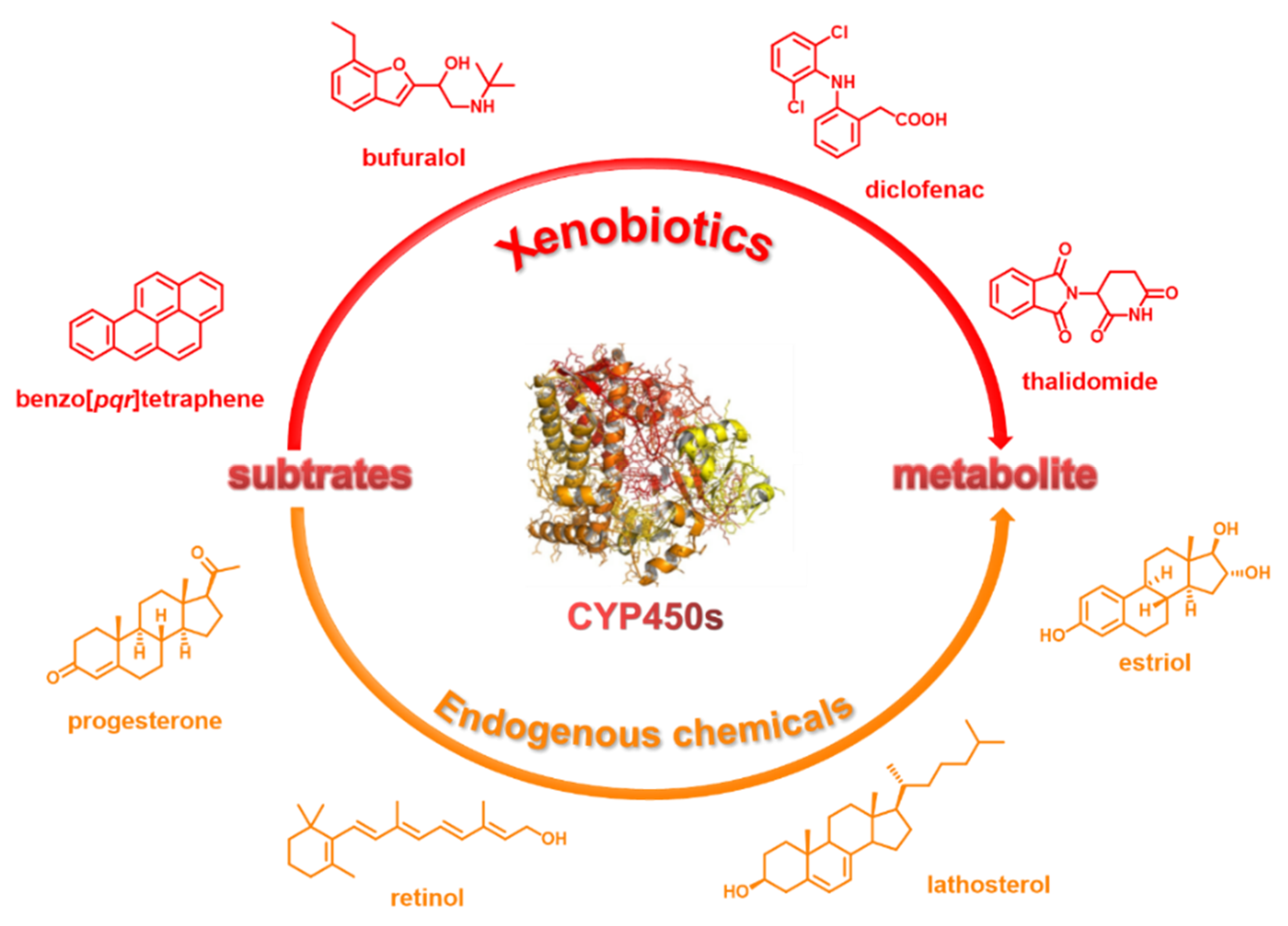
Figure 1. CYP450s are involved in the metabolism of xenobiotics and endogenous chemicals.
Chemosensors 10 00304 g001
Figure 2. CYP450 nomenclature.
Chemosensors 10 00304 g002
Figure 3. (A) Major CYP450 isoforms involved in drug metabolism. (B) Phase I metabolism by CYP450s. (C) The relationship between drugs and CYP450s.
Chemosensors 10 00304 g003
Figure 4. (A) The generalized catalytic cycle of CYP450. (B) CYP450-catalyzed various reactions.
Chemosensors 10 00304 g004
Figure 5. General design strategies for CYP450-catalyzed probes.
Chemosensors 10 00304 g005
Figure 6. (A) Structure of probe 1 (NCMN) and its fluorescence response toward CYP1A. (B) Docking simulation of NCMN into CYP1A2 (a) and CYP1A1 (b). (C) Two-photon confocal fluorescence images of HepG2 cells. (D) TPM images of a fresh rat liver slice stained with NCMNe (A methylester derivative of NCMN) at depth of ≈100 μm. Adapted with permission from Ref. [49]. Copyright 2015 American Chemical Society.
Chemosensors 10 00304 g006
Figure 7. (A) Proposed response mechanism of probe 4 (iPrBN) for sensing CYP1A1. (B) Two-photon confocal fluorescence images of the CYP1A1 in tumor tissue slices. (C) Fluorescence response of iPrBN at 525 nm upon the addition of various CYP450 isoforms in humans. (D) Two-photon confocal fluorescence images of CYP1A1 with iPrBN in living zebrafish. Adapted with permission from Ref. [56]. Copyright © 2018, copyright Royal Society of Chemistry.
Chemosensors 10 00304 g007
Figure 8. (A) Design of CYP1B1 targeted NIR probes from ANF derivatives. (B) Putative binding mode of compound probes 10 (6b). (C) In vitro cell binding studies on HCT-15 colon cancer cells of NIR probe 10 (6b). (D) In vivo NIR imaging with probe 10 (6b) for HCT-15 tumor-bearing mice and quantitative comparison of tumor uptake for imaging group and blocking group. Adapted, with permission from Ref. [67]. Copyright © 2018, American Chemical Society.
Chemosensors 10 00304 g008
Figure 9. (A) Proposed mechanism of CYP2J2 triggering the fluorescence response of probe 11 (BnXPI). (B) Fluorescence response of probe 11 at 718 nm (λex = 656 nm) upon the addition of various isoforms of human CYP450s. (C) In vivo NIR fluorescence imaging of CYP2J2 in tumor-bearing nude mice after tumor injection. The grown tumor tissue was taken from the mouse. Adapted, with permission, from Ref. [70]. Copyright © 2019, American Chemical Society.
Chemosensors 10 00304 g009
Figure 10. The double-filtering strategy consists of CYP450s activity-based screening (A) and reverses protein-ligand docking (B) to discover CYP450 isoenzyme-specific NIR fluorescent probes. Adapted, with permission, from Ref. [72]. Copyright © 2022 Elsevier Ltd.
Chemosensors 10 00304 g010
Figure 11. (A) Fluorescence response of probe 13 (MXMB). (B) Fluorescence bioimaging of CYP2J2 in vitro and in vivo. Adapted, with permission, from Ref. [72]. Copyright © 2022 Elsevier Ltd.
Chemosensors 10 00304 g011
Figure 12. (A) Fluorescence response of probe 14 (DDAM). (B) Inhibitory effects of various clinical drugs toward human CYP2C9. Adapted, with permission, from Ref. [72]. Copyright © 2022 Elsevier Ltd.
Chemosensors 10 00304 g012
Figure 13. (A) The response mechanism of probe 15 (McMe) and BnMcMe against CYP2D6. (B) Fluorescence spectra and intensity changes of probe 15 with various CYP450 enzymes in PBS buffer (pH 7.4) at 37 °C. (C) Confocal fluorescence images of HepG2 cells. (D) In vivo fluorescence imaging of probe 15 in tumor-bearing mice after injection probe. Adapted, with permission, from Ref. [78]. Copyright © 2021 Elsevier Ltd.
Chemosensors 10 00304 g013
Figure 14. Two-dimensional strategy for developing a two-photon fluorescent probe 16 (NEN) for CYP3A4. Adapted, with permission, from Ref. [87]. Copyright © 2019, John Wiley and Sons.
Chemosensors 10 00304 g014
Figure 15. (A) The proposed reaction mechanism of Hcy-Br (probe 17) to CYP3A5. Confocal fluorescence images of HepG2 (B) and A549 cells (C). (D) In vivo NIR fluorescence imaging of CYP3A5 in tumor-bearing nude mice after tumor injection. Adapted, with permission, from Ref. [91]. Copyright © 2021 Elsevier Ltd.
Chemosensors 10 00304 g015
Figure 16. (A) Structure optimization of BN fluorophore according to its key interactions with CYP3A. (B) High-throughput and visual characterization of mechanism-based CYP3A inactivation of herbal medicines. (C) MBI evaluation of Lim (limonin). (D) Two-photon fluorescence images of CYP3A activity in a living specimen using probe 18 (BN-1). Adapted, with permission, from Ref. [95] Copyright © 2021, John Wiley and Sons.
Chemosensors 10 00304 g016
Table 1. Key members of CYP1, 2, and 3 families.
FamilySubfamilyGene Identifier
CYP1CYP1ACYP1A1
CYP1A2
CYP2CYP2ACYP2A1
CYP2BCYP2B1, CYP2B6
CYP2CCYP2C8, CYP2C9, CYP2C18, CYP2C19
CYP2DCYP2D6
CYP2ECYP2E1
CYP2JCYP2J2
CYP3CYP3ACYP3A4, CYP3A5, CYP3A7
Table 2. Summary of CYP1 family-specific probes and their application for biomarkers.
Bio-MarkerNameStructure aLOD/nMKm/μMEx/Em b
nm/nm
Biological Application
CYP1ANCMN
Probe 1
[49]
Chemosensors 10 00304 i0015011.9450/564TPM c imaging identifies CYP1A induced by different concentrations of inducer and 3D Depth Imaging of CYP1A in rat liver tissues
NEMN
Probe 2
[50]
Chemosensors 10 00304 i0028.10/
15.75
/446/552Fluorescence imaging of endogenous CYP1A in living cells
CYP1A1NBCeN
Probe 3
[53]
Chemosensors 10 00304 i0032.50.84450/562Fluorescence imaging of endogenous CYP1A1 in living cells, TPM c images of a fresh rat liver slice
iPrBN
Probe 4
[56]
Chemosensors 10 00304 i0040.0360.29470/525TPM c imaging of CYP1A1 activities in living human cancer cells, tumor tissue slices, zebrafish
DPCl
Probe 5
[57]
Chemosensors 10 00304 i0050.026/555/673Fluorescence imaging of endogenous CYP1A1 in living cells, zebrafish
NEiPN
Probe 6
[58]
Chemosensors 10 00304 i0060.04874/374/446Fluorescence imaging of endogenous CYP1A1 in living cells, zebrafish
CHPO
Probe 7
[59]
Chemosensors 10 00304 i0070.0731.2540/590Fluorescence imaging of endogenous CYP1A1 in living cells
BCy-CYP
Probe 8
[60]
Chemosensors 10 00304 i0082.7/500/625Fluorescence imaging of endogenous CYP1A1 in living cells, effects of synergistic therapy of carnosol and cisplatin in the MCF-7 tumor-bearing mice
CYP1A2HBMN
Probe 9
[65]
Chemosensors 10 00304 i0090.05 pg/mL−16.01370/460Fluorescence imaging identifies CYP1A1 induced by different concentrations of TCDD d in living cells
CYP1B16b e
Probe 10
[67]
Chemosensors 10 00304 i010//675/720Fluorescence imaging CYP1A1 in HCT-15 colon cancer cells, in vivo and in vitro NIR imaging for HCT-15 tumor-bearing mice
a The red marked part indicates the dealkylation group; b The emission and excitation wavelengths are measured in the simulated physiological environment (100 mM PBS buffer, pH 7.4, NADPH reduction system, 37 °C); c TPM: Two-photon microscopy; d TCDD: 2,3,7,8-tetrachlorodibenzo-p-dioxin (a major threat carcinogen); e Probe 10 (6b) was designed inspired by enzyme-inhibitor binding strategies.
Table 3. Summary of CYP2 family-specific probes and their application for the biomarkers.
Bio-MarkerNameStructure aLOD/nMKm/μMEx/Em b
nm/nm
Biological Application
CYP2J2BnXPI
Probe 11
[70]
Chemosensors 10 00304 i0110.024 mg/mL4.2656/718Bioimaging and graphical quantification of average fluorescence intensity of CYP2J2 activities in living human cancer cells, bioimaging of angiogenesis in vivo and ex vivo, in vivo NIR fluorescence imaging of CYP2J2 in tumor-bearing nude mice
DPBM
Probe 12
[71]
Chemosensors 10 00304 i0120.09 nM/555/673Fluorescence imaging of endogenous CYP2J2 in living cells, BALB/c nude mice
MXMB
Probe 13
[72]
Chemosensors 10 00304 i013/0.51670/724Fluorescence bioimaging of CYP2J2 in vitro and in vivo, 2D and 3D confocal fluorescence imaging of tube formation
CYP2C9DDAM
Probe 14
[72]
Chemosensors 10 00304 i014/3.8600/658Fluorescence imaging of endogenous CYP2C9 in living cells, inhibitory effects of various clinical drugs toward human CYP2C9
CYP2D6McMe
Probe 15
[78]
Chemosensors 10 00304 i0150.052 nM/400/555Fluorescence imaging of endogenous CYP2D6 in living cells, tumor-bearing mice
a The red marked part indicates the dealkylation group; b The emission and excitation wavelengths are measured in the simulated physiological environment (100 mM PBS buffer, pH 7.4, NADPH reduction system, 37 °C).
Table 4. Summary of CYP3 family-specific probes and their application for the biomarkers.
Bio-MarkerNameStructure aLOD/nMKm/μMEx/Em b
nm/nm
Biological Application
CYP3A4NEN
Probe 16
[87]
Chemosensors 10 00304 i016/59.8450/558TPM c fluorescence imaging of CYP3A4 in human primary hepatocytes, Fluorescence imaging of CYP3A4 in living zebrafish
CYP3A5Hcy-Br
Probe 17
[91]
Chemosensors 10 00304 i0170.04903 nM/600/690Fluorescence imaging of endogenous CYP1A1 in living cells, in vivo NIR fluorescence imaging of CYP3A5 in tumor-bearing nude mice
CYP3A4/
CYP3A5
BN-1
Probe 18
[95]
Chemosensors 10 00304 i018/3.5/10.1470/526TPM c imaging of CYP3A activity in the living specimen, High-throughput and visual characterization of mechanism-based CYP3A inactivation of herbal medicines
a The red marked part indicates the dealkylation group, the red arrow indicates the major site of hydroxylation by the target CYP450(s); b The emission and excitation wavelengths are measured in the simulated physiological environment (100 mM PBS buffer, pH 7.4, NADPH reduction system, 37 °C); red arrow indicates the major site of hydroxylation by the target CYP450(s). c TPM: Two-photon microscopy.
Publisher’s Note: MDPI stays neutral with regard to jurisdictional claims in published maps and institutional affiliations.