Article

An Electrochemical Sensor Based on Reduced Graphene Oxide and Copper Nanoparticles for Monitoring Estriol Levels in Water Samples after Bioremediation

by
Francisco Contini Barreto
,
Martin Kassio Leme Silva
and
Ivana Cesarino
*
Department of Bioprocess and Biotechnology, School of Agriculture, São Paulo State University, Botucatu 18610-034, SP, Brazil
*
Author to whom correspondence should be addressed.
Chemosensors 2022, 10(10), 395; https://doi.org/10.3390/chemosensors10100395
Submission received: 7 September 2022 / Revised: 20 September 2022 / Accepted: 26 September 2022 / Published: 28 September 2022
(This article belongs to the Collection Sustainable Metal Oxide Materials for Sensing Applications)

Abstract

:
Water contamination from endocrine disruptors has become a major problem for health issues. Estriol is a hormone often detected in several aquatic matrices, due to the inefficient removal of such compounds through conventional water treatment methods. Therefore, there is a continuous need to develop new, efficient, and low-cost treatment methods for this hormone removal, as well as analytical devices able to detect estriol at low concentrations. In this present study, we report the use of the Eichhornia crassipes (water hyacinth) as a phytoremediation agent for estriol removal from aqueous matrices, in addition to a newly developed electrochemical sensor based on reduced graphene oxide and copper nanoparticles as a quantification and monitoring tool of the hormone. The developed sensor presented a linear detection region from 0.5 to 3.0 μmol L−1, with detection and quantification limits of 0.17 μmol L−1 and 0.56 μmol L−1, respectively. Phytoremediation experiments were conducted in 2 L beakers and the reducing levels of the hormone were studied. Water hyacinth was able to reduce contaminant levels by approximately 80.5% in 7 days and below detection limits in less than 9 days, which is a good alternative for water decontamination with this endocrine disruptor. Due to the hydrophobicity of estriol, the probable mechanism involved in the bioremediation process is rhizodegradation, and the decrease in pH in the beakers that contained the plants indicated a possible formation of biofilms on the roots.

1. Introduction

Currently, one of the biggest problems faced by government agencies is aquatic environments being contaminated with endocrine disruptors, which are capable of causing, even at low levels, disturbances in the endocrine system of animals and humans [1]. Among these disruptors is estriol, a steroid estrogenic hormone that has a relevant impact on sexual and reproductive functions, some organs and bone structure [2], and in aquatic organisms; it is also capable of generating feminization, cancer, and developing degenerations [3]. It can be found in mammals during pregnancy and is of great importance to women as it is related to menstruation and the reproductive cycle. It is also present in drugs used to treat menopausal urogenital diseases and in contraceptive pills, with urine being the main form of excretion. Due to this fact, estriol will be released into water sources and will cause the problems mentioned above, and this hormone persists even after water treatment [2,4]. When in the body, estriol is conjugated with other molecules in the liver, however, when excreted, it is rapidly hydrolyzed and restored [5]. Therefore, monitoring estriol in the aquatic environment is a priority for governments and regulatory agencies.
For the treatment of water with the most diverse contaminants, bioremediation has emerged as a promising technique due to its efficiency, lower cost, and lower environmental impact. It is based on the activity of a broad spectrum of fungi, bacteria, plants, and derivatives of these organisms, such as enzymes [6,7,8,9]. Among these techniques, phytoremediation, which consists of the use of plants and their associated microorganisms [10], has been widely used due to its low cost and high benefit that can be provided, being able to generate ecological sustainability, not requiring great strength of work, being more efficient than conventional methods, promoting biodiversity, and even improving air quality [11]. For organic contaminants, the main mechanisms involved in phytoremediation are phytostabilization, phytoextraction, rhizodegradation, rhizofiltration, phytodegradation, and phytovolatilization [12].
The Eichhornia crassipes plant is very common around the world and is often associated with being an invasive species due to its high speeds of reproduction and growth [13,14]. However, it has great potential as a phytoremediation agent due to its extensive roots (greater surface area in contact with contaminants), large biomass production, lower secondary waste generation, smaller carbon footprint, low energy requirement, and accelerated growth [14]. Water contaminated with the most diverse drugs, such as carbamazepine, ibuprofen, sulfadiazine, sulfamethoxazole, sulfamethazine, triclosan [15], naproxen, and diclofenac [16] have already been decontaminated by this species, indicating the potential for application and performance of this plant for the current problem of water resources contaminated with medicines.
Different methodologies to quantify the hormone estriol have been proposed, among which are several techniques of chromatography, immunoassays, electrophoresis, and an electroanalytical method [2]. Electrochemical sensors have been extensively researched and used for their simplicity of operation, low cost, fast response, high sensitivity, the potential to be miniaturized, automation, use of modified electrodes (improve sensitivity and lower detection and quantification limits), and the possibility of being developed and used in situ [1,2]. To increase sensitivity and selectivity, electrochemical sensors have been modified [5]. Metallic nanoparticles such as copper, antimony, silver, palladium, and gold are widely used as modifiers, largely due to their good conductivity, high surface area, and high chemical stability [4,17]. To quantify estriol, some studies used reduced graphene oxide modified with antimony nanoparticles and also modified with silver nanoparticles, and the performance was characterized by cyclic and differential pulse voltammetry, obtaining excellent results in the analyses [2,4].
Therefore, we aim in this work to decrease and monitor estriol hormone levels, using bioremediation and specific electroanalytical techniques, respectively. The Eicchornia crassipes plant and its associated microorganisms are used for the phytoremediation process, whereas glassy carbon electrodes modified with reduced graphene oxide and copper nanoparticles were produced for the electroanalytical analysis.

2. Materials and Methods

2.1. Instrumentation

A Metrohm potentiostat (PGSTAT-128N Autolab Electrochemical System, Utrecht, Netherlands) equipped with NOVA 2.1 software (Metrohm, Utrecht, Netherlands) was used for the cyclic voltammetry (CV) and differential pulse voltammetry (DPV) experiments. A conventional three-electrode glass electrochemical cell was used for the experiments. The working electrode was a glassy carbon modified with graphene oxide and copper nanoparticles (GC/rGO-CuNPs). The reference electrode was Ag/AgCl/KCl (3.0 mol L−1) and the auxiliary electrode was a platinum plate.
Morphologic characterization of the nanocomposites was performed by scanning electron microscopy (SEM) using a high-resolution field emission electron microscope (FEG-SEM) JEOL, model JSM-7500F at IQ-UNESP Araraquara, Brazil. For the energy-dispersive X-ray spectroscopy (EDS) measurements, the acceleration voltage was 10 kV, the e-beam current 3.2 nA, the time constant was 6400, and the magnification was set to x, using the Ultradry detector (Thermo Fisher Scientific, Waltham, MA, USA).

2.2. Solutions and Reagents

All solutions prepared in the electrochemical experiments used purified water in a Millipore Milli-Q system (resistivity ≥ 18 MΩcm−1). All reagents used were not previously purified and were of analytical grade. Graphene oxide, CuCl2, and estriol (standard) were purchased from Sigma-Aldrich® (Darmstadt, Germany). The estriol used for the phytoremediation study was manipulated in a local pharmacy in the city of Avaré, São Paulo, Brazil.

2.3. Synthesis of Reduced Graphene Oxide with Copper Nanoparticles (rGO-CuNPs)

The rGO-CuNPs synthesis was prepared by a procedure described by Fernandes et al. [17]. First, a suspension of GO (4.0 mg/mL) and sodium dodecyl sulfate (SDS) was prepared, in the proportion of 10:4, using ethanol as a medium. Then, the suspension was placed in the ultrasonic bath for 20 min. Sodium borohydride (NaBH4) was added in excess (16 mg), and again the mixture was taken to the ultrasonic homogenizer, this time for one hour. Then, copper chloride (CuCl2) is added at 30% (m/m) in relation to the mass of graphene oxide, and then it is diluted with ethanol. The CuCl2 solution is added under constant stirring at one drop per second to incorporate the nanoparticles into the graphene sheet. After finishing this step, the solution was sonicated for 20 min and then the material obtained was centrifuged and cleaned with ethanol. Prior to the modification of the electrodes, the material was sonicated for 5 min to guarantee the homogeneity of the film used in it.

2.4. Electrode Preparation

The glassy carbon electrodes had their surfaces polished by polystyrene with silicon carbide sandpaper and a 0.5 μm aqueous alumina suspension until a mirrored surface was obtained. After this step, the electrodes were sonicated in ethanol for 5 min and in water for the same time. After being polished, cleaned, and dried, the electrodes received 10 μL aliquot of a suspension of rGO-CuNPs at the optimized concentration. Then, when dried at room temperature, the electrodes were taken to the electrochemical procedures.

2.5. Obtaining the Plants and Setup of the Phytoremediation Experiment

The Eichhornia crassipes plants were obtained from the Tiete River dam in Botucatu, in the Rio Bonito condominium (22°57′24.9” S 48°24′09.1” W) (Figure 1A–C). After removing the plants from the water, they were placed in 50 L polypropylene boxes and transported to the greenhouse located at the School of Agriculture, UNESP Botucatu. The place is surrounded by glass, ensuring natural light and protecting the plants from rain. The plants were acclimatized in these same boxes, filled with tap water, and renewed every two days for ten days.
Three plants were selected according to size and weight criteria and were placed in 2 L beakers, with 1.8 L of estriol solution, at a concentration of 3.0 × 10⁻⁵ μmol L−1. The concentrations tested were based on the electrochemical experiments performed and whose results can be found in Section 3, considering the sample dilution in the electrolytic cell. Therefore, the expected concentration for the experiment is 1.0 μmol L−1. A beaker containing only the estriol solution was used as a control, and the experiment had a duration of 14 days. Samples were collected on the 4th, 7th, 9th, 12th, and 14th days of the experiment. The pH and temperature values were evaluated on the same days that the samples were collected.

2.6. Determination of Estriol in Real Samples

To determine the estriol concentration, 670 μL of the collected sample was added to the electrolytic cell with 20 mL of PBS 0.1 mol L−1 pH 7.0, and the concentration was evaluated by standard addition method (Figure 1D).

2.7. Statistical Analysis

R software version 3.6.3 (R core team, Vienna, Austria) [18] was used for the statistical analyses. Normality tests were performed using the Shapiro–Wilk test, and significance was observed using the t-test.

3. Results

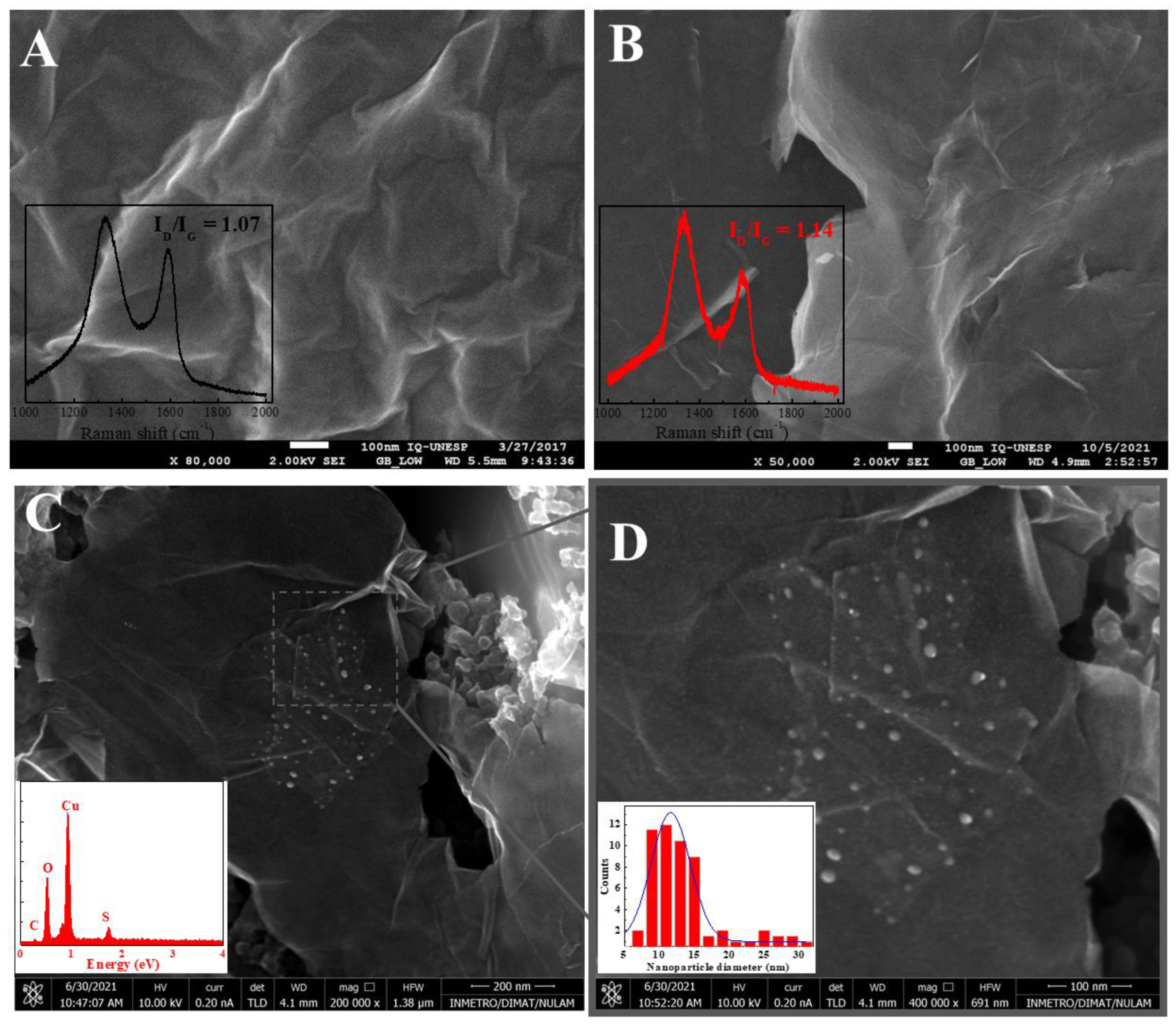
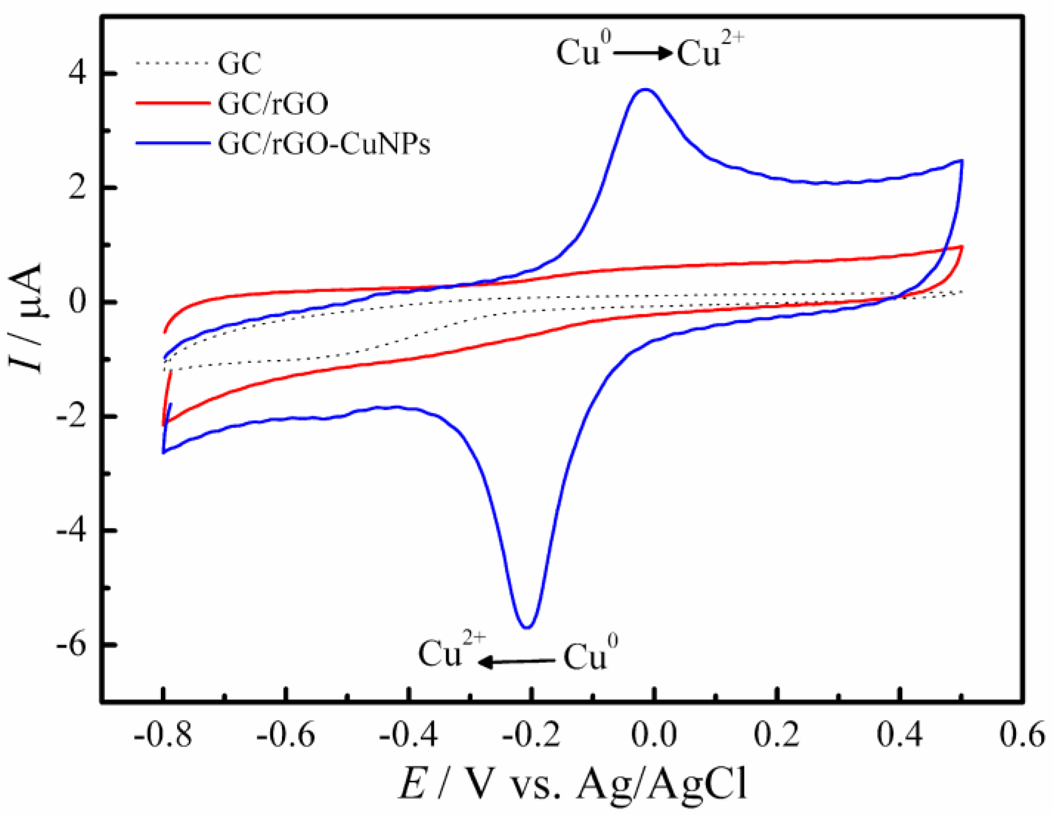
3.1. Morphological and Electrochemical Characterization of Materials

Scanning Electron Microscopy (SEM) characterized the nanomaterials in order to analyze the morphological changes on their surfaces. Figure 2A refers to commercial GO prior to chemical reduction. Figure 2B shows the rGO material, and it is possible to visualize a twisted, wavy, and wrinkled structure. The action of reducing graphene oxide causes sheets of carbon to be exposed, creating active sites [19]. Raman spectroscopy is a commonly used technique to characterize graphene-based materials’ chemical and physical properties, along with disordered and defective structures, defect density, and doping levels [20]. The reduction in GO leads to the unstacking of sheets and, subsequently, the unblocking of active sites. The insets of Figure 2A,B shows Raman spectra of GO and rGO, respectively. The reduction in GO can be characterized by analyzing the ratio between the G band (IG) and the D band (ID). The commercial GO showed a ratio of 1.07; after the chemical reduction, the ratio was 1.14. These findings and changes in the Raman spectra are attributed to a higher size of sp2 domains and edge associated with the chemical reduction in graphene oxide [21,22].
Figure 2C shows the rGO-CuNPs compound; it is possible to observe that the copper nanoparticles on the surface of the rGO sheet are non-uniformly distributed. The EDS spectra of the material were also obtained, as shown in Figure 2C inset, confirming the NPs composition. Figure 2D shows the rGO-CuNPs material at a higher magnitude, with nanoparticles ranging from 5–30 nm in length, evidencing the modification of the material.
The electrochemical characterization (Figure 3) of the GC/rGO-CuNPs composite was carried out in 0.1 mol L−1 PBS pH 7.0, with a scanning speed of 50 mVs−1, varying the potential from −0.8 to +0.5 V. No electrochemical process was observed for the GC and GC/rGO electrodes. On the other hand, in the cyclic voltammogram for the GC/rGO CuNPs electrode, there is an oxidation-reduction behavior with well-defined peaks in the electrode modified with copper nanoparticles, which refers to oxidation (Cu0 to Cu2+) and its reduction, confirming its incorporation in the electrode. The observed peaks are in agreement with previously published works [17,23].

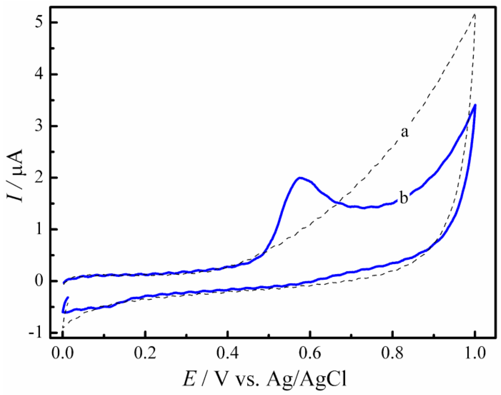
3.2. Electrochemical Behavior of GC/rGO-CuNPs Composite Electrode on the Estriol Oxidation Process

The electrochemical oxidation of estriol on the electrodes was carried out in a 0.1 mol L−1 PBS pH 7.0 solution by cyclic voltammetry, with a sweep speed of 50 mV s−1. The results are shown in Figure 4. No electrochemical process was observed in the cyclic voltammetry in the absence of the studied hormone (dotted line). However, with the addition of 10 μmol L−1 of estriol, an anodic peak at +0.54 V vs. Ag/AgCl/KCl refers to irreversible oxidation of the hormone at the phenolic hydroxyl group. The results are in accordance with the work reported by Cesarino et al. [2].

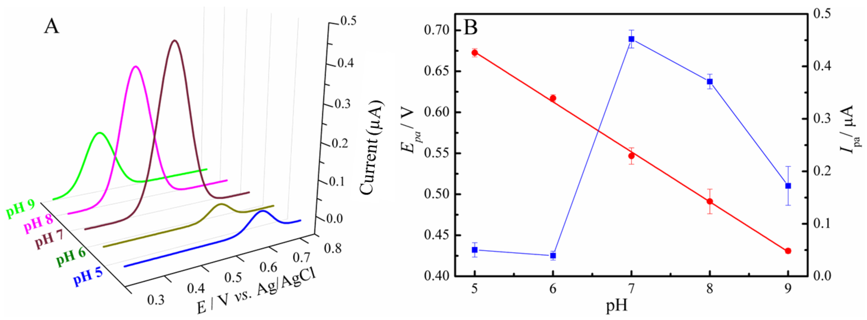
3.3. Optimization of Parameters

Experimental conditions were optimized according to Table 1. It has been reported in the literature that different concentrations of composite for the modification of the electrode interfere with the sensitivity of the evaluation of samples, and large amounts of the composite can make the analysis difficult by blocking binding sites. Therefore, the concentration of the rGO-CuNPs was evaluated by DPV, in PBS buffer 0.1 mol L−1 pH 7.0, with a sweep speed of 10 mV s−1, the amplitude of 25 mV, and a potential range from +0.4 to +0.8 V, with the addition of 10 μmol L−1 of estriol. As seen in Table 1, the best concentration for electrode modification was 0.02 mg/mL of rGO-CuNPs.
Estriol and/or its oxidation product have a tendency to be adsorbed on the electrode surface during voltammetry. Therefore, a pre-concentration study was carried out in order to determine the best potential to be applied, and then the estriol molecules can concentrate closer to the electrodes, promoting a greater analytical signal for their detection. The studied potentials were maintained for 30 s immediately before the start of the DPV and had the following values: −1.2 V; −1.1 V; −1.0 V; −0.9 V; −0.8 V; −0.7 V; −0.6 V, and −0.5 V. The potential that showed a higher sensitivity in the analysis was −1.1 V.
In order to verify the oxidation mechanisms of the molecule in question on the GC/rGO-CuNPs electrode, the dependence of the electrochemical oxidation of estriol at different pHs was studied using differential pulse voltammetry in PBS with pH ranging from 5.0 to 9.0, an amplitude of 25 mV, a sweep speed of 10 mV s−1, and a potential range from +0.3 to +0.8 V, as shown in Figure 5A. The highest peak obtained was at pH 7.0, so at this pH, we have greater sensitivity in the analyses, which is great for monitoring estriol in water. The plot Epa vs. pH showed a linear relationship, indicating a slope of −61 mV per pH unit, which indicates that the oxidation mechanism of estriol involves the same number of protons and electrons, as shown in Figure 5B. This mechanism was fully evaluated by previous works of our group, including molecular dynamics and computer simulations [2,4].

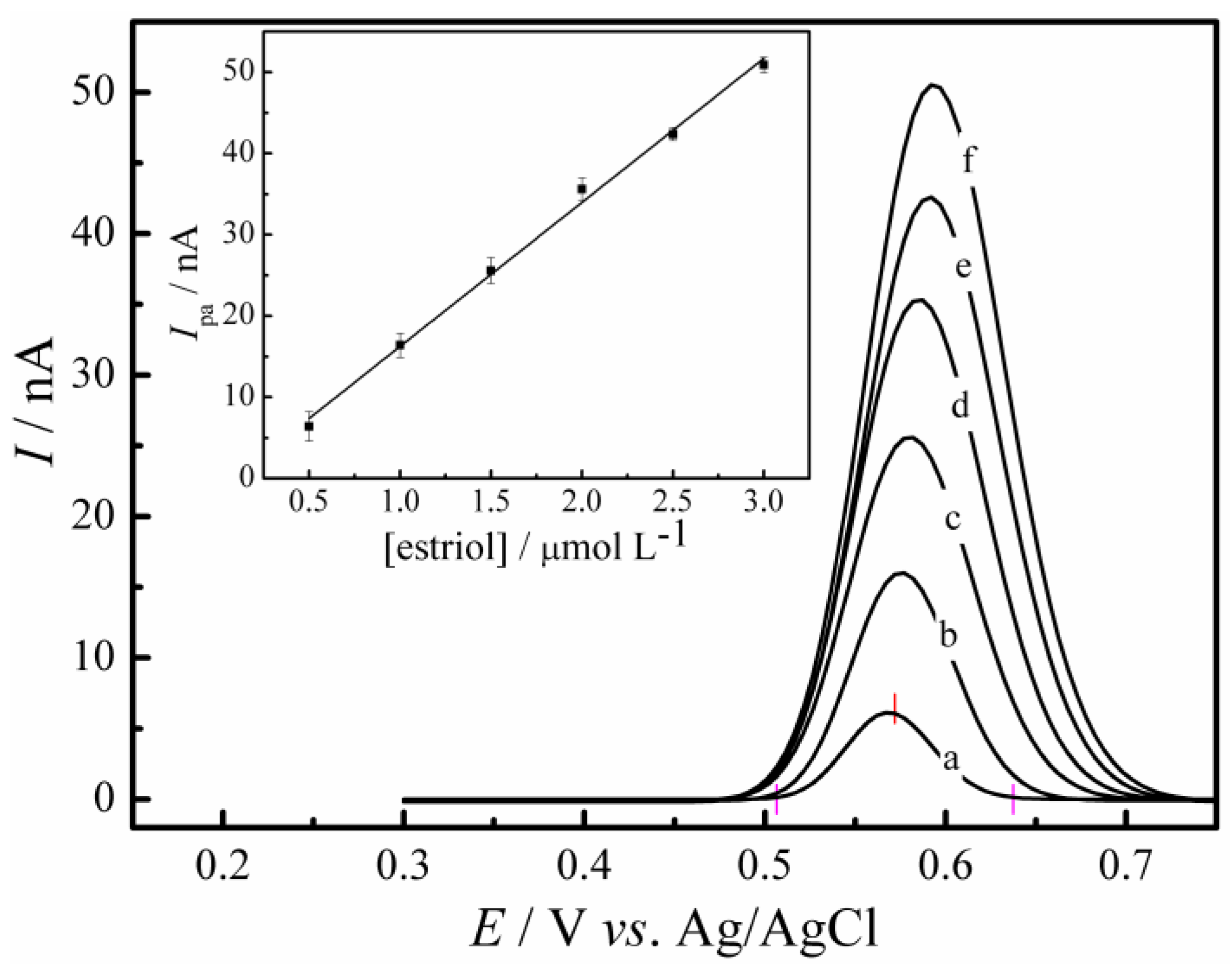
3.4. Analytical Characteristics

In order to verify the linearity intervals, an analytical curve was obtained. For this, the concentrations of estriol varied from 0.5 to 3.0 μmol L−1. Differential pulse voltammetry was used, with 25 mV amplitude, and 10 mV s−1 scanning speed, varying the potential from +0.3 to +0.8 V and using 0.1 mol L−1 PBS pH 7.0. The conditions were optimized according to Table 1. Then, the anodic peak current was plotted with the respective estriol concentrations. As can be seen in Figure 6, the technique showed a linear range between concentrations of 0.5 to 3.0 μmol L−1 of the analyte with the following equation:
Ipa (nA) = 1.49 (nA) + 17.74 (nA/μmol L−1) × Cestriol (μmol L−1)
The equation presented a coefficient of determination R2 = 0.996 (for n = 6 concentrations) and limits of detection and quantification of 0.17 μmol L−1 and 0.56 μmol L−1, respectively. These were calculated using a 3σ/slope ratio and 10σ/slope ratio, respectively, where σ is the standard deviation of the mean value for 10 voltammograms of the blank.
Table 2 shows a comparison of different electrodes reported in the literature and their respective linear ranges, detection limit, quantification limit, and target matrix sample for estriol detection. It is worth mentioning that several works in the literature mainly report the use of graphene materials/metallic nanoparticles for the detection of this hormone. However, most of them have applications aimed at detecting estriol in clinical samples (real or synthetic), samples from natural waters, and the evaluation of commercially available drugs. The article refers to the use of a sensor based on GC/rGO-CuNPs to monitor a bioremediation process of estriol by water hyacinth. Thus, the analyzed environmental matrix presents a great challenge for analytical chemistry due to the great variety of ions, molecules, and particles present throughout the process. Although some works in the literature report lower LODs by using materials such as rGO-AgNPs [4] and rGO-SbNPs [2] as the electrode modifier, CuNPs are relatively cheaper and less toxic than conventional nanoparticles. Note that 2D carbon-based materials, such as graphene, tend to have a higher performance in comparison to other materials. In addition, this tool proved to be quite versatile and expands the possibilities of application in the area of environmental monitoring with low-cost devices, and is easy to manufacture and apply.
Aiming to perform a repeatability test, the electrochemical measurements with estriol were triplicated with three different electrodes, given a repeatability value of 2.45%.

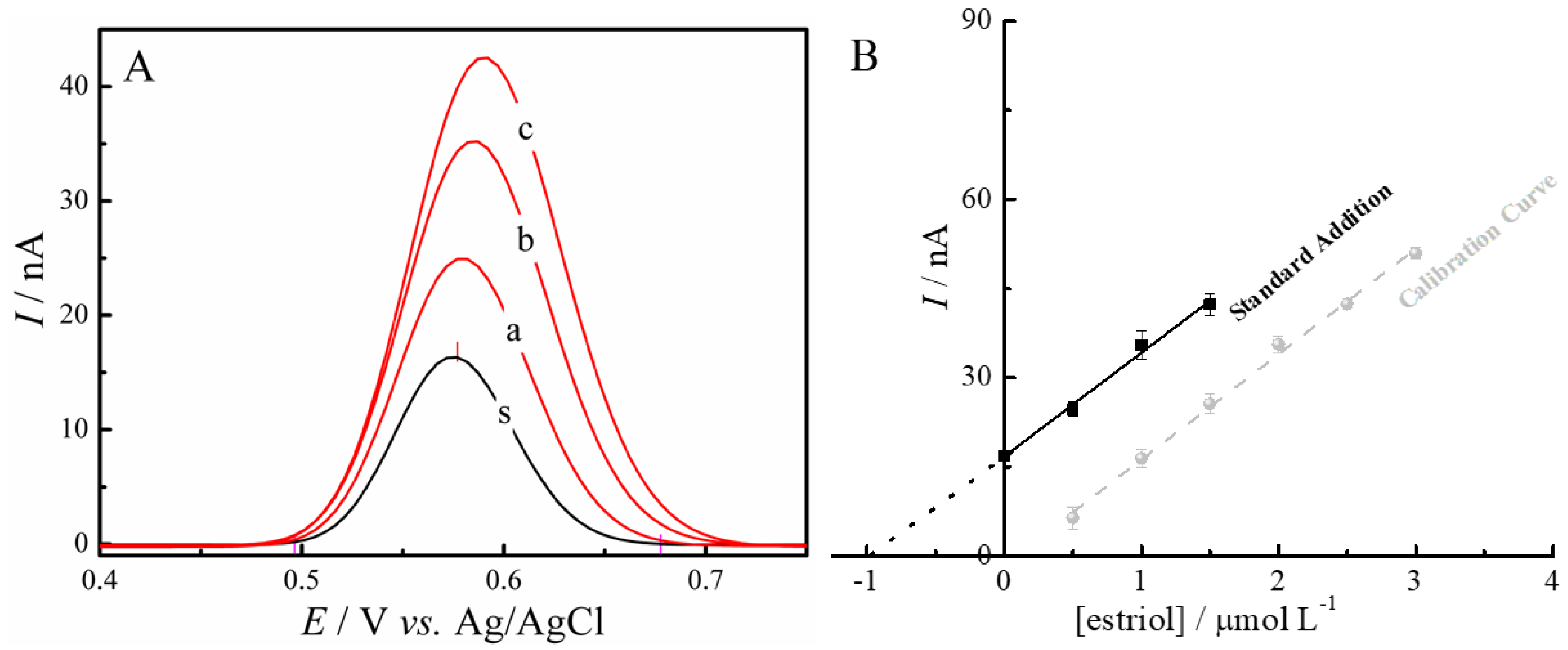
3.5. Determination of Estriol in Real Samples and Evaluation of the Efficiency of the Bioremediation Process

Figure 7A shows the DPV experiments carried out for the detection of estriol in the water samples during the phytoremediation experiment. Figure 7B shows the plot obtained of the concentration of estriol vs. the current response by the standard addition method. It is worth noticing that the water matrix did not influence the voltammetric response of estriol, which indicates that this approach is suitable for the monitoring of the hormone [26]. The standard addition method was able to quantify the amount of estriol in each experiment for 14 days of phytoremediation.
On the first day of sample collection (4th day of exposure of the plants to the hormone), there was an average reduction of approximately 52% in relation to the control. On the second day of collection (7th day of exposure), the reduction in relation to the control reached an average value close to 80%, and in beaker 1 this reduction was approximately 87%, the highest rate analyzed in the experiment. However, on the 9th day of exposure (3rd day of collection), the values were below the limits of detection, thus making it not possible to verify the concentration of the endocrine disruptor in the beakers with the macrophytes, suggesting decontamination above 90%, considering the limits of the developed sensor. During the experiment, no morphological changes were observed in the plants due to any toxic effect that estriol could cause [27]. These results are summarized in Table 3.
Another factor to be considered is the characteristics of each plant, which may react more or less depending on its physiology and metabolism, in addition to the microbial communities present in the roots [28], which may vary according to the sediments present in the aquatic environment [29].
There are still few studies that evaluate the ability of plants to remove contaminants from the endocrine disrupting class, and other processes are usually used in conjunction with phytoremediation to increase the efficiency of the methodology [30,31,32,33,34]. As a result, the comparison of the efficiency of E. crassipes with other studies is difficult and inaccurate, and the same difficulty occurs regarding the decontamination of waters with the hormone estriol, and the hormone is often not evaluated individually [33,34]. However, it is still possible to demonstrate the quality of the water hyacinth to act in bioremediation processes. Phouthavong-Murphy et al. [34] evaluated the potential of Panicum virgatum to act in the remediation of bisphenol A, an endocrine disruptor, at a concentration of 20 mg L−1, in water, using 1 L containers. For the tested plant varieties, the efficiency in compost removal was between 40 and 46% after 3 months of study. Loffredo et al. also studied the decontamination of bisphenol A and obtained an efficiency of 86 and 94% after 7 days of the experiment using Cannabis sativa L. [35]. Decontamination of three estrogens (17β-estradiol, 17α-ethinylestradiol, and zeranol) was evaluated using hybrid poplar hydroponics by Bircher et al. [36]. After 10 days of the experiment, using 300 mL containers with 2 mg L−1 of the contaminants, a decontamination rate above 97% was obtained.

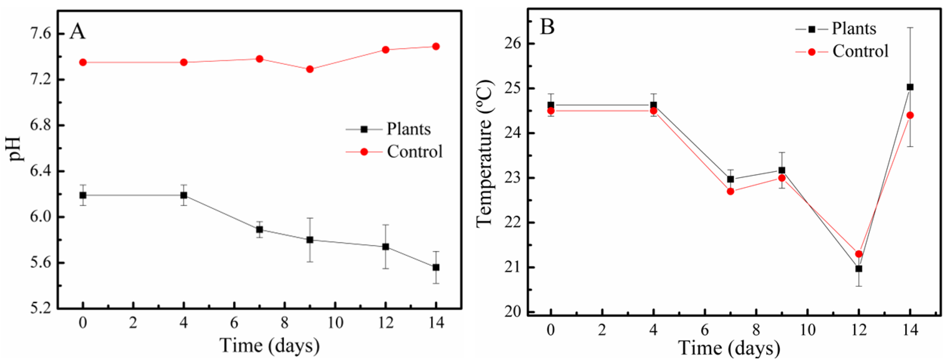
3.6. Physico-Chemical Parameters of the Bioremediation Process

The pH values of the solutions containing the plants were presented as moderately acidic, whereas the pH of the control remained close to neutral, as shown in Figure 8A. The occurrence of acidic pH in plant solutions can be explained by the release of several organic components and exudates from the roots, which can affect some biochemical processes [20]. Another justification is related to the development of biofilms in the roots, which act as a habitat for several bacteria and in the degradation of organic compounds, nitrification, and denitrification of nitrogen, and in the release of CO2 [28]. As for temperature, there were no significant differences between the control and the beaker samples with the plants, as shown in Figure 8B.

3.7. Morphology Assessment of Water Hyacinth

Table 4 shows the measurements of the plants immediately before and after being submitted to the phytoremediation experiment. The plants with the closest measurements were chosen, in order to try to standardize. All had healthy aspects and had no damage to their structures.
The root, of the evaluated parts of the plants, was the only one to show growth with a significant difference (p = 0.04752) in relation to the measurement immediately before the start of bioremediation. This fact can be related to the fact that this part in question is directly related to the absorption of nutrients and is the only one completely in contact with the estriol solution, reacting directly with the hormone. In a bioaccumulation experiment carried out by Pi et al., using E. crassipes and the endocrine disruptors 17β-estradiol, 17α-ethinylestradiol, estrone (E1), and bisphenol A, it was found that the highest concentration of these compounds is located in the roots of the macrophyte and that there is a tendency for endocrine disruptors, with the exception of more hydrophilic compounds, which are able to be transported to other parts of the plant, such as leaves [37]. However, this is not the case for estriol, which is naturally hydrophobic [38]. However, further studies are needed to understand the physicochemical properties and their relationship with the transport of these compounds to other parts of the plant [38].

4. Conclusions

As proposed, an electrochemical sensor based on reduced graphene oxide and copper nanoparticles was developed in order to detect the hormone estriol at low concentrations. As demonstrated, the sensor has a better response for the determination of estriol at pH 7.0, ideal for water analysis. Parameters were adjusted to obtain a lower detection limit. The estriol bioremediation experiment was conducted using the macrophyte E. crassipes, which showed good results for the decontamination of the hormone, taking the levels of the endocrine disruptor below the limits of detection in less than 9 days for the concentration studied. The decrease in the pH of the water in the experiments may indicate the formation of microbial biofilms on the roots, which may also have helped in the decontamination process. Therefore, E. crassipes shows up as a promising plant for use in larger-scale bioremediation devices, such as in floating constructed wetlands, in order to decontaminate water resources contaminated with this endocrine disruptor.

Author Contributions

Conceptualization, F.C.B. and I.C.; methodology, F.C.B.; software, F.C.B. and M.K.L.S.; validation, F.C.B.; formal analysis, F.C.B.; investigation, F.C.B. and I.C.; resources, I.C.; data curation, M.K.L.S.; writing—original draft preparation, F.C.B.; writing—review and editing, M.K.L.S. and I.C.; supervision, I.C.; project administration, I.C.; funding acquisition, I.C. All authors have read and agreed to the published version of the manuscript.

Funding

This research was funded by FAPESP, process 2019/25836-0.

Data Availability Statement

Data are available from the authors on reasonable request.

Acknowledgments

In memory of William de Melo Silva for his contribution to this article.

Conflicts of Interest

The authors declare no conflict of interest.

References

  1. Moreira, F.; Santana, E.R.; Spinelli, A. Ionic Liquid-Supported Magnetite Nanoparticles as Electrode Modifier Materials for Estrogens Sensing. Sci. Rep. 2020, 10, 1955. [Google Scholar] [CrossRef] [PubMed]
  2. Cesarino, I.; Cincotto, F.H.; Machado, S.A.S. A Synergistic Combination of Reduced Graphene Oxide and Antimony Nanoparticles for Estriol Hormone Detection. Sens. Actuators B Chem. 2015, 210, 453–459. [Google Scholar] [CrossRef]
  3. Manjunatha, J.G. Electroanalysis of Estriol Hormone Using Electrochemical Sensor. Sens. Bio-Sens. Res. 2017, 16, 79–84. [Google Scholar] [CrossRef]
  4. Donini, C.A.; da Silva, M.K.L.; Simões, R.P.; Cesarino, I. Reduced Graphene Oxide Modified with Silver Nanoparticles for the Electrochemical Detection of Estriol. J. Electroanal. Chem. 2018, 809, 67–73. [Google Scholar] [CrossRef]
  5. Gomes, E.S.; Leite, F.R.F.; Ferraz, B.R.L.; Mourão, H.A.J.L.; Malagutti, A.R. Voltammetric Sensor Based on Cobalt-Poly(Methionine)-Modified Glassy Carbon Electrode for Determination of Estriol Hormone in Pharmaceuticals and Urine. J. Pharm. Anal. 2019, 9, 347–357. [Google Scholar] [CrossRef] [PubMed]
  6. Sharma, R.; Malaviya, P. Constructed Wetlands for Textile Wastewater Remediation: A Review on Concept, Pollutant Removal Mechanisms, and Integrated Technologies for Efficiency Enhancement. Chemosphere 2022, 290, 133358. [Google Scholar] [CrossRef] [PubMed]
  7. Shah, A.; Shah, M. Characterisation and Bioremediation of Wastewater: A Review Exploring Bioremediation as a Sustainable Technique for Pharmaceutical Wastewater. Groundw. Sustain. Dev. 2020, 11, 100383. [Google Scholar] [CrossRef]
  8. Pinto, É.S.M.; Dorn, M.; Feltes, B.C. The Tale of a Versatile Enzyme: Alpha-Amylase Evolution, Structure, and Potential Biotechnological Applications for the Bioremediation of n-Alkanes. Chemosphere 2020, 250, 126202. [Google Scholar] [CrossRef] [PubMed]
  9. Sayara, T.; Sánchez, A. Bioremediation of PAH-Contaminated Soils: Process Enhancement through Composting/Compost. Appl. Sci. 2020, 10, 3684. [Google Scholar] [CrossRef]
  10. Agarry, S.E.; Oghenejoboh, K.M.; Aworanti, O.A.; Arinkoola, A.O. Biocorrosion Inhibition of Mild Steel in Crude Oil-Water Environment Using Extracts of Musa Paradisiaca Peels, Moringa Oleifera Leaves, and Carica Papaya Peels as Biocidal-Green Inhibitors: Kinetics and Adsorption Studies. Chem. Eng. Commun. 2019, 206, 98–124. [Google Scholar] [CrossRef]
  11. Kurniawan, S.B.; Abdullah, S.R.S.; Imron, M.F.; Said, N.S.M.; Ismail, N.‘I.; Hasan, H.A.; Othman, A.R.; Purwanti, I.F. Challenges and Opportunities of Biocoagulant/Bioflocculant Application for Drinking Water and Wastewater Treatment and Its Potential for Sludge Recovery. Int. J. Environ. Res. Public Health 2020, 17, 9312. [Google Scholar] [CrossRef]
  12. Madikizela, L.M. Removal of Organic Pollutants in Water Using Water Hyacinth (Eichhornia Crassipes). J. Environ. Manag. 2021, 295, 113153. [Google Scholar] [CrossRef] [PubMed]
  13. Correa dos Santos, N.M.; Monteiro, P.G.; Ferreira, E.A.; Alencar, B.T.B.; Cabral, C.M.; dos Santos, J.B. Use of Eichhornia Crassipes and Pistia Stratiotes for Environmental Services: Decontamination of Aquatic Environments with Atrazine Residues. Aquat. Bot. 2022, 176, 103470. [Google Scholar] [CrossRef]
  14. Mustafa, H.M.; Hayder, G. Recent Studies on Applications of Aquatic Weed Plants in Phytoremediation of Wastewater: A Review Article. Ain Shams Eng. J. 2021, 12, 355–365. [Google Scholar] [CrossRef]
  15. Lin, Y.L.; Li, B.K. Removal of Pharmaceuticals and Personal Care Products by Eichhornia Crassipe and Pistia Stratiotes. J. Taiwan Inst. Chem. Eng. 2016, 58, 318–323. [Google Scholar] [CrossRef]
  16. Amos Sibeko, P.; Naicker, D.; Mdluli, P.S.; Madikizela, L.M. Naproxen, Ibuprofen, and Diclofenac Residues in River Water, Sediments and Eichhornia Crassipes of Mbokodweni River in South Africa: An Initial Screening. Environ. Forensics 2019, 20, 129–138. [Google Scholar] [CrossRef]
  17. Fernandes, V.Q.; Silva, M.K.L.; Cesarino, I. Determination of Isotretinoin (13-Cis-Retinoic Acid) Using a Sensor Based on Reduced Graphene Oxide Modified with Copper Nanoparticles. J. Electroanal. Chem. 2020, 856, 113692. [Google Scholar] [CrossRef]
  18. R: A Language and Environment for Statistical Computing. Available online: https://www.gbif.org/pt/tool/81287/r-a-language-and-environment-for-statistical-computing (accessed on 2 September 2022).
  19. Pei, S.; Cheng, H.-M. The Reduction of Graphene Oxide. Carbon N. Y. 2012, 50, 3210–3228. [Google Scholar] [CrossRef]
  20. Wu, J.-B.; Lin, M.-L.; Cong, X.; Liu, H.-N.; Tan, P.-H. Raman Spectroscopy of Graphene-Based Materials and Its Applications in Related Devices. Chem. Soc. Rev. 2018, 47, 1822–1873. [Google Scholar] [CrossRef] [PubMed]
  21. Jabbar, A.; Yasin, G.; Khan, W.Q.; Anwar, M.Y.; Korai, R.M.; Nizam, M.N.; Muhyodin, G. Electrochemical Deposition of Nickel Graphene Composite Coatings Effect of Deposition Temperature on Its Surface Morphology and Corrosion Resistance. RSC Adv. 2017, 7, 31100–31109. [Google Scholar] [CrossRef] [Green Version]
  22. Xue, Y.; Zhu, L.; Chen, H.; Qu, J.; Dai, L. Multiscale Patterning of Graphene Oxide and Reduced Graphene Oxide for Flexible Supercapacitors. Carbon N. Y. 2015, 92, 305–310. [Google Scholar] [CrossRef]
  23. Setznagl, S.; Cesarino, I. Copper Nanoparticles and Reduced Graphene Oxide Modified a Glassy Carbon Electrode for the Determination of Glyphosate in Water Samples. Int. J. Environ. Anal. Chem. 2022, 102, 293–305. [Google Scholar] [CrossRef]
  24. da Silveira, J.P.; Piovesan, J.V.V.; Spinelli, A. Carbon Paste Electrode Modified with Ferrimagnetic Nanoparticles for Voltammetric Detection of the Hormone Estriol. Microchem. J. 2017, 133, 22–30. [Google Scholar] [CrossRef]
  25. Raymundo-Pereira, P.A.; Campos, A.M.; Vicentini, F.C.; Janegitz, B.C.; Mendonça, C.D.; Furini, L.N.; Boas, N.V.; Calegaro, M.L.; Constantino, C.J.L.; Machado, S.A.S.; et al. Sensitive Detection of Estriol Hormone in Creek Water Using a Sensor Platform Based on Carbon Black and Silver Nanoparticles. Talanta 2017, 174, 652–659. [Google Scholar] [CrossRef] [PubMed]
  26. Bader, M. A Systematic Approach to Standard Addition Methods in Instrumental Analysis. J. Chem. Educ. 1980, 57, 703–706. [Google Scholar] [CrossRef]
  27. Castillo Loría, K.; Emiliani, J.; Bergara, C.D.; Herrero, M.S.; Salvatierra, L.M.; Pérez, L.M. Effect of Daily Exposure to Pb-Contaminated Water on Salvinia Biloba Physiology and Phytoremediation Performance. Aquat. Toxicol. 2019, 210, 158–166. [Google Scholar] [CrossRef] [PubMed]
  28. Li, H.; He, W.; Qu, Y.; Li, C.; Tian, Y.; Feng, Y. Pilot-Scale Benthic Microbial Electrochemical System (BMES) for the Bioremediation of Polluted River Sediment. J. Power Sources 2017, 356, 430–437. [Google Scholar] [CrossRef]
  29. Dai, Y.N.; Dan, A.; Yang, Y.; Tam, N.F.Y.; Tai, Y.P.; Tang, X.Y. Factors Affecting Behavior of Phenolic Endocrine Disruptors, Estrone and Estradiol, in Constructed Wetlands for Domestic Sewage Treatment. Environ. Sci. Technol. 2016, 50, 11844–11852. [Google Scholar] [CrossRef]
  30. Franks, C.G.; Pearce, D.W.; Rood, S.B. A Prescription for Drug-Free Rivers: Uptake of Pharmaceuticals by a Widespread Streamside Willow. Environ. Manag. 2019, 63, 136–147. [Google Scholar] [CrossRef]
  31. Hakk, H.; Sikora, L.; Casey, F.X.M. Fate of Estrone in Laboratory-Scale Constructed Wetlands. Ecol. Eng. 2018, 111, 60–68. [Google Scholar] [CrossRef]
  32. Hsieh, C.Y.; Liaw, E.T.; Fan, K.M. Removal of Veterinary Antibiotics, Alkylphenolic Compounds, and Estrogens from the Wuluo Constructed Wetland in Southern Taiwan. J. Environ. Sci. Health—Part A Toxic/Hazard. Subst. Environ. Eng. 2015, 50, 151–160. [Google Scholar] [CrossRef]
  33. Herrera-Melián, J.A.; Guedes-Alonso, R.; Borreguero-Fabelo, A.; Santana-Rodríguez, J.J.; Sosa-Ferrera, Z. Study on the Removal of Hormones from Domestic Wastewaters with Lab-Scale Constructed Wetlands with Different Substrates and Flow Directions. Environ. Sci. Pollut. Res. 2018, 25, 20374–20384. [Google Scholar] [CrossRef]
  34. Phouthavong-Murphy, J.C.; Merrill, A.K.; Zamule, S.; Giacherio, D.; Brown, B.; Roote, C.; Das, P. Phytoremediation Potential of Switchgrass (Panicum Virgatum), Two United States Native Varieties, to Remove Bisphenol-A (BPA) from Aqueous Media. Sci. Rep. 2020, 10, 835. [Google Scholar] [CrossRef]
  35. Loffredo, E.; Picca, G.; Parlavecchia, M. Single and Combined Use of Cannabis Sativa L. and Carbon-Rich Materials for the Removal of Pesticides and Endocrine-Disrupting Chemicals from Water and Soil. Environ. Sci. Pollut. Res. 2021, 28, 3601–3616. [Google Scholar] [CrossRef] [PubMed]
  36. Bircher, S.; Card, M.L.; Zhai, G.; Chin, Y.P.; Schnoor, J.L. Sorption, Uptake, and Biotransformation of 17β-Estradiol, 17α-Ethinylestradiol, Zeranol, and Trenbolone Acetate by Hybrid Poplar. Environ. Toxicol. Chem. 2015, 34, 2906–2913. [Google Scholar] [CrossRef]
  37. Pi, N.; Ng, J.Z.; Kelly, B.C. Bioaccumulation of Pharmaceutically Active Compounds and Endocrine Disrupting Chemicals in Aquatic Macrophytes: Results of Hydroponic Experiments with Echinodorus Horemanii and Eichhornia Crassipes. Sci. Total Environ. 2017, 601–602, 812–820. [Google Scholar] [CrossRef]
  38. Santos, K.D.; Braga, O.C.; Vieira, I.C.; Spinelli, A. Electroanalytical Determination of Estriol Hormone Using a Boron-Doped Diamond Electrode. Talanta 2010, 80, 1999–2006. [Google Scholar] [CrossRef] [PubMed]
Figure 1. (A) Place of collection of plants in the Tiete River dam in Botucatu. (B) Acclimatization of the plants in a 50 L polypropylene box. (C) Arrangement of 3 × 10⁻⁵ mol L−1 estriol solutions containing Eichhornia crassipes and the solution used as control. (D) Schematic representation of bioremediation process and electrochemical monitoring approach.
Chemosensors 10 00395 g001
Figure 2. SEM images of (A) GO and (B) rGO materials (inset: Raman spectra). (C) SEM image of rGO−CuNPs nanocomposite. Inset: EDS spectrum. (D) rGO-CuNPs at higher magnitude. Inset: particle size distribution histogram.
Chemosensors 10 00395 g002
Figure 3. Electrochemical characterization of GC, GC/rGO, and GC/rGO-CuNPs electrodes. The observed peaks are in agreement with previously published works [17].
Chemosensors 10 00395 g003
Figure 4. Cyclic voltammetry in 0.1 mol L−1 PBS pH 7.0 in the absence (curve a) and presence (curve b) of 10 µmol L−1 estriol hormone.
Chemosensors 10 00395 g004
Figure 5. (A) DPV voltammograms of 10.0 µmol L−1 of estriol in 0.1 mol L−1 PBS at different pH from 5 to 9. (B) Effect of pH at the anodic peak potential (Epa) and current (Ipa) of estriol.
Chemosensors 10 00395 g005
Figure 6. DPV in 0.1 mol L−1 PBS pH 7.0 in the presence of different concentrations of estriol in μmol L−1: (a) 0.5; (b) 1.0; (c) 1.5; (d) 2.0; (e) 2.5; (f) 3.0. Inset: linear relationship of anodic peak current as a function of estriol concentration.
Chemosensors 10 00395 g006
Figure 7. (A) DPV responses obtained for the detection of estriol in the remediation samples, as follows: (s) sample, (a), (b), (c) represent three successive additions of 0.5 μmol L−1 of estriol. (B) Linear dependence of anodic peak current with estriol concentrations.
Chemosensors 10 00395 g007
Figure 8. Variation of (A) pH and (B) temperature (°C) throughout the bioremediation experiment.
Chemosensors 10 00395 g008
Table 1. Optimized values obtained for the voltammetry analysis with GC/rGO-CuNPs.
ParametersOptimization RangeOptimized Values
rGO-CuNPs concentration (mg/mL)0.01–0.040.02 mg/mL
pH5–97.0
Pre-concentration potential (V)−1.2–−0.5−1.1 V
Table 2. Comparison of different electrodes reported in the literature and application in real samples.
ElectrodeLinear RangeLOD (μmol L−1)LOQ (μmol L−1)Matrix SampleRef.
CPE/Fe3O4NPs 13.0–111.02.758.35Pharmaceutical samples[24]
GC/CNB/AgNP 20.2–3.00.160.50Creek water[25]
GC/rGO-SbNPs 30.2–1.40.005Not reportedNatural waters[2]
GC/rGO-AgNPs 40.1–3.00.02Not reportedSynthetic Urine and Tap Water[4]
GC/rGO-CuNPs0.5–3.00.170.56Bioremediation system waterThis work
1 Carbon paste electrode modified with ferrimagnetic nanoparticles. 2 Carbon black nanoballs decorated with silver nanoparticles. 3 Antimony nanoparticles. 4 Silver nanoparticles.
Table 3. Estriol concentrations in the collected samples. From the 9th day onwards, it was no longer possible to quantify the samples from the beakers containing the water hyacinth.
Days of BioremediationExp. 1 (μmol L−1)Exp. 2 (μmol L−1)Exp. 3 (μmol L−1)Control (μmol L−1)
1st day1.001.001.001.00
4th day0.33 ± 0.060.62 ± 0.020.73 ± 0.021.10 ± 0.10
7th day0.14 ± 0.050.15 ± 0.030.23 ± 0.020.96 ± 0.04
9th dayNo detectionNo detectionNo detection0.88 ± 0.03
Table 4. Plant measurements before and after 14 days of experiment in estriol solutions. Only the root showed growth with a significant difference in relation to the measurement performed immediately before the beginning of the experiment.
PlantsFresh Weight (g)Root Length (cm) 1Petiole Length (cm)Leaf Size (cm)
BeforeAfterBeforeAfterBeforeAfterBeforeAfter
Exp. 131.3538.715.019.08.38 ± 1.709.50 ± 3.425.40 ± 1.525.90 ± 1.60
Exp. 236.8852.116.019.59.40 ± 3.2110.70 ± 2.736.42 ± 1.466.58 ± 1.11
Exp. 337.9044.715.521.07.75 ± 2.068.25 ± 2.505.88 ± 0.636.5 ± 1.24
1 The largest length measured on the plant sample.
Publisher’s Note: MDPI stays neutral with regard to jurisdictional claims in published maps and institutional affiliations.

