
Effects of Two Foot-Ankle Interventions on Foot Structure, Function, and Balance Ability in Obese People with Pes Planus


Abstract
1. Introduction
2. Methods
2.1. Participants
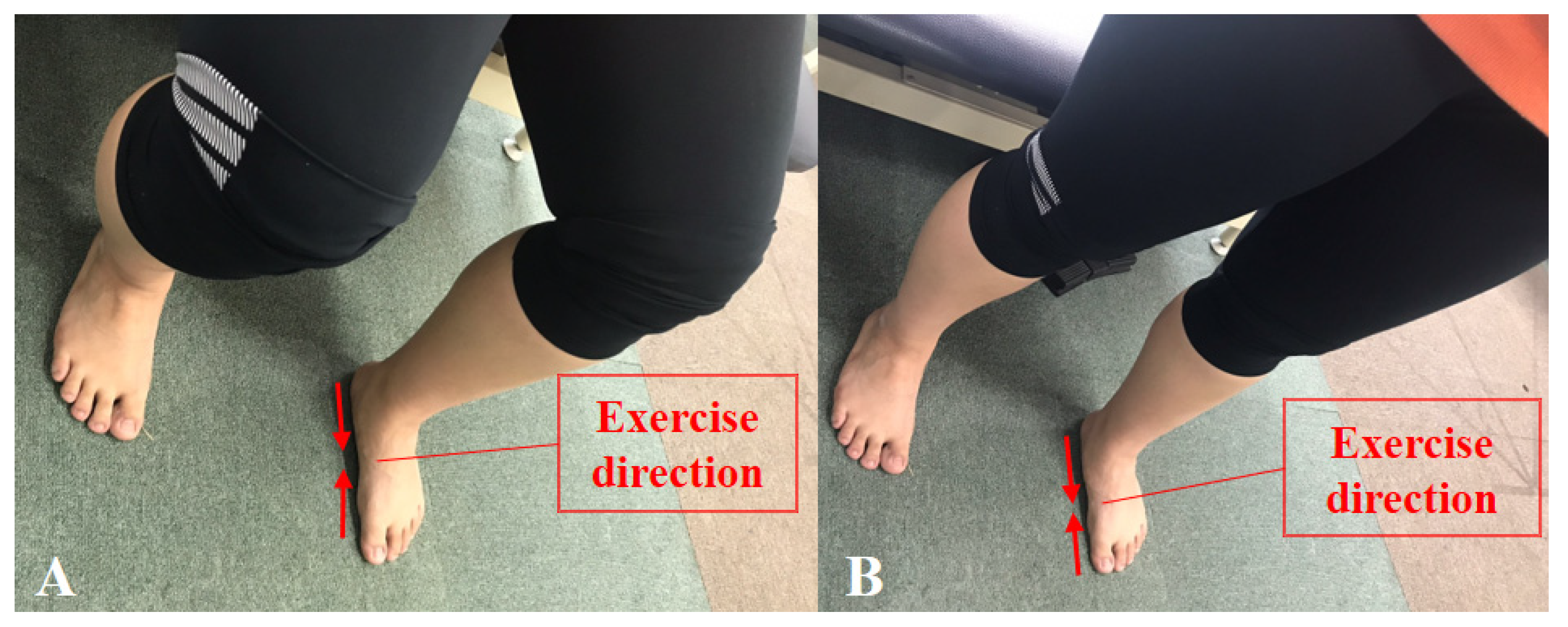
2.2. Study Design
2.3. Measurement Methods and Tools
2.3.1. Four-Way Ankle Strength Test
2.3.2. Navicular Drop Test
2.3.3. Balance Test
2.3.4. Plantar Fascia Thickness Test
2.3.5. Questionnaire for Foot Function
2.4. Experimental Procedure
2.5. Data Analysis
3. Results
3.1. General Characteristics of All Participants
3.2. Comparison of Results before and after Intervention
3.3. Comparison of Changes before and after Interventions
3.4. Intra and Inter-Rater Reliability of Measurements of Plantar Fascia
4. Discussion
5. Conclusions
Author Contributions
Funding
Institutional Review Board Statement
Informed Consent Statement
Data Availability Statement
Conflicts of Interest
References
- Doytch, N.; Dave, D.M.; Kelly, I.R. Global Evidence on Obesity and Related Outcomes: An Overview of Prevalence, Trends, and Determinants. East. Econ. J. 2014, 42, 7–28. [Google Scholar] [CrossRef]
- Korean Society for the Study of Obesity. Obesity Fact Sheet 2016. Available online: http://www.kosso.or.kr/file/factsheet_1006new6_web.pdf/ (accessed on 30 April 2017).
- Pourghasem, M.; Kamali, N.; Farsi, M.; Soltanpour, N. Prevalence of flatfoot among school students and its relationship with BMI. Acta Orthop. Traumatol. Turc. 2016, 50, 554–557. [Google Scholar] [CrossRef]
- World Health Organization (WHO). Obesity: Preventing and Managing the Global Epidemic (Report of a WHO Consultation on Obesity); World Health Organization: Geneva, Switzerland, 1997; p. 98. [Google Scholar]
- World Health Organization (WHO). Obesity and Overweight 2016. Available online: http://www.who.int/mediacentre/factsheets/fs311/en/ (accessed on 30 April 2017).
- Anandacoomarasamy, A.; Caterson, I.; Sambrook, P.; Fransen, M.; March, L. The impact of obesity on the musculoskeletal system. Int. J. Obes. 2007, 32, 211–222. [Google Scholar] [CrossRef]
- Paulis, W.D.; Silva, S.; Koes, B.W.; Van Middelkoop, M. Overweight and obesity are associated with musculoskeletal complaints as early as childhood: A systematic review. Obes. Rev. 2014, 15, 52–67. [Google Scholar] [CrossRef]
- Butterworth, P.; Menz, H.B.; Urquhart, D.M.; Cicuttini, F.M.; Pasco, J.; Brennan, S.L.; Wluka, A.; Strauss, B.J.; Proietto, J.; Dixon, J.B.; et al. The association between obesity and foot pain: Metabolic, biomechanical or both? J. Foot Ankle Res. 2015, 8, O5. [Google Scholar] [CrossRef]
- Stovitz, S.D.; Pardee, P.; Vazquez, G.; Duval, S.; Schwimmer, J.B. Musculoskeletal pain in obese children and adolescents. Acta Paediatr. 2008, 97, 489–493. [Google Scholar] [CrossRef]
- Chougala, A.; Phanse, V.; Khanna, E.; Panda, S. Screening of body mass index and functional flatfoot in adult: An observational study. Int. J. Physiother. Res. 2015, 3, 1037–1041. [Google Scholar] [CrossRef]
- Shibuya, N.; Jupiter, D.C.; Ciliberti, L.J.; VanBuren, V.; La Fontaine, J. Characteristics of Adult Flatfoot in the United States. J. Foot Ankle Surg. 2010, 49, 363–368. [Google Scholar] [CrossRef] [PubMed]
- Nielsen, M.D.; Dodson, E.E.; Shadrick, D.L.; Catanzariti, A.R.; Mendicino, R.W.; Malay, D.S. Nonoperative Care for the Treatment of Adult-acquired Flatfoot Deformity. J. Foot Ankle Surg. 2011, 50, 311–314. [Google Scholar] [CrossRef] [PubMed]
- Park, S.-Y.; Park, D.-J. Comparison of Foot Structure, Function, Plantar Pressure and Balance Ability According to the Body Mass Index of Young Adults. Osong Public Health Res. Perspect. 2019, 10, 102–107. [Google Scholar] [CrossRef] [PubMed]
- McKeon, P.; Hertel, J.; Bramble, D.; Davis, I. The foot core system: A new paradigm for understanding intrinsic foot muscle function. Br. J. Sports Med. 2014, 49, 290. [Google Scholar] [CrossRef]
- Unver, B.; Erdem, E.U.; Akbas, E. Effects of Short-Foot Exercises on Foot Posture, Pain, Disability, and Plantar Pressure in Pes Planus. J. Sport Rehabil. 2020, 29, 436–440. [Google Scholar] [CrossRef] [PubMed]
- Haun, C.; Brown, C.N.; Hannigan, K.; Johnson, S.T. The Effects of the Short Foot Exercise on Navicular Drop: A Critically Appraised Topic. J. Sport Rehabil. 2021, 30, 152–157. [Google Scholar] [CrossRef] [PubMed]
- Adler, S.S.; Beckers, D.; Buck, M. PNF in Practice: An Illustrated Guide, 4th ed.; Springer: Berlin/Heidelberg, Germany, 2014. [Google Scholar]
- Lee, K.-S.; Park, D.-J. Three-Dimensional Ankle Exercise with Combined Isotonic Technique for an Obese Subject with Plantar Fasciitis: A Case Study. Medicina 2020, 56, 190. [Google Scholar] [CrossRef] [PubMed]
- Park, D.-J.; Hwang, Y.-I. Comparison of the Intrinsic Foot Muscle Activities between Therapeutic and Three-Dimensional Foot-Ankle Exercises in Healthy Adults: An Explanatory Study. Int. J. Environ. Res. Public Health 2020, 17, 7189. [Google Scholar] [CrossRef] [PubMed]
- Munn, J.; Beard, D.J.; Refshauge, K.M.; Lee, R.Y.W. Eccentric Muscle Strength in Functional Ankle Instability. Med. Sci. Sports Exerc. 2003, 35, 245–250. [Google Scholar] [CrossRef]
- Park, D.-J.; Kim, B.-J.; Kim, Y.-H.; Park, S.-Y. A three-week intervention emphasized diagonal eccentric contraction on balance and joint position sense and ankle strength in subjects with ankle instability: A randomized controlled trial. J. Back Musculoskelet. Rehabil. 2021, 34, 95–101. [Google Scholar] [CrossRef]
- Hong, S.M.; Hur, Y.I. Relationship between obesity and depression in Korean adults: Korea National Health and Nutrition Examination Survey 2014. Medicine 2017, 96, e9478. [Google Scholar] [CrossRef]
- Lange, B.; Chipchase, L.; Evans, A. The Effect of Low-Dye Taping on Plantar Pressures, During Gait, in Subjects with Navicular Drop Exceeding 10 mm. J. Orthop. Sports Phys. Ther. 2004, 34, 201–209. [Google Scholar] [CrossRef]
- Langley, B.; Cramp, M.; Morrison, S.C. Clinical measures of static foot posture do not agree. J. Foot Ankle Res. 2016, 9, 1–6. [Google Scholar] [CrossRef]
- Hislop, H.; Avers, D.; Brown, M. Daniels and Wort Hingham’s Muscle Testing, 9th ed.; Saunders Elsevier: St. Louis, MO, USA, 2013. [Google Scholar]
- Granado, M.J.; Lohman, E.B.; Gordon, K.E.; Daher, N.S. Metatarsophalangeal joint extension changes ultrasound measurements for plantar fascia thickness. J. Foot Ankle Res. 2018, 11, 20. [Google Scholar] [CrossRef] [PubMed]
- Stratford, P.; Gill, C.; Westaway, M.; Binkley, J. Assessing disability and change on individual patients: A report of a patient specific measure. Physiother. Can. 1995, 47, 258–263. [Google Scholar] [CrossRef]
- Hefford, C.; Abbott, J.H.; Arnold, R.; Baxter, G.D. The Patient-Specific Functional Scale: Validity, Reliability, and Responsiveness in Patients with Upper Extremity Musculoskeletal Problems. J. Orthop. Sports Phys. Ther. 2012, 42, 56–65. [Google Scholar] [CrossRef]
- Alkhadhrawi, N.; Alshami, A. Effects of myofascial trigger point dry cupping on pain and function in patients with plantar heel pain: A randomized controlled trial. J. Bodyw. Mov. Ther. 2019, 23, 532–538. [Google Scholar] [CrossRef]
- Sulowska, I.; Oleksy, Ł.; Mika, A.; Bylina, D.; Sołtan, J. The Influence of Plantar Short Foot Muscle Exercises on Foot Posture and Fundamental Movement Patterns in Long-Distance Runners, a Non-Randomized, Non-Blinded Clinical Trial. PLoS ONE 2016, 11, e0157917. [Google Scholar] [CrossRef] [PubMed]
- Lynn, S.K.; Padilla, R.A.; Tsang, K.K. Differences in static- and dynamic-balance task performance after 4 weeks of intrinsic-foot-muscle training: The short-foot exercise versus the towel-curl exercise. J. Sport Rehabil. 2012, 21, 327–333. [Google Scholar] [CrossRef]
- Lee, E.; Cho, J.; Lee, S. Short-Foot Exercise Promotes Quantitative Somatosensory Function in Ankle Instability: A Randomized Controlled Trial. Med. Sci. Monit. 2019, 25, 618–626. [Google Scholar] [CrossRef]
- Janda, V.; Vavrova, M. Sensory Motor Stimulation; Liebenson, C., Ed.; Williams & Wilkins: Baltimore, MD, USA, 1990. [Google Scholar]
- Knellwolf, T.P.; Burton, A.R.; Hammam, E.; Macefield, V.G. Firing properties of muscle spindles supplying the intrinsic foot muscles of humans in unloaded and freestanding conditions. J. Neurophysiol. 2019, 121, 74–84. [Google Scholar] [CrossRef]
- Irving, D.B.; Cook, J.L.; Young, M.; Menz, H.B. Obesity and pronated foot type may increase the risk of chronic plantar heel pain: A matched case-control study. BMC Musculoskelet. Disord. 2007, 8, 41. [Google Scholar] [CrossRef] [PubMed]
- Dowling, G.J.; Murley, G.S.; Munteanu, S.; Smith, M.M.F.; Neal, B.S.; Griffiths, I.B.; Barton, C.J.; Collins, N.J. Dynamic foot function as a risk factor for lower limb overuse injury: A systematic review. J. Foot Ankle Res. 2014, 7, 53. [Google Scholar] [CrossRef] [PubMed]
- Snook, A.G. The Relationship between Excessive Pronation as Measured by Navicular Drop and Isokinetic Strength of the Ankle Musculature. Foot Ankle Int. 2001, 22, 234–240. [Google Scholar] [CrossRef]
- Mulligan, E.P.; Cook, P.G. Effect of plantar intrinsic muscle training on medial longitudinal arch morphology and dynamic function. Man. Ther. 2013, 18, 425–430. [Google Scholar] [CrossRef] [PubMed]
- Pabón-Carrasco, M.; Castro-Méndez, A.; Vilar-Palomo, S.; Jiménez-Cebrián, A.M.; García-Paya, I.; Palomo-Toucedo, I.C. Randomized Clinical Trial: The Effect of Exercise of the Intrinsic Muscle on Foot Pronation. Int. J. Environ. Res. Public Health 2020, 17, 4882. [Google Scholar] [CrossRef]
- Field, A.P. Discovering Statistics Using IBM SPSS Statistics: And Sex and Drugs and Rock ‘n’ Roll, 4th ed.; SAGE Publications: London, UK, 2015. [Google Scholar]
- Spink, M.J.; Fotoohabadi, M.R.; Wee, E.; Hill, K.; Lord, S.R.; Menz, H.B. Foot and Ankle Strength, Range of Motion, Posture, and Deformity are Associated with Balance and Functional Ability in Older Adults. Arch. Phys. Med. Rehabil. 2011, 92, 68–75. [Google Scholar] [CrossRef]
- Murley, G.S.; Menz, H.B.; Landorf, K.B. Foot posture influences the electromyographic activity of selected lower limb muscles during gait. J. Foot Ankle Res. 2009, 2, 35. [Google Scholar] [CrossRef] [PubMed]
- Park, D.J.; Park, S.Y. Comparison of subjects with and without pes planus during short foot exercises by measuring muscular activities of ankle and navicular drop height. J. Korean Soc. Phys. Med. 2018, 13, 107–113. [Google Scholar] [CrossRef]
- Johnson, C.H.; Christensen, J.C. Biomechanics of the first ray part I. The effects of peroneus longus function: A three-dimensional kinematic study on a cadaver model. J. Foot Ankle Surg. 1999, 38, 313–321. [Google Scholar] [CrossRef]
- Fiolkowski, P.; Brunt, D.; Bishop, M.; Woo, R.; Horodyski, M. Intrinsic pedal musculature support of the medial longitudinal arch: An electromyography study. J. Foot Ankle Surg. 2003, 42, 327–333. [Google Scholar] [CrossRef]
- Soysa, A.; Hiller, C.; Refshauge, K.; Burns, J. Importance and challenges of measuring intrinsic foot muscle strength. J. Foot Ankle Res. 2012, 5, 29. [Google Scholar] [CrossRef] [PubMed]
- Angin, S.; Crofts, G.; Mickle, K.J.; Nester, C.J. Ultrasound evaluation of foot muscles and plantar fascia in pes planus. Gait Posture 2014, 40, 48–52. [Google Scholar] [CrossRef] [PubMed]
- Hashimoto, T.; Sakuraba, K. Strength Training for the Intrinsic Flexor Muscles of the Foot: Effects on Muscle Strength, the Foot Arch, and Dynamic Parameters Before and After the Training. J. Phys. Ther. Sci. 2014, 26, 373–376. [Google Scholar] [CrossRef] [PubMed]
- Karabay, N.; Toros, T.; Hurel, C. Ultrasonographic Evaluation in Plantar Fasciitis. J. Foot Ankle Surg. 2007, 46, 442–446. [Google Scholar] [CrossRef] [PubMed]
- Simons, D.G.; Travell, J.G.; Simons, L.S. Travell and Simons’ Myofascial Pain and Dysfunction: The Trigger Point Manual, 2nd ed.; Williams & Wilkins: Baltimore, MD, USA, 1999. [Google Scholar]
- McPoil, T.G.; Martin, R.L.; Cornwall, M.W.; Wukich, D.K.; Irrgang, J.J.; Godges, J.J. Heel pain—Plantar fasciitis: Clinical practice guidelines linked to the international classification of function, disability, and health from the orthopaedic section of the American Physical Therapy Association. J. Orthop. Sports Phys. Ther. 2008, 38, A1–A18. [Google Scholar] [CrossRef]
- Ashton-Miller, J.A.; Ottaviani, R.A.; Hutchinson, C.; Wojtys, E.M. What best protects the inverted weight bearing ankle against further inversion? Evertor muscle strength compares favorably with shoe height, athletic tape, and three orthoses. Am. J. Sports Med. 1996, 24, 800–809. [Google Scholar] [CrossRef] [PubMed]


| Variable | SF (n = 12) | PNF (n = 12) | Z | p |
|---|---|---|---|---|
| Age (years) | 23.25 ± 1.22 | 24.00 ± 1.48 | −1.60 | 0.11 |
| Height (cm) | 168.50 ± 8.44 | 170.25 ± 10.06 | −0.46 | 0.64 |
| Weight (kg) | 83.83 ± 13.83 | 85.88 ± 19.99 | −0.12 | 0.91 |
| BMI (kg/m2) | 29.34 ± 2.81 | 29.39 ± 4.57 | −0.40 | 0.69 |
| PSFS (score) | 26.00 ± 3.44 | 26.25 ± 3.98 | −0.36 | 0.76 |
| Sex | M 7 (50%), F 5 (50%) | M 7 (50%), F 5 (50%) | - | - |
| Dominant foot | Rt 10 (83.3%), Lt 2 (16.7%) | Rt 10 (83.3%), Lt 2 (16.7%) | - | - |
| SF | Pre-Test | Post-Test | Z | p | Effect Size (r) |
|---|---|---|---|---|---|
| COP-LR (cm) | 3.20 ± 1.24 | 2.23 ± 0.72 | −2.43 | 0.02 | −0.70 |
| COP-AP (cm) | 2.21 ± 0.65 | 1.63 ± 0.48 | −2.98 | 0.01 | −0.86 |
| NDT (mm) | 11.83 ± 1.75 | 9.17 ± 1.64 | −2.73 | 0.01 | −0.79 |
| Dorsiflexor (N/kg) | 2.29 ± 0.66 | 2.32 ± 0.65 | −0.31 | 0.75 | −0.09 |
| Plantar flexor (N/kg) | 2.31 ± 0.42 | 2.76 ± 0.39 | −2.51 | 0.01 | −0.72 |
| Invertor (N/kg) | 1.10 ± 0.32 | 1.28 ± 0.25 | −2.59 | 0.01 | −0.75 |
| Evertor (N/kg) | 1.34 ± 0.37 | 1.39 ± 0.26 | −0.78 | 0.43 | −0.23 |
| PFT (cm) | 0.50 ± 0.07 | 0.48 ± 0.07 | −2.73 | 0.01 | −0.79 |
| PSFS (scores) | 26.00 ± 3.44 | 28.08 ± 1.83 | −2.39 | 0.02 | −0.69 |
| PNF | Pre-Test | Post-Test | Z | p | Effect Size (r) |
|---|---|---|---|---|---|
| COP-LR (cm) | 3.25 ± 1.02 | 2.18 ± 0.86 | −2.67 | 0.01 | −0.77 |
| COP-AP (cm) | 2.09 ± 0.56 | 1.66 ± 0.55 | −2.08 | 0.38 | −0.60 |
| NDT (mm) | 11.92 ± 1.68 | 8.67 ± 1.78 | −2.95 | 0.01 | −0.85 |
| Dorsiflexor (N/kg) | 2.37 ± 0.68 | 3.00 ± 0.78 | −2.75 | 0.01 | −0.79 |
| Plantar flexor (N/kg) | 2.81 ± 0.70 | 3.40 ± 0.49 | −2.67 | 0.01 | −0.77 |
| Invertor (N/kg) | 1.27 ± 0.41 | 1.51 ± 0.39 | −2.35 | 0.02 | −0.68 |
| Evertor (N/kg) | 1.49 ± 0.47 | 2.01 ± 0.56 | −2.75 | 0.01 | −0.79 |
| PFT (cm) | 0.51 ± 0.06 | 0.48 ± 0.06 | −3.01 | 0.01 | −0.87 |
| PSFS (scores) | 26.25 ± 3.98 | 28.83 ± 2.21 | −2.38 | 0.02 | −0.69 |
| Difference | SF | PNF | Z | p | Effect Size (r) |
|---|---|---|---|---|---|
| COP-LR (cm) | −0.98 ± 1.28 | −1.07 ± 1.06 | −0.69 | 0.51 | −0.20 |
| COP-AP (cm) | −0.58 ± 0.40 | −0.43 ± 0.57 | −0.49 | 0.63 | −0.14 |
| NDT (mm) | −2.67 ± 2.57 | −3.25 ± 2.01 | −0.88 | 0.41 | −0.25 |
| Dorsiflexor (N/kg) | 0.03 ± 0.39 | 0.63 ± 0.59 | −2.31 | 0.02 | −0.67 |
| Plantar flexor (N/kg) | 0.45 ± 0.48 | 0.59 ± 0.61 | −0.58 | 0.59 | −0.17 |
| Invertor (N/kg) | 0.18 ± 0.20 | 0.25 ± 0.28 | −0.64 | 0.55 | −0.18 |
| Evertor (N/kg) | 0.05 ± 0.27 | 0.52 ± 0.45 | −2.54 | 0.01 | −0.73 |
| PFT (cm) | −0.02 ± 0.02 | −0.03 ± 0.01 | −0.74 | 0.48 | −0.21 |
| PSFS (scores) | 2.08 ± 2.31 | 2.58 ± 2.68 | −0.33 | 0.76 | −0.10 |
| Reliability | Inter-Rater | Intra-Rater | ||
|---|---|---|---|---|
| ICC2,1 | 95% CI | ICC3,1 | 95% CI | |
| Plantar fascia | 0.89 | 0.70–0.98 | 0.93 | 0.78–0.98 |
Publisher’s Note: MDPI stays neutral with regard to jurisdictional claims in published maps and institutional affiliations. |
© 2021 by the authors. Licensee MDPI, Basel, Switzerland. This article is an open access article distributed under the terms and conditions of the Creative Commons Attribution (CC BY) license (https://creativecommons.org/licenses/by/4.0/).
Share and Cite
Park, D.-J.; Lee, K.-S.; Park, S.-Y. Effects of Two Foot-Ankle Interventions on Foot Structure, Function, and Balance Ability in Obese People with Pes Planus. Healthcare 2021, 9, 667. https://doi.org/10.3390/healthcare9060667
Park D-J, Lee K-S, Park S-Y. Effects of Two Foot-Ankle Interventions on Foot Structure, Function, and Balance Ability in Obese People with Pes Planus. Healthcare. 2021; 9(6):667. https://doi.org/10.3390/healthcare9060667
Chicago/Turabian StylePark, Du-Jin, Kyung-Sun Lee, and Se-Yeon Park. 2021. "Effects of Two Foot-Ankle Interventions on Foot Structure, Function, and Balance Ability in Obese People with Pes Planus" Healthcare 9, no. 6: 667. https://doi.org/10.3390/healthcare9060667
APA StylePark, D.-J., Lee, K.-S., & Park, S.-Y. (2021). Effects of Two Foot-Ankle Interventions on Foot Structure, Function, and Balance Ability in Obese People with Pes Planus. Healthcare, 9(6), 667. https://doi.org/10.3390/healthcare9060667