Perspective

Are We Going to Give Up Imaging in Cryptorchidism Management?

by
Cristina Gavrilovici
1,2,
Alma-Raluca Laptoiu
1,*,
Carmen-Iulia Ciongradi
1,2,
Petronela Pirtica
2,
Elena-Lia Spoiala
1,2,
Elena Hanganu
1,2,
Alexandru Pirvan
3 and
Monika Glass
4
1
Department of Mother and Child, “Grigore T. Popa” University of Medicine and Pharmacy, 700115 Iasi, Romania
2
“Sfânta Maria” Emergency Hospital for Children, 700309 Iasi, Romania
3
Department of Pediatric Surgery, “Iuliu Hațieganu” University of Medicine and Pharmacy, 400012 Cluj-Napoca, Romania
4
Kannerklinik, Center Hospitalier du Luxembourg, 4 Rue Nicolas Ernest Barblé, 1210 Luxembourg, Luxembourg
*
Author to whom correspondence should be addressed.
Healthcare 2025, 13(10), 1192; https://doi.org/10.3390/healthcare13101192

Submission received: 3 March 2025 / Revised: 12 May 2025 / Accepted: 19 May 2025 / Published: 20 May 2025

Abstract

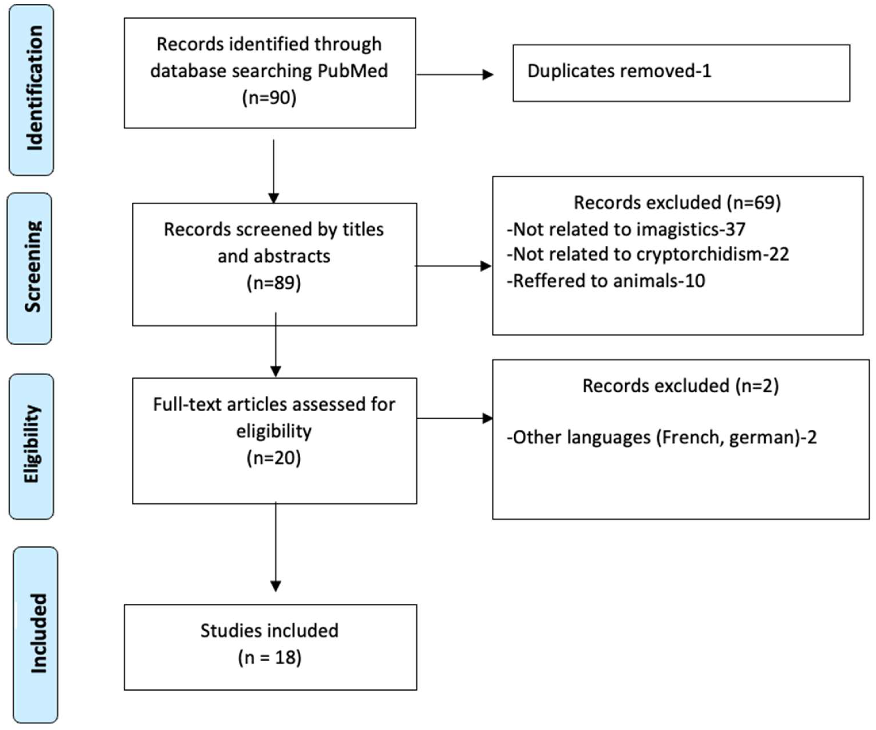
Background and Purpose: Undescended testes (UDT) is recognized as the most prevalent anomaly of the male genitalia and presents a significant risk factor for long-term complications, including infertility and testicular cancer. Currently, there is no consensus on the necessity of imaging in the management of UDT, nor is there agreement on which imaging modality is preferred or to what extent these tests offer real added value in the clinical setting. This review aims to evaluate the various imaging options available in the management of cryptorchidism, discussing their utility, advantages, and disadvantages compared to exploratory laparoscopy. Methods: We conducted a PubMed search using the following search terms: [“undescended testis”] OR [(“cryptorchidism”) OR (“diagnostic imaging”)] OR [(“Ultrasound”), OR (“CT scan”) OR (“MRI”)] AND [“laparoscopy”]. We analyzed 90 full articles, excluding irrelevant ones, and, in total, 18 publications were included in this review. Results: Ultrasound (US) is the most commonly used technique due to its non-invasive nature and absence of ionizing radiation. It is particularly beneficial in cases of non-palpable UDT. However, its main limitation lies in the difficulty in accurately locating UDT, especially when they are situated outside the inguinal region. Computed tomography (CT) scans serve as a crucial diagnostic tool, particularly for testes located below the internal inguinal ring. While CT exhibits comparable accuracy in detecting UDT, the need for sedation or general anesthesia, along with the costs and potential risks of secondary malignancy due to radiation exposure, does not favor its routine use. Magnetic resonance imaging (MRI) offers higher sensitivity than US and does not utilize ionizing radiation or intravascular contrast agents. It allows for the generation of multiplanar images, thereby providing improved tissue characterization. However, limitations include prolonged scan durations, the potential for motion artifacts during imaging, the need for sedation, and higher costs. Laparoscopy has been shown to provide better accuracy, offering both diagnostic and therapeutic benefits, particularly in cases of non-palpable UDT. It is widely regarded as the gold standard in achieving clear diagnostic and definitive therapeutic procedures and has demonstrated its utility in determining the anatomical position of intra-abdominal testes, owing to its magnification capabilities and minimally invasive approach. Conclusions: Achieving a correct and comprehensive diagnosis of cryptorchidism requires the medical team to decide on the appropriate imaging studies, as these will not significantly influence or alter the therapeutic decision-making process. It is unlikely that medical practice will eliminate imaging studies before a surgical decision is made in the near future. Therefore, a multidisciplinary approach that includes clinical examination, imaging, and diagnostic laparoscopy remains essential for the accurate management of UDT.
Keywords: undescended testis; cryptorchidism; ultrasound; computer tomography; MRI; laparoscopy undescended testis; cryptorchidism; ultrasound; computer tomography; MRI; laparoscopy