
The Functional Neuroimaging of Autobiographical Memory for Happy Events: A Coordinate-Based Meta-Analysis


Abstract
1. Introduction
2. Materials and Methods
2.1. Information Sources and Search Strategy
- -
- “autobiographical memory” OR “autobiographical recall”, AND “positive events”, OR “happy events”, AND “fMRI” OR “functional magnetic resonance imaging”.
- -
- “autobiographical memory” OR “autobiographical recall”, AND “positive events” OR “happy events”, AND “PET” OR “positron emission tomography”.
2.2. Eligibility Criteria
2.3. Coordinate-Based Meta-Analysis
2.4. Automated Regional Behavioral Analysis
3. Results
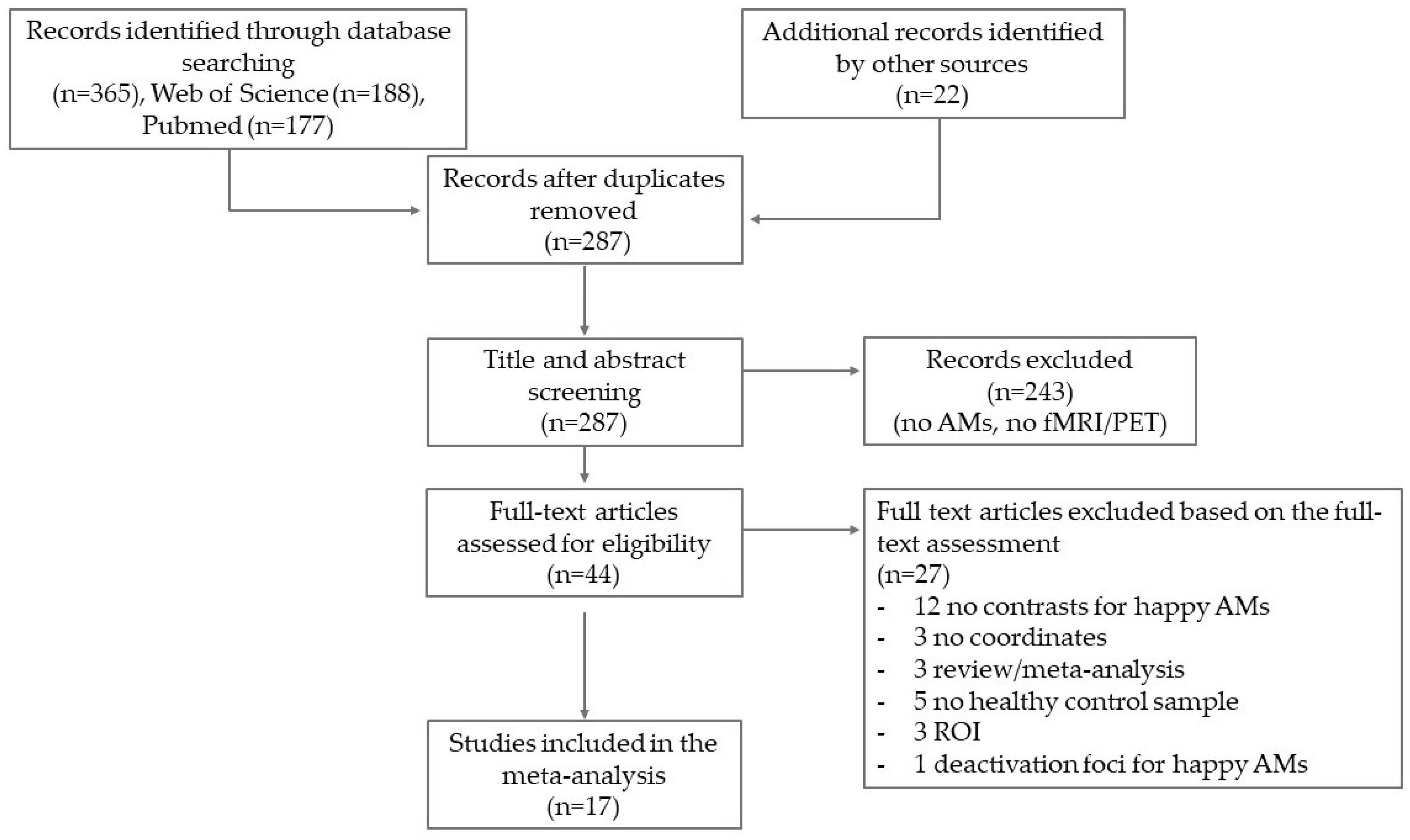
3.1. Study Selection
3.2. Characteristics of the Studies

| Year of Publication | First Author and Reference | Neuroimaging | Original Coordinates | Sample | Recall Induction Technique | Remoteness | Contrasted Conditions | Activation Foci |
|---|---|---|---|---|---|---|---|---|
| 1995 | George [63] | PET | Talairach | n = 11 (F, mean age: 33.3, SD: 12.3) | REC/REL Two events for condition cued with pictures of emotional faces | not defined | happiness > sadness | 5 |
| 1996 | George [64] | PET | Talairach | n = 20 (10 F, mean age: 34.5, SD: 12.1; 10 M, mean age: 35.5, SD: 8.8) | REC/REL Two events for condition cued with pictures of emotional faces | not defined | happiness > neutral | 8 |
| 1997 | Lane [65] | PET | Talairach | n = 12 (F, mean age: 23.3, SD: 3.2) | LIST.SCRIPT Three events for condition | last 6 months | happiness > neutral | 4 |
| 2000 | Damasio [78] | PET | Talairach | n = 41 (21 F, 20 M divided into four cohorts, age: from 23 to 42) | REC/REL One event for condition | not defined | happiness > neutral | 20 |
| 2003 | Markowitsch [36] | fMRI | MNI | n = 13 (7 F, 6 M, mean age: 30, from 19 to 43) | REC/REL 18 events for condition cued by keywords | before 12 years old; from 12 to 18 years; from 18 until now | happiness > rest | 10 |
| 2003 | Piefke [37] | fMRI | Talairach | n = 20 (10 F, 10 M, mean age: 26, SD: 3) | REC/REL 10 events for condition, cued by written sentences | childhood (up to 10 years); recent past (last 5 years) | happiness > negative | 4 |
| 2007 | Marci [71] | PET | MNI | n = 10 (5 F, 5 M, mean age: 33.9, SD: 11.9) | LIST.SCRIPT 2 events for condition | not defined | happiness > neutral | 4 |
| 2008 | Cerqueira [72] | fMRI | Talairach | n = 11 (5 F, 6 M, mean age: 32.4, SD: 7.2) | LIST.SCRIPT 3 events for condition | last 12 months | happiness > neutral happiness > irritability | 10 |
| 2010 | Cerqueira [73] | fMRI | Talairach | n = 11 (5 F, 6 M, mean age: 32.4, SD: 7.2) | LIST.SCRIPT 3 events for condition | last 6 months | happiness > neutral happiness > irritability | 5 |
| 2011 | Sitaram [74] | fMRI | MNI | n = 12 (mean age: 25 years, range: 22–26) | REC/REL 1 event for condition cued by emotional pictures | not defined | happiness > disgust | 112 |
| 2011 | Zotev [70] | fMRI | Talairach | n = 14 (M, mean age: 27.5, SD: 11.1) | REC/REL 3 happy events cued by the word “happy”; counting task as control condition | not defined | happiness > control | 22 |
| 2014 | Speer [79] | fMRI | Talairach | n = 19 (10 F, 9 M, mean age: 26.1, SD: 7.78) | REC/REL 21 episodes for condition cued by keywords | not defined | happiness > neutral | 27 |
| 2014 | Gong [66] | fMRI | MNI | n = 12 (F, mean age: 66.3, from 60 to 70) | REC/REL 10 events for condition cued by written sentences | before 12 years old; last 5 years (except the last month) | happiness > negative | 3 |
| 2014 | Ge [67] | fMRI | MNI | n = 27 (13 younger F, age from 18 to 22; 14 older F, age from 60 to 74) | REC/REL 5 events for condition cued by written sentences | last 5 years | happiness > negative | 3 |
| 2017 | Lempert [69] | fMRI | MNI | n = 35 (F; mean age: 20.86, SD: 2.9) | REC/REL 10 events for condition cued by written sentences | not defined | happiness > rest | 12 |
| 2018 | Xu [77] | fMRI | MNI | n = 25 (17 F, 8 M, mean age: 21.36, SD: 3.34) | REC/REL 9 events for condition cued by written sentences | before 18 years old | happiness > baseline happiness > negative | 21 |
| 2019 | Schie [68] | fMRI | MNI | n = 47 (F, mean age: 29.36; SD: 9.61) | REC/REL 4 events for condition cued by written sentences | not defined | happiness > neutral | 12 |
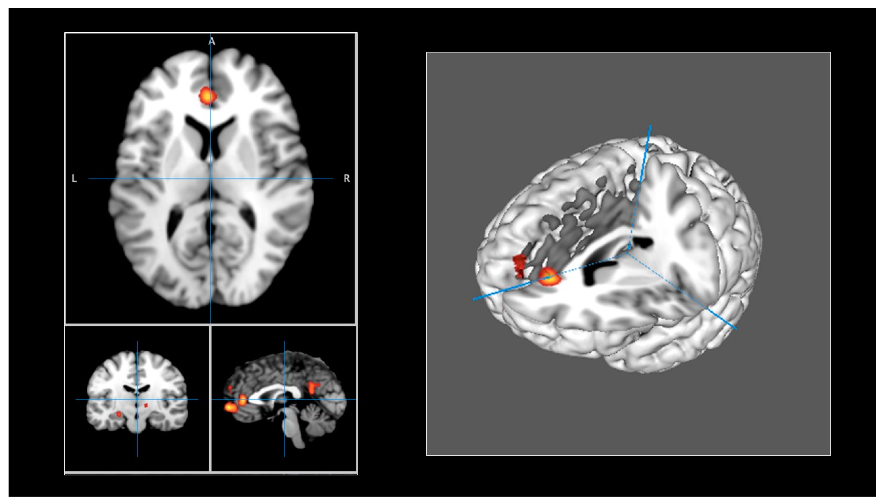
3.3. Clusters of Neural Activity Changes
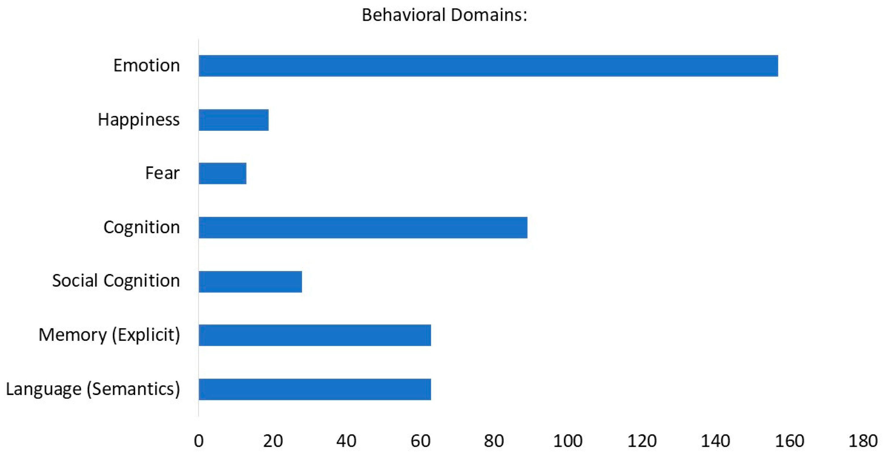
3.4. Characterization of the Clusters
4. Discussion
5. Conclusions
Author Contributions
Funding
Institutional Review Board Statement
Informed Consent Statement
Data Availability Statement
Conflicts of Interest
References
- Sotgiu, I. The Psychology of Autobiographical Memory: History, Theory, Research; Palgrave Macmillan: Cham, Switzerland, 2021. [Google Scholar]
- Conway, M.A. Memory and the self. J. Mem. Lang. 2005, 53, 594–628. [Google Scholar] [CrossRef]
- Conway, M.A.; Singer, J.A.; Tagini, A. The self and autobiographical memory: Correspondence and coherence. Soc. Cogn. 2004, 22, 491–529. [Google Scholar] [CrossRef]
- Berntsen, D.; Rubin, D.C. Introduction. In Understanding Autobiographical Memory: Theories and Approaches; Cambridge University Press: Cambridge, UK, 2012; pp. 1–8. [Google Scholar]
- Tulving, E. Episodic Memory: From Mind to Brain. Annu. Rev. Psychol. 2002, 53, 1–25. [Google Scholar] [CrossRef]
- Rubin, D.C. A Basic-Systems Approach to Autobiographical Memory. Curr. Dir. Psychol. Sci. 2005, 14, 79–83. [Google Scholar] [CrossRef]
- Rubin, D.C. The Basic-Systems Model of Episodic Memory. Perspect. Psychol. Sci. 2006, 1, 277–311. [Google Scholar] [CrossRef]
- Rubin, D.C. The basic systems model of autobiographical memory. In Understanding Autobiographical Memory: Theories and Approaches; Cambridge University Press: Cambridge, UK, 2012; pp. 11–32. [Google Scholar]
- Holland, A.C.; Kensinger, E.A. Emotion and autobiographical memory. Phys. Life Rev. 2010, 7, 88–131. [Google Scholar] [CrossRef] [PubMed]
- Bauer, P.J.; Pathman, T.; Inman, C.; Campanella, C.; Hamann, S. Neural correlates of autobiographical memory retrieval in children and adults. Memory 2017, 25, 450–466. [Google Scholar] [CrossRef]
- Sotgiu, I. How Do We Remember Happy Life Events? A Comparison Between Eudaimonic and Hedonic Autobiographical Memories. J. Psychol. Interdiscip. Appl. 2016, 150, 685–703. [Google Scholar] [CrossRef] [PubMed]
- Sotgiu, I. Gender Differences and Similarities in Autobiographical Memory for Eudaimonic Happy Events. J. Happiness Stud. 2019, 20, 1457–1479. [Google Scholar] [CrossRef]
- Huta, V.; Waterman, A.S. Eudaimonia and Its Distinction from Hedonia: Developing a Classification and Terminology for Understanding Conceptual and Operational Definitions. J. Happiness Stud. 2013, 15, 1425–1456. [Google Scholar] [CrossRef]
- Delle Fave, A.; Brdar, I.; Freire, T.; Vella-Brodrick, D.; Wissing, M.P. The Eudaimonic and Hedonic Components of Happiness: Qualitative and Quantitative Findings. Soc. Indic. Res. 2011, 100, 185–207. [Google Scholar] [CrossRef]
- Vittersø, J.; Søholt, Y. Life satisfaction goes with pleasure and personal growth goes with interest: Further arguments for separating hedonic and eudaimonic well-being. J. Posit. Psychol. 2011, 6, 326–335. [Google Scholar] [CrossRef]
- Berntsen, D.; Rubin, D.C.; Siegler, I.C. Two versions of life: Emotionally negative and positive life events have different roles in the organization of life story and identity. Emotion 2011, 11, 1190–1201. [Google Scholar] [CrossRef] [PubMed]
- Berntsen, D.; Rubin, D.C. Cultural life scripts structure recall from autobiographical memory. Mem. Cogn. 2004, 32, 427–442. [Google Scholar] [CrossRef] [PubMed]
- Cabeza, R.; St Jacques, P. Functional neuroimaging of autobiographical memory. Trends Cogn. Sci. 2007, 11, 219–227. [Google Scholar] [CrossRef] [PubMed]
- Svoboda, E.; McKinnon, M.C.; Levine, B. The functional neuroanatomy of autobiographical memory: A meta-analysis. Neuropsychologia 2006, 44, 2189–2208. [Google Scholar] [CrossRef] [PubMed]
- Maguire, E.A. Neuroimaging studies of autobiographical event memory. Philos. Trans. R. Soc. B Biol. Sci. 2001, 356, 1441–1451. [Google Scholar] [CrossRef] [PubMed]
- Andrews-Hanna, J.R.; Reidler, J.S.; Sepulcre, J.; Poulin, R.; Buckner, R.L. Functional-Anatomic Fractionation of the Brain’s Default Network. Neuron 2010, 65, 550–562. [Google Scholar] [CrossRef] [PubMed]
- Kelley, A.W.M.; Macrae, C.N.; Wyland, C.L.; Caglar, S.; Inati, S.; Heatherton, T.F. Finding the self? An event-related fMRI study. J. Cogn. Neurosci. 2002, 14, 785–794. [Google Scholar] [CrossRef]
- Macrae, C.N. Medial Prefrontal Activity Predicts Memory for Self. Cereb. Cortex 2004, 14, 647–654. [Google Scholar] [CrossRef]
- Cabeza, R.; Ciaramelli, E.; Olson, I.R.; Moscovitch, M. The parietal cortex and episodic memory: An attentional account. Nat. Rev. Neurosci. 2008, 9, 613–625. [Google Scholar] [CrossRef] [PubMed]
- Wagner, A.D.; Shannon, B.J.; Kahn, I.; Buckner, R.L. Parietal lobe contributions to episodic memory retrieval. Trends Cogn. Sci. 2005, 9, 445–453. [Google Scholar] [CrossRef] [PubMed]
- Cabeza, R.; Prince, S.E.; Daselaar, S.M.; Greenberg, D.L.; Budde, M.; Dolcos, F.; LaBar, K.S.; Rubin, D.C. Brain activity during episodic retrieval of autobiographical and laboratory events: An fMRI study using a novel photo paradigm. J. Cogn. Neurosci. 2004, 16, 1583–1594. [Google Scholar] [CrossRef] [PubMed]
- Greenberg, D.L.; Rubin, D.C. The neuropsychology of autobiographical memory. Cortex 2003, 39, 687–728. [Google Scholar] [CrossRef] [PubMed]
- Fink, G.R.; Markowitsch, H.J.; Reinkemeier, M.; Bruckbauer, T.; Kassler, J.; Heiss, W.D. Cerebral representation of one’s own past: Neural networks involved in autobiographical memory. J. Neurosci. 1996, 16, 4275–4282. [Google Scholar] [CrossRef] [PubMed]
- Maguire, E.A.; Frith, C.D. Lateral asymmetry in the hippocampal response to the remoteness of autobiographical memories. J. Neurosci. 2003, 23, 5302–5307. [Google Scholar] [CrossRef]
- Markowitsch, H.J.; Thiel, A.; Reinkemeier, M.; Kessler, J.; Koyuncu, A.; Heiss, W.-D. Right amygdalar and temporofrontal activation during autobiographic, but not during fictitious memory retrieval. Behav. Neurol. 2000, 12, 181–190. [Google Scholar] [CrossRef]
- Addis, D.R.; Moscovitch, M.; Crawley, A.P.; McAndrews, M.P. Recollective qualities modulate hippocampal activation during autobiographical memory retrieval. Hippocampus 2004, 14, 752–762. [Google Scholar] [CrossRef]
- Greenberg, D.L.; Rice, H.J.; Cooper, J.J.; Cabeza, R.; Rubin, D.C.; LaBar, K.S. Co-activation of the amygdala, hippocampus and inferior frontal gyrus during autobiographical memory retrieval. Neuropsychologia 2005, 43, 659–674. [Google Scholar] [CrossRef]
- Talarico, J.M.; Labar, K.S.; Rubin, D.C. Emotional intensity predicts autobiographical memory experience. Mem. Cogn. 2004, 32, 1118–1132. [Google Scholar] [CrossRef]
- Berntsen, D.; Rubin, D.C. Emotionally charged autobiographical memories across the life span: The recall of happy, sad, traumatic and involuntary memories. Psychol. Aging 2002, 17, 636–652. [Google Scholar] [CrossRef]
- Kensinger, E.A. What factors need to be considered to understand emotional memories? Emot. Rev. 2009, 1, 120–121. [Google Scholar] [CrossRef]
- Markowitsch, H.J.; Vandekerckhove, M.M.P.; Lanfermann, H.; Russ, M.O. Engagement of lateral and medial prefrontal areas in the ecphory of sad and happy autobiographical memories. Cortex 2003, 39, 643–665. [Google Scholar] [CrossRef]
- Piefke, M. Differential remoteness and emotional tone modulate the neural correlates of autobiographical memory. Brain 2003, 126, 650–668. [Google Scholar] [CrossRef]
- Jallais, C.; Gilet, A.L. Inducing changes in arousal and valence: Comparison of two mood induction procedures. Behav. Res. Methods 2010, 42, 318–325. [Google Scholar] [CrossRef] [PubMed]
- Westermann, R.; Spies, K.; Stahl, G.; Hesse, F.W. Relative effectiveness and validity of mood induction procedures: A meta-analysis. Eur. J. Soc. Psychol. 1996, 26, 557–580. [Google Scholar] [CrossRef]
- Phan, K.L.; Wager, T.; Taylor, S.F.; Liberzon, I. Functional neuroanatomy of emotion: A meta-analysis of emotion activation studies in PET and fMRI. Neuroimage 2002, 16, 331–348. [Google Scholar] [CrossRef] [PubMed]
- Lane, R.D.; Reiman, E.M.; Axelrod, B.; Yun, L.S.; Holmes, A.; Schwartz, G.E. Neural correlates of levels of emotional awareness: Evidence of an interaction between emotion and attention in the anterior cingulate cortex. J. Cogn. Neurosci. 1998, 10, 525–535. [Google Scholar] [CrossRef] [PubMed]
- Berridge, K.C.; Kringelbach, M.L. Towards a Neuroscience of Well-Being: Implications of Insights from Pleasure Research; Springer: Dordrecht, The Netherlands, 2013; pp. 81–100. [Google Scholar]
- Kringelbach, M.L.; Berridge, K.C. Towards a functional neuroanatomy of pleasure and happiness. Trends Cogn. Sci. 2009, 13, 479–487. [Google Scholar] [CrossRef] [PubMed]
- Burgdorf, J.; Panksepp, J. The neurobiology of positive emotions. Neurosci. Biobehav. Rev. 2006, 30, 173–187. [Google Scholar] [CrossRef] [PubMed]
- Chemali, Z.N.; Chahine, L.M.; Naassan, G. On happiness: A minimalist perspective on a complex neural circuitry and its psychosocial constructs. J. Happiness Stud. 2008, 9, 489–501. [Google Scholar] [CrossRef]
- Machado, L.; Cantilino, A. A systematic review of the neural correlates of positive emotions. Rev. Bras. Psiquiatr. 2017, 39, 172–179. [Google Scholar] [CrossRef]
- Tanzer, J.R.; Weyandt, L. Imaging Happiness: Meta Analysis and Review. J. Happiness Stud. 2020, 21, 2693–2734. [Google Scholar] [CrossRef]
- Dillon, D.G. The neuroscience of positive memory deficits in depression. Front. Psychol. 2015, 6, 1295. [Google Scholar] [CrossRef]
- Werner-Seidler, A.; Moulds, M.L. Autobiographical memory characteristics in depression vulnerability: Formerly depressed individuals recall less vivid positive memories. Cogn. Emot. 2010, 25, 1087–1103. [Google Scholar] [CrossRef] [PubMed]
- Suardi, A.; Sotgiu, I.; Costa, T.; Cauda, F.; Rusconi, M. The neural correlates of happiness: A review of PET and fMRI studies using autobiographical recall methods. Cogn. Affect. Behav. Neurosci. 2016, 16, 383–392. [Google Scholar] [CrossRef]
- Raemaekers, M.; Vink, M.; Zandbelt, B.; van Wezel, R.J.A.; Kahn, R.S.; Ramsey, N.F. Test-retest reliability of fMRI activation during prosaccades and antisaccades. Neuroimage 2007, 36, 532–542. [Google Scholar] [CrossRef]
- Wager, T.D.; Lindquist, M.A.; Nichols, T.E.; Kober, H.; Van Snellenberg, J.X. Evaluating the consistency and specificity of neuroimaging data using meta-analysis. Neuroimage 2009, 45, S210–S221. [Google Scholar] [CrossRef]
- Wager, T.D.; Lindquist, M.; Kaplan, L. Meta-analysis of functional neuroimaging data: Current and future directions. Soc. Cogn. Affect. Neurosci. 2007, 2, 150–158. [Google Scholar] [CrossRef]
- Laird, A.R.; Fox, P.M.; Price, C.J.; Glahn, D.C.; Uecker, A.M.; Lancaster, J.L.; Turkeltaub, P.E.; Kochunov, P.; Fox, P.T. ALE meta-analysis: Controlling the false discovery rate and performing statistical contrasts. Hum. Brain Mapp. 2005, 25, 155–164. [Google Scholar] [CrossRef]
- Turkeltaub, P.E.; Eden, G.F.; Jones, K.M.; Zeffiro, T.A. Meta-analysis of the functional neuroanatomy of single-word reading: Method and validation. Neuroimage 2002, 16, 765–780. [Google Scholar] [CrossRef]
- Liberati, A.; Altman, D.G.; Tetzlaff, J.; Mulrow, C.; Gøtzsche, P.C.; Ioannidis, J.P.A.; Clarke, M.; Devereaux, P.J.; Kleijnen, J.; Moher, D. The PRISMA statement for reporting systematic reviews and meta-analyses of studies that evaluate health care interventions: Explanation and elaboration. Ann. Intern. Med. 2009, 151, W-65–W-94. [Google Scholar] [CrossRef]
- Eickhoff, S.B.; Bzdok, D.; Laird, A.R.; Kurth, F.; Fox, P.T. Activation likelihood estimation meta-analysis revisited. Neuroimage 2012, 59, 2349–2361. [Google Scholar] [CrossRef]
- Eickhoff, S.B.; Laird, A.R.; Grefkes, C.; Wang, L.E.; Zilles, K.; Fox, P.T. Coordinate-based activation likelihood estimation meta-analysis of neuroimaging data: A random-effects approach based on empirical estimates of spatial uncertainty. Hum. Brain Mapp. 2009, 30, 2907–2926. [Google Scholar] [CrossRef]
- Turkeltaub, P.E.; Eickhoff, S.B.; Laird, A.R.; Fox, M.; Wiener, M.; Fox, P. Minimizing within-experiment and within-group effects in activation likelihood estimation meta-analyses. Hum. Brain Mapp. 2012, 33, 1–13. [Google Scholar] [CrossRef] [PubMed]
- Eickhoff, S.B.; Nichols, T.E.; Laird, A.R.; Hoffstaedter, F.; Amunts, K.; Fox, P.T.; Bzdok, D.; Eickhoff, C.R. Behavior, sensitivity, and power of activation likelihood estimation characterized by massive empirical simulation. Neuroimage 2016, 137, 70–85. [Google Scholar] [CrossRef] [PubMed]
- Eickhoff, S.B.; Laird, A.R.; Fox, P.M.; Lancaster, J.L.; Fox, P.T. Implementation errors in the GingerALE Software: Description and recommendations. Hum. Brain Mapp. 2017, 38, 7–11. [Google Scholar] [CrossRef] [PubMed]
- Lancaster, J.L.; Laird, A.R.; Eickhoff, S.B.; Martinez, M.J.; Fox, P.M.; Fox, P.T. Automated regional behavioral analysis for human brain images. Front. Neuroinform. 2012, 6, 23. [Google Scholar] [CrossRef] [PubMed]
- George, M.S.; Ketter, T.A.; Parekh, P.I.; Horwitz, B.; Herscovitch, P.; Post, R.M. Brain activity during transient sadness and happiness in healthy women. Am. J. Psychiatry 1995, 152, 341–351. [Google Scholar] [CrossRef] [PubMed]
- George, M.S.; Ketter, T.A.; Parekh, P.I.; Herscovitch, P.; Post, R.M. Gender differences in regional cerebral blood flow during transient self-induced sadness or happiness. Biol. Psychiatry 1996, 40, 859–871. [Google Scholar] [CrossRef] [PubMed]
- Lane, R.D.; Reiman, E.M.; Ahern, G.L.; Schwartz, G.; Davidson, R.E. Neuroanatomical correlates of happiness, sadness, and disgust. Am. J. Psychiatry 1997, 154, 926. [Google Scholar]
- Gong, X.; Fu, Y.; Wang, D.; Franz, E.; Long, Z. Remoteness Modulates the Effects of Emotional Valence on the Neural Network of Autobiographical Memory in Older Females. Int. J. Aging Hum. Dev. 2014, 79, 23–54. [Google Scholar] [CrossRef]
- Ge, R.; Fu, Y.; Wang, D.; Yao, L.; Long, Z. Age-related alterations of brain network underlying the retrieval of emotional autobiographical memories: An fMRI study using independent component analysis. Front. Hum. Neurosci. 2014, 8, 629. [Google Scholar] [CrossRef]
- Schie, C.C.; Chiu, C.; Rombouts, S.A.R.B.; Heiser, W.J.; Elzinga, B.M. When I relive a positive me: Vivid autobiographical memories facilitate autonoetic brain activation and enhance mood. Hum. Brain Mapp. 2019, 40, 4859–4871. [Google Scholar] [CrossRef]
- Lempert, K.M.; Speer, M.E.; Delgado, M.R.; Phelps, E.A. Positive autobiographical memory retrieval reduces temporal discounting. Soc. Cogn. Affect. Neurosci. 2017, 12, 1584–1593. [Google Scholar] [CrossRef]
- Zotev, V.; Krueger, F.; Phillips, R.; Alvarez, R.P.; Simmons, W.K.; Bellgowan, P.; Drevets, W.C.; Bodurka, J. Self-regulation of amygdala activation using real-time FMRI neurofeedback. PLoS ONE 2011, 6, e24522. [Google Scholar] [CrossRef] [PubMed]
- Marci, C.D.; Glick, D.M.; Loh, R.; Dougherty, D.D. Autonomic and prefrontal cortex responses to autobiographical recall of emotions. Cogn. Affect. Behav. Neurosci. 2007, 7, 243–250. [Google Scholar] [CrossRef] [PubMed]
- Cerqueira, C.T.; Almeida, J.R.C.; Gorenstein, C.; Gentil, V.; Leite, C.C.; Sato, J.R.; Amaro, E.; Busatto, G.F. Engagement of multifocal neural circuits during recall of autobiographical happy events. Braz. J. Med. Biol. Res. 2008, 41, 1076–1085. [Google Scholar] [CrossRef] [PubMed]
- Cerqueira, C.T.; Almeida, J.R.C.; Sato, J.R.; Gorenstein, C.; Gentil, V.; Leite, C.C.; Amaro, E.; Busatto, G.F. Cognitive control associated with irritability induction: An autobiographical recall fMRI study. Rev. Bras. Psiquiatr. 2010, 32, 109–118. [Google Scholar] [CrossRef][Green Version]
- Sitaram, R.; Lee, S.; Ruiz, S.; Rana, M.; Veit, R.; Birbaumer, N. Real-time support vector classification and feedback of multiple emotional brain states. Neuroimage 2011, 56, 753–765. [Google Scholar] [CrossRef]
- Lang, P.J. International Affective Picture System (IAPS): Affective Ratings of Pictures and Instruction Manual; Technical Report; University of Florida: Gainesville, FL, USA, 2005. [Google Scholar]
- Ford, J.H.; Kensinger, E.A. The role of the amygdala in emotional experience during retrieval of personal memories. Memory 2019, 27, 1362–1370. [Google Scholar] [CrossRef]
- Xu, R.; Yang, J.; Feng, C.; Wu, H.; Huang, R.; Yang, Q.; Li, Z.; Xu, P.; Gu, R.; Luo, Y. jia Time is nothing: Emotional consistency of autobiographical memory and its neural basis. Brain Imaging Behav. 2018, 12, 1053–1066. [Google Scholar] [CrossRef]
- Damasio, A.R.; Grabowski, T.J.; Bechara, A.; Damasio, H.; Ponto, L.L.B.; Parvizi, J.; Hichwa, R.D. Subcortical and cortical brain activity during the feeling of self-generated emotions. Nat. Neurosci. 2000, 3, 1049–1056. [Google Scholar] [CrossRef] [PubMed]
- Speer, M.E.; Bhanji, J.P.; Delgado, M.R. Savoring the Past: Positive Memories Evoke Value Representations in the Striatum. Neuron 2014, 84, 847–856. [Google Scholar] [CrossRef]
- Craik, F.I.M.; Moroz, T.M.; Moscovitch, M.; Stuss, D.T.; Winocur, G.; Tulving, E.; Kapur, S. In Search of the Self: A Positron Emission Tomography Study. Psychol. Sci. 1999, 10, 26–34. [Google Scholar] [CrossRef]
- Pelletier, M.; Bouthillier, A.; Lévesque, J.; Carrier, S.; Breault, C.; Paquette, V.; Mensour, B.; Leroux, J.M.; Beaudoin, G.; Bourgouin, P.; et al. Separate neural circuits for primary emotions? Brain activity during self-induced sadness and happiness in professional actors. Neuroreport 2003, 14, 1111–1116. [Google Scholar] [CrossRef]
- Funahashi, S. Brain mechanisms of happiness. Psychologia 2011, 54, 222–233. [Google Scholar] [CrossRef]
- Davidson, R.J. Well-being and affective style: Neural substrates and biobehavioural correlates. Philos. Trans. R. Soc. B Biol. Sci. 2004, 359, 1395–1411. [Google Scholar] [CrossRef] [PubMed]
- Levine, B.; Turner, G.R.; Tisserand, D.; Hevenor, S.J.; Graham, S.J.; McIntosh, A.R. The functional neuroanatomy of episodic and semantic autobiographical remembering: A prospective functional MRI study. J. Cogn. Neurosci. 2004, 16, 1633–1646. [Google Scholar] [CrossRef]
- Maddock, R.J.; Garrett, A.S.; Buonocore, M.H. Remembering familiar people: The posterior cingulate cortex and autobiographical memory retrieval. Neuroscience 2001, 104, 667–676. [Google Scholar] [CrossRef]
- Suzuki, W.L.; Amaral, D.G. Perirhinal and parahippocampal cortices of the macaque monkey: Cortical afferents. J. Comp. Neurol. 1994, 350, 497–533. [Google Scholar] [CrossRef]
- Gusnard, D.A.; Akbudak, E.; Shulman, G.L.; Raichle, M.E. Medial prefrontal cortex and self-referential mental activity: Relation to a default mode of brain function. Proc. Natl. Acad. Sci. USA 2001, 98, 4259–4264. [Google Scholar] [CrossRef] [PubMed]
- Buckner, R.L.; Andrews-Hanna, J.R.; Schacter, D.L. The brain’s default network: Anatomy, function, and relevance to disease. Ann. N. Y. Acad. Sci. 2008, 1124, 1–38. [Google Scholar] [CrossRef]
- Bush, G.; Luu, P.; Posner, M.I. Cognitive and emotional influences in anterior cingulate cortex. Trends Cogn. Sci. 2000, 4, 215–222. [Google Scholar] [CrossRef] [PubMed]
- Devinsky, O.; Morrell, M.J.; Vogt, B.A. Contributions of anterior cingulate cortex to behaviour. Brain 1995, 118, 279–306. [Google Scholar] [CrossRef] [PubMed]
- Whalen, P.J.; Rauch, S.L.; Etcoff, N.L.; McInerney, S.C.; Lee Michael, B.; Jenike, M.A. Masked presentations of emotional facial expressions modulate amygdala activity without explicit knowledge. J. Neurosci. 1998, 18, 411–418. [Google Scholar] [CrossRef]
- Hikosaka, O.; Bromberg-Martin, E.; Hong, S.; Matsumoto, M. New insights on the subcortical representation of reward. Curr. Opin. Neurobiol. 2008, 18, 203–208. [Google Scholar] [CrossRef]
- Espinosa-Parrilla, J.F.; Baunez, C.; Apicella, P. Modulation of neuronal activity by reward identity in the monkey subthalamic nucleus. Eur. J. Neurosci. 2015, 42, 1705–1717. [Google Scholar] [CrossRef]
- Espinosa-Parrilla, J.F.; Baunez, C.; Apicella, P. Linking reward processing to behavioral output: Motor and motivational integration in the primate subthalamic nucleus. Front. Comput. Neurosci. 2013, 7, 175. [Google Scholar] [CrossRef] [PubMed]
- Howell, N.A.; Prescott, I.A.; Lozano, A.M.; Hodaie, M.; Voon, V.; Hutchison, W.D. Preliminary evidence for human globus pallidus pars interna neurons signaling reward and sensory stimuli. Neuroscience 2016, 328, 30–39. [Google Scholar] [CrossRef] [PubMed]
- Campitelli, G.; Parker, A.; Head, K.; Gobet, F. LEFT LATERALIZATION IN AUTOBIOGRAPHICAL MEMORY: AN fMRI STUDY USING THE EXPERT ARCHIVAL PARADIGM. Int. J. Neurosci. 2008, 118, 191–209. [Google Scholar] [CrossRef]
- Dobbins, I.G.; Wagner, A.D. Domain-general and Domain-sensitive Prefrontal Mechanisms for Recollecting Events and Detecting Novelty. Cereb. Cortex 2005, 15, 1768–1778. [Google Scholar] [CrossRef]
- Poldrack, R.A.; Selco, S.L.; Field, J.E.; Cohen, N.J. The Relationship between Skill Learning and Repetition Priming: Experimental and Computational Analyses. J. Exp. Psychol. Learn. Mem. Cogn. 1999, 25, 208–235. [Google Scholar] [CrossRef]
- Lindquist, K.A.; Satpute, A.B.; Wager, T.D.; Weber, J.; Barrett, L.F. The Brain Basis of Positive and Negative Affect: Evidence from a Meta-Analysis of the Human Neuroimaging Literature. Cereb. Cortex 2016, 26, 1910–1922. [Google Scholar] [CrossRef]
- Müller, V.I.; Cieslik, E.C.; Laird, A.R.; Fox, P.T.; Radua, J.; Mataix-Cols, D.; Tench, C.R.; Yarkoni, T.; Nichols, T.E.; Turkeltaub, P.E.; et al. Ten simple rules for neuroimaging meta-analysis. Neurosci. Biobehav. Rev. 2018, 84, 151–161. [Google Scholar] [CrossRef]
- Haist, F.; Gore, J.B.; Mao, H. Consolidation of human memory over decades revealed by functional magnetic resonance imaging. Nat. Neurosci. 2001, 4, 1139–1145. [Google Scholar] [CrossRef] [PubMed]
- Niki, K.; Luo, J. An fMRI study on the time-limited role of the medial temporal lobe in long-term topographical autobiographic memory. J. Cogn. Neurosci. 2002, 14, 500–507. [Google Scholar] [CrossRef]
- Conway, M.A.; Turk, D.J.; Miller, S.L.; Logan, J.; Nebes, R.D.; Meltzer, C.C.; Becker, J.T.; Conway, M.A. A Positron Emission Tomography (PET) Study of Autobiographical Memory Retrieval. Memory 1999, 7, 679–703. [Google Scholar] [CrossRef] [PubMed]
- Gilboa, A.; Winocur, G.; Grady, C.L.; Hevenor, S.J.; Moscovitch, M. Remembering our past: Functional neuroanatomy of recollection of recent and very remote personal events. Cereb. Cortex 2004, 14, 1214–1225. [Google Scholar] [CrossRef] [PubMed]
- Mayes, A.R.; Mackay, C.E.; Montaldi, D.; Downes, J.J.; Singh, K.D.; Roberts, N. Does retrieving decades-old spatial memories activate the medial temporal lobes less than retrieving recently acquired spatial memories? Neuroimage 2000, 11, S421. [Google Scholar] [CrossRef]
- Ryan, L.; Nadel, L.; Keil, K.; Putnam, K.; Schnyer, D.; Trouard, T.; Moscovitch, M. Hippocampal complex and retrieval of recent and very remote autobiographical memories: Evidence from functional magnetic resonance imaging in neurologically intact people. Hippocampus 2001, 11, 707–714. [Google Scholar] [CrossRef]
- Steinvorth, S.; Corkin, S.; Halgren, E. Ecphory of autobiographical memories: An fMRI study of recent and remote memory retrieval. Neuroimage 2006, 30, 285–298. [Google Scholar] [CrossRef]
- Compère, L.; Sperduti, M.; Gallarda, T.; Anssens, A.; Lion, S.; Delhommeau, M.; Martinelli, P.; Devauchelle, A.-D.; Oppenheim, C.; Piolino, P. Sex Differences in the Neural Correlates of Specific and General Autobiographical Memory. Front. Hum. Neurosci. 2016, 10, 285. [Google Scholar] [CrossRef] [PubMed]
- Young, K.D.; Bodurka, J.; Drevets, W.C. Functional neuroimaging of sex differences in autobiographical memory recall in depression. Psychol. Med. 2017, 47, 2640–2652. [Google Scholar] [CrossRef] [PubMed]
- Grysman, A.; Hudson, J.A. Gender differences in autobiographical memory: Developmental and methodological considerations. Dev. Rev. 2013, 33, 239–272. [Google Scholar] [CrossRef]
- St. Jacques, P.L.; Rubin, D.C.; Cabeza, R. Age-related effects on the neural correlates of autobiographical memory retrieval. Neurobiol. Aging 2012, 33, 1298–1310. [Google Scholar] [CrossRef]
- Young, K.D.; Erickson, K.; Nugent, A.C.; Fromm, S.J.; Mallinger, A.G.; Furey, M.L.; Drevets, W.C. Functional anatomy of autobiographical memory recall deficits in depression. Psychol. Med. 2012, 42, 345–357. [Google Scholar] [CrossRef]
- Young, K.D.; Bellgowan, P.; Bodurka, J.; Drevets, W. Functional Neuroimaging Correlates of Autobiographical Memory Deficits in Subjects at Risk for Depression. Brain Sci. 2015, 5, 144–164. [Google Scholar] [CrossRef]
- Price, J.L.; Drevets, W.C. Neural circuits underlying the pathophysiology of mood disorders. Trends Cogn. Sci. 2012, 16, 61–71. [Google Scholar] [CrossRef]
- Atkinson, L.; Sankar, A.; Adams, T.M.; Fu, C.H.Y. Recent Advances in Neuroimaging of Mood Disorders: Structural and Functional Neural Correlates of Depression, Changes with Therapy, and Potential for Clinical Biomarkers. Curr. Treat. Options Psychiatry 2014, 1, 278–293. [Google Scholar] [CrossRef]


| Cluster | Extrema Value | Side | x | y | z | Label | BA |
|---|---|---|---|---|---|---|---|
| 1 | 0.0217316690 | Left | 0 | 58 | −2 | Medial frontal gyrus | 10 |
| 0.0100130019 | Left | −8 | 40 | 7 | Anterior cingulate | 32 | |
| 0.0117010213 | Left | −2 | 49 | 2 | Medial frontal gyrus | 10 | |
| 2 | 0.0196832641 | Left | −4 | −52 | 20 | Posterior cingulate | 23 |
| 0.0100133905 | Left | −5 | −58 | 15 | Posterior cingulate | 23 | |
| 0.0145051397 | Left | −5 | −53 | 25 | Posterior cingulate | 31 | |
| 3 | 0.0146129345 | Left | −8 | −6 | −6 | Hypothalamus | |
| 0.0100113600 | Left | −13 | −3 | −3 | Medial globus pallidus | ||
| 0.0133150452 | Left | −10 | −5 | −6 | Hypothalamus | ||
| 4 | 0.0119572980 | Left | −2 | 56 | 24 | Superior frontal gyrus | 9 |
| 0.0100121833 | Left | −8 | 56 | 24 | Superior frontal gyrus | 9 | |
| 0.0116624471 | Left | −4 | −57 | 23 | Superior frontal gyrus | 9 | |
| 5 | 0.0149053987 | Left | −30 | −28 | 16 | Parahippocampus | 36 |
| 0.0100117572 | Left | −26 | −25 | −18 | Parahippocampus | 35 | |
| 0.0142512666 | Left | −30 | −26 | −16 | Parahippocampus | 26 | |
| 6 | 0.0134308878 | Left | −24 | −14 | −10 | Hippocampus | |
| 0.0100120860 | Left | −21 | −15 | −9 | Lateral globus pallidus | ||
| 0.0134308878 | Left | −24 | −14 | −10 | Hippocampus | ||
| 7 | 0.0129249868 | Left | −14 | 20 | 47 | Anterior cingulate | 32 |
| 0.0100236097 | Left | −14 | 38 | 2 | Anterior cingulate | 32 | |
| 0.0123331807 | Left | −14 | 41 | 0 | Anterior cingulate | 32 | |
| 8 | 0.0127304997 | Right | 12 | −14 | 0 | Thalamus | |
| 0.0100203901 | Right | 12 | −11 | −1 | Subthalamic nucleus | ||
| 0.0117313978 | Right | 12 | −13 | 1 | Thalamus |
Disclaimer/Publisher’s Note: The statements, opinions and data contained in all publications are solely those of the individual author(s) and contributor(s) and not of MDPI and/or the editor(s). MDPI and/or the editor(s) disclaim responsibility for any injury to people or property resulting from any ideas, methods, instructions or products referred to in the content. |
© 2024 by the authors. Licensee MDPI, Basel, Switzerland. This article is an open access article distributed under the terms and conditions of the Creative Commons Attribution (CC BY) license (https://creativecommons.org/licenses/by/4.0/).
Share and Cite
Testa, G.; Sotgiu, I.; Rusconi, M.L.; Cauda, F.; Costa, T. The Functional Neuroimaging of Autobiographical Memory for Happy Events: A Coordinate-Based Meta-Analysis. Healthcare 2024, 12, 711. https://doi.org/10.3390/healthcare12070711
Testa G, Sotgiu I, Rusconi ML, Cauda F, Costa T. The Functional Neuroimaging of Autobiographical Memory for Happy Events: A Coordinate-Based Meta-Analysis. Healthcare. 2024; 12(7):711. https://doi.org/10.3390/healthcare12070711
Chicago/Turabian StyleTesta, Giulia, Igor Sotgiu, Maria Luisa Rusconi, Franco Cauda, and Tommaso Costa. 2024. "The Functional Neuroimaging of Autobiographical Memory for Happy Events: A Coordinate-Based Meta-Analysis" Healthcare 12, no. 7: 711. https://doi.org/10.3390/healthcare12070711
APA StyleTesta, G., Sotgiu, I., Rusconi, M. L., Cauda, F., & Costa, T. (2024). The Functional Neuroimaging of Autobiographical Memory for Happy Events: A Coordinate-Based Meta-Analysis. Healthcare, 12(7), 711. https://doi.org/10.3390/healthcare12070711