Article

Intraoral Scanning for Monitoring Dental Wear and Its Risk Factors: A Prospective Study

by
Víctor Díaz-Flores García
1,
Yolanda Freire
1,*,
Susana David Fernández
2,
Margarita Gómez Sánchez
1,
Beatriz Tomás Murillo
1 and
Ana Suárez
1
1
Department of Pre-Clinical Dentistry, School of Biomedical Sciences, Universidad Europea de Madrid, Calle Tajo s/n, Villaviciosa de Odón, 28670 Madrid, Spain
2
Department of Clinical Dentistry, School of Biomedical Sciences, Universidad Europea de Madrid, Calle Tajo s/n, Villaviciosa de Odón, 28670 Madrid, Spain
*
Author to whom correspondence should be addressed.
Healthcare 2024, 12(11), 1069; https://doi.org/10.3390/healthcare12111069
Submission received: 24 April 2024 / Revised: 13 May 2024 / Accepted: 22 May 2024 / Published: 24 May 2024

Abstract

:
Dental wear arises from mechanical (attrition or abrasion) and chemical (erosion) factors. Despite its prevalence and clinical significance, accurately measuring and understanding its causes remain challenging in everyday practice. This one-year study with 39 participants involved comprehensive examinations and full-arch intraoral scans at the start and after 12 months. Volume loss exceeding 100 µ on each tooth’s surfaces (buccal, lingual/palatine and incisal/occlusal) was measured by comparing three-dimensional scans from both time points. This study also assessed factors such as abrasion and erosion through clinical exams and questionnaires. There were no significant differences in dental wear in participants with sleep bruxism. However, noticeable wear occurred in the front teeth of those with waking bruxism and joint-related symptoms. Increased wear was associated with frequent consumption of acidic drinks, regular swimming, dry mouth, nocturnal drooling and heartburn, while no significant wear was found in patients with reflux. The used methodology proved effective in accurately assessing the progression of dental wear, which is important as many patients may initially be asymptomatic. The variability observed in dental wear patterns underscores the need to develop specific software applications that allow immediate and efficient comparison of wear areas based on extensive analysis of patient databases.

1. Introduction

Dental wear, defined as the irreversible loss of hard dental tissue in the absence of bacterial action or dental trauma [1,2,3], has a multifactorial aetiology [4]. It can be classified according to its mechanical or chemical origin [5] and further subdivided into intrinsic and extrinsic categories [6,7].
Attrition, known as intrinsic mechanical wear, is caused by tooth-to-tooth contact during chewing or by bruxism. The latter may occur during sleep (sleep bruxism), characterised by masticatory activity alternating between rhythmic (phasic) and non-rhythmic (tonic) phases, or during wakefulness (awake bruxism), where the individual is awake and there is sustained tooth contact, which may or may not be accompanied by jaw thrusting [6,8,9]. Bruxism movements have been associated with factors such as stress, anxiety [10] or psychiatric problems [11] such as depression in several population studies, particularly awake bruxism [12,13,14]. It is therefore important for dentists to understand the potential aetiology, pathophysiology and treatment strategies of bruxism [15,16], given its association with cumulative dental tissue wear over time [17,18,19].
Abrasion, or extrinsic mechanical wear, results from the frequent interposition of external objects between the teeth [20]. These objects can be hard, such as pencils, pens, nails, sunflower seeds, toothpicks, etc. [21,22,23,24], or soft, as when biting the buccal and labial mucosa [23]. Abrasion has also been reported due to inadequate washing of vegetables containing soil [24] or due to inappropriate brushing techniques or abrasive toothpastes [20,25,26].
On the other hand, erosion or chemical wear is caused by the action of non-bacterial acidic or chelating substances on the tooth surface [27,28]. An acidic environment with a pH ≤ 5.5 at the enamel surface predisposes to erosion [24], with gastroesophageal reflux disease (GERD) being one of the major intrinsic causes [3,27,29,30,31]. Extrinsic factors include exposure to acidic substances in the diet or environment [32,33] and high consumption of carbonated drinks, acidic foods, alcohol, dressings, fruit, and other foods [28,34,35,36,37,38,39]. Less common causes of dental erosion include prolonged environmental exposure to acids such as sulphuric acid and hydrochloric acid [39] and swimming in poorly chlorinated pools with a low pH and insufficient buffering system [40]. It is also important to note that some patients may have reduced salivary flow, which reduces the buffering capacity of saliva. This may be due to Sjögren’s syndrome, radiotherapy to the head and neck or the use of certain medications, making them more susceptible to dental erosion, so this condition should be considered in the risk assessment of the patient [23].
Dental wear has a high prevalence, affecting both primary and permanent teeth [41,42,43], with over 30% of the young population experiencing tooth surface volume loss, increasing with age [4].
In the context of dental wear, which is cumulative in nature, patients may either be asymptomatic or present with a wide range of clinical manifestations. These include tooth sensitivity due to exposure of dentinal tubules [24,44], aesthetic changes, loss of the vertical occlusal dimension [2,45] and disturbances in masticatory function [45]. Therefore, early diagnosis and careful monitoring of the progression of dental wear are essential [46]. This approach allows the implementation of effective preventive strategies, thereby avoiding more complex and costly treatments [47,48]. However, the clinical detection of dental tissue volume loss is a significant challenge in daily practice due to the difficulty in detecting subtle changes [4,7]. Recently, the use of intraoral scanners (IOSs) in in vivo studies has been explored as a tool for monitoring dental wear [34,46,49,50], using three-dimensional (3D) image superimposition [50]. This method has been shown to be acceptable for the measurement of dental wear [45] and is characterised by its high specificity and sensitivity [49]. Nevertheless, studies that have thoroughly investigated the factors involved in dental wear and its quantification using IOSs are still scarce [50]. In this regard, the present study focuses on quantifying dental wear using IOSs and investigating its relationship with both intrinsic and extrinsic aetiological factors.

2. Materials and Methods

2.1. Study Design

A one-year prospective clinical trial was conducted at the Dental Clinic of the European University of Madrid. The study was approved by the Regional Ethics Committee for Clinical Research of the Community of Madrid (project identification code: Desgaste-UE, 11 March 2018) and was conducted in accordance with the World Medical Association’s Declaration of Helsinki. This study was registered in Clinical Trials under ID NCT05843513. Participants were guaranteed a full understanding of the characteristics of the study to ensure their voluntary participation. Written informed consent was obtained from participants prior to their participation. They were fully informed of the aims of the study, the procedures involved, the potential risks and benefits, and their right to withdraw at any time. They were assured that all information collected would be kept confidential and used only for research purposes. Participation was voluntary and their decision would not affect their dental care. Patients received no financial compensation for their participation.

2.2. Patient Selection and Inclusion/Exclusion Criteria

Strict inclusion criteria were set for the study, including (i) patients over 18 years of age; (ii) those who would remain at the university for more than 18 months; and (iii) participants willing to sign an informed consent form. Although the recruitment campaign primarily targeted the university community to facilitate follow-up, participants did not necessarily have to be students. On the other hand, exclusion criteria included (i) pregnant women; (ii) patients planning to modify their oral condition through orthodontic, surgical and/or rehabilitative treatments; (iii) individuals with alterations in enamel and/or dentin development; and (iv) those with any personal or academic relationship with the study researchers.

2.3. Medical History and Physical Examination

Participants underwent a comprehensive medical history and detailed intraoral and extraoral examinations. Additional data on potential aetiological factors for dental wear were collected by means of a questionnaire divided into different thematic blocks. Both the examination and the questionnaire were administered at the first visit (T0) and after 12 months (T1). The items in the questionnaire were answered dichotomously (yes/no), with affirmative responses interpreted as indicative of common habits and therefore of clinical relevance. The thematic blocks of the questionnaire covered the following areas:
1
Endogenous abrasion (attrition): The use of occlusal splints, the existence of harmful oral habits and the incidence of daytime and/or nighttime grinding were considered.
2
Exogenous abrasion: Biting on hard or soft objects.
3
Exogenous erosion: Environmental factors, such as frequent swimming. Ingestion of acidic drinks or substances, such as carbonated beverages, juices, alcohol, dressings, etc.
4
Endogenous erosion: Reflux, vomiting, heartburn, nocturnal drooling, etc.
The assessment of potential bruxism was complemented with a detailed examination of the temporomandibular joint (TMJ), palpation of the muscles commonly involved in bruxism and/or TMJ disorders and the identification of the presence or absence of wear facets.
Additionally, the State Trait Anxiety Inventory (STAI) was used to measure state and trait anxiety, with scales ranging from 0 to 60 [51,52].

2.4. Clinical Procedure

Initially, all participants underwent bacterial plaque removal following extraoral and intraoral evaluations. This procedure was performed using a brush attached to a micromotor (KaVo Dental GmbH™, Bieberich, Germany) and prophylactic paste (Cleanic®, Kavo Kerr, Brea, CA, USA). In cases where dental calculus was detected, it was removed using a universal tip No. 1 (Satelec Acteon™, Merignac, France) attached to an ultrasonic device (Satelec Acteon™, Merignac, France), thus ensuring no interference with the digitization and subsequent measurement of dental wear.
Patients were then placed in an upright position with their backs straight and their heads aligned so that the occlusal plane was parallel to the floor. An Optragate® mouth opener (Ivoclar Vivadent AG, Schaan, Liechtenstein) was used to retract the mucosa and a suction device to remove saliva. A fine layer of titanium oxide powder (3M Espe, St. Paul, MN, USA) was then applied to all dental surfaces of both arches using an applicator gun with a 1 mm nozzle. This layer allowed the reflected image to be captured by a lens system and projected onto a sensor. Using an intraoral scanner (3M™ True Definition intraoral scanner, 3M ESPE, Seefeld, Germany) set to active wavefront sampling mode, the buccal, lingual or palatal, and incisal or occlusal surfaces of both arches were digitized by scanning in quadrants. This initial scan, referred to as T0, was repeated after 12 months (T1).

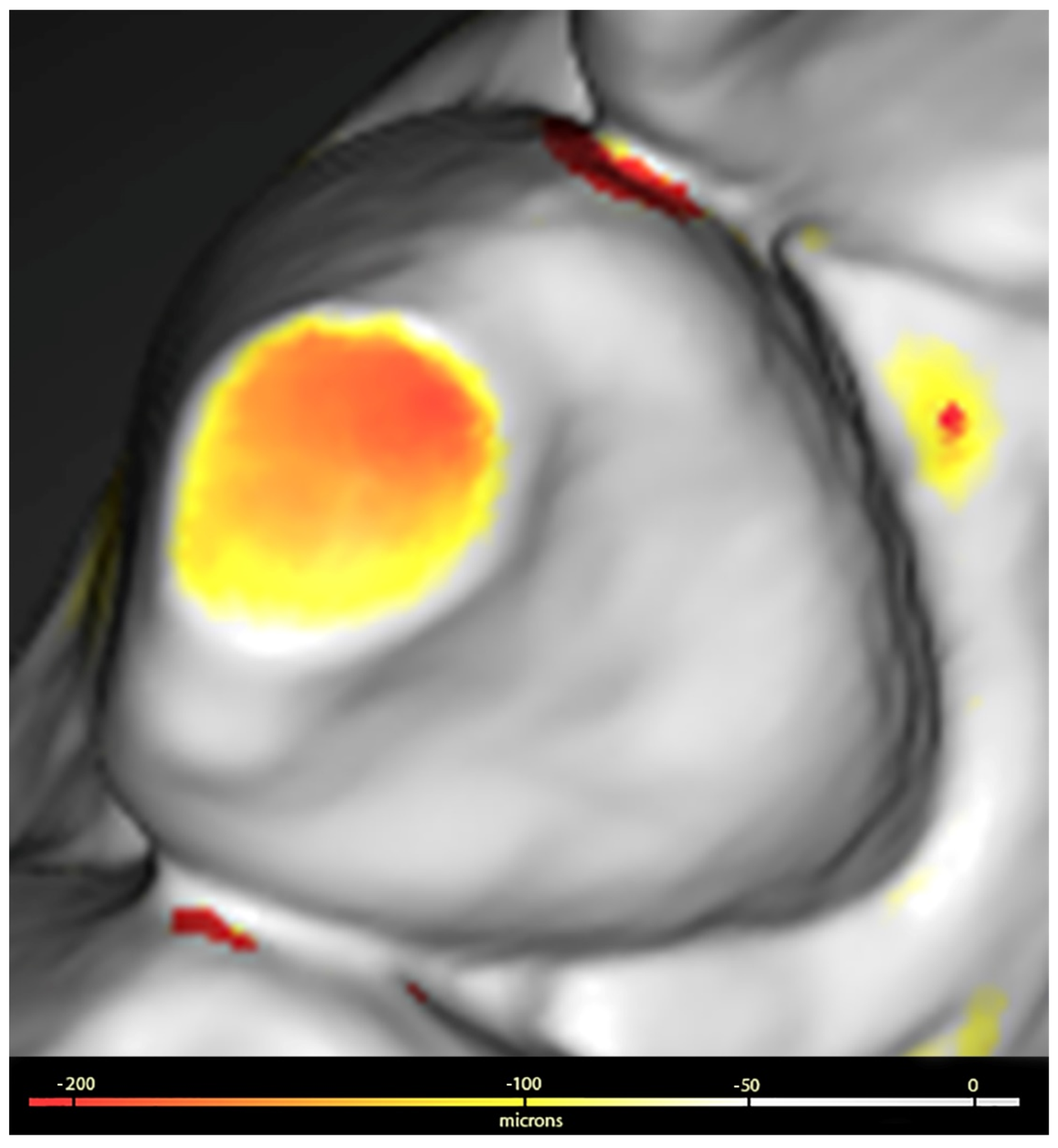
2.5. Image Processing

The surfaces digitised at T0 and T1 were saved in STL format and processed using Geomagic™ Control X software (3Dsystems, Darmstadt, Germany). This processing included the use of a colorimetric scale to facilitate the visualisation of dental wear, allowing the detection of discrepancies ranging from 20 microns (µ) to 5 millimetres (mm).
The procedure used to assess wear was as follows: the images corresponding to T0 and T1 were superimposed and aligned with Geomagic, which generated a 3D colour map that showed and quantified both the location and intensity of the volume differences on the surfaces of both sets. Volume losses (negative value) greater than 100 µ were identified on the three tooth surfaces (buccal, lingual or palatal, and incisal or occlusal). The values obtained were summed for each dental unit and for groups of teeth classified as anterosuperior (from upper canine to canine), posterosuperior (upper premolars and molars), anteroinferior (from lower canine to canine) and posteroinferior (lower premolars and molars), as shown in Figure 1.

2.6. Statistical Analysis

Descriptive analysis included both clinical and sociodemographic variables. For qualitative variables, absolute (n) and relative (%) frequencies were used, while for quantitative variables, mean ± standard deviation (SD) or median and interquartile range (IQR) were used, depending on the normality of their distribution.
In the inferential analysis, which aimed to study dental wear between T0 and T1, the Student’s t test or the Mann–Whitney U test were used for continuous variables, depending on their parametric nature. For qualitative variables, the Chi-square test or Fisher’s exact test was used.
All questionnaire answers and image processing data were compiled in an Excel spreadsheet (Microsoft, Albuquerque, NM, USA) and analysed using SPSS v.21.29 (IBM, SPSS Statistics, Version 21.29.0, Armonk, NY, USA: IBM Corp). Differences were considered statistically significant at p < 0.05.

3. Results

3.1. General Characteristics

Of the 65 participants who met the pre-defined criteria for the study, 39 agreed to participate in the study. Of these, 74.36% of the participants included in the study were male and 25.64% were female, with ages ranging from 20 to 34 years.

3.2. Dental Wear

The results are presented both individually for each tooth (considering its three surfaces and the total sum) and by sector (anterior and posterior). In particular, more pronounced wear was observed in the maxilla than in the mandible, as shown in Table 1 and Table 2.

3.3. Etiological Factors

1
Endogenous abrasion.
Table 3 shows the distribution of participants according to their self-reported symptoms related to endogenous abrasion.
The analysis revealed that patients with wear facets, nocturnal teeth grinding, clicking in the temporomandibular joint or pain on palpation of the masseter or external pterygoid muscles did not have significant levels of dental wear. Table 4 shows only the results that were statistically significant when comparing teeth with symptoms, specifying the specific surfaces.
The study also found no significant correlation between dental wear and State Trait Anxiety Inventory (STAI) scores for state and trait anxiety.
2
Possible etiological factors causing exogenous abrasion.
In relation to potential aetiological factors for exogenous abrasion, 51.28% of the participants reported engaging in parafunctional habits not related to bruxism. Of these, 90% reported biting hard objects, while 10% reported biting soft objects, as detailed in Table 5. In Table 5, only the results that were statistically significant when comparing the teeth of all patients who bit hard or soft objects are detailed, specifying the specific surfaces.
3
Potential aetiological factors for exogenous erosion.
Table 6 shows the distribution of participants who reported or denied engaging in activities that could contribute to dental wear due to exogenous erosion.
The correlation of these aetiological factors with dental wear, assessed using the Mann–Whitney U test, showed statistically significant results. In Table 7, only the results that were statistically significant when comparing the teeth of all patients with aetiological factors are detailed, specifying the specific surfaces.
4
Possible etiological factors causing endogenous erosion.
Table 8 shows the distribution of participants based on their self-reported symptoms associated with dental wear due to endogenous erosion.
In the analysis of aetiological factors, there was a notable association between nocturnal drooling and wear on the lower incisors, and between dry mouth and wear on the left lower canine. There was also a significant association between a burning sensation and dental wear on the lingual surfaces, as shown in Table 9, which details only the results that were statistically significant when comparing the teeth of all patients with aetiological factors, specifying the specific surfaces.

4. Discussion

The detection and monitoring of dental wear remains a challenge in clinical practice. A major obstacle in comparing studies on the relationship between potential aetiological factors and dental wear is the diversity of methodologies used, which vary between study models, intraoral photographs or a combination of both [53,54,55]. Although the Smith and Knight index [56] is widely recognised as the standard method for measuring wear, it has been criticised by some experts [57] for its limitations in differentiating wear by dentin exposure. In addition, the division of dentin exposure into thirds can lead to large variations in the quantification of wear severity, compromising the sensitivity of the results in favour of the reproducibility of the index [57]. These techniques, which often lack sensitivity, can be subjective [53]. In search of an effective solution to these challenges, recent research has validated the use of IOSs as reliable tools for monitoring and quantifying dental wear in vivo [2,34,49,50]. In this context, IOSs are emerging as tools that facilitate the implementation of an affordable and accurate wear quantification method [46]. In the present study, the use of the True Definition scanner for wear analysis was chosen due to its reliability and efficacy [58], a choice that is consistent with a systematic review published in 2023 showing that the most commonly used scanners for assessing dental wear are the True Definition, followed by the TRIOS 3, Cerec Omnicam and Planscan [59]. Furthermore, given the lack of in vivo studies [2,5,59] using this technology to quantify dental wear and its relationship to potential causes, the present study was developed.
Several methods can be used to effectively manage dental wear, including the use of splints to protect the teeth and reduce additional wear [60], medical treatments such as Botox to relax the jaw muscles and reduce the incidence of nocturnal grinding [61] and restorative materials to repair and protect teeth that are already affected and provide durable and aesthetic solutions [62].
In the assessment of dental wear attributed to endogenous abrasion, no significant association was found between nocturnal bruxism and the presence of dental wear, consistent with the findings of Bartolucci et al. [63]. However, a predominant effect on the anterior teeth was observed, particularly in participants who reported grinding during the day and in those with joint symptoms such as TMJ pain and crepitus or on palpation of the temporal or internal pterygoid muscles. Of note in this study, the presence of daytime or nighttime bruxism was subjectively determined by self-report, which requires cautious interpretation of signs of wear due to the multifactorial complexity of dental wear and its indirect implication in active bruxism [6,63]. Although bruxism has been associated in the literature with psychological problems such as anxiety and aggression [64,65,66], as well as stress at certain life stages [67,68], the relationship between sleep bruxism and general anxiety in adults remains controversial [69]. Similar to other studies, the identification of psychological factors and emotional stress was based on the STAI questionnaire, as it has reliable metric properties and is sensitive to environmental stressors [52]. However, it should be noted that it must be interpreted with caution without confirmation by sleep laboratory studies for the diagnosis of sleep bruxism [70].
The results did not show a significant relationship between dental wear and STAI scores, and there are no similar studies in the literature for comparison. Nevertheless, studies such as those by Al-Hiyasat et al. [11] and Winocur et al. [71] have found an association between dental wear and various psychiatric disorders; however, these differences should be interpreted with caution due to the nature of the group studied, which was diagnosed at the hospital level, and the methods used to measure wear.
With regard to exogenous abrasion, this type of dental wear, traditionally associated with toothbrushing, appears to be influenced more by the force applied during brushing than by the abrasive components in toothpaste [72]. Due to the complexity of measuring the brushing force applied by patients, studies in this area have mainly been carried out in vitro. In this context, our study evaluated parafunctional habits not associated with bruxism, such as biting hard and soft objects. Significant wear was observed in the anterior teeth, particularly on the entire surface, incisal edges and lingual surface. In addition, the study by Rusu Olaru et al. [22] reported cases where wear was limited to the incisal edges of anterior teeth, associated with habits such as nails or sunflower seeds. On the other hand, Nilner [73] did not observe wear in patients who bit their lips or cheeks, but did in those who bit their nails. Although it was noted that this wear occurred in the anterior teeth, no precise quantification was made.
Regarding dietary erosion, a two-year study by Schlenz et al. [50] found no correlation between tooth tissue loss and exposure to acidic substances. However, our study found more wear on certain surfaces in individuals who regularly consumed juices, energy drinks and dressings. Previous research using the Basic Erosive Wear Examination (BEWE) index to assess wear has also found an association between dental wear and the consumption of acidic foods, dairy products, fruit juices and alcohol [55,74]. Therefore, long-term studies are needed to assess the potential relationship between diet and dental wear using IOSs. Discrepancies between studies may be due to differences in dietary patterns between countries [55], as well as the methodologies used. Regarding environmental factors, our study observed increased wear on some buccal surfaces in participants who frequently swam in swimming pools, a finding consistent with previous studies [39,75] that used traditional methods to assess dental wear. Furthermore, as in our study, the most affected areas were the buccal surfaces of the incisors and canines, areas that have the most contact with pool water [39].
In this study, when investigating etiological factors potentially related to endogenous erosion, more pronounced dental wear was observed in participants who reported symptoms such as dry mouth, nocturnal drooling and heartburn. In contrast, no significant wear was found in those reporting reflux symptoms. Previous studies, such as those by Wetselaar et al. [6], have emphasised the multifactorial nature of dental wear, suggesting a possible coexistence of dry mouth and nocturnal bruxism. However, our analysis did not find a significant association between these factors. In a 2022 meta-analysis, Yanushevich et al. [30] associated dental erosion with GERD, a common gastrointestinal disorder. However, in our study, participants only reported reflux without a confirmed diagnosis of GERD, suggesting that dental wear associated with occasional reflux episodes may be lesser than that in patients with diagnosed GERD.
Regarding the methodology for analysing dental wear, some studies [76] have suggested focusing on “index teeth” to simplify the process and reduce costs. However, in our research we observed considerable variability in the wear results, possibly due to measuring each surface of each tooth and different groups of teeth. Rather than opting for simplification, it would be advisable to develop software applications that facilitate the comparison of intraoral scans and highlight surfaces with greater wear progression, using repositories of multiple scans from each patient.
The limitations of this study include the small sample size, which may limit the generalisation of the results to a larger population. In addition, the type of overbite of the patients was not taken into account, which could explain the higher wear in the anterior region in cases of deep bite and lower wear in cases of open bite. In addition, the combined interactions between factors were not considered, which may affect the full understanding of the causes of dental wear in the population studied.
Strengths of this study include the extensive use of IOSs to accurately monitor the progression of dental wear. This methodology allows for detailed and reliable monitoring, improving the ability to detect subtle changes in dental wear, which is often a challenge in daily clinical practice. In addition, this study addresses both intrinsic and extrinsic factors of dental wear, providing a comprehensive insight into the causes and progression of wear.

5. Conclusions

The used methodology proved effective in accurately assessing the progression of dental wear, which is important as many patients may initially be asymptomatic.
The present study also confirmed that dental wear is a multifactorial problem, influenced by both endogenous and exogenous factors. This understanding allows dental professionals to implement more specific preventive measures tailored to the individual needs of each patient, potentially improving clinical outcomes.
In addition, the findings suggest the need for improved diagnostic and monitoring tools, such as the development of advanced software capable of handling large repositories of scans and providing more accurate comparisons. This type of innovation could lead to better identification and management of risk factors associated with dental wear, allowing earlier and less invasive interventions.

Author Contributions

Conceptualization, Investigation, Formal Analysis, Software and Writing—original draft preparation (A.S., Y.F. and V.D.-F.G.); Investigation and Writing—review and editing (B.T.M., S.D.F. and M.G.S.). All authors have read and agreed to the published version of the manuscript.

Funding

This research received no external funding.

Institutional Review Board Statement

This study was conducted in accordance with the Declaration of Helsinki and approved by the Institutional Review Board Comité Ético de Investigación Clínica Regional de la Comunidad de Madrid (Project identification code Desgaste-UE, on 11 March 2018). This study was retrospectively registered in Clinical Trials under ID NCT05843513.

Informed Consent Statement

Informed consent was obtained from all subjects involved in the study.

Data Availability Statement

The data obtained in this study are available upon reasonable request.

Conflicts of Interest

The authors declare no conflicts of interest.

References

  1. Liu, B.; Zhang, M.; Chen, Y.; Yao, Y. Tooth Wear in Aging People: An Investigation of the Prevalence and the Influential Factors of Incisal/Occlusal Tooth Wear in Northwest China. BMC Oral Health 2014, 14, 65. [Google Scholar] [CrossRef] [PubMed]
  2. Travassos da Rosa Moreira Bastos, R.; Teixeira da Silva, P.; Normando, D. Reliability of Qualitative Occlusal Tooth Wear Evaluation Using an Intraoral Scanner: A Pilot Study. PLoS ONE 2021, 16, e0249119. [Google Scholar] [CrossRef] [PubMed]
  3. Nota, A.; Pittari, L.; Paggi, M.; Abati, S.; Tecco, S. Correlation between Bruxism and Gastroesophageal Reflux Disorder and Their Effects on Tooth Wear. A Systematic Review. J. Clin. Med. 2022, 11, 1107. [Google Scholar] [CrossRef] [PubMed]
  4. Bartlett, D.; O’Toole, S. Tooth Wear: Best Evidence Consensus Statement. J. Prosthodont. 2021, 30, 20–25. [Google Scholar] [CrossRef] [PubMed]
  5. Mitrirattanakul, S.; Neoh, S.P.; Chalarmchaichaloenkit, J.; Limthanabodi, C.; Trerayapiwat, C.; Pipatpajong, N.; Taechushong, N.; Chintavalakorn, R. Accuracy of the Intraoral Scanner for Detection of Tooth Wear. Int. Dent. J. 2023, 73, 56–62. [Google Scholar] [CrossRef] [PubMed]
  6. Wetselaar, P.; Manfredini, D.; Ahlberg, J.; Johansson, A.; Aarab, G.; Papagianni, C.E.; Reyes Sevilla, M.; Koutris, M.; Lobbezoo, F. Associations between Tooth Wear and Dental Sleep Disorders: A Narrative Overview. J. Oral Rehabil. 2019, 46, 765–775. [Google Scholar] [CrossRef] [PubMed]
  7. Kontaxopoulou, I.; Alam, S. Risk Assessment for Tooth Wear. Prim. Dent. J. 2015, 4, 25–29. [Google Scholar] [CrossRef] [PubMed]
  8. Kitagawa, K.; Kodama, N.; Manda, Y.; Mori, K.; Furutera, H.; Minagi, S. Effect of Masseter Muscle Activity during Wakefulness and Sleep on Tooth Wear. J. Prosthodont. Res. 2021, 66, 551–556. [Google Scholar] [CrossRef] [PubMed]
  9. Lobbezoo, F.; Ahlberg, J.; Raphael, K.G.; Wetselaar, P.; Glaros, A.G.; Kato, T.; Santiago, V.; Winocur, E.; De Laat, A.; De Leeuw, R.; et al. International Consensus on the Assessment of Bruxism: Report of a Work in Progress. J. Oral Rehabil. 2018, 45, 837–844. [Google Scholar] [CrossRef]
  10. Bandodkar, S.; Tripathi, S.; Chand, P.; Singh, S.V.; Arya, D.; Kumar, L.; Singh, M.; Singhal, R.; Tripathi, A. A Study to Evaluate Psychological and Occlusal Parameters in Bruxism. J. Oral Biol. Craniofac. Res. 2022, 12, 38–41. [Google Scholar] [CrossRef]
  11. Al-Hiyasat, A.S.; Khasawneh, S.F.; Khader, Y.S. Tooth Wear among Psychiatric Patients: Prevalence, Distribution, and Associated Factors. Int. J. Prosthodont. 2006, 19, 403–409. [Google Scholar] [PubMed]
  12. Osses-Anguita, Á.E.; Sánchez-Sánchez, T.; Soto-Goñi, X.A.; García-González, M.; Alén Fariñas, F.; Cid-Verdejo, R.; Sánchez Romero, E.A.; Jiménez-Ortega, L. Awake and Sleep Bruxism Prevalence and Their Associated Psychological Factors in First-Year University Students: A Pre-Mid-Post COVID-19 Pandemic Comparison. Int. J. Environ. Res. Public Health 2023, 20, 2452. [Google Scholar] [CrossRef] [PubMed]
  13. Goulart, A.C.; Arap, A.M.; Bufarah, H.B.; Bismarchi, D.; Rienzo, M.; Syllos, D.H.; Wang, Y.P. Anxiety, Depression, and Anger in Bruxism: A Cross-Sectional Study among Adult Attendees of a Preventive Center. Psychiatry Res. 2021, 299, 113844. [Google Scholar] [CrossRef] [PubMed]
  14. Rofaeel, M.; Chow, J.C.-F.; Cioffi, I. The Intensity of Awake Bruxism Episodes Is Increased in Individuals with High Trait Anxiety. Clin. Oral Investig. 2021, 25, 3197–3206. [Google Scholar] [CrossRef] [PubMed]
  15. Kuhn, M.; Türp, J.C. Risk Factors for Bruxism. Swiss Dent. J. 2018, 128, 118–124. [Google Scholar] [PubMed]
  16. Beddis, H.; Pemberton, M.; Davies, S. Sleep Bruxism: An Overview for Clinicians. Br. Dent. J. 2018, 225, 497–501. [Google Scholar] [CrossRef] [PubMed]
  17. Tao, J.; Liu, W.; Wu, J.; Zhang, X.; Zhang, Y. The Study of Grinding Patterns and Factors Influencing the Grinding Areas during Sleep Bruxism. Arch. Oral Biol. 2015, 60, 1595–1600. [Google Scholar] [CrossRef]
  18. Yagci, I.; Tasdelen, Y.; Kivrak, Y. Childhood Trauma, Quality of Life, Sleep Quality and Anxiety and Depression Levels in People with Bruxism. Arch. Neuropsychiatry 2019, 57, 131–135. [Google Scholar] [CrossRef]
  19. Dao, T.T.; Lund, J.P.; Lavigne, G.J. Comparison of Pain and Quality of Life in Bruxers and Patients with Myofascial Pain of the Masticatory Muscles. J. Orofac. Pain. 1994, 8, 350–356. [Google Scholar] [PubMed]
  20. Singh, T.; Nirola, A.; Brar, R. A Profilometric and Scanning Electron Microscopic Analysis of Tooth Surface Abrasion Caused by Rotary/Oscillatory, Linear Motion, Sonic, and Ultrasonic Toothbrushes: An in Vitro Study. J. Indian Soc. Periodontol. 2021, 25, 112. [Google Scholar] [CrossRef]
  21. Johansson, A.; Fareed, K.; Omar, R. Analysis of Possible Factors Influencing the Occurrence of Occlusal Tooth Wear in a Young Saudi Population. Acta Odontol. Scand. 1991, 49, 139–145. [Google Scholar] [CrossRef] [PubMed]
  22. Rusu Olaru, A.; Popescu, M.R.; Dragomir, L.P.; Popescu, D.M.; Arsenie, C.C.; Rauten, A.M. Identifying the Etiological Factors Involved in the Occurrence of Non-Carious Lesions. Curr. Health Sci. J. 2019, 45, 227–234. [Google Scholar] [PubMed]
  23. Panek, H.; Nawrot, P.; Mazan, M.; Bielicka, B.; Sumisławska, M.; Pomianowski, R. Coincidence and Awareness of Oral Parafunctions in College Students. Community Dent. Health 2012, 29, 74–77. [Google Scholar] [PubMed]
  24. Warreth, A.; Abuhijleh, E.; Almaghribi, M.A.; Mahwal, G.; Ashawish, A. Tooth Surface Loss: A Review of Literature. Saudi Dent. J. 2020, 32, 53–60. [Google Scholar] [CrossRef] [PubMed]
  25. Nam, J.; Nguyen, D.H.; Lee, S.; Heo, S.-M.; Park, J. Simulation of Non-Carious Cervical Lesions by Computational Toothbrush Model: A Novel Three-Dimensional Discrete Element Method. Sensors 2022, 22, 4183. [Google Scholar] [CrossRef] [PubMed]
  26. Hamza, B.; Martinola, L.; Körner, P.; Gubler, A.; Attin, T.; Wegehaupt, F.J. Effect of Brushing Force on the Abrasive Dentin Wear Using Slurries with Different Abrasivity Values. Int. J. Dent. Hyg. 2023, 21, 172–177. [Google Scholar] [CrossRef] [PubMed]
  27. Chauhan, N.; Manjunath, B.; Malhotra, P.; Yadav, V.; Sachin Kumar, J.; Muppalla, L.; Bhukal, S. Dietary Practices as a Potential Predictor for Dental Erosion among Patients Having Gastroesophageal Reflux Disease: An Analytical Cross-Sectional Study. J. Int. Soc. Prev. Community Dent. 2022, 12, 583. [Google Scholar] [CrossRef] [PubMed]
  28. Ramirez, V.; Lussi, A.; Marró Freitte, M.L.; Vasquez, P.; Aránguiz, V. Relationship between Intrinsic and Extrinsic Factors with Erosive Tooth Wear in Adults: A Cross-Sectional Study. Braz. Oral Res. 2022, 36, e0118. [Google Scholar] [CrossRef] [PubMed]
  29. Wilder-Smith, C.H.; Wilder-Smith, P.; Kawakami-Wong, H.; Voronets, J.; Osann, K.; Lussi, A. Quantification of Dental Erosions in Patients With GERD Using Optical Coherence Tomography Before and After Double-Blind, Randomized Treatment With Esomeprazole or Placebo. Am. J. Gastroenterol. 2009, 104, 2788–2795. [Google Scholar] [CrossRef]
  30. Yanushevich, O.O.; Maev, I.V.; Krikheli, N.I.; Andreev, D.N.; Lyamina, S.V.; Sokolov, F.S.; Bychkova, M.N.; Beliy, P.A.; Zaslavskaya, K.Y. Prevalence and Risk of Dental Erosion in Patients with Gastroesophageal Reflux Disease: A Meta-Analysis. Dent. J. 2022, 10, 126. [Google Scholar] [CrossRef]
  31. Leiman, D.A.; Metz, D.C. Gastroesophageal Reflux Disease. In Clinical Gastrointestinal Endoscopy; Elsevier: Amsterdam, The Netherlands, 2019; pp. 268–278.e3. [Google Scholar]
  32. Wetselaar, P.; Lobbezoo, F. The Tooth Wear Evaluation System: A Modular Clinical Guideline for the Diagnosis and Management Planning of Worn Dentitions. J. Oral Rehabil. 2016, 43, 69–80. [Google Scholar] [CrossRef] [PubMed]
  33. MercuŢ, V.; Popescu, S.M.; Scrieciu, M.; Amărăscu, M.O.; Vătu, M.; Diaconu, O.A.; Osiac, E.; Ghelase, Ş.M. Optical Coherence Tomography Applications in Tooth Wear Diagnosis. Rom. J. Morphol. Embryol. 2017, 58, 99–106. [Google Scholar] [PubMed]
  34. Schlenz, M.A.; Schlenz, M.B.; Wöstmann, B.; Jungert, A.; Ganss, C. Intraoral Scanner-Based Monitoring of Tooth Wear in Young Adults: 12-Month Results. Clin. Oral Investig. 2022, 26, 1869–1878. [Google Scholar] [CrossRef] [PubMed]
  35. Lan, Z.; Zhao, I.S.; Li, J.; Li, X.; Yuan, L.; Sha, O. Erosive Effects of Commercially Available Alcoholic Beverages on Enamel. Dent. Mater. J. 2023, 42, 236–240. [Google Scholar] [CrossRef] [PubMed]
  36. O’Toole, S.; Newton, T.; Moazzez, R.; Hasan, A.; Bartlett, D. Randomised Controlled Clinical Trial Investigating The Impact of Implementation Planning on Behaviour Related to The Diet. Sci. Rep. 2018, 8, 8024. [Google Scholar] [CrossRef] [PubMed]
  37. Khan, K.; Qadir, A.; Trakman, G.; Aziz, T.; Khattak, M.I.; Nabi, G.; Alharbi, M.; Alshammari, A.; Shahzad, M. Sports and Energy Drink Consumption, Oral Health Problems and Performance Impact among Elite Athletes. Nutrients 2022, 14, 5089. [Google Scholar] [CrossRef] [PubMed]
  38. Butera, A.; Gallo, S.; Pascadopoli, M.; Scardina, G.A.; Pezzullo, S.; Scribante, A. Home Oral Care Domiciliary Protocol for the Management of Dental Erosion in Rugby Players: A Randomized Clinical Trial. J. Clin. Med. 2022, 11, 4893. [Google Scholar] [CrossRef] [PubMed]
  39. Buczkowska-Radlińska, J.; Łagocka, R.; Kaczmarek, W.; Górski, M.; Nowicka, A. Prevalence of Dental Erosion in Adolescent Competitive Swimmers Exposed to Gas-Chlorinated Swimming Pool Water. Clin. Oral Investig. 2013, 17, 579–583. [Google Scholar] [CrossRef]
  40. Peampring, C. Restorative Management Using Hybrid Ceramic of a Patient with Severe Tooth Erosion from Swimming: A Clinical Report. J. Adv. Prosthodont. 2014, 6, 423. [Google Scholar] [CrossRef]
  41. Strużycka, I.; Lussi, A.; Bogusławska-Kapała, A.; Rusyan, E. Prevalence of Erosive Lesions with Respect to Risk Factors in a Young Adult Population in Poland—A Cross-Sectional Study. Clin. Oral Investig. 2017, 21, 2197–2203. [Google Scholar] [CrossRef]
  42. Mulic, A.; Fredriksen, Ø.; Jacobsen, I.D.; Tveit, A.B.; Espelid, I.; Crossner, C.G. Dental Erosion: Prevalence and Severity among 16-Year-Old Adolescents in Troms, Norway. Eur. J. Paediatr. Dent. 2016, 17, 197–201. [Google Scholar]
  43. Wei, Z.; Du, Y.; Zhang, J.; Tai, B.; Du, M.; Jiang, H. Prevalence and Indicators of Tooth Wear among Chinese Adults. PLoS ONE 2016, 11, e0162181. [Google Scholar] [CrossRef] [PubMed]
  44. Chawhuaveang, D.D.; Yu, O.Y.; Yin, I.X.; Lam, W.Y.H.; Chu, C.H. Topical Agents for Nonrestorative Management of Dental Erosion: A Narrative Review. Healthcare 2022, 10, 1413. [Google Scholar] [CrossRef] [PubMed]
  45. Esquivel-Upshaw, J.; Hsu, S.; Bohórquez, A.C.; Abdulhameed, N.; Scheiffele, G.W.; Kim, M.; Neal, D.; Chai, J.; Ren, F. Novel Methodology for Measuring Intraoral Wear in Enamel and Dental Restorative Materials. Clin. Exp. Dent. Res. 2020, 6, 677–685. [Google Scholar] [CrossRef]
  46. Bronkhorst, H.; Bronkhorst, E.; Kalaykova, S.; van der Meer, W.; Huysmans, M.-C.; Loomans, B. Precision of In Vivo Quantitative Tooth Wear Measurement Using Intra-Oral Scans. J. Vis. Exp. 2022, 185, e63680. [Google Scholar] [CrossRef]
  47. Passos, V.F.; Santiago, S.L. Methodologies to Analyze the Micromorphological Alterations of Enamel Subjected to Abrasion/Erosion. Dentistry 2014, 4, 1000255. [Google Scholar] [CrossRef]
  48. Katsoulis, J.; Nikitovic, S.G.; Spreng, S.; Neuhaus, K.; Mericske-Stern, R. Prosthetic Rehabilitation and Treatment Outcome of Partially Edentulous Patients with Severe Tooth Wear: 3-Years Results. J. Dent. 2011, 39, 662–671. [Google Scholar] [CrossRef]
  49. García, V.D.-F.; Freire, Y.; Fernández, S.D.; Murillo, B.T.; Sánchez, M.G. Application of the Intraoral Scanner in the Diagnosis of Dental Wear: An In Vivo Study of Tooth Wear Analysis. Int. J. Environ. Res. Public Health 2022, 19, 4481. [Google Scholar] [CrossRef] [PubMed]
  50. Schlenz, M.A.; Schlenz, M.B.; Wöstmann, B.; Glatt, A.S.; Ganss, C. Intraoral Scanner-Based Monitoring of Tooth Wear in Young Adults: 24-Month Results. Clin. Oral Investig. 2023, 27, 2775–2785. [Google Scholar] [CrossRef]
  51. Spielberger, C.D.; Gorsuch, R.L.; Lushen, R.E.; Vagg, P.R.; Jacobs, G.A. Manual for the State-Trait Anxiety Inventory; Spielberger, C.D., Gorsuch, R.L., Lushen, R.E., Vagg, P.R., Jacobs, G.A., Eds.; Consulting Psychologist Press: Palo Alto, CA, USA, 1983. [Google Scholar]
  52. Guillén-Riquelme, A.; Buela-Casal, G. [Psychometric Revision and Differential Item Functioning in the State Trait Anxiety Inventory (STAI)]. Psicothema 2011, 23, 510–515. [Google Scholar]
  53. Kumar, S.; Keeling, A.; Osnes, C.; Bartlett, D.; O’Toole, S. The Sensitivity of Digital Intraoral Scanners at Measuring Early Erosive Wear. J. Dent. 2019, 81, 39–42. [Google Scholar] [CrossRef] [PubMed]
  54. Marro, F.; De Lat, L.; Martens, L.; Jacquet, W.; Bottenberg, P. Monitoring the Progression of Erosive Tooth Wear (ETW) Using BEWE Index in Casts and Their 3D Images: A Retrospective Longitudinal Study. J. Dent. 2018, 73, 70–75. [Google Scholar] [CrossRef]
  55. Sezer, B.; Giritlioğlu, B.; Sıddıkoğlu, D.; Lussi, A.; Kargül, B. Relationship between Erosive Tooth Wear and Possible Etiological Factors among Dental Students. Clin. Oral Investig. 2022, 26, 4229–4238. [Google Scholar] [CrossRef]
  56. Smith, B.G.; Knight, J.K. An Index for Measuring the Wear of Teeth. Br. Dent. J. 1984, 156, 435–438. [Google Scholar] [CrossRef]
  57. AL-Omiri, M.K.; Sghaireen, M.G.; AlZarea, B.K.; Lynch, E. Quantification of Incisal Tooth Wear in Upper Anterior Teeth: Conventional vs New Method Using Toolmakers Microscope and a Three-Dimensional Measuring Technique. J. Dent. 2013, 41, 1214–1221. [Google Scholar] [CrossRef] [PubMed]
  58. Sacher, M.; Schulz, G.; Deyhle, H.; Jäger, K.; Müller, B. Accuracy of Commercial Intraoral Scanners. J. Med. Imaging 2021, 8, 035501. [Google Scholar] [CrossRef]
  59. Angelone, F.; Ponsiglione, A.M.; Ricciardi, C.; Cesarelli, G.; Sansone, M.; Amato, F. Diagnostic Applications of Intraoral Scanners: A Systematic Review. J. Imaging 2023, 9, 134. [Google Scholar] [CrossRef] [PubMed]
  60. Albagieh, H.; Alomran, I.; Binakresh, A.; Alhatarisha, N.; Almeteb, M.; Khalaf, Y.; Alqublan, A.; Alqahatany, M. Occlusal Splints-Types and Effectiveness in Temporomandibular Disorder Management. Saudi Dent. J. 2023, 35, 70–79. [Google Scholar] [CrossRef] [PubMed]
  61. Ågren, M.; Sahin, C.; Pettersson, M. The Effect of Botulinum Toxin Injections on Bruxism: A Systematic Review. J. Oral Rehabil. 2020, 47, 395–402. [Google Scholar] [CrossRef]
  62. Heintze, S.D.; Reichl, F.-X.; HICKEL, R. Wear of Dental Materials: Clinical Significance and Laboratory Wear Simulation Methods—A Review. Dent. Mater. J. 2019, 38, 343–353. [Google Scholar] [CrossRef]
  63. Bartolucci, M.L.; Incerti Parenti, S.; Bortolotti, F.; Della Godenza, V.; Vandi, S.; Pizza, F.; Plazzi, G.; Alessandri-Bonetti, G. Sleep Bruxism and Orofacial Pain in Patients with Sleep Disorders: A Controlled Cohort Study. J. Clin. Med. 2023, 12, 2997. [Google Scholar] [CrossRef]
  64. Olkinuora, M. Psychosocial Aspects in a Series of Bruxists Compared with a Group of Non-Bruxists. Proc. Finn. Dent. Soc. 1972, 68, 200–208. [Google Scholar]
  65. Schulte, J.K. Bruxism: A Review and Clinical Approach to Treatment. Northwest Dent. 1982, 61, 13–18. [Google Scholar]
  66. Rugh, J.D.; Harlan, J. Nocturnal Bruxism and Temporomandibular Disorders. Adv. Neurol. 1988, 49, 329–341. [Google Scholar]
  67. Funch, D.P.; Gale, E.N. Factors Associated with Nocturnal Bruxism and Its Treatment. J. Behav. Med. 1980, 3, 385–397. [Google Scholar] [CrossRef] [PubMed]
  68. Rugh, J.D.; Solberg, W.K. Psychological Implications in Temporomandibular Pain and Dysfunction. Oral Sci. Rev. 1976, 7, 3–30. [Google Scholar]
  69. Polmann, H.; Domingos, F.L.; Melo, G.; Stuginski-Barbosa, J.; Guerra, E.N.d.S.; Porporatti, A.L.; Dick, B.D.; Flores-Mir, C.; De Luca Canto, G. Association between Sleep Bruxism and Anxiety Symptoms in Adults: A Systematic Review. J. Oral Rehabil. 2019, 46, 482–491. [Google Scholar] [CrossRef] [PubMed]
  70. Alonso, J.; Angermeyer, M.C.; Bernert, S.; Bruffaerts, R.; Brugha, T.S.; Bryson, H.; Girolamo, G.; Graaf, R.; Demyttenaere, K.; Gasquet, I.; et al. Use of Mental Health Services in Europe: Results from the European Study of the Epidemiology of Mental Disorders (ESEMeD) Project. Acta Psychiatr. Scand. 2004, 109, 47–54. [Google Scholar] [CrossRef]
  71. Winocur, E.; Hermesh, H.; Littner, D.; Shiloh, R.; Peleg, L.; Eli, I. Signs of Bruxism and Temporomandibular Disorders among Psychiatric Patients. Oral Surg. Oral Med. Oral Pathol. Oral Radiol. Endodontol. 2007, 103, 60–63. [Google Scholar] [CrossRef]
  72. Hamza, B.; Niedzwiecki, M.; Körner, P.; Attin, T.; Wegehaupt, F.J. Effect of the Toothbrush Tuft Arrangement and Bristle Stiffness on the Abrasive Dentin Wear. Sci. Rep. 2022, 12, 840. [Google Scholar] [CrossRef]
  73. Nilner, M. Relationships Between Oral Parafunctions and Functional Disturbances and Diseases of the Stomatognathic System Among Children Aged 7–14 Years. Acta Odontol. Scand. 1983, 41, 167–172. [Google Scholar] [CrossRef] [PubMed]
  74. Vered, Y.; Lussi, A.; Zini, A.; Gleitman, J.; Sgan-Cohen, H.D. Dental Erosive Wear Assessment among Adolescents and Adults Utilizing the Basic Erosive Wear Examination (BEWE) Scoring System. Clin. Oral Investig. 2014, 18, 1985–1990. [Google Scholar] [CrossRef] [PubMed]
  75. Zhang, J.; Du, Y.; Wei, Z.; Tai, B.; Jiang, H.; Du, M. The Prevalence and Risk Indicators of Tooth Wear in 12- and 15-Year-Old Adolescents in Central China. BMC Oral Health 2015, 15, 120. [Google Scholar] [CrossRef]
  76. O’Toole, S.; Lau, J.S.; Rees, M.; Warburton, F.; Loomans, B.; Bartlett, D. Quantitative Tooth Wear Analysis of Index Teeth Compared to Complete Dentition. J. Dent. 2020, 97, 103342. [Google Scholar] [CrossRef]
Figure 1. Detail of the colour signal and its correspondence in microns.
Healthcare 12 01069 g001
Table 1. Quantification of the average wear of maxillary teeth.
Upper ArchSurfaceMean (µ)SD
Upper right central incisor (1.1)B38.4683.08
P33.3365.23
I124.36115.21
Total196.15160.75
Upper right lateral incisor (1.2)B48.7294.23
P66.6784.55
I89.74103.35
Total205.13188.41
Upper right canine (1.3)B61.5494.22
P69.23102.35
I106.41100.77
Total237.18197.26
Upper left central incisor (2.1)B35.9076.04
P47.4469.73
I114.1094.55
Total197.44149.10
Upper left lateral incisor (2.2)B19.2353.33
P52.5675.17
I102.56104.47
Total174.36135.66
Upper left canine (2.3)B41.0366.76
P43.5977.95
I65.3884.41
Total150.00129.27
Upper Anterior Teeth (1.1, 1.2, 1.3, 2.1, 2.2, 2.3)1160.26535.97
1st upper right premolar (1.4)B51.28104.81
P11.5435.28
O217.95134.01
Total280.77179.04
2nd upper right premolar (1.5)B35.9073.40
P21.7971.45
O242.31141.67
Total300.00185.67
1st upper right molar (1.6)B83.33115.47
P82.05101.62
O246.15118.87
Total411.54202.76
1st upper left premolar (2.4)B56.4174.50
P15.3853.99
O211.54149.32
Total283.33157.42
2nd upper left premolar (2.5)B41.0395.88
P14.1049.93
O246.15148.85
Total301.28186.55
1st upper left molar (2.6)B67.95108.51
P60.2699.46
O276.92109.33
Total405.13208.00
Upper Posterior Teeth (1.4, 1.5, 1.6, 2.4, 2.5, 2.6)1982.05730.40
B: buccal; L: lingual; P: palatal; I: incisal; O: oclusal; Total: sum of P/L, B and I/O; SD: Standard Deviation.
Table 2. Quantification of the average wear of mandibular teeth.
Lower ArchSurfaceMean (µ)SD
Lower left central incisor (3.1)B35.9068.78
L43.5977.95
I78.2183.35
Total157.69139.80
Lower left lateral incisor (3.2)B20.5150.94
L16.6746.36
I70.5173.20
Total107.6984.71
Lower left canine (3.3)B35.9073.40
L17.9547.97
I67.9577.36
Total121.79121.83
Lower right central incisor (4.1)B30.7764.50
L30.7770.35
I79.4992.99
Total141.03154.69
Lower right lateral incisor (4.2)B42.3189.25
L37.1870.45
I73.0864.73
Total152.56146.43
Lower right canine (4.3)B48.7299.00
L43.5973.61
I70.5186.39
Total162.82178.71
Lower Anterior Teeth (3.1, 3.2, 3.3, 4.1, 4.2, 4.3)843.59490.73
1st lower left premolar (3.4)B37.1874.09
L7.6935.43
O126.92133.70
Total171.79148.14
2nd lower left premolar (3.5)B16.6744.92
L24.3667.74
O175.64129.20
Total216.67165.57
1st lower left molar (3.6)B47.4480.25
L52.5681.88
O244.8797.19
Total344.87144.09
1st lower right premolar (4.4)B30.7781.61
L35.9086.56
O165.38102.05
Total232.05183.71
2nd lower right premolar (4.5)B14.1049.93
L34.6286.72
O173.08133.70
Total221.79170.45
1st lower right molar (4.6)B43.5983.65
L55.1399.20
O256.41135.33
Total355.13188.41
Lower Posterior Teeth (3.4, 3.5, 3.6, 4.4, 4.5, 4.6)1542.31658.50
B: buccal; L: lingual; I: incisal; O: oclusal; Total: sum of P/L, B and I/O; SD: Standard Deviation.
Table 3. Presence or absence of bruxism-related symptoms.
SymptomYesNo
N%N%
Teeth grinding while awake1641.032358.97
Teeth grinding during sleep2051.281948.72
Wear facets3186.11513.89
Pain on palpation of the masseter muscles717.953282.05
Pain on palpation of the temporalis muscles37.693692.31
Pain on palpation of the external pterygoid muscles820.513179.49
Pain on palpation of the internal pterygoid muscles1128.212871.79
Pain on palpation of the TMJ25.133794.87
Clicking in TMJ1435.92464.10
Crepitus in TMJ820.513179.49
Table 4. Quantification of mean tooth wear versus bruxism-related symptoms.
YesNo
SymptomToothLocationMean Wear (µ)SDMean Wear (µ)SDp-Value
Teeth grinding while awake2.1I156.398.184.881.80.022 *
4.2I10048.354.368.90.022 *
Pain on palpation of the temporalis muscles2.1I233.357.7104.290.50.026 *
1.1B166.7152.827.868.10.019 *
Total surfaces of 11500173.2170.8133.30.013 *
Pain on palpation of the internal pterygoid muscles1.1P63.677.821.456.80.047 *
4.1I131.8114.658.975.80.044 *
Pain on palpation of the TMJ1.1P150.070.727.059.60.008 *
3.1B175.035.428.461.80.005 *
Crepitus in TMJ4.1I162.5130.258.168.40.024 *
B: buccal; L: lingual; P: palatal; I: incisal; Total: sum of P/L, B and I/O. Yes: patients who reported symptoms; No: patients who did not report symptoms. SD: standard deviation. * p-value < 0.05- statistically significant.
Table 5. Quantification of mean tooth wear vs. parafunctional habits not related to bruxism.
YesNo
Etiological FactorToothLocationMean Wear (µ)SDMean Wear (µ)SDp-Value
BH2.3I105.698.428.950.90.033 *
BS5070.7
BHTotal surfaces of 2.3205.6125.997.4118.40.031 *
BS15070.7
BHTotal surfaces of 1.3322.2203.8181.6161.80.017 *
BS0.00.0
BH3.1L5078.618.444.80.012 *
BS225105.1
BH3.2L5.623.613.240.3<0.001 *
BS15070.7
BH4.2L33.364.228.969.40.042 *
BS15070.7
BH4.5B19.457.20.00.00.030 *
BS100141.4
BH4.6B63.9104.010.531.50.007 *
BS17535.4
BH: biting hard objects; BS: biting soft objects; B: buccal; L: lingual; I: incisal; Total: sum of P/L, B and I/O. Yes: patients who reported experiencing the aetiological factors; No: patients who did not report experiencing the aetiological factors. SD: standard deviation, * p-value < 0.05- statistically significant.
Table 6. Presence or absence of etiological factors that may cause exogenous erosion.
Etiological FactorYesNo
N%N%
Frequent swimming820.513179.49
Consumption of carbonated beverages2051.281948.72
Consumption of energy drinks1025.642974.36
Consumption of juice2666.671333.33
Consumption of Alcohol2461.541538.46
Consumption of fruit3692.3137.69
Consumption of dressing2769.231230.77
Table 7. Quantification of mean tooth wear versus etiological factors related to exogenous erosion.
YesNo
Etiological FactorToothLocationMean Wear (µ)SDMean Wear (µ)SDp-Value
Frequent swimming2.3B87.579.12958.80.024 *
1.1B93.882.124.278.40.003 *
3.3B112.595.416.152.30.001 *
Consumption of energy drinks3.2L5070.75.227.90.004 *
4.2L7582.52462.10.034 *
Consumption of juice2.3I86.587.823.159.90.018 *
Total surfaces of 2.3190.4126.569.294.70.004 *
3.2I88.575.234.655.50.031 *
3.4B55.885.200.00.011 *
Total surfaces of 3.4201.9135.3111.5159.60.040 *
Consumption of dressing1.6B109.3124.82462.20.041 *
B: buccal; L: lingual; P: palatal; I: incisal; Total: sum of P/L, B and I/O. Yes: patients who reported experiencing the aetiological factors; No: patients who did not report experiencing the aetiological factors. SD: standard deviation, * p-value < 0.05- statistically significant.
Table 8. Presence or absence of factors that may cause endogenous erosion.
Etiological FactorYesNo
N%N%
Night drooling923.083076.92
Reflux820.513179.49
Dry mouth489.743510.26
Vomiting0039100
Heartburn923.083076.92
Table 9. Quantification of mean tooth wear versus habits associated with exogenous erosion.
YesNo
Etiological FactorToothLocationMean Wear (µ)SDMean Wear (µ)SDp-Value
Night drooling3.1Total surfaces of 3.1250156.1130124.30.031 *
4.2I116.761.26060.70.026 *
Total surfaces of 4.2272.2195.4116.7108.50.016 *
Dry mouth3.3I12528.961.478.70.044 *
Heartburn3.3L44.468.21038.10.044 *
L: lingual; I: incisal; Total: sum of P/L, B and I/O. Yes: patients who reported experiencing the aetiological factors; No: patients who did not report experiencing the aetiological factors. SD: standard deviation. * p-value < 0.05- statistically significant.
Disclaimer/Publisher’s Note: The statements, opinions and data contained in all publications are solely those of the individual author(s) and contributor(s) and not of MDPI and/or the editor(s). MDPI and/or the editor(s) disclaim responsibility for any injury to people or property resulting from any ideas, methods, instructions or products referred to in the content.

