Article

Evaluation of Blood Cultures from SARS-CoV-2-Positive and Negative Adult Patients †

by
Bahar Akgün Karapınar
1,*,
İlvana Çaklovica Küçükkaya
1,
Yasemin Bölükbaşı
1,
Sertaç Küçükkaya
1,
Gonca Erköse Genç
2,
Zayre Erturan
2,
Ali Ağaçfidan
3 and
Betigül Öngen
1
1
İstanbul Faculty of Medicine, Medical Microbiology-Bacteriology Department, İstanbul University, 34093 İstanbul, Turkey
2
İstanbul Faculty of Medicine, Medical Microbiology-Mycology Department, İstanbul University, 34093 İstanbul, Turkey
3
İstanbul Faculty of Medicine, Medical Microbiology-Virology and İmmunology Department, İstanbul University, 34093 İstanbul, Turkey
*
Author to whom correspondence should be addressed.
The study was presented at the “32nd European Congress of Clinical Microbiology and Infectious Diseases, Lisbon, Portugal”, held on 23–26 April 2022. Poster number: 4568.
Healthcare 2023, 11(18), 2581; https://doi.org/10.3390/healthcare11182581
Submission received: 15 August 2023 / Revised: 6 September 2023 / Accepted: 11 September 2023 / Published: 19 September 2023
(This article belongs to the Section Coronaviruses (CoV) and COVID-19 Pandemic)

Abstract

:
Bacteremia and fungemia are significant causes of morbidity and mortality that frequently occur as co-infections with viral respiratory infections, including SARS-CoV-2. The aim of this study was to evaluate the microorganisms that were isolated from the blood cultures of SARS-CoV-2-positive and negative patients and investigate their antimicrobial resistance patterns. A retrospective analysis was performed of 22,944 blood cultures sent to the laboratory between November 2020 and December 2021. Blood culture analyses were performed using the BD Bactec FX automated system. Identification was carried out using conventional methods, namely, VITEK-2 and MALDI-TOF MS. Antibacterial/antifungal susceptibility tests were performed according to EUCAST/CLSI recommendations. SARS-CoV-2 tests were performed with RT-PCR. Culture positivity was detected in 1630 samples from 652 patients. Of these 652 patients, 633 were tested for SARS-CoV-2; 118 (18.6%) were positive and 515 (81.3%) were negative. The bacteria and fungi that were isolated at the highest rate in SARS-CoV-2-positive patients were methicillin-resistant coagulase-negative staphylococci (MR-CoNS) (21.5%), Escherichia coli (12.4%), Klebsiella pneumoniae (12.4%), Candida albicans (1.65%), and Candida glabrata complex (1.65%), while in the negative patients, the highest rates were for E. coli (21.3%), MR-CoNS (13.5%), K. pneumoniae (12.05%), C. albicans (2.1%), Candida parapsilosis (1.1%), and Candida tropicalis (0.9%). No statistically significant difference was determined between COVID-19-positive and negative patients in terms of detection, such as with the Pseudomonas spp., Enterococcus spp., and methicillin-resistant Staphylococcus aureus isolated from the blood cultures (p > 0.05). The most common isolate was MR-CoNS in SARS-CoV-2-positive patients (p = 0.028). Acinetobacter baumannii was more frequent (p = 0.004) and carbapenem-resistant K. pneumoniae was isolated at a higher rate (60% vs. 43%) in SARS-CoV-2-positive patients compared to SARS-CoV-2-negative patients (p > 0.05). These findings highlight the fact that isolation procedures should not be disregarded and the distribution of bacterial/fungal agents of bloodstream infections and their antibiotic resistance should be followed up during a pandemic, such as in the case of COVID-19.

1. Introduction

Viral respiratory tract infections cause high rates of morbidity and mortality throughout the world. Of the six largest outbreaks in the world in the last 20 years, four were caused by respiratory tract infections. Morbidity and mortality caused by bacterial, viral, and fungal infections are the main complications of viral infections, especially with viruses affecting the respiratory tract. As SARS-CoV-2 causes severe lung infections, similar to those seen in deaths by bacterial co-infection in the H1N1 influenza pandemic, the monitoring of morbidity and mortality is important [1]. SARS-CoV-2 infection can also occur as a co-infection secondary to bacterial infection or bacterial super-infection and may develop secondary to SARS-CoV-2 infection, depending on the host, virus, and bacterial factors [2]. Data have shown that culture-proven infections occur in 4–15% of hospitalized COVID-19 patients and are significantly associated with mortality [3].
SARS-CoV-2 infection may cause tissue destruction with the development of a combined infection that increases bacterial colonization and attachment in the relevant region. Airway dysfunction, cellular pathology, and tissue destruction are induced by the presence of SARS-CoV-2 and/or bacterial co-infection and can facilitate the systemic spread of pathogens, thereby significantly increasing the risk of bloodstream infections and sepsis [2]. Bloodstream infections (BSI) in COVID-19 patients may be associated with the systemic dissemination of co-pathogens caused by SARS-CoV-2-induced tissue destruction [2]. In severe COVID-19 lower respiratory tract infections, multidrug-resistant (MDR) bacteria such as Pseudomonas and Acinetobacter can colonize this damaged area and trigger secondary infections [4]. The risk of bacterial infection may be higher in patients with severe laboratory and clinical conditions. Co-infections or secondary infection positivity can vary between 0.6% and 45%. However, further studies involving bacterial species and infection sites are needed for detailed evaluations [5].
In patients with severe febrile illness, blood cultures are still essential for clinicians to be able to rule out bacterial/fungal BSI infections. However, there is a lack of sufficient data on the prevalence of bacterial and fungal agents causing BSI in patients infected with SARS-CoV-2 [6,7]. Recent studies have shown Escherichia coli, coagulase-negative staphylococci (CoNS), Klebsiella pneumoniae, methicillin-resistant Staphylococcus aureus (MRSA), and methicillin-sensitive Staphylococcus aureus (MSSA) to be the most frequently isolated agents in BSI [6,7]. The frequent use of more than one antimicrobial drug due to viral pneumonia and secondary infection results in an increased consumption of antimicrobials [8]. In addition, factors such as patient density, a prolonged stay in the ICU, the use of mechanical ventilators, and the expanded use of antimicrobials have resulted in the emergence and rapid spread of MDR bacteria [5,9]. Pathogens displaying a multidrug-resistant phenotype, such as carbapenem-resistant Enterobacterales (CRE), can cause problems in antimicrobial treatment processes [3]. Therefore, the determination of susceptibility and the prevalence of bacterial co-infections are likely to provide important information regarding the need for and choice of antibiotics [6].
Studies conducted during the COVID-19 pandemic on the distribution of microorganisms causing infections, the frequency of polymicrobial infections, and the effect of the pandemic on changes in antimicrobial resistance are important for ongoing and future processes. Therefore, the aim of this study was to evaluate the bacteria and fungi isolated from blood cultures of SARS-CoV-2-positive and negative patients, to investigate antimicrobial resistance patterns, and to compare the findings of these two patient groups in respect of the rates of bacteremia and fungemia that were determined.

2. Materials and Methods

2.1. Study Population

A retrospective analysis was performed with a total of 22,944 blood cultures sent to the laboratory between November 2020 and December 2021. When defining BSI, if a species belonging to the skin flora was found among the isolated microorganisms, it was considered that the same species should be recovered in at least 2 blood cultures collected from the same patient within 24 h, according to the Centers for Disease Control and Prevention (CDC) criteria [10]. The growth of these microorganisms in a single blood culture within 24 h was considered contamination, except in the neonatal period [11].

2.2. Blood Culture

Blood cultures were performed using the BD Bactec FX (Becton Dickinson, Sparks, MD, USA) automated system and cultures were incubated for up to 5 days. In special circumstances (for Brucella spp., etc.), the cultures were incubated for longer than in the standard procedure [12]. All positive blood cultures were subcultured on 5% Columbia sheep blood agar (Becton Dickinson, USA) and incubated at 35–37 °C under a 5–10% CO2 atmosphere for 48 h. Anaerobic positive blood cultures were also subcultured on anaerobic media and incubated at 35–37 °C under anaerobic conditions for 48 h. If there was no growth on the initial media, the blood cultures were subcultured on chocolate agar (incubated at 35–37 °C under 5–10% CO2 atmosphere for 48 h) for the isolation of fastidious microorganisms and on blood agar (incubated micro-aerobically at 35–37 °C for 48 h) for Campylobacter species.

2.2.1. Identification of Bacterial and Fungal Isolates

Bacterial identification was performed using conventional methods and with the VITEK-2 Compact system (bioMérieux, Marcy l’Etoile, France). Fungi isolated from Myco F or aerobic bottles were identified by morphological examination on cornmeal agar with Tween 80 and API ID 32C (bioMérieux, Marcy l’Etoile, France). Incompatible results were confirmed via MALDI-TOF MS (bioMérieux, Marcy l’Etoile, France).

2.2.2. Antimicrobial Susceptibility Testing

The antibacterial susceptibilities of the isolates were investigated using the standard Kirby–Bauer disk diffusion method and the VITEK-2 Compact system (bioMérieux, France) when necessary. Antifungal susceptibility was tested with the gradient test method using Roswell Park Memorial Institute medium (RPMI-1640) (Sigma-Aldrich, St. Louis, MO, USA), in order to detect the minimum inhibitory concentration (MIC) values based on elliptical growth around the antifungal gradient. The values were read on the higher MIC values side of the strips. Candida parapsilosis ATCC 22019 and Candida krusei ATCC 6258 were used for susceptibility testing as quality-control isolates. Antibacterial and antifungal susceptibility tests were performed and evaluated in accordance with the EUCAST/CLSI criteria [13,14,15,16]. “Intermediate (I): susceptible, increased exposure’’ strains of bacteria were considered susceptible. Since there are no clinical breakpoints for Candida auris, the MICs were evaluated according to the tentative breakpoints determined by the CDC [17].

2.3. The COVID-19 Diagnoses of the Patients

COVID-19 status was confirmed via SARS-CoV-2 real-time polymerase chain reaction (RT-PCR) test positivity in the nasopharyngeal and oral swabs.

2.4. Statistical Analysis

The software SPSS for Windows version 15.0 was used for statistical analysis. Descriptive statistics were given as numbers and percentages for categorical variables. Proportions were compared with the chi-square (χ2) test in independent groups. The Pearson chi-square test was used when there were no cells with expected values of < 1 and the number of cells with values of <5 was a maximum of 20%. When these conditions were not met, either Fisher’s exact test results for 2 × 2 tables were given, or Monte Carlo simulation results for 2 × 3 tables. Tests were performed within a 95% confidence interval. A value of p < 0.05 was considered statistically significant.

2.5. Ethical Approval
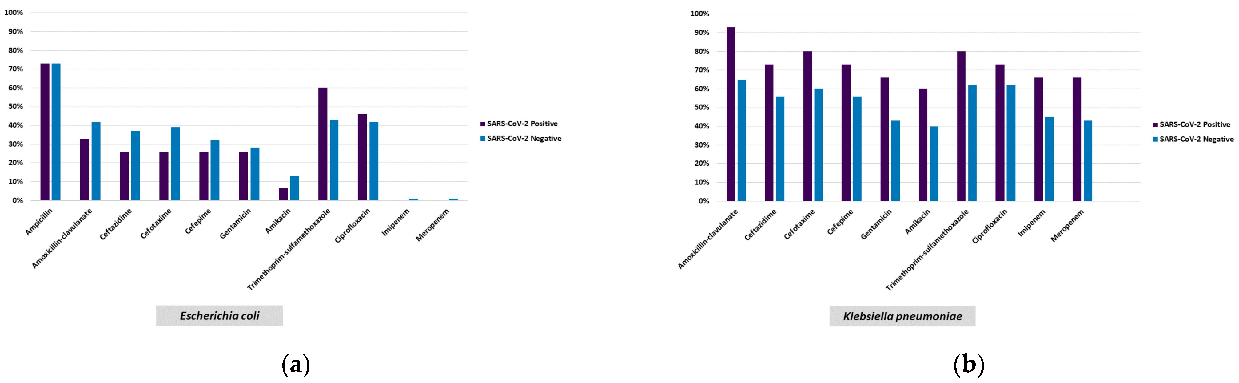
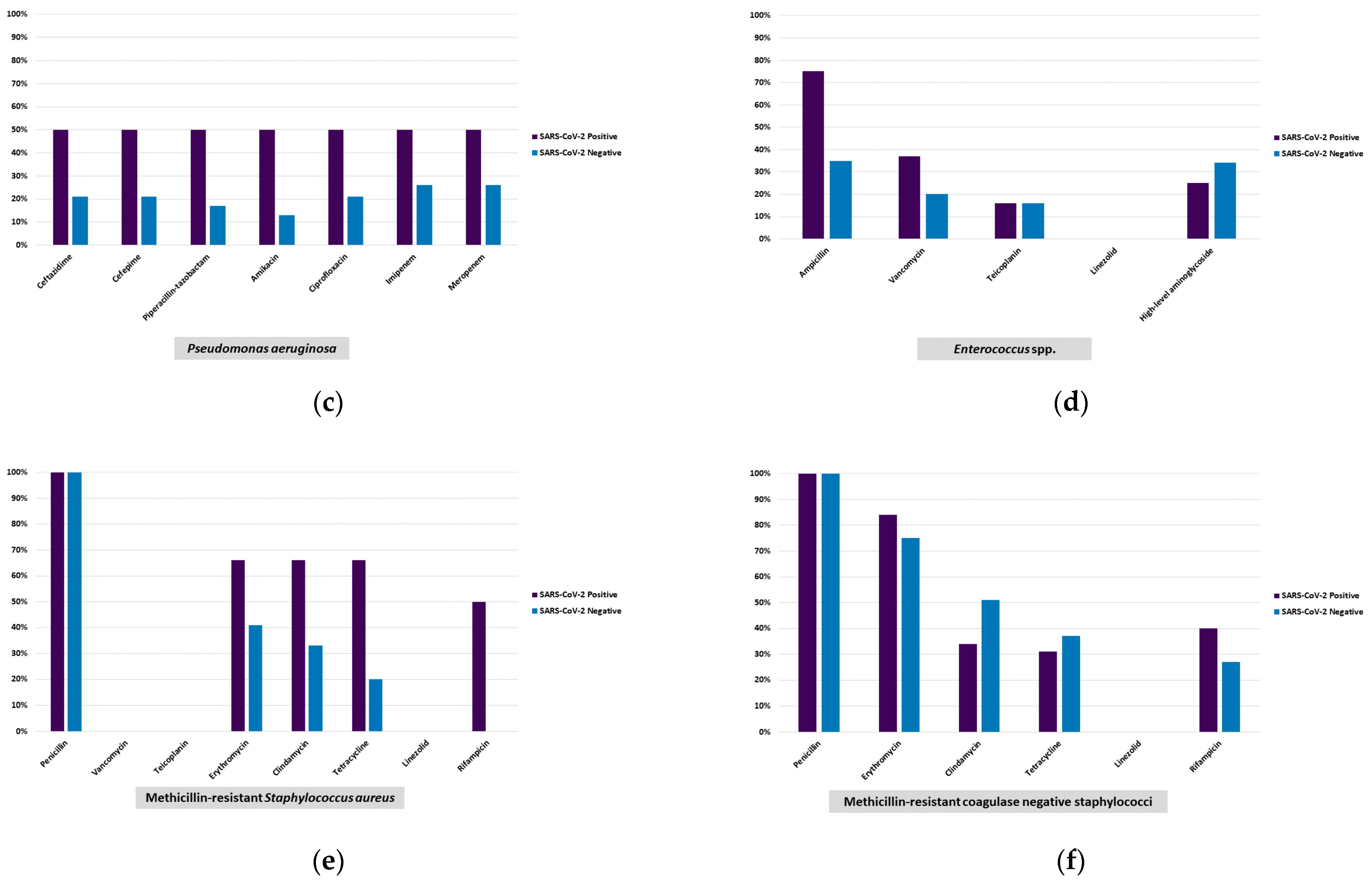
The research was approved by the University of Health Sciences İstanbul Training and Research Hospital Ethics Committee (protocol code: 63 and date of approval: 11 February 2022).

3. Results

The hospital where the study was conducted is a single-center university hospital with a burns unit, hematological units, an organ transplant center, and a bed capacity of 1183, of which 118 are intensive-care beds, with the hospital serving patients from different regions of Turkey, mostly from Istanbul.
Blood culture analysis was performed in the microbiology laboratory of our hospital. From a total of 22,944 blood cultures taken during the study period, culture positivity was determined in 1630 samples from 652 patients. During this process, a total of 200,180 SARS-CoV-2 tests were performed, of which 29,318 were positive and 170,862 were negative. Culture positivity was detected in 652 patients (321 outpatients and 331 inpatients) and 633 were tested for SARS-CoV-2; 118 were positive (18.6%; 45 females, 73 males; mean age: 62.7 years) and 515 were negative (81.3%; 247 females, 268 males; mean age: 61.7 years).
Internal medicine outpatients comprised 32% of the SARS-CoV-2-positive and 47% of the SARS-CoV-2-negative patients. A statistically highly significant difference was detected between the SARS CoV-2-positive and negative patients in the ICU (p < 0.001). The distribution of patients in the clinics and the ICU where the blood samples were collected is given in Table 1.
A total of 671 pathogens were isolated from the blood cultures of the patients. Of these, 252 were fermentative Gram-negative rods, 66 were non-fermentative Gram-negative rods, 300 were Gram-positive cocci, 3 were Gram-positive rods, 7 were anaerobic bacteria, and 5 were other bacterial species (Listeria monocytogenes (n:2), Campylobacter coli (n:1), Campylobacter jejuni (n:1), and Moraxella nonliquefaciens (n:1)), totaling 633 bacteria (94.3%). Fungi were isolated from the blood cultures of 38 (5.7%) patients. The isolated microorganisms are shown in Table 2 and Table 3.
The microorganisms isolated at the highest rate in SARS-CoV-2-positive patients were methicillin-resistant coagulase-negative staphylococci (MR-CoNS) (21.5%), E.coli (12.4%), K. pneumoniae (12.4%), Candida albicans (1.65%), and the Candida glabrata complex (1.65%), and in negative patients, they were E. coli (21.3%), MR-CoNS (13.5%), K. pneumoniae (12.05%), C. albicans (2.1%), C. parapsilosis (1.1%) and Candida tropicalis (0.9%). A. baumannii was detected at the rate of 5.8% in SARS-CoV-2-positive patients and at 1.1% in SARS-CoV-2-negative patients (p = 0.004). E. coli was more common (p = 0.026) in the PCR-negative group, while MR-CoNS were detected at a higher rate in the PCR-positive group (p = 0.028) (Table 2). No statistically significant difference was determined between the COVID-19-positive and negative patients in respect of Pseudomonas spp., Enterococcus spp., and methicillin-resistant Staphylococcus aureus isolated from the blood cultures (p > 0.05) (Table 2). Polymicrobial growth was determined in the cultures of 17 patients, of which 5 were SARS-CoV-2 positive (Table 4). The rate of carbapenem-resistant isolates among K. pneumoniae strains was 60% in SARS-CoV-2-positive patients, and 43.75% in negative patients (p > 0.05) (Table 2, Figure 1). No statistically significant difference was determined between the COVID-19-positive and negative patient groups with respect to the antibiotic susceptibility rates of the microorganisms detected (p > 0.05) (Figure 1). Resistance to imipenem and meropenem was determined in 13 of the 14 A. baumannii isolates, and of these 13 strains, 6 were from the cultures of SARS-CoV-2-positive patients, 5 of whom were being treated in the ICU (Table 4).
In the detection of MR-CoNS, MS-CoNS, E. coli, and K. pneumoniae, 45% (46/102), 34.7% (17/49), 40.4% (53/131), and 36.3% (29/80) were isolated from oncology/hematology patients, respectively. No underlying airway or other infections were detected in SARS-CoV-2-negative patients from whom E. coli was isolated (Table 4).
The three most frequently isolated species of fungi were C. albicans (39.47%; n:15), the C. parapsilosis complex (18.42%; n:7), and C. tropicalis (15.78%; n:6), and C. auris was isolated from one COVID-19 patient in the ICU [18]. Susceptibility tests were performed for 15 of the isolated fungi against antifungals requested by the clinician (Table 3). Of the seven C. parapsilosis complex isolates, three isolates were tested for antifungal susceptibilities, and in two, resistance to fluconazole was detected. Susceptibility to posaconazole and voriconazole was determined in three C. albicans isolates. One isolate was detected as resistant to voriconazole and three had MIC values above the ECVs for posaconazole. The C. auris isolate was resistant to fluconazole, and amphotericin B (Table 3).
A statistically significant difference was detected in the polymicrobial growth rates of SARS-CoV-2-positive and negative patients (p = 0.006) (Table 5).

4. Discussion

The COVID-19 pandemic imposed a devastating burden on healthcare systems around the world [19]. By affecting the epidemiology of other infections, the pandemic may have been reflected in healthcare services in the form of altered courses of bacteremia and fungemia. As a matter of fact, in recent studies examining the distribution of bacterial and fungal agents as well as their resistance profiles in BSI in COVID-19 patients; it has been stated that the agents isolated from COVID-19 patients were the organisms that most likely reflected the commensal skin microbiota at a high rate [20,21].
In the present study, the number of patients hospitalized in the ICU was 120, of whom 37 were SARS-CoV-2-positive. In another study performed during the pandemic period, although Enterococcus spp., Staphylococcus aureus, K. pneumoniae, and C. albicans were found at higher rates compared to the pre-pandemic period, community-acquired BSI cases were reported to be higher in individuals who were SARS-CoV-2-negative (15.8 per 1000 admissions) than those who tested positive (9.6 per 1000 admissions) [22]. In our study, microorganisms were detected in 79.1% and 18% of the blood cultures of SARS-CoV-2-negative and positive patients, respectively. In addition, lower rates of fermentative Gram-negative bacilli and E. coli (p = 0.013, p = 0.026, respectively), and higher rates of A. baumannii, MR-CoNS, Rhizobium radiobacter, and C. glabrata complex (p = 0.004, p = 0.028, p = 0.034, p = 0.034, respectively) were found in SARS-CoV-2-positive patients compared to SARS-CoV-2-negative patients in all clinics. In parallel to the other studies, MR-CoNS (21.5%) was isolated at the highest rate from the blood samples of SARS-CoV-2 patients. In a study by Michailides et al. [23] of patients with COVID-19 infection, CoNS, and K. pneumoniae, together with A. baumannii, were the most frequently isolated bacteria in early and late (> 5 days) nosocomial bacterial infections, respectively. Bahceci et al. [24] isolated CoNS (31%) and A. baumannii (27.5%) at higher rates. Michaelides et al. [23] stated that a prolonged hospital stay may increase CoNS isolation due to the development of superinfections. In the current study, the MR-CoNS (21.5%) and MS-CoNS (9.9%) isolation rates were found to be higher in patients determined to be SARS-CoV-2-positive. The blood culture contamination rate in our laboratory was 3%. The isolation rate (21.5%) of MR-CoNS, accepted as a pathogen according to the CDC recommendations [10], is very high in the current study compared to the contamination rate detected in the previous results of our laboratory. The higher isolation of CoNS in blood cultures from COVID-19-positive patients may be a result of possible concern felt by the staff during the collection of the sample, which was conducted in a stressful environment with isolation precautions on a COVID-19 ward.
Segala et al. [25] reported higher incidence rates of nosocomial BSI related to S. aureus and Acinetobacter spp. in the pre-pandemic period among COVID-19-negative patients in wards, compared to COVID-19-positive patients who were hospitalized in ICUs during the pandemic period [0.3 (95% CI 0.21–0.32) and 0.11 (0.08–0.16) new infections per 100 patient/day, respectively] but a 48% lower incident risk of E. coli infections in COVID-19-positive wards. In the current study, the E. coli isolation rate was found to be higher in patients who were SARS-CoV-2-negative (p = 0.026), and no underlying airway or other infection was detected in SARS-CoV-2-negative patients from whom E. coli was isolated (Table 4).
The presence of bacterial and fungal co-infections has been reported to increase the mortality of patients with severe COVID-19 [22,25]. CoNS (OR: 25.39), non-albicans Candida species (OR: 11.12), S. aureus (OR: 10.72), Acinetobacter spp. (OR: 6.88), Pseudomonas spp. (OR: 4.77), and C. albicans (OR: 3.97) have been isolated from these cases [26].
Taking into account the importance of antimicrobial management in preventing the emergence of antimicrobial resistance, an assessment of the prevalence and epidemiological characteristics of bacterial co-infection is crucial in the guidance of the appropriate empirical antibiotic therapy in the presence of an infection. Antimicrobial drugs can be prescribed either prophylactically or preemptively, especially for ICU patients. In a study of ICU patients hospitalized in ICUs from 88 different countries, despite the suspicion or the presence of bacterial co-infection in only 54% of the patients, treatment or prophylaxis with at least one antibiotic was administered in 70% of cases [27]. In another study conducted on COVID-19 patients, antibiotics were prescribed for 72% of the patients, although only 8% had confirmed bacterial or fungal co-infection [27]. The improper use of antibiotics may also lead to the emergence of resistance in bacteria and side effects in patients.
According to the COVID-19 special report published by the CDC in 2022, which investigated the impact of COVID-19 on antimicrobial resistance, there was a 35% increase in carbapenem-resistant Acinetobacter infections compared to 2019 and 2020 and a 78% increase in nosocomial infections, while a 35% increase in CRE infections was reported [28]. Mahmoudi et al. [29] found the resistance rates of co-trimoxazole, piperacillin, ceftazidime, and cefepime to be 74%, 67.5%, 47.5%, and 42.5% in Enterobacteriaceae strains and 90% for oxacillin, erythromycin and clindamycin in S. aureus, respectively. The sensitivity of imipenem in P. aeruginosa was 90% isolated from COVID-19 patients.
This situation shows that the isolation rates of carbapenem-resistant K. pneumoniae and A. baumannii strains should not be overlooked in the context of healthcare-associated infections in SARS-CoV-2-positive patients [30,31]. Following up the antibiotic resistance rates has been of great importance due to the increase in the isolation of multidrug-resistant strains. In a study of COVID-19-positive patients, 48% (n = 38/79) of S. aureus and 40% (n = 10/25) of K. pneumoniae isolates were found to be resistant to methicillin and carbapenems, respectively [25]. However, in the current study, MR-CoNS was isolated from SARS-CoV-2-positive patients at a significantly higher rate (21.5%) but no statistically significant difference was detected in the isolation rate of S. aureus and the methicillin resistance rate of the isolates between COVID-19-positive patients and SARS-CoV-2-negative individuals. In another study, meropenem resistance in K. pneumoniae strains isolated from patients in the ICU was reported to increase from 79.8% in 2019 to 92.4% in 2022. Moreover, the meropenem resistance rates of A. baumannii were determined to increase from 92.6% in 2018 to 97.9% in 2022 in the ICU and from 82.3% to 91.6% in the wards (p < 0.001) [30]. In the current study, the imipenem resistance of K. pneumoniae isolates was found to be 66.7% and 45.3% (p = 0.137), whereas meropenem resistance was 66.7% and 43.8% (p = 0.110) in SARS-CoV-2-positive and negative individuals, respectively. A great majority of the A. baumannii isolates were resistant to imipenem and meropenem in both SARS-CoV-2-positive and negative patients. No statistically significant difference was detected in the antibiotic susceptibility rates of the microorganisms grown in COVID-19-positive and negative patients in the current study (p > 0.05).
Since fastidious bacteria cannot grow on standard media, the isolation of such microorganisms from blood cultures is closely associated with the media used for subculture and incubation conditions [11]. The fastidious bacteria that were isolated in this study show the importance of using additional enriched media and various incubation conditions.
Species-level identification of not only bacteria but also fungi isolated from blood cultures is important in predicting the antifungal resistance of the isolates. As found in the present study, it is noteworthy that C. auris, a species that is resistant to numerous antifungal drugs and that can cause fatal outbreaks in the ICU, was reported during the COVID-19 pandemic [18,32,33,34,35]. Although the number of these isolates was low, detection is important in terms of applying the right treatment at the right time, especially considering the special health conditions of the patients in ICUs. In a previous study that investigated fungal colonization in the different body parts of COVID-19 patients hospitalized in the ICU, it was reported that the presence of colonization with non-albicans Candida species, which can be associated with treatment failures due to antifungal resistance, was significantly higher and more common in ICU patients compared to non-COVID-19 patients [36].

Limitations

The present study also has important limitations. Disease duration, the length of hospital stays, treatment of patients, and the rate of readmittances were not included. Whether blood samples are taken in or 48 h after admittance is not known, due to the high workloads during the COVID-19 pandemic. As the present study focused on BSI in COVID-19, data regarding other culture results were not analyzed, which precluded the analysis of other secondary infections such as pneumonia.

5. Conclusions

The results of this study show that the most common isolate was MR-CoNS in SARS-CoV-2-positive patients (p = 0.028); the detection of A. baumannii was more frequent (p = 0.004) and the isolation of carbapenem-resistant K. pneumoniae was at a higher rate (60% vs. 43%) than in SARS-CoV-2-negative patients (p > 0.05), which indicates that paying attention to isolation procedures and the major impact of measures to reduce mortality via reducing the risk of infection should not be disregarded while focusing on the outbreak. The presence of bacterial/fungal agents in bloodstream infections and their antibiotic resistance should still be followed up during a pandemic.

Author Contributions

Conceptualization, B.A.K. and B.Ö.; data curation, B.A.K., Z.E. and B.Ö.; formal analysis, B.A.K., Z.E., A.A. and B.Ö.; investigation, B.A.K., İ.Ç.K., Y.B. and S.K.; methodology, B.A.K., İ.Ç.K., Y.B., S.K., G.E.G., Z.E., A.A. and B.Ö.; writing—original draft, B.A.K., İ.Ç.K., Y.B., S.K., G.E.G., Z.E., A.A. and B.Ö.; writing—review and editing, B.A.K., G.E.G., Z.E. and B.Ö. All authors have read and agreed to the published version of the manuscript.

Funding

This research received no external funding.

Institutional Review Board Statement

The research was approved by the University of Health Sciences İstanbul Training and Research Hospital Ethics Committee (protocol code: 63 and date of approval: 11 February 2022).

Informed Consent Statement

Not applicable.

Data Availability Statement

All the data are included in the article by excluding personal data that may not comply with GDPR regulations.

Conflicts of Interest

The authors declare no conflict of interest.

References

  1. Manohar, P.; Loh, B.; Nachimuthu, R.; Hua, X.; Welburn, S.C.; Leptihn, S. Secondary bacterial infections in patients with viral pneumonia. Front. Med. 2020, 7, 420. [Google Scholar] [CrossRef]
  2. Bengoechea, J.A.; Bamford, C.G. SARS-CoV-2 bacterial co-infections and AMR: The deadly trio in COVID-19. EMBO. Mol. Med. 2020, 12, e12560. [Google Scholar] [CrossRef] [PubMed]
  3. Gomez-Simmonds, A.; Annavajhala, M.K.; McConville, T.H.; Dietz, D.E.; Shoucri, S.M.; Laracy, J.C.; Rozenberg, F.D.; Nelson, B.; Greendyke, W.G.; Furuya, E.Y.; et al. Carbapenemase-producing Enterobacterales causing secondary infections during the COVID-19 crisis at a New York City hospital. J. Antimicrob. Chemother. 2021, 76, 380–384. [Google Scholar] [CrossRef] [PubMed]
  4. Gaibani, P.; Viciani, E.; Bartoletti, M.; Lewis, R.E.; Tonetti, T.; Lombardo, D.; Castagnetti, A.; Bovo, F.; Horna, C.S.; Ranieri, M.; et al. The lower respiratory tract microbiome of critically ill patients with COVID-19. Sci. Rep. 2021, 12, 10103. [Google Scholar] [CrossRef] [PubMed]
  5. Bongiovanni, M.; Barda, B. Pseudomonas aeruginosa bloodstream infections in SARS-CoV-2 infected patients: A systematic review. J. Clin. Med. 2023, 12, 2252. [Google Scholar] [CrossRef]
  6. Wang, L.; Amin, A.K.; Khanna, P.; Aali, A.; McGregor, A.; Bassett, P.; Gopal Rao, G. An observational cohort study of bacterial coinfection and implications for empirical antibiotic therapy in patients presenting with COVID-19 to hospitals in North West London. J. Antimicrob. Chemother. 2021, 76, 796–803. [Google Scholar] [CrossRef]
  7. Yu, D.; Ininbergs, K.; Hedman, K.; Giske, C.G.; Stralin, K.; Özenci, V. Low prevalence of bloodstream infection and high blood culture contamination rates in patients with COVID-19. PLoS ONE 2020, 15, e0242533. [Google Scholar] [CrossRef]
  8. Nedel, W.; da Silveira, F.; da Silva, C.F.; Lisboa, T. Bacterial infection in coronavirus disease 2019 patients: Co-infection, super-infection and how it impacts on antimicrobial use. Curr. Opin. Crit. Care 2022, 28, 463–469. [Google Scholar] [CrossRef]
  9. Zhu, N.J.; Rawson, T.M.; Mookerjee, S.; Price, J.R.; Davies, F.; Otter, J.; Aylin, P.; Hope, R.; Gilchrist, M.; Shersing, Y.; et al. Changing patterns of bloodstream infections in the community and acute care across 2 Coronavirus disease 2019 epidemic waves: A retrospective analysis using data linkage. Clin. Infect. Dis. 2022, 24, e1082–e1091. [Google Scholar] [CrossRef]
  10. Centers for Disease Control and Prevention. Bloodstream Infection Event (Central Line-Associated Bloodstream Infection and Non-Central Line-Associated Bloodstream Infection). 2021. Available online: https://www.cdc.gov/nhsn/pdfs/pscmanual/4psc_clabscurrent.pdf (accessed on 12 December 2022).
  11. Gilligan, P.H.; Alby, K.; York, M.K. Blood cultures. In Clinical Microbiology Procedures Handbook, 4th ed.; Leber, A.L., Ed.; ASM Press: Washington, DC, USA, 2016; pp. 3.4.1.1–3.4.1.25. [Google Scholar]
  12. Gilligan, P.H.; York, M.K. Brucellosis-Brucella spp. In Clinical Microbiology Procedures Handbook, 4th ed.; Leber, A.L., Ed.; ASM Press: Washington, DC, USA, 2016; pp. 16.6.1–16.6.12. [Google Scholar]
  13. CLSI. Epidemiological Cutoff Values for Antifungal Susceptibility Testing, 3rd ed.; CLSI supplement M59; Clinical and Laboratory Standards Institute: Wayne, PA, USA, 2020. [Google Scholar]
  14. Clinical and Laboratory Standards Institute. Reference Method for Broth Dilution Antifungal Susceptibility Testing of Yeasts; 4th Informational Supplement M27-S4; CLSI: Wayne, PA, USA, 2012. [Google Scholar]
  15. The European Committee on Antimicrobial Susceptibility Testing. Breakpoint Tables for Interpretation of MICs and Zone Diameters. Version 10.0. 2020. Available online: http://www.eucast.org (accessed on 2 November 2020).
  16. The European Committee on Antimicrobial Susceptibility Testing. Breakpoint Tables for Interpretation of MICs and Zone Diameters. Version 11.0. 2021. Available online: http://www.eucast.org (accessed on 1 January 2021).
  17. Centers for Disease Control and Prevention (CDC). Candida auris. Available online: https://www.cdc.gov/fungal/candida-auris/index.html (accessed on 16 February 2021).
  18. Bölükbaşı, Y.; Erköse, G.G.; Orhun, G.; Kuşkucu, M.A.; Çağatay, A.; Önel, M.; Öngen, B.; Ağaçfidan, A.; Esen, F.; Erturan, Z. First case of COVID-19 positive Candida auris fungemia in Turkey. Mikrobiyol. Bul. 2021, 55, 648–655. [Google Scholar] [CrossRef]
  19. Willan, J.; King, A.J.; Jeffery, K.; Bienz, N. Challenges for NHS hospitals during Covid-19 epidemic. BMJ 2020, 368, m1117. [Google Scholar] [CrossRef] [PubMed]
  20. Sepulveda, J.; Westblade, L.F.; Whittier, S.; Satlin, M.J.; Greendyke, W.G.; Aaron, J.G.; Zucker, J.; Dietz, D.; Sobieszczyk, M.; Choi, J.J.; et al. Bacteremia and blood culture utilization during COVID-19 surge in New York City. J. Clin. Microbiol. 2020, 58, e00875-20. [Google Scholar] [CrossRef] [PubMed]
  21. Hughes, S.; Troise, O.; Donaldson, H.; Mughal, N.; Moore, L.S.P. Bacterial and fungal coinfection among hospitalized patients with COVID-19: A retrospective cohort study in a UK secondary-care setting. Clin. Microbiol. Infect. 2020, 26, 1395–1399. [Google Scholar] [CrossRef] [PubMed]
  22. Bauer, K.A.; Puzniak, L.A.; Yu, K.C.; Finelli, L.; Moise, P.; Ai, C.; Watts, J.A.; Gupta, V. Epidemiology and outcomes of culture-positive bloodstream pathogens prior to and during the SARS-CoV-2 pandemic: A multicenter evaluation. BMC Infect. Dis. 2022, 22, 841. [Google Scholar] [CrossRef]
  23. Michailides, C.; Paraskevas, T.; Karalis, I.; Koniari, I.; Pierrakos, C.; Karamouzos, V.; Marangos, M.; Velissaris, D. Impact of bacterial infections on COVID-19 patients: Is timing important? Antibiotics 2023, 12, 379. [Google Scholar] [CrossRef]
  24. Bahceci, I.; Yildiz, I.E.; Duran, O.F.; Soztanaci, U.S.; Kirdi Harbawi, Z.; Senol, F.F.; Demiral, G. Secondary bacterial infection rates among patients with COVID-19. Cureus 2022, 14, e22363. [Google Scholar] [CrossRef]
  25. Segala, F.V.; Pafundi, P.C.; Masciocchi, C.; Fiori, B.; Taddei, E.; Antenucci, L.; De Angelis, G.; Guerriero, S.; Pastorino, R.; Damiani, A.; et al. Incidence of bloodstream infections due to multidrug-resistant pathogens in ordinary wards and intensive care units before and during the COVID-19 pandemic: A real-life, retrospective observational study. Infection 2023, 51, 1061–1069. [Google Scholar] [CrossRef]
  26. Silva, D.L.; Lima, C.M.; Magalhães, V.C.R.; Baltazar, L.M.; Peres, N.T.A.; Caligiorne, R.B.; Moura, A.S.; Fereguetti, T.; Martins, J.C.; Rabelo, L.F.; et al. Fungal and bacterial coinfections increase mortality of severely ill COVID-19 patients. J. Hosp. Infect. 2021, 113, 145–154. [Google Scholar] [CrossRef]
  27. Getahun, H.; Smith, I.; Trivedi, K.; Paulin, S.; Balkhy, H.H. Tackling antimicrobial resistance in the COVID-19 pandemic. Bull. World Health Organ. 2020, 98, 442. [Google Scholar] [CrossRef]
  28. Center for Disease Control and Prevention. COVID-19: US Impact on Antimicrobial Resistance, Special Report 2022; US Department of Health and Human Services, CDC: Atlanta, GA, USA, 2022. Available online: https://www.cdc.gov/drugresistance/covid19.html (accessed on 10 August 2023).
  29. Mahmoudi, H. Bacterial co-infections and antibiotic resistance in patients with COVID-19. GMS Hyg. Infect. Control 2020, 15, Doc35. [Google Scholar]
  30. Petrakis, V.; Panopoulou, M.; Rafailidis, P.; Lemonakis, N.; Lazaridis, G.; Terzi, I.; Papazoglou, D.; Panagopoulos, P. The impact of the Covıd-19 pandemic on antimicrobial resistance and management of bloodstream infections. Pathogens 2023, 12, 780. [Google Scholar] [CrossRef] [PubMed]
  31. Sinto, R.; Lie, K.C.; Setiati, S.; Suwarto, S.; Nelwan, E.J.; Djumaryo, D.H.; Karyanti, M.R.; Prayitno, A.; Sumariyono, S.; Moore, C.E.; et al. Blood culture utilization and epidemiology of antimicrobial-resistant bloodstream infections before and during the COVID-19 pandemic in the Indonesian national referral hospital. Antimicrob. Resist. Infect. Control 2022, 11, 73. [Google Scholar] [CrossRef] [PubMed]
  32. Chowdhary, A.; Tarai, B.; Singh, A.; Sharma, A. Multidrug-resistant Candida auris infections in critically ill coronavirus disease patients, India, April–July 2020. Emerg. Infect. Dis. 2020, 26, 2694–2696. [Google Scholar] [CrossRef] [PubMed]
  33. Arastehfar, A.; Carvalho, A.; Nguyen, M.H.; Hedayati, M.T.; Netea, M.G.; Perlin, D.S.; Hoenigl, M. COVID-19-associated candidiasis (CAC): An underestimated complication in the absence of immunological predispositions? J. Fungi 2020, 6, 211. [Google Scholar] [CrossRef]
  34. Rodriguez, J.Y.; Le Pape, P.; Lopez, O.; Esquea, K.; Labiosa, A.L.; Alvarez-Moreno, C. Candida auris: A latent threat to critically ill patients with Coronavirus disease 2019. Clin. Infect. Dis. 2020, 73, e2836-7. [Google Scholar] [CrossRef]
  35. Magnasco, L.; Mikulska, M.; Giacobbe, D.R.; Taramasso, L.; Vena, A.; Dentone, C.; Dettori, S.; Tutino, S.; Labate, L.; Di Pilato, V.; et al. Spread of carbapenem-resistant gram-negatives and Candida auris during the COVID-19 pandemic in critically ill patients: One step back in antimicrobial stewardship? Microorganisms 2021, 9, 95. [Google Scholar] [CrossRef]
  36. Çaklovica-Küçükkaya, İ.; Orhun, G.; Çağatay, A.A.; Kalaycı, S.; Esen, F.; Şahin, F.; Ağaçfidan, A.; Erturan, Z. P494 Comparison of Candida colonization in intensive care unit patients with and without COVID-19: First prospective cohort study from Turkey. Med. Mycol. 2022, 60 (Suppl. S1), myac072P494. [Google Scholar] [CrossRef]
Figure 1. Antibiotic resistance profiles of the most frequently isolated bacteria. (a) Escherichia coli; (b) Klebsiella pneumoniae; (c) Pseudomonas aeruginosa; (d) Enterococcus spp.; (e) methicillin-resistant Staphylococcus aureus; (f) methicillin-resistant coagulase-negative staphylococcus.
Healthcare 11 02581 g001aHealthcare 11 02581 g001b
Table 1. Distribution of SARS-CoV-2 (+) and SARS-CoV-2 (−) patients according to clinics.
ClinicsUnitSARS CoV-2 (+) (n:118)SARS CoV-2 (−)
(n:515)
Not Tested
(n:19)
InpatientSurgical2496-
Internal19693
OutpatientSurgical-26-
Internal3824314
Intensive
Care Unit
Surgical5431
Internal32381
Table 2. Distribution of the bacteria and fungi isolated from the blood cultures of SARS-CoV-2-positive and negative patients.
MicroorganismsTotal
n (%)
SARS-
CoV-2
Positive
n (%)
SARS-
CoV-2
Negative
n (%)
SARS-
CoV-2
Non-Tested
n (%)
Positive
vs. Negative
All microorganisms a671 (100)121 (18.0)531 (79.1)19 (2.8)p-valueDifferences of two proportions 95% CI j
Fermentative
Gram-negative rods
252 (37.5)34 (28,0)214 (40.3)4 (21)0.013 h(3.2–21.2)
Escherichia coli131 (19.5)15 (12.4)113 (21.3)3 (15.8)0.026 h(2.1–5.7)
Klebsiella pneumoniaeb/
CRKP c
80/38 (11.9/5.6)15/9
(12.4/7.4)
64/28
(12.05/5.3)
1/1
(5.3/5.3)
0.917 b,h
0.353 c,h
(−6.1–0.8)
(−2.9–0.5)
Klebsiella oxytoca5 (0.7)0 (0.0)5 (0.9)-0.590 i(0.1–1.7)
Enterobacter spp.7 (1.1) 1 (0.8)6 (1.1)-1.000 i(−1.5–2.1)
Proteus mirabilis13 (1.9)1 (0.8)12 (2.3)-0.480 i(−0.5–3.5)
Non-Fermentative
Gram-negative rods
66 (9.8)16 (13.2)48 (9)2 (10.5)0.163 h(−1.7–10.1)
Pseudomonas aeruginosa25 (3.7)2 (1.7)23 (4.3)-0.199 i(−0.2–5.5)
Pseudomonas spp.5 (0.7)2 (1.7)3 (0.6)-0.233 i(−0.7–2.8)
Acinetobacter baumannii14 (2.1)7 (5.8)6 (1.1)1 (5.3)0.004 i(2.0–7.4)
Stenotrophomonas maltophilia7 (1.05)1 (0.8)6 (1.1)-1.000 i(−1.5–2.1)
Gram-positive cocci300 (44.7)59 (48.8)231 (40.1)10 (52.6)0.294 h(−4.5–15.1)
MRSA d 27 (4.0)3 (2.5)24 (4.5)-0.309 h(−1.3–5.3)
MSSA e53 (7.9)7 (5.8)43 (8.1)3 (15.8)0.388 h(−2.5–7.1)
MR-CoNS f102 (15.2)26 (21.5)72 (13.5)4 (21.1)0.028 h(1.0–15.0)
MS-CoNS g49 (7.3)12 (9.9)35 (6.6)2 (10.5)0.202 h(−1.8–8.4)
Enterococcus faecalis12 (1.8)2 (1.7)10 (1.9)-1.000 i(−2.3–2.8)
Enterococcus faecium12 (1.8)3 (2.5)9 (1.7)-0.474 i(−1.9–3.5)
Enterococcus spp.16 (2.4)2 (1.7)14 (2.6)-0.749 i(−1.7–3.6)
Alpha hemolytic streptococci7 (1.05)0 (0.0)7 (1.3)-0.359 i(0.3–2.3)
Fungi38 (5.7)8 (6.6)27 (5.1)3 (15.8)0.501 h(−3.0–6.0)
Candida albicans15 (2.2)2 (1.7)11 (2.1)2 (10.5)1.000 i(−2.1–3.0)
Candida parapsilosis complex7 (1.05)1 (0.8)6 (1.1)-1.000 i(−1.5–2.1)
Candida tropicalis6 (0.9)0 (0.0)5 (0.9)1 (5.3)0.590 i(0.1–1.7)
a. Numbers of ≤ 4 isolates were not specified in the table: Fermentative Gram-negative rods: Serratia marcescens (n:1), Serratia spp. (n:2), Citrobacter spp. (n:3), Citrobacter koseri (n:1), Morganella morganii (n:3), Raoultella planticola (n:1), Aeromonas spp. (n:1), and Salmonella Enteritidis (n:4). Non-fermentative Gram-negative rods: Pseudomonas stutzeri (n:1), Acinetobacter lwoffii (n:1), Acinetobacter spp. (n:4), Rhizobium radiobacter (n:2), Achromobacter xylosoxidans (n:1), Ochrobactrum anthropi (n:1), Sphingomonas paucimobilis (n:2), Burkholderia cepacia (n:1), and Pandoraea spp (n:1). Non-fermentative Gram-negative rod (n:1); Gram-positive cocci: Enterococcus avium (n:4), Enterococcus gallinarum (n:3), Streptococcus pneumoniae (n:4), Streptococcus agalactiae (n:3), Streptococcus gallolyticus (n:2), Streptococcus equi (n:1), and Leuconostoc pseudomesenteroides (n:2). Beta hemolytic streptococci (n:2). Non-hemolytic streptococci (n:1). Gram-positive rods: Corynebacterium jeikeium (n:1), Corynebacterium striatum (n:1), and Lactobacillus casei (n:1). Other bacteria: Listeria monocytogenes (n:2), Campylobacter coli (n:1), Campylobacter jejuni (n:1), and Moraxella nonliquefaciens (n:1). Anaerobic bacteria: Bacteroides fragilis (n:1), Bacteroides spp. (n:1), Prevotella spp. (n:2), Clostridium clostridioforme (n:1), and Fusobacterium nucleatum (n:1), anaerobic Gram-positive rod (n:1). Fungi: Candida kefyr (n:2), Candida glabrata complex (n:2), Candida metapsilosis (n:1), Candida krusei (n:1), Candida auris (n:1), Kodamaea ohmeri (n:1), Cryptococcus neoformans (n:1), and Rhodotorula spp. (n:1); b. All K. pneumoniae isolates; c. CRKP: Carbapenem-resistant Klebsiella pneumoniae; d. MRSA: Methicillin-resistant Staphylococcus aureus; e. MSSA: Methicillin-sensitive Staphylococcus aureus; f. MR-CoNS: Methicillin-resistant coagulase-negative staphylococci; g. MS-CoNS: Methicillin-sensitive coagulase-negative staphylococci; h. Pearson’s chi-square test; i. Fisher’s exact test; j. CI: confidence interval.
Table 3. Antifungal resistance profiles of fungi isolated from the blood cultures of SARS-CoV-2-positive and negative patients.
FungiPatientsAntifungal MIC, µg/mL
Species
(Tested n/Total n)
Strain NoSARS-CoV-2 StatusFluconazolePosaconazoleVoriconazoleItraconazoleAmphotericin BCaspofungin Anidulafungin
Candida albicans (7/15)1Positive2 (S)0.064 ᵃ (NWT)0.25 (I)-0.5 ᵃ (WT)0.016 (S)0.012 (S)
2Negative2 (S)0.064 ᵃ (NWT)0.047(S)----
3Not tested0.75 (S)----0.5 (I)-
4Negative2 (S)---0.25 ᵃ (WT)0.5 (I)0.012 (S)
5Positive1.5 (S)------
6Negative-0.25 ᵃ (NWT)1 (R)--0.065 (S)-
7Negative0.125 (S)---0.047 ᵃ (WT)0.096 (S)0.003 (S)
Candida parapsilosis complex (3/7)1Negative0.75 (S)---0.25 ᵃ (WT)0.75 (S)-
2Negative>256 (R) 0.19 ᵃ (WT)0.5 (I)2 c0.75 ᵃ (WT)0.38 (S)0.75 (S)
3Negative24 (R) 0.25 ᵃ (WT)0.75 (S)
Candida tropicalis (2/6)1Negative0.5 (S)-0.008 (S)-0.25 ᵃ (WT)0.094 (S)0.008 (S)
2Negative0.5 (S)---0.25 ᵃ (WT)-0.008 (S)
Candida glabrata complex (1/2)Positive1.5 (SDD)-0.032 ᵃ (WT)--0.25 (I)-
Candida auris (1/1) bPositive>256 (R) 0.016c0.19 c0.19 c3 (R)1 (S)0.094 (S)
Cryptococcus neoformans (1/1)Negative8 ᵃ (WT)---0.5 ᵃ (WT)--
MIC: Minimum inhibitory concentration; n: number of isolates; a: MICs were evaluated according to epidemiological cut-off values (ECVs); b: MICs were evaluated according to the breakpoints determined by the CDC; c: there are no clinical breakpoints or ECVs; S: susceptible; I: intermediate; R: resistant; SDD: susceptible-dose dependent; WT: wild type; NWT: non-wild type.
Table 4. Demographic information of the patients from whom some microorganisms were isolated.
Microorganisms (n)
Demographic InformationMR-CoNS a
(n:102)
MS-CoNS b
(n:49)
E. coli
(n:131)
K. pneumoniae
(n:80)
A. baumannii
(n:14)
C. albicans
(n:15)
C. glabrata complex
(n:2)
SARS-CoV-2 (+)26121564732
Inpatient94321122
Outpatient65929000
ICU113314610
SARS-CoV-2 (−) 7235113156100
Inpatient3418239380
Outpatient2513812010
ICU134104310
SARS-CoV-2—not tested 4231120
Inpatient1000010
Outpatient3230010
ICU0001100
Median age63.8563.4262.6960.7863.7168.471
Gender identity
Male442574441081
Female 58245736471
Oncology patient39114026360
Hematologic malignancy76133220
Hypertension33173633661
Diabetes mellitus22112719331
Coronary artery disease00100020
COPD c9200030
COVID-19 pneumonia22142412520
n: Number of patients, a. MR-CoNS: methicillin-resistant coagulase-negative staphylococci; b. MS-CoNS: methicillin-sensitive coagulase-negative staphylococci; c. COPD: chronic obstructive pulmonary disease.
Table 5. Distribution of the polymicrobial growths detected in patients a.
Clinic/UnitsMicroorganism
SARS-CoV-2 (+) [n:8]Inpatient (n:3)Surgical (n:1)Candida krusei, Kodamea ohmeri
Internal (n:2)Proteus mirabilis, Escherichia coli
Klebsiella pneumoniae, Candida glabrata complex
Outpatient (n:4)Surgical (n:1)Candida glabrata complex, Candida albicans
Internal (n:3)Streptococcus pneumoniae, Escherichia coli
Candida kefyr, Enterococcus gallinarum, Enterococcus faecium
Escherichia coli, Enterobacter spp.
Intensive Care Unit (n:1)Surgical (n:0)-
Internal (n:1)Enterococcus spp., Candida albicans
SARS-CoV-2 (-) [n:9]Inpatient (n:3)Surgical (n:2)Candida albicans, Candida parapsilosis
Escherichia coli, Candida parapsilosis
Internal (n:1)Pseudomonas aeruginosa, Acinetobacter spp.
Outpatient (n:5)Surgical (n:1)Citrobacter spp., Klebsiella oxytoca
Internal (n:4)Raoultella planticola, Escherichia coli
Klebsiella pneumoniae, Enterococcus spp.
Enterococcus spp., Escherichia coli, MSSA b
Enterococcus spp., Escherichia coli
Intensive Care Unit (n:1)Surgical (n:0)-
Internal (n:1)Proteus mirabilis, Klebsiella pneumoniae
a: Difference in proportion between SARS-CoV-2-positive and negative patients for polymicrobial growth (p = 0.006), b: methicillin-sensitive Staphylococcus aureus.
Disclaimer/Publisher’s Note: The statements, opinions and data contained in all publications are solely those of the individual author(s) and contributor(s) and not of MDPI and/or the editor(s). MDPI and/or the editor(s) disclaim responsibility for any injury to people or property resulting from any ideas, methods, instructions or products referred to in the content.

