
A Comparative Analysis of Post-Retraction Changes in Gingival Height after Conventional and Surgical Gingival Displacement: Rotary Curettage, Diode and Er:YAG Laser Troughing

 , , , , , , ,
  and
and 
Abstract
:1. Introduction
- Conventional mechanical, chemical, and mechano-chemical methods: These approaches involve techniques that use physical instruments, chemical agents, or a combination of both to displace the gingival tissues for impression taking. Examples may include retraction cords, astringent pastes, and retraction pastes containing specific compounds like aluminum chloride and kaolin.
- Surgical (troughing) methods: This group includes surgical techniques such as troughing, where a dental professional creates a trough or groove in the gingival tissue to facilitate proper retraction for accurate impression making.
2. Materials and Methods
2.1. Object of Observation
2.2. Units of Observation
2.3. Parameters of Observation
- ○
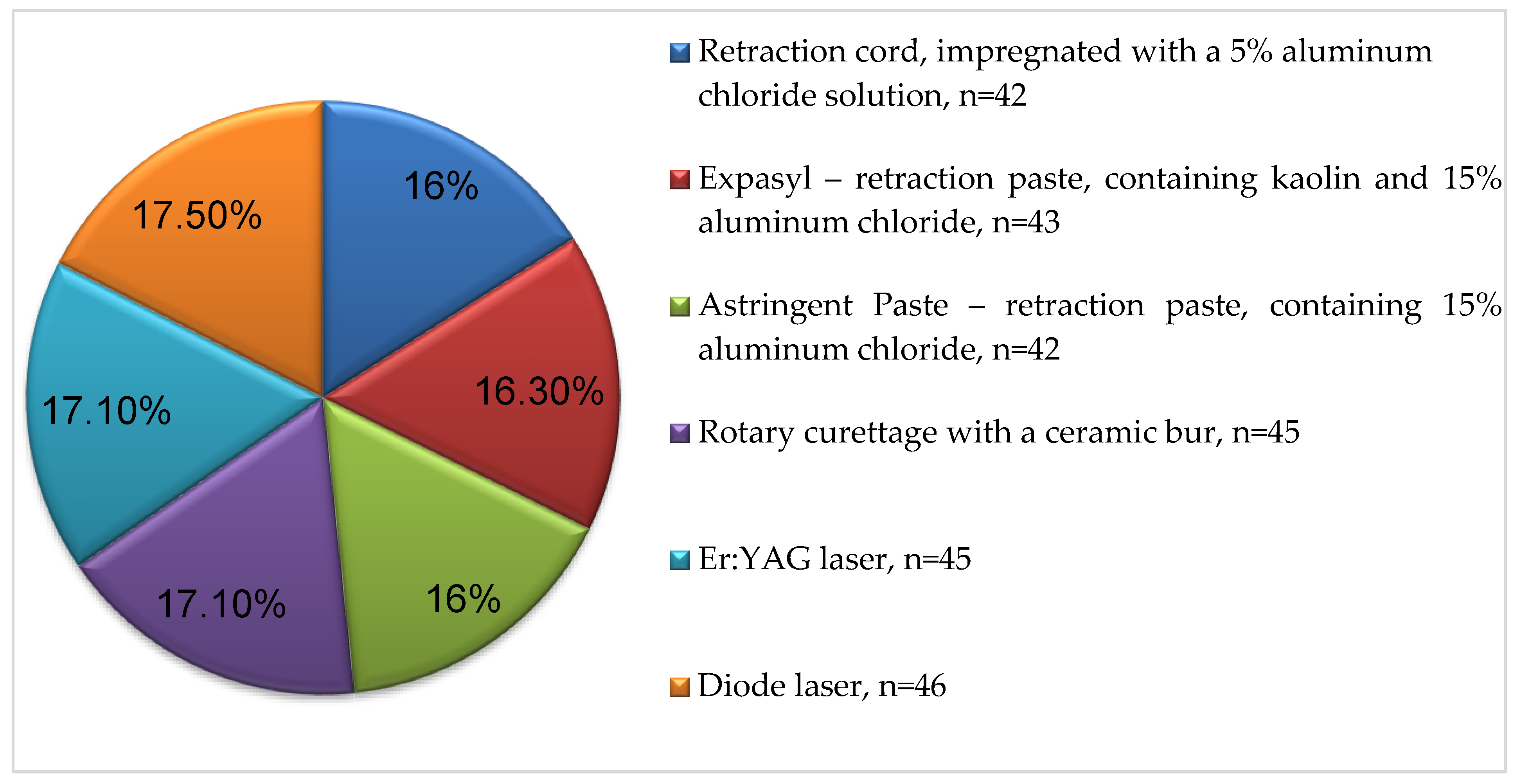
- The six tested retraction methods;
- ○
- Sex;
- ○
- Age;
- ○
- Group of teeth (frontal, premolars and molars);
- ○
- Time of gingival recovery (first week and second week).
- ○
- Presence of gingival recession (GR);
- ○
- Hyperplasia of the free gingival margin level.
2.4. Settings and Location Where the Data Were Collected (Venue of Observation)
- ○
- Department of Prosthetic Dentistry, Faculty of Dental Medicine, CAD/CAM Center of Dental Medicine, Research Institute, Medical University—Plovdiv (RIMUP).
- ○
- Department of Periodontology and Oral Mucosa Diseases, Laser Dental Center, Faculty of Dental Medicine, Research Institute, Medical University—Plovdiv, Bulgaria (RIMUP).
2.5. Eligibility Criteria for Participants in the Study
- ○
- ○
- They should not have taken medications in the last three months. Some medications may cause inflammation and/or bleeding, compromising the results [2].
- ○
- The Löe and Silness gingival index should be 0. The index assesses the prevalence and severity of gingivitis in populations, groups, and individuals. A score from 0.1 to 1.0 signifies mild inflammation, 1.1 to 2.0 moderate inflammation, and 2.1 to 3.0 severe inflammation [2].
- ○
- Patients subject to prosthetic restorations in more than one quadrant.
- ○
- Criteria for exclusion from the study are as follows: Patients with psychological disorders. The presence of inaccurate obturations can interfere with the biological width.
- ○
- Teeth with a periodontal probing depth (PPD) above 3 mm.
2.6. Entry of Primary Data
2.7. Trial Design
- Classic mechano-chemical:
- ○
- Retraction cords Elite cord (Zhermack, Badia Polesine, RO, Italy) in five different sizes (000, 00, 0, 1, 2) depending on the sulcus depth, impregnated with a 5% aluminium chloride solution.
- ○
- Retraction paste Expasyl (ACTEON Pharma – Pierre Rolland, Mérignac, France).
- ○
- Retraction paste Astringent (3M ESPE, Seefeld, Bayern, Germany).
- Surgical:
- ○
- Ceramic bur Soft Tissue Trimmer NTI (Kerr, Orange, CA, USA).
- ○
- Er:YAG laser with a wavelength of 2940 nm (Light Instruments, Yokne’am Illit, Israel).
- ○
- Diode laser with a wavelength of 810 nm and power of 8W (FOX, A.R.C. Lasers GmbH, Nürnberg, Germany).
- ○
- 1st measurement—immediately after the retraction—the value was considered 0.
- ○
- 2nd measurement—one week after the procedure.
- ○
- 3rd measurement—two weeks after that.
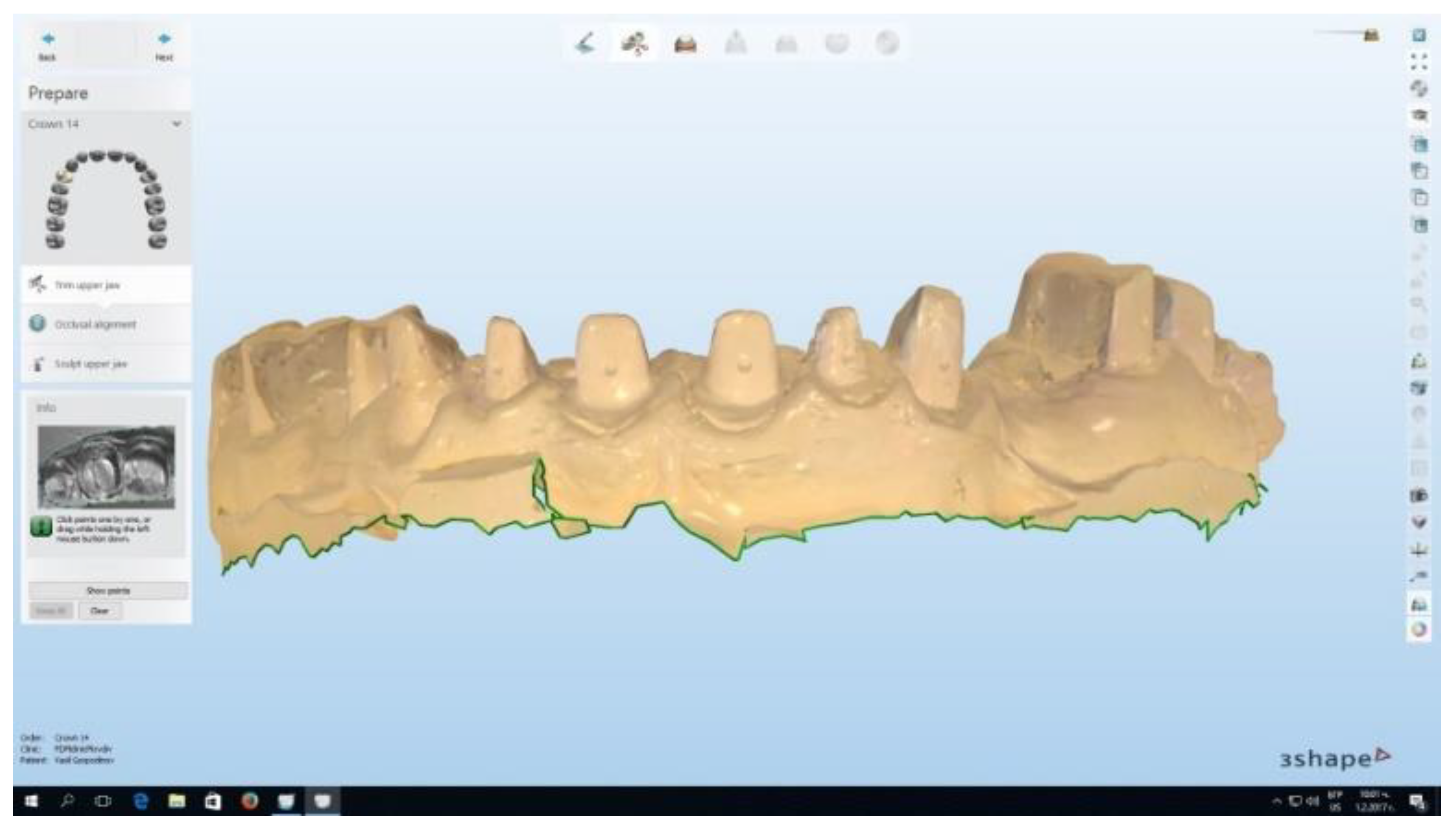
2.8. Technique of Implementation of the Applied Gingival Displacement Methods
2.9. Impression Materials
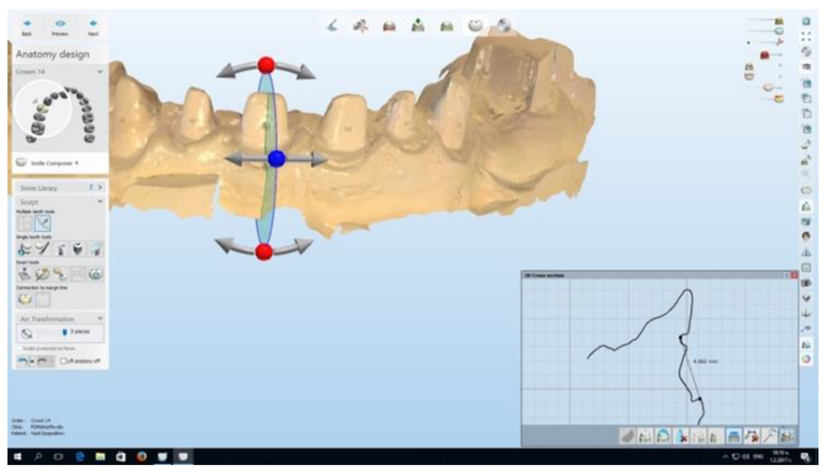
2.10. CAD/CAM System
2.11. Statistical Methods
3. Results
3.1. Sample Characteristics
3.2. Experimental Results
4. Discussion
- Astringent Paste: This retraction paste contains 15% aluminum chloride.
- Expasyl: This retraction paste contains kaolin and 15% aluminum chloride.
- Lack of subsequent gingival inflammation: The use of diode and erbium lasers results in reduced or negligible gingival inflammation following the procedure. This is beneficial as it helps in minimizing discomfort and promotes faster healing.
- Minimal to no pain during the procedure: Patients undergoing laser retraction experience minimal to no pain, which can be particularly advantageous as it often eliminates the need for local anesthesia.
- Reduced gingival recession: Laser troughing leads to less gingival recession compared to traditional methods. This means that the gingival margin remains more stable and less tissue is lost.
- Reduced tissue bleeding: Laser retraction techniques result in decreased tissue bleeding during the procedure, contributing to a smoother and more controlled clinical experience.
5. Conclusions
Author Contributions
Funding
Institutional Review Board Statement
Informed Consent Statement
Data Availability Statement
Conflicts of Interest
Abbreviations
| ADA | American Dental Association |
| A-silicones | addition silicones |
| BOP | bleeding on probing |
| GI | gingival index |
| GR | gingival recession |
| PD | probing depth |
| PPD | periodontal probing depth |
| PTFE | polytetrafluoroethylene |
| PVS | polyvinyl siloxane |
| VAS | Visual Analogue Scale |
Appendix A

| Number | % | ||
|---|---|---|---|
| Classic Mechano-chemical methods | Retraction cord, impregnated with a 5% aluminum chloride solution | 42 | 16.0 |
| Expasyl—retraction paste, containing kaolin and 15% aluminum chloride | 43 | 16.3 | |
| Astringent Paste—retraction paste, containing 15% aluminum chloride | 42 | 16.0 | |
| Surgical methods | Ceramic bur rotary curettage | 45 | 17.1 |
| Er:YAG laser | 45 | 17.1 | |
| Diode laser | 46 | 17.5 | |
| Total | 263 | 100.0 | |
Appendix B
| Number | % | ||
|---|---|---|---|
| Front teeth | 128 | 48.7 | |
| Premolars | 69 | 26.2 | |
| Molars | 66 | 25.1 | |
| Total | 263 | 100.0 | |
Appendix C
| Table Structural Distribution of the Examined Teeth | |||
|---|---|---|---|
| Teeth | Number | % | |
| 11 | 17 | 6.5 | |
| 12 | 14 | 5.3 | |
| 13 | 11 | 4.2 | |
| 14 | 10 | 3.8 | |
| 15 | 8 | 3.0 | |
| 16 | 8 | 3.0 | |
| 17 | 7 | 2.7 | |
| 18 | 2 | 0.8 | |
| 21 | 9 | 3.4 | |
| 22 | 7 | 2.7 | |
| 23 | 8 | 3.0 | |
| 24 | 8 | 3.0 | |
| 25 | 5 | 1.9 | |
| 26 | 7 | 2.7 | |
| 27 | 6 | 2.3 | |
| 28 | 2 | 0.8 | |
| 31 | 15 | 5.7 | |
| 32 | 11 | 4.2 | |
| 33 | 11 | 4.2 | |
| 34 | 12 | 4.6 | |
| 35 | 10 | 3.8 | |
| 36 | 6 | 2.3 | |
| 37 | 7 | 2.7 | |
| 38 | 2 | 0.8 | |
| 41 | 8 | 3.0 | |
| 42 | 9 | 3.4 | |
| 43 | 8 | 3.0 | |
| 44 | 9 | 3.4 | |
| 45 | 7 | 2.7 | |
| 46 | 9 | 3.4 | |
| 47 | 9 | 3.4 | |
| 48 | 1 | 0.4 | |
| Total | 263 | 10.0 | |
References
- Academy of Prosthodontics. Available online: https://www.academyofprosthodontics.org/lib_ap_articles_download/GPT9.pdf (accessed on 12 June 2023).
- Newman, M.; Takei, H.; Klokkevold, P.; Carranza, F. Carranza’s Clinical Periodontology, 12th ed.; Mosby Elsevier: Amsterdam, The Netherlands, 2014. [Google Scholar]
- Jepsen, S.; Caton, J.G.; Albandar, J.M.; Bissada, N.F.; Bouchard, P.; Cortellini, P.; Demirel, K.; de Sanctis, M.; Ercoli, C.; Fan, J.; et al. Periodontal manifestations of systemic diseases and developmental and acquired conditions: Consensus report of workgroup 3 of the 2017 World Workshop on the Classification of Periodontal and Peri-Implant Diseases and Conditions. J. Periodontol. 2018, 89 (Suppl. S1), S237–S248. [Google Scholar] [CrossRef]
- Hack, G.D.; Patzelt, S.B.M. Evaluation of the accuracy of six intraoral scanning devices: An in-vitro investigation. ADA Prof. Prod. Rev. 2015, 10, 1–5. Available online: https://teamziereis.de/api/datei/201705121114ybx.pdf (accessed on 31 May 2023).
- Abdel Gabbar, F.; Aboulazm, S.F. Comparative study on gingival retraction using mechanochemical procedure and pulsed Nd = YAG laser irradiation. Egypt Dent. J. 1995, 41, 1001–1006. Available online: https://pubmed.ncbi.nlm.nih.gov/9497632/ (accessed on 31 May 2023).
- Baba, N.Z.; Goodacre, C.J.; Jekki, R.; Won, J. Gingival displacement for impression making in fixed prosthodontics: Contemporary principles, materials, and techniques. Dent. Clin. N. Am. 2014, 58, 45–68. [Google Scholar] [CrossRef]
- Bowles, W.H.; Tardy, S.J.; Vahadi, A. Evaluation of new gingival retraction agents. J. Dent. Res. 1991, 70, 1447–1449. [Google Scholar] [CrossRef] [PubMed]
- Donovan, T.E.; Gandara, B.K.; Nemetz, H. Review and survey of medicaments used with gingival retraction cords. J. Prosthet. Dent. 1985, 53, 525–531. [Google Scholar] [CrossRef] [PubMed]
- Fisher, D.W. Conservative management of the gingival tissue for crowns. Dent. Clin. N. Am. 1976, 20, 273–284. Available online: https://pubmed.ncbi.nlm.nih.gov/1062318/ (accessed on 31 May 2023). [CrossRef]
- Hansen, P.A.; Tira, D.E.; Barlow, J. Current methods of finish-line exposure by practicing prosthodontists. J. Prosthodont. 1999, 8, 163–170. [Google Scholar] [CrossRef]
- Noble, W.H.; McClatchey, K.D.; Douglass, G.D. A histologic comparison of effects of electrosurgical resection using different electrodes. J. Prosthet. Dent. 1976, 35, 575–579. [Google Scholar] [CrossRef] [PubMed]
- Phatale, S.; Marawar, P.P.; Byakod, G.; Lagdive, S.B.; Kalburge, J.V. Effect of retraction materials on gingival health: A histopathological study. J. Indian Soc. Periodontol. 2010, 14, 35–39. [Google Scholar] [CrossRef]
- Ramadan, F.A.; el-Sadeek, M.; el-S, H. Histopathologic response of gingival tissues to hemodent and aluminum chloride solutions as tissue displacement materials. Egypt Dent. J. 1972, 18, 337–352. Available online: https://pubmed.ncbi.nlm.nih.gov/4524081/ (accessed on 31 May 2023). [PubMed]
- Robertson, P.B.; Lüscher, B.; Spangberg, L.S.; Levy, B.M. Pulpal and periodontal effects of electrosurgery involving cervical metallic restorations. Oral. Surg. Oral. Med. Oral. Pathol. 1978, 46, 702–710. [Google Scholar] [CrossRef] [PubMed]
- Runyan, D.A.; Reddy, T.G., Jr.; Shimoda, L.M. Fluid absorbency of retraction cords after soaking in aluminum chloride solution. J. Prosthet. Dent. 1988, 60, 676–678. [Google Scholar] [CrossRef] [PubMed]
- Scott, A. Use of an erbium laser in lieu of retraction cord: A modern technique. Gen. Dent. 2005, 53, 116–119. Available online: https://pubmed.ncbi.nlm.nih.gov/15833012/ (accessed on 31 May 2023).
- Silness, J. Periodontal conditions in patients treated with dental bridges. 3. The relationship between the location of the crown margin and the periodontal condition. J. Periodontal. Res. 1970, 5, 225–229. [Google Scholar] [CrossRef]
- Valderhaug, J.; Ellingsen, J.E.; Jokstad, A. Oral hygiene, periodontal conditions and carious lesions in patients treated with dental bridges. A 15-year clinical and radiographic follow-up study. J. Clin. Periodontol. 1993, 20, 482–489. [Google Scholar] [CrossRef]
- Weir, D.J.; Williams, B.H. Clinical effectiveness of mechanical-chemical tissue displacement methods. J. Prosthet. Dent. 1984, 51, 326–329. [Google Scholar] [CrossRef]
- Yang, J.C.; Tsai, C.; Chen, M.S.; Wei, J.Y.; Lee, S.Y.; Lin, C. Clinical study of a newly developed injection-type gingival retraction material. J. Dent. Sci. 2005, 24, 147–151. Available online: https://www.airitilibrary.com/Publication/alDetailedMesh?DocID=10103287-200509-24-3-147-151-a (accessed on 31 May 2023).
- Anneroth, G.; Nordenram, A. Reaction of the gingiva to the application of threads in the gingival pocket for taking impressions with elastic material. An experimental histological study. Odontol. Revy 1969, 20, 301–310. Available online: https://pubmed.ncbi.nlm.nih.gov/4903190/ (accessed on 31 May 2023).
- Manolakis, A.; Bartsch, N.; Hahn, P. Clinical comparison of a gingiva retraction paste and impregnated cords (abstract 1837). In Proceedings of the International Gingival Displacement for Definitive Impression 65 Association for Dental Research/American Association for Dental Research/Canadian Association for Dental Research 82nd General Session, Honolulu, HI, USA, 12 March 2004. [Google Scholar]
- Al Hamad, K.Q.; Azar, W.Z.; Alwaeli, H.A.; Said, K.N. A clinical study on the effects of cordless and conventional retraction techniques on the gingival and periodontal health. J. Clin. Periodontol. 2008, 35, 1053–1058. [Google Scholar] [CrossRef]
- Coelho, D.H.; Cavallaro, J.; Rothschild, E.A. Gingival recession with electrosurgery for impression making. J. Prosthet. Dent. 1975, 33, 422–426. [Google Scholar] [CrossRef] [PubMed]
- Kamansky, F.W.; Tempel, T.R.; Post, A.C. Gingival tissue response to rotary curettage. J. Prosthet. Dent. 1984, 52, 380–383. [Google Scholar] [CrossRef] [PubMed]
- Stuffken, M.; Vahidi, F. Preimpression troughing with the diode laser: A preliminary study. J. Prosthet. Dent. 2016, 115, 441–446. [Google Scholar] [CrossRef]
- Wilder-Smith, P.; Dang, J.; Kurosaki, T. Investigating the range of surgical effects on soft tissue produced by a carbon dioxide laser. J. Am. Dent. Assoc. 1997, 128, 583–588. [Google Scholar] [CrossRef]
- Ruel, J.; Schuessler, P.J.; Malament, K.; Mori, D. Effect of retraction procedures on the periodontium in humans. J. Prosthet. Dent. 1980, 44, 508–515. [Google Scholar] [CrossRef]
- Azzi, R.; Tsao, T.F.; Carranza, F.A., Jr.; Kenney, E.B. Comparative study of gingival retraction methods. J. Prosthet. Dent. 1983, 50, 561–565. [Google Scholar] [CrossRef] [PubMed]
- Moskow, B.S. The response of the gingival sulcus to instrumentation: A histological investigation 2. Gingival curettage. J. Periodontol. 1964, 35, 112–126. Available online: https://aap.onlinelibrary.wiley.com/doi/abs/10.1902/jop.1964.35.2.112 (accessed on 31 May 2023). [CrossRef]
- Qureshi, S.M.; Anasane, N.S.; Kakade, D. Comparative evaluation of the amount of gingival displacement using three recent gingival retraction systems—In vivo Study. Contemp. Clin. Dent. 2020, 11, 28–33. [Google Scholar] [CrossRef]
- Prasanna, G.S.; Reddy, K.; Kumar, R.K.; Shivaprakash, S. Evaluation of efficacy of different gingival displacement materials on gingival sulcus width. J. Contemp. Dent. Pract. 2013, 14, 217–221. [Google Scholar] [CrossRef]
- Shrivastava, K.J.; Bhoyar, A.; Agarwal, S.; Shrivastava, S.; Parlani, S.; Murthy, V. Comparative clinical efficacy evaluation of three gingival displacement systems. J. Nat. Sci. Biol. Med. 2015, 6 (Suppl. S1), S53–S57. [Google Scholar] [CrossRef] [Green Version]
- Thimmappa, M.; Bhatia, M.; Somani, P.; Kumar, D.R.V. Comparative evaluation of three noninvasive gingival displacement systems: An in vivo study. J. Indian Prosthodont. Soc. 2018, 18, 122–130. [Google Scholar] [CrossRef] [PubMed]
- Rathod, A.; Jacob, S.S.; MAlqahtani, A.; Valsan, I.; Majeed, R.; Premnath, A. Efficacy of different gingival displacement materials in the management of gingival sulcus width: A comparative study. J. Contemp. Dent. Pract. 2021, 22, 703–706. Available online: https://pubmed.ncbi.nlm.nih.gov/34393130/ (accessed on 31 May 2023). [CrossRef] [PubMed]
- Rayyan, M.M.; Hussien, A.N.M.; Sayed, N.M.; Abdallah, R.; Osman, E.; El Saad, N.A.; Ramadan, S. Comparison of four cordless gingival displacement systems: A clinical study. J. Prosthet. Dent. 2019, 121, 265–270. [Google Scholar] [CrossRef]
- de Carvalho, W.F.; Junior, L.C.V.; Junior, H.F.B.; Suguiura, T.P.D.S.; Previdelli, I.T.S.; Sábio, S. Evaluation of gingival displacement with aluminum chloride and naphazoline hydrochloride: A randomized controlled trial. Eur. J. Prosthodont. Restor. Dent. 2021, 29, 47–53. [Google Scholar] [CrossRef]
- Nasim, H.; Lone, M.A.; Kumar, B.; Ahmed, N.; Farooqui, W.A.; Alsahhaf, A.; Alresayes, S.; Vohra, F.; Abduljabbar, T. Evaluation of gingival displacement, bleeding and ease of application for polytetrafluoroethylene (PTFE) and conventional retraction cord—A clinical trial. Eur. Rev. Med. Pharmacol. Sci. 2023, 27, 2222–2231. [Google Scholar] [CrossRef]
- Gajbhiye, V.; Banerjee, R.; Jaiswal, P.; Chandak, A.; Radke, U. Comparative evaluation of three gingival displacement materials for efficacy in tissue management and dimensional accuracy. J. Indian Prosthodont. Soc. 2019, 19, 173–179. [Google Scholar] [CrossRef]
- Mehta, S.; Virani, H.; Memon, S.; Nirmal, N. A comparative evaluation of efficacy of gingival retraction using polyvinyl siloxane foam retraction system, vinyl polysiloxane paste retraction system, and copper wire reinforced retraction cord in endodontically treated teeth: An in vivo study. Contemp. Clin. Dent. 2019, 10, 428–432. [Google Scholar] [CrossRef] [PubMed]
- Kesari, Z.I.; Karani, J.T.; Mistry, S.S.; Pai, A.R. A comparative evaluation of amount of gingival displacement produced by four different gingival displacement agents—An in vivo study. J. Indian Prosthodont. Soc. 2019, 19, 313–323. [Google Scholar] [CrossRef]
- Bennani, V.; Aarts, J.M.; Schumayer, D. Correlation of pressure and displacement during gingival displacement: An in vitro study. J. Prosthet. Dent. 2016, 115, 296–300. [Google Scholar] [CrossRef]
- Hadjieva, H.; Dimova, M. Selective pressure impressions methods for total dentures by patients with loose and hypermobile mucosa on the alveolar ridges. J. IMAB 2005, 11, 51–53. Available online: https://www.journal-imab-bg.org/statii-05/48-50str-br2-05.pdf (accessed on 31 May 2023).
- Schmitz, J.H.; Valenti, M. Interim restoration technique for gingival displacement with a feather-edge preparation design and digital scan. J. Prosthet. Dent. 2020, 123, 580–583. [Google Scholar] [CrossRef] [PubMed]
- Tao, X.; Yao, J.W.; Wang, H.L.; Huang, C. Comparison of gingival troughing by laser and retraction cord. Int. J. Periodontics Restor. Dent. 2018, 38, 527–532. [Google Scholar] [CrossRef] [PubMed]
- Goutham, G.B.; Jayanti, I.; Jalaluddin, M.; Avijeeta, A.; Ramanna, P.K.; Joy, J. Clinical assessment of gingival sulcus width using various gingival displacement materials. J. Contemp. Dent. Pract. 2018, 19, 502–506. Available online: https://pubmed.ncbi.nlm.nih.gov/29807958/ (accessed on 31 May 2023). [PubMed]
- Thomas, M.S.; Joseph, R.M.; Parolia, A. Nonsurgical gingival displacement in restorative dentistry. Compend. Contin. Educ. Dent. 2011, 32, 26–34. Available online: https://pubmed.ncbi.nlm.nih.gov/21755893/ (accessed on 31 May 2023).
- Melilli, D.; Mauceri, R.; Albanese, A.; Matranga, D.; Pizzo, G. Gingival displacement using diode laser or retraction cords: A comparative clinical study. Am. J. Dent. 2018, 31, 131–134. Available online: https://pubmed.ncbi.nlm.nih.gov/30028930/ (accessed on 31 May 2023).
- Kurtzman, G.M.; Agarwal, T. Laser troughing to improve scanning and impressions. Dent. Today 2017, 36, 122–125. Available online: https://www.dentistrytoday.com/laser-troughing-to-improve-scanning-and-impressions/ (accessed on 31 May 2023).
- Lee, E.A. Laser-assisted gingival tissue procedures in esthetic dentistry. Pract. Proced. Aesthet. Dent. 2006, 18 (Suppl. 2–6). Available online: https://pubmed.ncbi.nlm.nih.gov/17139947/ (accessed on 31 May 2023).
- Wilhelmsen, N.R.; Ramfjord, S.P.; Blankenship, J.R. Effects of electrosurgery on the gingival attachment in rhesus monkeys. J. Periodontol. 1976, 47, 160–170. [Google Scholar] [CrossRef]
- Ünalan Değirmenci, B.; Karadağ Naldemir, B.; Değirmenci, A. Evaluation of gingival displacement methods in terms of periodontal health at crown restorations produced by digital scan: 1-year clinical follow-up. Lasers Med. Sci. 2021, 36, 1323–1335. [Google Scholar] [CrossRef]


| Sex | Number | Mean | Standard Deviation | Standard Error | U | p | Mean Difference | |
|---|---|---|---|---|---|---|---|---|
| Age | Men | 146 | 38.44 | 6.323 | 0.523 | 7.369 | 0.000 | 5.028 |
| Women | 117 | 33.41 | 4.269 | 0.395 |
| Methods | Number | Mean (mm) | Standard Deviation | u | p | Mean Difference | |
|---|---|---|---|---|---|---|---|
| Values first week after the retraction | Classic mechano-chemical methods | 127 | 0.011 | 0.170 | 8.954 | 0.000 | 0.239 |
| Surgical methods | 136 | 0.228 | 0.251 | ||||
| Values second week after the retraction | Classic mechano-chemical methods | 127 | 0.017 | 0.127 | 6.443 | 0.000 | 0.137 |
| Surgical methods | 136 | 0.155 | 0.206 |
| Methods | Number | Mean (mm) | Standard Deviation | u | p | Mean Difference | ||
|---|---|---|---|---|---|---|---|---|
| Frontal teeth | Values first week after the retraction | Classic mechano-chemical methods | 63 | −0.002 | 0.204 | 4.755 | 0.000 | 0.174 |
| Surgical methods | 65 | −0.176 | 0.209 | |||||
| Values second week after the retraction | Classic mechano-chemical methods | 63 | −0.031 | 0.162 | 3.022 | 0.003 | 0.091 | |
| Surgical methods | 65 | −0.122 | 0.179 | |||||
| Premolars | Values first week after the retraction | Classic mechano-chemical methods | 33 | 0.027 | 0.154 | 5.148 | 0.000 | 0.304 |
| Surgical methods | 36 | −0.277 | 0.305 | |||||
| Values second week after the retraction | Classic mechano-chemical methods | 33 | −0.004 | 0.092 | 4.082 | 0.000 | 0.188 | |
| Surgical methods | 36 | −0.192 | 0.249 | |||||
| Molars | Values first week after the retraction | Classic mechano-chemical methods | 31 | 0.018 | 0.097 | 6.149 | 0.000 | 0.292 |
| Surgical methods | 35 | −0.274 | 0.248 | |||||
| Values second week after the retraction | Classic mechano-chemical methods | 31 | −0.004 | 0.062 | 4.570 | 0.000 | 0.173 | |
| Surgical methods | 35 | −0.177 | 0.201 | |||||
| Methods Applied | Values First Week after the Retraction | Values Second Week after the Retraction | |
|---|---|---|---|
| Retraction cord, impregnated with a 5% aluminum chloride solution | Mean (mm) | −0.148 | −0.124 |
| Number | 42 | 42 | |
| Standard deviation | 0.185 | 0.167 | |
| Expasyl—retraction paste, containing kaolin and 15% aluminum chloride | Mean (mm) | 0.116 | 0.057 |
| Number | 43 | 43 | |
| Standard deviation | 0.110 | 0.056 | |
| Astringent—retraction paste, containing 15% aluminum chloride | Mean (mm) | 0.062 | 0.013 |
| Number | 42 | 42 | |
| Standard deviation | 0.047 | 0.012 | |
| Ceramic bur rotary curettage | Mean (mm) | −0.449 | −0.307 |
| Number | 45 | 45 | |
| Standard deviation | 0.269 | 0.227 | |
| Er:YAG laser | Mean (mm) | −0.100 | −0.060 |
| Number | 45 | 45 | |
| Standard deviation | 0.126 | 0.096 | |
| Diode laser | Mean (mm) | −0.137 | −0.099 |
| Number | 46 | 46 | |
| Standard deviation | 0.170 | 0.180 | |
| Total | Mean (mm) | −0.113 | −0.088 |
| Number | 263 | 263 | |
| Standard deviation | 0.246 | 0.185 | |
| F | 62.025 | 0.000 | |
| p | 33.923 | 0.000 | |
| Two Main Methods (Ceramic Bur Rotary Curettage Excluded) | Number | Mean (mm) | Standard Deviation | u | p | Mean Difference (mm) | |
|---|---|---|---|---|---|---|---|
| Values first week after the retraction | Classic mechano-chemical methods | 127 | 0.011 | 0.170 | 5.795 | 0.000 | 0.130 |
| Surgical methods | 91 | −0.118 | 0.150 | ||||
| Values second week after the retraction | Classic mechano-chemical methods | 127 | −0.017 | 0.127 | 3.341 | 0.001 | 0.062 |
| Surgical methods | 91 | −0.080 | 0.145 |
| Number | Mean (mm) | Standard Deviation | F | p | |||
|---|---|---|---|---|---|---|---|
| Frontal teeth | Values first week after the retraction | Retraction cord, impregnated with a 5% aluminum chloride solution | 21 | −0.193 | 0.237 | ||
| Expasyl—retraction paste, containing kaolin and 15% aluminum chloride | 20 | 0.109 | 0.115 | ||||
| Astringent Paste—retraction paste, containing 15% aluminum chloride | 22 | 0.080 | 0.048 | 25.689 | 0.000 | ||
| Ceramic bur rotary curettage | 22 | −0.361 | 0.254 | ||||
| Er:YAG laser | 22 | −0.075 | 0.072 | ||||
| Diode laser | 21 | −0.087 | 0.105 | ||||
| Total | 128 | −0.090 | 0.224 | ||||
| Values second week after the retraction | Retraction cord, impregnated with a 5% aluminum chloride solution | 21 | −0.168 | 0.215 | |||
| Expasyl—retraction paste, containing kaolin and 15% aluminum chloride | 20 | 0.063 | 0.069 | ||||
| Astringent Paste—retraction paste, containing 15% aluminum chloride | 22 | 0.015 | 0.011 | 12.099 | 0.000 | ||
| Ceramic bur rotary curettage | 22 | −0.235 | 0.170 | ||||
| Er:YAG laser | 22 | −0.057 | 0.101 | ||||
| Diode laser | 21 | −0.072 | 0.201 | ||||
| Total | 128 | −0.077 | 0.176 | ||||
| Premolars | Values first week after the retraction | Retraction cord, impregnated with a 5% aluminum chloride solution | 11 | −0.123 | 0.130 | ||
| Expasyl—retraction paste, containing kaolin and 15% aluminum chloride | 12 | 0.138 | 0.127 | ||||
| Astringent Paste—retraction paste, containing 15% aluminum chloride | 10 | 0.060 | 0.041 | 17.774 | 0.000 | ||
| Ceramic bur rotary curettage | 11 | −0.561 | 0.317 | ||||
| Er:YAG laser | 12 | −0.132 | 0.207 | ||||
| Diode laser | 13 | −0.170 | 0.202 | ||||
| Total | 69 | −0.131 | 0.287 | ||||
| Values second week after the retraction | Retraction cord, impregnated with a 5% aluminum chloride solution | 11 | −0.090 | 0.106 | |||
| Expasyl—retraction paste, containing kaolin and 15% aluminum chloride | 12 | 0.058 | 0.052 | ||||
| Astringent Paste—retraction paste, containing 15% aluminum chloride | 10 | 0.015 | 0.014 | 12.495 | 0.000 | ||
| Ceramic bur rotary curettage | 11 | −0.410 | 0.289 | ||||
| Er:YAG laser | 12 | −0.064 | 0.124 | ||||
| Diode laser | 13 | −0.125 | 0.179 | ||||
| Total | 69 | −0.102 | 0.212 | ||||
| Molars | Values first week after the retraction | Retraction cord, impregnated with a 5% aluminum chloride solution | 10 | −0.081 | 0.055 | ||
| Expasyl—retraction paste, containing kaolin and 15% aluminum chloride | 11 | 0.103 | 0.084 | ||||
| Astringent Paste—retraction paste, containing 15% aluminum chloride | 10 | 0.024 | 0.027 | 26.708 | 0.000 | ||
| Ceramic bur rotary curettage | 12 | −0.507 | 0.208 | ||||
| Er:YAG laser | 11 | −0.114 | 0.097 | ||||
| Diode laser | 12 | −0.187 | 0.213 | ||||
| Total | 66 | −0.137 | 0.241 | ||||
| Values second week after the retraction | Retraction cord, impregnated with a 5% aluminum chloride solution | 10 | −0.069 | 0.056 | |||
| Expasyl—retraction paste, containing kaolin and 15% aluminum chloride | 11 | 0.047 | 0.039 | ||||
| Astringent Paste—retraction paste, containing 15% aluminum chloride | 10 | 0.005 | 0.013 | 14.546 | 0.000 | ||
| Ceramic bur rotary curettage | 12 | −0.343 | 0.230 | ||||
| Er:YAG laser | 11 | −0.057 | 0.048 | ||||
| Diode laser | 12 | −0.119 | 0.151 | ||||
| Total | 66 | −0.095 | 0.175 | ||||
Disclaimer/Publisher’s Note: The statements, opinions and data contained in all publications are solely those of the individual author(s) and contributor(s) and not of MDPI and/or the editor(s). MDPI and/or the editor(s) disclaim responsibility for any injury to people or property resulting from any ideas, methods, instructions or products referred to in the content. |
© 2023 by the authors. Licensee MDPI, Basel, Switzerland. This article is an open access article distributed under the terms and conditions of the Creative Commons Attribution (CC BY) license (https://creativecommons.org/licenses/by/4.0/).
Share and Cite
Kazakova, R.; Vlahova, A.; Tomov, G.; Dimitrova, M.; Kazakov, S.; Zlatev, S.; Forte, M.; Barile, G.; Corsalini, M.; Capodiferro, S. A Comparative Analysis of Post-Retraction Changes in Gingival Height after Conventional and Surgical Gingival Displacement: Rotary Curettage, Diode and Er:YAG Laser Troughing. Healthcare 2023, 11, 2262. https://doi.org/10.3390/healthcare11162262
Kazakova R, Vlahova A, Tomov G, Dimitrova M, Kazakov S, Zlatev S, Forte M, Barile G, Corsalini M, Capodiferro S. A Comparative Analysis of Post-Retraction Changes in Gingival Height after Conventional and Surgical Gingival Displacement: Rotary Curettage, Diode and Er:YAG Laser Troughing. Healthcare. 2023; 11(16):2262. https://doi.org/10.3390/healthcare11162262
Chicago/Turabian StyleKazakova, Rada, Angelina Vlahova, Georgi Tomov, Mariya Dimitrova, Stoyan Kazakov, Stefan Zlatev, Marta Forte, Giuseppe Barile, Massimo Corsalini, and Saverio Capodiferro. 2023. "A Comparative Analysis of Post-Retraction Changes in Gingival Height after Conventional and Surgical Gingival Displacement: Rotary Curettage, Diode and Er:YAG Laser Troughing" Healthcare 11, no. 16: 2262. https://doi.org/10.3390/healthcare11162262
APA StyleKazakova, R., Vlahova, A., Tomov, G., Dimitrova, M., Kazakov, S., Zlatev, S., Forte, M., Barile, G., Corsalini, M., & Capodiferro, S. (2023). A Comparative Analysis of Post-Retraction Changes in Gingival Height after Conventional and Surgical Gingival Displacement: Rotary Curettage, Diode and Er:YAG Laser Troughing. Healthcare, 11(16), 2262. https://doi.org/10.3390/healthcare11162262