
A Systematic Review of Machine Learning and IoT Applied to the Prediction and Monitoring of Cardiovascular Diseases

 , , ,
, , ,  ,
,  , and
, and
Abstract
1. Introduction
2. Related Surveys
3. Methods
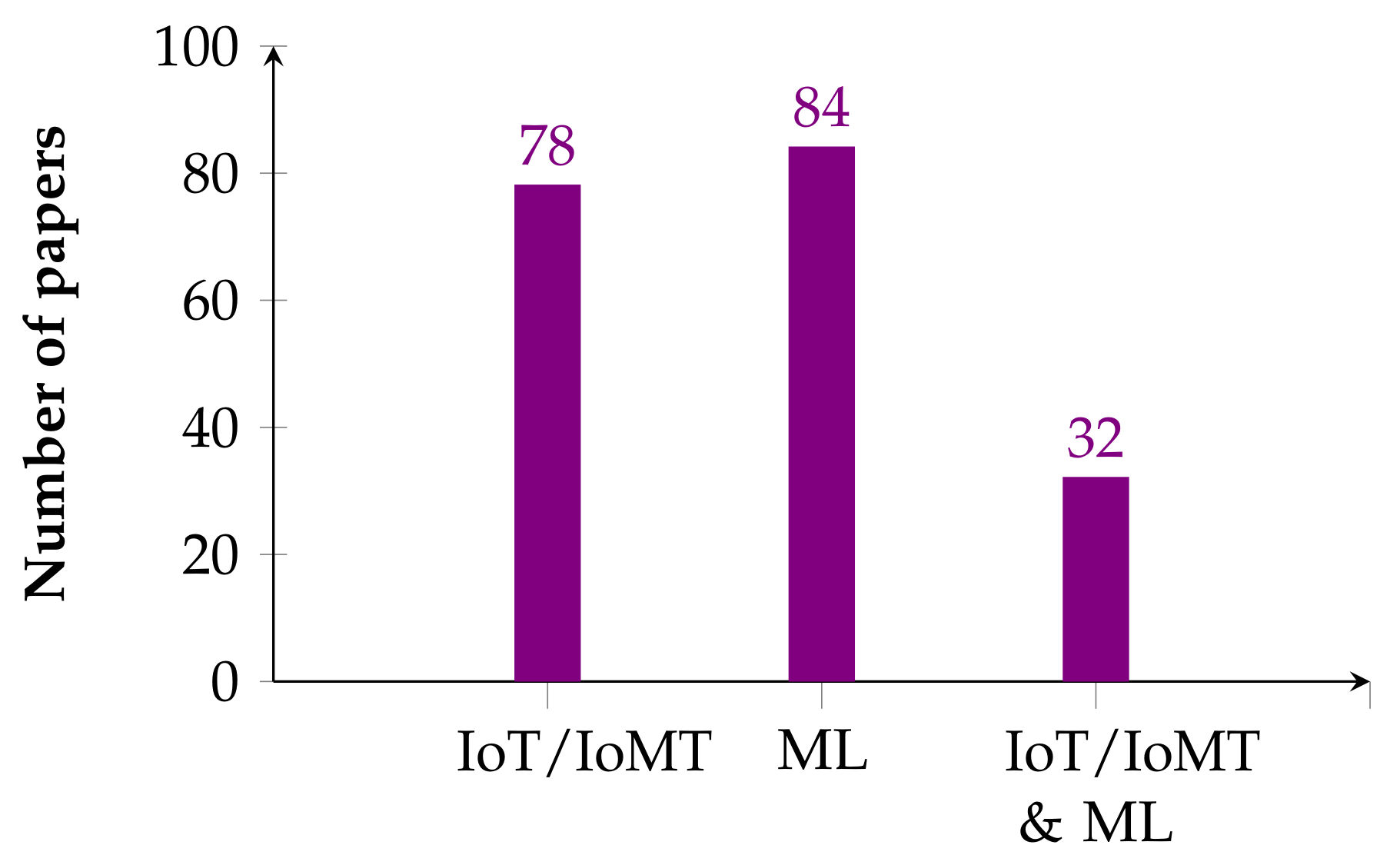
3.1. Data Sources
3.2. Study Selection
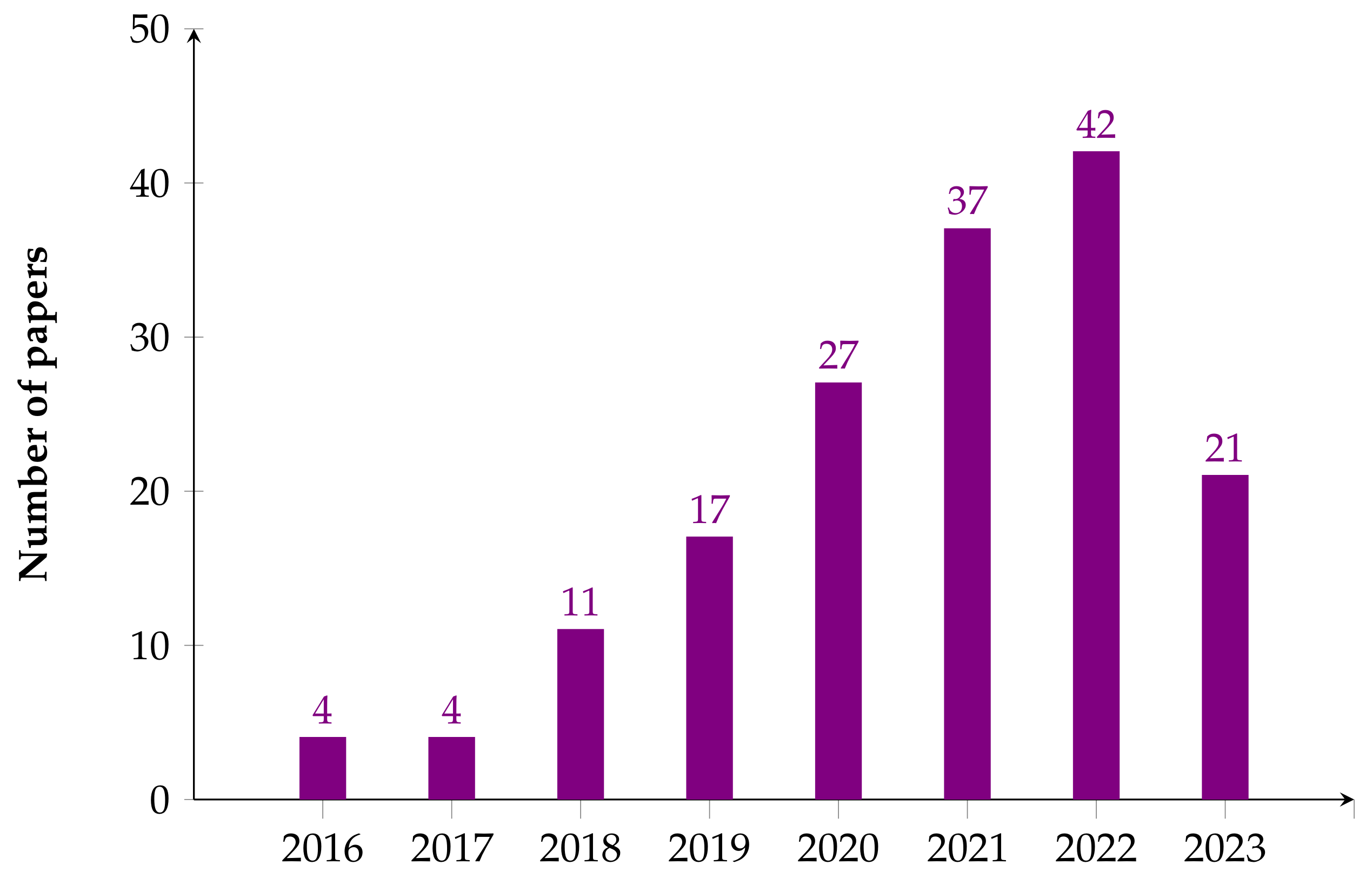
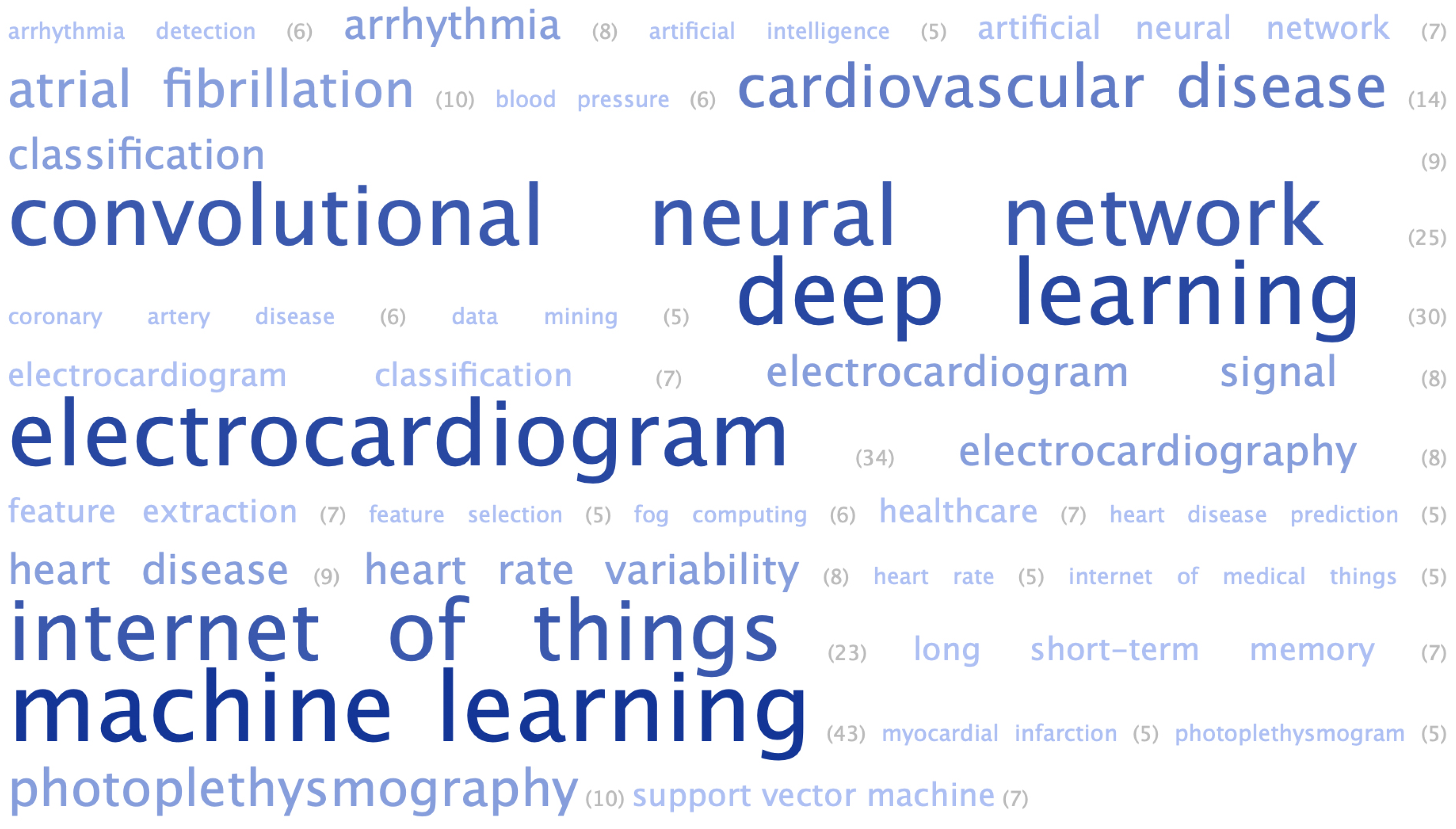
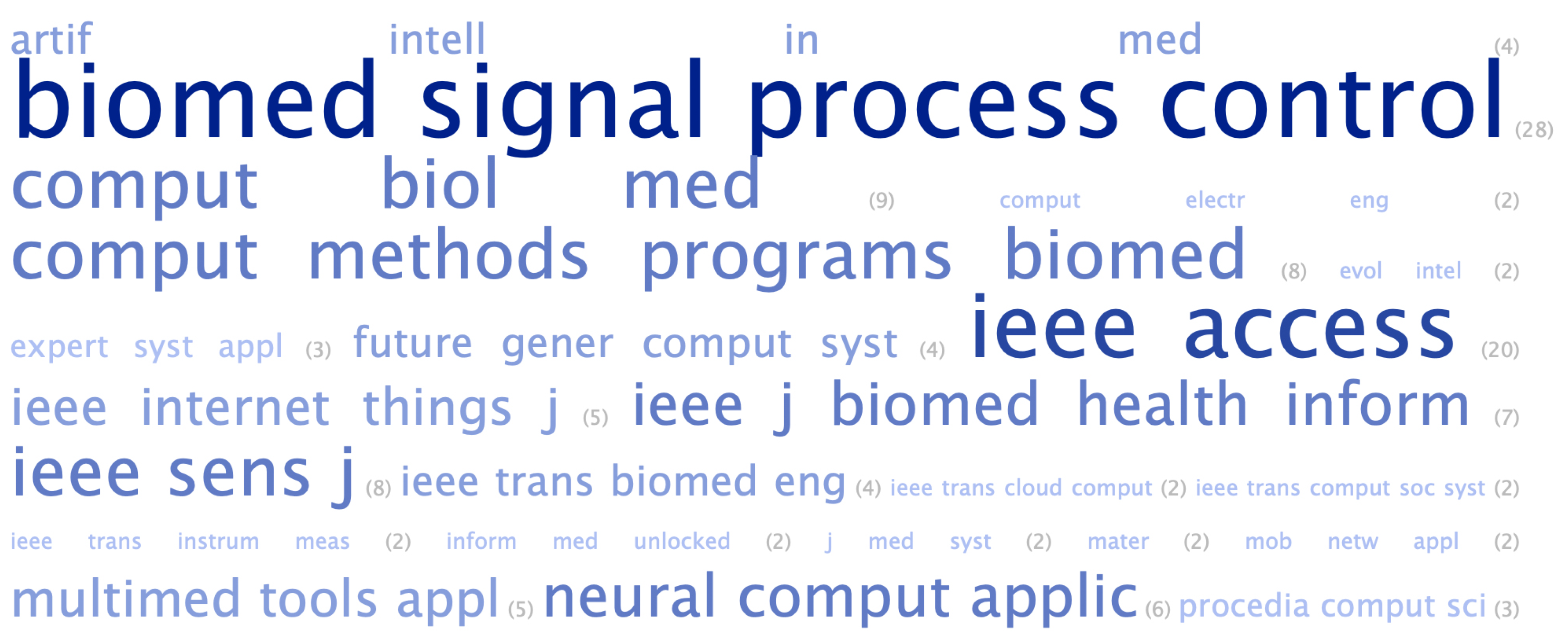
3.3. Bibliometric Analysis
4. Research on CVD Detection Using IoT/IoMT
4.1. Abnormality Detection and Arrhythmia
4.2. Aortic Stenosis
4.3. Arterial Stiffness
4.4. Blood Pressure and Hypertension
4.5. Cardiovascular Disease and Heart Disease
4.6. Others
5. Examples of CVD Detection Utilizing Machine Learning
5.1. Abnormality Detection and Arrhythmia
5.2. Blood Pressure and Hypertension
5.3. Cardiovascular Disease/Heart Disease
5.4. Myocardial Infarction
5.5. Coronary Artery Disease/Coronary Heart Disease
5.6. Others
6. Results and Discussion
6.1. Research Question 1
6.2. Research Question 2
6.3. Research Question 3
6.4. Future Trends
7. Conclusions
Author Contributions
Funding
Institutional Review Board Statement
Informed Consent Statement
Data Availability Statement
Conflicts of Interest
Abbreviations
| CVD | Cardiovascular disease |
| IoT | Internet of things |
| IoMT | Internet of medical things |
| SCG | Seismo-cardiography |
| GCG | Gyro-cardiography |
| PPG | Photoplethysmography |
| AFib | Atrial fibrillation |
| ADHF | Acute descompensated heart failure |
| BPM | Beats per minute |
| SBP | Systolic blood pressure |
| DBP | Diastolic blood pressure |
| SR | Systolic rate |
| DR | Diastolic rate |
| WT | Wavelet transform |
| MI | Miocardial infarction |
| BBB | Bundle branch block |
| BMI | Body mass index |
| FDA | Food and Drug Administration |
Appendix A

| Ref. | Title | Year | Cites |
|---|---|---|---|
| [47] | Ultra-Low Power, Secure IoT Platform for Predicting Cardiovascular Diseases | 2017 | 66 |
| [40] | A deep learning approach for ECG-based heartbeat classification for arrhythmia detection | 2018 | 351 |
| [48] | Towards collaborative intelligent IoT eHealth: From device to fog, and cloud | 2020 | 77 |
| [49] | Noise Rejection for Wearable ECGs Using Modified Frequency Slice Wavelet 739 Transform and Convolutional Neural Networks | 2019 | 54 |
| [50] | An IoT patient monitoring based on fog computing and data mining: Cardiac arrhythmia usecase | 2020 | 59 |
| [51] | ECG signal processing and KNN classifier-based abnormality detection by VH-doctor for remote cardiac healthcare monitoring | 2020 | 23 |
| [52] | Designing Very Fast and Accurate Convolutional Neural Networks with Application in ICD and Smart Electrocardiograph Devices | 2023 | 1 |
| [53] | Improving R Peak Detection in ECG Signal Using Dynamic Mode Selected Energy and Adaptive Window Sizing Algorithm with Decision Tree Algorithm | 2021 | 5 |
| [54] | A Self-Contained STFT CNN for ECG Classification and Arrhythmia Detection at the Edge | 2022 | 8 |
| [55] | An Adaptive Cognitive Sensor Node for ECG Monitoring in the Internet of Medical Things | 2021 | 9 |
| [56] | One-Dimensional CNN Approach for ECG Arrhythmia Analysis in Fog-Cloud Environments | 2021 | 39 |
| [57] | Classification and analysis of cardiac arrhythmia based on incremental support vector regression on IOT platform | 2021 | 9 |
| [41] | IoT-based ECG monitoring for arrhythmia classification using Coyote Grey Wolf optimization-based deep learning CNN classifier | 2022 | 12 |
| [59] | Deep Cardiac Telemonitoring for Clinical Cloud Healthcare Applications | 2022 | - |
| [60] | An IoT enabled secured clinical health care framework for diagnosis of heart diseases | 2023 | - |
| [61] | Dew-based offline computing architecture for healthcare IoT | 2022 | 8 |
| [62] | A novel convolutional neural network structure for differential diagnosis of Wide QRS Complex Tachycardia | 2023 | - |
| [63] | Hybrid optimized convolutional neural network for efficient classification of ECG signals in healthcare monitoring | 2022 | 5 |
| [64] | KEdge: Fuzzy-Based Multi-AI Model Coalescence Solution for Mobile Healthcare System | 2023 | 1 |
| [65] | Prediction of heart abnormalities using deep learning model and wearabledevices in smart health homes | 2022 | 6 |
| [66] | Atrial Fibrillation Detection via Accelerometer and Gyroscope of a Smartphone | 2017 | 130 |
| [67] | Using PPG Signals and Wearable Devices for Atrial Fibrillation Screening | 2019 | 49 |
| [68] | Accurate detection of atrial fibrillation from 12-lead ECG using deep neural | 2020 | 71 |
| [69] | Identification of undiagnosed atrial fibrillation patients using a machine learning risk predicting algorithm and diagnostic testing (PULsE-AI): Study protocol for a randomised controlled trial | 2020 | 9 |
| [70] | Classification of Atrial Fibrillation and Acute Decompensated Heart Failure Using Smartphone Mechanocardiography: A Multi-label Learning Approach | 2020 | 16 |
| [71] | Hardware implementation of 1D-CNN architecture for ECG arrhythmia classification | 2023 | - |
| [67] | Classification of Aortic Stenosis Using Time–Frequency Features From Chest Cardio-Mechanical Signals | 2019 | 27 |
| [73] | Cardiac Output Estimation: Online Implementation for Left Ventricular Assist Device Support | 2020 | 4 |
| [74] | Revealing Unforeseen Diagnostic Image Features with Deep Learning by Detecting Cardiovascular Diseases From Apical 4-Chamber Ultrasounds | 2022 | - |
| [75] | A Wearable Sensor for Arterial Stiffness Monitoring Based on Machine Learning Algorithms | 2018 | 23 |
| [76] | Predicting cardiovascular events with deep learning approach in the context of the internet of things | 2021 | 24 |
| [77] | Bridging Nano and Body Area Networks: A Full Architecture for Cardiovascular Health Applications | 2022 | - |
| [78] | BDCaM: Big Data for Context-Aware Monitoring—A Personalized Knowledge Discovery Framework for Assisted Healthcare | 2016* | 145 |
| [79] | Non-invasive cuffless blood pressure and heart rate monitoring using impedance cardiography | 2022 | 2 |
| [80] | A Machine Learning-Empowered System for Long-Term Motion-Tolerant Wearable Monitoring of Blood Pressure and Heart Rate with Ear-ECG/PPG | 2017 | 85 |
| [81] | Toward Hypertension Prediction Based on PPG-Derived HRV Signals: a Feasibility Study | 2018 | 48 |
| [82] | Blind, Cuff-less, Calibration-Free and Continuous Blood Pressure Estimation using Optimized Inductuve Group Method of Data Handling | 2022 | 31 |
| [83] | Pervasive blood pressure monitoring using Photoplethysmogram (PPG) sensor | 2017 | 48 |
| [84] | Cuffless Continuous Blood Pressure Estimation From Pulse Morphology of Photoplethysmograms | 2019 | 30 |
| [85] | Continuous blood pressure measurement from one-channel electrocardiogram signal using deep-learning techniques | 2020 | 72 |
| [86] | Resource-Aware Mobile-Based Health Monitoring | 2016 | 25 |
| [87] | Smart assisted diagnosis solution with multi-sensor Holter | 2017 | 7 |
| [88] | Machine Learning and Mobile Health Monitoring Platforms: A Case Study on Research and Implementation Challenges | 2018 | 11 |
| [39] | HealthFog: An ensemble deep learning based Smart Healthcare System for Automatic Diagnosis of Heart Diseases in integrated IoT and fog computing environments | 2020 | 432 |
| [89] | Utilizing IoT wearable medical device for heart disease prediction using higher order Boltzmann model: A classification approach | 2019 | 101 |
| [90] | Wireless high-frequency NLOS monitoring system for heart disease combined with hospital and home | 2020 | 13 |
| [91] | Construction and Application of a Medical-Grade Wireless Monitoring System for Physiological Signals at General Wards | 2020 | 22 |
| [98] | Ambient assisted living predictive model for cardiovascular disease prediction using supervised learning | 2021 | 23 |
| [43] | An IoT Framework for Heart Disease Prediction Based on MDCNN Classifier | 2020 | 188 |
| [41] | A novel three-tier Internet of Things architecture with machine learning algorithm for early detection of heart disease | 2018 | 258 |
| [92] | Adaptive Multi-Dimensional dual attentive DCNN for detecting Cardiac Morbidities using Fused ECG-PPG Signals | 2022 | - |
| [93] | Platform for Healthcare Promotion and Cardiovascular Disease Prevention | 2021 | 6 |
| [94] | MedAi: A Smartwatch-Based Application Framework for the Prediction of Common Diseases Using Machine Learning | 2023 | 1 |
| [95] | Detection of Cardiovascular Disease Based on PPG Signals Using Machine Learning with Cloud Computing | 2022 | 4 |
| [96] | A Predictive Analysis of Heart Rates Using Machine Learning Techniques | 2022 | 19 |
| [97] | An Open-Source Privacy-Preserving Large-Scale Mobile Framework for Cardiovascular Health Monitoring and Intervention Planning with an Urban African American Population of Young Adults: User-Centered Design Approach | 2022 | 2 |
| [98] | Ambient assisted living predictive model for cardiovascular disease prediction using supervised learning | 2021 | 23 |
| [99] | An Efcient AlexNet Deep Learning Architecture for Automatic Diagnosis of Cardio‑Vascular Diseases in Healthcare System | 2022 | 5 |
| [100] | Smart wearable model for predicting heart disease using machine learning | 2022 | 3 |
| [101] | Toward Real-Time, At-Home Patient Health Monitoring Using Reservoir Computing CMOS IC | 2021 | 2 |
| [102] | Portable and Real-Time IoT-Based Healthcare Monitoring System for Daily Medical Applications | 2022 | 3 |
| [103] | Dictionary Learning-Based Multichannel ECG Reconstruction Using Compressive Sensing | 2022 | 2 |
| [104] | Real-Time Cloud-Based Patient-Centric Monitoring Using Computational Health Systems | 2022 | 27 |
| [105] | A portable medical device for detecting diseases using Probabilistic Neural Network | 2022 | - |
| [106] | BeatClass: A Sustainable ECG Classification System in IoT-Based eHealth | 2021 | 34 |
| [107] | Non-contact Monitoring of Heart Rate Variability Using A Fiber Optic Sensor | 2023 | - |
| [108] | Remote Health Monitoring System for the Estimation of Blood Pressure, Heart Rate, and Blood Oxygen Saturation Level | 2023 | 1 |
| [109] | iKardo: An Intelligent ECG Device for Automatic Critical Beat Identification for Smart Healthcare | 2021 | 6 |
| [110] | Energy-Efficient Real-Time Heart Monitoring on Edge-Fog-Cloud Internet of Medical Things | 2021 | 15 |
| [111] | Real-Time Tele-Monitoring of Patients with Chronic Heart-Failure Using a Smartphone: Lessons Learned | 2016 | 58 |
| [112] | Fog based smart healthcare: a machine learning paradigms for IoT sector | 2022 | 2 |
| [113] | Real-Time Event-Driven Classification Technique for Early Detection and Prevention of Myocardial Infarction on Wearable Systems | 2018 | 75 |
| [114] | A High performance electronic nose system for the recognition of myocardial infarction and coronary artery diseases | 2021 | 24 |
| [115] | FETCH: A Deep Learning-Based Fog Computing and IoT Integrated Environment for Healthcare Monitoring and Diagnosis | 2022 | 32 |
| [116] | FedECG: A Federated Semi-supervised Learning Framework for Electrocardiogram Abnormalities Prediction | 2023 | - |
| [117] | Development of a PPG Sensor Array as a Wearable Device for Monitoring Cardiovascular Metrics | 2021 | 17 |
| [118] | AI-based stroke prediction system using body motion biosignals during walking | 2022 | 3 |
| [119] | A Machine Learning Pipeline for Measurement of Arterial Stiffness in A-Mode Ultrasound | 2021 | 2 |
| [120] | A Real-Time Tunable ECG Noise-Aware System for IoT-Enabled Devices | 2022 | 1 |
| [121] | Interpretable Rule Mining for Real-Time ECG Anomaly Detection in IoT Edge Sensors | 2023 | - |
| [129] | Proposition of novel classification approach and features for improved real-time arrhythmia monitoring | 2016 | 21 |
| [44] | A robust deep convolutional neural network with batch-weighted loss for heartbeat classification | 2019 | 158 |
| [38] | Arrhythmia detection using deep convolutional neural network with long duration ECG signals | 2018 | 579 |
| [42] | A new approach for arrhythmia classification using deep coded features and LSTM networks | 2019 | 256 |
| [130] | A Real-Time Arrhythmia Heartbeats Classification Algorithm Using Parallel Delta Modulations and Rotated Linear-Kernel Support Vector Machines | 2019 | 56 |
| [131] | Automated detection of shockable and non-shockable arrhythmia using novel wavelet-based ECG features | 2019 | 42 |
| [132] | Heart disease detection using hybrid of bacterial foraging and particle swarm optimization | 2020 | 23 |
| [133] | A Deep Biometric Recognition and Diagnosis Network with Residual Learning for Arrhythmia Screening Using Electrocardiogram Recordings | 2020 | 3 |
| [134] | Deep Multi-Scale Fusion Neural Network for Multi-Class Arrhythmia Detection | 2020 | 74 |
| [135] | Intelligent Health Vessel ABC-DE: An Electrocardiogram Cloud Computing Service | 2018 | 17 |
| [136] | Improving the safety of atrial fibrillation monitoring systems through human verification | 2017 | 11 |
| [137] | Validating the robustness of an internet of things based atrial fibrillation detection system | 2020 | 20 |
| [138] | Extracting deep features from short ECG signals for early atrial fibrillation detection | 2020 | 22 |
| [139] | Detection of Atrial Fibrillation in Compressively Sensed Electrocardiogram Measurements | 2020 | 15 |
| [140] | A Multi-tier Deep Learning Model for Arrhythmia Detection | 2020 | 115 |
| [141] | Arrhythmia classification from single-lead ECG signals using the inter-patient paradigm | 2021 | 50 |
| [142] | Time adaptive ECG driven cardiovascular disease detector | 2021 | 21 |
| [143] | ECG arrhythmia classification by using a recurrence plot and convolutional neural network | 2021 | 91 |
| [144] | Stacking segment-based CNN with SVM for recognition of atrial fibrillation from single-lead ECG recordings | 2021 | 33 |
| [145] | Exploring Deep Features and ECG Attributes to Detect Cardiac Rhythm Classes | 2021 | 91 |
| [146] | Automated ECG multi-class classification system based on combining deep learning features with HRV and ECG measures | 2022 | 17 |
| [147] | Deep learning-based multidimensional feature fusion for classification of ECG arrhythmia | 2021 | 15 |
| [148] | ECG signal classification to detect heart arrhythmia using ELM and CNN | 2022 | - |
| [149] | Multi-Lead ECG Classification via an Information-Based Attention Convolutional Neural Network | 2020 | 5 |
| [150] | Fuzz-ClustNet: Coupled fuzzy clustering and deep neural networks for Arrhythmia detection from ECG signals | 2023 | - |
| [151] | Inter-patient arrhythmia classification with improved deep residual convolutional neural network | 2022 | 21 |
| [152] | DeepArr: An investigative tool for arrhythmia detection using a contextual deep neural network from electrocardiograms (ECG) signals | 2023 | - |
| [153] | Two-stage detection method of supraventricular and ventricular ectopic beats based on sequential artificial features and heartbeats | 2023 | - |
| [154] | Automatic varied-length ECG classification using a lightweight DenseNet model | 2023 | - |
| [155] | Arrhythmia detection based on multi-scale fusion of hybrid deep models from single lead ECG recordings: A multicenter dataset study | 2022 | 2 |
| [151] | A deep learning approach to cardiovascular disease classification using empirical mode decomposition for ECG feature extraction | 2023 | 1 |
| [157] | Prediction of paroxysmal atrial fibrillation using new heart rate variability features | 2021 | 23 |
| [158] | Predicting Hypertensive Patients with Higher Risk of Developing Vascular Events Using Heart Rate Variability and Machine Learning | 2020 | 20 |
| [159] | Nonlinear Dynamic Modeling of Blood Pressure Waveform: Towards an Accurate Cuffless Monitoring System | 2020 | 22 |
| [160] | Predicting Systolic Blood Pressure in Real-Time Using Streaming Data and Deep Learning | 2021 | 15 |
| [161] | Cuffless blood pressure estimation based on composite neural network and graphics information | 2021 | 10 |
| [162] | PPG-based blood pressure estimation can benefit from scalable multi-scale fusion neural networks and multi-task learning | 2022 | 5 |
| [163] | A Refined Blood Pressure Estimation Model Based on Single Channel Photoplethysmography | 2022 | 4 |
| [164] | An advanced LAN model based on optimized feature algorithm: Towards hypertension interpretability | 2021 | 2 |
| [165] | Deep learning models for cuffless blood pressure monitoring from PPG signals using attention mechanism | 2021 | 51 |
| [166] | NABNet: A Nested Attention-guided BiConvLSTM network for a robust prediction of Blood Pressure components from reconstructed Arterial Blood Pressure waveforms using PPG and ECG signals | 2023 | 3 |
| [167] | DeepCNAP: A Deep Learning Approach for Continuous Noninvasive Arterial Blood Pressure Monitoring Using Photoplethysmography | 2022 | 5 |
| [168] | An IoT based efficient hybrid recommender system for cardiovascular disease | 2019 | 75 |
| [37] | Effective Heart Disease Prediction Using Hybrid Machine Learning Techniques | 2019 | 888 |
| [169] | An efficient IoT based patient monitoring and heart disease prediction system using Deep learning modified neural network | 2020 | 92 |
| [170] | HDPM: An Effective Heart Disease Prediction Model for a Clinical Decision Support System | 2020 | 130 |
| [45] | A Healthcare Monitoring System for the Diagnosis of Heart Disease in the IoMT Cloud Environment Using MSSO-ANFIS | 2020 | 153 |
| [46] | Comprenhensive electrocardiographic diagnosis based on deep learning | 2020 | 146 |
| [171] | An Efficient IoT-Based Platform for Remote Real-Time Cardiac Activity Monitoring | 2020 | 35 |
| [172] | Multi-disease big data analysis using beetle swarm optimization and an adaptive neuro-fuzzy inference system | 2021 | 17 |
| [173] | Cardiac disease detection using cuckoo search enabled deep belief network | 2022 | 2 |
| [174] | Automatic diagnosis of cardiovascular disorders by sub images of the ECG signal using multi-feature extraction methods and randomized neural network | 2021 | 20 |
| [175] | An intelligent heart disease prediction system based on swarm artificial neural network | 2021 | 23 |
| [176] | A multi-label learning prediction model for heart failure in patients with atrial fibrillation based on expert knowledge of disease duration | 2023 | - |
| [177] | WoM-based deep BiLSTM: smart disease prediction model using WoM-based deep BiLSTM classifier | 2023 | - |
| [178] | A scalable and real-time system for disease prediction using big data processing | 2023 | 3 |
| [179] | A novel blockchain-enabled heart disease prediction mechanism using machine learning | 2022 | 15 |
| [180] | Heart rate estimation network from facial videos using spatiotemporal feature image | 2022 | 2 |
| [181] | Predictive analysis of cardiovascular disease using gradient boosting based learning and recursive feature elimination technique | 2022 | 3 |
| [182] | Effective heart disease prediction using novel MLP-EBMDA approach | 2022 | 22 |
| [183] | LightX3ECG: A Lightweight and eXplainable Deep Learning System for 3-lead Electrocardiogram Classification | 2023 | 2 |
| [184] | Heart Diseases Prediction based on Stacking Classifiers Model | 2023 | - |
| [185] | Tuning Multi-Layer Perceptron by Hybridized Arithmetic Optimization Algorithm for Healthcare 4.0 | 2022 | - |
| [186] | Optimized Signal Quality Assessment for Photoplethysmogram Signals Using Feature Selection | 2022 | 8 |
| [187] | Photoplethysmography-Based Heart Action Monitoring Using a Growing Multilayer Network | 2022 | - |
| [188] | KD-Informer: A Cuff-Less Continuous Blood Pressure Waveform Estimation Approach Based on Single Photoplethysmography | 2022 | 6 |
| [189] | Multiclass classification of myocardial infarction with convolutional and recurrent neural networks for portable ECG devices | 2018 | 98 |
| [190] | ST-Net: Synthetic ECG tracing for diagnosing various cardiovascular diseases | 2020 | 6 |
| [191] | Explainable Prediction of Acute Myocardial Infarction Using Machine Learning and Shapley Values | 2020 | 47 |
| [192] | Prognostic Value of Machine Learning in Patients with Acute Myocardial Infarction | 2022 | 8 |
| [193] | Efficient detection of myocardial infarction from single lead ECG signal | 2021 | 21 |
| [194] | Near real-time single-beat myocardial infarction detection from single-lead electrocardiogram using Long Short-Term Memory Neural Network | 2021 | 10 |
| [195] | Predicting cardiac disease from interactions of simultaneously-acquired hemodynamic and cardiac signals | 2021 | 8 |
| [196] | Multilayer perceptron based deep neural network for early detection of coronary heart disease | 2021 | 17 |
| [197] | Early Detection of Coronary Heart Disease using Ensemble Techniques | 2021 | 37 |
| [198] | Non-invasive detection of coronary artery disease from photoplethysmograph using lumped parameter modelling | 2022 | 2 |
| [199] | Combining Convolutional Neural Network and Distance Distribution Matrix for Identification of Congestive Heart Failure | 2018 | 36 |
| [200] | Deep Learning Electrocardiographic Analysis for Detection of Left-Sided Valvular Heart Disease | 2022 | 10 |
| [201] | High-Throughput Precision Phenotyping of Left Ventricular Hypertrophy with Cardiovascular Deep Learning | 2022 | 32 |
| [202] | Convolutional neural network based automatic screening tool for cardiovascular diseases using different intervals of ECG signals | 2021 | 25 |
| [203] | Reaction-diffusion informed approach to determine myocardial ischemia using stochastic in-silico ECGs and CNNs | 2021 | 3 |
| [204] | A machine learning-based risk stratification model for ventricular tachycardia and heart failure in hypertrophic cardiomyopathy | 2021 | 19 |
| [205] | Identifying Stroke Indicators Using Rough Sets | 2020 | 18 |
Appendix B
| Refs. | Journal | Num. of Papers |
|---|---|---|
| [200] | Am. J. Cardiol. | 1 |
| [176] | Appl Intell | 1 |
| [79,85,138] | Artif Intell Med. | 3 |
| [46] | Artif Intell Med. | 1 |
| [38,68,129,131,150,157,180,203,204] | Comput Biol Med. | 9 |
| [41,179] | Comput Electr Eng | 2 |
| [24,33,42,141,151,195,202] | Comput Methods Programs Biomed | 7 |
| [98] | Evol Intel | 1 |
| [132] | Evol Syst. | 1 |
| [44,189] | Expert Syst. Appl | 2 |
| [39,40,83,90] | Future Gener Comput Syst. | 4 |
| [196] | Health Technol | 1 |
| [61] | ICT Express | 1 |
| [37,43,45,49,52,54,55,56,80,84,94,115,133,158,169,170,191,199,205] | IEEE Access | 18 |
| [77,106,107,110,121] | IEEE Internet Things J. | 5 |
| [66,86,134,163,167,188] | IEEE J. Biomed Health Inform | 6 |
| [101] | IEEE J. Emerg Sel Top Circuits | 1 |
| [70,75,103,108,117,120,159,187] | IEEE Sens. J. | 8 |
| [64] | IEEE Syst. J. | 1 |
| [171] | IEEE T Consum Electr | 1 |
| [47] | IEEE TCAS-I | 1 |
| [111] | IEEE Trans. Affect Comput | 1 |
| [73,113] | IEEE Trans. Biomed Circuits Syst. | 2 |
| [72,130,186] | IEEE Trans. Biomed Eng | 3 |
| [78,135] | IEEE Trans. Cloud Comput | 2 |
| [102,104] | IEEE Trans. Comput Soc Syst. | 2 |
| [109] | IEEE Trans. Consum | 1 |
| [67] | IEEE Trans. Ind Electron | 1 |
| [139,140] | IEEE Trans. Instrum Meas | 2 |
| [119] | IEEE Trans. Ultrason Ferroelectr | 1 |
| [92] | IEEE Trans. Artif Intell | 1 |
| [197] | Inform Med. Unlocked | 1 |
| [96] | Int. J. Environ. Res. Public Health | 1 |
| [50,77,106,107,110,121] | Internet Things | 6 |
| [74] | J. Am Heart Assoc | 1 |
| [192] | J. Cardiovasc. Dev. Dis. | 1 |
| [88] | J. Healthcare Inform. Res. | 1 |
| [116] | J. King Saud Univ. | 1 |
| [81,91] | J. Med. Syst. | 2 |
| [69] | J. Res. Health Sci. | 1 |
| [149] | Shanghai Jiaotong Univ Sci | 1 |
| [118] | J. Supercomput | 1 |
| [201] | JAMA Cardiol | 1 |
| [97] | JMIR Form Res | 1 |
| [145] | Knowl Based Syst. | 1 |
| [85] | Measurement | 1 |
| [48] | Microprocess Microsyst | 1 |
| [160] | Mob Netw Appl | 1 |
| [28,65,112,148,177,178] | Multimed Tools Appl | 6 |
| [26,76,146,147,172,175] | Neural Comput & Applic | 6 |
| [87] | Neurocomputing | 1 |
| [137] | Pattern Recognit. Lett. | 1 |
| [168] | Peer-to-Peer Netw. Appl. | 1 |
| [59] | Procedia Comput. Sci. | |
| [136] | Saf Sci | 1 |
| [23,53,121] | Sensors | 3 |
| [51] | Soft. Comput. | 1 |
| [99,100] | Wirel. Pers. Commun. | 2 |
References
- Pan American Health Organization. Available online: https://www.paho.org/en/topics/cardiovascular-diseases (accessed on 12 March 2022).
- Secretaría de Salud, Enfermedades no Transmisibles. Available online: https://www.gob.mx/cms/uploads/attachment/file/416454/Enfermedades_No_Transmisibles_ebook.pdf (accessed on 12 March 2022).
- World Health Organization. Available online: https://www.who.int/health-topics/cardiovascular-diseases/#tab=tab_1 (accessed on 12 March 2022).
- Pizarro, J. Internet de las Cosas (IoT) con Esp. Manual Práctico, 1st ed.; Ediciones Paraninfo: Madrid, Spain, 2020; p. 1. [Google Scholar]
- Singh, R.P.; Javaid, M.; Haleem, A.; Vaishya, R.; Ali, S. Internet of Medical Things (IoMT) for orthopedic in COVID-19 pandemic: Roles, challenges, and applications. J. Clin. Orthop. Trauma 2020, 11, 713–717. [Google Scholar] [CrossRef]
- Müller, C.A.; Guido, S. Introduction to Machine Learning with Python, 1st ed.; O’Reilly Media: Sebastopol, CA, USA, 2017; pp. 1–2. [Google Scholar]
- Kim, P. MATLAB Deep Learning: With Machine Learning, Neural Networks, and Artificial Intelligence, 1st ed.; Apress: Seoul, Republic of Korea, 2017; pp. 1–2. [Google Scholar]
- Vayena, E.; Blasimme, A.; Cohen, I.G. Machine learning in medicine: Addressing ethical challenges. PLoS Med. 2018, 15, e1002689. [Google Scholar] [CrossRef]
- Page, M.J.; McKenzie, J.E.; Bossuyt, P.M.; Boutron, I.; Hoffmann, T.C.; Mulrow, C.D.; Shamseer, L.; Tetzlaff, J.M.; Akl, E.A.; Brennan, S.E.; et al. The PRISMA 2020 statement: An updated guideline for reporting systematic reviews. BMJ 2021, 88, 105906. [Google Scholar]
- Pettricrew, M.; Roberts, H. Systematic Reviews in the Social Sciences, A Practical Guide; Blackwell Publishing: Malden, NJ, USA, 2006; p. 38. [Google Scholar]
- Friedrich, S.; Groß, S.; König, I.R.; Engelhardt, S.; Bahls, M.; Heinz, J.; Huber, C.; Kaderali, L.; Kelm, M.; Leha, A.; et al. Applications of artificial intelligence/machine learning approaches in cardiovascular medicine: A systematic review with recommendations. Eur. Heart. J. Digit. Health 2021, 2, 424–436. [Google Scholar] [CrossRef] [PubMed]
- Hazra, A.; Mandal, S.K.; Gupta, A.; Mukherjee, A.; Mukherjee, A. Heart disease diagnosis and prediction using machine learning and data mining techniques: A review. Adv. Comput. Sci. Technol. 2017, 10, 2137–2159. [Google Scholar]
- Shameer, K.; Johnson, K.W.; Glicksberg, B.S.; Dudley, J.T.; Sengupta, P.P. Machine learning in cardiovascular medicine: Are we there yet? Heart 2018, 104, 1156–1164. [Google Scholar] [CrossRef]
- Bolhasani, H.; Mohseni, M.; Rahmani, A.M. Deep learning applications for IoT in health care: A systematic review. Inform. Med. 2021, 23, 100550. [Google Scholar] [CrossRef]
- Huang, J.; Wu, X.; Huang, W.; Wu, X.; Wang, S. Internet of things in health management systems: A review. Int. J. Commun. Syst. 2020, 34, e4683. [Google Scholar] [CrossRef]
- Lin, J.; Fu, R.; Zhong, X.; Yu, P.; Tan, G.; Li, W.; Zhang, H.; Li, Y.; Zhou, L.; Ning, C. Wearable sensors and devices for real-time cardiovascular disease monitoring. Cell Rep. Cell Phys. Sci. 2021, 2, 1–25. [Google Scholar] [CrossRef]
- Rahaman, A.; Islam, M.; Islam, R.; Sadi, M.S.; Nooruddin, S. Developing IoT Based Smart Health Monitoring Systems: A Review. Rev. D’Intell. Artif. 2019, 33, 435–440. [Google Scholar] [CrossRef]
- Panicker, S.; Gayathri, P. Use of machine learning techniques in healthcare: A brief review of cardiovascular disease classification. In Proceedings of the 2nd International Conference on Communication & Information Processing (ICCIP), Pune, India, 26–27 June 2020; Volume 2. [Google Scholar]
- Dadkhah, M.; Mehraeen, M.; Rahimnia, F.; Kimiafar, K. Use of Internet of Things for Chronic Disease Management: An Overview. J. Med. Signals Sens. 2021, 11, 138. [Google Scholar]
- Lamonaca, F.; Balestrieri, E.; Tudosa, I.; Picariello, F.; Bonavolontà, F. An Overview on Internet of Medical Things in Blood Pressure Monitoring. In Proceedings of the 2019 IEEE International Symposium on Medical Measurements and Applications (MeMeA), Istanbul, Turkey, 26–28 June 2019; pp. 1–6. [Google Scholar]
- Argha, A.; Celler, B.G.; Lovell, N.H. Artificial Intelligence Based Blood Pressure Estimation FRom Auscultatory and Oscillometric Waveforms: A Methodological Review. IEEE Rev. Biomed. Eng. 2020, 15, 152–168. [Google Scholar] [CrossRef]
- Huang, J.-D.; Wang, J.; Ramsey, E.; Leavey, G.; Chico, T.J.A.; Condell, J. Applying Artificial Intelligence to Wearable Sensor Data to Diagnose and Predict Cardiovascular Disease: A Review. Sensors 2021, 22, 8002. [Google Scholar] [CrossRef]
- Hinai, G.A.; Jammoul, S.; Vajihi, Z.; Afilalo, J. Deep learning analysis of resting electrocardiograms for the detection of myocardial dysfunction, hypertrophy, and ischaemia: A systematic review. Eur. Heart J. 2022, 3, 115–116. [Google Scholar]
- Faizal, A.S.M.; Thevarajah, M.T.; Khor, S.M.; Chang, S.-W. A review of risk prediction models in cardiovascular disease: Conventional approach vs. artificial intelligence approach. Comput. Methods Programs Biomed. 2021, 207, 106190. [Google Scholar] [CrossRef]
- Chen, S.W.; Wang, S.L.; Qi, X.Z.; Samuri, S.M.; Yang, C. Review of ECG detection and classification based on deep learning: Coherent taxonomy, motivation, open challenges and recommendations. Biomed. Signal Procress. Control 2021, 129, 104163. [Google Scholar] [CrossRef]
- Qureshi, M.A.; Qureshi, K.N.; Jeon, G.; Piccialli, F. Deep learning-based ambient assisted living for self-management of cardiovascular conditions. Neural Comput. Appl. 2022, 34, 10449–10467. [Google Scholar] [CrossRef]
- Bhushan, M.; Pandit, A.; Garg, A. Machine learning and deep learning techniques for the analysis of heart disease: A systematic literature review, open challenges and future directions. Artif. Intell. Rev. 2023. [Google Scholar] [CrossRef]
- Rath, A.; Mishra, D.; Panda, G.; Satapathy, S.C. An exhaustive review of machine learning and deep learning based diagnosis of heart diseases. Multimed. Tools Appl. 2022, 81, 36069–36127. [Google Scholar] [CrossRef]
- Maurya, M.R.; Riyaz, N.U.S.; Reddy, M.S.B.; Yalcin, H.C.; Ouakad, H.M.; Bahadur, I.; Al-Maadeed, S.; Sadasivuni, K.K. A review of smart sensors coupled with Internet of Things and Artificial Intelligence approach for heart failure monitoring. Med. Biol. Eng. Comput. 2021, 59, 2185–2203. [Google Scholar] [CrossRef] [PubMed]
- Kumar, Y.; Koul, A.; Singla, R.; Ijaz, M.F. Artificial intelligence in disease diagnosis: A systematic literature review, synthesizing framework and future research agenda. J. Ambient Intell. Humaniz. Comput. 2022, 14, 8459–8486. [Google Scholar] [CrossRef] [PubMed]
- Jasinska-Piadlo, Q.; Bond, R.; Biglarbeigi, P.; Brisk, R.; Campbell, P.; McEneaneny, D. What can machines learn about heart failure? A systematic literature review. J. Ambient Intell. Humaniz. Comput. 2022, 13, 163–183. [Google Scholar] [CrossRef]
- Chakrabarti, S.; Biswas, N.; Jones, L.D.; Kesari, S.; Ashili, S. Smart Consumer Wearables as Digital Diagnostic Tools: A Review. Diagnostics 2022, 12, 2110. [Google Scholar] [CrossRef]
- Guo, Y.; Liu, X.; Peng, S.; Jiang, X.; Xu, K.; Chen, C.; Wang, Z.; Dai, C.; Chen, W. A review of wearable and unobtrusive sensing technologies for chronic disease management. Comput. Methods Programs Biomed. 2021, 129, 104163. [Google Scholar] [CrossRef]
- Xiao, Y.; Watson, M. Guidance on Conducting a Systematic Literature Review. J. Plan. Educ. Res. 2017, 39, 93–112. [Google Scholar] [CrossRef]
- MacMillan, F.; McBride, K.A.; George, E.S.; Steiner, G.Z. Conducting a Systematic Review: A Practical Guide. In Handbook of Research Methods in Health Social Sciences; Springer: Berlin, Heidelberg, 2019; pp. 805–826. [Google Scholar]
- Harris, J.D.; Quatman, C.E.; Manring, M.M.; Siston, R.A.; Flanigan, D.C. How to Write a Systematic Review. Am. J. Sports Med. 2014, 42, 2761–2768. [Google Scholar] [CrossRef]
- Mohan, S.; Thirumalai, C.; Srivastava, G. Effective Heart Disease Prediction Using Hybrid Machine Learning Techniques. IEEE Access 2019, 7, 81542–81554. [Google Scholar] [CrossRef]
- Yildirim, Ö.; Plawiak, P.; Tan, R.-S.; Acharya, U.R. Arrhythmia detection using deep convolutional neural network with long duration ECG signals. Comput. Biol. Med. 2018, 102, 411–420. [Google Scholar] [CrossRef]
- Tuli, S.; Basumatary, N.; Gill, S.S.; Kahani, M.; Arya, R.C.; Wander, G.S.; Buyya, R. HealthFog: An ensemble deep learning based Smart Healthcare System for Automatic Diagnosis of Heart Diseases in inte-grated IoT and fog computing environments. Future Gener. Comput. Syst. 2020, 104, 187–200. [Google Scholar] [CrossRef]
- Sannino, G.; De Pietro, G. A deep learning approach for ECG-based heartbeat classification for arrhythmia detection. Future Gener. Comput. Syst. 2018, 86, 446–455. [Google Scholar] [CrossRef]
- Kumar, P.M.; Gandhi, U.D. A novel three-tier Internet of Things architecture with machine learning algorithm for early detection of heart diseases. Comput. Electr. Eng. 2018, 65, 222–235. [Google Scholar] [CrossRef]
- Yildirim, O.; Baloglu, U.B.; Tan, R.-S.; Ciaccio, E.J.; Acharya, U.R. A new approach for arrhythmia classification using deep coded features and LSTM networks. Comput. Methods Programs Biomed. 2019, 176, 121–133. [Google Scholar] [CrossRef] [PubMed]
- Khan, M.A. An IoT Framework for Heart Disease Prediction Based on MDCNN Classifier. IEEE Access 2020, 8, 34717–34727. [Google Scholar] [CrossRef]
- Sellami, A.; Hwang, H. A robust deep convolutional neural network with batch-weighted loss for heartbeat classification. Expert Syst. Appl. 2019, 122, 75–84. [Google Scholar] [CrossRef]
- Khan, M.A.; Algarni, F. A Healthcare Monitoring System for the Diagnosis of Heart Disease in the IoMT Cloud Environment Using MSSO-ANFIS. IEEE Access 2020, 8, 122259–122269. [Google Scholar] [CrossRef]
- Lih, O.S.; Jahmunah, V.; San, T.R.; Ciaccio, E.J.; Yamakawa, T.; Tanabe, M.; Kobayashi, M.; Faust, O.; Acharya, U.R. Comprenhensive electrocardiographic diagnosis base don deep learning. Artif. Intell. Med. 2020, 103, 101789. [Google Scholar] [CrossRef]
- Yasin, M.; Tekeste, T.; Saleh, H.; Mohammad, B.; Sinanoglu, O.; Ismail, M. Ultra-Low Power, Secure IoT Platform for Predicting Cardiovascular Diseases. IEEE TCAS-I 2017, 64, 2624–2637. [Google Scholar] [CrossRef]
- Farahani, B.; Barzegari, M.; Aliee, F.S.; Shaik, K.A. Towards collaborative intelligent IoT eHealth: From device to fog, and cloud. Microprocess. Microsyst 2020, 72, 102938. [Google Scholar] [CrossRef]
- Zhao, Z.; Liu, C.; Li, Y.; Li, Y.; Wang, J.; Lin, B.S.; Li, J. Noise Rejection for Wearable ECGs Using Modified Frequency Slice Wavelet Transform and Convolutional Neural Networks. IEEE Access 2019, 7, 34060–34067. [Google Scholar] [CrossRef]
- Moghadas, E.; Rezazadeh, J.; Farahbakhsh, R. An IoT patient monitoring based on fog computing and data mining: Cardiac arrhythmia usecase. Internet Things 2020, 11, 100251. [Google Scholar] [CrossRef]
- Venkataramanaiah, B.; Kamala, J. ECG signal processing and KNN classifier-based abnormality detection by VH-doctor for remote cardiac healthcare monitoring. Soft Comput. 2020, 24, 17457–17466. [Google Scholar] [CrossRef]
- Keyanfar, A.; Ghaderi, R.; Nazari, S.; Hajimoradi, B.; Kamalzadeh, L. Designing Very Fast and Accurate Convolutional Neural Networks with Application in ICD and Smart Electrocardiograph Devices. IEEE Access 2023, 11, 5502–5516. [Google Scholar] [CrossRef]
- Al, Z.M.A.; Thapa, K.; Yang, S.-H. Improving R Peak Detection in ECG Signal Using Dynamic Mode Selected Energy and Adaptive Window Sizing Algorithm with Decision Tree Algorithm. Sensors 2021, 21, 6682. [Google Scholar]
- Farag, M.M. A Self-Contained STFT CNN for ECG Classification and Arrhythmia Detection at the Edge. IEEE Access 2022, 10, 94469–94486. [Google Scholar] [CrossRef]
- Scrugli, M.A.; Loi, D.; Raffo, L.; Meloni, P. An Adaptive Cognitive Sensor Node for ECG Monitoring in the Internet of Medical Things. IEEE Access 2021, 10, 1688–1705. [Google Scholar] [CrossRef]
- Cheikhrouhou, O.; MAhmud, R.; Zouari, R.; Ibrahim, M.; Zaguia, A.; Gia, T.N. One-dimensional CNN approach for ECG arrhythmia analysis in fog-cloud environments. IEEE Access 2021, 9, 103513–103523. [Google Scholar] [CrossRef]
- Sanamdikar, S.T.; Hamde, S.T.; Asutkar, V.G. Classification and analysis of cardiac arrhythmia based on incremental support vector regression on IOT platform. Biomed. Signal Process. Control 2021, 64, 102324. [Google Scholar] [CrossRef]
- Kumar, A.; Kumar, S.; Dutt, V.; Dubey, A.K.; García-Díaz, V. IoT-based ECG monitoring for arrhythmia classification using Coyote Grey Wolf optimization-based deep learning CNN classifier. Biomed. Signal Process. Control 2022, 76, 103638. [Google Scholar] [CrossRef]
- Belaid, S.; Hattay, J.; Mohamed, H.H.; Rezgui, R. Deep Cardiac Telemonitoring for Clinical Cloud Healthcare Applications. Procedia Comput. Sci. 2022, 207, 2843–2852. [Google Scholar] [CrossRef]
- Raheja, N.; Manocha, A.K. An IoT enabled secured clinical health care framework for diagnosis of heart diseases. Biomed. Signal Process. Control 2023, 80, 104368. [Google Scholar] [CrossRef]
- Medhi, K.; Ahmed, N.; Hussain, M.I. Dew-based offline computing architecture for healthcare IoT. ICT Express 2023, 80, 371–378. [Google Scholar] [CrossRef]
- Fayyazifar, N.; Dwivedi, G.; Suter, D.; Ahderom, S.; Maiorana, A.; Clarkin, O.; Balamane, S.; Saha, N.; King, B.; Green, M.S.; et al. A novel convolutional neural network structure for differential diagnosis of Wide QRS Complex Tachycardia. Biomed. Signal Process. Control 2023, 81, 104506. [Google Scholar] [CrossRef]
- Karthiga, M.; Santhi, V.; Sountharrajan, S. Hybrid optimized convolutional neural network for efficient classification of ECG signals in healthcare monitoring. Biomed. Signal Process. Control 2022, 76, 371–378. [Google Scholar] [CrossRef]
- Misra, S.; Pal, S.; Deb, P.K.; Gupta, E.S. KEdge: Fuzzy-Based Multi-AI Model Coalescence Solution for Mobile Healthcare System. IEEE Syst. J. 2023, 17, 1721–1728. [Google Scholar] [CrossRef]
- Shafi, J.; Obaidat, M.S.; Krishna, P.V.; Sadoun, B.; Punambal, M.; Gitanjali, J. Prediction of heart abnormalities using deep learning model and wearabledevices in smart health homes. Multimed. Tools Appl. 2022, 81, 543–557. [Google Scholar] [CrossRef]
- Lahdenoja, O.; Hurnanen, T.; Iftikhar, Z.; Nieminen, S.; Knuutila, T.; Saraste, A.; Kiviniemi, T.; Vasankari, T.; Airaksinen, J.; Pänkäälä, M.; et al. Atrial Fibrillation Detection via Accelerometer and Gyroscope of a Smartphone. IEEE J. Biomed. Health Inform. 2018, 22, 108–118. [Google Scholar] [CrossRef] [PubMed]
- Yang, C.; Veiga, C.; Rodriguez-Andina, J.J.; Farina, J.; Iniguez, A.; Yin, S. Using PPG Signals and Wearable Devices for Atrial Fibrillation Screening. IEEE Trans. Ind. Electron. 2019, 66, 8832–8842. [Google Scholar] [CrossRef]
- Cai, W.; Chen, Y.; Guo, J.; Han, B.; Shi, Y.; Ji, L.; Wang, J.; Zhang, G.; Luo, J. Accurate detection of atrial fibrillation from 12-lead ECG using deep neural network. Comput. Biol. Med. 2020, 116, 103378. [Google Scholar] [CrossRef]
- Hill, N.R.; Arden, C.; Beresford-Hulme, L.; Camm, A.J.; Clifton, D.; Davies, D.W.; Farooqui, U.; Gordon, J.; Groves, L.; Hurst, M.; et al. Identification of undiagnosed atrial fibrillation patients using a machine learning risk predicting algorithm and diagnostic testing (PULsE-AI): Study protocol for a randomised controlled trial. J. Res. Health Sci. 2020, 99, 106191. [Google Scholar]
- Mehrang, S.; Lahdenoja, O.; Kaisti, M.; Tadi, M.J.; Hurnanen, T.; Airola, A.; Knuutila, T.; Jaakkola, J.; Jaakkola, S.; Vasankari, T.; et al. Classification of Atrial Fibrillation and Acute Decompensated Heart Failure Using Smartphone Mechanocardiography: A Multilabel Learning Approach. IEEE Sens. J. 2020, 20, 7957–7968. [Google Scholar] [CrossRef]
- Rawal, V.; Prajapati, P.; Darji, A. Hardware implementation of 1D-CNN architecture for ECG arrhythmia classification. Biomed. Signal Process. Control 2023, 85, 104865. [Google Scholar] [CrossRef]
- Yang, C.; Aranoff, N.D.; Green, P.; Tavassolian, N. Classification of Aortic Stenosis Using Time–Frequency Features From Chest Cardio-Mechanical Signals. IEEE Trans. Biomed. Eng. 2020, 67, 1672–1683. [Google Scholar] [CrossRef]
- Petrou, A.; Kanakis, M.; Magkoutas, L.; Vries, B.D.; Merboldt, M.; Daners, M.S. Cardiac Output Estimation: Online Implementation for Left Ventricular Assist Device Support. IEEE Trans. Biomed. Circuits Syst. 2020, 66, 1990–1998. [Google Scholar]
- Cheng, L.H.; Bosch, P.B.; Hofman, R.F.; Brakenhoff, T.B.; Bruggemans, E.F.; van der Geest, R.J.; Holman, E.R. Revealing Unforeseen Diagnostic Image Features with Deep Learning by Detecting Cardiovascular Diseases From Apical 4-Chamber Ultrasounds. J. Am. Heart Assoc. 2022, 11, e024168. [Google Scholar] [CrossRef] [PubMed]
- Miao, F.; Wang, X.; Yin, L.; Li, Y. A Wearable Sensor for Arterial Stiffness Monitoring Based on Machine Learning Algorithms. IEEE Sens. J. 2019, 19, 1426–1434. [Google Scholar] [CrossRef]
- Dami, S.; Yahaghizadeh, M. Predicting cardiovascular events with deep learning approach in the context of the internet of things. Neural Comput. Appl. 2021, 33, 7979–7996. [Google Scholar] [CrossRef]
- Asorey-Cacheda, R.; Correia, L.M.; Garcia-Pardo, C.; Wojcik, K.; Turbic, K.; Kulakowski, P. Bridging Nano and Body Area Networks: A Full Architecture for Cardiovascular Health Applications. IEEE Internet Things J. 2022, 10, 4307–4323. [Google Scholar] [CrossRef]
- Forkan, A.R.M.; Khalil, A.; Ibaida, A.; Tari, Z. BDCaM: Big Data for Context-Aware Monitoring—A Personalized Knowledge Discovery Framework for Assisted Healthcare. IEEE Trans. Cloud Comput. 2017, 5, 628–641. [Google Scholar] [CrossRef]
- Ghosh, S.; Chattopadhyay, B.P.; Roy, R.M.; Mukherjee, J.; Mahadevappa, M. Non-invasive cuffless blood pressure and heart rate monitoring using impedance cardiography. Intell. Med. 2022, 2, 199–208. [Google Scholar] [CrossRef]
- Zhang, Q.; Zeng, W.; Hu, W.; Zhou, D. A Machine Learning-Empowered System for Long-Term Motion-Tolerant Wearable Monitoring of Blood Pressure and Heart Rate with Ear-ECG/PPG. IEEE Access 2017, 5, 10547–10561. [Google Scholar] [CrossRef]
- Lan, K.C.; Raknim, P.; Kao, W.F.; Huang, J.H. Toward Hypertension Prediction Based on PPG-Derived HRV Signals: A Feasibility Study. J. Med. Syst. 2018, 42, 103. [Google Scholar] [CrossRef] [PubMed]
- Mohebbian, M.R.; Dinh, A.; Wahid, K.; Alam, M.S. Blind, Cuff-less, Calibration-Free and Continuous Blood Pressure Estimation using Optimized Inductuve Group Method of Data Handling. Biomed. Signal Process. Control 2020, 57, 101682. [Google Scholar] [CrossRef]
- Riaz, F.; Azad, M.A.; Arshad, J.; Imran, M.; Hassan, A.; Rehman, S. Pervasive blood pressure monitoring using Photoplethysmogram (PPG) sensor. Future Gener. Comput. Syst. 2019, 98, 120–130. [Google Scholar] [CrossRef]
- Yan, W.-R.; Peng, R.-C.; Zhang, Y.-T.; Ho, D. Cuffless Continuous Blood Pressure Estimation From Pulse Morphology of Photoplethysmograms. IEEE Access 2019, 7, 141970–141977. [Google Scholar] [CrossRef]
- Miao, F.; Wen, B.; Hu, Z.; Fortino, G.; Wang, X.P.; Liu, Z.D.; Tang, M.; Li, Y. Continuous blood pressure measurement from one-channel electrocardiogram signal using deep-learning techniques. Artif. Intell. Med. 2020, 108, 101919. [Google Scholar] [CrossRef]
- Masud, M.M.; Serhani, M.A.; Navaz, A. Resource-Aware Mobile-Based Health Monitoring. IEEE J. Biomed. Health Inform. 2017, 21, 349–360. [Google Scholar] [CrossRef]
- Bie, R.; Zhang, G.; Sun, Y.; Xu, S.; Li, Z.; Song, H. Smart assisted diagnosis solution with multi-sensor Holter. Neurocomputing 2017, 220, 67–75. [Google Scholar] [CrossRef]
- Boursalie, O.; Samavi, R.; Doyle, T.E. Machine Learning and Mobile Health Monitoring Platforms: A Case Study on Research and Implementation Challenges. J. Healthc. Inform. Res. 2018, 2, 179–203. [Google Scholar] [PubMed]
- Al-Makhadmeh, Z.; Tolba, A. Utilizing IoT wearable medical device for heart disease prediction using higher order Boltzmann model: A classification approach. Measurement 2019, 147, 106815. [Google Scholar]
- Yang, J.; Xiao, W.; Lu, H.; Barnawi, A. Wireless high-frequency NLOS monitoring system for heart disease combined with hospital and home. Future Gener. Comput. Syst. 2020, 110, 772–780. [Google Scholar] [CrossRef]
- Xu, H.; Li, P.; Yang, Z.; Liu, X.; Wang, Z.; Yan, W.; He, M.; Chu, W.; She, Y.; Li, Y.; et al. Construction and Application of a Medical-Grade Wireless Monitoring System for Physiological Signals at General Wards. J. Med. Syst. 2020, 44, 182. [Google Scholar] [CrossRef]
- Pal, P.; Mahadevappa, M. Adaptive Multi-Dimensional dual attentive DCNN for detecting Cardiac Morbidities using Fused ECG-PPG Signals. IEEE Trans. Artif. Intell. 2022. [Google Scholar] [CrossRef]
- Gómez-García, C.A.; Askar-Rodriguez, M.; Velasco-Medina, J. Platform for Healthcare Promotion and Cardiovascular Disease Prevention. IEEE J. Biomed. Health Inform. 2021, 25, 2758–2767. [Google Scholar] [CrossRef] [PubMed]
- Himi, S.T.; Monalisa, N.T.; Whaiduzzaman, M.D.; Barros, A.; Uddin, M.S. MedAi: A Smartwatch-Based Application Framework for the Prediction of Common Diseases Using Machine Learning. IEEE Access 2023, 11, 12342–12359. [Google Scholar] [CrossRef]
- Sadad, T.; Bukhari, S.A.C.; Munir, A.; Ghani, A.; El-Sherbeeny, A.M.; Rauf, H.T. Detection of Cardiovascular Disease Based on PPG Signals Using Machine Learning with Cloud Computing. Comput. Intell. Neurosci. 2022, 2022, 1672677. [Google Scholar] [CrossRef] [PubMed]
- Oyeleye, M.; Chen, T.; Titarenko, S.; Antoniou, G. A Predictive Analysis of Heart Rates Using Machine Learning Techniques. Int. J. Environ. Res. Public Health 2022, 19, 2417. [Google Scholar] [CrossRef] [PubMed]
- Clifford, G.; Nguyen, T.; Shaw, C.; Newton, B.; Francis, S.; Salari, M.; Evans, C.; Jones, C.; Akintobi, T.H.; Taylor, H., Jr. An Open-Source Privacy-Preserving Large-Scale Mobile Framework for Cardiovascular Health Monitoring and Intervention Planning with an Urban African American Population of Young Adults: User-Centered Design Approach. JMIR Form. Res. 2022, 6, e25444. [Google Scholar]
- Patro, S.P.; Padhy, N.; Chiranjevi, D. Ambient assisted living predictive model for cardiovascular disease prediction using supervised learning. Evol. Intell. 2021, 14, 941–969. [Google Scholar] [CrossRef]
- Nelson, I.; Annadurai, C.; Devi, K.N. An EfFicient AlexNet Deep Learning Architecture for Automatic Diagnosis of Cardio-Vascular Diseases in Healthcare System. Wirel. Pers. Commun. 2022, 126, 493–509. [Google Scholar] [CrossRef]
- Rani, S.V.J.; Chandran, K.R.S.; Ranganathan, A.; Chandrasekharan, M.; Janani, B.; Deepsheka, G. Smart wearable model for predicting heart disease using machine learning. Wirel. Pers. Commun. 2022, 13, 4321–4332. [Google Scholar]
- Chandrasekaran, S.T.; Bhanushali, S.P.; Banerjee, I.; Sanyal, A. Toward Real-Time, At-Home Patient Health Monitoring Using Reservoir Computing CMOS IC. IEEE J. Emerg. Sel. Top Circuits Syst. 2021, 11, 829–839. [Google Scholar] [CrossRef]
- Siam, A.I.; El-Affendi, M.A.; Elazm, A.A.; El-Banby, G.M.; El-Bahnasawy, N.A.; El-Samie, F.E.A.; El-Latif, A.A.A. Portable and Real-Time IoT-Based Healthcare Monitoring System for Daily Medical Applications. IEEE Trans. Comput. Soc. Syst. 2022, 10, 1629–1641. [Google Scholar] [CrossRef]
- Deka, B.; Kumar, S.; Datta, S. Dictionary Learning-Based Multichannel ECG Reconstruction Using Compressive Sensing. IEEE Sens. J. 2022, 22, 16359–16369. [Google Scholar] [CrossRef]
- Chakraborty, C.; Kishor, A. Real-Time Cloud-Based Patient-Centric Monitoring Using Computational Health Systems. IEEE Trans. Comput. Soc. Syst. 2022, 9, 1613–1623. [Google Scholar] [CrossRef]
- Moradkhani, A.; Broumandnia, A.; Mirabedini, S.J. A portable medical device for detecting diseases using Probabilistic Neural Network. Biomed. Signal Process. Control 2022, 71, 103142. [Google Scholar] [CrossRef]
- Sun, L.; Wang, Y.; Qu, Z.; Xiong, N.N. BeatClass: A Sustainable ECG Classification System in IoT-Based eHealth. IEEE Internet Things J. 2021, 9, 7178–7195. [Google Scholar] [CrossRef]
- Zhao, T.; Fu, X.; Zhou, Y.; Zhan, J.; Chen, K.; Li, Z. Non-contact Monitoring of Heart Rate Variability Using A Fiber Optic Sensor. IEEE Internet Things J. 2023. [Google Scholar] [CrossRef]
- Nwibor, C.; Haxha, S.; Ali, M.M.; Sakel, M.; Haxha, A.R.; Saunders, K.; Nabakooza, S. Remote Health Monitoring System for the Estimation of Blood Pressure, Heart Rate, and Blood Oxygen Saturation Level. IEEE Sens. J. 2023, 23, 5401–5411. [Google Scholar] [CrossRef]
- Maji, P.; Mondal, H.K.; Roy, A.P.; Poddar, S.; Mohanty, S.P. iKardo: An Intelligent ECG Device for Automatic Critical Beat Identification for Smart Healthcare. IEEE Trans. Consum. Electron. 2021, 67, 235–243. [Google Scholar] [CrossRef]
- Demirel, B.U.; Bayoumy, I.A.; Faruque, M.A.A. Energy-Efficient Real-Time Heart Monitoring on Edge–Fog–Cloud Internet of Medical Things. IEEE Internet Things J. 2021, 9, 12472–12481. [Google Scholar] [CrossRef]
- Aranki, D.; Kurillo, P.; Yan, D.; Liebovitz, D.M.; Bajcsy, R. Real Time Tele-Monitoring of Patients with Chronic Heart-Failure Using a Smartphone: Lessons Learned. IEEE Trans. Affect. Comput. 2016, 7, 206–219. [Google Scholar] [CrossRef]
- Hanumantharaju, R.; Shreenath, K.N.; Sowmya, B.; Srinivasa, K.G. Fog based smart healthcare: A machine learning paradigms for IoT sector. Multimed. Tools Appl. 2022, 81, 37299–37318. [Google Scholar] [CrossRef]
- Sopic, D.; Aminifar, A.; Aminifar, A.; Atienza, D. Real-Time Event-Driven Classification Technique for Early Detection and Prevention of Myocardial Infarction on Wearable Systems. IEEE Trans. Biomed. Circuits Syst. 2018, 12, 982–992. [Google Scholar] [CrossRef]
- Tozlu, B.H.; Simsek, C.; Aydemir, O.; Karavelioglu, Y. A High performance electronic nose system for the recognition of myocardial infarction and coronary artery diseases. Biomed. Signal Process. Control 2021, 64, 102247. [Google Scholar] [CrossRef]
- Verma, P.; Tiwari, R.; Hong, W.-B.; Upadhyah, S.; Yeh, Y.-H. FETCH: A Deep Learning-Based Fog Computing and IoT Integrated Environment for Healthcare Monitoring and Diagnosis. IEEE Access 2022, 10, 12548–12563. [Google Scholar] [CrossRef]
- Ying, Z.; Zhang, G.; Pan, Z.; Chu, C.; Liu, X. FedECG: A Federated Semi-supervised Learning Framework for Electrocardiogram Abnormalities Prediction. J. King Saud. Univ. Comput. Inf. Sci. 2022, 35, 101568. [Google Scholar] [CrossRef]
- Rodriguez-Labra, J.I.; Kosik, C.; Maddipatla, D.; Narakathu, B.B. Development of a PPG Sensor Array as a Wearable Device for Monitoring Cardiovascular Metrics. IEEE Sens. J. 2022, 21, 26320–26327. [Google Scholar] [CrossRef]
- Yu, J.; Park, S.; Ho, C.M.B.; Kwon, S.-H.; Cho, K.-H.; Lee, Y.S. AI-based stroke prediction system using body motion biosignals during walking. J. Supercomput. 2022, 78, 8867–8889. [Google Scholar] [CrossRef]
- Sahani, A.K.; Srivastava, D.; Sivaprakasam, M.; Joseph, J. A Machine Learning Pipeline for Measurement of Arterial Stiffness in A-Mode Ultrasound. IEEE Trans. Ultrason. Ferroelectr. Freq. Control 2022, 69, 106–113. [Google Scholar] [CrossRef]
- Rahman, S.; Karmakar, C.; Yearwood, J.; Palaniswami, M. A Real-Time Tunable ECG Noise-Aware System for IoT-Enabled Devices. IEEE Sens. J. 2022, 22, 23277–23285. [Google Scholar] [CrossRef]
- Sivapalan, G.; Nundy, K.K.; James, A.; Cardiff, B.; John, D. Interpretable Rule Mining for Real-Time ECG Anomaly Detection in IoT Edge Sensors. IEEE Internet Things J. 2022. [Google Scholar] [CrossRef]
- National Heart, Lung, and Blood Institute. Available online: https://www.nhlbi.nih.gov/health-topics/arrhythmia (accessed on 12 March 2022).
- Kastor, J.A. You and Your Arrhythmia: A Guide to Heart Rhythm Problems for Patients and Their Families; Jones and Bartlett Publishers: Boston, MA, USA, 2006; p. 16. [Google Scholar]
- Hill, N.R.; Ayoubkhani, D.; McEwan, P.; Sugrue, D.M.; Farooqui, U.; Lister, S.; Lumley, M.; Bakhai, A.; Cohen, A.T.; O’Neill, M.; et al. Predicting atrial fibrillation in primary care using machine learning. PLoS ONE 2019, 14, e0224582. [Google Scholar] [CrossRef] [PubMed]
- Abbas, A.E. Aortic Stenosis: Case-Based Diagnosis and Therapy; Springer: Berlin/Heidelberg, Germany, 2015; pp. 32–58. [Google Scholar]
- Fleenor, B.S.; Berrones, A.J. Arterial Stiffness: Implications and Interventions; Springer: Berlin/Heidelberg, Germany, 2015; pp. 4–5. [Google Scholar]
- National Heart, Lung, and Blood Institute. Available online: https://www.nhlbi.nih.gov/health-topics/high-blood-pressure (accessed on 12 March 2022).
- Battegay, E.J.; Lip, G.Y.H.; Bakris, G.L. Hypertension: Principles and Practice; Jones and Bartlett Publishers: Burlington, MA, USA; Taylor & Francis Group: Abingdon, UK, 2005; p. 16. [Google Scholar]
- Kim, J.Y.; Heo, J.; Park, K.S.; Kim, S. Proposition of novel classification approach and features for improved real-time arrhythmia monitoring. Comput. Biol. Med. 2016, 75, 190–202. [Google Scholar] [CrossRef]
- Tang, X.; Ma, Z.; Hu, Q.; Tang, W. A Real-Time Arrhythmia Heartbeats Classification Algorithm Using Parallel Delta Modulations and Rotated Linear-Kernel Support Vector Machines. IEEE Trans. Biomed. Eng. 2020, 67, 978–986. [Google Scholar] [CrossRef]
- Sharma, M.; Singh, S.; Kumar, A.; Tan, R.-S.; Acharya, U.R. Automated detection of shockable and non-shockable arrhythmia using novel wavelet-based ECG features. Comput. Biol. Med. 2019, 115, 103446. [Google Scholar] [CrossRef]
- Kora, P.; Abraham, A.; Meenakshi, K. Heart disease detection using hybrid of bacterial foraging and particle swarm optimization. Evol. Syst. 2020, 11, 15–28. [Google Scholar] [CrossRef]
- Dang, H.; Yue, Y.; Xiong, D.; Zhou, X.; Xu, X.; Tao, X. A Deep Biometric Recognition and Diagnosis Network with Residual Learning for Arrhythmia Screening Using Electrocardiogram Recordings. IEEE Access 2020, 8, 153436–153454. [Google Scholar] [CrossRef]
- Wang, R.; Fan, J.; Li, Y.A. Deep Multi-Scale Fusion Neural Network for Multi-Class Arrhythmia Detection. IEEE J. Biomed. Health Inform. 2020, 24, 2461–2472. [Google Scholar] [CrossRef]
- Jin, L.-P.; Dong, J. Intelligent Health Vessel ABC-DE: An Electrocardiogram Cloud Computing Service. IEEE Trans. Cloud Comput. 2020, 8, 861–874. [Google Scholar] [CrossRef]
- Faust, O.; Ciaccio, E.J.; Majid, A.; Acharya, R. Improving the safety of atrial fibrillation monitoring systems through human verification. Saf. Sci. 2019, 118, 881–886. [Google Scholar] [CrossRef]
- Faust, O.; Kareem, M.; Shenfield, A.; Ali, A.; Rajendra, A.U. Validating the robustness of an internet of things based atrial fibrillation detection system. Pattern Recognit. Lett. 2020, 133, 55–61. [Google Scholar] [CrossRef]
- Wu, X.; Zheng, Y.; Chu, C.; He, Z. Extracting deep features from short ECG signals for early atrial fibrillation detection. Artif. Intell. Med. 2020, 109, 101896. [Google Scholar] [CrossRef]
- Abdelazez, M.; Rajan, S.; Chan, D.C. Detection of Atrial Fibrillation in Compressively Sensed Electrocardiogram Measurements. IEEE Trans. Instrum. Meas. 2021, 70, 1–9. [Google Scholar] [CrossRef]
- Hammad, M.; Iliyasu, A.M.; Subasi, A.; Ho, E.S.L.; El-Latif, A.A. A Multitier Deep Learning Model for Arrhythmia Detection. IEEE Trans. Instrum. Meas. 2021, 70, 2502809. [Google Scholar] [CrossRef]
- Dias, F.M.; Monteiro, H.L.M.; Cabral, T.W.; Naji, R.; Kuehni, M.; Luz, E.J.S. Arrhythmia classification from single-lead ECG signals using the interpatient paradigm. Comput. Methods Programs Biomed. 2021, 202, 105948. [Google Scholar] [CrossRef] [PubMed]
- Haleem, M.S.; Castaldo, R.; Pagliara, S.M.; Petretta, M.; Salvatore, M.; Franzese, M.; Pecchia, L. Time adaptive ECG driven cardiovascular disease detector. Biomed. Signal Process. Control 2021, 70, 102968. [Google Scholar] [CrossRef]
- Mathunjwa, B.M.; Lin, Y.T.; Lin, C.H.; Abbod, M.F.; Shieh, J.S. ECG arrhythmia classification by using a recurrence plot and convolutional neural network. Biomed. Signal Process. Control 2021, 64, 102262. [Google Scholar] [CrossRef]
- Nguyen, Q.H.; Nguyen, B.P.; Nguyen, T.B.; Do, T.T.; Mbinta, J.F.; Simpson, C.R. Stacking segment-based CNN with SVM for recognition of atrial fibrillation from single-lead ECG recordings. Biomed. Signal Process. Control 2021, 68, 102672. [Google Scholar] [CrossRef]
- Murat, F.; Yildirim, O.; Talo, M.; Demir, Y.; Tan, R.S.; Ciaccio, E.J.; Acharya, U.R. Exploring deep features and ecg attributes to detect cardiac rhythm classes. Knowl.-Based Syst. 2021, 232, 107473. [Google Scholar] [CrossRef]
- Eltrass, A.S.; Tayel, M.B.; Ammar, A.I. Automated ECG multi-class classification system based on combining deep learning features with HRV and ECG measures. Neural Comput. Appl. 2022, 34, 8755–8775. [Google Scholar] [CrossRef]
- Cui, J.; Wang, L.; He, X.; De Albuquerque, V.H.; AlQahtani, S.A.; Hassan, M.M. Deep learning-based multidimensional feature fusion for classification of ECG arrhythmia. Neural Comput. Appl. 2021, 35, 16073–16087. [Google Scholar] [CrossRef]
- Kuila, S.; Dhanda, N.; Joardar, S. ECG signal classification to detect heart arrhythmia using ELM and CNN. Multimed. Tools Appl. 2022, 82, 29857–29881. [Google Scholar] [CrossRef]
- Tung, H.; Zheng, C.; Mao, X.; Qian, D. Multi-Lead ECG Classification via an Information-Based Attention Convolutional Neural Network. J. Shanghai Jiaotong Univ. Sci. 2022, 27, 55–69. [Google Scholar] [CrossRef]
- Kumar, S.; Mallik, A.; Kumar, A.; Del Ser, J.; Yang, G. Fuzz-ClustNet: Coupled fuzzy clustering and deep neural networks for Arrhythmia detection from ECG signals. Comput. Biol. Med. 2023, 153, 106511. [Google Scholar] [CrossRef]
- Li, Y.; Qian, R.; Li, K. Inter-patient arrhythmia classification with improved deep residual convolutional neural network. Comput. Methods Programs Biomed. 2022, 214, 106582. [Google Scholar] [CrossRef] [PubMed]
- Midani, W.; Ouarda, W.; Ben, A.M. DeepArr: An investigative tool for arrhythmia detection using a contextual deep neural network from electrocardiograms (ECG) signals. Biomed. Signal Process. Control 2023, 85, 104954. [Google Scholar] [CrossRef]
- Zhu, W.; Zheng, L.; Cheng, A.; Qiu, L.; Chen, Y.; Wang, L. Two-stage detection method of supraventricular and ventricular ectopic beats based on sequential artificial features and heartbeats. Biomed. Signal Process. Control 2023, 85, 104804. [Google Scholar] [CrossRef]
- Bui, H.T.; Hoang, M.V.; Pham, T.M. Automatic varied-length ECG classification using a lightweight DenseNet model. Biomed. Signal Process. Control 2023, 82, 104529. [Google Scholar] [CrossRef]
- Ma, C.; Lan, K.; Wang, J.; Yang, Z.; Zhang, Z. Arrhythmia detection based on multi-scale fusion of hybrid deep models from single lead ECG recordings: A multicenter dataset study. Biomed. Signal Process. Control 2022, 77, 103753. [Google Scholar]
- Li, Y.; Luo, J.H.; Dai, Q.Y.; Eshraghian, J.K.; Ling, B.W.K.; Zheng, C.Y.; Wang, X.L. A deep learning approach to cardiovascular disease classification using empirical mode decomposition for ECG feature extraction. Biomed. Signal Process. Control 2023, 79, 104188. [Google Scholar] [CrossRef]
- Parsi, A.; Glavin, M.; Jones, E.; Byrne, D. Prediction of paroxysmal atrial fibrillation using new heart rate variability features. Comput. Biol. Med. 2021, 133, 104367. [Google Scholar] [CrossRef] [PubMed]
- Alkhodari, M.; Islayem, D.K.; Alskafi, F.A.; Khandoker, H. Predicting Hypertensive Patients with Higher Risk of Developing Vascular Events Using Heart Rate Variability and Machine Learning. IEEE Access 2020, 8, 192727–192739. [Google Scholar] [CrossRef]
- Landry, C.; Peterson, S.D.; Arami, A. Nonlinear Dynamic Modeling of Blood Pressure Waveform: Towards an Accurate Cuffless Monitoring System. IEEE Sens. J. 2020, 20, 5368–5378. [Google Scholar] [CrossRef]
- Saleh, H.; Younis, E.M.G.; Sahal, R.; Ali, A.A. Predicting Systolic Blood Pressure in Real-Time Using Streaming Data and Deep Learning. Mob. Netw. Appl. 2021, 26, 326–335. [Google Scholar] [CrossRef]
- Qiu, Y.; Liu, D.; Yang, G.; Qi, D.; Lu, Y.; He, Q.; Qian, X.; Li, X.; Cao, Y.; Shuai, J. Cuffless blood pressure estimation based on composite neural network and graphics information. Biomed. Signal Process. Control 2021, 70, 103001. [Google Scholar] [CrossRef]
- Hu, Q.; Wang, D.; Yang, C. PPG-based blood pressure estimation can benefit from scalable multi-scale fusion neural networks and multi-task learning. Biomed. Signal Process. Control 2022, 78, 103891. [Google Scholar] [CrossRef]
- Zhang, Y.; Ren, X.; Liang, X.; Ye, X.; Zhou, C. A Refined Blood Pressure Estimation Model Based on Single Channel Photoplethysmography. IEEE J. Biomed. Health Inform. 2022, 26, 5907–5917. [Google Scholar] [CrossRef]
- Agham, N.D.; Chaskar, U.M. An advanced LAN model based on optimized feature algorithm: Towards hypertension interpretability. Biomed. Signal Process. Control 2021, 68, 102760. [Google Scholar] [CrossRef]
- El-Hajj, C.; Kyriacou, P.A. Deep learning models for cuffless blood pressure monitoring from PPG signals using attention mechanism. Biomed. Signal Process. Control 2021, 65, 102301. [Google Scholar] [CrossRef]
- Mahmud, S.; Ibtehaz, N.; Khandakar, A.; Rahman, M.S.; Gonzales, A.J.; Rahman, T.; Hossain, M.S.; Hossain, M.S.A.; Faisal, M.A.A.; Abir, F.F.; et al. NABNet: A Nested Attention-guided BiConvLSTM network for a robust prediction of Blood Pressure components from reconstructed Arterial Blood Pressure waveforms using PPG and ECG signals. Biomed. Signal Process. Control 2023, 79, 104247. [Google Scholar] [CrossRef]
- Kim, D.K.; Kim, Y.T.; Kim, H.; Kim, D.J. DeepCNAP: A Deep Learning Approach for Continuous Noninvasive Arterial Blood Pressure Monitoring Using Photoplethysmography. IEEE J. Biomed. Health Inform. 2022, 26, 3697–3707. [Google Scholar] [CrossRef]
- Jabeen, F.; Maqsood, M.; Ghazanfar, M.A.; Aadil, F.; Khan, S.; Khan, M.F.; Mehmood, I. An IoT based efficient hybrid recommender system for cardiovascular disease. Peer-to-Peer Netw. Appl. 2019, 12, 1263–1276. [Google Scholar] [CrossRef]
- Sarmah, S.S. An Efficient IoT-Based Patient Monitoring and Heart Disease Prediction System Using Deep Learning Modified Neural Network. IEEE Access 2020, 8, 135784–135797. [Google Scholar] [CrossRef]
- Fitriyani, N.L.; Syafrudin, M.; Alfian, G.; Rhee, J. HDPM: An Effective Heart Disease Prediction Model for a Clinical Decision Support System. IEEE Access 2020, 8, 133034–133050. [Google Scholar] [CrossRef]
- Raj, S. An Efficient IoT-Based Platform for Remote Real-Time Cardiac Activity Monitoring. IEEE Trans. Consum. Electr. 2020, 66, 106–114. [Google Scholar] [CrossRef]
- Singh, P.; Kaur, A.; Batth, R.S.; Kaur, S.; Gianini, G. Multi-disease big data analysis using beetle swarm optimization and an adaptive neuro-fuzzy inference system. Neural Comput. Appl. 2021, 33, 10403–10414. [Google Scholar] [CrossRef]
- Nandakumar, P.; Subhashini, N. Cardiac disease detection using cuckoo search enabled deep belief network. Intell. Syst. Appl. 2022, 16, 200131. [Google Scholar]
- Ertuğrul, Ö.F.; Acar, E.; Aldemir, E.; Öztekin, A. Automatic diagnosis of cardiovascular disorders by sub images of the ECG signal using multi-feature extraction methods and randomized neural network. Biomed. Signal Process. Control 2021, 64, 102260. [Google Scholar] [CrossRef]
- Nandy, S.; Adhikari, M.; Balasubramanian, V.; Menon, V.; Li, X.; Zakarya, M. An intelligent heart disease prediction system based on swarm-artificial neural network. Neural Comput. Appl. 2023, 35, 14723–14737. [Google Scholar] [CrossRef]
- Huang, Y.; Zhang, R.; Li, H.; Xia, Y.; Yu, X.; Liu, S.; Yang, Y. A multi-label learning prediction model for heart failure in patients with atrial fibrillation based on expert knowledge of disease duration. Appl. Intell. 2023. [Google Scholar] [CrossRef]
- Dhaka, P.; Nagpal, B. WoM-based deep BiLSTM: Smart disease prediction model using WoM-based deep BiLSTM classifier. Multimed. Tools Appl. 2023, 82, 25061–25082. [Google Scholar] [CrossRef]
- Ed-daoudy, A.; Maalmi, K.; El Ouaazizi, A. A scalable and real-time system for disease prediction using big data processing. Multimed. Tools Appl. 2023. [Google Scholar] [CrossRef]
- Hasanova, H.; Tufail, M.; Baek, U.J.; Park, J.T.; Kim, M.S. A novel blockchain-enabled heart disease prediction mechanism using machine learning. Comput. Electr. Eng. 2022, 101, 108086. [Google Scholar] [CrossRef]
- Jaiswal, K.B.; Meenpal, T. Heart rate estimation network from facial videos using spatiotemporal feature image. Comput. Biol. Med. 2023, 151, 106307. [Google Scholar] [CrossRef]
- Theerthagiri, P. Predictive analysis of cardiovascular disease using gradient boosting based learning and recursive feature elimination technique. Intell. Syst. Appl. 2022, 16, 200121. [Google Scholar] [CrossRef]
- Deepika, D.; Balaji, N. Effective heart disease prediction using novel MLP-EBMDA approach. Biomed. Signal Process. Control 2022, 7, 103318. [Google Scholar] [CrossRef]
- Le, K.H.; Pham, H.H.; Nguyen, T.B.; Nguyen, T.A.; Thanh, T.N.; Do, C.D. LightX3ECG: A Lightweight and eXplainable Deep Learning System for 3-lead Electrocardiogram Classification. Biomed. Signal Process. Control 2023, 85, 104963. [Google Scholar] [CrossRef]
- Mohapatra, S.; Maneesha, S.; Kumar Patra, P.; Mohanty, S. Heart Diseases Prediction based on Stacking Classifiers Model. Proc. Comput. Sci. 2023, 218, 1621–1630. [Google Scholar] [CrossRef]
- Stankovic, M.; Gavrilovic, J.; Jovanovic, D.; Zivkovic, M.; Antonijevic, M.; Bacanin, N.; Stankovic, M. Tuning Multi-Layer Perceptron by Hybridized Arithmetic Optimization Algorithm for Healthcare 4.0. Proc. Comput. Sci. 2022, 215, 51–60. [Google Scholar] [CrossRef]
- Mohagheghian, F.; Han, D.; Peitzsch, A.; Nishita, N.; Ding, E.; Dickson, E.L.; DiMezza, D.; Otabil, E.M.; Noorishirazi, K.; Scott, J.; et al. Optimized Signal Quality Assessment for Photoplethysmogram Signals Using Feature Selection. IEEE Trans. Biomed. Eng. 2022, 69, 2982–2993. [Google Scholar] [CrossRef] [PubMed]
- Wang, L.; Zhao, C.; Huang, L.; Mathiopoulos, P.T. Photoplethysmography-Based Heart Action Monitoring Using a Growing Multilayer Network. IEEE Sens. J. 2022, 23, 3756–3765. [Google Scholar] [CrossRef]
- Ma, C.; Zhang, P.; Song, F.; Sun, Y.; Fan, G.; Zhang, T.; Feng, Y.; Zhang, G. KD-Informer: A Cuff-Less Continuous Blood Pressure Waveform Estimation Approach Based on Single Photoplethysmography. IEEE J. Biomed. Health Inform. 2022, 27, 2219–2230. [Google Scholar] [CrossRef]
- Lui, H.W.; Chow, K.L. Multiclass classification of myocardial infarction with convolutional and recurrent neural networks for portable ECG devices. Expert Syst. Appl. 2018, 13, 26–33. [Google Scholar] [CrossRef]
- Deng, Y.; Gao, Z.; Xu, S.; Ren, P.; Wen, Y.; Mao, Y.; Li, Z. ST-Net: Synthetic ECG tracing for diagnosing various cardiovascular diseases. Biomed. Signal Process. Control 2020, 61, 101997. [Google Scholar] [CrossRef]
- Ibrahim, L.; Mesinovic, M.; Yang, K.W.; Eid, M.A. Explainable Prediction of Acute Myocardial Infarction Using Machine Learning and Shapley Values. IEEE Access 2020, 8, 210410–210417. [Google Scholar] [CrossRef]
- Xiao, C.; Guo, Y.; Zhao, K.; Liu, S.; He, N.; He, Y.; Guo, S.; Chen, Z. Prognostic Value of Machine Learning in Patients with Acute Myocardial Infarction. J. Cardiovasc. Dev. Dis. 2022, 9, 56. [Google Scholar] [CrossRef] [PubMed]
- Fatimah, B.; Singh, P.; Singhal, A.; Pramanick, D.; Pranav, S.; Pachori, R.B. Efficient detection of myocardial infarction from single lead ECG signal. Biomed. Signal Process. Control 2021, 68, 102678. [Google Scholar] [CrossRef]
- Martin, H.; Izquierdo, W.; Cabrerizo, M.; Cabrera, A.; Adjouadi, M. Near real-time single-beat myocardial infarction detection from single-lead electrocardiogram using Long Short-Term Memory Neural Network. Biomed. Signal Process. Control 2021, 68, 102683. [Google Scholar] [CrossRef]
- Fathieh, F.; Paak, M.; Khosousi, A.; Burton, T.; Sanders, W.E.; Doomra, A.; Lange, E.; Khedraki, R.; Bhavnani, S.; Ramchandani, S. Predicting cardiac disease from interactions of simultaneously-acquired hemodynamic and cardiac signals. Comput. Methods Programs Biomed. 2021, 202, 105970. [Google Scholar] [CrossRef]
- Masih, N.; Naz, H.; Ahuja, S. Multilayer perceptron based deep neural network for early detection of coronary heart disease. Health Techno.l 2021, 11, 127–138. [Google Scholar] [CrossRef]
- Shorewala, V. Early detection of coronary heart disease using ensemble techniques. Inform. Med. Unlocked 2021, 26, 100655. [Google Scholar] [CrossRef]
- Dash, A.; Jain, K.; Ghosh, N.; Patra, A. Non-invasive detection of coronary artery disease from photoplethysmograph using lumped parameter modelling. Biomed. Signal Process. Control 2022, 77, 103781. [Google Scholar] [CrossRef]
- Li, Y.; Zhang, Y.; Zhao, L.; Zhang, Y.; Liu, C.; Zhang, L.; Zhang, L.; Li, Z.; Wang, B.; Ng, E.Y.K.; et al. Combining Convolutional Neural Network and Distance Distribution Matrix for Identification of Congestive Heart Failure. IEEE Access 2018, 6, 39734–39744. [Google Scholar] [CrossRef]
- Elias, P.; Poterucha, T.J.; Rajaram, V.; Moller, L.M.; Rodriguez, V.; Bhave, S.; Hahn, R.T.; Tison, G.; Abreau, S.A.; Barrios, J.; et al. Deep Learning Electrocardiographic Analysis for Detection of Left-Sided Valvular Heart Disease. J. Am. Coll. Cardiol. 2022, 80, 613–626. [Google Scholar] [CrossRef] [PubMed]
- Duffy, G.; Cheng, P.P.; Yuan, N.; He, B.; Kwan, A.C.; Shun-Shin, M.J.; Alexander, K.M.; Ebinger, J.; Lungren, M.P.; Rader, F.; et al. High-Throughput Precision Phenotyping of Left Ventricular Hypertrophy with Cardiovascular Deep Learning. JAMA Cardiol. 2022, 7, 386–395. [Google Scholar] [CrossRef]
- Dai, H.; Hwang, H.G.; Tseng, V.S. Convolutional neural network based automatic screening tool for cardiovascular diseases using different intervals of ECG signals. Comput. Methods Programs Biomed. 2023, 203, 106035. [Google Scholar] [CrossRef] [PubMed]
- Loeffler, S.; Starobin, J. Reaction-diffusion informed approach to determine myocardial ischemia using stochastic in-silico ECGs and CNNs. Comput. Biol. Med. 2021, 136, 104635. [Google Scholar] [CrossRef]
- Smole, T.; Žunkovič, B.; Pičulin, M.; Kokalj, E.; Robnik-Šikonja, M.; Kukar, M.; Fotiadis, D.I.; Pezoulas, V.C.; Tachos, N.S.; Barlocco, F.; et al. A machine learning-based risk stratification model for ventricular tachycardia and heart failure in hypertrophic cardiomyopathy. Comput. Biol. Med. 2021, 135, 104648. [Google Scholar] [CrossRef]
- Pathan, M.S.; Jianbiao, Z.; John, D.; Nag, A.; Dev, S. Identifying Stroke Indicators Using Rough Sets. IEEE Access 2020, 8, 210318–210327. [Google Scholar] [CrossRef]
- National Heart, Lung, and Blood Institute. Available online: https://www.nhlbi.nih.gov/health-topics/heart-attack (accessed on 12 March 2022).
- Calnan, M. Preventing Coronary Heart Disease: Prospects, Policies, and Politics; Routledge: London, UK, 2002; pp. 1–2. [Google Scholar]
- Stepaniuk, J. Rough–Granular Computing in Knowledge Discovery and Data Mining; Springer: Berlin/Heidelberg, Germany, 2008; p. 3. [Google Scholar]


| RQ1 | What types of devices with IoT and IoMT technologies have been used to detect and predict cardiovascular disease using machine learning? |
| RQ2 | What machine learning techniques have been used to detect and predict cardiovascular disease? |
| RQ3 | What diseases were detected and predicted? |
| Population | Formal publications on detection or prediction of cardiovascular disease |
| Intervention | Techniques, methods, or machine learning algorithms that are implemented through IoT or IoMT |
| Comparison | Comparison of technologies, methods, or algorithms implemented by IoT or IoMT |
| Outcome | Assessing the proposals analyzed for early detection of cardiovascular disease |
| Context | Technologies, techniques, and methods by means of computational mechanisms for the monitoring of people with cardiovascular diseases |
| P | Cardiovascular disease, heart disease, cardiovascular events, heart illness, heart condition |
| I | IoT, IoMT |
| C | Machine learning, deep learning, data mining |
| O | Early detection, detect, predict, monitor |
| C | Wearable devices, devices |
| Year | Authors | Database | Name of the Journal | Number of Citations | Reference |
|---|---|---|---|---|---|
| 2019 | Mohan, S.; Thirumalai, G.; Srivastava, G. | IEEE | IEEE Access | 888 | [37] |
| 2018 | Yildirim, Ö. et al. | Science Direct | Comput. Biol. Med. | 579 | [38] |
| 2020 | Tuli et al. | Science Direct | Future Gener. Comput. Syst. | 432 | [39] |
| 2018 | Sannino, G.; De Pietro, G. | Science Direct | Future Gener. Comput. Syst. | 351 | [40] |
| 2018 | Kumar, P.M.; Gandhi, U.D. | Science Direct | Comput. Electr. Eng. | 258 | [41] |
| 2019 | Yildirim et al. | Science Direct | Comput. Methods Programs Biomed. | 256 | [42] |
| 2020 | Khan, M.A. | IEEE | IEEE Access | 188 | [43] |
| 2019 | Sellami, A.; Hwang, H. | Science Direct | Expert Syst. Appl. | 158 | [44] |
| 2020 | Khan, M.A.; Algarni, F. | IEEE | IEEE Access | 153 | [45] |
| 2020 | Lih et al. | Science Direct | Artif. Intell. Med. | 146 | [46] |
| A | B | C | D | E | F | Number of Proposals |
|---|---|---|---|---|---|---|
| [47,48,49,50,51,52,53,54,55,56,57,58,59,60,61,62,63,64,65,66,67,68,69,70,71] | 25 (33.78%) | |||||
| [72,73,74] | 3 (4.05%) | |||||
| [75,76,77] | 3 (4.05%) | |||||
| [78,79,80,81,82,83,84,85] | 8 (10.81%) | |||||
| [39,41,43,86,87,88,89,90,91,92,93,94,95,96,97,98,99,100,101,102,103,104,105,106,107,108,109,110] | 29 (37.83%) | |||||
| [111,112,113,114,115,116,117,118,119,120,121] | 10 (13.51%) |
| Wearable/Smart/Medical Device | Approach | Results |
|---|---|---|
| VA Processor/SoC, (Custom-made) [47] | Naive Bayes | Accuracy: 86%, Power consumption reduction: 62.2% |
| Electrodes (3 unipolar limb leads, 3 bipolar limb leads, 6 unipolar chest leads) [48] | Convolutional Neural Network | Accuracy: 98%, Sensitivity: 96% |
| Lenovo Smart ECG vest, (Lenovo Group Ltd., Beijing, China) [49] | Convolutional Neural Network | Accuracy: 86.3% |
| Arduino Uno, (Arduino, Scarmagno, Italy), Raspberry Pi 3B, (Raspberry Pi Foundation, Cambridge, UK) AD8232 ECG sensor (DFRobot, Shanghai, China), [50] | k-NN | Accuracy: 94.44% |
| Biomedical sensors, ARM processor, FPGA [51] | k-NN | Accuracy: 99% |
| Intelligent electrocardiograph device [52] | Neural network architecture based on deep learning | 1st network Accuracy: 91%, 2nd network Accuracy: 100%, 3rd network Accuracy: 90% |
| AD8232 EKG sensor, (SparkFun Electronics, Niwot, CO, USA), Arduino board, (Arduino, Scarmagno, Italy), Jetson Nano microcomputer, (Nvidia Corporate, Santa Clara, CA, USA) [53] | Dynamic mode selected energy, adaptive window sizing, R location correction algorithm for detecting R-peaks with better efficiency | Accuracy: 99.94%, Sensitivity: 99.98%, Precision: 99.96% Specificity: 99.98% AUC: 99.89% Detection error rate: 0.06% |
| Raspberry Pi 3B (Raspberry Pi Foundation, Cambridge, UK) [54] | Fourier Transform, Convolutional Neural network (CNN) | Accuracy: 99.91% F1-Score: 95% Average inference time: 9 ms Maximun memory usage: 12 mb% |
| SensorTile (STEVAL-STLKT01V1), (STMicroelectronics, Grenoble, France), AD8232 (DFRobot, Shanghai, China), Raspberry Pi (Raspberry Pi Foundation, Cambridge, England, UK) [55] | Convolutional Neural network (CNN) | Accuracy: 97% Sensitivity: 96.92% Precision: 91.50% F1-Score: 94.89% |
| Raspberry Pi 4 (Raspberry Pi Foundation, Cambridge, UK) [56] | 1D Convolutional Neural network (1D-CNN) GridSearch | Accuracy: 99.46% |
| Arduino Uno, (Arduino, Scarmagno, Italy), ATMEGA328P Microcontroller, (Microchip, AZ, USA) Raspberry Pi (Raspberry Pi Foundation, Cambridge, UK) [57] | Incremental Support vector Regression | Accuracy: 98.5% Sensitivity: 88% Precision: 90% Specificity: 99% |
| Sensor nodes [58] | Convolutional Neural network (CNN) | Accuracy: 95% Sensitivity: 94.63% Specificity: 94.63% ROC: 96.53% |
| Diagnosis and Tracking Shield, (Custom-made), ADS1298 (TX Instruments, Dallas, TX, USA), Raspberry Pi (Raspberry Pi Foundation, Cambridge, UK) [59] | Depth Convolutional Neural Network | Accuracy: 96.67% Sensitivity: 96.63% Specificity: 96.67% |
| ECG Machine [60] | Convolutional Neural network (CNN) | Accuracy: 99.12% Sensitivity: 100% Specificity: 99.12% |
| Smartphone device [61] | Convolutional Neural network (CNN) | Accuracy: 93% |
| MAC 5500 HD, (GE Healthcare, Chicago, IL, USA), MUSE v9, (GE Healthcare, Chicago, Illinois, USA) [62] | Convolutional Neural network (CNN) | Sensitivity: 88.50% Specificity: 88.54% Positive Predictive: 88.54% Negative Predictive: 88.54% F1-Score: 88.49% |
| Wearable sensors [63] | Convolutional Neural network (CNN), Artificial Bee Colony, Grey Wolf Optimizer | Accuracy: 94% Recall: 94.5% Precision: 96% Specificity: 95.4% |
| Noninvasive healthcare sensor, SkopEdge (Custom-made, India), Raspberry Pi, (Raspberry Pi Foundation, Cambridge, UK) [64] | Randon Forest | MIT-BIH Accuracy: 98.53% PTB Accuracy: 99% RF Accuracy: 98.68% |
| BH1790GLC (Rohm, Kyoto, Japan) [65] | Convolutional Neural network (CNN) | Sensitivity: 99.5% Specificity: 98.7% F1-Score: 99.1% Time: 19 s% |
| Sony Xperia Z-series, (Sony, Tokyo, Japan) [66] | Kernel SVM | Accuracy: 97.4%, Sensitivity: 93.8%, Specificity: 100% |
| AFE4403 (TX Instruments, Dallas, TX, USA) [67] | Linear Kernel SVM | TPR : 70.10%, TNR : 88.61%, Accuracy: 80.37% |
| Mason-Likar ECG 12-lead system (CardioCloud Medical Technology, Beijing, China) [68] | Deep Densely Connected Neural Network (DDNN) | Accuracy: 96.73%, Sensitivity: 96.67%, Specificity: 96.93% |
| KardiaMobile EKG Monitor (AliveCor Inc., CA, USA) [69] | Neural Network | AUC : 82.7%, Specificity: 74.9% |
| Sony Xperia Z1/Z5, (Sony, Tokyo, Japan), Philips IntelliBue MX40 (Philips, Amsterdam, Netherlands) [70] | Random Forest, XGBoost, Logistic Regression | AUC AFib : 98%, 98%, 96%, AUC ADHF: 85%, 82%, 83% |
| ZYNQ Ultrascale ZCU106 FPGA, (Advanced Micro Devices, Inc., Santa Clara, CA, USA) [71] | 1D Convolutional Neural network (1D-CNN) | Accuracy: 99.17%, Sensitivity: 97.03%, Specificity: 99.37%, Precision: 93.72%, F1-score: 97.90% |
| Device | Machine Learning Approach | Results |
|---|---|---|
| 3-axis MEMS accelerometer, Kionix KXRB5-2042, (Kionix, Inc., New York, USA), 3-axis MEMS gyroscope, Invensense MPU9150, (Invensense, Inc. San Jose, CA, USA) [72] | Decision Tree, Random Forest, Neural Network | Accuracy SCG 1: 94.79%, 95.94%, 93.54%, Accuracy GCG 2: 96.98%, 97.40%, 96.04%, Accuracy SCG + GCG 3: 96.98%, 98.96%, 97.08% |
| Non-implantable-mixed flow turbodynamic blood pump, Deltastream DP2 (Xenios AG, Helibronn, Germany) [73] | Cardiac output estimation pipeline utilizing a PIP sensor | Linear/quadratic discriminant analysis: Matthews correlation coefficient: 0.771 Sensititivity: 91.3% Specificity: 87.1% |
| GE Vingmed Ultrasound AS, (Norway Health Tech Horten, Norway) [74] | Two 3D Convolutional Neural Nerwork | LV function detection Accuracy: 86% AV regurgitation detection Accuracy: 83% |
| Wearable/Smart/Medical Device | Machine Learning Approach | Results |
|---|---|---|
| OMRON BP-203RPE III, (OMRON Industrial Automation, Kyoto, Japan) [75] | Multiple Linear Regression, Back Propagation Neural Network | Accuracy: 89%, 94% |
| Heart rate monitoring, respiratory sensor, optical sensor [76] | PCA, Deep Belief Networks Long Short-Term Memory | Accuracy: 88.42% Sensitivity: 85.13% Specificity: 85.54% |
| Bio-sensors [77] | Diagnostic system based on implanted devices and nano-nodes circulating in the cardiovascular system | Diagnosis of blocked artery in 3 h, and medication released by another 3 h |
| Wearable/Smart/Medical Device | Machine Learning Approach | Results |
|---|---|---|
| Accelerometer, GPS, ECG, Blood Pressure Monitor [78] | Multilayer Perceptron, Decision Tree J48, Decision Table, Radial Basis Function, Bayes Network | Accuracy for three different types of patient Accuracy MLP: 91.46%, 95.54%, 90.75%, Accuracy J48: 99.14%, 99.78%, 99.1%, Accuracy DTable: 95.91%, 97.08%, 96.33%, Accuracy RBF: 81.52%, 83.95%, 84.78%, Accuracy BN: 86.58%, 95.11%, 88.55% |
| Impedance cardiography sensor, (Custom-made, India) [79] | Auto-adaptive algorithm based on Impedance Cardiography signals for non-invasive, cuffless, continous monitoring of blood pressure and heart rate | Systolic BP: ±2.33 mmHg Diastolic BP: ±3.60 mmHg Heart rate: ±2.88 beats |
| ADS1299EEG-FE, (TX Instruments, Dallas, TX, USA), AFE4490SPO2, (TX Instruments, Dallas, TX, USA), MSP430F55291PN, (TX Instruments, Dallas, TX, USA) [80] | SVM, Dynamic Time Warping (DTW), K-medoids clustering | ME STD : BPM , MAE : 1.8 BPM, RMSE : 2.8 BPM For HR estimation |
| Ring PPG, Accelerometer, ZigBee, (Custom-made device, Taiwan) [81] | MIL (Multiplate instance learning algorithm) | Accuracy Standard Deviation of all RR (NN) intervals: 85.74%, Specificity: 83.33%, Precision: 92.11%, Sensitivity: 86.42% |
| Raspberry Pi 2, (Raspberry Pi Foundation, Cambridge, UK) [82] | Random Forest, Decision Tree, SVM, AdaBoost | SBP RMSE : mmHg, DBP RMSE : mmHg, SBP MAE : mmHg, DBP MAE : mmHg |
| Pulse oximeter, (Arduino, Scarmagno, Italy) [83] | k-NN, SVM, Decision Tree, Neural Network | 10 fold cross k-NN Precision: 91%, SVM Precision: 96%, DT Precision: 95%, NN Precision: 96%, LOOCV k-NN Precision: 90%, SVM Precision: 93%, DT Precision: 94%, NN Precision: 95% |
| CMS50FW Pulse Oximeter, (Contec Inc., Qinhuangdao, China) Finometer MIDI Model II, (Finapres Medical Systems B.V., Amsterdam, The Netherlands) [84] | SVM | MAE : systolic 0.043 mmHg, diastolic 0.011 mmHg, mean blood pressure 0.008 mmHg |
| Mindray N12, (Mindray, Shenzhen, China) [85] | Residual Network Long Short-Term Memory Network (Res-LSTM) | SBP Mean difference ± Standard deviation accuracy: −0.2 ± 5.82 mmHg, Mean Arterial Pressure Mean difference ± Standard deviation accuracy: −0.57 ± 4.39 mmHg DBP , Mean difference ± Standard deviation accuracy: −0.75 ± 5.62 mmHg |
| Wearable/Smart/Medical Device | Approach | Results |
|---|---|---|
| Zephyr HxM heart rate/BioHarness (Zephyr Technology Corporation Annapolis, Maryland, USA) [86] | Pan Tompkins algorithm | 10 times faster and consumed 90% less energy |
| Holter [87] | Decision Tree, SVM | ECG accuracy: 90.77%, AP clustering accuracy: 87.40%, Action recognition accuracy: 93.36%, AP clustering accuracy: 94.49% |
| Raspberry Pi 2, (Raspberry Pi Foundation, Cambridge, England, UK), 2014 Motorola Moto G, (Motorola Mobility LLC Chicago, IL, USA) [88] | SVM, Multilayer Perceptron (MLP) | Accuracy: 71.30%, 77.50%, Sensitivity: 45.5%, 60.37%, Specificity: 76.21%, 61.03% |
| Raspberry Pi 3B, (Raspberry Pi Foundation, Cambridge, UK) [39] | Deep Neural Network, Bagging Classifier | Accuracy 1 edge node: 78%, 2 edge node: 78%, 3 edge node: 72%, 4 edge node: 74%, 5 edge node: 74% |
| Wearable watch [89] | Boltzmann Deep Belief Neural Network (HOBDBNN), Genetic Algorithm-Based Trained Recurrent Fuzzy Neural Networks (GA-TRFNN), Swarm Optimized Convolutional Neural Network-Support Vector (SCNN-SVM), Particle Optimized Feed Forward Back Propagated Neural Network (PFFBPNN), Particle Swarm Optimized Radial Basis Function Network (PSRBFN) | Accuracy: 99.03% |
| Distance 2Go radar, INFINEON entry-level kit (Infineon Technologies AG, Munich, Germany) [90] | Long Short-Term Memory (LSTM) | Train loss: 00086, Valid loss: 0.0054 |
| SensEcho, (Beijing SensEcho Science & Technology Co, Ltd., Beijing, China) [91] | Bidirectional Long Short- Term Memory (BI-LSTM) | Bradycardia sensitivity: 92.86%, Bradycardia specificity: 99.92%, Bradycardia precision: 85.53%, Tachycardia sensitivity: 81.44%, Tachycardia specificity: 99.80%, Tachycardia precision: 84.24% |
| Wearable sensor for Smart Healthcare Monitoring System (SHMS) [98] | SVM, k-NN, Naive Bayes, Decision Tree | Accuracy: 92%, 72%, 83%, 75%, F1-score: 85%, 72%, 84%, 76% |
| Smartwatch, OMRON HeartGuid bp8000m, (OMRON Industrial Automation, Kyoto, Japan), AD8232 SparkFun Single Lead Heart Rate Monitor, (SparkFun Electronics, Niwot, CO, USA) Raspberry Pi, (Raspberry Pi Foundation, Cambridge, UK) [43] | Modified Deep Convolutional Neural Network (MDCNN) | Accuracy: 98.2% |
| Arduino Uno, (Arduino, Scarmagno, Italy), Finger tip heart rate sensor | Linear Regression | ———— |
| Wearable IoT device [41] | Logistic Regression | Sensitivity Respiratory Rate: 92.06%, Heart rate: 72.38%, Blood pressure SR 1: 85.71%, Blood pressure DR 2: 48.25%, Body temperature: 82.54%, Blood sugar fasting: 60.63%, Blood sugar post-meal: 25.8% |
| IR Plethysmograph Velcro Strap, MLT 1020 PPG, (AD Instruments, Sydney, Australia), Bio-amplifier, Dual Bio-AMP-FE 232, (AD Instruments, Sydney, Australia), DAQ, Power Lab 8/35, ML135, (AD Instruments, Sydney, Australia) [92] | Deep Neural Network | Accuracy: 80%, Recall: 75%, Precision: 73%, F1-Score: 78%, |
| Raspberry Pi, (Raspberry Pi Foundation, Cambridge, UK) [93] | Deep Neural Network (DNN), Logistic Regression, Random Forest | LR F1-Score: 84% LR Precision: 87% LR Accuracy: 83% LR Recall: 82% LR Specificity: 84% RF F1-Score: 85% RF Precision: 86% RF Accuracy: 82% LR Recall: 83% RF Specificity: 83% |
| Sense O’Clock smartwatch, (Custom-made device, Australia) [94] | SVM, k-NN, XGBoost, Support Vector Regression | RF Accuracy: 99% k-NN Accuracy: 99.3% XGBoost Accuracy: 98.56% |
| IoT-enabled WPM devices [95] | Decision Tree, One-dimensional convolutional neural network-long short-term memory(1D CNN-LSTM) | PPG-BP dataset DT Accuracy: 99.5% PPG-DaLiA CNN Accuracy: 97.56% |
| Polar H7 heart rate monitor, (Polar Electro Inc., Bethpage, NY, USA), Actigraph data, ACTi Graph wGT3X-BT, (ACTi Graph LLC, Pensacola, FL, USA) [96] | Linear Regression, SVM, k-NN, LSTM, Decision Tree, Random Forest | k-NN regressor and LSTM performed the worst, with SI scores of 41.36% and 34.15%, respectively |
| MOYO mobile platform, (Custom-made platform), Omron M7 (OMRON Industrial Automation, Kyoto, Japan), Jawbone UP3, (Jawbone Health, San Francisco, CA, USA) [97] | Electronic cohort study, HealthTech Events | The research team collected 13 prototypes, consisting of 297 screens |
| ECG sensor, temperature sensor Electroencephalagram sensor, electromyography sensor, oxygen level sensor, respiration rate sensor, glucose level [99] | Deep Learning | Accuracy: 89.98% Precision: 88.8% Specificity: 89.72% Recall: 89.72% F1-Score: 89.96% |
| AD8232 SparkFun Single Lead Heart Rate Monitor, (SparkFun Electronics, Niwot, CO, USA), Arduino Uno, (Arduino, Scarmagno, Italy) [100] | Random Forest | Accuracy: 88.10% Precision: 93.75% Recall: 78.95% F1-Score: 85.71% |
| On-sensor, (Custom-made, Buffalo, NY, USA) [101] | Mixed-signal neural network and reservoir-computation (RC-NN) | Heart Disease Accuracy: 86.8% Sensitivity: 83% Specificity: 89% |
| ESP8266 NodeMCU Wi-Fi Devkit, (Arduino, Scarmagno, Italy), MAX30102 board, (DFRobot, Shanghai, China), DS18b20 sensor, (DFRobot, Shanghai, China), DHT22 sensor, (DFRobot, Shanghai, China), AD8232 ECG sensor (DFRobot, Shanghai, China) [102] | Portable IoT-based health monitoring system | Error percentage Body temperature: 2.67% Heart rate: 2.04% SpO: 1.58% |
| Server nodes, smartphone nodes [103] | Adaptive multiple dictionary learning-based joint compressive sensing for MECG compression | Percent root mean-squared difference: 3.942 |
| Medtonic sensor, (Medtronic, Minneapolis, USA), Heartbeat sensor, (Sunrom Electronics, Ahmedabad, Gujarat) [104] | Healthcare monitoring system based on IoMT and cloud-fog environment | Accuracy: 97.32% Recall: 97.58% Precision: 97.16% F1-Score: 97.37% Specificity: 96.87% |
| ECG electrodes, Microcontroller PIC24FJ64GB002 (Microchip Technology Inc., Chandler, AZ, USA) [105] | Probabilistic Neural Network | Heart disease Accuracy: 60.98% Precision: 58.32% Sensitivity: 68% Specificity: 55.1% |
| Holter monitor [106] | Bi-directional Short Term Memory Network | Accuracy: 98.7% Precision: 99.1% Recall: 99.9% |
| Ballistocardiogram sensor, (Custom-made, Wuhan, China), Polysomnography equipment (SOMNO medics GmbH, Randersacker, Germany) [107] | Ballistocardiogram (BCG)-based system | Median error of 4.4 ms |
| Pulse Express Pulse-Ox, & Heart Rate Sensor (ProtoCentral Electronics Pvt Ltd., Bengaluru, Karnataka, India), Arduino 1010 WIFI MKR, (Arduino, Scarmagno, Italy) [108] | Automatic multiscale-based peak algorithm | Accuracy: 98.7% |
| ECG sensor, (Custom-made) [109] | Residual Convolutional Neural Network | Accuracy: 99.58% Precision: 98.5% Recall: 99% AUC: 99.8% |
| SmartCardia INYU, (SmartCardia Inc., Lausanne, Switzerland) [110] | Convolutional Neural Network (CNN) | Accuracy: 99.2% Precision: 99.5% Specificity: 99.4% Sensitivity: 99.2% |
| Wearable/Smart/Medical Device | Machine Learning Approach | Results |
|---|---|---|
| Smartphone Samsung Galaxy Young (Samsung Electronics Co., Yeongtong-gu, Suwon-si, South Korea.) [111] | SVM | Accuracy: 97.74%, Precision: 92.21% |
| EEG sensors, ECG sensors, accelerometer, gateway module [112] | Random Forest | Accuracy: 83.35%, Precision: 91.32% Recall: 91.32%, F1-Score: 65% |
| SmartCardia INYU, (SmartCardia Inc., Lausanne, Switzerland) [113] | Random Forest | Sensitivity: 87.95%, Specificity: 78.82% |
| Electronic nose: 1 humidity sensor 18 electrochemical gas sensors [114] | SVM | Accuracy: 97.19%, Sensitivity: 93.37% Specificity: 99.07% |
| Network on body-area sensor (BAS) Raspberry Pi 3B+, (Raspberry Pi Foundation, Cambridge, UK) [115] | Deep Neural Network (DNN) | Accuracy: 90% |
| Smart device sensors [116] | ResNet-9, federated semi-supervised learning (FSSL) | Accuracy: 95.9% |
| Photoplethysmography sensor, temperature sensor, accelerometer 12C slave sensor, microcontroller 12C master, (Custom-made, Miami, USA) [117] | Long Short-Term Memory (LSTM) | Root mean square: 0.07% Accuracy: 99.5% |
| Motion sensor, ECG sensor EMG sensor, Foot sensor [118] | Long Short-Term Memory (LSTM) | Accuracy: 98.99% |
| Raspberry Pi 3B, (Raspberry Pi Foundation, Cambridge, UK) NVidia Jetson Nano, (NVidia, Santa Clara, CA, USA) [119] | Deep Neural Network (DNN) | Accuracy: 99.8% |
| ECG sensors [120] | R-peak detection algorithm | Reduction of the data dropout rate, by average of 21.09% Number of R-peak detections increased by 15.33% compared to the existing classification system |
| NRF52 cortex ARM M4F microcontroller (NRF52DK), (Nordic semiconductor, Trondheim, Norway) [121] | Artificial Neural Network (ANN) | INCART Accuracy: 93% INCART Sensitivity: 88% INCART Specificity: 94% INCART Precision: 67% |
| A | B | C | D | E | F | Number of Proposals |
|---|---|---|---|---|---|---|
| [38,40,42,44,129,130,131,132,133,134,135,136,137,138,139,140,141,142,143,144,145,146,147,148,149,150,151,152,153,154,155,156,157] | 33 (39.28%) | |||||
| [158,159,160,161,162,163,164,165,166,167] | 10 (11.90%) | |||||
| [37,45,46,168,169,170,171,172,173,174,175,176,177,178,179,180,181,182,183,184,185,186,187,188] | 25 (28.57%) | |||||
| [189,190,191,192,193,194] | 6 (7.14%) | |||||
| [195,196,197,198] | 4 (4.76%) | |||||
| [199,200,201,202,203,204,205] | 7 (8.33%) |
| Data Set | Approach | Results |
|---|---|---|
| MIT-BIH (MLII) [38] | Convolutional Neural Network (CNN) | Accuracy: 91.33% |
| MIT-BIH Arrhythmia [40] | Deep Neural Network | Accuracy: 99.68%, Sensitivity: 99.48%, Specificity: 99.83% |
| MIT-BIH [42] | Long Short-Term Memory (LSTM) | Accuracy: 99% |
| MIT-BIH Arrhythmia [44] | Convolutional Neural Network (CNN) | Intra-patient Accuracy: 99.79%, Positive productivity: 97.71%, Sensitivity: 94.65%, Specificity: 99.36% Inter-patient Accuracy: 88.34%%, Positive productivity: 48.25%, Sensitivity: 90.90%, Specificity: 88.51% |
| MIT-BIH Arrhythmia [129] | SVM | Accuracy (Gaussian linear, Polynomial kernel): 91.69%, 88.14%, 88.74% |
| MIT-BIH Arrhythmia, Robust Detection of Heart Beats in Multimodal Data (RDHBMD) [130] | Linear-Kernel SVM | Supraventricular ectopic beat (SVEB) F1-score: 83%, Sensitivity: 79.3%, Specificity: 99.6%, Positive predictive value: 88.3% Ventricular ectopic beat (VEB) F1-score: 92%, Sensitivity: 92.8%, Specificity: 99.4% |
| MIT-BIH MITDBA, MIT-BIH FVADB, Ventricular Tachyarrhythmia from Creighton University (VTADB) [131] | SVM | Accuracy: 98.9%, Sensitivity: 99.08%, Specificity: 97.11% |
| MIT-BIH Atrial Fibrillation, MIT-BIH Arrhythmia [132] | Wavelet Transform, Bacterial-Foraging Optimization (BFO), Particle Swarm Optimization (PSO) | AFib 1-WT 2 accuracy: 99.1%, MI 3-SVM accuracy: 98.9%, BBB 4-SVM accuracy: 99.3% |
| MIT-BIH Arrhythmia [133] | Multi-scale Fusion-Convolutional Neural Network | Accuracy; 98%, Sensitivity: 96.17%, Specificity: 96.38% |
| CPSC_2018, PhysioNet/CinC_2017 [134] | End-to-End Deep Multi-Scale Fusion Convolutional Neural Network (DMSFNet) | F1-score CPSC: 82.8%, F1-score CinC: 84.01%, Accuracy CPSC: 83%, Accuracy CinC: 85% |
| MIT-BIH Atrial Fibrillation Database (AFDB) [135] | Lead Convolutional Neural Network (LCNN) | AUC: 93.17%, Sensitivity: 98.51%, Specificity: 98.26% |
| PhysioNet Atrial Fibrillation [136] | Long Short-Term Memory (LSMT) | Accuracy: 98.15% |
| MIT-BIH [137] | Long Short-Term Memory (LSMT) | Accuracy: 94%, AUC: 96.58%, Precision: 95%, Sensitivity: 95% |
| MIT-BIH Atrial Fibrillation [138] | Linear regression, k-NN, CART, SVM, Random Forest, XGBoost | Random Forest Accuracy: 95.47%, Sensitivity: 94.54%, Specificity: 96.40%, Precision: 96.55%, F1-score: 95.56% |
| Long-Term Atrial Fibrillation Database (LTAFDB) [139] | Random Forest | AUC/AP (Average Precision): 50% compression: 91%, 90%, 75% compression: 92%, 91%, 95% compression: 82%, 91% |
| MIT-BIH Arrhythmia [140] | Genetic Algorithm, Deep Neural Network, k-NN | Accuracy: 98% |
| MIT-BIH Arrhythmia [141] | Linear Discriminant Analysis | Normal Sensitivity: 93.7% Precision: 99.2% Supraventricular ectopic beat Sensitivity: 89.7% Precision: 36.8% Entricular ectopic beat Sensitivity: 87.9% Precision: 93.9% |
| MIT-BIH Arrhythmia, The European Society of Cardiology ST-T, Boston’s Beth Israel Deaconess Medical Center [142] | Convolutional Bidirectional Long Short-Term Memory Neural Networks Time adaptive Convolutional Neural Networks | Congestive heart failure events Accuracy: 100% Arrhtythmia events Accuracy: 97.9% Sudden cardiac deaths Accuracy: 100% |
| MIT-BIH Arrhythmia, Creighton University Ventricular Tachyarrhythmia, MIT-BIH Atrial Fibrillation, MIT-BIH Malignant Ventricular Ectopy [143] | Convolutional Neural Network (CNN) (AlexNet, VGG16, VGG19) | First stage Accuracy: 98.41% Second stage Accuracy: 95.3% |
| PhysioNet 2017 [144] | Convolutional Neural Network (CNN), SVM | F1-Score: 84.19% Precision: 81.65% Recall: 75.88% |
| ECG Rhythm [145] | Deep Neural Network, k-NN, SVM, Random Forest, Naive Bayes, GBoost, AdaBoost, Decision Tree, Multilayer Perceptron | RF Accuracy: 98% RF Sensitivity: 97.69% RF Specificity: 99.34% RF Precision: 97.77% RF F1-Score: 97.72% |
| MIT-BIH, MIT-BIH NSR, BIDMC [146] | Hybrid Deep CNN | Accuracy: 98.75% Specificity: 99% Sensitivity: 98.18% Time: 0.15 seg |
| MIT-BIH Arrhythmia [147] | One-Dimensional Neural Network (1D-CNN) | Accuracy: 98.35% Precision: 99.36% Sensitivity: 98.18% |
| MIT-BIH Arrhythmia [148] | Convolutional Neural Network (CNN) | Accuracy: 98.82% Sensitivity: 93.14% Specificity: 94.73% F1-Score: 93.52% |
| MIT-BIH Arrhythmia [149] | Convolutional Neural Network (CNN) | Accuracy: 99.4% Precision: 97.6% Specificity: 99.7% Sensitivity: 97.1% |
| MIT-BIH Arrhythmia PTB Diagnostic ECG [150] | Deep Learning and fuzzy clustering (Fuzz-ClustNet) | Accuracy: 98.66% Precision: 98.92% Recall: 93.88% F1-Score: 96.34% |
| MIT-BIH Arrhythmia [151] | Deep Residual Convolutional Neural Network | Normal segments Sensitivity: 94.54% Precision: 93.33% Specificity: 80.80% Supraventricular segment Sensitivity: 35.22% Precision: 65.88% Specificity: 98.83% |
| MIT-BIH Arrhythmia [152] | Feedforward and recurrent deep neural networks | Accuracy: 99.46% Specificity: 99.57% Sensitivity: 99.46% Precision: 98.26% F1-Score: 97.63% |
| MIT-BIH Arrhythmia [153] | Sequential Artificial Features | F1-Score: 98.96% Precision: 98.93% Sensitivity: 99% |
| CinC2017, ICBEB2018 [154] | Densely Connected Convolutional Network | CinC2017 F1-Score: 83.10% ICBEB2018: 82.60% |
| PLA General Hospital, CPSC 2018, MIT-BIH Arrhythmia [155] | Convolutional Neural Network (CNN) | F1-Score: 99.57% Accuracy: 99.89% Precision: 99.28% Specificity: 99.93% Sensitivity: 99.86% |
| MIT-BIH Arrhythmia [156] | Convolutional Neural Network (CNN) | Accuracy: 99.01% Sensitivity: 99.11% Precision: 99.02% |
| Atrial Fibrillation Prediction PyshioNet [157] | SVM, k-NN | Sensitivity: 98.8% Specificity: 96.7% Accuracy: 97.7% |
| Data Set | Approach | Results |
|---|---|---|
| PhysioNet [158] | ANOVA, Chi-squared, Decision Tree, Random Undersampling Boosting (RUSBOOST) | Accuracy: 97.08%, Precision: 81.25%, F1-score: 86.67% |
| MIMIC-II Database [159] | Nonlinear Auto-regressive Model with Exogenous Input (NARX), ANN Perceptron | Mean Absolute Error (MAE) 10 beat ECG: , PPG: , ECG+PPG: |
| MIMIC-II Database [160] | Recurrent Neural Network (RNN), Long Short-Term Memory (LSTM), Bidirectional Long Short-Term Memory (BI-LSTM), Grated Recurrent Units (GRU) | Averages using 1, 2, and 3 hidden layers (RMSE) RNN: 3.025, 3.065, 3.055 LSTM: 3.106, 3.074, 3.06 BI-LSTM: 3.101, 3.069, 2.840 GRU: 3.077, 3.084, 3.061 |
| MIMIC-II Database [161] | Convolutional Neural Network (CNN) | Large db MAE Systolic BP: 3.70 Diastolic BP: 2.81 Small db MAE Systolic BP: 1.37 Diastolic BP: 0.93 |
| UCI Machine Learning Repository [162] | Multi-scale fusion neural networks and multi-task learning | Mean and std Systolic BP: Diastolic BP: |
| MIMIC-II Database [163] | Deep Learning (BiLSTM-At) | Classification: 92.19% Normal Subjects Accuracy Systolic BP: 2.815 mmHG and 1.876 mmHG AF Subjects Accuracy Systolic BP: 3.024 mmHG and 1.334 mmHG CA Subjects Accuracy Systolic BP: 4.444 mmHG and 2.549 mmHG |
| MIMIC-II Database [164] | Deep Learning | Diastolic BP Root mean square error: 1.17 Mean absolute error: 1.04 Systolic BP Root mean square error: 1.06 Mean absolute error: 1.02 |
| MIMIC-II Database [165] | Recurrent Neural Network with bidirectional connections | Systolic BP 7-feature set Diastolic BP |
| MIMIC-III Database [166] | BiConvLSTM | MAE: 2.29 Mean: 0.075 |
| MIMIC-II Database [167] | Deep Learning | Systolic BP Accuracy: 91.44% Sensitivity: 70.17% Specificity: 94.20% F1-Score:70.07% Diastolic BP Accuracy: 94.66% Sensitivity: 83.10% Specificity: 94.88% F1-Score: 84.67% |
| Data set | Approach | Results |
|---|---|---|
| Cleveland, Hungarian, Long-beach VA, Switzerland (UCI) [168] | SVM, Naive Bayes, Random Forest, Multilayer Perceptron | Accuracy: 98% |
| Cleveland (UCI) [37] | Hybrid Random Forest with a Linear Model | Accuracy: 95.87% |
| Hungarian HD (UCI) [169] | Deep Learning Modified Neural Network (DLMNN) | Security: 95.87% |
| Statlog Cleveland (UCI) [170] | Density-Based Spatial Clustering of Application with Noise (DBSCAN)/SMOTE-ENN/XGBoost | STATLOG dataset Accuracy: 95.90 ± 5.55, Precision: 97.14 ± 5.71, Sensitivity: 94.67 ± 11.08, Cleveland dataset Accuracy: 98.40 ± 3.21, Precision: 98.57 ± 4.29, Sensitivity: 98.33 ± 5.00 |
| Cleveland (UCI) [45] | Modified Salp-Swarm Optimization-Adaptative Neuro-Fuzzy Inference System (MSSO-ANFIS) | Accuracy: 99.45%, Precision: 96.54% |
| PTB Diagnostic ECG, Fantasia Database, St. Petersburg Institute of Cardiological Technics 12-lead Arrhythmia Database, PTB Diagnostic ECG Database, BIDMC Congestive Heart Failure Database [46] | Convolutional Neural Network (CNN), Long Short-Term Memory (LSTM) | Accuracy: 98.51%, Specificity: 97.89%, Sensitivity: 99.30%, Positive predictive value: 97.33% |
| PhysioNet [171] | Particle Swarm Optimization (PSO), Twin Support Vector Machine (TSVM) | Accuracy: 96.68% |
| Framingham and Hungarian Kaggle, Health Dataset USA Health site [172] | Beetle Swarm Optimization-Adaptive Neuro-fuzzy inference system (BSO-ANFIS) | BSO-ANFIS of heart disease classification Accuracy: 99.1%, Precision: 99.37%, Specificity: 99.4%, Sensitivity: 99.21% BSO-ANFIS of multi-disease identification Accuracy: 96.08%, Precision: 98.63% |
| MIT-BIH Arrhythmia, BIDMC Congestive Heart Failure, MIT-BIH Normal Sinus Rhythm | Deep Neural Network | Accuracy: 99% |
| Cleveland, South Africa, Z-Alizadeh Sani, Framingham, Statlog [173] | Deep Belief Network | Cleveland Accuracy: 89.2% South Africa Accuracy: 89.5% Z-Alizadeh Sani Accuracy: 89.7% Framingham Accuracy: 90.2% Statlog cardiac disease Accuracy: 91.2% |
| Computing in Cardiology Challenge [174] | Neural Network | Accuracy: 97% |
| UCI Machine Learning Repository [175] | Artificial Neural Network (ANN) | Accuracy: 95.78% Precision: 95.2% Recall: 95.2% Equal rate of error: 4.32% |
| Dalian Medical University and Northeastern University [176] | Three Decision Tree-based multilabel learning methods | F1-score: 86.73% AUC: 90.80% Accuracy: 92.72% |
| Statlog, Cleveland, Hungary [177] | Deep Bidirectional Long Short-Term Memory with Elliptic Curve Cryptography dependent Diffi-Huffman algorithm | Accuracy: 97.53% Sensitivity: 97.93% Specificity: 97.52% F1-Score: 7.65% |
| Cleveland [178] | Big data processing Apache Spark Apache Kafka | Accuracy: 92.05% Sensitivity: 88.10% Specificity: 95.65% |
| UCI public repository [179] | Sine Cosine Weighted k-NN | Accuracy: 92.13% Precision: 88.21% Recall: 93.27% F1-Score: 90.60% RMSE: 0.1115 |
| MAHNOB-HCI, MMSE-HR, UBFC-Rppg, VIPL-HR [180] | Convolutional Neural Network (CNN) | VIPL-HR Accuracy: 90% MAE: 5.23 RMSE: 7.21 |
| Kaggle repository [181] | Recursive feature elimination based Gradient Boosting (RFE-GB) | Accuracy: 88.8% Precision: 88.8% Recall: 85% F1-score: 83% AUC: 84% MSE: 0.20 RMSE: 0.44 |
| Cleveland [182] | Multi-layer Perceptron for Enhanced Brownian Motion based on Dragonfly Algorithm | Accuracy: 97.47% Sensitivity: 98.92% F1-score: 96.45% Specificity: 96.47% Kappa: 96.75% |
| Chapman University and Shaoxing People’s Hospital, China Physiological Signal Challenge [183] | Deep Learning System | F1-score: 97.18% Precision: 97.36% Recall: 97.03% Accuracy: 98.73% |
| UCI Heart Disease [184] | Stacking Classifiers Model | Accuracy: 91.8% Precision: 92.6% Sensitivity: 92.6% Specificity: 91% |
| Breast Cancer, Heart Disease, Pima of UCI repository [185] | Multi-layer Perceptron | Heart Disease prediction with MLP-AOA-AE Accuracy: 83.90% Precision: 84.69% F1-score: 83.84% Recall: 83.90% |
| Pulsewatch, UMMC Simband Stanford University’s PPG, MIMIC-III [186] | Ensemble based feature selection | MIMIC-III Accuracy: 99% Sensitivity: 83% Specificity: 98% UMMC Accuracy: 94% Sensitivity: 95% Specificity: 91% |
| CapnoBase, MIMIC-II [187] | Growing Multilayer Network | CapnoBase Sensitivity: 98.49% Precision: 98.60% F1-score: 98.55% MIMIC-II Sensitivity: 96.01% Precision: 98.35% F1-score: 97.17% |
| Mindray, MIMIC [188] | Knowledge Distillation Strategies | Estimation error Systolic BP: mmHG Diastolic BP: mmHG |
| Data Set | Machine Learning Approach | Results |
|---|---|---|
| Physikalisch-Technische Bundesanstalt (PTB) diagnostic ECG, AF Classification [PhysioNet] [189] | Convolutional Neural Network–Long Short-term memory (CNN-LSTM) | Sensitivity: 92.4%, Specificity: 97.7%, PPV 1: 97.2%, F1-score: 94.6% |
| Physikalisch-Technische Bundesanstalt [190] | Convolutional Neural Network | Accuracy: 98.13%, Sensitivity: 98.19%, Specificity: 98.09% |
| MIT-BIH, Electrocardiogram Vigilance with Electronic data Warehouse (ECG-ViEW II) [191] | Convolutional Neural Network (CNN), Recurrent Neural Network, XGBoost | Accuracy: 89.8%, 84.6%, 97.5%, Sensitivity: 93.2%, 78%, 93.5%, Specificity: 88.1%, 87.8%, 99.4%, F1-score: 89%, 82.8%, 97.1%, AUROC: 90.7%, 82.9%, 96.5% |
| Medical records from the hospital information system [192] | Random Forest | AUC: 85%, Accuracy: 82% |
| MIT PhysioNet PTB diagnostic ECG [193] | k-NN | Accuracy: 99.96% Sensitivity: 99.96% Specificity: 99.95% |
| Physikalisch-Technische Bundesanstalt diagnostic ECG [194] | Long Short-Term Memory (LSTM) | Accuracy: 89.56% Recall: 91.88% Specificity: 80.81% |
| Data Set/Study | Machine Learning Approach | Results |
|---|---|---|
| ——— [195] | Logistic regression, Elastic Net, SVM, Random Forest, XGBoost | AUC CAD 1 classification LR: 0.79 ± 0.03, EN: 0.90 ± 0.03, SVM: 0.82 ± 0.03, RF: 0.83 ± 0.03, XGBoost: 0.85 ± 0.03 AUC LVEDP 2 classification LR: 0.77 ± 0.05, EN: 0.89 ± 0.03, SVM: 0.76 ± 0.04, RF: 0.73 ± 0.05, XGBoost: 0.81 ± 0.04, Sensitivity CAD EN: 80%, Specificity CAD EN: 80%, Sensitivity LVEDP EN: 91%, Specificity LVEDP EN: 81% |
| Framingham Heart Study [196] | Multilayer Perceptron | Accuracy: 96.50%, Sensitivity: 91.90%, Specificity: 98.28% |
| Cardiovascular Disease Dataset [197] | k-NN, Bagging, Binary Logistic Classification, Naive Bayes, Boosting | Bagged Decision Accuracy: 73.9% Gradient Boosting Recall: 73.39% Neural Network F1-score: 72% XGB AUC: 73% AdaBoost Precision: 77.8% |
| MIMIC-II [198] | Random Forest | Accuracy: 96% Sensitivity: 100% Specificity: 85% |
| Data Set/Study | Machine Learning Approach | Results |
|---|---|---|
| MIT-BIH RR Interval [199] | Convolutional Neural Network (CNN) | Accuracy: 81.85% |
| Data Hospital ECG echocardiographic dataset at New York Presbyterian Lawrence Hospital [200] | Convolutional Neural Network (CNN) | Aortic stenosis AUC-ROC: 88% Aortic regurgitation AUC-ROC: 77% Mitral regurgitation AUC-ROC: 83% |
| Physician-curated cohorts Stanford [201] | 3D Convolutional Neural Network | Cardiac amyloidosis AUC: 83% Hypertrophic cardiomyopathy AUC: 98% |
| Physiobank [202] | Deep Convolutional Neural Network (ResNet) | Three-seconds ECGsignals Accuracy: 99.84% Sensitivity: 99.52% Specificity: 99.95% |
| In-silico ECGs [203] | 1D Convolutional Neural Network | MSE: 0.2404 |
| Careggi University Hospital [204] | Random Forest, SVM, Boosted Trees, Neural Networks, Bayesian Optimization, Random Search | Boosted Trees Accuracy: 75% F1-score: 82% AUC: 82% Neural Networks Specificity: 79% Precision: 79% |
| Electronic Health Record (EHR) by McKinsey Company [205] | SVM | Gender precision: 50%, Gender sensitivity: 32%, Gender F1-score: 30%, Gender accuracy: 50%, Age precision: 56%, Age sensitivity: 43%, Age F1-score: 49%, Age accuracy: 55%, Hypertension precision: 51%, Hypertension sensitivity: 52%, Hypertension F1-score: 49%, Hypertension accuracy: 51%, Heart disease precision: 49%, Heart disease sensitivity: 50%, Heart disease F1-score: 37%, Heart disease accuracy: 49%, Ever married precision: 63%, Ever married sensitivity: 6%, Ever married F1-score: 11%, Ever married accuracy: 51%, Work type precision: 52%, Work type sensitivity: 76%, Work type F1-score: 62%, Work type accuracy: 53%, Residence type precision: 51%, Residence type sensitivity: 84%, Residence type F1-score: 63%, Residence type accuracy: 51%, Glucose level precision: 51%, Glucose level sensitivity: 51%, Glucose level F1-score: 48%, Glucose level accuracy: 50%, BMI precision: 52%, BMI sensitivity: 38%, BMI F1-score: 34%, BMI accuracy: 50%, Smoking status precision: 49%, Smoking status sensitivity: 39%, Smoking status F1-score: 32%, Smoking status accuracy: 49% |
Disclaimer/Publisher’s Note: The statements, opinions and data contained in all publications are solely those of the individual author(s) and contributor(s) and not of MDPI and/or the editor(s). MDPI and/or the editor(s) disclaim responsibility for any injury to people or property resulting from any ideas, methods, instructions or products referred to in the content. |
© 2023 by the authors. Licensee MDPI, Basel, Switzerland. This article is an open access article distributed under the terms and conditions of the Creative Commons Attribution (CC BY) license (https://creativecommons.org/licenses/by/4.0/).
Share and Cite
Cuevas-Chávez, A.; Hernández, Y.; Ortiz-Hernandez, J.; Sánchez-Jiménez, E.; Ochoa-Ruiz, G.; Pérez, J.; González-Serna, G. A Systematic Review of Machine Learning and IoT Applied to the Prediction and Monitoring of Cardiovascular Diseases. Healthcare 2023, 11, 2240. https://doi.org/10.3390/healthcare11162240
Cuevas-Chávez A, Hernández Y, Ortiz-Hernandez J, Sánchez-Jiménez E, Ochoa-Ruiz G, Pérez J, González-Serna G. A Systematic Review of Machine Learning and IoT Applied to the Prediction and Monitoring of Cardiovascular Diseases. Healthcare. 2023; 11(16):2240. https://doi.org/10.3390/healthcare11162240
Chicago/Turabian StyleCuevas-Chávez, Alejandra, Yasmín Hernández, Javier Ortiz-Hernandez, Eduardo Sánchez-Jiménez, Gilberto Ochoa-Ruiz, Joaquín Pérez, and Gabriel González-Serna. 2023. "A Systematic Review of Machine Learning and IoT Applied to the Prediction and Monitoring of Cardiovascular Diseases" Healthcare 11, no. 16: 2240. https://doi.org/10.3390/healthcare11162240
APA StyleCuevas-Chávez, A., Hernández, Y., Ortiz-Hernandez, J., Sánchez-Jiménez, E., Ochoa-Ruiz, G., Pérez, J., & González-Serna, G. (2023). A Systematic Review of Machine Learning and IoT Applied to the Prediction and Monitoring of Cardiovascular Diseases. Healthcare, 11(16), 2240. https://doi.org/10.3390/healthcare11162240