Abstract
This work presents the development of a Z-depth system for Confocal Raman Spectroscopy (CRS), which allows for the acquisition of Raman spectra both at the surface and at depth profile in heterogeneous samples. The proposed CRS system consists of the coupling of a commercial 785 nm Raman Probe Bifurcated (RPB) with a 20x/0.40 infinity plan achromatic polarizing microscope objective, a Long Working Distance (LWD) of 1.2 cm, and a 50 μm core-multimode optical fiber used as a pinhole filter. With this implementation, it is possible to achieve both a high spatial resolution of approximately 16.2 μm and a spectral resolution of ∼14 cm−1, which is determined by the FWHM of the thin 1004 cm−1 Raman profile band. The system is configured to operate within 400–1800 cm−1 spectral windows. The implementation of a system of this nature offers a favorable cost–benefit ratio, as commercial CRS is typically found in high-cost environments such as cosmetics, pharmaceutical, and biological laboratories. The proposed system is low-cost and employs a minimal set of optical components to achieve functionality comparable to that of a confocal Raman microscope. High signal-to-noise ratio (SNR) Raman spectra (∼660.05 at 1447 cm−1) can be obtained with short integration times (∼25 s) and low laser power (30–35 mW) when analyzing biological samples such as in vivo human fingernails and fingertips. This power level is significantly lower than the exposure limits established by the American National Standards Institute (ANSI) for human laser experiments. Raman spectra were recorded from the surface of both the nails and fingertips of three volunteers, in order to characterize their biological samples at different depths. The measurements were performed in 50 μm steps to obtain molecular structural information from both surface and subsurface tissue layers. The proposed CRS enables the identification of differences between two closely spaced, centered, and narrow Raman bands. Additionally, broad Raman bands observed at the skin surface can be deconvolved into at least three sub-bands, which can be quantitatively characterized in terms of intensity, peak position, and bandwidth, as the confocal plane advances in depth. Moreover, the CRS system enables the detection of subtle, low-intensity features that appear at the surface but disappear beyond specific depth layers.
1. Introduction
Raman Spectroscopy (RS) enables the acquisition of molecular and structural information from materials in any physical state—solid, liquid, or gas—whether organic or inorganic. In a typical RS analysis, laser radiation is directed onto the sample, and the scattered light is collected by a highly sensitive spectrometer. The interaction between the laser and the molecular structure of the sample leads to vibrational energy changes, resulting in frequency shifts in the scattered light. The portion of scattered light that exhibits such shifts is known as inelastic scattering and carries the Raman spectral information. These frequency shifts are highly specific to the chemical composition of the material, providing a spectra unique to the molecular structure [1]. Raman Spectroscopy is combined with confocal microscopy and high spatial resolution at the micrometer scale, allowing high-resolution detection of Raman spectra in biological systems such as skin tissue [2].
A confocal microscope includes a micron-sized aperture known as a pinhole, which spatially filters out signals originating from planes above and below the focal plane. As a result, only the light originating from the focal plane reaches the spectrograph, where the Raman spectral bands are recorded using a highly sensitive electronic detector, typically a Charge-Coupled Device (CCD) camera [3,4].
The first Confocal Raman Spectroscopy (CRS) systems designed for in vivo human testing—primarily aimed at studying the molecular characteristics of skin at various depths—were based on configurations using optical fiber arrays [5] or microscope objectives with a wide field of view [6]. However, these systems typically required laser powers exceeding 100 mW, surpassing the maximum continuous exposure limits recommended by the American National Standards Institute (ANSI). According to these and other recent studies employing multiple optical fibers [7,8] or inverted microscope designs [9,10] there were no reported adverse effects on volunteers, such as photobleaching or noticeable temperature changes. Therefore, this technique is considered non-invasive and safe for human application. Nevertheless, it is worth noting that these studies did not report measurements at depths greater than 100 μm [6,11,12,13,14,15,16].
To implement a Z-depth Confocal Raman Spectroscopy (CRS) system, the initial approach was to use the Standard Raman System (SRS), which consists of a commercial Ocean Optics QE65000 Raman spectrometer, a 785 nm laser, and an RPB, Table 1. The design is complemented with a 20x/0.40 Plan microscope lens with a Long Working Distance (LWD = 1.2 cm) and a 50 μm core multimode optical fiber as a pinhole filter at the spectrometer entrance.

Table 1.
List of optical components used in the assembled CRS system.
The objective of this project was to design and assemble a confocal system that allows better depth measurements comparable to those of the Standard Raman System. This setup is expected to provide Raman spectra capable of revealing molecular structures within the stratum corneum, epidermis, and dermis of the index finger and nail.
2. Materials and Methods
2.1. Experimental Setup
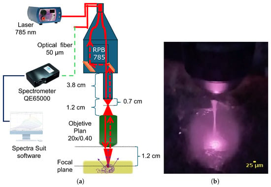
The optical configuration of the confocal design consists of a Raman Probe Bifurcated (RPB-785) equipped with two optical fibers—a 105 μm excitation fiber and a standard 200 μm collection fiber. The excitation fiber is connected to the 785 nm laser module, while the collection fiber is coupled to the QE65000 spectrometer (Ocean Optics, Edinburgh, UK) equipped with a symmetric crossed Czerny-Turner optical bench, 101 mm focal length, an RPB 785 fiber optic prove, and Hamamatsu S7031-1006 detector with a spectral range between 780–940 nm. The solid line in the diagram represents the propagation path of the laser radiation, which passes through the RPB-785 and the microscope objective before interacting with the sample. Laser excitation induces elastic Rayleigh scattering and inelastic Raman scattering. In a backscatter configuration, the same microscope objective collects the scattered signals and directs them back through the RPB-785. These signals, represented as dashed lines, are finally transmitted through the 50 μm pinhole fiber to the QE65000 spectrometer. The spectra were analyzed using the SpectraSuit Software (version 2.0.162, Ocean Optics, Edinburgh, UK) (see Figure 1a). Matlab software (version R2022a academic, MathWorks Inc.)

Figure 1.
(a) Schematic layout of the Z-depth confocal Raman microscope design. The system excites the sample under study and collects both elastic Rayleigh scattering and inelastic Raman scattering in a backscattering configuration. The solid line represents the 785 nm laser excitation path passing through the RPB-785 and the 20x/0.40 microscope objective before reaching the sample. The dashed line indicates the path of the Rayleigh and Raman signals as they travel back through the RPB-785 and, finally, arrive at the QE65000 spectrograph. (b) Photograph of the conical shape formed by the CRS illuminating a transparent aerogel block with a 785 nm laser. The confocal volume within the sample is clearly visible.
The optical components used in the assembly are listed in Table 1.
A collimating lens is typically included in conventional confocal Raman designs to cover the rear lens of the microscope objective. In the present configuration, however, the collimating lens was excluded because the RPB-785 detected a significant portion of the reflected beam from this lens, which saturated the CCD camera. As a result, only about 50% of the microscope objective lens was illuminated. Experimental tests showed that the best separation efficiency between the RPB lens and the objective was achieved at 0.7 cm, which eliminated detector oversaturation. Proper alignment of these lenses ensured high-quality Raman spectra. The focal length of the microscope objective was determined experimentally and found to be 1.2 cm.
Once the CRS is assembled, it can be mounted on a CNC to perform measurements along the XY plane or the Z component, while the sample remains stationary. Alternatively, the CRS remains stationary and the sample is placed on a mobile platform. For measurements involving volunteers, the first option is preferred because it ensures stability and minimizes movement by the volunteer. Figure 1b shows a photograph of the CRS illuminated by the 785 nm laser, showing the confocal volume at a given depth inside a transparent aerogel block.
2.2. Instrumental Test
Hypothesis: Is our CRS suitable for performing measurements at depths greater than 150 μm in human nails and fingers in vivo?
Objective Test: The main goal of this study was to determine whether the proposed CRS system is able to find differences in the molecular content between the surface and subsurface layers of human skin tissues in vivo. To achieve this, it is necessary to identify the principal Raman spectral bands associated with protein structures—reported both in vivo and ex vivo—at the skin surface (Z = 0) and at various subdermal depths (Z > 0).
Volunteers: CRS measurements were performed on the index fingernail and fingertip of three male volunteers. The two healthy participants, aged 40 and 59, were contributing authors of this work. The third participant was a 45-year-old male diagnosed with Type II diabetes, managed under medical treatment.
2.3. Anatomy and Optical Characteristics of the Index Fingernail and Fingertip
Spectral measurements were obtained from the index fingertip and fingernail. The outer layer of the skin, called the stratum corneum, is 10-to-40 μm thick and made up of layers of dead cells called corneocytes [22]. The epidermis, which is 120-to-1000 μm thick, depending on the area of the body, is composed primarily of keratin. This layer also contains melanocytes, which are responsible for the formation of melanin. The dermis is 15-to-40-times-thicker than the epidermis and is composed mostly of collagen, which gives the skin strength and support [23].
The index fingernail is 500-to-700 μm thick and is composed primarily of keratin, which, along with the amino acid cysteine, gives it its characteristic rigidity [24].
It is known that an infrared wavelength laser can penetrate the skin, passing through both the epidermis [25] and the dermis and potentially reaching the hypodermis [26]. Visible light can also penetrate far enough to stimulate melanin activity, resulting in a darkening of the skin tone [27].
3. Results
3.1. Confocal Volume
The confocal volume is the region in the sample from which Raman scattered light is efficiently collected by the microscope objective. It is influenced by the excitation laser beam, the focus, and the pinhole size in front of the detector. Determining the measurement volume of the CRS is crucial for accurate and reliable analysis, especially in applications like depth profiling, single molecule studies, and 2D material characterization. It is essentially the three-dimensional region from which the Raman signal is collected. The confocal volume is characterized by its lateral (XY) and axial (Z) dimensions, which are influenced by factors like the laser wavelength, the objective lens numerical aperture, and the pinhole.
Before acquiring the Raman spectra, the axial and lateral resolution of the system were determined experimentally.
3.1.1. Spatial Resolution
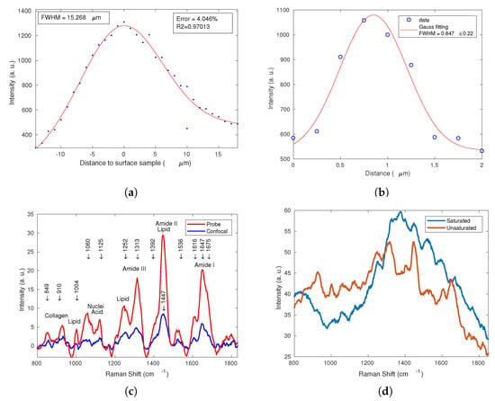
A response curve was constructed using Raman spectra collected from a thin, semi-flat quartz sample by monitoring the intensity of the 465 cm−1 quartz band while shifting the focal plane of the microscope objective in 1 μm steps along the Z-axis. Measurements were taken below the surface (Z < 0), at the surface (Z = 0), and within the sample (Z > 0). Figure 2a shows the experimental Z-positions and the corresponding Gaussian fit, which yielded a Full Width at Half Maximum (FWHM) of 15.268 μm. This FWHM value was adopted as the spatial resolution of the CRS setup.

Figure 2.
(a) Intensity profile of the 464 cm−1 quartz band acquired with the CRS system while translating the focal plane in 1 μm steps from above the sample surface (Z < 0) to inside the quartz (Z > 0). The dashed line represents the Gaussian fit (FWHM = 15.268 μm). (b) Intensity profile of the 1585 cm−1 G band of CNTs acquired with the CRS system. (c) Overlaid Raman spectra of the fingernail recorded with the commercial RPB (red) and the CRS configuration (blue), illustrating the expected reduction in signal intensity while preserving band positions and relative intensities. (c) Raw spectra showing the effect of detector saturation when a collimating lens is included (blue) and when it is absent (red).(d) Notably, saturated spectra prevented the identification of Raman bands, whereas in the spectra acquired without a collimating lens the bands were clearly distinguishable.
3.1.2. Lateral Resolution
To assess the spatial resolution, well-defined samples such as Carbon Nanotubes (CNTs) [28,29] were used to measure the Full Width at Half Maximum (FWHM) of the Raman signal. CNTs were deposited onto aluminum foil, and Raman measurements were subsequently performed as follows: first, the focal height was fixed, and then the sample was laterally shifted in 0.25 μm steps along the X-axis of the focal plane across the CNTs. Figure 2b shows the experimental XY-position data together with the corresponding Gaussian fit, which yielded an FWHM of 0.847 μm. This value was adopted as the lateral resolution of the CRS system setup. The result is comparable to the value obtained from Equation (1) in [28], which was 0.915 μm. Table 2 presents a summary of the parameters experimentally determined for the confocal volume of the CRS setup.

Table 2.
Confocal parameters determined experimentally.
3.2. Comparison of Raman Spectra: RPB-785 vs. CRS System
Figure 2b compares the Raman spectra collected with the RPB-785 and those obtained using our CRS configuration. Because CRS integrates only the contribution from the focal plane—while a pinhole rejects signals originating from planes above or below that plane—the CRS spectrum exhibited lower overall intensity. Nevertheless, Figure 2b shows that the positions of the Raman bands, as well as their relative intensity ratios, were preserved in the CRS spectrum despite the reduced signal level.
Using the RPB-785, only superficial skin information can be obtained; sub-surface investigation is not possible with this configuration. In contrast, our CRS setup—although it delivers lower signal intensity—provides spectra of adequate signal-to-noise ratio, and, as demonstrated previously, superior spatial (axial) resolution. Thanks to the incorporation of a wide-field microscope objective, the CRS system enables measurements at multiple depths beneath the skin surface, allowing the detection of variations in Raman bands arising from the inner epidermal and dermal layers.
Figure 2c also highlights several molecular bands associated with the skin surface: collagen (800–1100 cm−1), lipids and nucleic acids (1000–1200 cm−1), amide III (1200–1400 cm−1), amide II (1400–1600 cm−1), and amide I (1600–1750 cm−1).
3.3. Protocol for the Acquisition of Raman Spectra In Vivo
To ensure safe and reliable in vivo measurements, a protocol was established to minimize temperature-related effects and prevent photodegradation of the tissue. Raman Spectroscopy is generally considered safe and painless for human studies; nevertheless, ANSI guidelines recommend a maximum laser power of 100 mW for human subjects [30]. In this work, the laser power was kept well below that limit to prevent local overheating caused by prolonged irradiation. Prior to each depth scan, the focal position corresponding to the surface (Z = 0) was determined. The volunteers required no special preparation or training. The following criteria were applied during spectral acquisition:
- Laser power was limited to 30 mW for each acquired spectrum.
- Exposure time per spectrum was set to 25 s.
- The volunteers remained motionless, relaxed and breathing gently.
- The index finger was placed on a pad to ensure stability.
- The maximum intensity recorded was defined as the signal from the sample surface (Z = 0).
- At Z = 0, three consecutive spectra were acquired to estimate variability, yielding 5.75%.
- Only one spectrum was acquired at each depth step to avoid prolonged exposure at the same focal point.
- If a volunteer moved and the laser spot shifted from the target area then the measurement was interrupted and resumed only after the subject had relaxed.
3.4. Processing of Spectral Data
Raman spectra are highly affected by fluorescence in the case of the fingertip and moderately affected in the case of the fingernail. The fluorescence background in both cases is normalized using a 5-order polynomial fitting method, and noise is suppressed with a 3-order Savitzky–Golay filter as a numerical method for smoothing of data.
3.5. Generation of 2D and 3D Images from Raw Raman Spectra
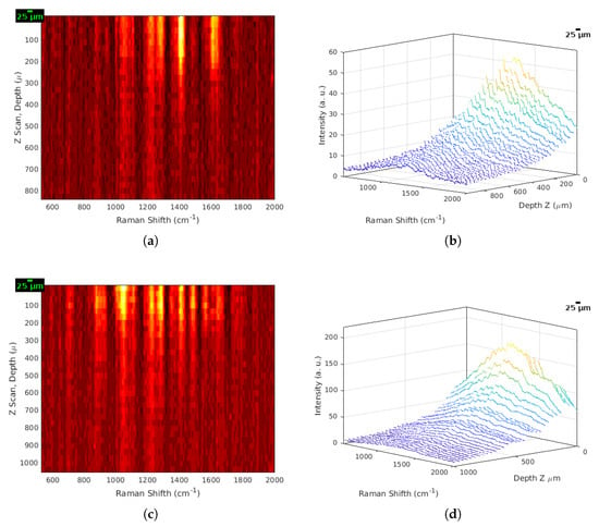
One of the key advantages of the CRS system is its ability to produce Raman spectral images. Unlike conventional optical images obtained with a light microscope, these images are generated from a series of spectral acquisitions that can be assembled into depth-resolved maps. In this study, spectra were acquired along the Z-axis from Z = 0 down to 850 μm below the skin surface, producing an array that reflected spectral variations with depth. Estas imágenes Raman son valiosas para visualizar la composición de la muestra y evaluar la distribución espacial de componentes específicos, and tracking depth-dependent changes in band intensities [11]. Figure 3a presents a 2D representation of the sequential raw spectra collected from a human fingernail in vivo, while Figure 3b shows the corresponding 3D plot. The 3D view clearly reveals the progressive decrease in overall signal intensity as depth increased.

Figure 3.
(a) 2D Raman spectral map of the index fingernail, showing intensity and structural variations from the skin surface (Z = 0) down to 800 μm. Notable features include the broadening and de-blending of bands below 350 μm, particularly in the amide I region (1580–1690 cm−1). (b) 3D Raman surface plot of the same dataset, illustrating the sequential decrease in overall signal intensity with increasing depth. (c) 2D Raman spectral map of the index fingertip, highlighting depth-dependent changes in band intensities and profiles. (d) 3D Raman surface plot for the index fingertip, confirming the progressive attenuation of the full spectral window as depth increased.
Figure 3a (2D heat-map) reveals how band intensities decreased with depth in the fingernail. The amide II band (∼1445 cm−1) was the most intense from the surface down to ∼300 μm; beyond this point its intensity dropped by roughly ∼30 at Z ∼600 μm, and it was no longer detectable below 750 μm. The amide I envelope (1600–1750 cm−1) was the second-brightest region. It appeared as two dominant components at the surface, but it split into three or four sub-bands below 300 μm, consistent with the -helix, -sheet, and water-bending contributions (∼1640 cm−1) observed in two volunteers. In contrast, the 1060 cm−1 and 1250 cm−1 bands remained relatively constant in intensity throughout the entire 0–850 μm range. Figure 3b (3D rendering of the same data) underscores the uniform decrease in signal intensity as depth increased. Figure 3c,d show the corresponding depth scan for the fingertip. Here, the 1313 cm−1 band was dominant at the surface, lost ∼50% of its intensity by 300 μm and became negligible below 700 μm. The 1060 cm−1 band was the second-strongest, followed by amide II (∼1445 cm−1). Two additional surface-confined bands at 1525 cm−1 and 1603 cm−1 were intense but vanished beyond 150 μm. The amide I region was comparatively weak and, below 300 μm, resolved into at least three secondary components. As in the nail, the 3D plot (Figure 3d) highlights the depth-dependent attenuation of all the bands.
Figure 3c also shows that the 1060 cm−1 and 1250 cm−1 bands were brighter from the surface (Z = 0) down to approximately 200 μm. Below this depth their intensity diminished and then remained nearly constant to the maximum depth accessible with the CRS. Although Figure 3a,c display a similar set of bands, their relative intensities differed. In particular, the bands at 1525 cm−1 and 1603 cm−1 were weak in the fingernail but noticeably stronger in the fingertip; however, both bands vanished beyond 200 μm. The 1525 cm−1 feature is attributable to carotenoids—skin pigments located predominantly near the surface—while the 1600 cm−1 band arose from the C=O stretching and N-H bending modes of the peptide backbone; see Table 3.

Table 3.
Peak assignment of the Raman spectra of fingernail and finger tip.
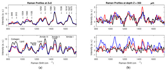
Figure 4a presents the Raman spectra of the human fingertip region acquired at the surface (Z = 0). The blue, red, and black traces correspond to the three volunteers. The upper panel shows the spectra from the fingernail, while the lower panel displays those from the fingertip. In both cases, the most intense band was the amide II peak at 1447 cm−1, with a full width at half maximum (FWHM) of ∼42 cm−1. The peak intensities and profiles were comparable among the volunteers; the fingertip spectra exhibited minor intensity differences but only slight variations in peak shape. Figure 4b shows the corresponding spectra at a depth of Z = 350 μm. The signal intensity was already 40% lower than at the surface. Both the nail and fingertip panels reveal additional sub-bands—secondary-structure components of amide I and II—with FWHM∼12 cm−1 and small shifts in the peak center. These features are well resolved, due to the spectral resolution of the CRS system.

Figure 4.
(a) Raman spectra of the human index finger acquired at the surface (Z = 0). The blue, red, and black lines correspond to the three volunteers. The top panel shows the spectra of the fingernail, while the bottom panel shows the spectra of the fingertip. (b) Raman spectra recorded at a depth of Z = 350 μm. The top panel contains the data from the fingernail, and the bottom panel those from the fingertip, highlighting the ∼40% reduction in overall Raman intensity relative to the surface measurements.
Amide I Secondary Components as Biomarkers for Distinguishing Healthy and Diabetic Patients
Farhan et al. [34], Liu et al. [35], Sadat et al. [36], reported that diabetes alters nail composition, and they proposed that secondary components of the amide I band can be used as diagnostic markers. According to these studies, healthy subjects show secondary-band maxima ≤ 1645 cm−1 (bands at ≈1620, 1635, 1638, 1640, and 1645 cm−1), characteristic of -sheet structures, while patients with type 2 diabetes present secondary bands in the 1648–1660 cm−1 interval, characteristic of -helix structures. The presence of an amide I -helix structures band near 1650 cm−1 (Raman) has been proposed as indicative of type 2 diabetes.
In order to validate the health status of the participants, Gaussian fitting of the amide I profiles was performed at Z = 0 to deconvolute overlapping secondary components. At Z = 350 μm, where the secondary bands were well resolved, Gaussian fitting was applied directly to the individual peaks rather than performing deconvolution. Table 3 presents the intensity and designation of the secondary bands registered from the nail samples, while Table 4 summarizes the bands that defined the healthy or diabetic status of the volunteers, based on both their fingernails and fingertips, as shown in Figure 4. According to Table 4, volunteer 3 met the diabetic criterion, since the fingernail spectra included the -helix 1650 cm−1 amide I secondary component, whereas the other volunteers were classified as healthy. However, further measurements with a larger number of volunteers are necessary to confirm this observation.

Table 4.
Secondary components used to define the healthy or diabetic volunteer status of the volunteers.
4. Discussion
This optical design has considerable technological potential in science, engineering, and medically sensitive bio-spectroscopic applications. Raman spectra were acquired on the surface of and at successive depths beneath the index fingernail and fingertip of the three male volunteers, using 50 μm steps to obtain depth-resolved molecular information. Although the surface spectra of all three volunteers appeared similar at first glance, the configuration of the CRS system revealed subtle differences: closely spaced narrow bands can be distinguished, and broad bands deconvolved into at least three resolvable sub-bands whose intensity, center position, and width varied with depth. The CRS configuration also detected weak features that were present at the surface but disappeared at specific subsurface layers.
Comparing the fingertip and fingernail spectra shows clear contrasts. Fluorescence dominated the fingertip signal at the surface but decreased progressively with depth, whereas fluorescence was lower and more uniform in the nail. Gaussian fitting applied to spectra from both sampling sites quantified shifts in band position and intensity, highlighting those Raman features that differed between nail and skin; further measurements are required to characterize variations in the contribution of secondary molecular components.
The earliest CRS prototypes also incorporated microscope objectives [6,16], but their studies were limited to depths no greater than 100 μm and did not report the detection of secondary molecular components. Moreover, the strong water signal in the 2500–4000 cm−1 region led researchers to focus primarily on shallow depths and on quantifying epidermal water content. As a result, analyses in the fingerprint region (400–1800 cm−1) were restricted to the skin surface, with secondary protein bands inferred through Gaussian model fitting. In contrast, the present CRS system yields clearly separated sub-profiles at depths greater than 300 μm.
Recent advances in detector sensitivity and grating design have driven several CRS configurations into commercial Raman modules used for skin-water analysis [37]. A very recent handheld dual-laser confocal Raman device has also been reported [38]; however, there is no reported information regarding secondary protein bands.
Our CRS system, as presented here, can be employed to address a variety of challenges in the medical field. It can be used to assess health or diabetic status via amide I secondary molecular components, as well as other biomarkers such as the ratio, in fingernails and at well-defined skin depths [39]. The system can also study nail diseases caused by dermatophyte T. rubrum or Candida species, the most common onychomycosis pathogens, using fingernail clippings [33,39,40]. Furthermore, the CRS can be adapted to study brain diseases leading to tumor formation [41], allowing spectra acquisition at both the surface and systematically at desired depths, instead of relying on ex vivo brain tumor slices. Future work will focus on extending these studies to larger cohorts of volunteers and additional biomarkers to further validate the system’s diagnostic potential.
5. Conclusions
A wide-field 20x/0.40 microscope objective and a 50 μm multimode optical-fiber pinhole were integrated into a QE65000 Raman spectrometer to create a confocal Raman system with high axial and high lateral resolution and an adequate signal-to-noise ratio. Configured for depth-resolved (Z-axis) measurements, the instrument successfully retrieved structural and molecular information from in vivo biological samples—namely, the index fingernail and fingertip. To protect the volunteers, a dedicated protocol was adopted: a single 25 s acquisition per 50 μm depth step at 30 mW laser power. Under these conditions no discomfort or adverse effects were reported, which was in agreement with previous studies on similar tissues.
Significant differences were observed in the spectra of the three volunteers, particularly at depths greater than 300 μm, likely associated with variations in the secondary components of the amide I band. These differences may have reflected individual health status, underscoring the need for further testing to validate this hypothesis. The demonstrated CRS configuration thus provides a non-invasive, depth-resolved tool for analyzing molecular changes in skin tissues, paving the way for future biomedical and technological applications such as CNT studies and material characterization.
Author Contributions
All authors contributed to the writing and preparation of the manuscript; the experimental setup was performed by E.U.A. under the supervision of A.F.G. and L.d.l.C.M. Sample measurements were performed by E.U.A., L.d.l.C.M., O.B. and M.B.G., under the supervision of A.F.G. All authors have read and agreed to the published version of the manuscript.
Funding
This research received support from SECIHTI.
Institutional Review Board Statement
The study was conducted in accordance with the Declaration of Helsinki, and the protocol was approved by the Ethics Committee of the Autonomous University of Carmen (Project identification code FCING/2019/02) on 19 August 2019.
Informed Consent Statement
Volunteer consent is included, due to the use of exclusively archival and anonymized data from Raman depth profile measurements on skin and under skin.
Data Availability Statement
The data presented in this study are available on request from the corresponding author. The data are not publicly available due to privacy and ethics.
Conflicts of Interest
The authors declare no conflicts of interest. The funders had no role in the design of the studies in the collection, in the analyses and interpretation of the data, in the writing of the manuscript, or in the decision to publish the results.
Abbreviations
The following abbreviations are used in this manuscript:
| CRS | Confocal Raman Spectroscopy |
| RPB | Raman Probe |
| LWD | Long Working Distance |
| ANSI | American National Standards Institute |
| RS | Raman Spectroscopy |
| CCD | Charge Coupled Device |
| SRS | Standard Raman System |
| FWHM | Full Width at Half Maximum |
| CNTs | Carbon Nanotubes |
References
- Smith, E.; Dent, G. Modern Raman Spectroscopy: A Practical Approach, 2nd ed.; John Wiley & Sons Ltd.: Hoboken, NJ, USA, 2019. [Google Scholar] [CrossRef]
- Briancon, S.; Bolzinger, M.-A.; Chevalier, Y. Confocal Raman Spectroscopy as a Tool to Investigate the Action of Penetration Enhancers Inside the Skin. In Percutaneous Penetration Enhancers Drug Penetration Into/Through the Skin: Methodology and General Considerations; Dragicevic, E.N., Maibach, H.I., Eds.; Springer: Berlin/Heidelberg, Germany, 2017; pp. 229–246. [Google Scholar] [CrossRef]
- Toporski, J.; Dieing, T.; Hollricher, O. (Eds.) Confocal Raman Microscopy; Springer International Publishing: Berlin/Heidelberg, Germany, 2018; Volume 66. [Google Scholar] [CrossRef]
- Pereira, A.F.M.; Rodrigues, B.V.M.; Neto, L.P.M.; Lopes, L.d.; da Costa, A.L.F.; Santos, A.S.; Viana, B.C.; Tosato, M.G.; Silva, G.C.d.; Gusmão, G.O.M.; et al. Confocal Raman spectroscopy as a tool to assess advanced glycation end products on solar-exposed human skin. Vib. Spectrosc. 2021, 114, 103234. [Google Scholar] [CrossRef]
- Shim, M.G.; Wilson, B.C. Development of an In Vivo Raman Spectroscopic System for Diagnostic Applications. J. Raman Spectrosc. 1997, 28, 131–142. [Google Scholar] [CrossRef]
- Chrit, L.; Hadjur, C.; Morel, S.; Sockalingum, G.D.; Lebourdon, G.; Leroy, F.; Manfait, M. In vivo chemical investigation of human skin using a confocal Raman fiber optic microprobe. J. Biomed. Opt. 2005, 10, 044007. [Google Scholar] [CrossRef]
- Ramírez-Elías, M.G.; González, F.J. Raman Spectroscopy for In Vivo Medical Diagnosis. In Raman Spectroscopy; InTech: London, UK, 2018. [Google Scholar] [CrossRef]
- Abdala, J.M.d.; Lemos, F.R.; Almeida, R.M.d.; Tippavajhala, V.K.; da Silva, G.C.; Neto, L.P.M.; Fávero, P.P.; Martin, A.A. Noninvasive in vivo application of confocal Raman spectroscopy in identifying age-related biochemical changes in human stratum corneum and epidermis. Vib. Spectrosc. 2024, 130, 103627. [Google Scholar] [CrossRef]
- Ghita, A.; Pascut, F.C.; Sottile, V.; Notingher, I. Monitoring the mineralisation of bone nodules in vitro by space- and time-resolved Raman micro-spectroscopy. Analyst 2013, 139, 55–58. [Google Scholar] [CrossRef] [PubMed]
- Available online: https://static.horiba.com/fileadmin/Horiba/Application/Health_Care/Pharmaceuticals_and_Medicine_Manufacturing/Cosmetics/In_Vivo_Raman_measurements_of_Human_Skin.pdf (accessed on 1 February 2025).
- Darvin, M.E.; Schleusener, J.; Parenz, F.; Seidel, O.; Krafft, C.; Popp, J.; Lademann, J. Confocal Raman microscopy combined with optical clearing for identification of inks in multicolored tattooed skin in vivo. Analyst 2018, 143, 4990–4999. [Google Scholar] [CrossRef] [PubMed]
- Tfayli, A.; Piot, O.; Manfait, M. Confocal Raman microspectroscopy on excised human skin: Uncertainties in depth profiling and mathematical correction applied to dermatological drug permeation. J. Biophotonics 2008, 1, 140–153. [Google Scholar] [CrossRef] [PubMed]
- Choe, C.; Lademann, J.; Darvin, M.E. Depth profiles of hydrogen bound water molecule types and their relation to lipid and protein interaction in the human stratum corneum in vivo. Analyst 2016, 141, 6329–6337. [Google Scholar] [CrossRef]
- Choe, C.; Schleusener, J.; Lademann, J.; Darvin, M.E. Keratin-water-NMF interaction as a three layer model in the human stratum corneum using in vivo confocal Raman microscopy. Sci. Rep. 2017, 7, 15900. [Google Scholar] [CrossRef]
- Tippavajhala, V.K.; de Oliveira Mendes, T.; Martin, A.A. In Vivo Human Skin Penetration Study of Sunscreens by Confocal Raman Spectroscopy. AAPS PharmSciTech 2018, 19, 753–760. [Google Scholar] [CrossRef]
- Caspers, P.J.; Bruining, H.A.; Puppels, G.J.; Lucassen, G.W.; Carter, E.A. In Vivo Confocal Raman Microspectroscopy of the Skin: Noninvasive Determination of Molecular Concentration Profiles. J. Investig. Dermatol. 2001, 116, 434–442. [Google Scholar] [CrossRef] [PubMed]
- Ocean Optics. Raman Lasers. Available online: https://www.oceanoptics.com/accessories/light-sources/raman-lasers/ (accessed on 23 July 2025).
- InPhotonics. Low-Cost Laboratory Fiber Optic Raman Probe (Datasheet). Available online: https://www.inphotonics.com/probeRPB.htm (accessed on 23 July 2025).
- Ocean Optics. Legacy Spectrometers: A Historical Archive of Ocean Optics Spectrometers. Available online: https://www.oceanoptics.com/resources/legacy-spectrometers/ (accessed on 23 July 2025).
- BoliOptics. 20X Infinity-Corrected Plan Achromatic POL Polarizing Microscope Objective Lens Working Distance 2.71 mm with Black Finish. Available online: https://bolioptics.com/20x-infinity-corrected-plan-achromatic-pol-polarizing-microscope-objective-lens-working-distance-2-71mm-with-black-finish/ (accessed on 23 July 2025).
- BestScope. BS-5070BTR Binocular Polarizing Microscope. Available online: https://www.bestscope.net/bs-5070btr-polarizing-microscope/ (accessed on 24 July 2025).
- Moll, R.; Divo, M.; Langbein, L. The human keratins: Biology and pathology. Histochem. Cell Biol. 2008, 129, 705–733. [Google Scholar] [CrossRef] [PubMed]
- Conejo-Mir, J.; Jiménez, J.C.M.; Martínez, F.M.C. Manual de Dermatología; Grupo Aula Médica S.L.: Madrid, Spain, 2018; Available online: https://dialnet.unirioja.es/servlet/libro?codigo=762726 (accessed on 23 July 2025).
- Shin, M.K.; Kim, T.I.; Kim, W.S.; Park, H.; Kim, K.S. Changes in nail keratin observed by Raman spectroscopy after Nd:YAG laser treatment. Microsc. Res. Tech. 2017, 80, 338–343. [Google Scholar] [CrossRef] [PubMed]
- Crowther, J.M.; Matts, P.J. Molecular Concentration Profiling in the Skin Using Confocal Raman Spectroscopy. In Textbook of Aging Skin; Farage, E.M.A., Miller, K.W., Maibach, H.I., Eds.; Springer: Berlin/Heidelberg, Germany, 2017; pp. 1171–1187. [Google Scholar] [CrossRef]
- Cho, S.; Shin, M.H.; Kim, Y.K.; Seo, J.-E.; Lee, Y.M.; Park, C.-H.; Chung, J.H. Effects of Infrared Radiation and Heat on Human Skin Aging in vivo. J. Investig. Dermatol. Symp. Proc. 2009, 14, 15–19. [Google Scholar] [CrossRef]
- Mahmoud, B.H.; Ruvolo, E.; Hexsel, C.L.; Liu, Y.; Owen, M.R.; Kollias, N.; Lim, H.W.; Hamzavi, I.H. Impact of Long-Wavelength UVA and Visible Light on Melanocompetent Skin. J. Investig. Dermatol. 2010, 130, 2092–2097. [Google Scholar] [CrossRef]
- Kim, Y.; Lee, E.J.; Roy, S.; Sharbirin, A.S.; Ranz, L.-G.; Dieing, T.; Kim, J. Measurement of lateral and axial resolution of confocal Raman microscope using dispersed carbon nanotubes and suspended graphene. Curr. Appl. Phys. 2020, 20, 71–77. [Google Scholar] [CrossRef]
- Itoh, N. Comparison of Lateral Resolutions Obtained by Different Methods for Confocal Raman Microscopes. J. Raman Spectrosc. 2025, 56, 389–397. [Google Scholar] [CrossRef]
- ANSI Z136.1-2022; American National Standard for Safe Use of Lasers. The American National Standards Institute: Washington, DC, USA, 2022. Available online: https://webstore.ansi.org/ (accessed on 23 July 2025).
- Veras, J.M.d.M.F.; Coelho, L.d.S.; Neto, L.P.M.; de Almeida, R.M.; da Silva, G.C.; de Santana, F.B.; Garcia, L.A.; Martin, A.A.; Favero, P.P. Identification of biomarkers in diabetic nails by Raman spectroscopy. Clin. Chim. Acta 2023, 544, 117363. [Google Scholar] [CrossRef]
- Kast, R.E.; Serhatkulu, G.K.; Cao, A.; Pandya, A.K.; Dai, H.; Thakur, J.S.; Naik, V.M.; Naik, R.; Klein, M.D.; Auner, G.W.; et al. Raman spectroscopy can differentiate malignant tumors from normal breast tissue and detect early neoplastic changes in a mouse model. Biopolymers 2008, 89, 235–241. [Google Scholar] [CrossRef]
- Kourkoumelis, N.; Gaitanis, G.; Velegraki, A.; Bassukas, I.D. Nail Raman spectroscopy: A promising method for the diagnosis of onychomycosis. An ex vivo pilot study. Med. Mycol. 2018, 56, 551–558. [Google Scholar] [CrossRef]
- Farhan, K.M.; Sastry, T.P.; Mandal, A.B. Comparative study on secondary structural changes in diabetic and non-diabetic human finger nail specimen by using FTIR spectra. Clin. Chim. Acta 2011, 412, 386–389. [Google Scholar] [CrossRef]
- Liu, J.; Chu, J.; Xu, J.; Zhang, Z.; Wang, S. In vivo Raman spectroscopy for non-invasive transcutaneous glucose monitoring on animal models and human subjects. Spectrochim. Acta Part A Mol. Biomol. Spectrosc. 2025, 329, 125584. [Google Scholar] [CrossRef]
- Azin, S.; Joye, I.J. Peak Fitting Applied to Fourier Transform Infrared and Raman Spectroscopic Analysis of Proteins. Appl. Sci. 2020, 10, 5918. [Google Scholar] [CrossRef]
- Téllez-Soto, C.A.; Silva, M.G.P.; dos Santos, L.; de OMendes, T.; Singh, P.; Fortes, S.A.; Favero, P.; Martin, A.A. In vivo determination of dermal water content in chronological skin aging by confocal Raman spectroscopy. Vib. Spectrosc. 2021, 112, 103196. [Google Scholar] [CrossRef]
- Qi, Y.; Zhang, R.; Rajarahm, P.; Zhang, S.; Attia, A.B.E.; Bi, R.; Olivo, M. Simultaneous Dual-Wavelength Source Raman Spectroscopy with a Handheld Confocal Probe for Analysis of the Chemical Composition of In Vivo Human Skin. Anal. Chem. 2023, 95, 5240–5247. [Google Scholar] [CrossRef] [PubMed]
- Tabasz, T.; Szymańska, N.; Bąk-Drabik, K.; Damasiewicz-Bodzek, A.; Nowak, A. Is Raman Spectroscopy of Fingernails a Promising Tool for Diagnosing Systemic and Dermatological Diseases in Adult and Pediatric Populations? Medicina 2024, 60, 1283. [Google Scholar] [CrossRef]
- Yin, N.-H.; Griffiths, F.; Mann, C.; Dawes, H.; van Arkel, R.; Bukhari, M.; Kerns, J.G. Raman spectroscopy identified fingernail compositional differences between sexes and age-related changes but not handedness or fingers in a healthy cohort. PLoS ONE 2025, 20, e0329092. [Google Scholar] [CrossRef]
- Ranasinghe, J.C.; Wang, Z.; Huang, S. Unveiling brain disorders using liquid biopsy and Raman spectroscopy. Nanoscale 2024, 16, 11879–11913. [Google Scholar] [CrossRef]
Disclaimer/Publisher’s Note: The statements, opinions and data contained in all publications are solely those of the individual author(s) and contributor(s) and not of MDPI and/or the editor(s). MDPI and/or the editor(s) disclaim responsibility for any injury to people or property resulting from any ideas, methods, instructions or products referred to in the content. |
© 2025 by the authors. Licensee MDPI, Basel, Switzerland. This article is an open access article distributed under the terms and conditions of the Creative Commons Attribution (CC BY) license (https://creativecommons.org/licenses/by/4.0/).