Article

Chemical Composition of a Supercritical Fluid (Sfe-CO2) Extract from Baeckea frutescens L. Leaves and Its Bioactivity Against Two Pathogenic Fungi Isolated from the Tea Plant (Camellia sinensis (L.) O. Kuntze)

1
State Key Laboratory of Tea Plant Biology and Utilization, Anhui Agricultural University, 130 West Changjiang Road, Hefei 230036, China
2
Anhui Provincial Key Laboratory of Microbial Pest Control, Anhui Agricultural University, 130 West Changjiang Road, Hefei 230036, China
3
School of Grain Science and Technology, Jiangsu University of Science and Technology, Zhenjiang 212003, China
4
Guangxi Forestry Research Institute, Guangxi Engineering and Technology Research Center for Woody Spices, Guangxi Key Laboratory for Cultivation and Utilization of Special Non-Timber Forest Crops, 23 Yongwu Road, Nanning 530002, China
5
Food Processing and Quality, Natural Resources Institute Finland, Tietotie 2, 02150 Espoo, Finland
*
Authors to whom correspondence should be addressed.
These authors contributed equally to this work.
Plants 2020, 9(9), 1119; https://doi.org/10.3390/plants9091119
Submission received: 5 August 2020 / Revised: 25 August 2020 / Accepted: 26 August 2020 / Published: 29 August 2020

Abstract

:
Colletotrichum gloeosporioides and Pseudopestalotiopsis camelliae-sinensis are the two most important tea plant (Camellia sinensis L.) pathogenic fungi. Interest in natural plant extracts as alternatives to synthetic chemical fungicides to control plant pathogens is growing. In this study, the volatile fraction of Baeckea frutescens L. was extracted by supercritical fluid extraction (SFE-CO2), and its chemical composition was analyzed, and investigated for its antifungal activity against C. gloeosporioides and P. camelliae. The major constituents of the volatile fraction were β-caryophyllene (28.05%), α-caryophyllene (24.02%), δ-cadinene (6.29%) and eucalyptol (5.46%) in B. frutescens SFE-CO2 extracts. The terpineol, linalool, terpinen-4-ol and eucalyptol showed strong contact antifungal activity against P. camelliae and C. gloeosporioides with median inhibitory concentration (MIC50) in the range of 0.69 μL/mL to 2.79 μL/mL and 0.62 μL/mL to 2.18 μL/mL, respectively. Additionally, the volatile fraction had high fumigation antifungal activity against P. camelliae and C. gloeosporioides with an inhibition rate between 20.87% and 92.91%. Terpineol presented the highest antifungal activity in the contact and fumigation toxicity assays. Terpineol, linalool, terpinen-4-ol and eucalyptol were associated with the most active chemical compounds in the volatile fraction against the fungi. The results suggest that B. frutescens SFE-CO2 extracts are potential ingredients to develop a natural fungicide for control of tea plant pathogens.

Graphical Abstract

1. Introduction

As an alternative to using chemical fungicides, plant extracts have attracted the attention of chemical companies as they may be used as botanical fungicides. Baeckea frutescens L. is an important medicinal plant belonging to the Myrtaceae family [1] and is found in Peninsular Malaysia and Sumatra and distributed along coastal areas of southeastern China and Australia [2]. B. frutescens has various bioactive properties, such as antibacterial [1,3], antioxidant [2], anticancer [4], anti-inflammatory [5] and insecticidal activities [1]. The leaves of B. frutescens are rich in various volatile constituents such as terpenoids, sesquiterpenes and phenylpropanoids, which are secondary metabolites associated with numerous bioactivities [1,5,6].
The tea plant (Camellia sinensis L.) is a crucial commercial crop in world [7]. Tea, made from fresh leaves of C. sinensis, is a prominent beverage worldwide and is the second most consumed nonalcoholic beverage after water [8]. The tea plant suffers from biotic stresses of some pathogenic fungi, which is a serious concern for the tea industry as this condition impacts on reduced tea yields, decreased quality and damaged human health [9,10]. Several Pestalotiopsis-like species are common phytopathogens that cause tea grey blight disease and result in severe losses (10 to 20%) in tea production [11,12]. Pseudopestalotiopsis camelliae-sinensis was shown to cause grey blight disease on tea plants in China [13]. Some research has showed that planting disease-resistant varieties of C. sinensis has a certain effect on controlling the tea plant disease infected by Pestalotiopsis species [14]. In addition, the Colletotrichum species is also a common group of plant pathogens that are responsible for anthracnose diseases [15,16]. Colletotrichum gloeosporioides could lead to tea anthracnose disease, which a is serious foliar disease of the tea plant and causes severe damage accompanied by high yield losses [17]. Meanwhile, C. gloeosporioides-contaminated tea beverage may be harmful to human health [10]. It was recently reported that the leaves of tea plants infected by C. gloeosporioides were reduced in 30–60% of the tea products [18,19]. Altogether, P. camelliae and C. gloeosporioides are two of most destructive pathogens of foliar disease on tea plants and strategies to inhibit their growth are highly necessary.
At present, synthetic chemical fungicides are still the primary control approach to prevent fungal-related diseases in tea leaves. However, the indiscriminate use of these chemical fungicides not only reduces the quality of tea products and causes tea security crises, but also represents a harmful condition for human health and the natural environment [20,21,22]. Nowadays, effective and environmentally safe plant protection products which protect against phytopathogenic fungi are urgently needed in modern agriculture [23,24]. Therefore, replacing synthetic chemical fungicides by botanical alternatives could be a suitable choice to avoid toxicological and environmental-related issues. For instance, the botanical secondary metabolites, such as alkaloids [25], flavonoids [26] and volatile organic compounds [27], may act as antibacterial active substances to inhibit plant diseases. Therefore, botanical fungicides have been considered as environmentally friendly alternatives to synthetic chemical fungicides.
Traditionally, the volatile fraction of plants is extracted by steam distillation and organic solvent extraction. Supercritical fluid extraction (SFE) utilizes supercritical fluids, which exhibit liquid-like as well as gas-like properties, and has become an effective method for separating and extracting more apolar compounds, as well as the extraction of polyphenols, lipids, and essential oils [28,29,30]. Furthermore, conventional extraction with organic solvents has some disadvantages, such as high energy costs, and the possible loss of volatile compounds during the removal of the solvent [31]. Therefore, supercritical fluids are attractive for extracting flavors present in natural materials [28]. The supercritical fluid extraction with carbon dioxide (SFE-CO2), a green extraction method, provides the final product without organic solvent residues [32]. Due to the properties of CO2, such as being odorless, colorless, safe, nontoxic and recyclable [28], SFE-CO2 has been widely used for the extraction of volatile compounds [28]. To the best of our knowledge, there are still no reports on the chemical composition of the supercritical fluid (SFE-CO2) extract of B. frutescens leaves and its bioactivity against P. camelliae and C. gloeosporioides. Therefore, the aims of this study were to investigate the chemical composition of the SFE-CO2 extract from B. frutescens using GC-MS. In addition, the antifungal effects of SFE-CO2 extract from B. frutescens against P. camelliae and C. gloeosporioides were also evaluated. The main research outline applied in this study is shown in Figure 1.

2. Results and Discussion

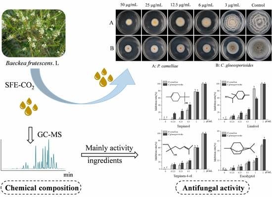
2.1. Chemical Composition of the B. Frutescens Leaf SFE-CO2 Extract

The yield of the extract obtained by SFE-CO2 was 2.2% (w/w relative to dry material weight). The major compounds of the SFE-CO2 extract of were analyzed using GC-MS, and the peaks with matching similarly of more than 80% were accepted as candidate compounds. These compounds were further confirmed and identified by comparing the mass spectra with those from National Institute of Standards and Technology (NIST)mass spectral (http://webbook.nist.gov/chemistry/) and National Institutes of Health (NIH) databases, as well as by calculating the retention index (RI) and comparing RI from some of the previous literature records. Some major compounds were identified by co-injection of available standard compounds, such as α-pinene, β-pinene, eucalyptol, γ-terpinene, linalool, terpinen-4-ol, terpineol, β-caryophyllene and α-caryophyllene. The GC-MS separation of SFE-CO2 extract is shown in supplementary material Figure S1.The chemical composition of the SFE-CO2 extract is listed in Table 1, along with their retention indices, relative contents and identification.
As observed in Table 1, a total of 27 compounds were identified in the SFE-CO2 extract, accounting for 98.99% of total volatile oil. The major constituents of the volatile SFE-CO2 extract were β-caryophyllene (28.05%), α-caryophyllene (24.02%), δ-cadinene (6.29%), eucalyptol (5.46%) and β-pinene (5.21%), followed by terpineol (4.54%), cubenol (2.81%) and γ-terpinene (2.49%). The volatile organic metabolites from B. frutescens have been investigated elsewhere and α-pinene, β-caryophyllene, γ-terpinene, terpineol, 1,8-cineole and linalool were the main components of B. frutescens volatile oils [1,33]. Furtnemore, Myrtaceae was divided into different chemotypes, according to the single most abundant compound, called dominant terpenes, in its plant volatile oils [33,34]. In most cases, 1,8-cineole or α-pinene are the most abundant and most common terpenes compounds in the majority of Myrtaceae foliar terpene profile, so in the vast majority of species (80%) there is an α-pinene or 1,8-cineole foliar terpene chemotype [34]. In this study, β-caryophyllene and α-caryophyllene were the most abundant terpene compounds. Compared with previous observations, it may represent a new or another chemical type of B. frutescens based on the main chemical composition of the volatile oil.
The hydrodistillation volatile oil of B. frutescens leaves grown in Vietnam was found to contain β-pinene (19.0%), γ-terpinene (11.7%), α-pinene (11.1%) and (E)-caryophyllene (7.1%) [1]. Similarly, the hydrodistillation volatile oil of B. frutescens leaves collected in Vietnam was shown to contain α-humulene (19.2%), β-caryophyllene (17.3%), baeckeol (13.8%), α-thujene (8.8%), linalool (5.6%) and eucalyptol (5.6%) [33]. From these results, it seems the essential oil composition of B. frutescens leaves exhibits high chemical variability [35]. Several factors may be related to these differences, such as geographic location, harvest time, local climate and the plant physiological status. In addition, the method of essential oil extraction also becomes an important factor. The B. frutescens volatile essential oil obtained by solid-phase micro extraction (SPME) presented γ-terpinene, o-cymene, α-pinene and Eucalyptol as major compounds. However, head-space extraction (HS) and conventional hydro distillation (HD) detected β-pinene, γ-terpinene, α-pinene and o-cymene as major compounds [33]. Meanwhile, the particle size of plant material is also an important factor in SFE and HD [28,36]. Some results reported that decrease of particle size increased the extract yield and extraction rate [28,36]. The above findings suggest that further studies on plant materials and extraction methods are needed.
This is the first report in which the chemical composition of a supercritical fluid (SFE-CO2) extract of B. frutescens leaves is provided. β-Caryophyllene, α-caryophyllene, δ-cadinene and eucalyptol were the major components.

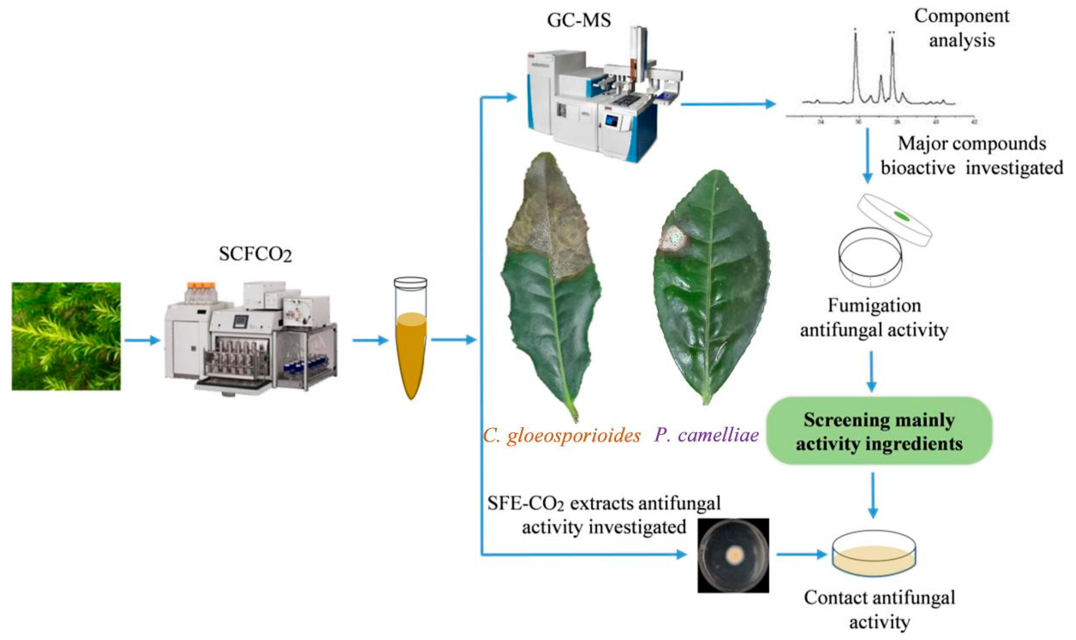
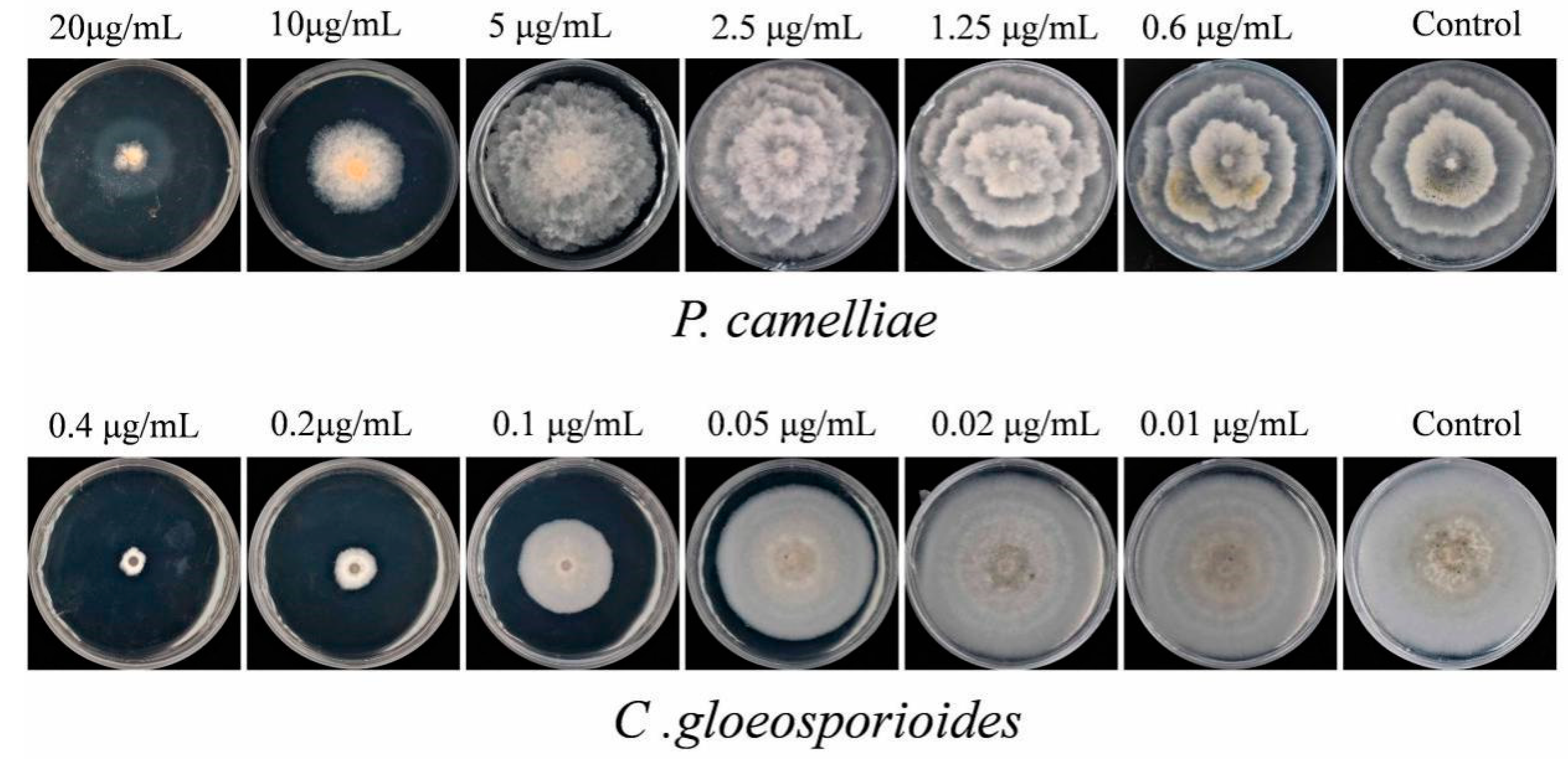
2.2. Contact Antifungal Activity of B. frutescens SFE-CO2 Extract

The SFE-CO2 extract of B. frutescens leaves exhibited an inhibitory effect on mycelial growth of two tea plant pathogenic fungi as shown in Figure 2. The mycelial growth of P. camelliae and C. gloeosporioides was strongly inhibited and this effect was dose-dependent as shown in Figure 2 and Table 2.
As seen in Figure 2, from concentrations of 6 μg/mL to 50 μg/mL, the SFE-CO2 extract of B. frutescens leaves showed a significantly limited mycelial growth of both fungi. In addition, the extract presented stronger inhibited activity against C. gloeosporioides than against P. camelliae (Figure 2) as also observed by the lower MIC50 value for C. gloeosporioides.
The median inhibitory concentration (MIC50) of the B. frutescens leaf SFE-CO2 extract against two phytopathogenics was also evaluated (Table 2). The inhibitory activity of B. frutescens SFE-CO2 extract against C. gloeosporioides exhibited the lowest MIC50 value of 4.79 µg/mL, while the MIC50 against P. camelliae was 5.11 µg/mL. However, compared with other previously published papers, our research exhibited that the B. frutescens SFE-CO2 extracts have stronger antifungal activity. For example, three plants (oregano, thyme and ajwain) SFE-CO2 extracts showed broad-spectrum antifungal activity against four Aspergillus species fungi with MIC values in the range of 128–1024 µg/mL [37]. In addition, the SFE-CO2 extract of Prunus persica leaves presented MIC50 of 62.50 µg/mL against Candida albicans [38]. In our study, the B. frutescens SFE-CO2 extract revealed a stronger antifungal activity against two fungi (MIC50 values of 4.79 µg/mL and 5.11 µg/mL, respectively). Overall, results showed that C. gloeosporioides may be the most sensitive micro-organism for B. frutescens SFE-CO2 extract.

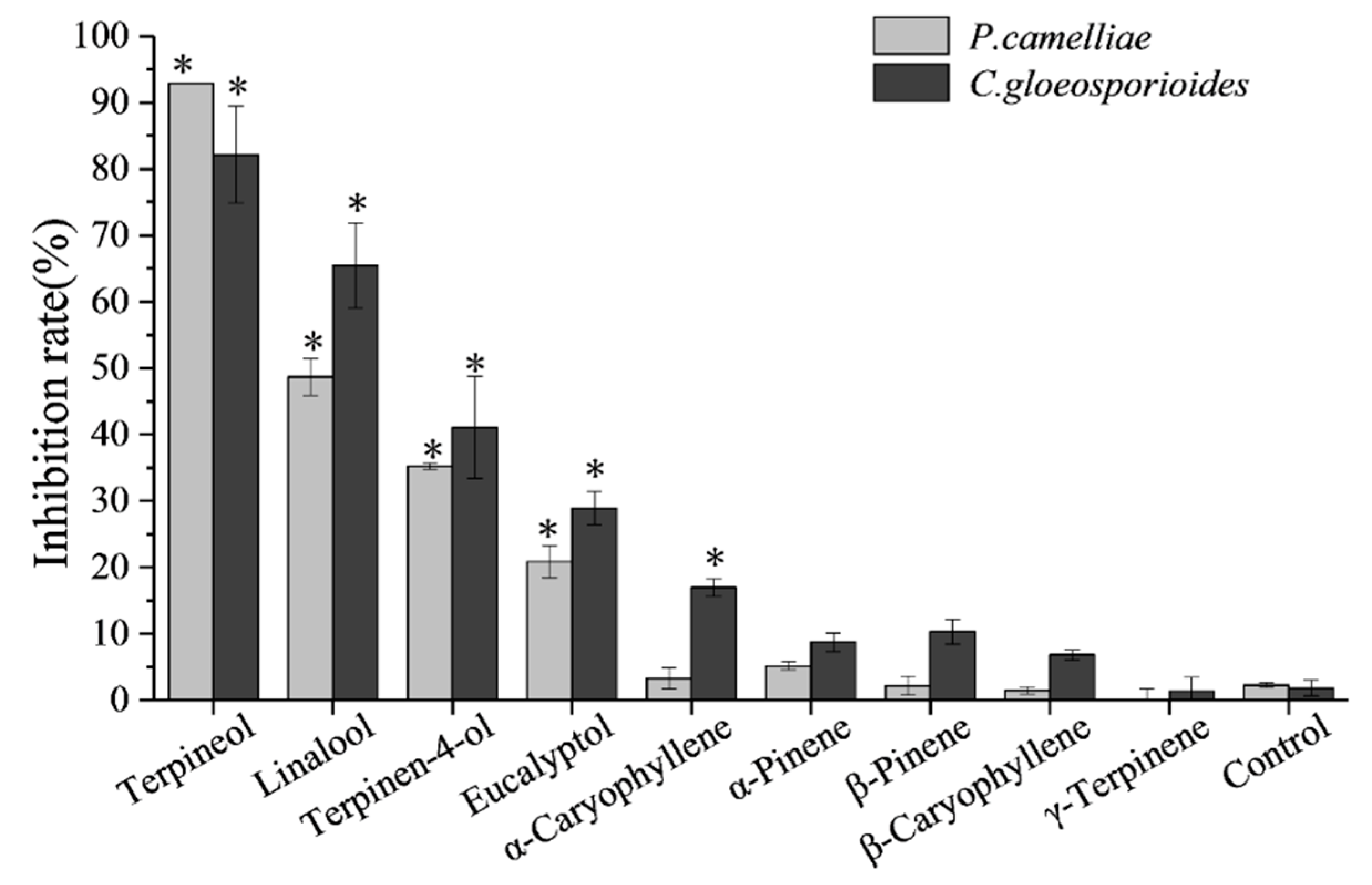
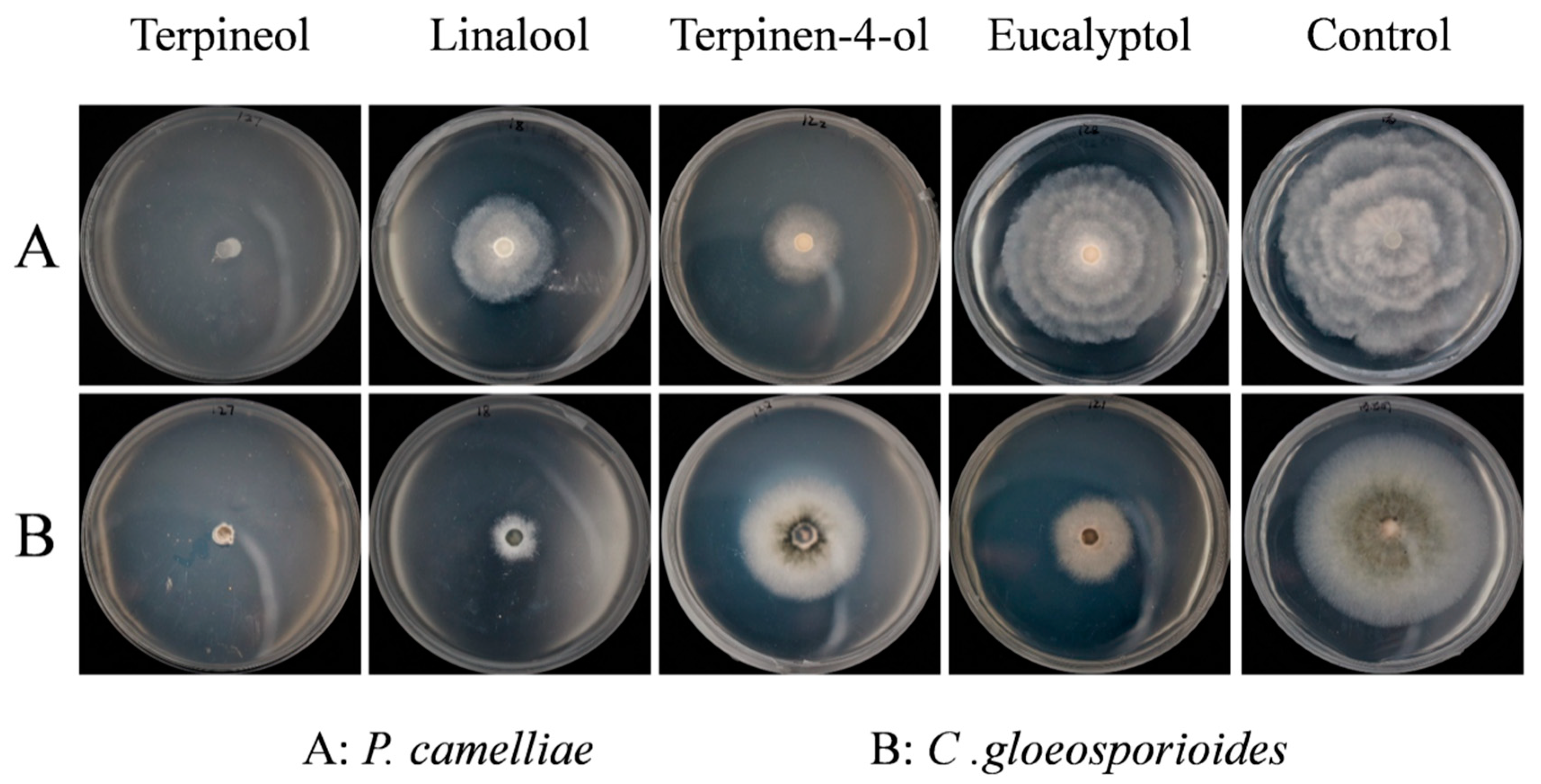
2.3. Screening for Fumigation Antifungal Activities of Major Chemical Compounds

In order to quickly screen the antifungal activity of the nine major chemical compounds present in the SFE-CO2 extract the fumigation method against the mycelial growth of P. camelliae and C. gloeosporioides was used. The results are presented in Figure 3.
In Figure 3, it can be seen that the four primary compounds (terpineol, linalool, terpinen-4-ol and eucalyptol) had stronger fumigation activity against P. camelliae and C. gloeosporioides, respectively. By contrast, the other five major compounds (α-pinene, β-pinene, γ-terpinene, β-caryophyllene and α-caryophyllene) only exhibited weak fumigation activity against two tea plant pathogenic fungi, respectively. In the fumigation activity assay, the mycelial growth of P. camelliae and C. gloeosporioides was inhibited by the four relatively higher activity standards (terpineol, linalool, terpinen-4-ol and eucalyptol) as shown in Figure 4.
As shown in Figure 3 and Figure 4, terpineol showed the strongest fumigation inhibitory effects against P. camelliae, presenting an inhibition rate of 92.91%, followed by linalool, terpinen-4-ol and eucalyptol which inhibited 48.68%, 35.20% and 20.87%, respectively. Terpineol also exhibited the highest fumigation inhibitory effect (82.15%) against C. gloeosporioides, followed by linalool, terpinen-4-ol and eucalyptol that inhibited 65.51%, 41.06% and 28.92%, respectively. In addition, α-caryophyllene showed weak fumigation activity (16.93%) against C. gloeosporioides. In a previous report, linalool showed fumigation inhibitory effect (83.7% inhibition) against, Aspergillus ochraceus at an air concentration of 56 µg/mL [39].
However, as seen in Figure 3, the other five major compounds (α-pinene, β-pinene, γ-terpinene, β-caryophyllene and α-caryophyllene) showed less than 17% inhibition rate against mycelial growth of P. camelliae and C. gloeosporioides, respectively. These results are in-line with a previous report that showed that α-pinene, β-pinene and β-caryophyllene did not present fumigation inhibitory effects against Aspergillus ochraceus, A. flavus, and A. niger at an air concentration of 56 µg/mL [39].
Conversely, many plant essential oils have been evaluated for fumigation activity against plant pathogens [39,40]. For example, the antifungal activity of cinnamon oil, clove oil, eugenol and geraniol was dose-dependent, and relatively higher doses (8 μL/disc) presented stronger antifungal activities [41]. At a concentration of 10 μL/L air, cinnamon oil, cinnamon bark oil, Litsea cubeba oil, Angelica dahurica oil and thyme oil showed high fumigation inhibitory effects (inhibition rate of 100%) against Villosiclava virens [40]. Trans-cinnamaldehyde, the major component of cinnamon oil and cinnamon bark oil, exhibited strong fumigation activity against Villosiclava virens with an effective medium concentration EC50 of 0.5 µL/L air [40].

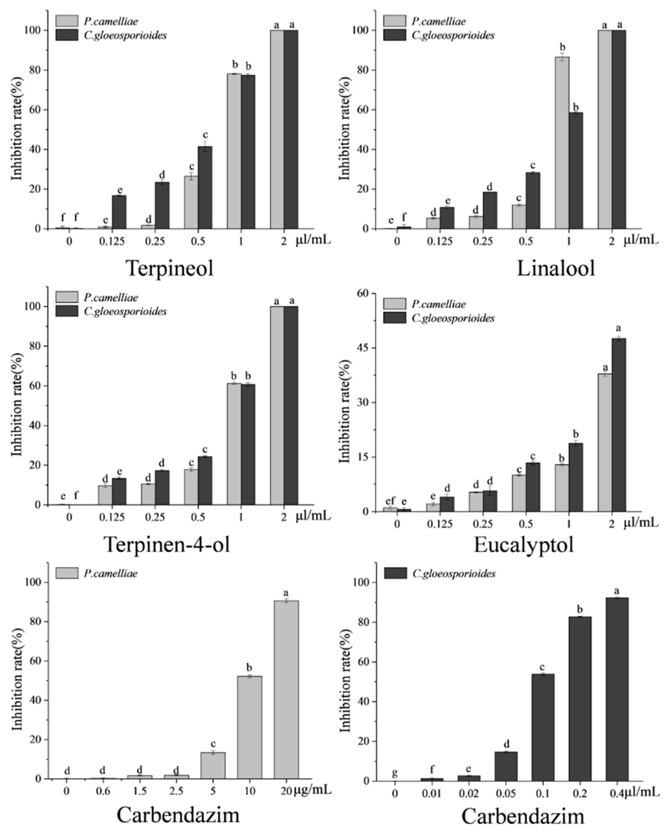
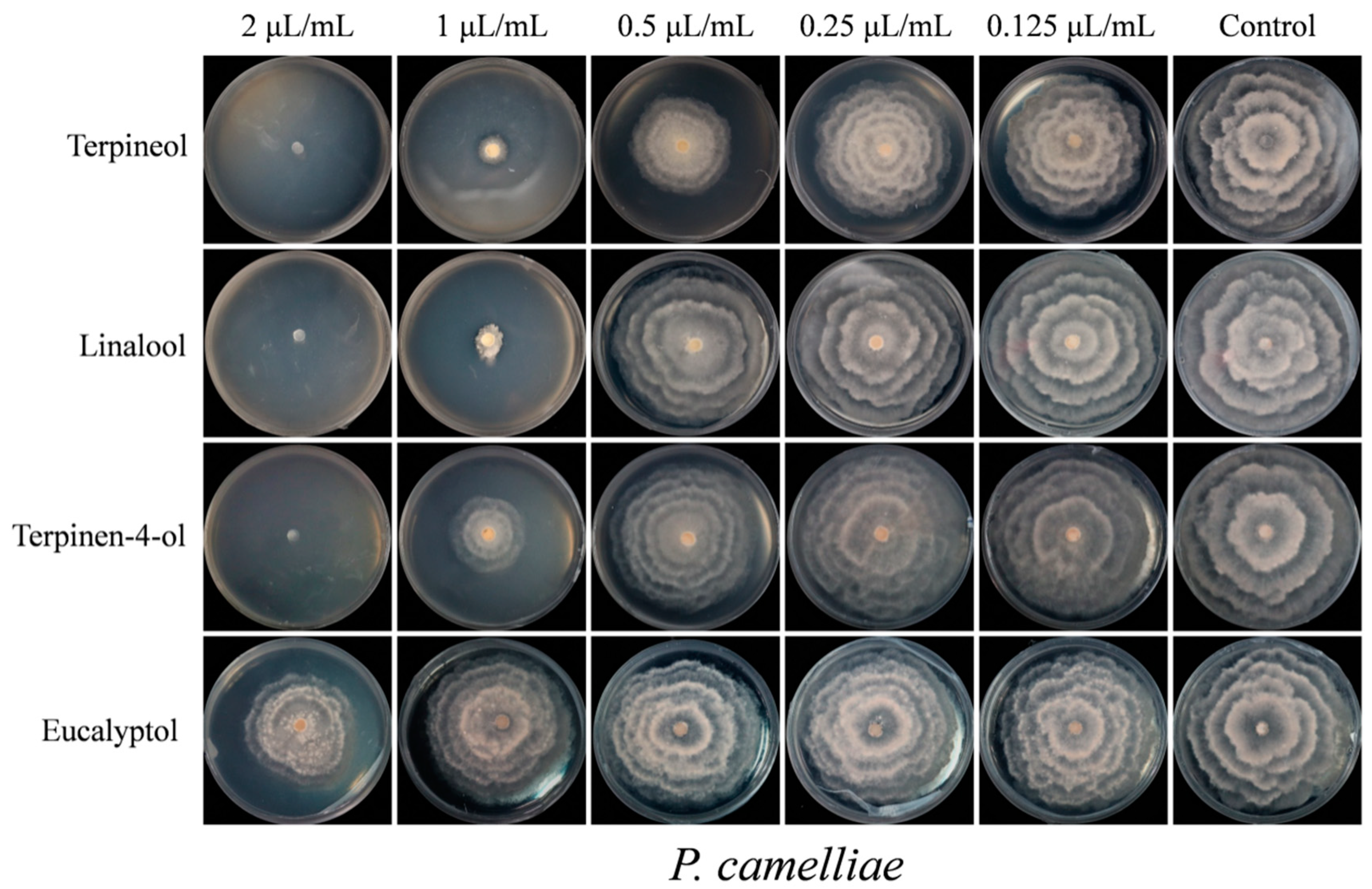
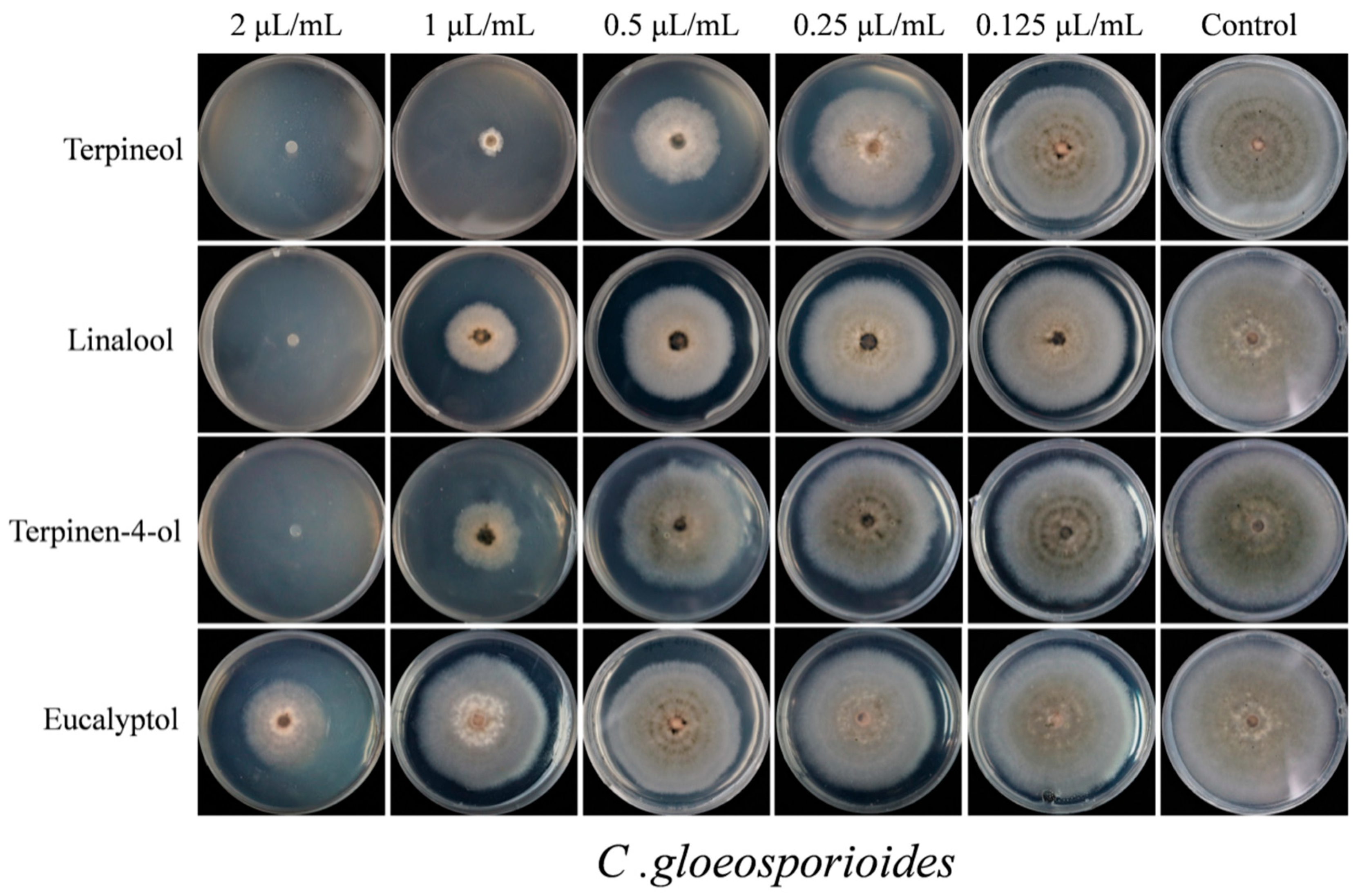
2.4. Contact Antifungal Activity of Major Activity Components

According to the results of the fumigation activity assay, the four compounds with the highest activity (terpineol, linalool, terpinen-4-ol and eucalyptol) were thoroughly evaluated in relation to their contact antifungal activity against P. camelliae and C. gloeosporioides. The results of contact antifungal activity of the chemical compounds compared to that of carbendazin are presented in Figure 5. The contact antifungal toxicity of terpineol, linalool, terpinen-4-ol and eucalyptol against P. camelliae and C. gloeosporioides was concentration-dependent.
Terpineol, linalool, terpinen-4-ol and eucalyptol showed inhibitory effects on mycelial growth of P. camelliae at the five different concentrations, Figure 6. As shown in Figure 5 and Figure 6, at 2 µL/mL, terpineol, linalool and terpinen-4-ol exhibited the strongest contact activity with 100% inhibition of P. camelliae. In contrast, at 20 µL/mL, the commercial fungicide carbendazim inhibited 90.64% the growth of P. camelliae (Figure 5). Linalool, terpineol and terpinen-4-ol exhibited the highest inhibitory effects on the mycelial growth of P. camelliae at 1 µL/mL (Figure 6). Terpineol presented antifungal activity (26.48% inhibition) at 0.5 µL/mL (Figure 6), whereas eucalyptol showed moderate antifungal activity (37.9% inhibition) against P. camelliae at 2 µL/mL. In addition, compared with the control, other concentrations of the linalool and terpinen-4-ol also showed significantly contact antifungal activity (Figure 5). Eucalyptol also showed antifungal activity and inhibited mycelial growth of P. camelliae at concentrations higher than 0.25 µL/mL.
However, as expected, carbendazim showed a strong inhibitory effect on the mycelial growth of P. camelliae at concentrations higher than 10 µL/mL (Figure 7). Carbendazim presented the highest antifungal activity (90.64% inhibition) at 20 µL/mL, and it also showed moderate antifungal activity with inhibition rate of 52.17% at 10 µL/mL (Figure 5 and Figure 7). In summary, compared with carbendazim, the tested compounds (terpineol, linalool, terpinen-4-ol and eucalyptol) showed stronger antifungal activity and inhibited mycelial growth of P. camelliae.
The effects of terpineol, linalool, terpinen-4-ol and eucalyptol on the mycelial growth of C. gloeosporioides at the five different concentrations are shown in Figure 8. Compared with the control, all the four chemical compounds had significant difference in the contact antifungal activity and inhibition of mycelial growth of C. gloeosporioides (Figure 5 and Figure 8). The results show that terpineol, linalool and terpinen-4-ol were able to inhibit 100% of mycelial growth (Figure 8) and at 2 µL/mL it inhibited 100% of C. gloeosporioides growth. On the other hand, eucalyptol inhibited 47.63% of the C. gloeosporioides growth (Figure 5). Terpineol showed the strongest contact inhibition of C. gloeosporioides mycelial growth, followed by linalool, terpinen-4-ol and eucalyptol (Figure 8). However, compared with the tested four compounds, carbendazim exhibited the strongest contact antifungal activity and inhibition of mycelial growth of C. gloeosporioides (Figure 5 and Figure 7). Carbendazim exhibited the strongest contact activity with 92.27% inhibition of C. gloeosporioides at 0.4 µL/mL (Figure 5). In addition, at 0.2 µL/mL and 0.1 µL/mL, carbendazim also inhibited 82.76% and 53.8%of C. gloeosporioides mycelial growth, respectively (Figure 7).
This result is in-line with other studies that showed that the treatment with an 8 μL/disc of linalool dramatically decreased the conidial germination of C. gloeosporioides to 3.4%, but eugenol showed moderate anti-germination activities with the conidial germination of C. gloeosporioides was 50.4% [41]. At 1 µL/mL, terpineol, linalool, and terpinen-4-ol inhibitedthe mycelial growth by 77.42%, 58.53% and 60.62%, respectively. However, eucalyptol at 1 µL/mL showed relatively weak activity (<20% inhibition) in relation to the mycelial growth of C. gloeosporioides [41].
The median inhibitory concentration (MIC50) values of terpineol, linalool, terpinen-4-ol and eucalyptol compounds were estimated using the logistic analyses model and the results are displayed in Table 3.
The high determination coefficient values (R2 ≥ 0.90) indicate that the logistic analyses model is suitable to fit the antimicrobial data. Terpineol, linalool, terpinen-4-ol and eucalyptol showed strong contact antifungal activity against P. camelliae with remarkable MIC50 values of 0.69, 0.73, 0.86 and 2.79μL/mL, respectively (Table 3). Compared with the commercial fungicide carbendazim (MIC50 values of 9.70 μL/mL), terpineol, linalool, terpinen-4-ol and eucalyptol had 3.5 to 14-fold more antifungal activity against P. camelliae (Table 3). Terpineol, linalool, terpinen-4-ol and eucalyptol also exhibited strong inhibitory effect against C. gloeosporioides, with MIC50 values of 0.62, 0.93, 0.94 and 2.18 μL/mL, respectively (Table 3). The carbendazim showed acute contact antifungal activity (MIC50 = 0.095 μL/mL) in relation to C. gloeosporioides. Overall, terpineol had the highest antifungal activity against P. camelliae and C. gloeosporioides, followed by linalool, terpinen-4-ol and eucalyptol. This observation is consistent with other research stating that linalool and terpinen-4-ol were more active than eucalyptol [42,43]. In fact, the contact antifungal activity of terpineol was approximately four times higher than that of eucalyptol against P. camelliae and C. gloeosporioides.
Some terpenoids, such as terpineol, linalool, terpinen-4-ol and eucalyptol have antifungal activity against plant pathogens. For example, terpineol could strongly inhibit the mycelial growth of Penicillium digitatum, with the minimum inhibitory concentration of 2.00 µL/mL [44]. Terpinen-4-ol and eucalyptol exhibited antifungal activity against Candida albicans with MIC50 values of 0.06% (v/v) and 4% (v/v), respectively [42]. Linalool and eucalyptol inhibited the mycelial growth of Fusarium oxysporum, Fusarium solani, and Cylindrocarpon destrutans [45], whereas capsidiol exhibited antifungal activity against C. gloeosporioides [46]. Regarding plant essential oils, the antifungal activity against C. gloeosporioides or P. camelliae has also been reported. For instance, Engenia caryophyllus and Cinnamomum cassia essential oils showed good contact antifungal toxicity against C. gloeosporioides, with 100 and 189 μL/L, respectively [47]. Cinnamon oil (8 μL/disc) exhibited strong contact antifungal efficacies against C. gloeosporioides [41]. Thus, it seems that our samples are very effective against plant pathogens compared with other plants.
In general, the fumigation and contact antifungal activity of plant extracts could not be easily correlated with one specific component. In this study, terpineol, linalool, terpinen-4-ol and eucalyptol showed strong fumigation antifungal activity against P. camelliae and C. gloeosporioides with inhibition rates in the range of 20.87% and 92.91%. Additionally, terpineol, linalool, terpinen-4-ol and eucalyptol also exhibited notable contact antifungal activity against P. camelliae and C. gloeosporioides with MIC50 values in the range of 0.62 μL/mL to 2.79 μL/mL. The commercial fungicide carbendazim showed significantly contact antifungal activity against P. camelliae and C. gloeosporioides with MIC50 values of 0.095 μL/mL and 9.70 μL/mL, respectively. Therefore, it seems that terpineol, linalool, terpinen-4-ol and eucalyptol could be considered as the main active ingredients and are responsible for antifungal activities of the SFE-CO2 extract from B. frutescens leaves.

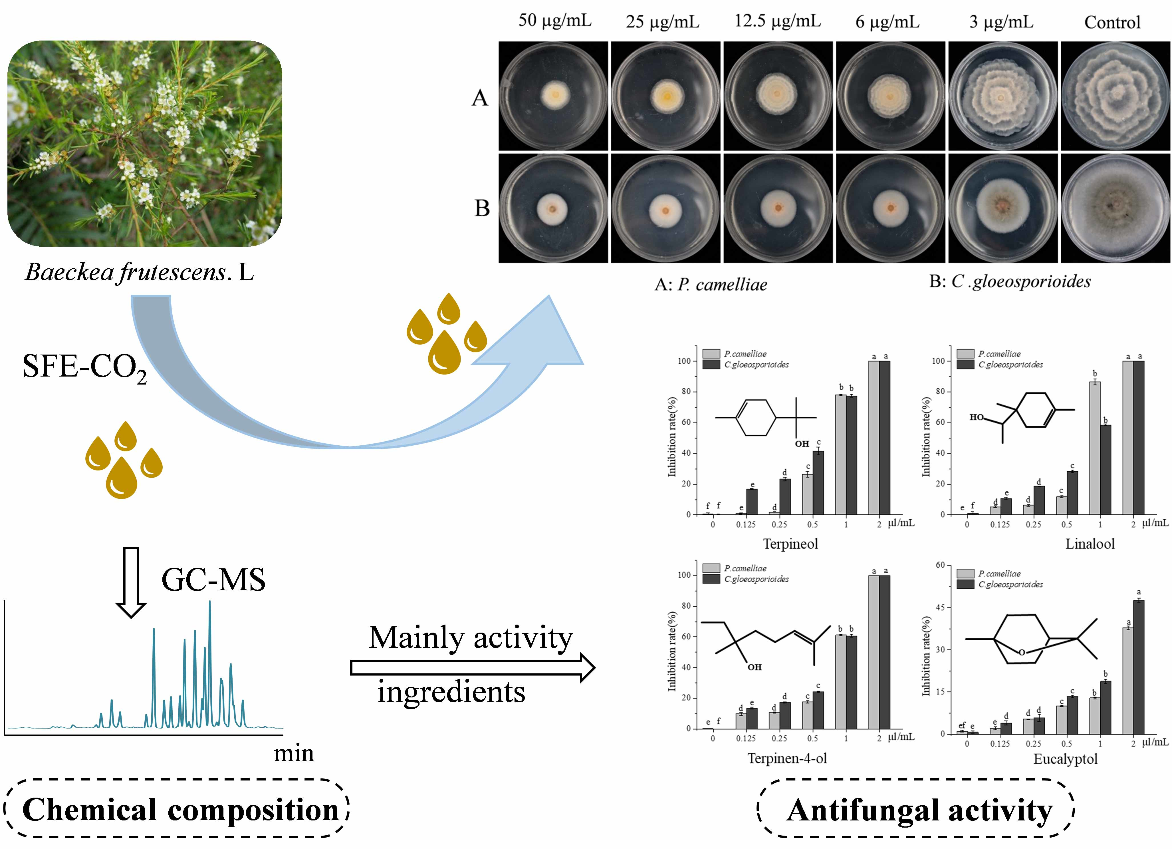
3. Materials and Methods

3.1. Plant Materials

The leaves of Baeckea frutescens were collected at the Guangxi Forestry Research Institute, Nanning, Guangxi Province, China in September of 2018. The B. frutescens trees were cultivated in the resource garden of Guangxi Forestry Research Institute. B. frutescens were authenticated by Prof. Wen-Hui Liang (Guangxi Forestry Research Institute). A voucher specimen (No. GX201816) has been deposited in the State Key Laboratory of Tea Plant Biology and Utilization, Anhui Agricultural University. A total of 2160 g B. frutescens leaves were collected from nine randomly selected trees. The leaves were freeze-dried in a freeze dryer (model Martin Christ ALPHA 1-4 LD, Osterode am Harz, Germany). The dried leaves (10 mm × 1 mm, 4% humidity) were ground into powder (filtered through a 200-mesh sieve) and stored at −20 °C.

3.2. Chemicals

The standard chemicals—α-pinene, β-pinene, eucalyptol (1.8-cineol), γ-terpinene, linalool, terpinen-4-ol, terpineol, β-caryophyllene and α-caryophyllene—with a purity of ≥99% were purchased from Sigma Chemical Co. (Fairfield, OH, USA). The homologous series of (C5–C36) alkanes were purchased from Sigma Chemical Co. (Fairfield, OH, USA). HPLC-grade methanol and N-hexane were obtained from Fisher Scientific (Fair Lawn, NJ, USA). Ultrapure water was used in the experiments (Purelab Plus, Pall, Show Low, AZ, USA). All other chemicals were of analytical grade and purchased from Beijing Chemical Works (Beijing, China).

3.3. Isolation and Identification of Fungal Pathogens

Diseased tea leaves were collected from the tea plantation located in Cuona County, Shannan city, Tibet Autonomous Region in China. The pathogenic fungi were isolated from diseased leaves that presented visible grey blight disease and anthracnose symptoms using traditional method as previously described [12,48] and with slight modification. Briefly, the symptomatic leaves were surface-sterilized in 1% NaClO for 2 min, then in 70% ethanol for 1 min, rinsed three times in sterile water, and then margins of lesions were cut into small pieces and transferred into potato dextrose agar medium (PDA, Difco Company). The culture was incubated at 25 °C for 3 to 5 days until fungal hyphae started to grow from the pieces. Single-hyphal tip was transferred to a new PDA plate to purify isolates and two strains were obtained. To identify the two fungal isolates, DNA was extracted, amplified, and sequenced using universal primers (Internal Transcribed Spacer, ITS1/ITS4). BLAST (Basic Local Alignment Search Tool) analysis of GenBank data showed 100% sequence homology with the ITS sequence of strain for Colletotrichum gloeosporioides (Accession No. KF836743.1) and Pseudopestalotiopsis camelliae-sinensis (Accession No. MK909901.1), respectively. The two pathogenic fungi C. gloeosporioides and P. camelliae cultured using potato dextrose agar medium at 4 °C and stored for future use.

3.4. Supercritical Fluid (SFE-CO2) Extract of Baeckea frutescens L.

The extraction was performed with a laboratory scale multi-vessel accelerated supercritical fluid extraction system (model MV-10 ASFE Waters Corporation, Milford, MA, USA). The dried B. frutescens leaf powder (5.0 g) was placed in a 25 mL supercritical fluid extractor vessel and was extracted using supercritical carbon dioxide without any cosolvents. The temperature and pressure were set at 40 °C and 180 bar, respectively. The extract laden Supercritical CO2 (flow rate 13 min/mL) was sent to extractor vessel through a pressure pump for 70 min, with 20 min dynamic duration, 30 min of static duration and 20 min of dynamic duration, respectively. The extraction efficiency (yield) was 2.2% (110 mg), and the SFE-CO2 extract was stored for future use.

3.5. Contact Antifungal Activities Bioassay

The contact antifungal activity of the SFE-CO2 extract was determined using the toxic medium method. Median inhibitory concentration (MIC50) of the extract was determined using a serial two-fold micro dilution method against the tea plant pathogenic fungi. The stock solution (100 mg/mL) was serially diluted in 30 mL PDA medium at 45–50 °C and mixed to provide different concentrations (50 µg/mL, 25 µg/mL, 12.5 µg/mL, 6 µg/mL, 3 µg/mL) to evaluate inhibitory activities against C. gloeosporioides and P. camelliae. Based on preliminary screening activities, the four individual chemical components with higher antifungal activity were determined, and which were adjusted to a series of concentration gradients medium (2.0 μL/mL, 1.0 μL/mL, 0.5 μL/mL, 0.25 μL/mL and 0.125 μL/mL) to evaluate inhibitory activities against C. gloeosporioides and P. camelliae. Carbendazim, a broad-spectrum fungicide, was used as a positive control and different concentrations were tested. The negative control received the same quantity of acetone mixed with PDA. The 10 mL toxic medium were poured onto aseptic 9 cm plastic Petri dishes. A 5 mm diameter fungal disc of C. gloeosporioides or P. camelliae was immediately inoculated in the center of each PDA plate and plates were incubated in the dark at 25 °C.
After 7 days of incubation at 25 °C in the dark, the colony growth diameter (mm) was measured using a digital caliper. Each test was repeated three times. The growth inhibition was calculated using the formula.
Inhibition rate (%) = (Dc − Dt)/Dc × 100,
where: DC, DT—average diameter (mm) of the fungal colony of the control and the treatment, respectively.

3.6. Fumigation Antifungal Activities Bioassay

The fumigation activity of the SFE-CO2 extract was determined as previously described [49] and with slight modifications. A 5 mm diameter disc of C. gloeosporioides or P. camelliae was inoculated in the center of each PDA plate (9 cm, the volume about 60 mL air spaces), and a 6 mm filter paper containing 10 μL of the extract or isolated substance was placed on the center of the inner surface of the Petri dish lid. Plastic Petri dishes (60 mL air spaces) offer 50 mL air spaces after the addition of 10 mL PDA medium, and the final concentration was 200 μL/L air. The negative control was composed of acetone. The plate was immediately sealed with parafilm to prevent any leakage of the standards. Three replicates were used for each concentration, and the PDA plates were placed upside down in the incubator. After 5 days of incubation at 25 °C in the dark, the colony growth diameter (mm) was measured using a digital caliper. The growth inhibition was calculated using the above Formula (1).

3.7. B. frutescens Extract GC-MS Analysis

The constituents SFE-CO2 Extract of B. frutescens were analyzed by Thermo Fisher trace 1300 gas chromatography system, equipped with ISQ 7000 MS (Thermo Fisher Scientific, San Jose, CA, USA). Briefly, the stock solution of SFE-CO2 extract (10 uL) or the isolated compounds (terpineol, linalool, terpinen-4-ol, eucalyptol, α-caryophllene, (1R)-(+)- α-pinene, (1S)-(-)- α-pinene, α-phellandrene, β-caryophyllene, γ-terpinene) were diluted with n-hexane (1 mL). A DB-5MS column (60 m × 0.25 mm, film thickness 0.25 μm, J&W Scientific, Folsom, CA, USA) was used in the separation of chemical compounds. Helium ( >99.99%) was used as the carrier gas at a flow rate of 1 mL/min. The injector temperature was 250 °C using a splitless injection mode with a sampling time of 1.00 min. The oven program was set as follows: Temperatures and times of 50 °C (5 min), 20 °C/min to 180 °C (5 min), 5 °C/min to 230 °C (5 min), and 10 °C/min to 280 °C (5 min). The electron-impact mass spectra were generated at 70 eV, with a scan range from 30 to 600 m/z, the ion source temperature was 230 °C, and the MS interface temperature was 250 °C.

3.8. Statistical Analysis

Statistical significance was carried out by applying one-way ANOVA followed by Duncan’s test with the acceptance level of significance p = 0.05, using SPSS version 22.0 (IBM Corp., Armonk, NY, USA). Logistic analysis was performed using Origin version 2017 software (OriginLab., Northampton, MA, USA) and GraphPad Prism 7 software (GraphPad Software, Inc., La Jolla, CA, USA).

4. Conclusions

To the best of the authors’ knowledge, this is first report that investigates the volatile chemical composition of the SFE-CO2 extract from B. frutescens leaves. β-Caryophyllene, α-caryophyllene, δ-cadinene, eucalyptol and terpineol are the major compounds of the extract. The extract presented antifungal activity against P. camelliae and C. gloeosporioides. Terpineol, linalool, terpinen-4-ol and eucalyptol are the main active ingredients. Therefore, the SFE-CO2 extract from B. frutescens leaves could be a potential alternative to traditional synthetic chemical fungicides of tea plant pathogens (P. camelliae and C. gloeosporioides).

Supplementary Materials

The following are available online at https://www.mdpi.com/2223-7747/9/9/1119/s1, Figure S1: The total ion chromatogram of volatile fraction of Baeckea frutescens L (SFE-CO2) extract by GC-MS.

Author Contributions

Conceptualization, H.J. and L.Q.; methodology and software, M.Z.; formal analysis, M.Z. and H.J.; investigation, H.J. and D.W.; plant materials resources, W.L.; data curation, M.Z. and F.Y.; writing—original draft preparation, H.J.; writing—review and editing, H.J. and D.G.; project administration, H.J.; funding acquisition, H.J. and C.S. All authors have read and agreed to the published version of the manuscript.

Funding

This research was funded by National Natural Science Foundation of China (31902075), Anhui Key Research and Development Program (202004e11020006), Anhui Provincial Postdoctoral Fund (2017B232), Anhui Provincial Department of Education (KJ20198A0196), Anhui Agricultural University Young Talent Fund (YJ2017-32). In addition, thankful to the financial support from Earmarked Fund for Modern Agro-industry Technology Research System in Tea Industry (NO. CARS-19, the Ministry of Agriculture of P. R. China), the Changjiang Scholars and Innovative Research Team in University (NO. IRT1101), Guangxi Forestry Research and Extension Project (gl2017kt19) and The Fundamental Research Funds for Guangxi Forestry Research Institute (No. 201823).

Conflicts of Interest

The authors declare no conflict of interest.

References

  1. An, N.T.G.; Huong, L.T.; Satyal, P.; Tai, T.A.; Dai, D.N.; Hung, N.H.; Ngoc, N.T.B.; Setzer, W.N. Mosquito Larvicidal Activity, Antimicrobial Activity, and Chemical Compositions of Essential Oils from Four Species of Myrtaceae from Central Vietnam. Plants 2020, 9, 544. [Google Scholar] [CrossRef] [PubMed] [Green Version]
  2. Shahruzaman, S.H.; Mustafa, M.F.; Ramli, S.; Maniam, S.; Fakurazi, S.; Maniam, S. The Cytotoxic Properties of Baeckea frutescens Branches Extracts in Eliminating Breast Cancer Cells. Evid. Based Complement. Altern. Med. 2019, 2019, 9607590. [Google Scholar] [CrossRef]
  3. Vu, T.T.; Kim, H.; Tran, V.K.; Le Dang, Q.; Nguyen, H.T.; Kim, H.; Kim, I.S.; Choi, G.J.; Kim, J.C. In vitro antibacterial activity of selected medicinal plants traditionally used in Vietnam against human pathogenic bacteria. BMC Complement. Altern. Med. 2016, 16, 32. [Google Scholar] [CrossRef] [Green Version]
  4. Shahruzaman, S.H.; Mustafa, M.F.; Ramli, S.; Maniam, S.; Fakurazi, S.; Maniam, S. The cytotoxic effect and glucose uptake modulation of Baeckea frutescens on breast cancer cells. BMC Complement. Altern. Med. 2019, 19, 220. [Google Scholar] [CrossRef] [PubMed] [Green Version]
  5. Hou, J.Q.; Guo, C.; Zhao, J.J.; Dong, Y.Y.; Hu, X.L.; He, Q.W.; Zhang, B.B.; Yan, M.; Wang, H. Anti-inflammatory Meroterpenoids from Baeckea frutescens. J. Nat. Prod. 2017, 80, 2204–2214. [Google Scholar] [CrossRef] [PubMed]
  6. Zhi, Y.E.; Qi, X.J.; Liu, H.; Zeng, Y.; Ni, W.; He, L.; Wang, Z.D.; Liu, H.Y. Structurally Diverse Polymethylated Phloroglucinol Meroterpenoids from Baeckea frutescens. Nat. Prod. Bioprospect. 2018, 8, 431–439. [Google Scholar] [CrossRef] [Green Version]
  7. Wei, C.L.; Yang, H.; Wang, S.B.; Zhao, J.; Liu, C.; Gao, L.P.; Xia, E.H.; Lu, Y.; Tai, Y.L.; She, G.B.; et al. Draft genome sequence of Camellia sinensis var. sinensis provides insights into the evolution of the tea genome and tea quality. Proc. Natl. Acad. Sci. USA 2018. [Google Scholar] [CrossRef] [Green Version]
  8. Jiang, H.; Yu, F.; Qin, L.; Zhang, N.; Cao, Q.; Schwab, W.; Li, D.X.; Song, C.K. Dynamic change in amino acids, catechins, alkaloids, and gallic acid in six types of tea processed from the same batch of fresh tea (Camellia sinensis L.) leaves. J. Food Compos. Anal. 2019, 77, 28–38. [Google Scholar] [CrossRef]
  9. Chen, Y.J.; Tong, H.R.; Wei, X.; Yuan, L.Y. First Report of Brown Blight Disease on Camellia sinensis Caused by Colletotrichum acutatum in China. Plant Dis. 2016, 100, 227–228. [Google Scholar] [CrossRef]
  10. Li, J.; Sun, K.; Ma, Q.P.; Chen, J.; Wang, L.; Yang, D.J.; Chen, X.; Li, X.H. Colletotrichum gloeosporioides–contaminated tea infusion blocks lipids reduction and induces kidney damage in mice. Front. Microbiol. 2017, 8. [Google Scholar] [CrossRef] [Green Version]
  11. Chen, Y.J.; Zeng, L.; Shu, N.; Jiang, M.Y.; Wang, H.; Huang, Y.J.; Tong, H.R. Pestalotiopsis-Like Species Causing Gray Blight Disease on Camellia sinensis in China. Plant Dis. 2018, 102, 98–106. [Google Scholar] [CrossRef] [PubMed] [Green Version]
  12. Thangaraj, K.; Deng, C.; Cheng, L.L.; Deng, W.W.; Zhang, Z.Z. Report of Phoma herbarum Causing Leaf Spot Disease of Camellia sinensis in China. Plant Dis. 2018, 102, 2373. [Google Scholar] [CrossRef] [PubMed]
  13. Chen, Y.J.; Zeng, L.; Shu, N.; Wang, H.; Tong, H.R. First Report of Pestalotiopsis camelliae causing Grey Blight Disease on Camellia sinensis in China. Plant Dis. 2017, 101, 1034. [Google Scholar] [CrossRef]
  14. Akbar, A.; Ali, G.S.; Pearson, B.J.; Hamid, F.S.; Sumreen, S. Screening Camelia sinensis Germplasm Against Grey Leaf Blight of Tea. J. Agric. Stud. 2017, 5, 123–135. [Google Scholar] [CrossRef]
  15. Wang, Y.X.; Chen, J.Y.; Xu, X.W.; Cheng, J.Y.; Zheng, L.; Huang, J.B.; Li, D.W. Identification and Characterization of Colletotrichum Species Associated with Anthracnose Disease of Camellia oleifera in China. Plant Dis. 2020, 104, 474–482. [Google Scholar] [CrossRef]
  16. He, L.F.; Li, X.X.; Gao, Y.Y.; Li, B.X.; Mu, W.; Liu, F. Characterization and Fungicide Sensitivity of Colletotrichum spp. from Different Hosts in Shandong, China. Plant Dis. 2019, 103, 34–43. [Google Scholar] [CrossRef] [Green Version]
  17. Jeyaraj, A.; Wang, X.W.; Wang, S.S.; Liu, S.R.; Zhang, R.; Wu, A.; Wei, C.T. Identification of Regulatory Networks of MicroRNAs and Their Targets in Response to Colletotrichum gloeosporioides in Tea Plant (Camellia sinensis L.). Front. Plant Sci. 2019, 10. [Google Scholar] [CrossRef] [Green Version]
  18. Guo, M.; Pan, Y.M.; Dai, Y.L.; Gao, Z.M. First Report of Brown Blight Disease Caused by Colletotrichum gloeosporioides on Camellia sinensis in Anhui Province, China. Plant Dis. 2014, 98, 284. [Google Scholar] [CrossRef]
  19. Chen, Y.J.; Qiao, W.J.; Zeng, L.; Shen, D.H.; Liu, Z.; Wang, X.S.; Tong, H.R. Characterization, Pathogenicity, and Phylogenetic Analyses of Colletotrichum Species Associated with Brown Blight Disease on Camellia sinensis in China. Plant Dis. 2017, 101, 1022–1028. [Google Scholar] [CrossRef] [Green Version]
  20. Wari, D.; Okada, R.; Takagi, M.; Yaguchi, M.; Kashima, T.; Ogawara, T. Augmentation and compatibility of Beauveria bassiana with pesticides against different growth stages of Bemisia tabaci (Gennadius); an in-vitro and field approach. Pest Manag. Sci. 2020. [Google Scholar] [CrossRef]
  21. Bai, Y.B.; Gao, Y.Q.; Nie, X.D.; Tuong, T.M.; Li, D.; Gao, J.M. Antifungal Activity of Griseofulvin Derivatives against Phytopathogenic Fungi in Vitro and in Vivo and Three-Dimensional Quantitative Structure-Activity Relationship Analysis. J. Agric. Food Chem. 2019, 67, 6125–6132. [Google Scholar] [CrossRef] [PubMed]
  22. Wang, Z.; Mei, X.; Du, M.; Chen, K.; Jiang, M.; Wang, K.; Zalan, Z.; Kan, J. Potential modes of action of Pseudomonas fluorescens ZX during biocontrol of blue mold decay on postharvest citrus. J. Sci. Food Agric. 2020, 100, 744–754. [Google Scholar] [CrossRef] [PubMed]
  23. Shuping, D.S.S.; Eloff, J.N. The use of plants to protect plants and food against fungal pathogens: A review. Afr. J. Tradit. Complement. Altern. Med. 2017, 14, 120–127. [Google Scholar] [CrossRef] [PubMed] [Green Version]
  24. Tyskiewicz, K.; Tyskiewicz, R.; Konkol, M.; Roj, E.; Jaroszuk-Scisel, J.; Skalicka-Wozniak, K. Antifungal Properties of Fucus vesiculosus L. Supercritical Fluid Extract Against Fusarium culmorum and Fusarium oxysporum. Molecules 2019, 24, 3518. [Google Scholar] [CrossRef] [Green Version]
  25. Hu, Z.; Wang, Z.; Liu, Y.; Wang, Q. Leveraging botanical resources for crop protection: The isolation, bioactivity and structure-activity relationships of lycoris alkaloids. Pest Manag. Sci. 2018, 74, 2783–2792. [Google Scholar] [CrossRef]
  26. Li, Y.; Wei, W.; Zhang, J.; Li, G.; Gao, K. Structures and antipathogenic fungi activities of flavonoids from pathogen-infected Astragalus adsurgens. Nat. Prod. Res. 2019, 33, 822–826. [Google Scholar] [CrossRef]
  27. Han, X.B.; Zhao, J.; Cao, J.M.; Zhang, C.S. Essential oil of Chrysanthemum indicum L.: Potential biocontrol agent against plant pathogen Phytophthora nicotianae. Environ. Sci. Pollut. Res. Int. 2019, 26, 7013–7023. [Google Scholar] [CrossRef]
  28. Capuzzo, A.; Maffei, M.E.; Occhipinti, A. Supercritical fluid extraction of plant flavors and fragrances. Molecules 2013, 18, 7194–7238. [Google Scholar] [CrossRef] [Green Version]
  29. Messyasz, B.; Michalak, I.; Leska, B.; Schroeder, G.; Gorka, B.; Korzeniowska, K.; Lipok, J.; Wieczorek, P.; Roj, E.; Wilk, R.; et al. Valuable natural products from marine and freshwater macroalgae obtained from supercritical fluid extracts. J. Appl. Phycol. 2018, 30, 591–603. [Google Scholar] [CrossRef]
  30. Tyskiewicz, K.; Konkol, M.; Roj, E. The Application of Supercritical Fluid Extraction in Phenolic Compounds Isolation from Natural Plant Materials. Molecules 2018, 23, 2625. [Google Scholar] [CrossRef] [Green Version]
  31. Malaman, F.S.; Moraes, L.A.B.; West, C.; Ferreira, N.J.; Oliveira, A.L. Supercritical fluid extracts from the Brazilian cherry (Eugenia uniflora L.): Relationship between the extracted compounds and the characteristic flavour intensity of the fruit. Food Chem. 2011, 124, 85–92. [Google Scholar] [CrossRef]
  32. Tyskiewicz, K.; Konkol, M.; Roj, E. Supercritical Carbon Dioxide (scCO2) Extraction of Phenolic Compounds from Lavender (Lavandula angustifolia) Flowers: A Box-Behnken Experimental Optimization. Molecules 2019, 24, 3354. [Google Scholar] [CrossRef] [PubMed] [Green Version]
  33. Wang, S.S.; Mi, X.Z.; Wu, Z.R.; Zhang, L.X.; Wei, C.L. Characterization and Pathogenicity of Pestalotiopsis-Like Species Associated With Gray Blight Disease on Camellia sinensis in Anhui Province, China. Plant Dis. 2019, 103, 2786–2797. [Google Scholar] [CrossRef]
  34. Soylu, E.M.; Soylu, S.; Kurt, S. Antimicrobial activities of the essential oils of various plants against tomato late blight disease agent Phytophthora infestans. Mycopathologia 2006, 161, 119–128. [Google Scholar] [CrossRef]
  35. Dai, D.N.; Thang, T.D.; Olayiwola, T.; Ogunwande, I.A. Chemical Composition of Essential Oil of Baeckea frutescens L. Int. Res. J. Pure Appl. Chem. 2015, 8, 26–32. [Google Scholar] [CrossRef]
  36. Padovan, A.; Keszei, A.; Külheim, C.; Foley, W.J. The evolution of foliar terpene diversity in Myrtaceae. Phytochem. Rev. 2014, 13, 695–716. [Google Scholar] [CrossRef]
  37. Tam, N.T.; Thuam, D.T.; Bighelli, A.; Castola, V.; Muselli, A.; Richomme, P.; Casanova, J. Baeckea frutescens leaf oil from Vietnam: Composition and chemical variability. Flavour Fragr. J. 2004, 19, 217–220. [Google Scholar] [CrossRef]
  38. Ouzzar, M.L.; Louaer, W.; Zemane, A.; Meniai, A.H. Comparison of the performances of hydrodistillation and supercritical CO2 extraction processes for essential oil extraction from Rosemary (Rosmarinus officinalis L.). Chem. Eng. Trans. 2015, 43, 1129–1134. [Google Scholar]
  39. Bhavya, M.L.; Chandu, A.G.S.; Devi, S.S.; Quirin, K.W.; Pasha, A.; Vijayendra, S.V.N. In-vitro evaluation of antimicrobial and insect repellent potential of supercritical-carbon dioxide (SCF-CO(2)) extracts of selected botanicals against stored product pests and foodborne pathogens. J. Food Sci. Technol. 2020, 57, 1071–1079. [Google Scholar] [CrossRef]
  40. Koyu, H.; Kazan, A.; Nalbantsoy, A.; Yalcin, H.T.; Yesil-Celiktas, O. Cytotoxic, antimicrobial and nitric oxide inhibitory activities of supercritical carbon dioxide extracted Prunus persica leaves. Mol. Biol. Rep. 2020, 47, 569–581. [Google Scholar] [CrossRef]
  41. Kim, E.; Park, I.K. Fumigant antifungal activity of Myrtaceae essential oils and constituents from Leptospermum petersonii against three Aspergillus species. Molecules 2012, 17, 10459–10469. [Google Scholar] [CrossRef] [PubMed] [Green Version]
  42. Zheng, J.; Liu, T.; Guo, Z.; Zhang, L.; Mao, L.; Zhang, Y.; Jiang, H. Fumigation and contact activities of 18 plant essential oils on Villosiclava virens, the pathogenic fungus of rice false smut. Sci. Rep. 2019, 9, 7330. [Google Scholar] [CrossRef] [PubMed] [Green Version]
  43. Hong, J.K.; Yang, H.J.; Jung, H.; Yoon, D.J.; Sang, M.K.; Jeun, Y.C. Application of Volatile Antifungal Plant Essential Oils for Controlling Pepper Fruit Anthracnose by Colletotrichum gloeosporioides. Plant Pathol. J. 2015, 31, 269–277. [Google Scholar] [CrossRef] [PubMed] [Green Version]
  44. Mondello, F.; De Bernardis, F.; Girolamo, A.; Cassone, A.; Salvatore, G. In vivo activity of terpinen-4-ol, the main bioactive component of Melaleuca alternifolia Cheel (tea tree) oil against azole-susceptible and -resistant human pathogenic Candida species. BMC Infect. Dis. 2006, 6. [Google Scholar] [CrossRef] [PubMed] [Green Version]
  45. Zengin, H.; Baysal, A.H. Antibacterial and Antioxidant Activity of Essential Oil Terpenes against Pathogenic and Spoilage-Forming Bacteria and Cell Structure-Activity Relationships Evaluated by SEM Microscopy. Molecules 2014, 19, 17773–17798. [Google Scholar] [CrossRef] [Green Version]
  46. Jing, G.-X.; Tao, N.-G.; Jia, L.; Zhou, H.-E. Influence of α-terpineol on the growth and morphogenesis of Penicillium digitatum. Bot. Stud. 2015, 56, 35. [Google Scholar] [CrossRef] [Green Version]
  47. Sun, W.M.; Ma, Y.N.; Yin, Y.J.; Chen, C.J.; Xu, F.R.; Dong, X.; Cheng, Y.X. Effects of Essential Oils from Zingiberaceae Plants on Root-Rot Disease of Panax notoginseng. Molecules 2018, 23, 1021. [Google Scholar] [CrossRef] [Green Version]
  48. Park, S.; Park, A.R.; Im, S.; Han, Y.J.; Lee, S.; Back, K.; Kim, J.I.; Kim, Y.S. Developmentally Regulated Sesquiterpene Production Confers Resistance to Colletotrichum gloeosporioides in Ripe Pepper Fruits. PLoS ONE 2014, 9. [Google Scholar] [CrossRef]
  49. Wang, D.; Zhang, J.; Jia, X.M.; Xin, L.; Zhai, H. Antifungal Effects and Potential Mechanism of Essential Oils on Collelotrichum gloeosporioides In Vitro and In Vivo. Molecules 2019, 24, 3386. [Google Scholar] [CrossRef] [Green Version]
Figure 1. The mainly schematic diagram of the research process.
Plants 09 01119 g001
Figure 2. The mycelial growth of P. camelliae (A) and C. gloeosporioides (B) on different concentration of toxic potato dextrose agar medium (PDA) medium after 7 days.
Plants 09 01119 g002
Figure 3. Fumigation activity of the ten main constituents available against P. camelliae and C. gloeosporioides at 200 μL/L air after 5 days. * Data are presented as mean (±standard error) representative of significantly different versus control group at p ≤ 0.05 (Duncan’s test).
Plants 09 01119 g003
Figure 4. Fumigation activity of five mainly constituents on mycelial growth of P. camelliae (A) and C. gloeosporioides (B) at 200 μL/L air of concentration after 5 days, respectively.
Plants 09 01119 g004
Figure 5. The contact antifungal activity of four major compounds and carbendazim (positive control) against P. camelliae and C. gloeosporioides after 7 days. Percentage values followed by the same letter are not significantly different in the same group at p ≤ 0.05 (Duncan’s test).
Plants 09 01119 g005
Figure 6. The mycelial growth of P. camelliae treatment by four mainly activity compounds with contact antifungal activity after 7 days.
Plants 09 01119 g006
Figure 7. The mycelial growth of C. gloeosporioides and P. camelliae treatment by commercial fungicide carbendazim (positive control) with contact antifungal activity after 7 days.
Plants 09 01119 g007
Figure 8. The mycelial growth of C. gloeosporioides after treatment by four mainly activity compounds with contact antifungal activity after 7 days.
Plants 09 01119 g008
Table 1. Chemical compounds identified in the volatile supercritical fluid extraction (SFE-CO2) extract of B. frutescens.
Peak NOCompound NameRI (C) 1RI (L) 2Relative Area (%) 3Identification Methods
1α-Pinene9409442.11RI, MS, STD 4
2β-Pinene9879795.21RI, MS, STD
3o- Cymene104410421.18RI, MS
4Eucalyptol104910475.46RI, MS, STD
5γ-Terpinene107410632.49RI, MS, STD
6α-Terpinolene110410970.38RI, MS
7Linalool111511041.47RI, MS, STD
8Terpinen-4-ol119411820.83RI, MS, STD
9Terpineol120912004.54RI, MS, STD
10 Epicubebol143314940.19RI, MS
11β-Caryophyllene1448143928.05RI, MS, STD
12α-Caryophyllene1485145224.02RI, MS, STD
13α-Muurolene152015001.60RI, MS
14δ-Guaiene152315020.57RI, MS
15δ-Cadinene153615246.29RI, MS
16trans-Calamenene154215271.28RI, MS
176-epi-Shyobunol154815480.29RI, MS
18Cadine-1,4-diene155215460.64RI, MS
19Isoshyobunone160515621.63RI, MS
20Caryophyllene oxide161015810.70RI, MS
21Unkown1618-0.77RI, MS
22Humulene epoxide II162416160.86RI, MS
23Unkown1633-0.38RI, MS
24α-Acorenol164316300.73RI, MS
25Cubenol164816372.81RI, MS
26Longifolenaldehyde165616312.40RI, MS
27β-Acorenol166116492.11RI, MS
Total 98.99
1 RI (C): Retention index was calculated with relative to the homologous series of (C5–C36) alkanes under the same operating conditions. 2 RI (L): Retention index reported in the relative literature for equivalent capillary column. 3 Relative Area (%): Relative area (peak area relative to the total peak area); 4 STD: co-injection with standard compound have the same mass spectrum (MS) and RI.
Table 2. Contact antifungal toxicity of B. frutescens leaf SFE-CO2 extracts against P. camelliae and C. gloeosporioides.
Content (μg/mL)Inhibitory Rate (%) 1
P. camelliaeC. gloeosporioides
5066.63 ± 2.05 a,264.59 ± 1.59 a
2565.77 ± 1.35 a63.94 ± 0.39 a
12.563.09 ± 2.84 a61.69 ± 0.46 a
654.11 ± 1.18 b53.05 ± 0.87 b
332.48 ± 4.37 c40.29 ± 1.65 c
Control 0.00 ± 1.08 d0.00 ± 0.16 d
MIC505.11 μg/mL4.79 μg/mL
1 Mean (±standard error) of three replicates for each sample. 2 Percentage values followed by the same letter are not significantly different in the same group at p ≤ 0.05 (Duncan’s test).
Table 3. Contact antifungal toxicity of the four major chemical compounds of B. frutescens leaf SFE-CO2 extracts against P. camelliae and C. gloeosporioides.
CompoundP. camelliaeC. gloeosporioides
MIC50 (μL/mL)95% CI 1 (μL/mL)Chi Square (χ2)R2MIC50 (μL/mL)95% CI (μL/mL)Chi Square (χ2)R2
Terpineol0.690.66–0.721.400.9990.620.49–0.759.880.988
Linalool0.730.63–0.8210.610.9940.930.74–1.1312.320.990
Terpinen-4-ol0.860.69–1.0925.820.9860.940.66–1.2123.900.987
Eucalyptol2.792.62–2.976.500.9802.182.05–2.35.070.981
Carbendazim9.709.41–10.010.550.9990.0950.091–0.1001.220.999
1 95%CI: 95% confidence interval for each of three replicates for each median inhibitory concentration (MIC50) value.

