Article

Insights into the Role of Streptomyces hydrogenans as the Plant Growth Promoter, Photosynthetic Pigment Enhancer and Biocontrol Agent against Meloidogyne incognita in Solanum lycopersicum Seedlings

1
Department of Zoology, Guru Nanak Dev University, Amritsar, Punjab 143005, India
2
Department of Botanical and Environmental Sciences, Guru Nanak Dev University, Amritsar, Punjab 143005, India
3
Department of Microbiology, Guru Nanak Dev University, Amritsar, Punjab 143005, India
4
Botany and Microbiology Department, College of Science, King Saud University, Riyadh 11451, Saudi Arabia
5
Department of Botany, S.P. College Srinagar, Jammu and Kashmir 190001, India
*
Authors to whom correspondence should be addressed.
Plants 2020, 9(9), 1109; https://doi.org/10.3390/plants9091109
Submission received: 12 July 2020 / Revised: 20 August 2020 / Accepted: 21 August 2020 / Published: 27 August 2020
(This article belongs to the Special Issue Photosynthetic Responses to Biotic and Abiotic Stress)

Abstract

:
Root-knot nematodes (RKN), Meloidogyne sp. hinders functioning of crops and causes global losses in terms of productivity and yield. Meloidogyne sp. are microscopic, obligatory endoparasites with ubiquitous distribution in different parts of the world. Taking into consideration these aspects, the present study was conducted to explore nematicidal activity of the Streptomyces hydrogenans strain DH-16 against M. incognita to regulate its pathogenicity in plants. In-vitro experimentation revealed that pretreated seeds with solvent and culture supernatant lowered root galls in infested plants and promoted growth of Solanum lycopersicum seedlings, revealed through the morphological analysis. Additionally, antioxidative defense responses were induced with microbes. However, oxidative stress markers were considerably reduced after microbial inoculations. Apart from this, secondary metabolites were assessed and modulated in RKN infested plants on microbial supplementations. Confocal studies evaluated glutathione accumulation within root apices and its enhancement was directly proportional to defense responses. Therefore, the current study concluded the role of S. hydrogenans in stimulating antioxidant potential against RKN along with growth promoting aids. Thus, the outcome of the current study endorses that metabolites produced by S. hydrogenans can be used as safe biocontrol agents against M. incognita and also as plant growth promoting agents.

1. Introduction

Nematodes form a highly diverse group comprising free-living as well as plant and animal parasitic nematodes that can be found worldwide in various habitats [1]. Many species of plant parasitic nematodes (PPNs) can act as pests on a wide range of important agricultural crops. Among PPNs, root knot nematodes (RKNs), are the major destructive pests that infect various agricultural crops globally, especially in tropical and subtropical areas, and cause significant yield losses annually [2]. Plant–parasitic nematodes caused annual yield loss estimated as 8.8–14.6% of the total crop production [3]. In India, the crops susceptible to RKNs include okra [4], tomato [5], cowpea [6], Withania somnifera [7], etc. Basically, the Meloidogyne spp. are highly destructive due to their rapid multiplication rate and wide host range. Meloidogyne spp. have been found to penetrate the epidermal tissue of plant root with the help of several hydrolytic enzymes secreted from the stylet [8,9]. After invasion, the juveniles migrate from root tips to apical meristem where they become established as sedentary root endoparasites [10,11,12,13,14]. This leads to the disruption of root morphology, which also has an impact on root exudation [15]. Generally, root exudates constitute various essential nutrients [16], which gets affected by the infestation of RKNs into the host plants [17]. The visible symptoms of RKNs infected plants include growth impairment, chlorosis and reduction in the level of photosynthetic pigments [18]. Infestation of these nematodes inside the plant tissue causes the oxidative burst that results in the production and generation of reactive oxygen species (ROS), like hydrogen peroxide (H2O2), superoxide anions, malondialdehyde (MDA), hydroxyl radicals, etc. ROS affects the plant tissue by inducing signalling pathways inside infected plants. These ROS are capable of reacting with membrane proteins, lipids, carbohydrates and thus resulting in the death of cells. In order to neutralise the effect of ROS, various antioxidative enzymes as well as non-enzymatic antioxidants are activated in infected plants. Antioxidative enzymes include catalase (CAT), superoxide dismutase (SOD), ascorbate peroxidise (APOX), polyphenol oxidase (PPO), glutathione-S-transferase (GST), etc. Non-enzymatic antioxidants include total glutathione content, total tocopherol content, flavonoid content, anthocyanin content, etc. [19,20].
S. lycopersicum, an important agricultural crop due to its high nutrient content, is grown globally [21]. This crop is highly nutritious and also an important source of essential amino acids, carbohydrates, minerals, various vitamins (A, C and E) and various antioxidants, which are involved in strengthening the defense system of organisms [22]. About 50% of tomato yield losses in India are attributed to PPNs [23,24]. There are various methods that are used for controlling the PPN’s population. Crop rotation is one of the management strategies but is not suitable for controlling RKNs because of the wide distribution of this pathogen. So, another option is the use of chemical nematicides, which are available in the market. These chemicals have been found to work quickly and efficiently, but they also have negative impact on non-target organisms. Thus, side effects of these hazardous chemical nematicides have emphasised the need for new suitable biological methods to control nematodes. It has been found that rhizospheric organisms act as an initial barrier against pathogens attacking plant roots [25]. These agents can act beneficially against RKNs [26,27]. These microbes can act on a variety of nematode hosts that exists in different environmental conditions [28]. Rhizospheric agents include various microorganisms like fungi, bacteria and arbuscular fungi, which are already present in the soil biota [29]. Studies have been reported in which different fungal species like Lecanicillium muscarium, Trichoderma sp., Arthrobotrys oligospora, etc., were used as active nematophagous agents [30,31,32,33]. Arbuscular mycorrhizal fungi like Scutellospora heterogama, Funneliformis mosseae and F. versiforme have also been found as important agents that act as nematode antagonists [34,35,36]. In addition to these, many bacterial species have been reported to act as potential nematode killers. These bacterial species belonging to genera Bacillus, Pseudomonas, Burkholderia, Streptomyces, etc., show active responses against RKNs [37,38,39,40,41,42,43,44]. Various Streptomyces species including S. hydrogenans strain DH-16, S. antibioticus strain M7 have been reported to produce active secondary biometabolites that have the capability to act as potential biocontrol agents against M. incognita. These biometabolites also have the potential to promote plant growth and also help in water and nutrient uptake and promote plant health [45]. Production of secondary metabolites by microbes having nematicidal properties has also been reported by Borah, et al. [46] and Zhai, et al. [47]. These metabolites have also been found to enhance the defense system and antioxidant potential in plants exposed to RKNs [33,48,49]. All these studies indicate that these biocontrol agents are suitable for the management of RKNs.
The current work was designed to control RKN pathogenesis in S. lycopersicum plants using an efficient microbe, the S. hydrogenans strain DH16. Presently, a report cites the mortality of J2 (infective stage) of M. incognita using the metabolites produced by S. hydrogenans strain DH-16 [40]. Thus, carrying forward, the present study was designed to test the biocontrol effects of S. hydrogenans strain DH-16 on the RKN disease in tomato and to assess the role of metabolites produced by these microbes in modulating the defense mechanism in 7-day-old M. incognita infected S. lycopersicum seedlings.

2. Results

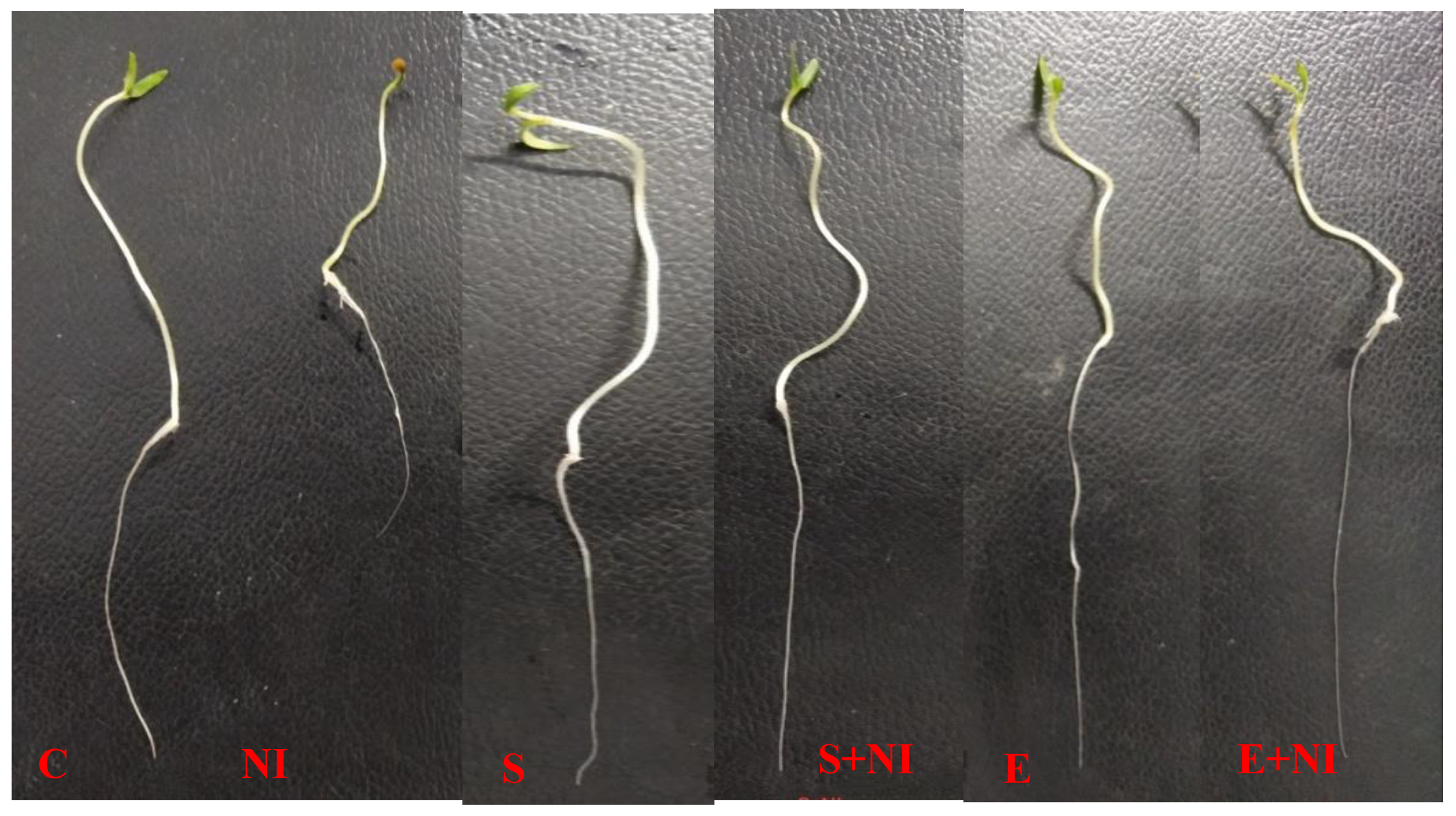
2.1. Morphological Parameters

The influence of S. hydrogenans strain DH-16 was investigated on various growth parameters like germination %, root length, shoot length, root weight, shoot weight and number of galls (Table 1). All these parameters except the number of galls showed a significant difference in the present study. Nematode infestation decreased the root length, shoot length, root weight and shoot weight by 22%, 5.4%, 13% and 5% respectively, as compared to control seedlings. However, plants that were pretreated with supernatant improved germination %, root length, shoot length, root weight and shoot weight of seedlings by 23%, 3.9%, 24%, 23% and 30.2%, respectively when compared with untreated nematode inoculated seedlings. Moreover, amendment of solvent extract also increased the germination %, root length, shoot length, root weight and shoot weight by 21%, 6%, 3%, 25% and 9% respectively as compared to the seedlings subjected to nematode infection. Supernatant and solvent extract pre-treated seedlings showed a reduced number of galls in roots by 19.9% and 16% respectively, when compared with untreated nematode inoculated seedlings (Table 1, Figure 1).

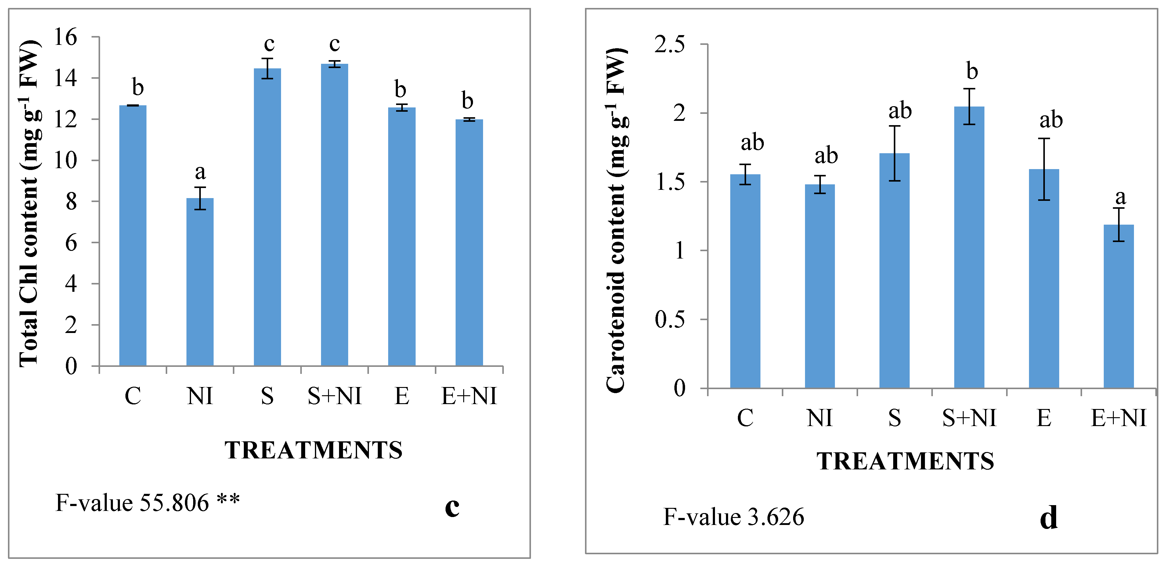
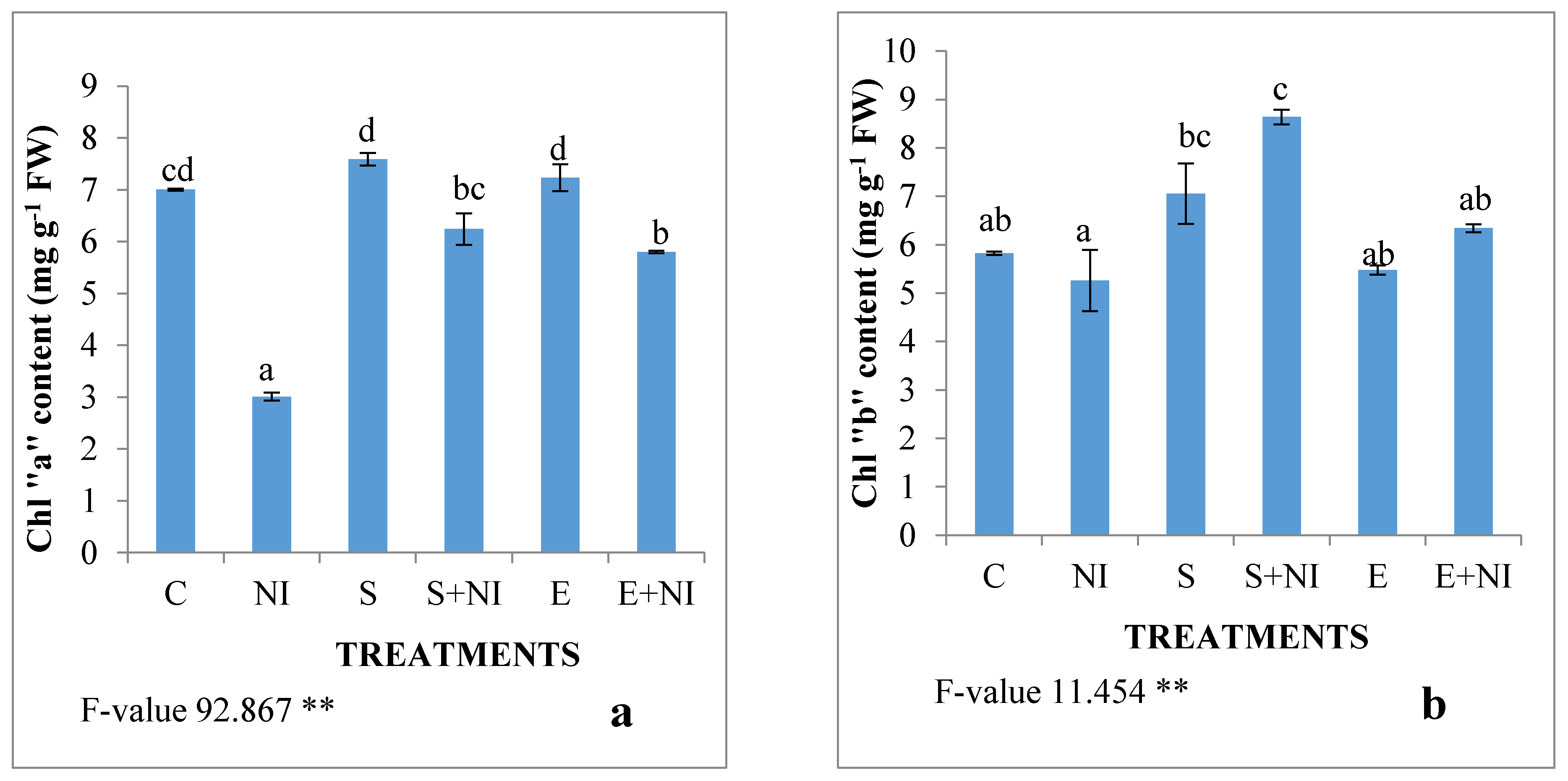
2.2. Effect of Biometabolites Produced by S. hydrogenans Strain DH-16 on the Photosynthetic Pigment of S. lycopersicum Seedlings 7 DAI

Photosynthetic pigments such as chlorophyll “a”, chlorophyll “b”, total chlorophyll and carotenoid content were assessed (Figure 2a–d). The content of all these pigments was reduced in nematode infested seedlings by 57%, 9.7%, 35.6% and 4% respectively when compared with the control seedlings. The application of the supernatant resulted into increased levels of all the pigments by 107%, 64%, 80% and 37% respectively. The seedlings pre-treated with solvent extract enhanced the levels by 92%, 20%, 47% and 20% respectively when compared with untreated nematode infested seedlings.

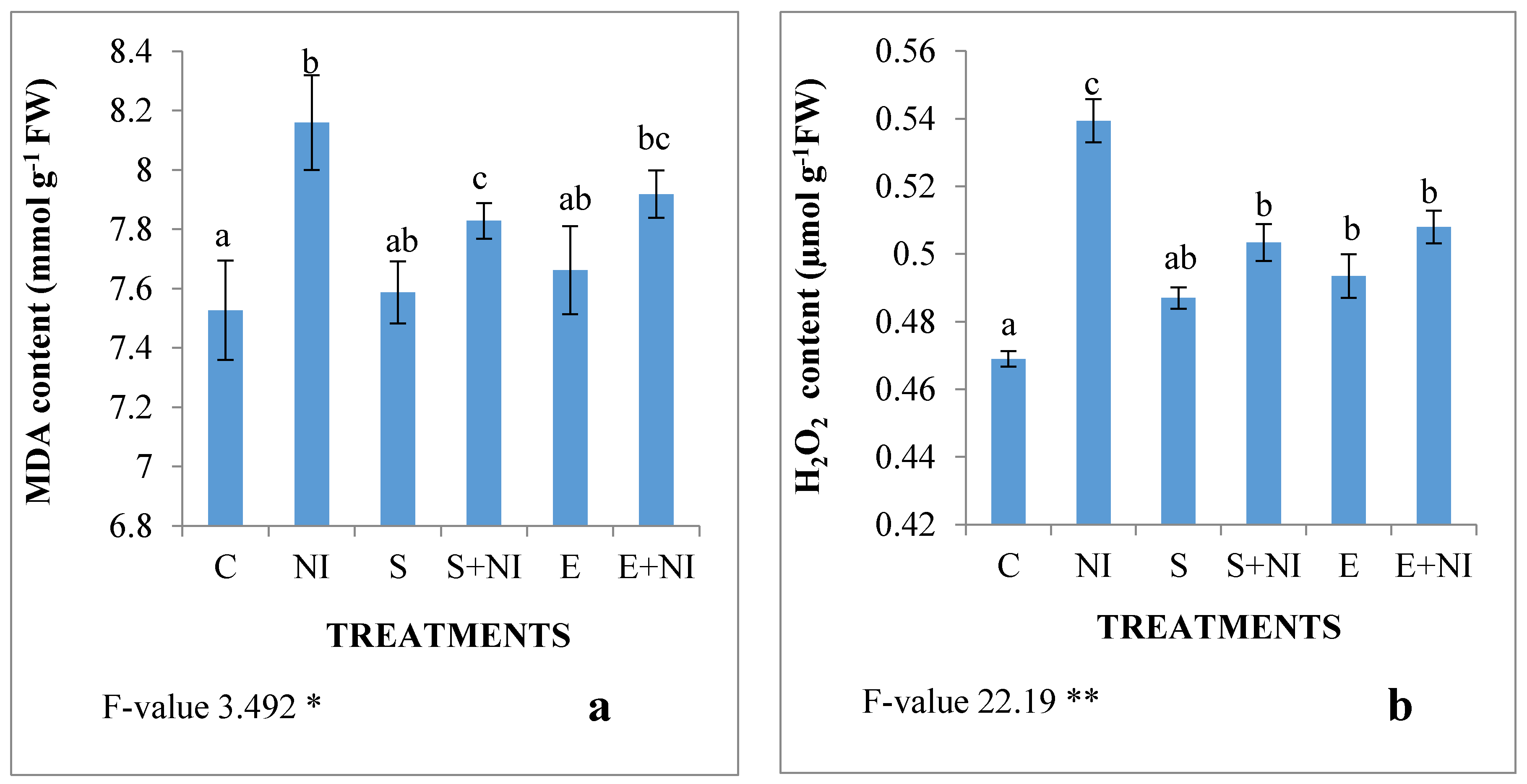
2.3. Malondialdehyde (MDA) and H2O2 Content

The oxidative damage in nematode infected seedlings was assessed in the form of hydrogen peroxide and MDA content. Nematode infestation resulted in the increased level of MDA by 8% and H2O2 by 15% (Figure 3a,b). Treatment of the supernatant lowered the contents of MDA by 4% and H2O2 by 7% respectively in nematode infected seedlings. However, treatment of solvent extract reduced the levels of MDA and H2O2 by 3% and 7.4% respectively.

2.4. Antioxidative Enzyme Activities

Different antioxidative enzymes like CAT, SOD, APOX, GuPOX, PPO and GST were also assessed and it was found that the specific activity of all these enzymes was enhanced in nematode infested S. lycopersicum seedlings by 8%, 33%, 37%, 15%, 56.6% and 40% respectively in comparison to control seedlings, whereas supernatant pretreated nematode infected seedlings showed further increase in the specific activity of all these antioxidative enzymes by 3%, 15%, 4%, 1%, 15% and 20% respectively when compared with nematode infested untreated seedlings.
Supplementation of solvent extract in nematode infested seedlings resulted in enhanced specific activities of CAT (1.9%), APOX (by 2%), PPO (by 1%), GPOX (by 14%) and GST (by 13.6%) when compared with nematode infected S. lycopersicum seedlings (Table 2).

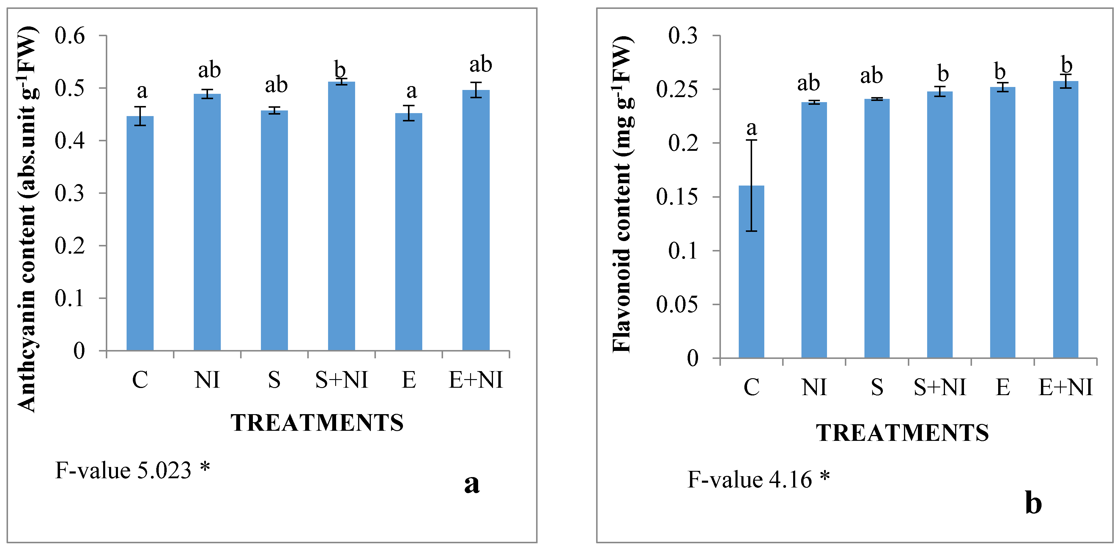
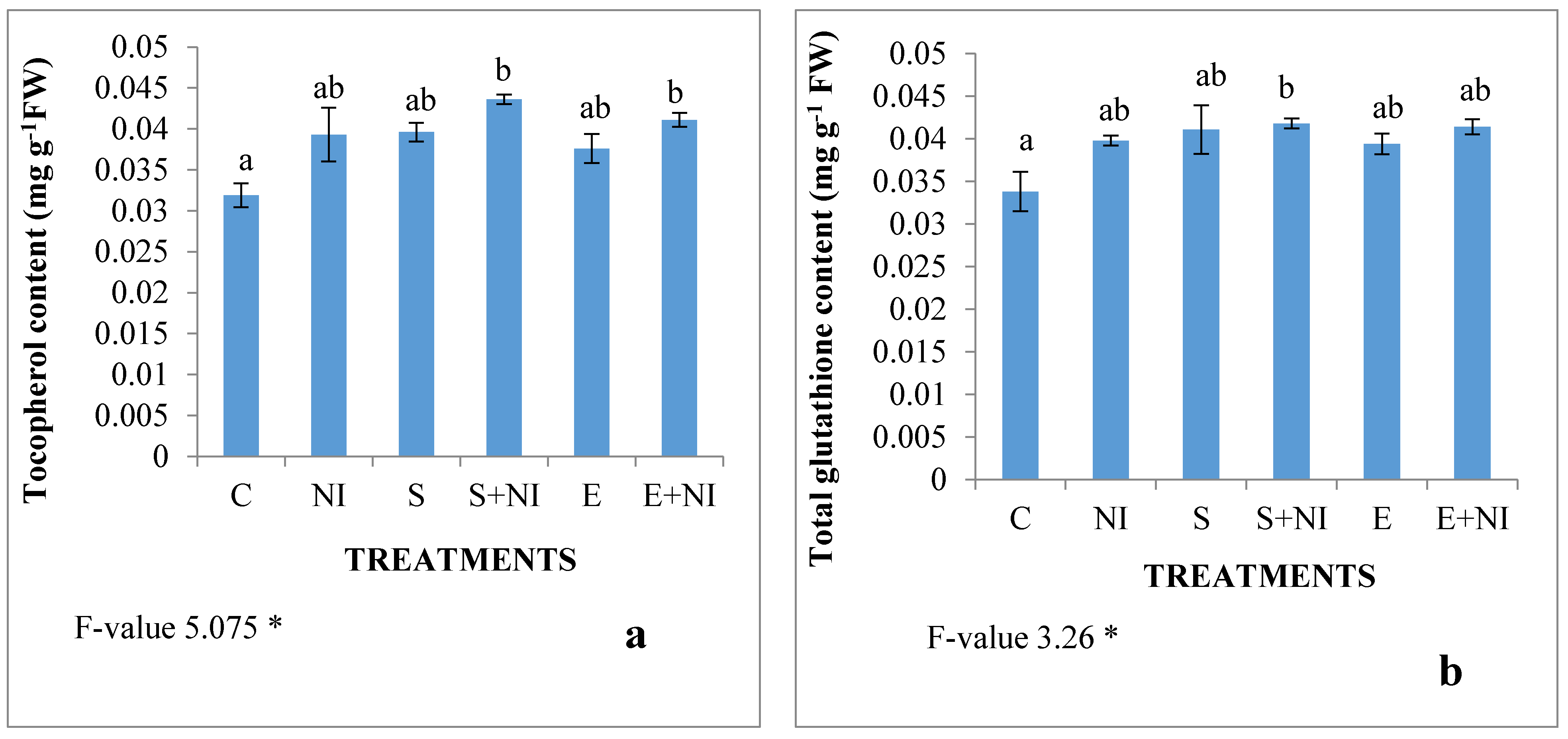
2.5. Non-Enzymatic Antioxidants

The levels of non-enzymatic antioxidants like glutathione and tocopherol content were assessed and were found to play an important function in nematode infested plants and the inoculation of biometabolites produced by microbes boosted their activities. The level of tocopherol and glutathione was enhanced by 23% and 18% respectively in nematode infected seedlings. These contents further increased in nematode infested seedlings that were pretreated with supernatant by 11% and 5% respectively, when compared with untreated nematode infected seedlings. However, the level of tocopherol and glutathione was enhanced in the nematode infected solvent extract treated seedlings by 12% and 4% respectively in comparison to the nematode contaminated S. lycopersicum seedlings (Figure 4a,b).

2.6. Phenolic Compounds

Effect of S. hydrogenans on phenolic compounds such as the anthocyanin content, flavonoid content was also studied in the present work. The levels of anthocyanins and flavonoids increased in the M. incognita infected seedlings by 9% and 48% respectively. Furthermore, in the supernatant treated nematode infested seedlings, the level of anthocyanin and flavonoid content increased by 5% and 4% respectively. Whereas, in the seedlings pretreated with solvent extract, the level of anthocyanin and flavonoid content was enhanced by 2% and 10% respectively, when compared with the untreated nematode inoculated seedlings (Figure 5a,b).

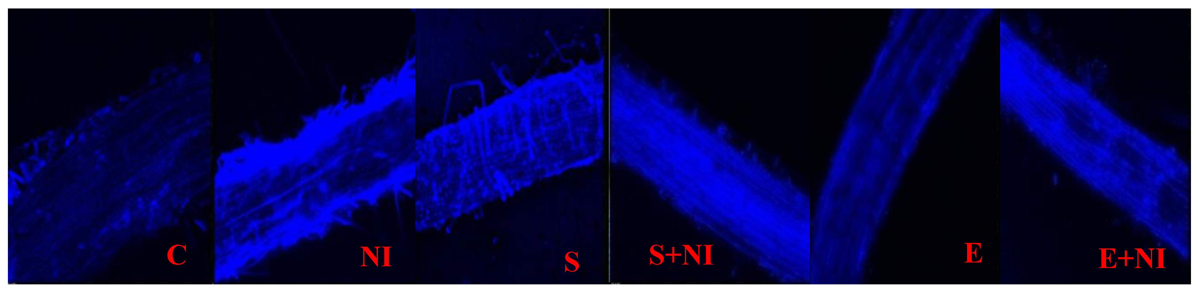
2.7. Confocal Microscopy

Tagging of glutathione was performed by dipping tips of roots of S. lycopersicum seedlings in MCB dye. Tagging was noticed on the basis of intensity of the blue colour in comparison to control seedlings. It was found that the root tissue of nematode infected seedlings revealed high intensity of blue colour, i.e., high level of glutathione content when compared to untreated control seedlings. Moreover, this intensity was further increased in the seedlings pretreated with biometabolites present in culture filtrate and extract, indicating the antioxidative features of biometabolites produced by S. hydrogenans strain DH-16 (Figure 6).

3. Discussion

The present study revealed a remarkable effect of biometabolites produced by S. hydrogenans strain DH-16 in mitigating nematode stress in S. lycopersicum seedlings. M. incognita had a negative impact on the morphological parameters of infected S. lycopersicum seedlings. In the presence of M. incognita, there was an overall decline in the length and weight of S. lycopersicum seedlings. Our studies are in agreement to the previous studies in which reduction in morphological attributes was depicted in Medicago truncatula plants. They observed an enhanced formation of galls over root surfaces due to nematode infestation [50], which in turn impaired the vascular transportation in plants. In addition, Venkatesan, et al. [51] observed a hindrance in the mineral uptake of Oryza sativa plants due to nematode infections. They concluded that nematodes clogged the vascular pathways of plants by their rapid multiplication within the tissues leading to overall impairment in their metabolic activities [51]. A similar decline in the fresh weight was observed in Ocimum basilicum plants infected with M. incognita [52]. According to them, nematode infestation had a negative impact on the uptake of nutrients by plants. The plants overcome such harsh situations via developing the lateral root elongation in order to increase the nutrient uptake from the soils [53]. This characteristic feature not only balances the nutritional homeostasis, but also restricts the nematodes towards outer zones [54]. The retardation of growth parameters in the present study could be due to the reason of root damage, which act as a hurdle in mineral absorption from the root system and their translocation towards the shoot, thus disrupting the complete physiology of plant. Moreover, earlier studies carried out by Gupta, et al. [7] in Withania somnifera also demonstrated similar results where morphological traits were reduced in terms of height as well as biomass. Our study also coincides with Khajuria and Ohri [55], Khanna, et al. [56] and Khanna, et al. [57] in tomato plants in terms of decreased root length, shoot length, fresh weight and dry weight on nematode infestations. They inferred that the blockage of pathways associated with nutrition and water transport via nematodes leads to retarded growth attributes [56,57].
Treatment with biometabolites produced by S. hydrogenans strain DH-16 in the form of the supernatant and extract resulted in a decreased number of galls, and stimulated root length, shoot length and fresh and dry weights of M. incognita infected S. lycopersicum seedlings. Similar results were found in cucumber plants pretreated with the culture broth of Streptomyces isolates NA-303 [58]. They found a remarkable suppression in the number of galls in microbe treated seedlings, directly influencing growth and development of plants. According to them, nematode motility causing substances were produced by Streptomyces isolates respectively. Our results were also in agreement with Khanna, et al. [56] and Khanna, et al. [57] who revealed that inoculations of Pseudomonas aeruginosa and Burkholderia gladioli promoted growth parameters in M. incognita infected S. lycopersicum plants and also reduced gall formation. They speculated that microbes restrained the nematode entry within the plant cells by forming a uniform layer over root surfaces to avoid their entry within the plants [56,57]. The most possible mechanism behind microbial mediated nematicidal activity in the present study could be due to production of toxic nematicidal compounds by these microorganisms, which caused nematode mortality and ultimately resulted in the plant growth promotion.
The results obtained from present study also determined a decline in the content of photosynthetic pigments in M. incognita infected S. lycopersicum seedlings. The similar reduction in the levels of pigments like chlorophyll, carotenoid and β-carotene was revealed earlier by Vasil’Eva, et al. [59]. This decline in plant pigments is mainly because of the inhibition of crucial enzymes required in the Violoxanthin pathway, this in turn impairs the stability of the photosynthetic apparatus [59]. Likewise, Sharma, et al. [18] reported lowered or disrupted photosynthetic pigments in tomato plants subjected to nematode infections. Reduction in the plant pigments are attributed to the formation of galls over root surfaces, leading to disrupted water transportation and chlorophyll a and b levels [60]. Our study also revealed inclined levels of photosynthetic pigments in S. hydrogenans treated S. lycoperscum seedlings infested with M. incognita. Our findings are supported from the previous studies conducted by Sharma, et al. [18] in tomato plants. They observed that plants treated with S. antibioticus strain M7 showed an enhancement in the levels of pigments due to triggered photosynthetic activities of plants. Moreover, a study reported by Jiang, et al. [61], also revealed that Trichosanthes kirilowii plants infested with nematodes showed an increase in pigment levels upon microbial inoculations. Another study reported by Khanna, et al. [57] suggested that stimulatory effects in their photosynthetic apparatus are due to microbe mediated upregulation in the enzymes responsible for pigment synthesis. Apart from this, they also concluded that the role of microbes in plant growth promotion in terms of nitrogen fixation, siderophore synthesis, etc., also leads to their better metabolic functions [57]. Furthermore, da Silva, et al. [62] observed better photosynthesis in nematode inhabited Pinus pinaster under the treatment of microbial strains. Present study upholds the fact that treated plants show better functions due to their ability to suppress nematode functioning or might be due to its ability to enhance enzymes associated with pigment production during nematode infection.
Our work also revealed an enhancement in the oxidative damage in nematode infected S. lycopersicum seedlings in the form of increased oxidative stress markers in terms of MDA and H2O2 content in M. incognita stressed plants. A similar increase in the level of MDA was also reported in case of sugar beet plants inoculated with nematodes [63]. Likewise, enhancement in the H2O2 content was also assessed by Sharma and Sharma [64] in tomato plants. They suggested that nematode infestation led to the generation of ROS in host plants. It is attributable to the pathogenicity of nematodes in the form of elevated ROS within stressed plants. Moreover, a tremendous increase in ROS species in the Bacopa monnieri plants has also been reported upon nematode infestations [39]. Along with this, studies conducted by Khanna, et al. [56] also formulated a paramount increase in oxidative stress markers in tomato plants infested with nematodes. However, the present work also assessed the role of microbes on oxidative damage in plants inhabited with nematodes. It was observed that the seedlings pretreated with biometabolites reduced the level of oxidative stress markers. Our findings were in agreement with Khanna, et al. [56] in S. lycopersicum plants treated with P. aeruginosa and B. gladioli who revealed declined levels of MDA and lipid peroxidation in plants. According to their study, this reduction is due to enhanced activities of defense enzymes in microbe inoculated plants [56]. Similarly, Gupta, et al. [39] conducted research in nematode infected B. monnieri plants inoculated with Streptomyces sp. and Chitinphilus sp. in which H2O2 and MDA content was found to be considerably reduced. They speculated that this decrease is mainly due to the production of immunity developed signals via microbes in order to combat pathogen accumulated within plants [39]. Furthermore, microbes possess the ability to scavenge the ROS produced by plants through the ability to secrete chelating molecules like organic acids, siderophores, and other secondary metabolites. Moreover, plants have the inbuilt capability to protect themselves from different stresses by inducing the activities of the antioxidative defense system in terms of enzymatic and non-enzymatic antioxidants [65]. The present study revealed an increase in the activities of various enzymes (CAT, SOD, APOX, GPOX, PPO and GST) in M. incognita infected S. lycopersicum seedlings. Similar results were also reported by Khajuria and Ohri [55] in the case of tomato plants, where activities of SOD, POD, CAT, GPOX, etc., were enhanced during nematode infection. As per the reports of Alscher, et al. [66] the activities of defensive enzymes occurred in response to increased oxidative stress markers in plants. In addition to this, enhancement in the activities of SOD, POD and CAT was also found in tomato plants under nematode influence [67]. This mainly relied on the basis of modulation in the defensive pathways. Our study also demonstrated a further incline in the enzymatic activities after the treatment with the biometabolites produced by S. hydrogenans in the form of the supernatant and extract. A rise in the activity of PPO was also reported by Ma, et al. [68] in Streptomyces sp. treated nematode infected tomato plants. According to their speculation, this inflation in enzymatic activity is correlated to tolerance towards the pathogenic response via systematic resistance [68]. Additionally, the results of Gupta, et al. [7] reported an increase in the activity of SOD in Withania somnifera plants, treated with Streptomyces sp. and Chitiniphilus sp. respectively, due to the internal immune response activation along with induced systemic resistance. The activities of CAT, SOD, PPO and GuPOX were also triggered in nematode infected tomato plants treated with P. aeruginosa and B. gladioli as revealed by Khanna, et al. [56]. The reason behind this remarkable increase in antioxidative enzyme activities might be due to the increased protein content in plants after supplementation of beneficial microbes, which further resulted in upregulation of these defense enzymes in the host plants.
In addition to antioxidative enzymes, non-enzymatic antioxidants like glutathione content and tocopherol content were also found to be enhanced in nematode infected S. lycopersicum seedlings. As depicted in the previous reports, glutathione content acts actively in reducing the contents of thiol-sulfide and sulphur present inside the cells and has also found to help the plants in mitigating abiotic and biotic stresses [69]. Tocopherol, mainly present in the membranes of chloroplast, acts as a lipid peroxyl scavenger in most of the plants [70]. The levels of these non-enzymatic antioxidants were enhanced in infected plants like tomato, eggplant, pea, papaya and grapevine under the incidence of nematode infection [56,57,70,71]. Moreover, an elevation in the levels of tocopherol was also observed in Helianthus annuus plants subjected to nematode infestation [72]. They found this enhancement in close linkage with the defense potential of plants against inhabiting nematodes [72]. Further, the glutathione and tocopherol levels were found to be enhanced in seedlings treated with biometabolites produced by S. hydrogenans. A study conducted by Khanna, et al. [56] also revealed similar trends in which nematode infected tomato seedlings demonstrated raised levels of non-enzymatic antioxidants after inoculation with P. aeruginosa and B. gladioli respectively. According to their perception, these increased levels might be due to the activation of antioxidants by soil microbes and in order to act as scavengers by reducing H2O2 to H2O [73,74]. This further reduces the oxidative stress from the cells through the growth promoting action of microbes in complicated situations. Moreover, the induction in the level of antioxidants in nematode stressed plants in the present study might be due to the fact that microbes boosted the internal immune system within the plants to form a shield against the adversities caused by RKNs.
The current study also determined an elevation in the levels of phenolic compounds like flavonoids and anthocyanins in M. incognita infected S. lycopersicum seedlings. It has been elucidated since previous times that phenols act as buffers to regulate the stress levels in plants [75]. Moreover, many different categories of phenolic compounds act as nematicidal elements to cope up with the RKN infections [76]. Along with this, they also possess the characteristics to subdue the gall formation in plants and maintain the proper regulatory actions of the plants [77]. Induction in the levels of phenols have been directly in proportion to the stress levels as it gets deposited onto the cell wall in order to toughen them against a pathogen attack [77]. Various studies revealed that enhancement in the levels of different phenolic compounds in tomato plants against nematicidal attack to counteract the responses [78]. Our study also found support by the findings of Treutter [79], who confirmed that this rise is associated with the phytopathogen infection. Apart from this, the enhanced level of phenolic compounds also reduces the infestation of pathogens within plants [5,80]. Our results also revealed that the level of these compounds was further increased in the seedlings pretreated with biometabolites produced by S. hydrogenans. This enhancement or accumulation might be linked with the biotic stress tolerance as indicated by a reduction in the nematode infestation by impairing gall formation. Our results were also on similar lines as with Gupta, et al. [7], in the case of nematode infected W. somnifera treated with Chitiniphilus sp., Streptomyces sp. and Cellulosimicrobium sp. respectively. They depicted that this increase in the phenolic compounds led to normal functioning of plants during a pathogenic attack. Additionally, inclined levels of phenolic compounds, flavonoids, anthocyanins and polyphenols were also found in nematode infested tomato plants under the incidence of a microbial treatment [57]. They also depicted an upregulation in the activities of genes encoding phenolic compounds that is PAL and CS during microbial treatment in infected plants. Their studies recapitulated that phenolic compounds triggered the immune responses within plants in order to combat the mechanical injuries caused to them by pathogens along with mediating normal metabolic activities of the plants [57]. The induction in the level of phenolic compounds such as anthocyanins and flavonoids in the present study might be due to the chelating action of these compounds against pathogenic responses. Moreover, the synthesis of these compounds also hinders the movement of nematodes, thereby, directly targeting them to cause their morbidity. All these observations indicated the efficiency of biometabolites produced by Streptomyces sp. In developing stress tolerance in M. incognita infected host plants, thereby, making them the perfect candidates as biocontrol agents against nematode infections.

4. Materials and Methods

Nematode culture: Culture of nematodes was collected from infected roots of tomato and brinjal from different sites of Guru Nanak Dev University campus. Females were isolated and identified. The identified culture was then used for maintaining the pure cultures in the glass house of the department and then used for further studies. Egg masses of M. incognita were extracted from the pure culture, washed with 1.5% sodium hypochlorite solution [81], and then subsequently washed with distilled water. These egg masses were then kept at temperature 26 ± 2 °C in a Biological Oxygen Demand (B.O.D) incubator (CALTAN, NSW, New Delhi, India) to stimulate hatching. After 2–3 days, second stage juveniles (J2) were collected and were counted using alight microscope and used for further experimental work.
Microbial inoculations: S. hydrogenans strain DH-16 (Gene Bank accession no. JX123130) was obtained from the Department of Microbiology, Guru Nanak Dev University, Amritsar (Punjab) [82]. The bacterial strain was prepared on starch casein nitrate agar (SCNA) plates at a temperature of 4 °C and was then stored at 4 °C for further studies.

4.1. Production of Secondary Metabolites

The bioactive metabolites production by S. hydrogenans strain DH-16 was carried out as described by Kaur and Manhas [82]. The production of metabolites was carried out in the Orbital shaker incubator at 28 °C temperature and 180 rpm. After incubation of 72 h under the required conditions, the culture broth was collected and centrifuged at 10,000 rpm at 4 °C for 20 min. The filterate/supernatant (free from cells) were used for experimental studies.
The bioactive metabolites in the filtrate were extracted using ethyl acetate and were concentrated using rota evaporator (IKA India Pvt. Ltd., RV 10 digital V, Bengaluru, India). The resulted brown coloured compound thus produced was redissolved in 0.5% DMSO and stored at 4°C for further experimental work.

4.2. Plant Material and Treatments

Surface sterilized S. lycopersicum seeds var. Pusa Ruby (susceptible variety) were soaked for 5 h in the supernatant (cell free filtrate) as well as in the crude extract (480 µg/mL). The seeds were grown in autoclaved petri plates lined with whatman filter paper grade 1. Each petri plate contained 25 seeds with treatment of 0.5% DMSO solution (control) and the crude extract (E) as well as the cell free supernatant (S). After germination, the seedlings were inoculated with 5 juveniles per seedling in all the treatments. The petri plates were kept in B.O.D incubator “CALTAN” (Super Deluxe Automatic) for 7 days with a light period of 16 h per day. After 7 days of nematode inoculation, the experiment was terminated and various morphological and physiological parameters were studied. For each set of experiment, treated and inoculated plants were compared with untreated, uninoculated plants as well as untreated, inoculated plants.

4.3. Morphological Parameters

After 7 days of nematode inoculation, various morphological parameters were studied. The parameters include percentage germination, root length, shoot length, root weight, shoot weight and number of galls.

4.4. Determination of Carotenoid and Chlorophyll Contents

Extraction of carotenoid and chlorophyll content was done using 80% acetone [83,84]. Fresh leaf tissue of seedlings was crushed in 80% acetone in the 1:4 ratio and then subjected to centrifugation at 4 °C at 10,000 rpm for 20 min. The supernatant was removed and its absorbance was recorded at 480 and 510 nm for carotenoid content and 645 and 663 nm for chlorophyll content using a UV-VIS spectrophotometer (GENESYS 108, Waltham, MA, USA).

4.5. Determination of Malondialdehyde (MDA) Content and H2O2 Content

MDA content was used for the measurement of lipid peroxidation [85]. Homogenate was prepared by crushing fresh plant seedling in 0.1% (w/v) trichloroacetic acid (TCA), followed by centrifugation at 4 °C at 5000 rpm. The supernatant thus produced was used for further analysis. Of the supernatant 1 mL was mixed with 6 mL of 20% (w/v) TCA containing 0.5% (w/v) TBA and was heated at 95 °C for 30 min in a water bath and then the reaction was terminated in ice cold conditions. The optical density of the supernatant was taken at 532 nm. The nonspecific absorbance was corrected by subtracting absorbance at 600 nm. The level of MDA content was determined by using 155 mM cm−1 as an extinction coefficient.
For H2O2 content, the homogenate was prepared by crushing 0.5 g whole seedling in 1% trichloroacetic acid (TCA) and centrifuged at 12,000 rcf at 4 °C for 15 min. For further analysis, 0.5 mL of supernatant was mixed with 0.5 mL potassium phosphate buffer of pH 7 (PPB) and 1 mL potassium iodide (KI, 1 M). The absorbance of the mixture was then measured at 390 nm. A standard curve of H2O2 was used for evaluation [86].

4.6. Protein Content and Antioxidative Enzymes

Sample preparation: Fresh S. lycopersicum seedlings were homogenised in 0.1 M potassium phosphate buffer having pH 7.0. Homogenate was then subjected to centrifugation at 13,000 rpm at 4 °C for 20 min. The protein content was estimated by using the Folin Ciocalteu phenol reagent [87] and bovine serum albumen (BSA) was used as a standard for the evaluation of protein content. The activities of various antioxidative enzymes were determined by standard protocols reported by Aebi [88] for catalase (CAT; E.C. 1.11.16), Kono [89] for superoxide dismutase (SOD; E.C. 1.15.1.1), Nakano and Asada [90] for ascorbate peroxidase (APOX; E.C. 1.11.11.1), Esterbauer, et al. [91] for polyphenol oxidase (PPO; E.C. 1.10.3.1), Pütter [92] for guaiacol peroxidise (GuPOX; E.C. 1.11.1.7) and Habig and Jakoby [93] for glutathione-S-transferase (GST; E.C. 2.5.1.18).

4.7. Non Enzymatic Antioxidants

Total glutathione content: Total glutathione content was determined using the method given by Sedlak and Lindsay [94]. The homogenate was prepared by crushing whole seedling in 50 mM Tris buffer (pH 10), followed by centrifugation at 12,000× g at 4 °C for 15 min, the supernatant thus formed was mixed with DTNB and methanol and was kept at room temperature for 15 min. Incubation was followed by centrifugation at 3000× g for 15 min and the absorbance of the supernatant was measured at 412 nm. A standard plot was generated using the known concentration of reduced glutathione for evaluation.
Tocopherol content: The homogenate was prepared in a similar way as in the case of the total glutathione content. The supernatant was mixed with absolute ethanol and double distilled water, and the mixture thus formed was shaken. Then xylene was added, followed by centrifugation at 3000× g at 4 °C for 10 min. Then the upper xylene layer was separated and was mixed with 2,4,6-tripyridyl-s-triazine and absorbance was measured at 600 nm. A standard plot was generated using the known concentration of tocopherol for evaluation [95].

4.8. Estimation of Phenolic Compounds

Flavonoid content: The homogenate was prepared by crushing fresh seedlings in 80% methanol, followed by centrifugation at 4 °C and at 10,000 rpm for 20 min. The supernatant thus formed was used for estimation of flavonoid content. The 100 µL of the supernatant was taken in a test tube and then with the addition of methanol final volume of 3 mL was made. To this mixture 100 µL of AlCl3·6H2O, 100 µL of 5% sodium potassium tartarate and 0.5 mL of distilled water were added sequentially. The final mixture thus formed was shaken vigorously and was then the absorbance was recorded at 415 nm after 30 min of incubation. A standard plot was generated using known concentration of rutin. [96].
Anthocyanin content: The anthocyanin levels were estimated by crushing fresh S. lycopersicum seedlings in acidified methanol that had been prepared by mixing methanol, distilled water and hydrochloric acid at a ratio of 79:20:1. The extract was kept overnight at 4 °C and was then centrifuged at 10,000× g for 15–20 min at 4 °C. The absorbance was read at 530 and 657 nm [97].

4.9. Confocal Microscopy

Confocal microscopy was used for studying glutathione content. For glutathione tagging, root tips of S. lycopersicum seedlings were dipped in 25 mM monochlorobimane (MCB) solution and were kept in dark conditions for 20 min and extra stain was then washed with deionized water [98]. The samples were further examined by a Nikon A1R confocal laser scanning microscope (CLSM, New York, USA) at an excitation wavelength of 351–364 nm and emission wavelength of 477 nm.

4.10. Statictical Analysis

All the calculations (one-way ANOVA and Tukey’s test) were performed by using SPSS software (IBM SPSS, Version 24.0, IBM Corp., Armonk, NY, USA) at p ≤ 0.01 and p ≤ 0.05 and by self-coded Microsoft office excel software.

5. Conclusions

The current study showed alterations in the morphological and physiological characteristics of M. incognita infected S. lycopersicum seedlings. S. hydrogenans strain DH-16 release substantial metabolites had nematicidal activity that also led to activation of defense parameters in the RKN infected host plants. Basically, metabolites produced by these microbes enhanced the stress tolerance level by modulating the defense responses. Moreover, our results also showed an increase in the growth parameters, photosynthetic pigments, phenolic compounds of the nematode infested plants treated with biometabolites in the form of the cell filterate (supernatant) and extract. Furthermore, these metabolites are involved in regulating the level of ROS under stress by elevating the activities of enzymatic and non-enzymatic antioxidants. As a result, the present study suggests that Streptomyces sp. play a vital role in biotic stress mitigation in S. lycopersicum seedlings. Thus, the biometabolites produced by S. hydrogenans strain DH-16 can be used as a potential agent for the management of M. incognita along with the benefit of increasing germination as well as growth parameters of infected plants. Moreover, further research is required to elucidate enhanced tolerance of host plants against nematodes by microbial strains.

Author Contributions

Conceptualization, N.S., R.K.M., R.B. and P.O.; methodology, N.S. and K.K.; software, J.A. and M.S.A.; validation, R.B., P.O. and P.A.; formal analysis, N.S. and P.O.; investigation, N.S.; resources, P.O.; data curation, P.O. an M.S.A.; writing—original draft preparation, N.S., K.K., J.A. and R.K.M.; writing—review and editing, P.O. and P.A.; visualization, R.B. and P.O.; supervision, P.O.; project administration, P.O.; funding acquisition, J.A., M.S.A. and P.A. All authors have read and agreed to the published version of the manuscript.

Funding

This research was funded by the Researchers Supporting Project Number (RSP-2020/193), King Saud University, Riyadh, Saudi Arabia. The authors are also funded by DST-PURSE scheme for providing financial assistance.

Acknowledgments

The authors would like to extend their sincere appreciation to the Researchers Supporting Project Number (RSP-2020/193), King Saud University, Riyadh, Saudi Arabia. We are also thankful to DST-PURSE scheme for providing financial assistance.

Conflicts of Interest

The authors declare no conflict of interest.

References

  1. Ferraz, L.C.; Brown, D.J. An Introduction to Nematodes: Plant Nematology; Pensoft, Sofia-Moscow: Sofia, Bulgaria, 2002. [Google Scholar]
  2. Sikora, R.A.; Fernandez, E. Nematode parasites of vegetables. Plant Parasit. Nematodes Subtrop. Trop. Agric. 2005, 2, 319–392. [Google Scholar]
  3. Nicol, J.; Turner, S.; Coyne, D.; Den Nijs, L.; Hockland, S.; Maafi, Z.T. Current nematode threats to world agriculture. In Genomics and Molecular Genetics of Plant-Nematode Interactions; Springer: Dordrecht, The Netherlands, 2011; pp. 21–43. [Google Scholar]
  4. Hussain, M.A.; Mukhtar, T.; Kayani, M.Z. Characterization of susceptibility and resistance responses to root-knot nematode (Meloidogyne incognita) infection in okra germplasm. Pak. J. Agric. Sci. 2014, 51, 309–314. [Google Scholar]
  5. Singh, A.; Jain, A.; Sarma, B.K.; Upadhyay, R.S.; Singh, H.B. Rhizosphere competent microbial consortium mediates rapid changes in phenolic profiles in chickpea during Sclerotium rolfsii infection. Microbiol. Res. 2014, 169, 353–360. [Google Scholar] [CrossRef] [PubMed]
  6. Singh, K.; Khan, A.; Khan, I.; Rizvi, R.; Saquib, M. Morphological and biochemical resposes of cow pea (cv. pusa barsati) grown on fly ash amended soil in presence and absence of Meloidogyne Javanica and Rhizobium leguminosarum. Ecoprint Int. J. Ecol. 2010, 17, 17–22. [Google Scholar] [CrossRef] [Green Version]
  7. Gupta, R.; Singh, A.; Srivastava, M.; Gupta, M.; Pandey, R. Augmentation of systemic resistance and secondary metabolites by chitinolytic microbes in Withania somnifera against Meloidogyne incognita. Biocontrol. Sci. Technol. 2016, 26, 1626–1642. [Google Scholar] [CrossRef]
  8. Adam, M.; Heuer, H.; Hallmann, J. Bacterial antagonists of fungal pathogens also control root-knot nematodes by induced systemic resistance of tomato plants. PLoS ONE 2014, 9, e90402. [Google Scholar] [CrossRef] [Green Version]
  9. Elhady, A.; Adss, S.; Hallmann, J.; Heuer, H. Rhizosphere microbiomes modulated by pre-crops assisted plants in defense against plant-parasitic nematodes. Front. Microbiol. 2018, 9, 1133. [Google Scholar] [CrossRef] [Green Version]
  10. Goellner, M.; Wang, X.; Davis, E.L. Endo-β-1, 4-glucanase expression in compatible plant–nematode interactions. Plant Cell 2001, 13, 2241–2255. [Google Scholar]
  11. Williamson, V.M.; Hussey, R.S. Nematode pathogenesis and resistance in plants. Plant Cell 1996, 8, 1735. [Google Scholar]
  12. Abad, P.; Favery, B.; Rosso, M.N.; Castagnone-Sereno, P. Root-knot nematode parasitism and host response: Molecular basis of a sophisticated interaction. Mol. Plant Pathol. 2003, 4, 217–224. [Google Scholar] [CrossRef]
  13. Celik, I.; Sogut, M.A.; Ozkaynak, E.; Doganlar, S.; Frary, A. Physical mapping of NBS-coding resistance genes to the Me-gene cluster on chromosome P9 reveals markers tightly linked to the N gene for root-knot nematode resistance in pepper. Mol. Breed. 2016, 36, 137. [Google Scholar] [CrossRef] [Green Version]
  14. Jang, J.Y.; Le Dang, Q.; Choi, Y.H.; Choi, G.J.; Jang, K.S.; Cha, B.; Luu, N.H.; Kim, J.-C. Nematicidal activities of 4-quinolone alkaloids isolated from the aerial part of Triumfetta grandidens against Meloidogyne incognita. J. Agric. Food Chem. 2014, 63, 68–74. [Google Scholar] [CrossRef] [PubMed]
  15. Bais, H.P.; Weir, T.L.; Perry, L.G.; Gilroy, S.; Vivanco, J.M. The role of root exudates in rhizosphere interactions with plants and other organisms. Annu. Rev. Plant Biol. 2006, 57, 233–266. [Google Scholar] [CrossRef] [PubMed] [Green Version]
  16. Hirsch, P.R.; Miller, A.J.; Dennis, P.G. Do root exudates exert more influence on rhizosphere bacterial community structure than other rhizodeposits? Mol. Microb. Ecol. Rhizosphere 2013, 1, 229–242. [Google Scholar]
  17. Hallmann, J. Plant Interactions with Endophytic Bacteria; CABI Publishing: New York, NY, USA, 2001. [Google Scholar]
  18. Sharma, M.; Jasrotia, S.; Ohri, P.; Manhas, R.K. Nematicidal potential of Streptomyces antibioticus strain M7 against Meloidogyne incognita. AMB Express 2019, 9, 168. [Google Scholar] [CrossRef] [Green Version]
  19. Ahmad, P.; Sarwat, M.; Sharma, S. Reactive oxygen species, antioxidants and signaling in plants. J. Plant Biol. 2008, 51, 167–173. [Google Scholar] [CrossRef]
  20. Zhang, F.; Shi, W.; Jin, Z.; Shen, Z. Response of antioxidative enzymes in cucumber chloroplasts to cadmium toxicity. J. Plant Nutr. 2003, 26, 1779–1788. [Google Scholar] [CrossRef]
  21. Kimura, S.; Sinha, N. Tomato (Solanum lycopersicum): A model fruit-bearing crop. Cold Spring Harb. Protoc. 2008, 2008, pdb. emo105. [Google Scholar] [CrossRef]
  22. de la Torre-González, A.; Navarro-León, E.; Albacete, A.; Blasco, B.; Ruiz, J.M. Study of phytohormone profile and oxidative metabolism as key process to identification of salinity response in tomato commercial genotypes. J. Plant Physiol. 2017, 216, 164–173. [Google Scholar] [CrossRef]
  23. Darekar, K.; Mhase, N. Assessment of yield losses due to root-knot nematode Meloidogyne incognita race 3 in tomato, brinjal and bittergourd. Int. Nematol. Netw. Newsl. (USA) 1988, 5, 7–9. [Google Scholar]
  24. Williamson, V.M.; Roberts, P.A.; Perry, R. Mechanisms and genetics of resistance. In Root-Knot Nematodes; CAB International: Wallingford, IA, USA, 2009; Volume 301. [Google Scholar]
  25. Weller, D.M. Biological control of soilborne plant pathogens in the rhizosphere with bacteria. Annu. Rev. Phytopathol. 1988, 26, 379–407. [Google Scholar] [CrossRef]
  26. Wang, C.; Hu, H.-J.; Li, X.; Wang, Y.-F.; Tang, Y.-Y.; Chen, S.-L.; Yan, S.-Z. Effects of varying environmental factors on the biological control of Meloidogyne incognita in tomato by Bacillus cereus strain BCM2. Biocontrol. Sci. Technol. 2018, 28, 359–376. [Google Scholar] [CrossRef]
  27. Zhou, L.; Yuen, G.; Wang, Y.; Wei, L.; Ji, G. Evaluation of bacterial biological control agents for control of root-knot nematode disease on tomato. Crop Prot. 2016, 84, 8–13. [Google Scholar] [CrossRef]
  28. Sayre, R.; Starr, M. Bacterial diseases and antagonisms of nematodes. In Nematode Pathology; Poinar, G.O., Jansson, H.B., Eds.; CRC Press: Boca Raton, FL, USA, 1988; Volume 1, pp. 69–101. [Google Scholar]
  29. Crawford, J.M.; Clardy, J. Bacterial symbionts and natural products. Chem. Commun. 2011, 47, 7559–7566. [Google Scholar] [CrossRef] [PubMed] [Green Version]
  30. Cuthbertson, A.; Blackburn, L.; Northing, P.; Luo, W.; Cannon, R.; Walters, K. Chemical compatibility testing of the entomopathogenic fungus Lecanicillium muscarium to control Bemisia tabaci in glasshouse environment. Int. J. Environ. Sci. Technol. 2010, 7, 405–409. [Google Scholar] [CrossRef] [Green Version]
  31. Larralde-Corona, C.P.; Santiago-Mena, M.; Sifuentes-Rincon, A.M.; Rodríguez-Luna, I.; Rodriguez-Perez, M.A.; Shirai, K.; Narvaez-Zapata, J.A. Biocontrol potential and polyphasic characterization of novel native Trichoderma strains against Macrophomina phaseolina isolated from sorghum and common bean. Appl. Microbiol. Biotechnol. 2008, 80, 167. [Google Scholar] [CrossRef]
  32. Monga, D. Effect of carbon and nitrogen sources on spore germination, bio-mass production and antifungal metabolites by species of Trichoderma and Gliocladium. Indian Phytopathol. 2012, 54, 435–437. [Google Scholar]
  33. Sharma, P.; Pandey, R. Biological control of root-knot nematode; Meloidogyne incognita in the medicinal plant; Withania somnifera and the effect of biocontrol agents on plant growth. Afr. J. Agric. Res. 2009, 4, 564–567. [Google Scholar]
  34. Anjos, É.C.T.d.; Cavalcante, U.M.T.; Gonçalves, D.M.C.; Pedrosa, E.M.R.; Santos, V.F.d.; Maia, L.C. Interactions between an arbuscular mycorrhizal fungus (Scutellospora heterogama) and the root-knot nematode (Meloidogyne incognita) on sweet passion fruit (Passiflora alata). Braz. Arch. Biol. Technol. 2010, 53, 801–809. [Google Scholar] [CrossRef]
  35. Li, H.-Y.; Yang, G.-D.; Shu, H.-R.; Yang, Y.-T.; Ye, B.-X.; Nishida, I.; Zheng, C.-C. Colonization by the arbuscular mycorrhizal fungus Glomus versiforme induces a defense response against the root-knot nematode Meloidogyne incognita in the grapevine (Vitis amurensis Rupr.), which includes transcriptional activation of the class III chitinase gene VCH3. Plant Cell Physiol. 2006, 47, 154–163. [Google Scholar]
  36. Vos, C.; Claerhout, S.; Mkandawire, R.; Panis, B.; De Waele, D.; Elsen, A. Arbuscular mycorrhizal fungi reduce root-knot nematode penetration through altered root exudation of their host. Plant Soil 2012, 354, 335–345. [Google Scholar] [CrossRef]
  37. Goyal, D.; Pandey, J.; Prakash, O. Role of plant growth-promoting Rhizobacteria (PGPR) in degradation of xenobiotic compounds and Allelochemicals. In Advances in PGPR Research; CAB International: Wallingford, IA, USA, 2017; Volume 330. [Google Scholar]
  38. Gupta, G.; Snehi, S.K.; Singh, V. Role of PGPR in biofilm formations and its importance in plant health. Biofilms Plant Soil Health 2017, 27, 27–40. [Google Scholar]
  39. Gupta, R.; Singh, A.; Ajayakumar, P.; Pandey, R. Microbial interference mitigates Meloidogyne incognita mediated oxidative stress and augments bacoside content in Bacopa monnieri L. Microbiol. Res. 2017, 199, 67–78. [Google Scholar] [CrossRef] [PubMed]
  40. Kaur, T.; Jasrotia, S.; Ohri, P.; Manhas, R.K. Evaluation of in vitro and in vivo nematicidal potential of a multifunctional streptomycete, Streptomyces hydrogenans strain DH16 against Meloidogyne incognita. Microbiol. Res. 2016, 192, 247–252. [Google Scholar] [CrossRef] [PubMed]
  41. Mehmood, U.; Inam-ul-Haq, M.; Saeed, M.; Altaf, A.; Azam, F.; Hayat, S. A brief review on plant growth promoting Rhizobacteria (PGPR): A key role in plant growth promotion. Plant Prot. 2018, 2, 77–82. [Google Scholar]
  42. Prasad, M.; Srinivasan, R.; Chaudhary, M.; Choudhary, M.; Jat, L.K. Plant Growth Promoting Rhizobacteria (PGPR) for Sustainable Agriculture: Perspectives and Challenges. In PGPR Amelioration in Sustainable Agriculture; Woodhead Publishing; Elsevier: Amsterdam, The Netherlands, 2019; pp. 129–157. [Google Scholar]
  43. Sidhu, H.S. Potential of plant growth-promoting rhizobacteria in the management of nematodes: A review. J. Entomol. Zool. Stud. 2018, 6, 1536–1545. [Google Scholar]
  44. Verma, R.K.; Sachan, M.; Vishwakarma, K.; Upadhyay, N.; Mishra, R.K.; Tripathi, D.K.; Sharma, S. Role of PGPR in sustainable agriculture: Molecular approach toward disease suppression and growth promotion. In Role of Rhizospheric Microbes in Soil; Springer: Singapore, 2018; pp. 259–290. [Google Scholar]
  45. Siddiqui, Z.A. PGPR: Prospective biocontrol agents of plant pathogens. In PGPR: Biocontrol and Biofertilization; Springer: Singapore, 2005; pp. 111–142. [Google Scholar]
  46. Borah, B.; Ahmed, R.; Hussain, M.; Phukon, P.; Wann, S.B.; Sarmah, D.K.; Bhau, B.S. Suppression of root-knot disease in Pogostemon cablin caused by Meloidogyne incognita in a rhizobacteria mediated activation of phenylpropanoid pathway. Biol. Control 2018, 119, 43–50. [Google Scholar] [CrossRef]
  47. Zhai, Y.; Shao, Z.; Cai, M.; Zheng, L.; Li, G.; Huang, D.; Cheng, W.; Thomashow, L.S.; Weller, D.M.; Yu, Z. Multiple modes of nematode control by volatiles of Pseudomonas putida 1A00316 from Antarctic soil against Meloidogyne incognita. Front. Microbiol. 2018, 9, 253. [Google Scholar] [CrossRef] [Green Version]
  48. Sahebani, N.; Hadavi, N. Induction of H2O2 and related enzymes in tomato roots infected with root knot nematode (M. javanica) by several chemical and microbial elicitors. Biocontrol. Sci. Technol. 2009, 19, 301–313. [Google Scholar] [CrossRef]
  49. Mostafanezhad, H.; Sahebani, N.; Nourinejhad Zarghani, S. Control of root-knot nematode (Meloidogyne javanica) with combination of Arthrobotrys oligospora and salicylic acid and study of some plant defense responses. Biocontrol. Sci. Technol. 2014, 24, 203–215. [Google Scholar] [CrossRef]
  50. Čepulytė, R.; Danquah, W.B.; Bruening, G.; Williamson, V.M. Potent attractant for root-knot nematodes in exudates from seedling root tips of two host species. Sci. Rep. 2018, 8, 1–10. [Google Scholar] [CrossRef] [PubMed] [Green Version]
  51. Venkatesan, M.; Gaur, H.; Datta, S. Effect of root-knot nematode, Meloidogyne graminicola on the uptake of macronutrients and arsenic and plant growth of rice. Vegetos 2013, 26, 112–120. [Google Scholar] [CrossRef]
  52. Tiwari, S.; Pandey, S.; Chauhan, P.S.; Pandey, R. Biocontrol agents in co-inoculation manages root knot nematode [Meloidogyne incognita (Kofoid & White) Chitwood] and enhances essential oil content in Ocimum basilicum L. Ind. Crop. Prod. 2017, 97, 292–301. [Google Scholar]
  53. Shane, M.W.; Lambers, H. Systemic suppression of cluster-root formation and net P-uptake rates in Grevillea crithmifolia at elevated P supply: A proteacean with resistance for developing symptoms of ‘P toxicity’. J. Exp. Bot. 2005, 57, 413–423. [Google Scholar] [CrossRef] [PubMed]
  54. Gilbert, G.; Knight, J.; Vance, C.; Allan, D. Acid phosphatase activity in phosphorus-deficient white lupin roots. Plant Cell Environ. 1999, 22, 801–810. [Google Scholar] [CrossRef]
  55. Khajuria, A.; Ohri, P. Exogenously applied putrescine improves the physiological responses of tomato plant during nematode pathogenesis. Sci. Hortic. 2018, 230, 35–42. [Google Scholar] [CrossRef]
  56. Khanna, K.; Jamwal, V.L.; Kohli, S.K.; Gandhi, S.G.; Ohri, P.; Bhardwaj, R.; Wijaya, L.; Alyemeni, M.N.; Ahmad, P. Role of plant growth promoting Bacteria (PGPRs) as biocontrol agents of Meloidogyne incognita through improved plant defense of Lycopersicon esculentum. Plant Soil 2019, 436, 325–345. [Google Scholar] [CrossRef]
  57. Khanna, K.; Jamwal, V.L.; Sharma, A.; Gandhi, S.G.; Ohri, P.; Bhardwaj, R.; Al-Huqail, A.A.; Siddiqui, M.H.; Marraiki, N.; Ahmad, P. Evaluation of the role of Rhizobacteria in controlling root knot nematode (RKN) infection in Lycopersicon esculentum plants by modulation in the secondary metabolite profiles. AoB PLANTS 2019, 11, 1–14. [Google Scholar] [CrossRef]
  58. Chubachi, K.; Furukawa, M.; Fukuda, S.; Matsumura, S.; Yanagisawa, T.; Itagawa, H.; Shimizu, T.; Nakagawa, A. Suppressive effects of antinematodal Streptomyces spp. on root-knot nematodes of cucumbers caused by Meloidogyne incognita. Biocontrol. Sci. 2002, 7, 25–29. [Google Scholar] [CrossRef]
  59. Vasil’Eva, I.; Vanyushkin, S.; Zinov’Eva, S.; Udalova, Z.V.; Bolychevtseva, Y.V.; Paseshnichenko, V. Photosynthetic pigments of tomato plants under conditions of biotic stress and effects of furostanol glycosides. Appl. Biochem. Microbiol. 2003, 39, 606–612. [Google Scholar] [CrossRef]
  60. Yang, C.-M.; Yang, M.-M.; Hsu, J.-M.; Jane, W.-N. Herbivorous insect causes deficiency of pigment-protein complexes in an oval-pointed cecidomyiid gall of Machilus thunbergii leaf. Bot. Bull. Acad. Sin. 2003, 44, 315–321. [Google Scholar]
  61. Jiang, C.-H.; Xie, P.; Li, K.; Xie, Y.-S.; Chen, L.-J.; Wang, J.-S.; Xu, Q.; Guo, J.-H. Evaluation of root-knot nematode disease control and plant growth promotion potential of biofertilizer Ning shield on Trichosanthes kirilowii in the field. Braz. J. Microbiol. 2018, 49, 232–239. [Google Scholar] [CrossRef] [PubMed]
  62. da Silva, M.N.; Pintado, M.; Sarmento, B.; Stamford, N.; Vasconcelos, M.W. A biofertilizer with diazotrophic bacteria and a filamentous fungus increases Pinus pinaster tolerance to the pinewood nematode (Bursaphelenchus xylophilus). Biol. Control 2019, 132, 72–80. [Google Scholar] [CrossRef]
  63. Korayem, A.; El-Bassiouny, H.; El-Monem, A.A.A.; Mohamed, M. Physiological and biochemical changes in different sugar beet genotypes infected with root-knot nematode. Acta Physiol. Plant. 2012, 34, 1847–1861. [Google Scholar] [CrossRef]
  64. Sharma, I.P.; Sharma, A. Physiological and biochemical changes in tomato cultivar PT-3 with dual inoculation of mycorrhiza and PGPR against root-knot nematode. Symbiosis 2017, 71, 175–183. [Google Scholar] [CrossRef]
  65. Kuźniak, E.; Skłodowska, M. Ascorbate, glutathione and related enzymes in chloroplasts of tomato leaves infected by Botrytis cinerea. Plant Sci. 2001, 160, 723–731. [Google Scholar] [CrossRef]
  66. Alscher, R.G.; Erturk, N.; Heath, L.S. Role of superoxide dismutases (SODs) in controlling oxidative stress in plants. J. Exp. Bot. 2002, 53, 1331–1341. [Google Scholar] [CrossRef]
  67. Nikoo, F.S.; Sahebani, N.; Aminian, H.; Mokhtarnejad, L.; Ghaderi, R. Induction of systemic resistance and defense-related enzymes in tomato plants using Pseudomonas fluorescens CHAO and salicylic acid against root-knot nematode Meloidogyne javanica. J. Plant Prot. Res. 2014, 54, 383–389. [Google Scholar] [CrossRef]
  68. Ma, Y.-Y.; Li, Y.-L.; Lai, H.-X.; Guo, Q.; Xue, Q.-H. Effects of two strains of Streptomyces on root-zone microbes and nematodes for biocontrol of root-knot nematode disease in tomato. Appl. Soil Ecol. 2017, 112, 34–41. [Google Scholar] [CrossRef]
  69. Jimenez, A.; Hernandez, J.A.; del Río, L.A.; Sevilla, F. Evidence for the presence of the ascorbate-glutathione cycle in mitochondria and peroxisomes of pea leaves. Plant Physiol. 1997, 114, 275–284. [Google Scholar] [CrossRef] [Green Version]
  70. Kesba, H.H.; El-Beltagi, H.S. Biochemical changes in grape rootstocks resulted from humic acid treatments in relation to nematode infection. Asian Pac. J. Trop. Biomed. 2012, 2, 287–293. [Google Scholar] [CrossRef] [Green Version]
  71. Afifi, A.; Al-Sayed, A.; Mahfoud, N.; Farahat, A. Enzymatic and non-enzymatic oxidants and antioxidants involved in defense mechanisms against root-knot, reniform and citrus nematodes in their hosts. Egypt. J. Agron. 2014, 13, 172–188. [Google Scholar] [CrossRef] [Green Version]
  72. Korayem, A.; Mohamed, M.; El-Ashry, S. Yield and oil quality of sunflower infected with the root-knot nematode, Meloidogyne arenaria. Int. J. Chem. Tech. Res. 2016, 3, 207–2014. [Google Scholar]
  73. Mollavali, M.; Bolandnazar, S.A.; Schwarz, D.; Rohn, S.; Riehle, P.; Zaare Nahandi, F. Flavonol glucoside and antioxidant enzyme biosynthesis affected by mycorrhizal fungi in various cultivars of onion (Allium cepa L.). J. Agric. Food Chem. 2016, 64, 71–77. [Google Scholar] [CrossRef]
  74. Ray, S.; Singh, V.; Singh, S.; Sarma, B.K.; Singh, H.B. Biochemical and histochemical analyses revealing endophytic Alcaligenes faecalis mediated suppression of oxidative stress in Abelmoschus esculentus challenged with Sclerotium rolfsii. Plant Physiol. Biochem. 2016, 109, 430–441. [Google Scholar] [CrossRef]
  75. Nayak, D. Effects of nematode infection on contents of phenolic substances as influenced by root-knot nematode, Meloidogyne incognita in susceptible and resistant brinjal cultivars. Agric. Sci. Dig. Res. J. 2015, 35, 163–164. [Google Scholar] [CrossRef]
  76. Mahajan, R.; Kaur, D.; Bajaj, K. Nematicidal activity of phenolic compounds against Meloidogyne incognita. Nematol. Mediterr. 1992, 20, 217–219. [Google Scholar]
  77. Malik, M.S.; Dhindsa, K.S.; Pal, V.; Verma, K.; Sangwan, N.K.; Bhatti, D. Nematicidal efficacy of substituted phenols, phenoxyacetic acid esters and hydrazides: A structure-activity relationship study. Nematologica 1989, 35, 366–370. [Google Scholar] [CrossRef]
  78. Patel, V.S.; Shukla, Y.; Dhruve, J. Influence of root knot nematode (Meloidogyne spp.) on phenolic acid profile in root of tomato (Solanum lycopersicum L.). Int. J. Curr. Microbiol. Appl. Sci. 2017, 6, 840–848. [Google Scholar] [CrossRef]
  79. Treutter, D. Significance of flavonoids in plant resistance and enhancement of their biosynthesis. Plant Biol. 2005, 7, 581–591. [Google Scholar] [CrossRef]
  80. Gupta, R.; Pandey, R. Microbial interference ameliorates essential oil yield and diminishes root-knot infestation in sweet basil under field conditions. Biocontrol. Sci. Technol. 2015, 25, 1165–1179. [Google Scholar] [CrossRef]
  81. Hussey, R.; Barker, K. A comparison of nematodes of collecting inocula for Meloidogyne spp., including a new technique Pl. Dis. Rep. 1973, 61, 328–331. [Google Scholar]
  82. Kaur, T.; Manhas, R.K. Antifungal, insecticidal, and plant growth promoting potential of Streptomyces hydrogenans DH16. J. Basic Microbiol. 2014, 54, 1175–1185. [Google Scholar] [CrossRef] [PubMed]
  83. Arnon, D.I. Copper enzymes in isolated chloroplasts. Polyphenoloxidase in Beta vulgaris. Plant Physiol. 1949, 24, 1–5. [Google Scholar] [CrossRef] [PubMed] [Green Version]
  84. Maclachlan, S.; Zalik, S. Plastid structure, chlorophyll concentration, and free amino acid composition of a chlorophyll mutant of barley. Can. J. Bot. 1963, 41, 1053–1062. [Google Scholar] [CrossRef]
  85. Heath, R.L.; Packer, L. Photoperoxidation in isolated chloroplasts: I. Kinetics and stoichiometry of fatty acid peroxidation. Arch. Biochem. Biophys. 1968, 125, 189–198. [Google Scholar] [CrossRef]
  86. Velikova, V.; Yordanov, I.; Edreva, A. Oxidative stress and some antioxidant systems in acid rain-treated bean plants: Protective role of exogenous polyamines. Plant Sci. 2000, 151, 59–66. [Google Scholar] [CrossRef]
  87. Lowry, O.H.; Rosebrough, N.J.; Farr, A.L.; Randall, R.J. Protein measurement with the Folin phenol reagent. J. Biol. Chem. 1951, 193, 265–275. [Google Scholar]
  88. Aebi, H. Catalase. In Methods of Enzymatic Analysis; Academic Press: New York, NY, USA, 1974; pp. 673–680. [Google Scholar]
  89. Kono, Y. Generation of superoxide radical during autoxidation of hydroxylamine and an assay for superoxide dismutase. Arch. Biochem. Biophys. 1978, 186, 189–195. [Google Scholar] [CrossRef]
  90. Nakano, Y.; Asada, K. Hydrogen peroxide is scavenged by ascorbate-specific peroxidase in spinach chloroplasts. Plant Cell Physiol. 1981, 22, 867–880. [Google Scholar]
  91. Esterbauer, H.; Schwarzl, E.; Hayn, M. A rapid assay for catechol oxidase and laccase using 2-nitro-5-thiobenzoic acid. Anal. Biochem. 1977, 77, 486–494. [Google Scholar] [CrossRef]
  92. Pütter, J. Peroxidases. In Methods of Enzymatic Analysis; Academic Press: New York, NY, USA, 1974; pp. 685–690. [Google Scholar]
  93. Habig, W.H.; Jakoby, W.B. Assays for differentiation of glutathione S-Transferases. Methods Enzymol. 1981, 77, 398–405. [Google Scholar] [PubMed]
  94. Sedlak, J.; Lindsay, R.H. Estimation of total, protein-bound, and nonprotein sulfhydryl groups in tissue with Ellman’s reagent. Anal. Biochem. 1968, 25, 192–205. [Google Scholar] [CrossRef]
  95. Martinek, R.G. Method for the determination of vitamin E (total tocopherols) in serum. Clin. Chem. 1964, 10, 1078–1086. [Google Scholar] [CrossRef]
  96. Lamaison, J.; Carnet, A. Contents of main flavonoids flowers Crataegeus monogyna Jacq and Crataegeus laevigata (Poiret DC) at different developmental stages. Pharm. Acta Helv. 1990, 65, 315–320. [Google Scholar]
  97. Mancinelli, A.L. Photoregulation of anthocyanin synthesis: VIII. Effect of light pretreatments. Plant Physiol. 1984, 75, 447–453. [Google Scholar] [CrossRef] [Green Version]
  98. Meyer, A.J.; May, M.J.; Fricker, M. Quantitative in vivo measurement of glutathione in Arabidopsis cells. Plant J. 2001, 27, 67–78. [Google Scholar] [CrossRef] [Green Version]
Figure 1. This figure depicts the change in morphometric parameters of tomato seedlings that were pretreated with a supernatant and extract. Different treatments are in order C (control), NI (nematode inoculated), S (supernatant), S + NI (supernatant + nematode inoculated), E (extract) and E + NI (extract + nematode inoculated).
Plants 09 01109 g001
Figure 2. Effect of the supernatant and extract containing biometabolites on the photosynthetic pigments (a) Chl “a”, (b) Chl “b”, (c) Total chl content and (d) Total Carotenoid content in 7-day-old L. esculetum nematode infected seedlings. Values are presented as means ± standard error (S.E.). F-values ** indicates significance at p ≤ 0.01 and * indicates significance at p ≤ 0.05. Various alphabets on the graphs indicate that the average mean values of treatments are significantly different according to Tukey’s multiple comparison test (C—Control, NI—Nematode, S—Supernatant, S + NI—Supernatant + Nematode inoculated, E—Extract, E + NI—Extract + Nematode inoculated).
Plants 09 01109 g002aPlants 09 01109 g002b
Figure 3. Effect of the supernatant and extract containing biometabolites on the oxidative damage (a) MDA and (b) H2O2 content in 7-day-old S. lycopersicum nematode infected seedlings. Values are presented as means ± standard error (S.E.). F-values ** indicates significance at p ≤ 0.01 and * indicates significance at p ≤ 0.05. Various alphabets on the graphs indicate that the average mean values of different treatments are significantly different according to Tukey’s multiple comparison test (C—Control, NI—Nematode inoculated, S—Supernatant, S + NI—Supernatant + Nematode inoculated, E—Extract, E + NI—Extract + Nematode inoculated).
Plants 09 01109 g003
Figure 4. Effect of the supernatant and extract containing biometabolites on the non-enzymatic antioxidants (a) Tocopherol and (b) Glutathione content) in 7-day-old S. lycopersicum nematode infected seedlings. Values are presented as means ± standard error (S.E.). F-values ** indicates significance at p ≤ 0.01 and * indicates significance at p < 0.05. Various alphabets on the graphs indicate that the average mean values of different treatments are significantly different according to the Tukey’s multiple comparison test (C—Control, NI—Nematode inoculated, S—Supernatant, S + NI—Supernatant + Nematode inoculated, E—Extract, E + NI—Extract + Nematode inoculated).
Plants 09 01109 g004
Figure 5. Effect of the supernatant and extract containing biometabolites on the phenolic compounds: (a) anthocyanin and (b) flavonoid content in 7-day-old S. lycopersicum nematode infected seedlings. Values are presented as means ± standard error (S.E.). F-values ** indicates significance at p ≤ 0.01 and * indicates significance at p < 0.05. Various alphabets on the graphs indicate that the average mean values of different treatments are significantly different at p < 0.5 according to the Tukey’s multiple comparison test (C—Control, NI—Nematode inoculated, S—Supernatant, S + NI—Supernatant + Nematode inoculated, E—Extract, E + NI—Extract + Nematode inoculated).
Plants 09 01109 g005
Figure 6. Confocal micrographs showing images of glutathione tagging in differently treated control (C), nematode inoculates (NI), supernatant (S), supernatant (S + NI), extract (E) and extract + nematode inoculated (E + NI) S. lycopersicum roots 7 DAI by staining them with MCB.
Plants 09 01109 g006
Table 1. Morphological parameters of S. lycopersicum 7 days after nematode inoculations (DAI) under different treatments of biometabolites produced by S. hydrogenans in the form of a supernatant and solvent extract. Different treatments are represented in tables as C (control), NI (nematode inoculated), S (supernatant), S + NI (supernatant + nematode inoculated), E (extract) and E + NI (extract + nematode inoculated).
TreatmentsParameters (Mean ± S.E.)
Germination %Root Length (cm)Shoot Length (cm)Root Weight (g)Shoot Weight (g)No. of Galls
C70.67 ± 1.453 a8.827 ± 0.361 bcd7.337 ± 0.130 a0.150 ± 0.012 ab0.5600 ± 0.021 a-
NI69.33 ± 1.202 a6.897 ± 0.529 a6.943 ± 0.135 a0.127 ± 0.008 a0.533 ± 0.015 a8 ± 0.333 a
S87 ± 1.527 b9.130 ± 0.460 cd7.9167 ± 0.202 ab0.170 ± 0.006 b0.693 ± 0.009 b-
S + NI85.67 ± 1.202 b7.173 ± 0.061 ab8.620 ± 0.399 b0.1633 ± 0.003 ab0.690 ± 0.023 b6 ± 1.202 a
E86.67 ± 1.333 b9.150 ± 0.105 d7.456 ± 0.059 a0.170 ± 0.100 b0.690 ± 0.006 b-
E + NI84 ± 1.732 b7.287 ± 0.525 abc7.157 ± 0.206 a0.153 ± 0.008 ab0.580 ± 0.012 a7 ± 0.575 a
F-value33.747 **7.440 **7.903 **3.733 *23.431 **52.235 (ns)
The results are presented in the form of mean ± standard error (S.E.) and F-value ** indicates significance at p ≤ 0.01, * indicates p ≤ 0.05 and ns indicates non-significant difference according to Tukey’s comparison test. Different letters a, b, c and d on table represents significant difference whereas the same letters represent no significant difference.
Table 2. Effect of biometabolites produced by S. hydrogenans strain DH-16 in the form of supernatant and solvent extract on S. lycopersicum seedlings 7 days after nematode inoculation. Different treatments are represented in tables as C (control), NI inoculated (nematode inoculated), S (supernatant), S + NI (supernatant + nematode inoculated), E (extract) and E + NI (extract + nematode inoculated).
TreatmentsParameters (Mean ± S.E.)
Catalase (CAT) (U/mg Protein)Superoxide Dismutase (SOD) (U/mg Protein)Ascorbate Peroxidise (APOX) (U/mg Protein)Polyphenol Oxidise (PPO) (U/mg Protein)Guaiacol Peroxidise (GuPOX) (U/mg Protein)Glutathione-S-Transferase (GST) (U/mg Protein)
C0.1249 ± 0.0195 a0.0030 ± 0.00028 a0.0651 ± 0.00451 a0.0119 ± 0.00040 a0.0534 ± 0.00266 a0.0471 ± 0.00375 a
NI0.1353 ± 0.0175 a0.0039 ± 0.00028 abc0.0886 ± 0.00426 b0.0138 ± 0.00048 a0.0827 ± 0.00632 b0.0659 ± 0.01176 ab
S0.1487 ± 0.0106 a0.0043 ± 0.00017 bc0.0809 ± 0.00330 ab0.0130 ± 0.00024 a0.0715 ± 0.00576 ab0.0591 ± 0.00452 ab
S + NI0.1407 ± 0.0133 a0.0045 ± 0.00032 c0.0918 ± 0.00070 b0.0140 ± 0.00033 a0.0954 ± 0.00796 b0.0791 ± 0.00279 b
E0.1357 ± 0.0042 a0.0032 ± 0.00010 ab0.0777 ± 0.00509 ab0.0128 ± 0.00083 a0.699 ± 0.00829 ab0.0503 ± 0.00321 ab
E + NI0.138 ± 0.00493 a0.0037 ± 0.00015 abc0.0908 ± 0.00474 b0.0139 ± 0.00016 a0.0950 ± 0.00111 b0.0753 ± 0.00890 ab
F-value0.355 (ns)6.632 **6.372 **3.174 (ns)7.452 **3.761 *
The results are presented in the form of mean ± standard error (S.E.) and F-value ** indicates significance at p ≤ 0.01, * indicates p ≤ 0.05 and ns indicates non–significant difference according to Tukey’s comparison test. Different letters a, b and c on table represents significant difference whereas the same letters represent no significant difference.

