Article

Substrate and Plant Genotype Strongly Influence the Growth and Gene Expression Response to Trichoderma afroharzianum T22 in Sugar Beet

1
Department of Biology, Lund University, Sölvegatan 35B, SE–223 62 Lund, Sweden
2
MariboHilleshög AB, Säbyholmsv. 24, 261 91 Landskrona, Sweden
3
Department of Plant Protection Biology, Swedish University of Agricultural Sciences, Box 102, SE–23053 Alnarp, Sweden
*
Author to whom correspondence should be addressed.
Plants 2020, 9(8), 1005; https://doi.org/10.3390/plants9081005
Submission received: 14 July 2020 / Revised: 29 July 2020 / Accepted: 3 August 2020 / Published: 7 August 2020
(This article belongs to the Special Issue Biostimulants in Plants Science)

Abstract

:
Many strains of Trichoderma fungi have beneficial effects on plant growth and pathogen control, but little is known about the importance of plant genotype, nor the underlying mechanisms. We aimed to determine the effect of sugar beet genotypic variation on Trichoderma biostimulation. The effect of Trichoderma afroharzianum T22 on sugar beet inbred genotypes were investigated in soil and on sterile agar medium regarding plant growth, and by quantitative reverse transcriptase-linked polymerase chain reaction (qRT-PCR) analysis for gene expression. In soil, T22 application induced up to 30% increase or decrease in biomass, depending on plant genotype. In contrast, T22 treatment of sterile-grown seedlings resulted in a general decrease in fresh weight and root length across all sugar beet genotypes. Root colonization of T22 did not vary between the sugar beet genotypes. Sand- and sterile-grown roots were investigated by qRT-PCR for expression of marker genes for pathogen response pathways. Genotype-dependent effects of T22 on, especially, the jasmonic acid/ethylene expression marker PR3 were observed, and the effects were further dependent on the growth system used. Thus, both growth substrate and sugar beet genotype strongly affect the outcome of inoculation with T. afroharzianum T22.

Graphical Abstract

1. Introduction

Trichoderma spp. are filamentous fungi that are commonly found in soil, rhizosphere, and foliar environments, and some species have been found to grow endophytically [1,2]. Strains of several species are beneficial to plants. They are used for both growth promotion and as biocontrol agents against a broad variety of diseases in crops, including cereals, oil seeds, and even trees [3,4,5,6]. Substantial growth promotion by Trichoderma harzianum strains has been observed for a number of horticultural species, such as petunia, chrysanthemum, tomato, and red pepper, including an up to 80% increase in root area and dry weight (DW) in cucumber seedlings [7,8]. Trichoderma can exert beneficial effects to plants by a combination of several mechanisms, including: competition with pathogens for space and nutrients, direct antibiotic damage to the pathogens (e.g., as part of mycoparasitism), eliciting plant pathogen defense systems, and directly stimulating plant growth and nutrient uptake [5,9]. Most of these interactions will demand active participation of both the plant and the Trichoderma. The direct fungal attack on pathogens involves secretion of enzymes and metabolites, including peptaibols that are membrane-permeabilizing peptides [10]. Sterile-grown plant cells have been shown susceptible to permeabilization by the model peptaibol alamethicin [11] from Trichoderma viride (reidentified as T. arundinaceum) [12]. However, pre-exposure to Trichoderma cellulase elicits the plant cell to modify its plasma membrane, rendering it alamethicin-resistant [13,14]. Similarly, the direct interaction between roots and Trichoderma hyphae involves local plant formation of novel structural and metabolic components, plant recognition of the fungal elicitors, the global orchestration of plant pathogen defense, especially via jasmonic acid (JA) and ethylene (ET) response pathways, and modification of the plant nutrient uptake processes and hormonal systems for growth regulation [3,5,15]. Examples of plant growth promotion cases investigated include the interaction of Trichoderma afroharzianum T22 and Trichoderma asperelloides T203 (both previously called Trichoderma harzianum) with maize and cucumber, respectively [8,16,17,18,19], and the interaction of Trichoderma virens and Trichoderma atroviride with Arabidopsis thaliana [20]. In the most studied system, the interaction of T22 and maize, the growth effects are, however, variable, indicating an incomplete understanding of the factors governing the outcome for the plant [5]. Also, tomato and lentil cultivars have displayed variable growth stimulation and inhibition by Trichoderma [21,22]. Overall, the present knowledge indicates that a large number of plant functions will be important for the outcome of plant-Trichoderma co-cultivation.
Sugar beet (Beta vulgaris) is an important crop, which is related to the crops quinoa and spinach within the amaranth family. It is relatively salt and drought tolerant, probably due to its origin from the beach plant Beta maritima, yet is subject to a range of diseases [23]. The diploid genome has been sequenced and found to be relatively small for a crop [24], making sugar beet an attractive species for biomolecular investigations. Research on Trichoderma effects on sugar beet has focused on describing induced resistance to pathogens that are common in sugar beet agriculture. Field trials have shown an increased resistance to leaf spot disease, caused by the fungus Cercospora beticola, and amelioration of damping-off and root rot infections after Trichoderma spp. treatment [25,26,27]. However, the direct effect of Trichoderma on growth in sugar beet has not been studied.
As an initial step for characterizing sugar beet–Trichoderma interaction, this work investigated whether there is a variable effect of the commonly used single strain biocontrol and biostimulant agent T. afroharzianum T22 on inbred genotypes of sugar beet, and if gene expression in response to T22 differs between genotypes. We report a pronounced effect of the sugar beet genome on the outcome of the interaction, including both stimulation and inhibition of plant growth, and that absolute changes also depended on the plant growth systems used. Two lines, with different growth responses to T22, were further shown to have a pronounced variation in gene expression responses for marker genes for different pathogen response pathways. Similar to growth outcome, the gene responses also depend on the plant growth substrate used.

2. Results

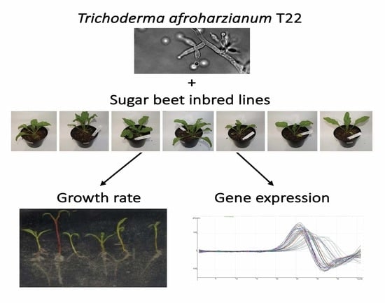
2.1. Presence of Trichoderma Promotes or Decreases Sugar Beet Growth in Soil

Seven inbred sugar beet genotypes, representing a wide variation in susceptibility to various pathogens, were grown in soil and treated with T. afroharzianum T22 conidiospores. A general trend of increased total, shoot, and root biomass accumulation was observed (Figure 1A), but without an obvious difference in plant vegetative morphology (Figure 1B). However, a 1.8-fold difference (0.85 in log2 units) between genotypes in the effect of T22 on the total DW was observed. Specifically, one line, designated F, displayed a negative effect by T22 treatment on total and shoot DW, and deviated significantly from three genotypes (D, G, and I), which were positively affected by T22. Three genotypes showed no tendency to be affected by T22. Root DW displayed a similar trend as shoot and total DW, though differences were not significant. Root length was unaffected by T22 (Figure 1). T22 also had no effect (less than 13%) on shoot to root DW ratio in any of the lines.

2.2. Roots of Genotype A and F Show a Difference in Gene Response to T22

For investigating potential differences in gene responses, we grew sugar beet seedlings of lines A and F on a sand/loam mix and treated with germinated T22 conidiospores for 8 h. The sand/loam mix was chosen because it could be rapidly removed by washing (<30 s per plant), allowing RNA isolation from relatively unperturbed roots without contaminating soil. Sugar beet orthologues to marker genes for different pathogen response pathways characterized in A. thaliana [28,29] were identified by two-way BLASTP searches (Table 1). The investigated genes included markers for the salicylic acid (SA) pathway (PATHOGENESIS-RELATED1; PR1), and genes associated with the ET (RELATED TO AP2–3; RAP2–3), JA (LIPOXYGENASE2; LOX2), and JA/ET (PATHOGENESIS-RELATED3/BASIC CHITINASE; PR3/CHIB) pathways.
Genotypes A and F were found to have similar transcript levels for all four pathogenesis-associated genes under control conditions (Figure 2). For most of the genes, treatment with T22 did not induce changes; minor upwards trends (27–60%) for PR1 in line A and PR1, RAP2–3, and LOX2 in line F were not significant. However, whereas genotype A maintained PR3 mRNA levels similar to the control also after T22 treatment, the PR3 transcript disappeared after T22 treatment of genotype F (Figure 2). The transcript could no longer be detected by real-time or by endpoint RT-PCR analysis.

2.3. T22 Decreases the Growth of Sugar Beet Seedlings in Sterile Culture

To study the plant–fungal interaction in isolation, we grew sugar beet seedlings sterile on agar and treated with T22 by root neck application of germinated conidiospores. Generally, across the genotypes, a negative effect of T22 was observed for the fresh weight (FW) of roots and shoots and for lengths of root and hypocotyl (Figure 3). The T22-induced inhibition of leaf and root FW was significant in most lines, and quantitatively varying between approximately 30% (line F) and 50% (line A). Root length was lowered by 30–50%, except for in two lines (F and G) whose root lengths were unaffected (Figure 3). Hypocotyl length was significantly decreased in genotypes B and G.

2.4. Genotypes A and F Are Similarly Colonised by T22

Differences in T22 effects on sugar beet genotypes may be caused by different abilities for the roots to harbor T22. We therefore investigated root colonization by visual estimation of adhering hyphae. The sugar beet lines A and F were analyzed in the sterile growth system over 8–48 h after treatment with germinated conidiospores of T22. Fungal hyphae were visible in all parts of the roots (Figure 4), except for the growing root tip, and the presence of hyphae increased during the time of co-cultivation. However, the two genotypes were colonized to a similar extent by the T22.

2.5. Sugar Beet Genotypes Show Major Differences in Gene Expression Responses Upon Sterile Co-Cultivation with T22

We extracted total RNA from roots of sterile-grown seedlings 8–48 h after root neck treatment with germinated T22 conidiospores or water control. A major genotype-specific difference in gene expression responses was observed in the sugar beet seedlings. Genotype A displayed a rapid and significant upregulation of PR3 and a slower downregulation of PR1, leading to a more than a 5-fold change in the PR3 to PR1 ratio 48 h after the application of T22 (Figure 5). In contrast, genotype F displayed a small and slow increase in PR3 expression in T22-treated roots as compared to the control (circa 50% over 48 h), and no decrease in PR1, though the control expression was higher in line F than line A (p < 10−4). Furthermore, a small and slow decrease in RAP2–3 (circa 50%; p < 10−3) was observed in control but not T22-treated genotype F, whereas line A was unaffected.
The results thus show that in sterile culture, roots of the two sugar beet genotypes have pronounced differences in the expressional changes of pathogen response genes induced by the presence of T. afroharzianum T22.

3. Discussion

A large number of Trichoderma species occur naturally in soils, and colonize roots of both monocot and eudicot plants [2]. Application of particular Trichoderma strains offers pathogen protection and will frequently also improve growth and performance of both healthy plants and plants under biotic or abiotic stress [1,3,5,6,9,28,31]. However, a variation between plant genotypes in the growth effect triggered by T22 has been observed, e.g., in maize. Specifically, one maize genotype showed seed yield reductions of up to 7% by T22 application in field trials [5]. Another genotype displayed up to a twofold increase in root and shoot length, although biomass accumulation was not specified [16]. In tomato, four inbred cultivars were compared for the growth effect of T22 and Trichoderma atroviride P1 [21]. Positive and negative effects on biomass accumulation were observed, and T22 and P1 affected individual tomato lines differently, with up to twofold differences in the T22 effect on shoot or root DW being observed.
We here found that treating soil-grown sugar beet plants with T22 affected growth (DW accumulation) of shoot and root (Figure 1), in agreement with previously published results on Trichoderma-enhanced plant growth in other plant species [5,7]. The response in sugar beet varied 1.8-fold between the different inbred genotypes, with one line (F) being growth-inhibited and markedly varying from three stimulated lines, and three lines being unaffected. In contrast to maize [5], the variation in the T22 effect on sugar beet biomass was not reflected in root length effects (Figure 1), suggesting instead a change in root architecture in the sugar beet. Trichoderma effects on DW accumulation in tomato were not coupled to changes in root length [21], and a similar case was seen in the sugar beet data (Figure 1). However, whereas Trichoderma induced different, or even reverse, effects on the shoot and root DW in individual tomato lines [21], the sugar beet data presented here (Figure 1) indicate similar T22-effects on shoot and root. Taken together, the variation in growth response to Trichoderma between plant genotypes that was earlier described for maize and tomato was also observed in sugar beet, but the patterns of response to Trichoderma differ between the plant species, and the negative effect in genotype F is stronger than previously reported. Putative mechanisms for decreased plant growth include cellular damage by peptaibols [11,14] or hydrolytic enzymes [32] released by Trichoderma, adverse effects of hormone-mimicking substances [33], or resource allocations to pathogen response pathways induced by the Trichoderma priming of plant immunity [34,35], and it is presently not possible to distinguish between these factors.
The above results were obtained using non-sterile growth systems that, besides Trichoderma and the plant, will contain various other microorganisms, even if using steam-treated soil. The growth effects could therefore be indirect and reflect Trichoderma effects on other microorganisms. This might be a reason why soil-grown sugar beet genotypes were generally stimulated by T22 (Figure 1), whereas sterile-grown sugar beet was mainly inhibited (Figure 3). Independent of the direction of the effects, however, the sterile system confirmed the major conclusion from the soil system, i.e., that the genotypes differed in their response to T22.
Sterile conditions have also been used when co-cultivating Arabidopsis, tomato, and cucumber with Trichoderma spp. [20,30,36]. In these experiments, growth promotions were found, again showing the variability of responses in different systems. Besides the different plant species and Trichoderma strains used, which may explain much of the variability, the inoculation methods cannot be neglected. In our investigations, a germinated conidiospore suspension was added directly to the root neck to allow analyses at early and defined time points after application, whereas in work by others, Trichoderma usually was applied at some distance below the growing root tip, also allowing plant–fungal interaction via growth-stimulating volatiles, as previously suggested [37]. In the sterile growth experiments, a small inoculum of 10–20 colony-forming units (cfu) per seedling was used and shown to be efficient when applied directly to the root neck (Figure 3). Direct application of similar numbers of cfu of T. atroviride has been shown to affect gene expression in Arabidopsis, but growth rates were not reported [38].
The presence of pathogens and beneficial microbes can induce mechanisms such as systemic acquired resistance (SAR) or induced systemic resistance (ISR). Changes in SA, JA, and ET response pathways upon Trichoderma treatment have been observed in multiple plant species, including, for example, A. thaliana, tomato, melon, cucumber, and grapevines [17,21,28,35,39,40]. In sugar beet (Figure 5), orthologues to A. thaliana markers for the SA (PR1), ET (RAP2–3), JA (LOX2), and JA/ET (PR3/CHIB) response pathways displayed variant T22-responses in two genotypes, and also depended on the plant growth substrate used. Specifically, the increase in the PR3 chitinase gene in genotype A after treatment with T22 indicates a rapid activation of the JA/ET pathway, which is lacking in T22-treated genotype F (Figure 5). In sand-grown plants, the PR3 transcript is unchanged by T22 in genotype A, yet disappears completely in genotype F (Figure 2). Thus, the relative T22 effect on the expression of PR3 is relatively similar in the plant genotypes (i.e., after T22 treatment, the PR3 expression is for both growth substrates higher in genotype A than in genotype F), yet the overall orientation of the signal changes differs between the two growth substrates. In conjunction with the opposite T22 effects on growth (Figure 1 and Figure 3), the observed gene expression changes indicate that T22 has a stronger damaging effect on the plants in the sterile system. Regarding growth effects, a similar case of damage in sterile growth systems has been observed in the sugar beet relative Chenopodium quinoa [37]. Trichoderma applied to soil for two months induced both SA and JA marker genes in tomato leaves, but relatively little in the most growth-promoted genotypes [21], consistent with a pattern where the pathogen response genes are most highly expressed under conditions where plants are little stimulated, or even inhibited, by Trichoderma.
PR1 and PR3 are both antifungal proteins and for both, examples of enhanced pathogen resistance upon transgenic transfer between plant species have been reported [41]. Thus, changes in the expression of these genes (Figure 2 and Figure 5) may be involved in the different growth responses to T22 displayed by the sugar beet lines (Figure 1 and Figure 3). Since PR1 and PR3 are also reporters for the SA and JA/ET pathways, respectively [41], associated response genes may also produce a differential effect. PR3 expression depends on the action of several other response genes, and SA markedly inhibits it [41,42,43]. These mechanisms may have a part in causing the shifts observed in PR3 and PR1 (Figure 2 and Figure 5), consistent with the generally observed antagonism between SA and JA/ET pathways in regulating SAR and ISR responses to pathogens [3,29]. Differential effects have also been reported for beneficial microbes, e.g., application of T. asperelloides T203 to roots of sterile-grown cucumber seedlings induced root and leaf expression of genes involved in JA/ET signaling without affecting SA [17]. Consistently, inoculation of tomato seedlings with spores of T. virens TriV_JSB100 displayed a specific induction of the JA marker gene PDF1, similarly in two cultivars [44]. However, treatment of A. thaliana sterile-grown seedlings with T. virens induced both SA and JA marker gene reporter constructs over an 8-day period in leaves [45], as has also been observed with other growth-promoting soil microorganisms, such as Bacillus cereus AR156 [46]. In contrast, treatment of A. thaliana roots with T. hamatum T382 activated SA-induced genes in leaves after 2 days, but JA/ET pathway genes only after multiple treatments [35]. The differences in responses between plant species are, however, confounded by experimental variations in time of treatment and analysis. For example, T. atroviride-induced responses of SA, JA, and ET markers in tomato vary over time [47], and can even reciprocally change direction of induction and suppression during treatment [48]. SA, JA, and ET pathway sensitivity may also differ between roots and leaves, and be affected by the site of microbial application [49,50]. Especially, transcriptional changes in roots have been little analyzed, and then only in sterile systems, probably due to the technical difficulties of analyzing roots in soil. The observations presented here, where sugar beet growth and root gene expression responses to Trichoderma are highly dependent on both growth substrate and plant genotype, is a step forward towards explaining the variation observed in Trichoderma effects, and emphasize a need for more strict and natural experimental systems.
Several plant processes are likely involved in governing the stimulation and inhibition of plant growth. Genotypes with contrasting responses may upon molecular analyses allow mechanistic characterization of the response to Trichoderma. This may also lay the ground for crop breeding to enhance Trichoderma stimulation, thus avoiding the variability in growth response that has been found for different plants and growth conditions.

4. Materials and Methods

4.1. Biological Material

A set of sugar beet inbred genotypes (MariboHilleshög AB, Landskrona, Sweden) were selected to constitute a diversity in pathogen resistance and sensitivity, and were given a random identification letter. After excluding lines with low germination or slow or aberrant growth, seven genotypes were included in soil experiments. Preferences for the genotypes used, according to Hilleshög AB, were as follows; Genotype A is susceptible to plant pathogens, especially to Fusarium spp.; Genotype B is mildly resistant to Aphanomyces spp.; Genotype D is resistant to Aphanomyces spp.; Genotype F is susceptible to beet yellows virus; Genotype G is generally susceptible to plant pathogens; Genotype H is resistant to Rhizomania and Cercospora spp.; Genotype I is resistant to Rhizomania.
Trichoderma afroharzianum T22 (Rifai; ATCC20847) was obtained from ATCC (Manassas, VA, USA) and grown and maintained on potato dextrose agar, according to instructions (ATCC). For obtaining conidiospore suspensions, sterile distilled water was added to 2-week-old sporulating cultures of T. afroharzianum T22. The suspension was gently scraped from the agar surface, filtered through a cotton ball, and washed twice by centrifugation (2850 g for 5 min in 50 mL tubes in a table-top swing-out centrifuge) and resuspension in water. Conidiospores were germinated by incubation in a synthetic medium [51] for 18 h at 30 °C with 150 rpm shaking in a TR−225 orbital shaker (Infors, Bottmingen, Switzerland). The germinated conidiospores were washed and resuspended in water as above, and then directly used for inoculation. The procedure was carried out at 25 °C.

4.2. Greenhouse Growth

Sugar beet fruits, including pericarp, were consecutively washed in tap water for 30 min each at 25, 55, and 25 °C and dried on paper towels overnight. They were then sown individually in polystyrene cultivation trays containing steamed potting soil and allowed to grow in a greenhouse with supplemental metal halide light (Osram Powerstar HQI-BT, 16 h/day; approximately 150 µmol m−2 s−1) for 12 days at 23 °C. The plants were transplanted to pots (one plant per pot) and further grown under the same conditions. Twenty days after sowing, 2.5 mL of T22 conidiospore suspension that contained 2.5 × 10⁶ colony-forming units (cfu), or 2.5 mL of water (control) was added to the root neck (i.e., the hypocotyl/root transition zone) of each plant. Genotypes were grown in a diagonal setup of the pots with rotation three times per week. Root lengths were determined after rinsing in water, and DW after drying tissues for 40 h at 70 °C. Damages to minor roots upon extraction from soil were assumed not to bias weight determinations. Data points deviating more than 3 SDs from the average of their biological replicates were considered outliers and removed from the analysis. T22/control-ratios were converted to log2 and normalized to the average ratio in each experiment. Statistical analysis was made by one-way ANOVA with Tukey–Kramer’s post-hoc test (p < 0.05) in Kaleidagraph 4.1.3 (Synergy Software, Reading, PA, USA).
For sand growth, seeds were washed as for soil growth and additionally for 12 h at 10 °C, and then germinated on filter paper soaked with water. Seedlings with fully expanded cotyledons (5–7 days after germination) were transferred to polyethylene boxes (16 × 25 × 8.5 cm) containing a 3 cm layer of a heat-treated (80 °C for 8 h) sand/loam mix (75/25) under conditions as described for soil growth. Genotypes A and F were cultivated side-by-side within each box. The boxes were supplemented with 150 mL of ½ Murashige–Skoog (MS) media, and deionized H2O was added for appropriate moisture. After two days of acclimation, each seedling was treated with 0.5 mL of T22 conidiospore suspension (20,000 cfu/mL) or 0.5 mL H2O to the root neck. After 8 h of treatment, the seedlings were rapidly (<30 s) harvested by removal of the sand under running water, drying between paper towels, and submerging cut off roots in liquid nitrogen.

4.3. Sterile Growth of Sugar Beet Seedlings

For each genotype, approximately 0.5 mL of seeds were extracted from the fruits by manual removal of the pericarp. Seeds were sterilized by treatment with 25 mL 1.8% (w/v) NaClO, 5% (w/v) Tween 20 for 5 min under slow stirring, and washed twice in 25 mL sterile distilled water for 15 min with slow stirring. Seeds were then placed on the surface of 1/2 Murashige–Skoog (MS) medium [52] with 0.8% (w/v) agar in Petri plates, which were sealed with Parafilm. The Petri plates were incubated at 23.5 °C with 50 µmol s−1m−2 white fluorescent light (Polylux XLRFT8/50 W/840 4000 K; GE Lighting, East Cleveland, OH, USA) for 16 h per day. Sterile seedlings of similar sizes were transplanted after 2–3 days to 12 cm square Petri plates with the same medium, each plate thus containing one seedling of each genotype. Petri dishes were then incubated as above, but in a vertical position. Genotype I was not investigated, because seedlings from extracted and sterilized seeds displayed developmental growth defects, probably due to mechanical damages during extraction.
For growth analyses, treatments of seedlings were made 1 day after transplantation by adding 10–20 cfu of T22 conidiospores to the root neck. Control seedlings were treated with the corresponding volume of sterile water. The plates were incubated at conditions as mentioned above. Results are shown for pooled data from two independent experiments. Another experiment using only genotypes A and F, at lower nutrient level (1/10 MS medium), and using root tip application of the T22 conidiospores, gave similar results as those presented. Box plots were made in Kaleidagraph 4.1. Control and T22-treated seedlings were compared by Student’s t-test (p < 0.01).
For transcript analysis of sterile-grown material, genotypes were grown together in the same Petri dishes. The sugar beet seedlings were treated with 40 cfu of germinated T22 conidiospores applied to the root neck 14 days after transplantation.

4.4. Colonization Analysis

Sterile-grown seedlings were treated 1 day after transplantation with germinated T22 conidiospores (60–100 cfu/seedling) added to the root neck, and were, after the incubation, stained with lactophenol blue, as described previously [53]. The colonization degree was determined using visual scoring of coded and randomized microscopy images taken at 40 times magnification using an Optiphot−2 microscope (Nikon Corporation, Tokyo, Japan) equipped with a DP70 camera (Olympus Optical, Tokyo, Japan). Scoring was made according to a scale from 0–5, where 0 denotes absence of hyphae, 1 denotes scarce presence of short hyphae, and 5 denotes thick layers of long hyphae covering the root surface.

4.5. qRT-PCR

A. thaliana marker genes for SA, ET, and JA response pathways [28,29] were used for finding orthologues by two-way BLASTP against the sugar beet genome [24]. Single (non-paralogous) sugar beet genes, for which specific exon-exon border qRT-PCR primer pairs could be designed, were selected. Sugar beet genes encoding glyceraldehyde−3-phosphate dehydrogenase (GAPDH) and isocitrate dehydrogenase (ICDH) were used as references [30,54].
RNA was isolated from roots as previously described [55]. For sterile-grown material, each RNA preparation was made using roots from three seedlings, each one grown in a different Petri dish. Isolated RNA was DNase-treated with the DNA-free DNA Removal Kit (Ambion, Foster City, CA, USA), and cDNA was synthesized using the Maxima First Strand cDNA Synthesis Kit (Thermo Fisher Scientific, Waltham, MA, USA). Transcripts were analyzed by qRT-PCR, principally as in Wallström, Aidemark, Escobar and Rasmusson [55], but using the Maxima SYBR Green qPCR Master Mix (Thermo Fisher Scientific). Annealing temperature was 57 °C for all primer pairs. Each 20 µL PCR reaction contained cDNA corresponding to 0.16 ng total RNA. Transcript levels in T22-treated and control seedlings were compared using Student’s t-test and corrected for multiple testing by the Benjamini–Hochberg method [56] at a false discovery rate of <0.05. For sand-grown material, differences between individual growth boxes were corrected for by mean centering [57] before normalization to the reference genes.

Author Contributions

Conceptualization, S.M., K.M.F., S.W., and A.G.R.; data curation, A.G.R.; funding acquisition, S.W. and A.G.R.; investigation, J.S., B.R.D., L.S., A.V.T., and B.K.; methodology, J.S., B.R.D., S.M., K.M.F., S.W., and A.G.R.; project administration, A.G.R.; resources, K.M.F. and A.G.R.; supervision, K.M.F., S.W., and A.G.R.; writing—original draft, J.S. and A.G.R.; writing—review and editing, J.S., B.R.D., L.S., A.V.T., B.K., S.M., K.M.F., S.W., and A.G.R. All authors have read and agreed to the published version of the manuscript.

Funding

The research was financially supported by PlantLink (20135) and Svenska Forskningsrådet Formas FORMAS (2017–01263).

Acknowledgments

We thank Ms. Chatarina Mattsson for maintaining growth setups.

Conflicts of Interest

The authors declare no conflicts of interest. The funders had no role in the design of the study; in the collection, analyses, or interpretation of data; in the writing of the manuscript; or in the decision to publish the results.

References

  1. Harman, G.E.; Howell, C.R.; Viterbo, A.; Chet, I.; Lorito, M. Trichoderma species-opportunistic, avirulent plant symbionts. Nat. Rev. Microbiol. 2004, 2, 43–56. [Google Scholar] [CrossRef] [PubMed]
  2. Druzhinina, I.S.; Seidl-Seiboth, V.; Herrera-Estrella, A.; Horwitz, B.A.; Kenerley, C.M.; Monte, E.; Mukherjee, P.K.; Zeilinger, S.; Grigoriev, I.V.; Kubicek, C.P. Trichoderma: The genomics of opportunistic success. Nat. Rev. Microbiol. 2011, 9, 749–759. [Google Scholar] [CrossRef] [PubMed]
  3. Shoresh, M.; Harman, G.E.; Mastouri, F. Induced systemic resistance and plant responses to fungal biocontrol agents. Annu. Rev. Phytopathol. 2010, 48, 21–43. [Google Scholar] [CrossRef] [Green Version]
  4. Monte, E. Understanding Trichoderma: Between biotechnology and microbial ecology. Int. Microbiol. 2001, 4, 1–4. [Google Scholar] [CrossRef] [PubMed]
  5. Harman, G.E. Overview of mechanisms and uses of Trichoderma spp. Phytopathology 2006, 96, 190–194. [Google Scholar] [CrossRef] [Green Version]
  6. Schuster, A.; Schmoll, M. Biology and biotechnology of Trichoderma. Appl. Microbiol. Biotechnol. 2010, 87, 787–799. [Google Scholar] [CrossRef] [Green Version]
  7. Chang, Y.C.; Chang, Y.C.; Baker, R.; Kleifeld, O.; Chet, I. Increased growth of plants in the presence of the biological control agent Trichoderma harzianum. Plant Dis. 1986, 70, 145–148. [Google Scholar] [CrossRef]
  8. Yedidia, I.; Srivastva, A.K.; Kapulnik, Y.; Chet, I. Effect of Trichoderma harzianum on microelement concentrations and increased growth of cucumber plants. Plant Soil 2001, 235, 235–242. [Google Scholar] [CrossRef]
  9. Harman, G.E. Trichoderma-not just for biocontrol anymore. Phytoparasitica 2011, 39, 103–108. [Google Scholar] [CrossRef] [Green Version]
  10. Leitgeb, B.; Szekeres, A.; Manczinger, L.; Vagvolgyi, C.; Kredics, L. The history of alamethicin: A review of the most extensively studied peptaibol. Chem. Biodivers. 2007, 4, 1027–1051. [Google Scholar] [CrossRef]
  11. Matic, S.; Geisler, D.A.; Møller, I.M.; Widell, S.; Rasmusson, A.G. Alamethicin permeabilizes the plasma membrane and mitochondria but not the tonoplast in tobacco (Nicotiana tabacum L. cv. Bright Yellow) suspension cells. Biochem. J. 2005, 389, 695–704. [Google Scholar] [CrossRef] [PubMed] [Green Version]
  12. Degenkolb, T.; Dieckmann, R.; Nielsen, K.F.; Grafenhan, T.; Theis, C.; Zafari, D.; Chaverri, P.; Ismaiel, A.; Bruckner, H.; von Dohren, H.; et al. The Trichoderma brevicompactum clade: A separate lineage with new species, new peptaibiotics, and mycotoxins. Mycol. Prog. 2008, 7, 177–219. [Google Scholar] [CrossRef] [Green Version]
  13. Aidemark, M.; Tjellström, H.; Sandelius, A.S.; Stålbrand, H.; Andreasson, E.; Rasmusson, A.G.; Widell, S. Trichoderma viride cellulase induces resistance to the antibiotic pore-forming peptide alamethicin associated with changes in the plasma membrane lipid composition of tobacco BY-2 cells. Bmc. Plant Biol. 2010, 10, 274. [Google Scholar] [CrossRef] [PubMed] [Green Version]
  14. Dotson, B.R.; Soltan, D.; Schmidt, J.; Areskoug, M.; Rabe, K.; Swart, C.; Widell, S.; Rasmusson, A.G. The antibiotic peptaibol alamethicin from Trichoderma permeabilises Arabidopsis root apical meristem and epidermis but is antagonised by cellulase-induced resistance to alamethicin. BMC Plant Biol. 2018, 18, 165. [Google Scholar] [CrossRef]
  15. Yedidia, I.; Shoresh, M.; Kerem, Z.; Benhamou, N.; Kapulnik, Y.; Chet, I. Concomitant induction of systemic resistance to Pseudomonas syringae pv. lachrymans in cucumber by Trichoderma asperellum (T-203) and accumulation of phytoalexins. Appl. Environ. Microbiol. 2003, 69, 7343–7353. [Google Scholar] [CrossRef] [Green Version]
  16. Harman, G.E.; Petzoldt, R.; Comis, A.; Chen, J. Interactions between Trichoderma harzianum strain T22 and maize inbred line Mo17 and effects of these interactions on diseases caused by Pythium ultimum and Colletotrichum graminicola. Phytopathology 2004, 94, 147–153. [Google Scholar] [CrossRef] [Green Version]
  17. Shoresh, M.; Yedidia, I.; Chet, I. Involvement of jasmonic acid/ethylene signaling pathway in the systemic resistance induced in cucumber by Trichoderma asperellum T203. Phytopathology 2005, 95, 76–84. [Google Scholar] [CrossRef] [Green Version]
  18. Samuels, G.J.; Ismaiel, A.; Bon, M.C.; De Respinis, S.; Petrini, O. Trichoderma asperellum sensu lato consists of two cryptic species. Mycologia 2010, 102, 944–966. [Google Scholar] [CrossRef]
  19. Chaverri, P.; Branco-Rocha, F.; Jaklitsch, W.; Gazis, R.; Degenkolb, T.; Samuels, G.J. Systematics of the Trichoderma harzianum species complex and the re-identification of commercial biocontrol strains. Mycologia 2015, 107, 558–590. [Google Scholar] [CrossRef] [Green Version]
  20. Contreras-Cornejo, H.A.; Macías-Rodríguez, L.; Cortes-Penagos, C.; López-Bucio, J. Trichoderma virens, a plant beneficial fungus, enhances biomass production and promotes lateral root growth through an auxin-dependent mechanism in Arabidopsis. Plant Physiol. 2009, 149, 1579–1592. [Google Scholar] [CrossRef] [Green Version]
  21. Tucci, M.; Ruocco, M.; De Masi, L.; De Palma, M.; Lorito, M. The beneficial effect of Trichoderma spp. on tomato is modulated by the plant genotype. Mol. Plant Pathol. 2011, 12, 341–354. [Google Scholar] [CrossRef] [PubMed]
  22. Prashar, P.; Vandenberg, A. Genotype-specific responses to the effects of commercial Trichoderma formulations in lentil (Lens culinaris ssp. culinaris) in the presence and absence of the oomycete pathogen Aphanomyces euteiches. Biocontrol Sci. Techn. 2017, 27, 1123–1144. [Google Scholar] [CrossRef]
  23. Draycott, A.P. Sugar Beet; Blackwell: Oxford, UK, 2006. [Google Scholar]
  24. Dohm, J.C.; Minoche, A.E.; Holtgräwe, D.; Capella-Gutierrez, S.; Zakrzewski, F.; Tafer, H.; Rupp, O.; Sorensen, T.; Stracke, R.; Reinhardt, R.; et al. The genome of the recently domesticated crop plant sugar beet (Beta vulgaris). Nature 2014, 505, 546–549. [Google Scholar] [CrossRef] [PubMed] [Green Version]
  25. Abada, K.A. Fungi causing damping-off and root-rot on sugar-beet and their biological control with Trichoderma harzianum. Agric. Ecosyst. Environ. 1994, 51, 333–337. [Google Scholar] [CrossRef]
  26. Galletti, S.; Burzi, P.L.; Cerato, C.; Marinello, S.; Sala, E. Trichoderma as a potential biocontrol agent for Cercospora leaf spot of sugar beet. Biocontrol 2008, 53, 917–930. [Google Scholar] [CrossRef]
  27. Kakvan, N.; Heydari, A.; Zamanizadeh, H.R.; Rezaee, S.; Naraghi, L. Development of new bioformulations using Trichoderma and Talaromyces fungal antagonists for biological control of sugar beet damping-off disease. Crop Prot. 2013, 53, 80–84. [Google Scholar] [CrossRef]
  28. Pieterse, C.M.; Zamioudis, C.; Berendsen, R.L.; Weller, D.M.; Van Wees, S.C.; Bakker, P.A. Induced systemic resistance by beneficial microbes. Annu. Rev. Phytopathol. 2014, 52, 347–375. [Google Scholar] [CrossRef] [Green Version]
  29. Pieterse, C.M.; Leon-Reyes, A.; Van der Ent, S.; Van Wees, S.C. Networking by small-molecule hormones in plant immunity. Nat. Chem. Biol. 2009, 5, 308–316. [Google Scholar] [CrossRef] [Green Version]
  30. Pin, P.A.; Benlloch, R.; Bonnet, D.; Wremerth-Weich, E.; Kraft, T.; Gielen, J.J.; Nilsson, O. An antagonistic pair of FT homologs mediates the control of flowering time in sugar beet. Science 2010, 330, 1397–1400. [Google Scholar] [CrossRef]
  31. Contreras-Cornejo, H.A.; Macias-Rodriguez, L.; Alfaro-Cuevas, R.; López-Bucio, J. Trichoderma spp. improve growth of Arabidopsis seedlings under salt stress through enhanced root development, osmolite production, and Na+ elimination through root exudates. Mol. Plant Microbe Interact. 2014, 27, 503–514. [Google Scholar] [CrossRef] [Green Version]
  32. Lorito, M.; Woo, S.L.; Harman, G.E.; Monte, E. Translational research on Trichoderma: From ‘omics to the field. Annu. Rev. Phytopathol. 2010, 48, 395–417. [Google Scholar] [CrossRef] [PubMed] [Green Version]
  33. Kottb, M.; Gigolashvili, T.; Großkinsky, D.K.; Piechulla, B. Trichoderma volatiles effecting Arabidopsis: From inhibition to protection against phytopathogenic fungi. Front. Microbiol. 2015, 6, 995. [Google Scholar] [CrossRef] [PubMed] [Green Version]
  34. Brotman, Y.; Landau, U.; Cuadros-Inostroza, Á.; Tohge, T.; Fernie, A.R.; Chet, I.; Viterbo, A.; Willmitzer, L. Trichoderma-plant root colonization: Escaping early plant defense responses and activation of the antioxidant machinery for saline stress tolerance. PLoS Pathog. 2013, 9, e1003221. [Google Scholar] [CrossRef]
  35. Mathys, J.; De Cremer, K.; Timmermans, P.; Van Kerckhove, S.; Lievens, B.; Vanhaecke, M.; Cammue, B.P.; De Coninck, B. Genome-wide characterization of ISR induced in Arabidopsis thaliana by Trichoderma hamatum T382 against Botrytis cinerea infection. Front. Plant Sci. 2012, 3, 108. [Google Scholar] [CrossRef] [PubMed] [Green Version]
  36. Samolski, I.; Rincón, A.M.; Pinzón, L.M.; Viterbo, A.; Monte, E. The qid74 gene from Trichoderma harzianum has a role in root architecture and plant biofertilization. Microbiology 2012, 158, 129–138. [Google Scholar] [CrossRef] [PubMed]
  37. Rollano-Penaloza, O.M.; Widell, S.; Mollinedo, P.; Rasmusson, A.G. Trichoderma harzianum T-22 and BOL-12QD inhibit lateral root development of Chenopodium quinoa in axenic co-culture. Cogent. Biol. 2018, 4. [Google Scholar] [CrossRef]
  38. Saenz-Mata, J.; Jimenez-Bremont, J.F. HR4 gene is induced in the Arabidopsis-Trichoderma atroviride beneficial interaction. Int. J. Mol. Sci. 2012, 13, 9110–9128. [Google Scholar] [CrossRef] [Green Version]
  39. Perazzolli, M.; Moretto, M.; Fontana, P.; Ferrarini, A.; Velasco, R.; Moser, C.; Delledonne, M.; Pertot, I. Downy mildew resistance induced by Trichoderma harzianum T39 in susceptible grapevines partially mimics transcriptional changes of resistant genotypes. BMC Genom. 2012, 13, 660. [Google Scholar] [CrossRef]
  40. Martinez-Medina, A.; Alguacil, M.D.; Pascual, J.A.; Van Wees, S.C.M. Phytohormone profiles induced by Trichoderma isolates correspond with their biocontrol and plant growth-promoting activity on melon plants. J. Chem. Ecol. 2014, 40, 804–815. [Google Scholar] [CrossRef]
  41. Ali, S.; Ganai, B.A.; Kamili, A.N.; Bhat, A.A.; Mir, Z.A.; Bhat, J.A.; Tyagi, A.; Islam, S.T.; Mushtaq, M.; Yadav, P.; et al. Pathogenesis-related proteins and peptides as promising tools for engineering plants with multiple stress tolerance. Microbiol. Res. 2018, 212, 29–37. [Google Scholar] [CrossRef]
  42. Norman-Setterblad, C.; Vidal, S.; Palva, E.T. Interacting signal pathways control defense gene expression in Arabidopsis in response to cell wall-degrading enzymes from Erwinia carotovora. Mol. Plant Microbe Interact. 2000, 13, 430–438. [Google Scholar] [CrossRef] [PubMed] [Green Version]
  43. Ali, S.; Mir, Z.A.; Tyagi, A.; Bhat, J.A.; Chandrashekar, N.; Papolu, P.K.; Rawat, S.; Grover, A. Identification and comparative analysis of Brassica juncea pathogenesis-related genes in response to hormonal, biotic and abiotic stresses. Acta. Physiol. Plant 2017, 39. [Google Scholar] [CrossRef]
  44. Jogaiah, S.; Abdelrahman, M.; Tran, L.S.P.; Ito, S.I. Different mechanisms of Trichoderma virens-mediated resistance in tomato against Fusarium wilt involve the jasmonic and salicylic acid pathways. Mol. Plant Pathol. 2018, 19, 870–882. [Google Scholar] [CrossRef] [PubMed] [Green Version]
  45. Contreras-Cornejo, H.A.; Macías-Rodríguez, L.; Beltrán-Peña, E.; Herrera-Estrella, A.; López-Bucio, J. Trichoderma-induced plant immunity likely involves both hormonal- and camalexin-dependent mechanisms in Arabidopsis thaliana and confers resistance against necrotrophic fungi Botrytis cinerea. Plant Signal Behav. 2011, 6, 1554–1563. [Google Scholar] [CrossRef] [PubMed] [Green Version]
  46. Niu, D.D.; Liu, H.X.; Jiang, C.H.; Wang, Y.P.; Wang, Q.Y.; Jin, H.L.; Guo, J.H. The plant growth-promoting rhizobacterium Bacillus cereus AR156 induces systemic resistance in Arabidopsis thaliana by simultaneously activating salicylate- and jasmonate/ethylene-dependent signaling pathways. Mol. Plant Microbe Interact. 2011, 24, 533–542. [Google Scholar] [CrossRef] [Green Version]
  47. Rubio, M.B.; Quijada, N.M.; Perez, E.; Dominguez, S.; Monte, E.; Hermosa, R. Identifying beneficial qualities of Trichoderma parareesei for plants. Appl. Environ. Microbiol. 2014, 80, 1864–1873. [Google Scholar] [CrossRef] [Green Version]
  48. de Medeiros, H.A.; de Araujo, J.V.; de Freitas, L.G.; Castillo, P.; Rubio, M.B.; Hermosa, R.; Monte, E. Tomato progeny inherit resistance to the nematode Meloidogyne javanica linked to plant growth induced by the biocontrol fungus Trichoderma atroviride. Sci. Rep. UK 2017, 7. [Google Scholar] [CrossRef] [Green Version]
  49. Martinez-Medina, A.; Fernandez, I.; Lok, G.B.; Pozo, M.J.; Pieterse, C.M.J.; Van Wees, S.C.M. Shifting from priming of salicylic acid- to jasmonic acid-regulated defences by Trichoderma protects tomato against the root knot nematode Meloidogyne incognita. New Phytol. 2017, 213, 1363–1377. [Google Scholar] [CrossRef] [Green Version]
  50. Tytgat, T.O.G.; Verhoeven, K.J.F.; Jansen, J.J.; Raaijmakers, C.E.; Bakx-Schotman, T.; McIntyre, L.M.; van der Putten, W.H.; Biere, A.; van Dam, N.M. Plants know where it hurts: Root and shoot jasmonic acid induction elicit differential responses in Brassica oleracea. PLoS ONE 2013, 8. [Google Scholar] [CrossRef]
  51. Yedidia, I.; Benhamou, N.; Chet, I. Induction of defense responses in cucumber plants (Cucumis sativus L.) by the biocontrol agent Trichoderma harzianum. Appl. Environ. Microbiol. 1999, 65, 1061–1070. [Google Scholar] [CrossRef] [Green Version]
  52. Murashige, T.; Skoog, F. A revised medium for rapid growth and bio assays with tobacco tissue cultures. Physiol. Plant 1962, 15, 473–497. [Google Scholar] [CrossRef]
  53. Dalpé, Y.; Séguin, S.M. Microwave-assisted technology for the clearing and staining of arbuscular mycorrhizal fungi in roots. Mycorrhiza 2013, 23, 333–340. [Google Scholar] [CrossRef] [PubMed]
  54. Schmidt, K.; Pflugmacher, M.; Klages, S.; Maser, A.; Mock, A.; Stahl, D.J. Accumulation of the hormone abscisic acid (ABA) at the infection site of the fungus Cercospora beticola supports the role of ABA as a repressor of plant defence in sugar beet. Mol. Plant Pathol. 2008, 9, 661–673. [Google Scholar] [CrossRef] [PubMed]
  55. Wallström, S.V.; Aidemark, M.; Escobar, M.A.; Rasmusson, A.G. An alternatively spliced domain of the NDC1 NAD(P)H dehydrogenase gene strongly influences the expression of the ACTIN2 reference gene in Arabidopsis thaliana. Plant Sci. 2012, 183, 190–196. [Google Scholar] [CrossRef]
  56. Benjamini, Y.; Hochberg, Y. Controlling the false discovery rate: A practical and powerful approach to multiple testing. J. R. Stat. Soc. B 1995, 57, 289–300. [Google Scholar] [CrossRef]
  57. Willems, E.; Leyns, L.; Vandesompele, J. Standardization of real-time PCR gene expression data from independent biological replicates. Anal. Biochem. 2008, 379, 127–129. [Google Scholar] [CrossRef]
Figure 1. Effect of T22 on the growth of inbred sugar beet genotypes in soil. Twenty-day-old plants were treated with T22 or water, and grown in parallel for 2 or 3 weeks in two separate experiments. (A) shows the average effect of T22 ± SE (n = 9–12 plants) for both experiments pooled. The average total fresh weight and dry weight (DW) at harvest were 9.0 ± 0.4 and 0.71 ± 0.03 g, respectively. Significant differences are denoted by different letters. In (B), representative plants from selected lines 2 weeks after treatment are shown.
Plants 09 01005 g001
Figure 2. Gene responses to T22 treatment. Transcript levels in sand-grown roots of genotypes A (A-C, A-T22) and F (F-C, F-T22) are shown at 8 h after treatment with germinated T22 conidiospores (A-T22, F-T22) and water control (A-C, F-C). Data are presented as averages ± SE for three biological replicates, and ND denotes not detected. The complete disappearance of the PR3 transcript upon T22-treatment of genotype F was confirmed by agarose gel analysis, where no amplified band could be found present.
Plants 09 01005 g002
Figure 3. Effects of T22 on sugar beet seedling growth under sterile conditions. Box and whiskers plots show the effects on (A) shoot FW, (B) hypocotyl length, (C) root FW and (D) root length by Trichoderma afroharzianum T22 on six genotypes of sugar beet seedlings 9 days after T22 application. Sugar beet seedlings treated with T22 and the water control are represented by filled and open boxes, respectively, and n is 18–24 seedlings. Significant differences between T22 and the corresponding control are denoted with asterisks, outliers are depicted as circles.
Plants 09 01005 g003
Figure 4. Colonization of sugar beet roots by T22. (A) After 48 h of co-cultivation with T22, roots were scored (1–5) for hyphae remaining adhering to the roots after the removal of the agar gel. Score 0 means lack of detectable hyphae on the roots. The bar corresponds to 200 µm. (B) The degree of colonization of sterile-grown sugar beet roots of genotypes A (light grey) and F (dark grey) at different time points after infection with T22. Images from lactophenol blue-stained roots were collected from early root hair zone, late root hair zone, and root base from 3–4 roots for each time point and genotype. Average scoring across all three zones ± SE (n = 6–7 seedlings) is shown. No colonization was observed at root tips.
Plants 09 01005 g004
Figure 5. Gene responses to T22 treatment in sterile culture. Transcript levels for control and T22-treated sterile-grown roots of genotypes A and F are shown. Data are presented as average expression ± SE for 3–4 biological replicates. Values are standardized to line A controls 8 h, which were given the value zero. Significant differences between T22 treatment and control are denoted with asterisks.
Plants 09 01005 g005
Table 1. Primer pairs used.
GeneRefbeet Code 1A. thaliana Orthologue 2Primers (5′−3′)
Forward/Reverse
Reference
GAPDHBv5_107870_ygnn CATCAAGGCGGAATCAGAAGG/ACGAGCTTTGCGAAGTGGTCThis work
ICDHBv3u_070630 CACACCAGATGAAGGCCGT/CCCTGAAGACCGTGCCATPin et al. (2010) [30]
LOX2Bv4_072290_uwjaAt3g45140CCAAGATGTTTGATCGGGATCG/ATTCCGTGACACGCTTGATGThis work
PR1Bv9_228910_cfgqAt2g14610CAAGTAGTGTGGAGAGAATCGG/TGTAATTGCCAGGAGGATCATAAThis work
PR3Bv1_008140_uzgxAt3g12500AAAGCCAATGTTCGCCTAGC/CAGTAGCCCATCCTCCAGTGThis work
RAP2–3Bv_33470_gcqzAt3g16770CCGACCTTCTCTCCTCTCATTC/CCGCCCATCCGAGTTGTGThis work
1 The Beta vulgaris Resource as described at http://bvseq.molgen.mpg.de [24]; 2 TAIR10; www.arabidopsis.org.

