Article

Screening of Bioactive Metabolites and Biological Activities of Calli, Shoots, and Seedlings of Mertensia maritima (L.) Gray

1
Department of Bioresources Engineering, Sejong University, 209 Neungdong-ro, Gwangjin-gu, Seoul 05006, Korea
2
Department of Bioresources and Food Science, Institute of Natural Science and Agriculture, Konkuk University, Seoul 05029, Korea
3
Department of Biology, Faculty of Science, Selcuk University, 42130 Konya, Turkey
4
Agricultural and Molecular Research and Service Institute, University of Nyíregyháza, 4400 Nyíregyháza, Hungary
5
Institute of Research and Development, Duy Tan University, Da Nang 550000, Vietnam
6
Faculty of Environment and Chemical Engineering, Duy Tan University, Da Nang 550000, Vietnam
7
Indigenous Knowledge Systems Centre, Faculty of Natural and Agricultural Sciences, North-West University, Private Bag X2046, Mmabatho 2745, North West Province, South Africa
8
Department of Bioindustry and Bioresource Engineering, Sejong University, 209 Neungdong-ro, Gwangjin-gu, Seoul 05006, Korea
*
Author to whom correspondence should be addressed.
Plants 2020, 9(11), 1551; https://doi.org/10.3390/plants9111551
Submission received: 29 October 2020 / Revised: 10 November 2020 / Accepted: 11 November 2020 / Published: 12 November 2020
(This article belongs to the Special Issue Plant Tissue Culture)

Abstract

:
Mertensia maritima (L.) Gray is threatened with extinction owing to climate change, poor seed germination, and ocean warming. In vitro explant-culture is used for ex situ preservation and plantlet massive production. In vitro cell and organ cultures serve as an alternative plant material source to investigate the biological activities and phytochemical profiles of rare plants. We aimed to develop an efficient callus and shoot production protocol and investigate bioactive metabolites, antioxidants, and enzyme inhibitory potential of M. maritima calli, shoots, and in vivo seedlings. The effects of combinations of different plant growth regulators, 6-BA (N6-benzyladenine), 6-KN (Kinetin), TDZ (Thidiazuron), and NAA (1-Naphthylacetic acid), in MS (Murashige and Skoog) nutrient medium were studied. The highest callus proliferation was obtained after 5-week cultivation over a 16-h photoperiod on growth medium MS enriched with 4 µM each of 6-BA and NAA. The medium with 2 µM 6-BA and 4 µM 6-KN had the best shoot induction rate (91.1%) with a mean of 13.4 shoots. The combination of two cytokinins (6-BA and 6-KN) was found to be effective in M. maritima shoot regeneration. The rooting frequency was 100% in ½ MS with Indole-3-butyric acid (IBA 2 µM). The number of detected compounds and chemical composition in the M. maritima shoots and seedlings extracts were similar. The total amount of phenolics in the shoots was 216.4% and 369.5% higher than in seedlings and calli, respectively. The total amount of flavonoids in the shoots was 241.1% and 429.3% higher than in seedlings and calli, respectively. The best antioxidant activity was obtained in the shoots, followed by seedlings and calli. However, the order was seedlings > calli > shoots regarding metal chelating ability. The strongest acetylcholinesterase inhibition properties were obtained in the calli, followed by seedlings and shoots. However, the tested samples can be ranked as seedlings > shoots > calli in butylcholinestrase inhibition assay. This study is the first report on the enzyme inhibitory effects of M. maritima extracts, providing valuable contributions to the scientific community.

1. Introduction

The genus Mertensia of the family Boraginaceae comprises 62 species of perennial herbs widely distributed in Europe, North and Central America, and Northern Asia [1]. Several Mertensia species are traditionally used to treat tuberculosis, venereal diseases, and whooping cough [2]. Pyrrolizidine alkaloids such as lycopsamine and intermedine were found in M. bakeri (Greene) and M. ciliata (James) G. Don [3]. M. maritima (L.) Gray, also recognized as an oyster plant, is largely found in the northern hemisphere. It has striking blue-green leaves and blooms from June to September with pink and blue flowers; thus, it has high ornamental value. The fresh leaves, taproots, and flowers of M. maritima are eaten by the Iñupiat of Alaska [2]. However, the presence of hepatotoxic pyrrolizidine alkaloids in the tissues of M. maritima has not yet been disclosed. It contains several bioactive metabolites such as carotenoids, phenolic acid, terpenoids, tocopherol, fatty acids, and volatile compounds [4,5,6], which are well-known to have many biological activities. In addition, the pharmacological activities of Mertensia species have not yet been documented.
M. maritima is at risk of extinction owing to climate change and ocean warming [7]. Although the cultivation of oyster plants in nurseries is often difficult [7], it has been successfully grown in southwestern France and Northern Scotland [4]. M. maritima is naturally propagated by seeds. However, the mass production of M. maritima using conventional methods is hampered because of its poor seed germination. Therefore, an alternative mass production method for M. maritima would be valuable. In vitro explant-culture methods are effectively used for ex situ germplasm maintenance and the massive production of plantlets and bioactive metabolites. Recently, the in vitro micropropagation of M. maritima was reported [6]; the authors used TDZ (Thidiazuron) for micropropagation. However, the mass production of healthy 
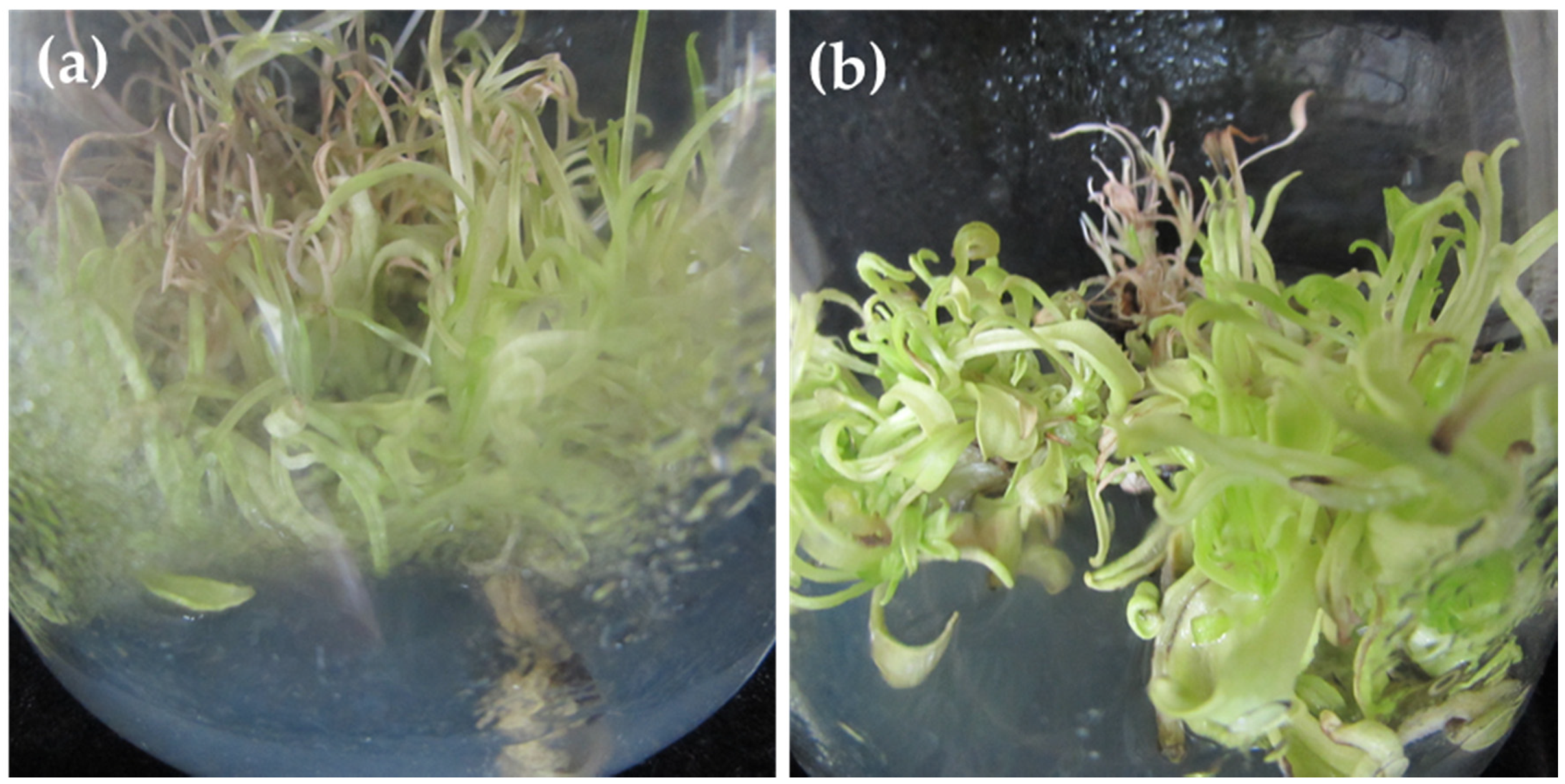
M. maritima shoots is affected by TDZ. Continuous TDZ exposure resulted in shoot tip necrosis and hyperhydricity (Figure 1a,b). Hence, the identification of an efficient plant growth regulator (PGR) is necessary for the in vitro multiplication of M. maritima shoots.
Several bioactive metabolites such as allantoin, rabdosiin, rosmarinic acid, all-E-lutein, all-E-β-carotene, all-E-violaxanthin, 9-Z-neoxanthin, (Z)-β-carotene, all-E-zeaxanthin, α-tocopherol, α-linolenic acid, palmitic acid, linoleic acid, γ-linolenic acid, stearidonic acid, stearic acid, lignoceric acid, behenic acid, and arachidic acid were obtained from M. maritima callus and shoot cultures [5,6]. However, the screening of bioactive metabolites, including phenolics, in M. maritima calli, shoots, and in vivo seedlings, has not yet been reported. Thus, the aims of this work were to (a) determine the effects of PGRs on callus and shoot proliferation, (b) assess the bioactive metabolite profile of in vitro-developed calli and shoots and in vivo seedlings, and (c) estimate the antioxidant capacity and enzyme inhibitory activity of M. maritima.

2. Results

2.1. In Vitro Propagation

2.1.1. Callus Induction

Leaf explants of M. maritima inoculated on control medium (MS medium devoid of hormones) failed to develop calli after 5 weeks of cultivation. Calli were induced in the inoculated explants on medium with combinations of 6-BA (N6-benzyladenine) and NAA (2-(1-naphthyl) acetic acid) (Figure 2a). Callus initiation was detected within 2 weeks of cultivation. Of the six different combinations of 6-BA and NAA studied, 4 µM of each 6-BA and NAA was found to be the most useful for callusing with 89.6% frequency (Figure 3a). Calli induced on this media thrived compared with those induced using other treatments; therefore, the above medium was selected to study callus growth. The lowest percentage of callus initiation (44.7%) was noticed in the medium with 2 µM of 6-BA and NAA (Figure 2a). Light green, friable calli obtained from leaf explants of M. maritima were inoculated on medium containing 6-BA and NAA each at 4 µM, and growth was monitored at weekly intervals up to 7 weeks. The growth of M. maritima calli displayed a normal sigmoidal curve having log, lag, and stationary phases from 0 to 1, 1 to 5, and 5 to 7 weeks, respectively. The results disclosed that the biomass of the callus intensified slowly with an increase in the cultivation period. The maximum fresh weight (FW) and dry weight (DW) of the calli were obtained after 5 and 6 weeks of cultivation, respectively (Figure 2b).

2.1.2. Shoot Multiplication

Nodal explants of M. maritima cultivated on MS without phytohormones (control medium) did not produce axillary shoots and died after 4 weeks of incubation. Shoots developed from explants of M. maritima after 2 weeks of culturing on MS with different 6-BA and 6-KN (N6-furfuryladenine) combinations. Significant (p < 0.05) differences were noted in multiple shoot production among the different 6-BA and 6-KN combinations (Table 1). The medium with 2 µM 6-BA and 4 µM 6-KN had the best shoot induction rate (91.1%) with a mean of 13.4 shoots (Figure 3b, Table 1). However, the medium containing 6-BA and 6-KN at 8 µM was the least effective in promoting the induction of multiple shoots from explants of M. maritima.
Various 6-BA and NAA combinations were studied for axillary shoot production. Approximately 38.4–72.0% of nodal explants produced axillary shoots with an average shoot number of 1.7–6.3 after 4 weeks of cultivation on MS with different 6-BA and NAA combinations. The rate of shoot initiation and induced shoot number per node were increased in the medium augmented with 1 µM of NAA and all three levels of 6-BA. However, shoot production decreased when the M. maritima nodal segments were cultivated on MS augmented with 2 µM of NAA and all three levels of 6-BA. The best combination for axillary shoot production was 8 µM 6-BA and 1 µM NAA (Table 2). A combination of 6-KN and NAA was also studied for massive axillary shoots regeneration from the nodal segments of M. maritima. Approximately 53.7–80.1% of nodal explants produced axillary shoots with an average shoot number of 2.3–8.4 after 4 weeks of cultivation on MS with various 6-BA and NAA combinations. Out of the six combined treatments studied, the medium containing 8 µM 6-KN and 1 µM NAA induced the highest shoot number (8.4 per node) with an induction rate of 80.1% (Table 2). Calli formed from the base of the nodal segments when they were cultivated on MS augmented with combinations of 6-BA or 6-KN and NAA (Figure 3c). However, the calli induced on all media were unable to differentiate into shoots.

2.1.3. Shoot Elongation and Root Induction

Shoots (4 weeks old) obtained from the multiplication medium were elongated on MS medium without any PGR (Figure 3d). The shoots developed adventitious roots after 7 days of cultivation on rooting medium 1/2 MS plus Indole-3-butyric acid (IBA 0–8 µM). The rooting frequency of M. maritima was 56.7% in the half-strength medium. The rooting rate, number of adventitious roots produced, and mean length varied (Table 3). The inclusion of 1–8 µM IBA in the half-strength medium stimulated adventitious rooting compared to the control (devoid of hormone). The highest number of adventitious roots number (16.2) and their length (7.6 cm) were significantly higher on rooting medium ½ MS with 2 µM IBA (Figure 3e,f, Table 3) compared with other treatments. A further increase in the level of IBA declined the number of formed adventitious roots per explant of M. maritima and the root growth. However, the rate of root induction was not significantly (p < 0.05) different (Table 3).

2.2. Chemical Composition

The total amount of phenolic (TAP) and flavonoid (TAF) in the M. maritima tissue extracts was estimated using colorimetric methods, and the data are provided in Table 4. The shoots (41.98 mg GAE/g of extract) contained the highest TAP, followed by the seedlings (19.40 mg GAE/g of extract) and calli (11.36 mg GAE/g of extract). Regarding the TAF, we observed the same order: shoots (1.76 mg RE/g of extract) > seedlings (0.73 mg RE/g of extract) > calli (0.41 mg RE/g of extract).
Both positive and negative ion electrospray ionization mass spectra were acquired over the mass range 100–1500 Da. Data acquisition was performed in the data-dependent acquisition (DDA) mode. The application of this technique allows the determination of the exact molecular mass, and the fragments were also recoded with high accuracy. The identification of the detected compounds in the calli, shoot, or seedling extracts of M. maritima was based on their fragmentation patterns and chromatographic behaviors (The figures are in Supplementary file: Figures S1–S6). The results showed that, in some cases, the negative mode was more sensitive for the identification of these compounds.
The number of detected compounds and chemical composition in the extracts of the shoots and seedlings of M. maritima were similar. In total, 39 compounds were tentatively or unambiguously detected in the shoot extract (Table 5), and 33 compounds were found in the seedling extract (Table 6). Only 18 components were detected in the callus extract (Table 7).
Further, pantothenic; undecanedioic; dodecanedioic; tetradecanedioic; hexadecanedioic; stearidonic acid and their derivatives; abscisic acid; intermedine or lycopsamine and their N-oxide; heliotrine; caffeic acid and its derivatives; rosmarinic acid and its di-O-hexoside isomers; 3-O-methylrosmarinic acid; apigenin-C-pentoside-O-hexoside; echimidine or heliosupine; quercetin and isoquercitine; 3,4-dihydrocinnamic acid derivatives; and di-, tri-, tetra-, pentamethoxy(iso)flavone isomers were assigned to the extracts.
In some cases, two or three isomers of compounds could be tentatively identified in the extracts. For example, two isomers of rosmarinic acid di-O-hexoside were identified in the shoot extract. A typical extracted ion chromatogram of these compounds at m/z 683.1823 and Figure 4, Figure 5 and Figure 6 shows the MS2 spectra (negative ion mode).

2.3. Biological Activities

The antioxidant activities of M. maritima calli, shoot, or seedling extracts were evaluated using different test systems, and the results are provided in Table 8. The best antioxidant capacity was obtained in the shoots, followed by the seedlings and calli in the radical scavenging ABTS (2,2-azino-bis(3-ethylbenzothiazoline-6-sulphonic acid) and DPPH (2,2-diphenyl-1-picrylhydrazyl), reducing power FRAP (ferric reducing antioxidant power) and CUPRAC (cupric reducing antioxidant capacity), and phosphomolybdenum (PBD) assays. However, the order was seedlings > calli > shoots with regards to the MCA (metal chelating ability).
The enzyme inhibitory effects of M. maritima calli, shoot, or seedling extracts were investigated against AChE (acetylcholinesterase) and BChE (butylcholinestrase), tyrosinase, and amylase. Values are presented in Table 9. The strongest AChE inhibition properties were obtained by the calli, followed by seedlings and shoots. However, the tested samples were ranked as seedlings > shoots > calli in the BChE inhibition assay. Regarding the antityrosinase inhibition ability, the strongest ability was detected in the seedlings (IC50: 0.74 mg/mL), followed by the calli (IC50: 0.80 mg/mL) and shoots (IC50: 0.87 mg/mL). The anti-amylase activities of the samples were almost the same (IC50: 1.40–147 mg/mL).

3. Discussion

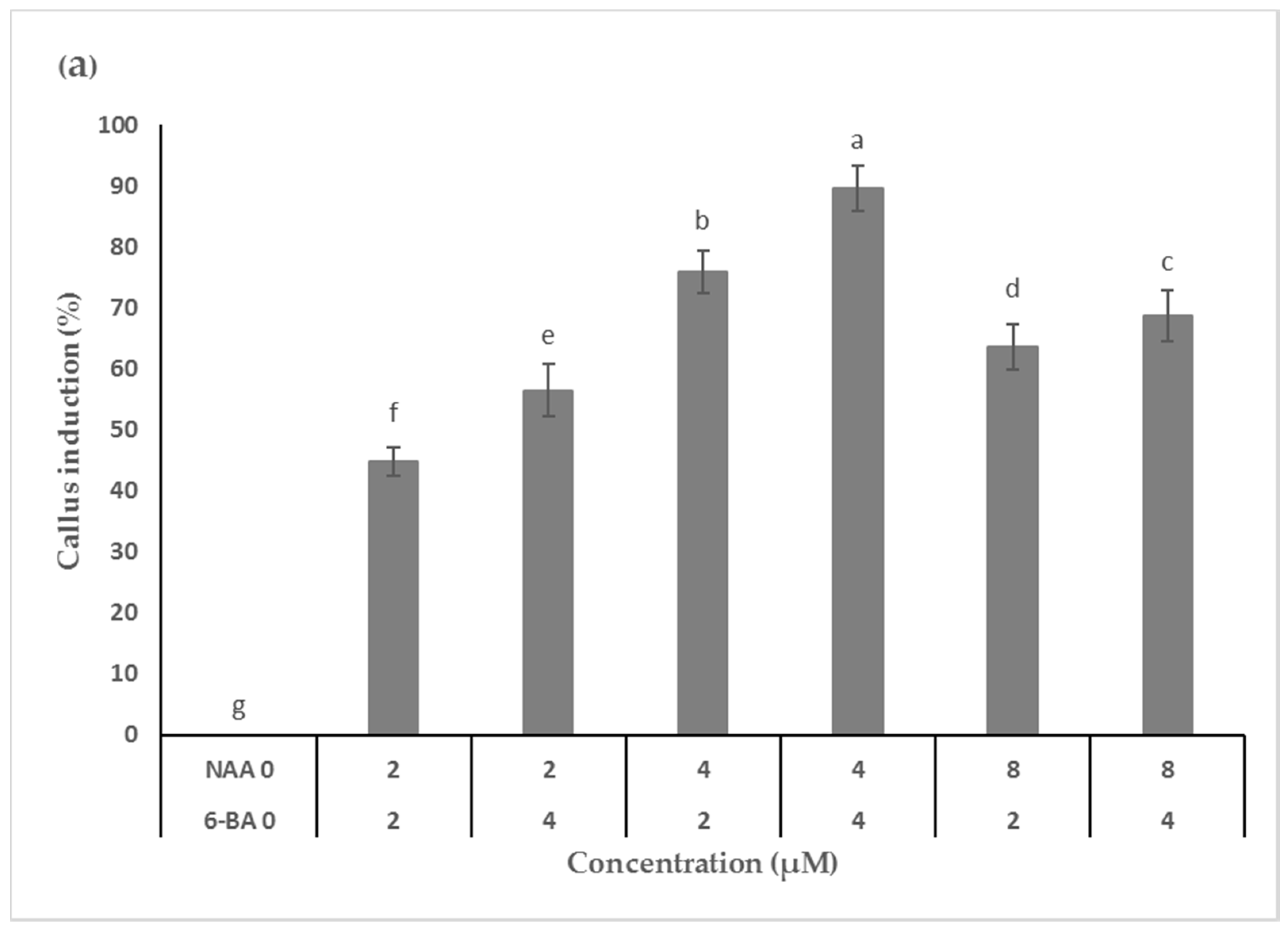
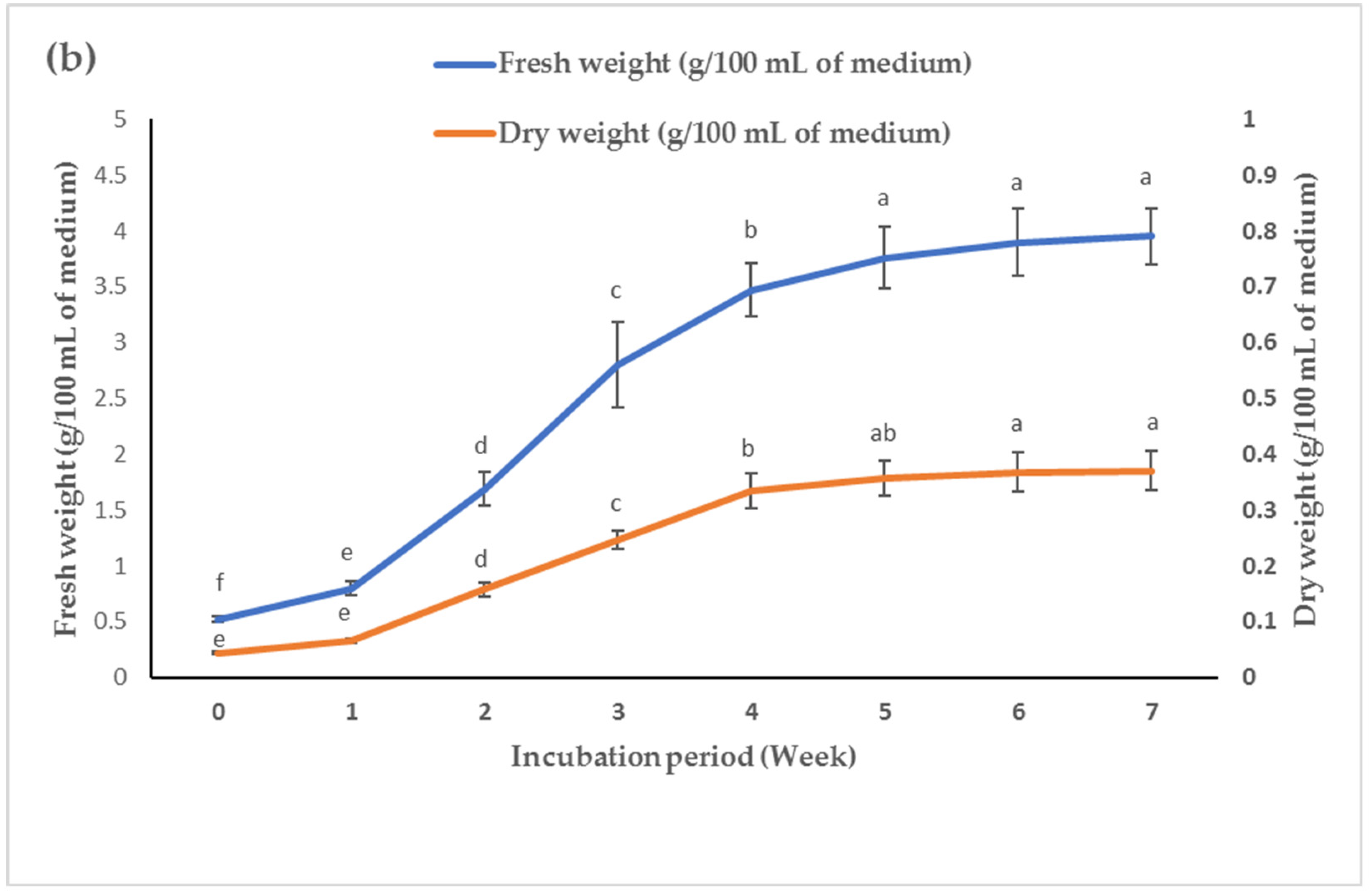
Callus induction and axillary shoot multiplication are significant in vitro technologies for the rapid commercial production of plantlets and bioactive metabolites. Leaf explants from M. maritima initiated calli only on PGR (6-BA and NAA) supplemented MS medium. A combination of PGRs such as cytokinin and auxin are generally used to induce callus initiation in M. maritima [5] and several other Boraginaceae members such as Alkanna orientalis (L.) Boiss. and A. sieheana Rech. f. [8], Arnebia euchroma (Royle) Jonst. [9], Arnebia hispidissima (Lehm.) A. DC. [10], Echium italicum L. [11], Eritrichium sericeum (Lehm.) A. DC. [12], Onosma bulbotrichum [13], and Onosma sericeum Willd [14]. In this study, the highest callus proliferation was attained after 5 weeks of cultivation under a 16-h photoperiod on MS medium augmented with 4 µM of each 6-BA and NAA. Fedoreyev et al. [5] also obtained maximum callus growth in M. maritima on WB/A medium containing 2.2 µM 6-BA and 10.8 µM NAA after 30 days of cultivation in the dark. However, there was a considerable difference in the optimal levels of 6-BA and NAA. The differences in the optimal concentration of 6-BA and NAA required for the best callus growth may be due to culture conditions, callus type, and growth media.
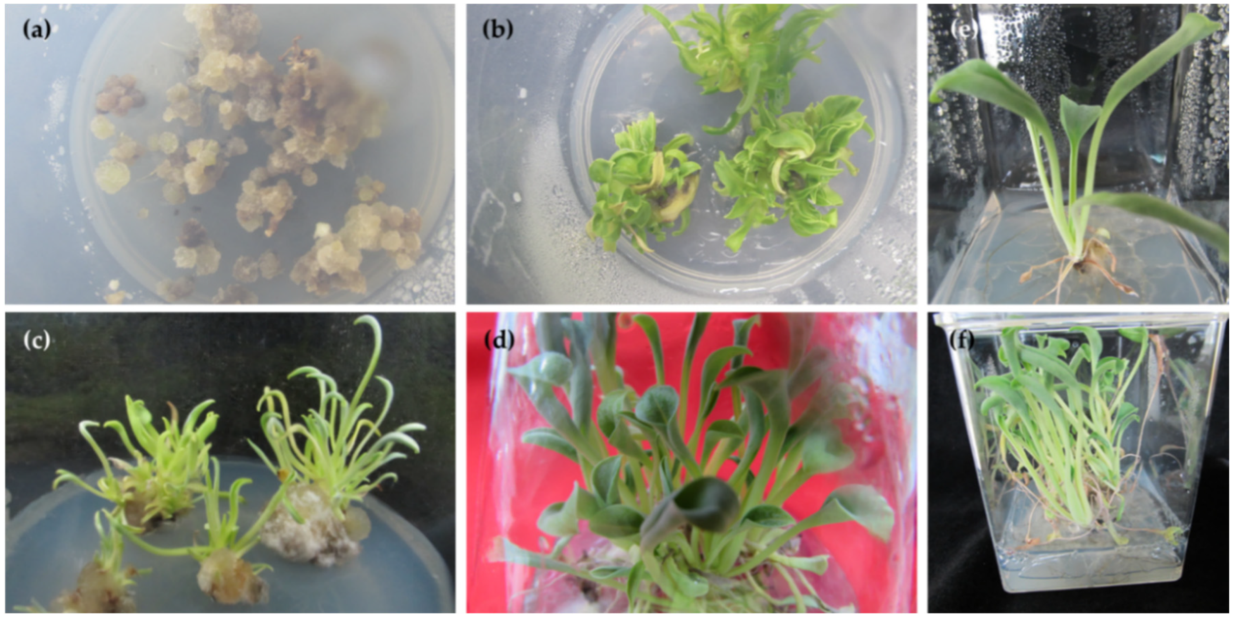
Nodal explants of M. maritima failed to regenerate in MS medium lacking PGR. This is consistent with an earlier study of M. maritima [6]. Similar results have been reported for Boraginaceae members such as Hackelia venusta (Piper) H.St.John [15] and Trichodesma indicum (Linn) R. Br. [16]. Axillary shoot multiplication can be achieved by supplementing the growth media with optimal levels of PGRs. Cytokinin is a class of PGR that promotes axillary shoot multiplication by antagonizing apical shoot dominance. Nodal segments of M. maritima cultivated on medium with 6-BA and 6-KN produced axillary shoots (Table 1). Cytokinins (for shoot production) and their concentrations were chosen based on our previous study [6]. The explants cultivated on medium with 2 µM 6-BA and 4 µM 6-KN developed more shoots (13.4) than those on media containing the other five 6-BA and 6-KN combinations (Table 1). The combinations of two cytokinins (6-BA and 6-KN) were found to be effective in the shoot regeneration of Boraginaceae members [10,17]. Kumar and Rao [17] reported that Heliotropium indicum L. axillary buds inoculated on medium with 4.7 µM 6-KN and 2.2 µM 6-BA developed 11.8 shoots.
Several studies have shown that media with cytokinin and auxin enhance shoot proliferation in numerous members of the Boraginaceae family, including M. maritima [6,16,17,18,19]. The ratio of cytokinin and auxin is crucial for the production of multiple shoots. In general, high cytokinin/auxin ratio promotes shoot proliferation in many plants [20]. In this study, supplementing MS with a high level of 6-BA or 6-KN (8 µM) and low level of NAA (1 µM) yielded the maximum number of shoots. However, the incorporation of a high concentration of NAA (2 µM) in media containing 6-BA or 6-KN inhibited shoot proliferation. Similar results have been described for A. hispidissima [18], H. indicum [17], and T. indicum [16]. The MS added with 8 µM 6-KN and 1 µM NAA produced the maximum (8.4) number of shoots (Table 2). This number was lower than that reported for oyster plant [6]. Maximal shoot production was 17.7 shoots per node for M. maritima and was stimulated by 4 µM TDZ and 1 µM NAA [6].
The incorporation of IBA in ½ MS enhanced the rooting response of the explants (Table 3). IBA was chosen for in vitro rooting because it has been reported to be beneficial for adventitious root induction in M. maritima [6] and other Boraginaceae members [10,16,17,18]. However, the rooting response of shoots under in vitro conditions varied with the IBA concentration. The optimal level of IBA required for the rooting of micro shoots depends on the rooting medium strength, the composition of shoot production medium, plant species, and genotype [20]. In this study, the best rooting was attained with 2 µM IBA in M. maritima. However, the rooting of M. maritima was the most effective in half-strength medium with 4 µM IBA [6]. MS with 9.8 µM IBA exhibited the best rooting in A. hispidissima [18], half-strength medium with 0.49 µM IBA showed the best rooting in H. indicum [17], and MS with 2.46 µM IBA displayed the best rooting in T. indicum [16].
Phenolics are unique compounds with significant biological properties, including antioxidant, anti-microbial, and anti-cancer activities [21]. In this context, the determination of the TAP in plant extracts is the first step; for this purpose, a colorimetric method was developed by Folin and Ciocalteu [22]. The TAP in the shoots was 216.4% and 369.5% higher than in seedlings and calli, respectively. The TAP in M. maritima shoots extract was also larger (41.98 mg GAE/g of extract) than in calli (9.20 GAE/g of extract), in vivo leaves (16.06 GAE/g of extract), stems (14.55 GAE/g of extract), inflorescences (21.70 GAE/g of extract) and roots (3.17 GAE/g of extract) of Heliotropium indicum [23], flowers (18.43 GAE/g of extract) and roots (13.11 GAE/g of extract) of Cynoglossum creticum [24], and aerial parts (32.7 GAE/g of extract) [25] and roots (11.45 GAE/g of extract) of Symphytum anatolicum [26]. The TAP in the shoots was 241.1% and 429.3% higher than in seedlings and calli, respectively. However, TAP in M. maritima shoot tissues was lower (1.76 mg RE/g of extract) compared to in vivo leaves (4.39 RE/g of extract) and flowers (21.77 RE/g of extract) of C. creticum [24], aerial parts (20.9 RE/g of extract) of Cynoglottis barrelieri [25], leaves (3.32 QE/g of extract), stems (3.23 QE/g of extract) and inflorescences (4.90 QE/g of extract) of H. indicum [23], and aerial parts (13.3 RE/g of extract) [25] and roots (2.74 RE/g of extract) of S. anatolicum [26]. Folin and Ciocalteu method, one of the most popular, is simple and requires inexpensive reagents. However, in recent times, there have been some concerns regarding the use of colorimetric methods, and some authors reported that these assays do not accurately reflect the levels of phytochemicals in extracts. This fact is related to the reduction of the Folin–Ciocalteu reagent by not only phenolics but also non-phenolic compounds [
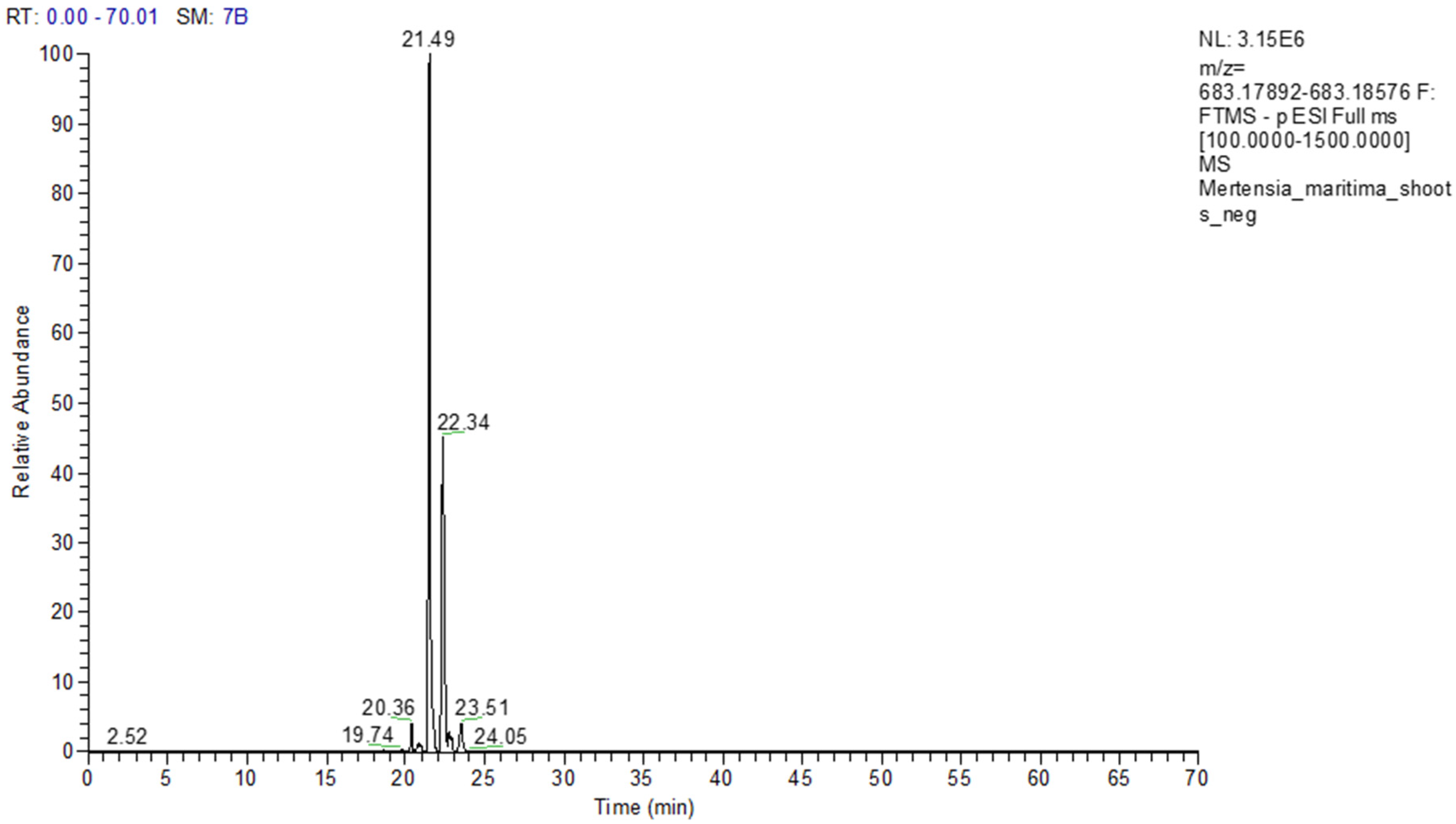
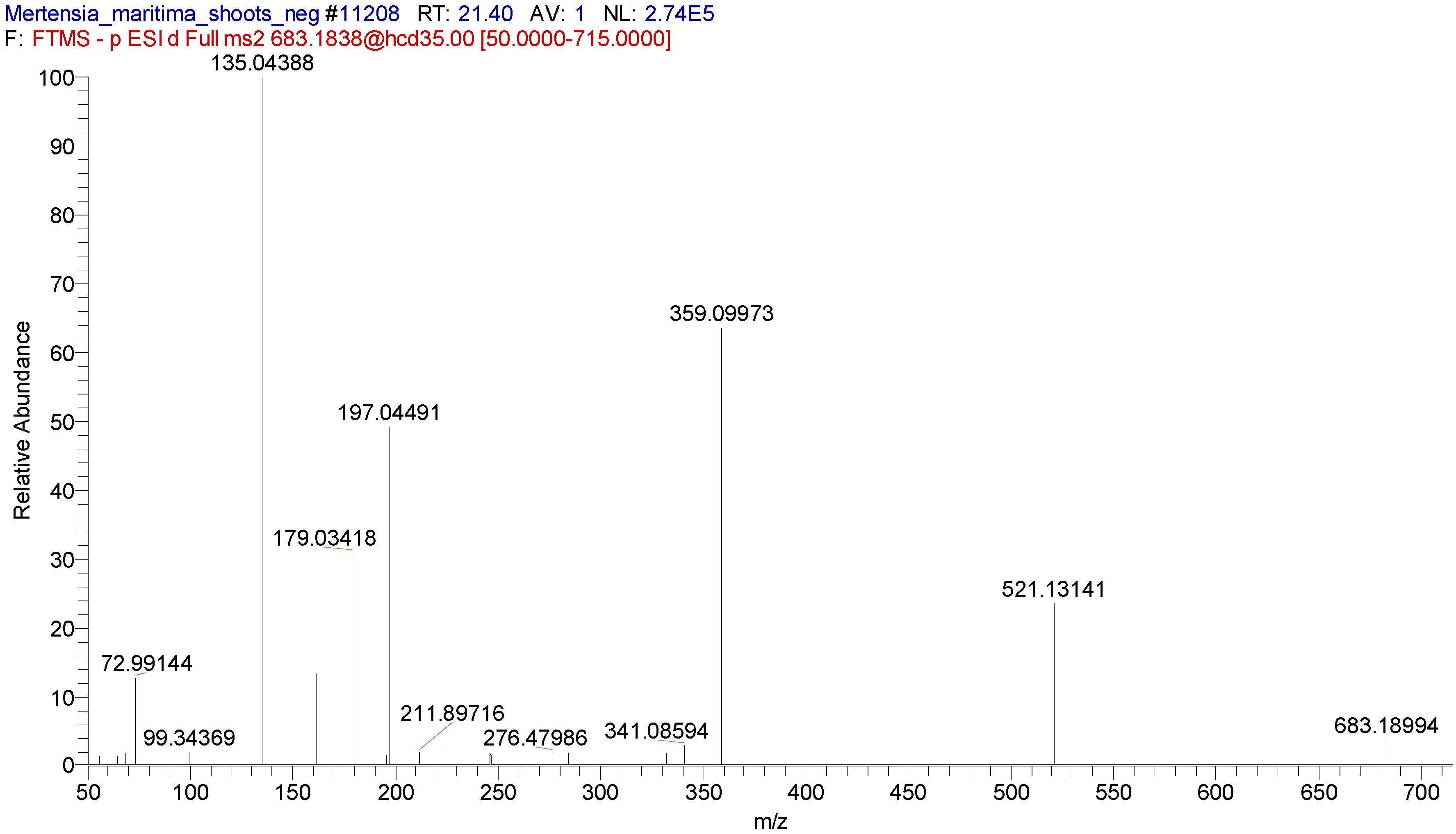
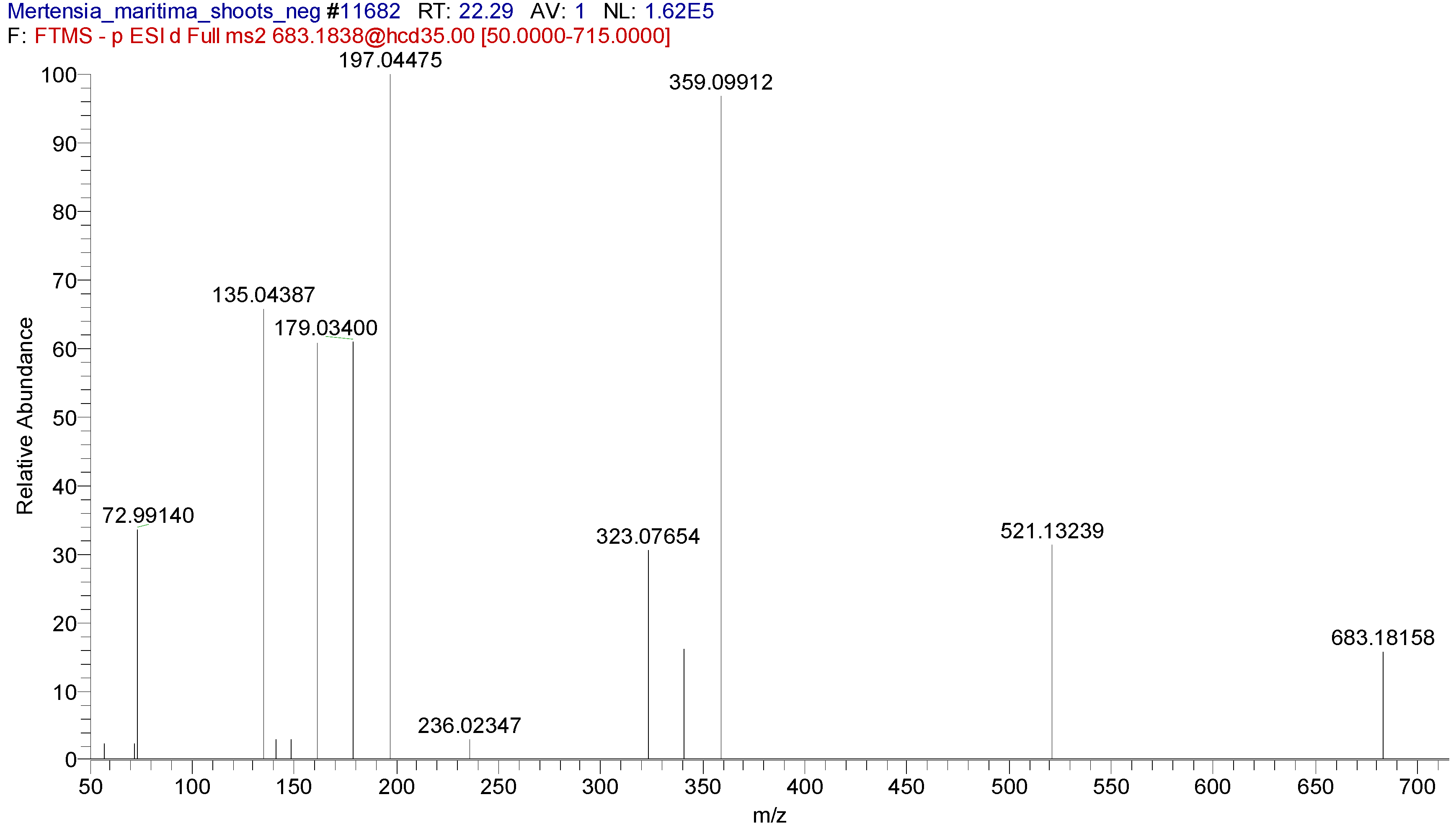
27]. In this sense, at least one chromatographic technique (HPLC, LC-MS, or LC-MS/MS) is important for determining accurate levels of bioactive compounds. Hence, we determined the chemical profiles of M. maritima extracts using the UHPLC-MS/MS technique.
The oyster plant is eaten by the Iñupiat of Alaska. It is cultivated as an edible plant in southwestern France and Northern Scotland [4] for their fragrant leaves. Thus, knowledge of its chemical composition is necessary. Pyrrolizidine alkaloids are one of the main bioactive compounds of Boraginaceae members [28]. For the first time, seven pyrrolizidine alkaloids, such as echimidine, heliosupine, heliotrine, intermedine or lycopsamine, and their N-oxides were identified in M. maritima extracts by UHPLC-MS/MS (Table 5, Table 6 and Table 7). The occurrence of these compounds was also disclosed in numerous members of the Boraginaceae family, including Mertensia species [3,28,29]. Four and six fatty acids were identified in callus and tissue extracts of M. maritima by UHPLC-MS/MS (Table 5–7). Among the fatty acids detected, α-linolenic acid and stearidonic acid were reported in M. maritima [6]. In this study, undecanedioic, dodecanedioic, tetradecanedioic, and hexadecanedioic acids were identified in M. maritima, for the first time. Of the phenolic compounds detected, only rosmarinic acid was reported in the callus extract of M. maritima [5]. Whereas other phenolic compounds were found in members of the Boraginaceae [30,31,32,33,34].
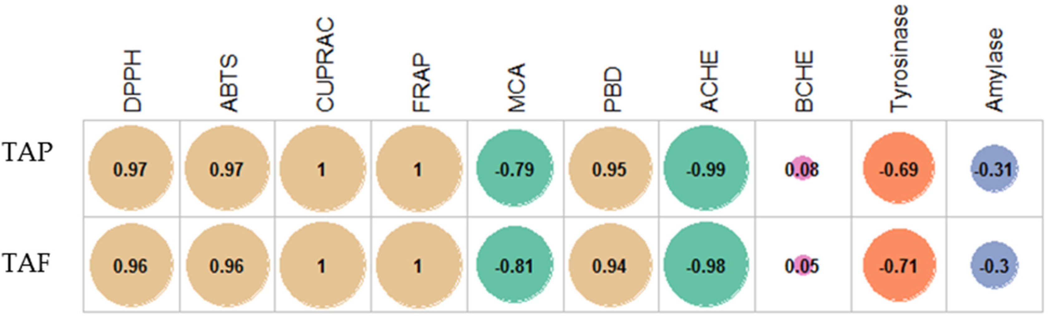
In the last few decades, the terms “antioxidant” and “oxidative stress” have become popular terms in the scientific community [35]. This fact could be explained by the role of oxidative stress in the progression of chronic and degenerative diseases [36]. In light of this information, we determined the antioxidant profiles of M. maritima extracts using different chemical methods. Except for the metal chelation assay, the best antioxidant properties were obtained in the shoot, followed by the seedling and callus. The order is in line with the levels of TAP and TAF. These findings showed that phenolics were the main contributors of the antioxidant capacities of the M. maritima extract. This fact also was confirmed by correlation analysis, and the results are given in Figure 7. In addition, this approach was also confirmed by several authors who reported a linear relationship between total phenolics and antioxidant properties [37,38,39]. In addition, individual compounds, including caffeic acid [40], rosmarinic acid [41], and rutin [42], have been reported as significant antioxidants. The contents of these compounds are relatively high in members of the Boraginaceae family, including M. maritima [5,30,32,33,34]. Phenolic compounds with one or more hydroxyl groups are effective hydrogen or electron donors. Regarding the metal chelating ability, the contradictory results could be explained by the presence of non-phenolic chelators in M. maritima shoot extracts, including polysaccharides, peptides, or vitamin C [43].
According to WHO reports, diseases such as heart disease, stroke, chronic pulmonary disease, Alzheimer’s disease, and diabetes mellitus are the biggest killers worldwide [44]. The inhibition of cholinesterase increases the level of acetylcholine, which could enhance the memory capacity in patients with Alzheimer’s [45]. Similarly, the inhibition of carbohydrate-hydrolyzing enzymes can retard the increase in blood glucose levels in patients with diabetes [46]. Hence, several compounds are synthetically produced as enzyme inhibitors in the pharmaceutical industry. However, several studies have reported that synthetic compounds have unpleasant side effects [47,48]. Thus, enzyme inhibitors from natural sources are needed to replace these synthetic ones. In recent investigations, several researchers reported enzyme inhibition properties of several Boraginaceae members such as, Alkanna sfikasiana [49], Cynoglossum creticum [24], Cynoglottis barrelieri [25], Echium confusum [50], Onosma aucheriana, Onosma sieheana, Onosma frutescens, Onosma stenoloba, and Onosma sericea [51,52] Symphytum anatolicum [25,26]. In the present study, the enzyme inhibitory effects of M. maritima extracts were investigated using different enzymes. Among the three M. martima tissue extracts studied, calli extracts exhibited ideal AChE (IC50: 0.74 mg/mL) inhibition, seedlings extracts exhibited ideal BChE (IC50: 0.74 mg/mL) inhibition and tyrosinase (IC50: 0.74 mg/mL) inhibition, while all three extracts exhibited similar amylase (IC50: 1.40–1.47 mg/mL) inhibition activity (Table 9). The IC50 values attained from tyrosinase and amylase inhibition assays were lower compared to several other Boraginaceae members [51,52]. We observed different results for each enzyme inhibition ability. The observed enzyme inhibitory effects could be explained by the chemical profiles of the extracts. Some compounds in the chemical profiles, including caffeic acid [53,54], rosmarinic acid [55,56], and rutin [57,58] have been reported as significant enzyme inhibitor agents in previous studies. To our knowledge, the present study is the first report on the enzyme inhibitory effects of M. maritima extracts, and these findings could provide valuable contributions to the scientific community.

4. Materials and Methods

4.1. In Vitro Propagation

4.1.1. Surface Disinfection, Media, and Culture Conditions

Leaf and node explants of M. maritima used in callus and shoot regeneration studies were harvested from plants cultivated in a greenhouse. The shoots were cautiously rinsed with water for 15 min and then surface decontaminated in ethanol (70% v/v) for 90 s; rinsed twice in sterilized distilled water and sodium hypochlorite solution (2% v/v) with 2–3 drops of nonionic detergent (Tween 20) for 10 min; washed three to five times in sterilized distilled water and ethanol (70% v/v) for 90 s; and rinsed twice in sterilized distilled water [6]. The cultivation media consisted of MS [59] minerals, vitamins, PGRs, sugar (30 g/L sucrose), and solidifying agent (8 g/L plant agar). The pH of all cultivation media was adjusted to 5.75 ± 0.25 before being autoclaved for 20 min at 121 °C. Leaf (callus), callus (proliferation), node (multiple shoot), micro shoot (elongation), and well-developed shoot (rooting) explant cultures were incubated for 5, 7, 4, 5, and 6 weeks, respectively, at 22–24 °C under a 16-h photoperiod (55 ± 5 µmol m−2 s−1).

4.1.2. Callus Induction

Leaf explants (5–10 mm) were prepared from surface-disinfected M. maritima shoots placed on medium with 0, 2, or 4 µM 6-BA in combination with 0, 2, 4, or 8 µM of NAA for callus formation. To study the growth of M. maritima callus, approximately 50 mg of callus was inoculated on a growth medium containing 4 µM of each 6-BA and NAA. Each callus initiation or callus growth treatment consisted of 15 or 25 leaf or callus explants with three replicates. Callusing percentages and fresh and dry weights of calluses were recorded.

4.1.3. Shoot Multiplication

Node explants (approximately 5 mm) were prepared from surface-disinfected M. maritima shoots placed on medium with 6-BA and 6-KN (Table 1), 6-BA and NAA, or 6-KN and NAA combinations (Table 2) for multiple shoot induction. Each multiple shoot production treatment consisted of 25 nodes with three replicates. Shooting percentages and shoot numbers were recorded.

4.1.4. Shoot Elongation and Root Induction

For elongation of M. maritima shoots, in vitro-induced shoot masses obtained from medium with 2 µM 6-BA and 4 µM 6-KN were transferred to PGRs-free growth medium. After 5 weeks, well-developed shoots (≥2 cm in height) were cultured on medium ½ MS with 0–8 µM IBA for rooting. Each root induction treatment consisted of 25 shoots with three replicates. Rooting percentages, number, and length of roots were recorded.

4.2. Phytochemical Analysis

4.2.1. Extract Preparation

Calli (obtained from the medium with 4 µM each of 6-BA and NAA), leaves (obtained from shoots developed in PGR-free medium), and seedlings (obtained from the greenhouse) were collected after 5 weeks of cultivation; they were lyophilized and then powdered. The samples (50 mg) were extracted with methanol (80%) using an Ultraturrax at 6000 g for 20 min. After, filtration solvents were removed using a rotary evaporator and kept at plus 4 °C until further use.

4.2.2. Determination of TAP and TAF

TAP in calli, shoot, or seedling extracts of M. maritima was assessed using the Folin–Ciocalteu method, as adapted by Slinkard and Singleton [60] and calculated as gallic acid equivalent (GAE). TAF in calli, shoot, or seedling extracts of M. maritima was estimated using the aluminum chloride (AlCl3) method, as adapted by Zengin et al. [61] and was calculated as RE. All assays were performed in three analytical replications.

4.2.3. Identification and Quantification of Phytochemicals

The qualitative analysis of phytochemicals in calli, shoot, or seedling extracts of M. maritima was carried out using an UHPLC instrument (Dionex Ultimate 3000 RS, Thermo Scientific, MA, 01001 USA) connected to a mass spectrometer (Q Exactive Orbitrap, Thermo Scientific, USA). Thermo Accucore C18 column (100 mm length × 2.1 mm column I.D., 2.6 µm) was used for chromatographic separation [62]. Analytical details are presented in the supplementary material.

4.3. Biological Activities

4.3.1. Antioxidant Assay

ABTS, DPPH, FRAP, CUPRAC, MCA, and PBD assays were conducted to estimate the antioxidant ability of calli, shoot, or seedling extracts of M. maritima and standards (Trolox and EDTA) [63]. All assays were performed in three analytical replications.

4.3.2. Enzyme Inhibitory Assay

Amylase-, AChE-, BChE-, and tyrosinase-inhibitory activity of calli, shoot, or seedling extracts of M. maritima was determined according to Uysal et al. [63]. All assays were performed in three analytical replications.

4.4. Statistical Analysis

Data obtained such as callusing, shooting, rooting, TAP, TAF, and antioxidant and enzyme inhibition were analyzed in SAS version 9.1. Analysis of variance and Duncan (at 5% level) were used to test the influence and significance of treatments, respectively. For correlation analysis (Pearson coefficient), R software v. 3.5.1 was used.

5. Conclusions

An improved in vitro micropropagation method for M. maritima was established. This in vitro callus and shoot proliferation method will be useful for the extraction of bioactive metabolites from M. maritima. The synthesis of bioactive metabolites in M. maritima cell and shoot cultures is affected by chemical factors. Thus, further research on the optimization of growth media, culture environment, and elicitation are needed to maximize bioactive metabolite production in M. maritima in vitro cultures.

Supplementary Materials

The following are available online at https://www.mdpi.com/2223-7747/9/11/1551/s1, UHPLC methods and Figures S1–S6.

Author Contributions

Conceptualization, K.S., D.H.K. and I.S.; methodology, I.S., G.A., G.Z., Z.C., J.J.; formal analysis, K.S., K.R.R.; investigation, I.S., G.A., Z.C.; data curation, O.N.L.; writing—original draft preparation, K.S., G.A., G.Z., Z.C., J.J., I.S.; writing—review and editing, K.R.R., G.A., G.Z., I.S.; funding acquisition, I.S. All authors have read and agreed to the published version of the manuscript.

Funding

This research was funded by Konkuk University.

Acknowledgments

This article was supported by the KU Research Professor Program of Konkuk University.

Conflicts of Interest

The authors declare no conflict of interest.

References

  1. Nazaire, M.; Wang, X.Q.; Hufford, L. Geographic origins and patterns of radiation of Mertensia (Boraginaceae). Am. J. Bot. 2014, 101, 104–118. [Google Scholar] [CrossRef] [Green Version]
  2. Gracie, C. Spring Wildflowers of the Northeast; Princeton University Press: Princeton, NJ, USA, 2012; pp. 222–227. [Google Scholar]
  3. Li, Y.; Stermitz, F.R. Pyrrolizidine alkaloids from Mertensia species of Colorado. J. Nat. Prod. 1988, 51, 1289–1290. [Google Scholar] [CrossRef]
  4. Delort, E.; Jaquier, A.; Chapuis, C.; Rubin, M.; Starken-mann, C.J. Volatile composition of oyster leaf (Mertensia maritima (L.) Gray). J. Agric. Food Chem. 2012, 60, 11681–11690. [Google Scholar] [CrossRef] [PubMed]
  5. Fedoreyev, S.A.; Inyushkina, Y.V.; Bulgakov, V.P.; Veselova, M.V.; Tchernoded, G.K.; Gerasimenko, A.V.; Zhuravlev, Y.N. Production of allantoin, rabdosiin and rosmarinic acid in callus cultures of the seacostal plant Mertensia maritima (Boraginaceae). Plant. Cell Tissue Organ. Cult. 2012, 110, 183–188. [Google Scholar] [CrossRef]
  6. Park, H.Y.; Kim, D.H.; Saini, R.K.; Gopal, J.; Keum, Y.S.; Sivanesan, I. Micropropagation and quantification of bioactive compounds in Mertensia maritima (L.) Gray. Int. J. Mol. Sci. 2019, 20, 2141. [Google Scholar] [CrossRef] [PubMed] [Green Version]
  7. Alton, S.; FitzGerald, R. Mertensia maritima Boraginaceae. Curtis’s Bot. Mag. 2009, 26, 96–110. [Google Scholar] [CrossRef]
  8. Yaman, C.; Uranbey, S.; Ahmed, H.; Özcan, S.; Tugay, O.; Başalma, D. Callus induction and regeneration of Alkanna orientalis var. Orientalis and A. sieheana. Bangladesh J. Bot. 2019, 48, 633–640. [Google Scholar] [CrossRef]
  9. Damianakos, H.; Kretschmer, N.; Syklowska-Baranek, K.; Pietrosiuk, A.; Bauer, R.; Chinou, I. Antimicrobial and cytotoxic isohexenylnaphthazarins from Arnebia euchroma (Royle) Jonst. (Boraginaceae) callus and cell suspension culture. Molecules 2012, 17, 14310–14322. [Google Scholar] [CrossRef] [Green Version]
  10. Phulwaria, M.; Shekhawat, N.S. An efficient in vitro shoot regeneration from immature inflorescence and ex vitro rooting of Arnebia hispidissima (Lehm). DC.—A red dye (Alkannin) yielding plant. Physiol. Mol. Biol. Plants 2013, 19, 435–441. [Google Scholar] [CrossRef] [Green Version]
  11. Zare, K.; Khosrowshahli, M.; Nazemiyeh, H.; Movafeghi, A.; Motallebi Azar, A.; Omidi, Y. Callus culture of Echium italicum L. towards production of a shikonin derivative. Nat. Prod. Res. 2011, 25, 1480–1487. [Google Scholar] [CrossRef]
  12. Fedoreyev, S.A.; Veselova, M.V.; Krivoschekova, O.E.; Mischenko, N.P.; Denisenko, V.A.; Dmitrenok, P.S.; Glazunov, V.P.; Bulgakov, V.P.; Tchernoded, G.K.; Zhuravlev, Y.N. Caffeic acid metabolites from Eritrichium sericeum cell cultures. Planta Med. 2005, 71, 446–451. [Google Scholar] [CrossRef] [PubMed]
  13. Bagheri, F.; Tahvilian, R.; Karimi, N.; Chalabi, M.; Azam, M. Shikonin production by callus culture of Onosma bulbotrichom as active pharmaceutical ingredient. Iran J. Pharm. Res. 2018, 17, 495–504. [Google Scholar] [PubMed]
  14. Gharehmatrossian, S.; Popov, Y.; Ghorbanli, M.; Safaeian, S.; Iranbakhsh, A. Phytochemical and morphological evidences for shikonin production by plant cell cultures of Onosma sericeum Willd. Braz. Arch. Biol. Technol. 2016, 59, e16160235. [Google Scholar] [CrossRef] [Green Version]
  15. Edson, J.L.; Leege-Brusven, A.D.; Everett, R.L.; Wenny, D.L. Minimizing growth regulators in shoot cultures of an endangered plant, Hackelia venusta (Boraginaceae). In Vitro Cell. Dev. Biol. Plant 1996, 32, 267–271. [Google Scholar] [CrossRef]
  16. Mahesh, A.; Jeyachandran, R. Influence of plant growth regulators on micropropagation and in vitro flowering of Trichodesma indicum (Linn) R. Br. Plant. Biosyst. 2013, 147, 493–499. [Google Scholar] [CrossRef]
  17. Kumar, M.S.; Rao, M.V. In vitro micropropagation of Heliotropium indicum L.—An ayurvedic herb. Indian J. Biotechnol. 2007, 6, 245–249. [Google Scholar]
  18. Pal, M.; Chaudhury, A. High frequency direct plant regeneration, micropropagation and shikonin induction in Arnebia hispidissima. J. Crop. Sci. Biotech. 2010, 13, 13–19. [Google Scholar] [CrossRef]
  19. Lameira, O.A.; Pinto, J.E.B.P. In vitro propagation of Cordia verbenacea (Boraginaceae). Rev. Bras. Plant Med. Botucatu. 2006, 8, 102–104. [Google Scholar]
  20. Park, H.Y.; Kim, D.H.; Sivanesan, I. Micropropagation of Ajuga species: A mini review. Biotechnol. Lett. 2017, 39, 1291–1298. [Google Scholar] [CrossRef]
  21. Wang, Z.; Li, S.; Ge, S.; Lin, S. Review of distribution, extraction methods, and health benefits of bound phenolics in food plants. J. Agric. Food Chem. 2020, 68, 3330–3343. [Google Scholar] [CrossRef]
  22. Folin, O.; Ciocalteu, V. On tyrosine and tryptophane determinations in proteins. J. Biol. Chem. 1927, 73, 627–650. [Google Scholar]
  23. Kumar, M.S.; Chaudhury, S.; Balachandran, S. In vitro callus culture of Heliotropium indicum Linn. for assessment of total phenolic and flavonoid content and antioxidant activity. Appl. Biochem. Biotechnol. 2014, 174, 2897–2909. [Google Scholar] [CrossRef] [PubMed]
  24. Menghini, L.; Ferrante, C.; Zengin, G.; Mahomoodally, M.F.; Leporini, L.; Locatelli, M.; Cacciagrano, F.; Recinella, L.; Chiavaroli, A.; Leone, S.; et al. Multiple pharmacological approaches on hydroalcoholic extracts from different parts of Cynoglossum creticum Mill. (Boraginaceae). Plant Biosyst. 2019, 153, 633–639. [Google Scholar] [CrossRef]
  25. Varvouni, E.-F.; Zengin, G.; Graikou, K.; Ganos, C.; Mroczek, T.; Chinou, I. Phytochemical analysis and biological evaluation of the aerial parts from Symphytum anatolicum Boiss. and Cynoglottis barrelieri (All.) Vural & Kit Tan (Boraginaceae). Biochem. Syst. Ecol. 2020, 92, 104128. [Google Scholar]
  26. Sarikurkcu, C.; Ozer, M.S.; Tlili, N. LC–ESI–MS/MS characterization of phytochemical and enzyme inhibitory effects of different solvent extract of Symphytum anatolicum. Ind. Crop. Prod. 2019, 140, 111666. [Google Scholar] [CrossRef]
  27. Sánchez-Rangel, J.C.; Benavides, J.; Heredia, J.B.; Cisneros-Zevallos, L.; Jacobo-Velázquez, D.A. The Folin–Ciocalteu assay revisited: Improvement of its specificity for total phenolic content determination. Anal. Methods 2013, 5, 5990–5999. [Google Scholar] [CrossRef]
  28. El-Shazly, A.; Wink, M. Diversity of pyrrolizidine alkaloids in the Boraginaceae structures, distribution, and biological properties. Diversity 2014, 6, 188–282. [Google Scholar] [CrossRef] [Green Version]
  29. Schramm, S.; Köhler, N.; Rozhon, W. Pyrrolizidine Alkaloids: Biosynthesis, Biological Activities and Occurrence in Crop Plants. Molecules 2019, 24, 498. [Google Scholar] [CrossRef] [Green Version]
  30. Zemmouri, H.; Ammar, S.; Boumendjel, A.; Messarah, M.; El Feki, A.; Bouaziz, M. Chemical composition and antioxidant activity of Borago officinalis L. leaf extract growing in Algeria. Arab. J. Chem. 2019, 12, 1954–1963. [Google Scholar] [CrossRef]
  31. Dresler, S.; Szymczak, G.; Wójcik, M. Comparison of some secondary metabolite content in the seventeen species of the Boraginaceae family. Pharm. Biol. 2017, 55, 691–695. [Google Scholar] [CrossRef] [Green Version]
  32. Li, L.; Li, M.-H.; Xu, L.-J.; Guo, N.; Wu-Lan, T.-N.; Shi, R.-B.; Peng, Y.; Xiao, P.-G. Distribution of seven polyphenols in several medicinal plants of Boraginaceae in China. J. Med. Plants Res. 2010, 4, 1216–1221. [Google Scholar]
  33. Bai, X.R.; Zhang, N.; Ren, K.; Xu, J.P.; Li, Z.Y.; Zhang, C.H.; Li, M.H. Chemical constituents from Stenosolenium saxatile (Pall.) turcz. (Boraginaceae) Biochem. Systemat. Ecol. 2017, 74, 30–32. [Google Scholar] [CrossRef]
  34. Trifan, A.; Skalicka-Woźniak, K.; Granica, S.; Czerwińska, M.E.; Kruk, A.; Marcourt, L.; Wolfender, J.-L.; Wolfram, E.; Esslinger, N.; Grubelnik, A.; et al. Symphytum officinale L.: Liquid-liquid chromatography isolation of caffeic acid oligomers and evaluation of their influence on pro-inflammatory cytokine release in LPS-stimulated neutrophils. J. Ethnopharmacol. 2020, 262, 113169. [Google Scholar] [CrossRef]
  35. Yeung, A.W.K.; Tzvetkov, N.T.; El-Tawil, O.S.; Bungǎu, S.G.; Abdel-Daim, M.M.; Atanasov, A.G. Antioxidants: Scientific literature landscape analysis. Oxidative Med. Cell. Longev. 2019, 2019. [Google Scholar] [CrossRef]
  36. Bjørklund, G.; Chirumbolo, S. Role of oxidative stress and antioxidants in daily nutrition and human health. Nutrition 2017, 33, 311–321. [Google Scholar] [CrossRef]
  37. Ghafoor, K.; Al Juhaimi, F.; Özcan, M.M.; Uslu, N.; Babiker, E.E.; Ahmed, I.A.M. Total phenolics, total carotenoids, individual phenolics and antioxidant activity of ginger (Zingiber officinale) rhizome as affected by drying methods. LWT 2020, 126, 109354. [Google Scholar] [CrossRef]
  38. Muhammad, H.; Qasim, M.; Ikram, A.; Versiani, M.A.; Tahiri, I.A.; Yasmeen, K.; Abbasi, M.W.; Azeem, M.; Ali, S.T.; Gul, B. Antioxidant and antimicrobial activities of Ixora coccinea root and quantification of phenolic compounds using HPLC. S. Afr. J. Bot. 2020, 135, 71–79. [Google Scholar] [CrossRef]
  39. Vidal-Gutiérrez, M.; Robles-Zepeda, R.E.; Vilegas, W.; Gonzalez-Aguilar, G.A.; Torres-Moreno, H.; López-Romero, J.C. Phenolic composition and antioxidant activity of Bursera microphylla A. Gray. Ind. Crop. Prod. 2020, 152, 112412. [Google Scholar] [CrossRef]
  40. Espíndola, K.M.M.; Ferreira, R.G.; Narvaez, L.E.M.; Silva Rosario, A.C.R.; da Silva, A.H.M.; Silva, A.G.B.; Vieira, A.P.O.; Monteiro, M.C. Chemical and pharmacological aspects of caffeic acid and its activity in hepatocarcinoma. Front. Oncol. 2019, 9, 541. [Google Scholar] [CrossRef] [Green Version]
  41. Nadeem, M.; Imran, M.; Aslam Gondal, T.; Imran, A.; Shahbaz, M.; Muhammad Amir, R.; Wasim Sajid, M.; Batool Qaisrani, T.; Atif, M.; Hussain, G.; et al. Therapeutic potential of rosmarinic acid: A comprehensive review. Appl. Sci. 2019, 9, 3139. [Google Scholar] [CrossRef] [Green Version]
  42. Gullón, B.; Lú-Chau, T.A.; Moreira, M.T.; Lema, J.M.; Eibes, G. Rutin: A review on extraction, identification and purification methods, biological activities and approaches to enhance its bioavailability. Trends Food Sci. Technol. 2017, 67, 220–235. [Google Scholar] [CrossRef]
  43. Sarikurkcu, C.; Zengin, G. Polyphenol profile and biological activity comparisons of different parts of Astragalus macrocephalus subsp. finitimus from Turkey. Biology 2020, 9, 231. [Google Scholar] [CrossRef]
  44. WHO. 2020. Available online: https://www.who.int/news-room/fact-sheets/detail/the-top-10-causes-of-death (accessed on 12 September 2020).
  45. Mishra, P.; Kumar, A.; Panda, G. Anti-cholinesterase hybrids as multi-target-directed ligands against Alzheimer’s disease (1998–2018). Bioorgan. Med. Chem. 2019, 27, 895–930. [Google Scholar] [CrossRef]
  46. Sun, L.; Wang, Y.; Miao, M. Inhibition of α-amylase by polyphenolic compounds: Substrate digestion, binding interactions and nutritional intervention. Trends Food Sci. Technol. 2020, 104, 190–207. [Google Scholar] [CrossRef]
  47. Dorababu, A. Critical evaluation of current Alzheimer’s drug discovery (2018–19) & futuristic Alzheimer drug model approach. Bioorgan. Chem. 2019, 93, 103299. [Google Scholar]
  48. Ni, M.; Hu, X.; Gong, D.; Zhang, G. Inhibitory mechanism of vitexin on α-glucosidase and its synergy with acarbose. Food Hydrocoll. 2020, 105, 105824. [Google Scholar] [CrossRef]
  49. Tufa, T.; Damianakos, H.; Zengin, G.; Graikou, K.; Chinou, I. Antioxidant and enzyme inhibitory activities of disodium rabdosiin isolated from Alkanna sfikasiana Tan, Vold and Strid. S. Afr. J. Bot. 2019, 120, 157–162. [Google Scholar] [CrossRef]
  50. Benamar, H.; Tomassini, L.; Venditti, A.; Marouf, A.; Bennaceur, M.; Serafini, M.; Nicoletti, M. Acetylcholinesterase inhibitory activity of pyrrolizidine alkaloids from Echium confusum Coincy. Nat. Prod. Res. 2017, 31, 1277–1285. [Google Scholar] [CrossRef]
  51. Sarikurkcu, C.; Sahinler, S.S.; Tepe, B. Onosma aucheriana, O. frutescens, and O. sericea: Phytochemical profiling and biological activity. Ind. Crop. Prod. 2020, 154, 112633. [Google Scholar] [CrossRef]
  52. Sarikurkcu, C.; Sahinler, S.S.; Husunet, M.T.; Istifli, E.S.; Tepe, B. Two endemic Onosma species (O. sieheana and O. stenoloba): A comparative study including docking data on biological activity and phenolic composition. Ind. Crop. Prod. 2020, 154, 112656. [Google Scholar] [CrossRef]
  53. Oboh, G.; Agunloye, O.M.; Adefegha, S.A.; Akinyemi, A.J.; Ademiluyi, A.O. Caffeic and chlorogenic acids inhibit key enzymes linked to type 2 diabetes (in vitro): A comparative study. J. Basic Clin. Physiol. Pharmacol. 2015, 26, 165–170. [Google Scholar] [CrossRef]
  54. Agunloye, O.M.; Oboh, G. Modulatory effect of caffeic acid on cholinesterases inhibitory properties of donepezil. J. Complement. Integr. Med. 2017, 15, 20170016. [Google Scholar] [CrossRef] [PubMed]
  55. Gülçin, İ.; Scozzafava, A.; Supuran, C.T.; Koksal, Z.; Turkan, F.; Çetinkaya, S.; Bingöl, Z.; Huyut, Z.; Alwasel, S.H. Rosmarinic acid inhibits some metabolic enzymes including glutathione S-transferase, lactoperoxidase, acetylcholinesterase, butyrylcholinesterase, and carbonic anhydrase isoenzymes. J. Enzyme Inhib. Med. Chem. 2016, 31, 1698–1702. [Google Scholar] [CrossRef] [PubMed] [Green Version]
  56. Mccue, P.; Shetty, K. Inhibitory effects of rosmarinic acid extracts on porcine pancreatic amylase in vitro. Asia Pac. J. Clin. Nutr. 2004, 13, 101–106. [Google Scholar] [PubMed]
  57. Oboh, G.; Ademosun, A.O.; Ayeni, P.O.; Omojokun, O.S.; Bello, F. Comparative effect of quercetin and rutin on α-amylase, α-glucosidase, and some pro-oxidant-induced lipid peroxidation in rat pancreas. Comp. Clin. Pathol. 2015, 24, 1103–1110. [Google Scholar] [CrossRef]
  58. Ademosun, A.O.; Oboh, G.; Bello, F.; Ayeni, P.O. Antioxidative properties and effect of quercetin and its glycosylated form (Rutin) on acetylcholinesterase and butyrylcholinesterase Activities. J. Evid. Based Complement. Altern. Med. 2016, 21, Np11–Np17. [Google Scholar]
  59. Murashige, T.; Skoog, F. A revised medium for rapid growth and bioassays with tobacco tissue cultures. Physiol. Plant. 1962, 15, 473–497. [Google Scholar] [CrossRef]
  60. Slinkard, K.; Singleton, V.L. Total phenol analysis: Automation and comparison with manual methods. Am. J. Enol. Viticult. 1977, 28, 49–55. [Google Scholar]
  61. Zengin, G.; Sarikurkcu, C.; Aktumsek, A.; Ceylan, R.; Ceylan, O. A comprehensive study on phytochemical characterization of Haplophyllum myrtifolium Boiss. endemic to Turkey and its inhibitory potential against key enzymes involved in Alzheimer, skin diseases and type II diabetes. Ind. Crop. Prod. 2014, 53, 244–251. [Google Scholar] [CrossRef]
  62. Zengin, G.; Uysal, A.; Diuzheva, A.; Gunes, E.; Jekő, J.; Cziáky, Z.; Picot-Allain, C.M.N.; Mahomoodally, M.F. Characterization of phytochemical components of Ferula halophila extracts using HPLC-MS/MS and their pharmacological potentials: A multi-functional insight. J. Pharm. Biomed. Anal. 2018, 160, 374–382. [Google Scholar] [CrossRef]
  63. Uysal, S.; Zengin, G.; Locatelli, M.; Bahadori, M.B.; Mocan, A.; Bellagamba, G.; De Luca, E.; Mollica, A.; Aktumsek, A. Cytotoxic and enzyme inhibitory potential of two Potentilla species (P. speciosa L. and P. reptans Willd.) and their chemical composition. Front. Pharmacol. 2017, 8, 290. [Google Scholar] [CrossRef] [PubMed]
Figure 1. (a) Shoot tip necrosis occurred after 5-week cultivation over a 16-h photoperiod in a culture medium containing MS (Murashige and Skoog) + 1 µM NAA (1-Naphthylacetic acid) + 4 µM TDZ (Thidiazuron) [6]. (b) Hyperhydricity occurred after 5-week cultivation over a 16-h photoperiod in a culture medium containing MS + 1 µM NAA + 2 µM TDZ [6].
Plants 09 01551 g001
Figure 2. Influence of PGRs on callus induction and growth. (a) Effect of 6-BA (N6-benzyladenine) and NAA (1-Naphthylacetic acid) combination on callus induction; (b) growth of callus culture in MS (Murashige and Skoog) medium with 4 µM each of 6-BA and NAA. Different letters in the graph indicate significant differences at p < 0.05 (DMRT).
Plants 09 01551 g002aPlants 09 01551 g002b
Figure 3. Micropropagation of M. maritima. (a) Callus formed on a culture medium MS (Murashige and Skoog) + 4 µM of each 6-BA (N6-benzyladenine) and NAA (1-Naphthylacetic acid) after 5 weeks; (b) multiple shoots regenerated on a culture medium MS + 2 µM 6-BA and 4 µM 6-KN (Kinetin) after 4 weeks; (c) multiple shoots regenerated on culture medium MS + 8 µM 6-KN and 1 µM NAA after 4 weeks; (d) shoots elongated on PGR-free MS medium after 5 weeks; tooted shoots on half-strength medium with 2 µM Indole-3-butyric acid (e) after 3 weeks and (f) 6 weeks of cultivation.
Plants 09 01551 g003
Figure 4. Extracted Ion Chromatogram (XIC) of rosmarinic acid di-O-hexoside isomers at m/z 683.1823 ([M − H]) in the shoots extract.
Plants 09 01551 g004
Figure 5. MS2 spectrum of rosmarinic acid di-O-hexoside isomer 1 at 21.49 min (ESI-).
Plants 09 01551 g005
Figure 6. MS2 spectrum of rosmarinic acid di-O-hexoside isomer 2 at 22.34 min (ESI-).
Plants 09 01551 g006
Figure 7. Pearson correlation between TAP, TAF and biological activities (p < 0.05). TAP: Total amount of phenolic; TAF: Total amount of flavonoid; DPPH: 2,2-Diphenyl-1-picrylhydrazyl; ABTS: 2,2-Azino-bis(3-ethylbenzothiazoline-6-sulphonic acid); CUPRAC: Cupric reducing antioxidant capacity; FRAP: Ferric reducing antioxidant power; MCA: Metal chelating ability; PBD: Phosphomolybdenum; AChE: Acetylcholinesterase; BChE: Butylcholinestrase.
Plants 09 01551 g007
Table 1. Influence of 6-BA and 6-KN combinations on shoot proliferation of M. maritima after 4 weeks.
6-BA (µM)6-KN (µM)Shoot Induction (%)Number of Shoots
000.0 ± 0.0 i0.0 ± 0.0 f
2254.4 ± 2.1 g4.6 ± 0.9 d
4262.1 ± 2.9 e7.2 ± 1.2 c
8268.3 ± 2.5 d6.4 ± 1.1 c
2491.1 ± 2.5 a13.4 ± 1.9 a
4483.4 ± 3.2 b9.8 ± 1.6 b
8471.0 ± 2.9 c5.9 ± 1.4 c
2858.6 ± 2.4 f6.7 ± 1.2 c
4852.2 ± 2.6 g4.1 ± 1.4 de
8839.3 ± 2.3 h2.9 ± 0.9 e
Mean ± SD with different letters (a–i) are significantly different at p < 0.05 (DMRT). 6-BA: N6-benzyladenine; 6-KN: Kinetin.
Table 2. Effects of PGRs combinations on shoot multiplication of M. maritima after 4 weeks.
6-BA (µM)6-KN (µM)NAA (µM)Shoot Induction (%)Number of Shoots
0000.0 ± 0.0 j0.0 ± 0.0 j
20145.9 ± 2.6 h3.6 ± 0.9 ef
40164.8 ± 3.3 cd4.7 ± 0.7 c
80172.0 ± 4.0 b6.3 ± 1.1 b
20238.4 ± 3.9 i1.7 ± 0.5 i
40241.1 ± 3.8 i2.7 ± 0.7 gh
80251.9 ± 2.8 g3.1 ± 0.8 efg
02159.6 ± 3.6 f2.9 ± 0.8 fgh
04167.6 ± 2.7 c5.7 ± 0.7 b
08180.1 ± 3.1 a8.4 ± 0.9 a
02253.7 ± 3.4 g2.3 ± 0.5 hi
04260.3 ± 3.6 ef3.8 ± 0.7 de
08263.0 ± 3.8 de4.4 ± 0.5 cd
Mean ± SD with different letters (a–j) are significantly different at p < 0.05 (DMRT). PGRs: Plant growth regulators; 6-BA: N6-benzyladenine; 6-KN: Kinetin; NAA: 1-Naphthylacetic acid.
Table 3. Rooting of M. maritima shoots after 6 weeks of incubation on half strength MS medium with IBA.
IBA (µM)Root Induction (%)Number of RootsRoot Length (cm)
056.7 ± 2.3 c3.1 ± 0.8 d1.6 ± 0.4 d
178.8 ± 3.9 b6.8 ± 1.1 c3.2 ± 0.5 c
2100 ± 0.0 a16.2 ± 1.9 a7.6 ± 0.6 a
497.5 ± 3.1 a11.3 ± 1.2 b5.1 ± 0.6 b
899.1 ± 1.1 a7.5 ± 1.3 c4.7 ± 0.7 b
Mean ± SD with different letters (a–d) are significantly different at p < 0.05 (DMRT). MS: Murashige and Skoog; IBA: Indole-3-butyric acid.
Table 4. TAP and TAF in the M. maritima tissue extracts.
SamplesTAP (mg GAE/g of Extract)TAF (mg RE/g of Extract)
Callus11.36 ± 0.16 c0.41 ± 0.04 c
Shoots41.98 ± 0.37 a1.76 ± 0.04 a
Seedling19.40 ± 0.14 b0.73 ± 0.04 b
TAP: Total amount of phenolic; TAF: Total amount of flavonoid; GAE: Gallic acid equivalent; RE: Rutin equivalent. Values are expressed as mean ± SD. Different letters indicate significant differences in the extracts (p < 0.05).
Table 5. Chemical inventory of shoots of Mertensia maritima.
No.NameFormulaRt[M + H]+[M − H]Fragment 1Fragment 2Fragment 3Fragment 4Fragment 5
1Pantothenic acidC9H17NO56.09220.11850 202.1075184.0970116.034590.055472.0451
2Intermedine or LycopsamineC15H25NO57.67300.18110 156.1020138.0915120.081194.065682.0657
3Intermedine N-oxide or Lycopsamine N-oxideC15H25NO612.37316.17602 226.1438172.0967155.0942138.091494.0655
4Heliotrine or isomerC16H27NO514.55314.19675 156.1019138.0915120.081096.0812
5Caffeic acidC9H8O415.17 179.03444135.0439107.0485
6Caffeoylshikimic acidC16H16O818.50 335.07670179.0340161.0233135.0439111.043893.0330
7RiboflavinC17H20N4O619.07377.14611 359.1364243.0878200.0826172.086869.0343
8Rosmarinic acid di-O-hexoside isomer 1C30H36O1821.49 683.18234521.1293359.0996197.0449179.0341135.0439
9Rosmarinic acid di-O-hexoside isomer 2C30H36O1821.49 683.18234521.1324359.0991197.0448179.0340135.0439
10Apigenin-C-pentoside-O-hexosideC26H28O1421.67565.15574 403.1020385.0918367.0815337.0709283.0602
11Rosmarinic acid O-hexoside isomer 1C24H26O1321.69 521.12952359.0779341.0863179.0340161.0232135.0439
12Rosmarinic acid O-hexoside isomer 2C24H26O1322.43 521.12952359.0774341.0880179.0339161.0232135.0439
13Echimidine or HeliosupineC20H31NO722.63398.21788 220.1330120.081183.0498
14Echimidine or HeliosupineC20H31NO723.02398.21788 380.2054220.1331120.081083.0497
15Rosmarinic acid O-hexoside isomer 3C24H26O1323.42 521.12952359.0753341.0877179.0340161.0232135.0439
16 1Isoquercitrin (Quercetin-3-O-glucoside)C21H20O1223.45 463.08765301.0356300.0276271.0250255.0299151.0022
17 1Rutin (Quercetin-3-O-rutinoside)C27H30O1623.53 609.14557300.0277271.0249255.0296178.9977151.0024
18Methyl caffeateC10H10O424.71195.06574 163.0390145.0285135.0442117.0337107.0495
19Rosmarinic acid (Labiatenic acid)C18H16O824.73 359.07670197.0448179.0340161.0232135.043872.9915
20Kaempferol-3-O-rutinoside (Nicotiflorin)C27H30O1525.38 593.15065285.0407284.0329255.0298227.0345
21Abscisic acidC15H20O425.81 263.12834219.1383204.1149201.1278152.0830151.0751
22Ethyl caffeateC11H12O426.52209.08139 163.0390145.0285135.0443117.033889.0391
233-O-Methylrosmarinic acidC19H18O826.65 373.09235197.0448179.0340175.0389160.0154135.0439
24 1Quercetin (3,3′,4′,5,7-Pentahydroxyflavone)C15H10O727.56 301.03483178.9979151.0024121.0279107.0123
253,4-Dihydroxycinnamoyl-2-(3,4-dihydroxyphenyl) ethenol (cis isomer)C17H14O629.08315.08687 205.0500163.0390145.0285135.0442123.0443
263,4-Dihydroxycinnamoyl-2-(3,4-dihydroxyphenyl) ethenol (trans isomer)C17H14O630.06315.08687 205.0499163.0390145.0285135.0443123.0443
27Undecanedioic acidC11H20O431.36 215.12834197.1177153.1273125.095957.0332
28Pentamethoxy(iso)flavoneC20H20O732.92373.12873 358.1035357.1026343.0814327.0867312.0992
29Dodecanedioic acidC12H22O433.80 229.14399211.1332167.1430
30Dimethoxy(iso)flavoneC17H14O434.14283.09704 268.0730267.0648239.0702225.0550
31Trimethoxy(iso)flavone isomer 1C18H16O534.81313.10760 298.0835297.0754269.0809268.0728255.0655
32Trimethoxy(iso)flavone isomer 2C18H16O534.99313.10760 298.0837297.0760269.0807267.0648252.0781
33Tetramethoxy(iso)flavoneC19H18O635.44343.11817 328.0934327.0858314.0780313.0704299.0905
34Tetradecanedioic acidC14H26O437.73 257.17529239.1649195.1747
35Dimethoxy-hydroxy(iso)flavoneC17H14O538.94299.09195 284.0678283.0600256.0729255.0648
36Hexadecanedioic acidC16H30O440.78 285.20659267.1967223.2062
37Stearidonic acid methyl esterC19H30O242.16291.23241 259.2058241.1961199.1487135.117093.0704
38Stearidonic acid ethyl esterC20H32O243.06305.24806 259.2055241.1955199.1483135.117093.0703
39 1α-Linolenic acidC18H30O245.13 277.21676259.2064233.2268205.195759.0122
1 Confirmed by standard. Rt: Retention time.
Table 6. Chemical inventory of seedling of Mertensia maritima.
No.NameFormulaRt[M + H]+[M − H]Fragment 1Fragment 2Fragment 3Fragment 4Fragment 5
1Pantothenic acidC9H17NO56.09220.11850 202.1075184.0970116.034790.055572.0450
2Intermedine or LycopsamineC15H25NO57.87300.18110 156.1020138.0915120.081094.065782.0657
3Intermedine N-oxide or Lycopsamine N-oxideC15H25NO612.44316.17602 226.1441172.0968155.0944138.091594.0656
4Heliotrine or isomerC16H27NO514.55314.19675 156.1021138.0916120.080696.0812
5Caffeic acidC9H8O415.18 179.03444135.0438107.0489
6Caffeoylshikimic acidC16H16O818.52 335.07670179.0340161.0232135.0439111.043693.0330
7RiboflavinC17H20N4O619.07377.14611 359.1354243.0877200.0820172.087069.0342
8Rosmarinic acid O-hexoside isomer 2C24H26O1322.44 521.12952359.0774341.0889179.0338161.0231135.0439
9Echimidine or HeliosupineC20H31NO722.63398.21788 220.1331120.080983.0498
10Echimidine or HeliosupineC20H31NO722.99398.21788 380.2090220.1333120.081183.0498
11Rosmarinic acid O-hexoside isomer 3C24H26O1323.46 521.12952359.0769341.0883179.0340161.0232135.0439
12 1Isoquercitrin (Quercetin-3-O-glucoside)C21H20O1223.47 463.08765301.0358300.0277271.0252255.0296151.0024
13 1Rutin (Quercetin-3-O-rutinoside)C27H30O1623.55 609.14557300.0276271.0249255.0297178.9972151.0025
14Methyl caffeateC10H10O424.76195.06574 163.0390145.0285135.0442117.0338107.0496
15Rosmarinic acid (Labiatenic acid)C18H16O824.80 359.07670197.0448179.0339161.0231135.043872.9915
16Kaempferol-3-O-rutinoside (Nicotiflorin)C27H30O1525.40 593.15065285.0405284.0328255.0296227.0346
17Abscisic acidC15H20O425.83 263.12834219.1382204.1143201.1274152.0833151.0750
18Ethyl caffeateC11H12O426.45209.08139 163.0390145.0285135.0442117.033789.0390
193-O-Methylrosmarinic acidC19H18O826.65 373.09235197.0448179.0339175.0389160.0153135.0439
20 1Quercetin (3,3′,4′,5,7-Pentahydroxyflavone)C15H10O727.58 301.03483178.9981151.0025121.0280107.0123
213,4-Dihydroxycinnamoyl-2-(3,4-dihydroxyphenyl) ethenol (cis isomer)C17H14O629.07315.08687 205.0495163.0391145.0287135.0444123.0442
223,4-Dihydroxycinnamoyl-2-(3,4-dihydroxyphenyl) ethenol (trans isomer)C17H14O630.07315.08687 205.0498163.0391145.0286135.0443123.0443
23Undecanedioic acidC11H20O431.37 215.12834197.1176153.1272125.095657.0331
24Pentamethoxy(iso)flavoneC20H20O732.92373.12873 358.1046357.1057343.0808327.0869312.0989
25Dodecanedioic acidC12H22O433.80 229.14399211.1333167.1430
26Dimethoxy(iso)flavoneC17H14O434.14283.09704 268.0731267.0650239.0706225.0552
27Trimethoxy(iso)flavone isomer 1C18H16O534.81313.10760 298.0840297.0751269.0808268.0738255.0655
28Trimethoxy(iso)flavone isomer 2C18H16O535.00313.10760 298.0837297.0762269.0817267.0648252.0775
29Tetramethoxy(iso)flavoneC19H18O635.44343.11817 328.0948327.0864314.0786313.0704299.0929
30Tetradecanedioic acidC14H26O437.73 257.17529239.1647195.1747
31Hexadecanedioic acidC16H30O440.77 285.20659267.1966223.2061
32Stearidonic acid methyl esterC19H30O242.15291.23241 259.2060241.1949199.1483135.117093.0703
33 1α-Linolenic acidC18H30O245.13 277.21676259.2064233.2272205.197759.0123
1 Confirmed by standard. Rt: Retention time.
Table 7. Chemical inventory of callus of Mertensia maritima.
No.NameFormulaRt[M + H]+[M − H]Fragment 1Fragment 2Fragment 3Fragment 4Fragment 5
1Intermedine N-oxide or Lycopsamine N-oxideC15H25NO612.47316.17602 226.1437172.0968155.0940138.091594.0657
2Vanillic acidC8H8O414.42 167.03444152.0103123.0437108.0202
3Caffeic acidC9H8O415.19 179.03444135.0438107.0489
4RiboflavinC17H20N4O619.06377.14611 359.1311243.0876200.0819172.086769.0341
5Ferulic acidC10H10O419.91 193.05009178.0261149.0596137.0232134.0361121.0279
6Isoferulic acidC10H10O420.93 193.05009178.0263149.0596137.0228134.0360
7 1Rutin (Quercetin-3-O-rutinoside)C27H30O1623.55 609.14557300.0279271.0255255.0288178.9974151.0022
8Unidentified alkaloidC16H15NO524.39302.10285 284.0915141.0699134.0449116.034588.0398
9Rosmarinic acid (Labiatenic acid)C18H16O824.78 359.07670197.0449179.0341161.0232135.043972.9915
10Methoxy-methylcoumarin isomer 1C11H10O325.75191.07082 176.0468148.0519135.0805131.0491105.0702
11Camphanic acid or isomerC10H14O4 27.41 197.08139153.090985.0279
12Methoxy-methylcoumarin isomer 2C11H10O328.09191.07082 176.0468148.0519135.0806131.0492105.0703
13Undecanedioic acidC11H20O431.35 215.12834197.1177153.1273125.095957.0332
14Dodecanedioic acidC12H22O433.81 229.14399211.1332167.1429
15Dimethoxy(iso)flavoneC17H14O434,14283.09704 268.0729267.0650239.0701225.0539
16Tetramethoxy(iso)flavoneC19H18O635.43343.11817 328.0938327.0863314.0780313.0704299.0905
17Tetradecanedioic acidC14H26O437.73 257.17529239.1650195.1748
18Hexadecanedioic acidC16H30O440.78 285.20659267.1966223.2061
1 Confirmed by standard. Rt: Retention time.
Table 8. Antioxidant properties of the extracts (IC50 (mg/mL)).
SamplesDPPHABTSCUPRACFRAPPBDChelating
Callus>3>32.91 ± 0.01 d1.77 ± 0.01 d>31.94 ± 0.15 c
Shoots0.57 ± 0.01 b0.78 ± 0.01 b0.55 ± 0.01 b0.35 ± 0.01 b1.60 ± 0.05 b>3
Seedling1.18 ± 0.01 c1.63 ± 0.03 c1.33 ± 0.01 c0.81 ± 0.01 c2.91 ± 0.07 c1.23 ± 0.20 b
Trolox0.06 ± 0.01 a0.09 ± 0.01 a0.11 ± 0.01 a0.04 ± 0.01 a0.52 ± 0.02 ant
EDTAntntntntnt0.02 ± 0.001 a
DPPH: 2,2-Diphenyl-1-picrylhydrazyl; ABTS: 2,2-Azino-bis(3-ethylbenzothiazoline-6-sulphonic acid); CUPRAC: Cupric reducing antioxidant capacity; FRAP: Ferric reducing antioxidant power; PBD: Phosphomolybdenum; nt: no tested. Values are expressed as mean ± SD. Different letters indicate significant differences in the extracts (p < 0.05).
Table 9. Enzyme inhibitory properties of the extracts (IC50 (mg/mL)).
SamplesAChEBChETyrosinaseAmylase
Callus0.78 ± 0.01 b1.92 ± 0.17 d0.80 ± 0.01 c1.40 ± 0.08 b
Shoots1.21 ± 0.07 d1.66 ± 0.08 bc0.87 ± 0.01 d1.45 ± 0.02 b
Seedling0.89 ± 0.02 c1.35 ± 0.16 b0.74 ± 0.01 b1.47 ± 0.05 b
Galantamine0.003 ± 0.001 a0.007 ± 0.002 antnt
Kojic acidntnt0.08 ± 0.001 ant
Acarbosentntnt0.68 ± 0.01 a
AChE: Acetylcholinesterase; BChE: Butylcholinestrase; nt: no tested. Values are expressed as mean ± SD. Different letters indicate significant differences in the extracts (p < 0.05).
Publisher’s Note: MDPI stays neutral with regard to jurisdictional claims in published maps and institutional affiliations.

