Article

Enzymatic Properties of Recombinant Phospho-Mimetic Photorespiratory Glycolate Oxidases from Arabidopsis thaliana and Zea mays

Institute of Plant Sciences Paris-Saclay, CNRS, Université Paris-Sud, INRA, Université d’Evry, Université Paris-Diderot, Université Paris-Saclay, 91405 Orsay CEDEX, France
*
Author to whom correspondence should be addressed.
Plants 2020, 9(1), 27; https://doi.org/10.3390/plants9010027
Submission received: 22 November 2019 / Revised: 19 December 2019 / Accepted: 21 December 2019 / Published: 24 December 2019
(This article belongs to the Special Issue Regulation of Central Carbon and Amino Acid Metabolism in Plants)

Abstract

:
In photosynthetic organisms, the photorespiratory cycle is an essential pathway leading to the recycling of 2-phosphoglycolate, produced by the oxygenase activity of ribulose-1,5-bisphosphate carboxylase/oxygenase, to 3-phosphoglycerate. Although photorespiration is a widely studied process, its regulation remains poorly understood. In this context, phosphoproteomics studies have detected six phosphorylation sites associated with photorespiratory glycolate oxidases from Arabidopsis thaliana (AtGOX1 and AtGOX2). Phosphorylation sites at T4, T158, S212 and T265 were selected and studied using Arabidopsis and maize recombinant glycolate oxidase (GOX) proteins mutated to produce either phospho-dead or phospho-mimetic enzymes in order to compare their kinetic parameters. Phospho-mimetic mutations (T4D, T158D and T265D) led to a severe inhibition of GOX activity without altering the KM glycolate. In two cases (T4D and T158D), this was associated with the loss of the cofactor, flavin mononucleotide. Phospho-dead versions exhibited different modifications according to the phospho-site and/or the GOX mutated. Indeed, all T4V and T265A enzymes had kinetic parameters similar to wild-type GOX and all T158V proteins showed low activities while S212A and S212D mutations had no effect on AtGOX1 activity and AtGOX2/ZmGO1 activities were 50% reduced. Taken together, our results suggest that GOX phosphorylation has the potential to modulate GOX activity.

1. Introduction

Photorespiration begins with the fixation of O2 to ribulose-1,5-bisphosphate by ribulose-1,5-bisphosphate carboxylase/oxygenase (RuBisCO) leading to the formation of one molecule of 2-phosphoglycolate (2PG) and one molecule of 3-phosphoglycerate (3PGA). 2PG is then metabolized to produce 3PGA by the photorespiratory cycle which occurs in four subcellular compartments (chloroplasts, peroxisomes, mitochondria and cytosol), and involves eight core enzymes and several transporters [1]. Photorespiration has a negative impact on plant yield since it limits photosynthetic CO2 assimilation due to competition at the RuBisCO active site, and it releases assimilated carbon and nitrogen as CO2 and ammonium that have to be either reassimilated at an energetic cost or lost. This has led to efforts to minimize the negative effects of photorespiration to improve plant yield by producing plants containing a chloroplastic bypass to metabolize photorespiratory glycolate (the most recent examples being [2,3]). However, photorespiratory glycolate produced in the chloroplast from toxic 2PG [4] is normally metabolized in peroxisomes by glycolate oxidase (GOX), a flavin mononucleotide (FMN) containing enzyme that catalyzes the transformation of glycolate to glyoxylate with the production of hydrogen peroxide [5]. This enzyme evolved from a bacterial lactate oxidase and it is a member of the α-hydroxy-acid oxidase superfamily [6]. In Arabidopsis, there are five GOX-related genes: At3g14420, At3g14415, At4g18360, At3g14130 and At3g14150 (encoding AtGOX1, AtGOX2, AtGOX3, AtHAOX1 and AtHAOX2, respectively). According to transcriptomic analyses, At3g14415 (AtGOX2) and At3g14420 (AtGOX1) are highly expressed in leaves and they represent the major GOX isoforms in Arabidopsis thaliana [7]. AtGOX3 is mainly expressed in senescing leaves and roots, where it has been proposed to also function as a lactate oxidase and thus play a role in lactate metabolism [8]. AtHAOX1 and AtHAOX2 are expressed in seeds and encode proteins preferring medium- and long-chain hydroxyl acids as substrates [6]. Knock-out mutants of each Arabidopsis GOX gene do not exhibit a photorespiratory growth phenotype although they were all more sensitive to Pseudomonas syringae and to ozone [9,10]. A photorespiratory phenotype in air that was reversed by elevated CO2 (3000 ppm) was observed however in an Arabidopsis artificial miRNA GOX line (amiRgox1/2) with both AtGOX1 and AtGOX2 knocked-down and only 5%–10% of wild-type Arabidopsis leaf GOX activity [7]. The transfer of amiRgox1/2 plants from high CO2 to ambient air led to a 700-fold accumulation of glycolate and a reduced carbon allocation to sugars, organic acids and amino acids that induced an early senescence of older leaves [7]. Even though AtGOX1 and AtGOX2 showed a redundant photorespiratory function, AtGOX1 appeared to have a more predominant role in photorespiration since it attenuated the phenotype of a cat2 mutant [11]. The importance of GOX has been observed also in tobacco, rice and even maize (a C4-plant), where a reduction of GOX activity in RNAi, antisense or mutant lines led to delayed growth in air associated with a decrease of net CO2 assimilation rate [12,13,14,15].
Since photorespiration interacts with several metabolic processes including photosynthesis, nitrogen metabolism, respiration, C1 metabolism as well as H2O2 production by GOX [16], it might be expected that the photorespiratory cycle would be coordinated with these processes and modulated according to metabolic needs and perhaps environmental cues. Even though photorespiration has been widely studied over the last few decades, regulation of this C2-cycle and its enzymes is still poorly understood. It has been proposed that serine acts as a signal to regulate the expression of photorespiratory genes [17]. Several peroxisomal photorespiratory enzymes have been associated with putative post-translational modifications such as ubiquitination, nitration, persulfidation, and acetylation (for a review see [18]). Indeed, the activity of pea GOX was found to be inhibited by S-nitrosylation [19] as was Arabidopsis glycine decarboxylase [20]. The glycerate kinase of maize was shown to be redox-regulated by thioredoxin f; however, this was not the case for the Arabidopsis enzyme [21]. Arabidopsis mitochondrial glycine decarboxylase L-protein activity was found to be redox regulated by thioredoxin [22,23]. Protein phosphorylation could be another post-translational mechanism involved in the regulation of the photorespiratory cycle as phosphoproteomics studies have identified a number of phosphopeptides associated with all but one of the core photorespiratory enzymes [24]. Concerning photorespiratory AtGOX1 and AtGOX2, several phosphorylation sites (T4, T155, T158, S212, T265 and T355) have been reported (see Table 1).
In this study, the consequences of T4, T158, S212 and T265 phosphorylation on AtGOX1 and AtGOX2 enzymatic activities and kinetic properties were analysed using purified recombinant wild-type, phospho-dead and phosphorylation-mimetic GOX proteins. This was also carried out to test the equivalent residues (T5, T159, S213 and T266) of maize photorespiratory GOX (ZmGO1) [15]. Our results are discussed in terms of the possible consequences of each GOX phosphorylation on enzyme structure and function and in response to environmental stresses and future avenues to explore to better understand photorespiratory GOX phosphorylation are proposed.

2. Results

2.1. Phosphopeptides and Phosphorylated Residues of Arabidopsis GOX1 and GOX2

In the context of a possible regulation of photorespiratory GOX by protein phosphorylation, a first step was to retrieve potential phosphorylated residues from phosphopeptides associated with AtGOX1 and AtGOX2 from published phosphoproteomics studies and the PhosPhAt 4.0 database (http://phosphat.mpimp-golm.mpg.de/) (see Table 1). In this way, six phosphopeptides were found with six different phosphorylated residues; T4, T155, T158, S212, T265 phosphopeptides associated with both AtGOX1 and AtGOX2 and T355 that was in a peptide associated only with AtGOX1 (Table 1).
All of these phosphorylated amino acids were conserved among AtGOX1, AtGOX2 and AtGOX3 proteins while T4 and T158 were replaced by a valine in AtHAOX1 and AtHOAX2 (Figure S1). To analyse the conservation of these phosphorylated residues amongst (S)-2-hydroxy-acid oxidases (EC 1.1.3.15) of bacteria, cyanobacteria, algae, human and plants, we compared amino acid sequences of lactate oxidase (LOX) from Aerococcus viridians, Lactococcus lactis, Nostoc and Chlamydomonas reinhardtii, GOX from Zea mays, Arabidopsis thaliana, Brassica napus, Vitis vinifera, Nicotiana benthamiana, Spinacia oleracea, Populus alba and Homo sapiens and HAOX from Arabidopsis thaliana and Homo sapiens (Figure S2). The residues corresponding to T155, S212 and T265 of Arabidopsis GOX were conserved as either a serine or a threonine in all species (except for T265 in Chlamydomonas reinhardtii LOX, S212 in Lactococcus LOX and HAOX2 of Homo sapiens, T265 in Chlamydomonas reinhardtii LOX), whereas T4 was conserved only in the plant GOX proteins while T158 was conserved in all GOX proteins, human HAOX2, Aerococcus viridians LOX and Lactococcus lactis LOX (Figure S2). Based on these observations, it was decided to investigate further the phosphoregulation of photorespiratory GOX by characterising phospho-mimetic recombinant AtGOX1 and AtGOX2 as well as the C4-plant enzyme, ZmGO1 that had been shown to be important for growth in air (400 ppm CO2) [15]. Among the six phosphorylation sites, the highly conserved plant GOX residues, T4, T158, S212 and T265 of AtGOX1 and AtGOX2 were chosen for this study that corresponded to T5, T159, S213, T266 of ZmGO1 (Figure S2).

2.2. Phospho-Mimetic AtGOX1, AtGOX2 and ZmGO1 Exhibit Altered Glycolate Oxidase Activities

To investigate the potential regulation of AtGOX1 and AtGOX2 activity by protein phosphorylation and to see if this was conserved in maize photorespiratory GO1, the kinetic parameters of purified recombinant N-terminal His-tagged GOX proteins (see Figure S3) was undertaken. All selected phosphorylated residues were replaced by an aspartate to mimic a constitutive phosphorylation. To produce phospho-dead GOX proteins, S212/213 and T265/266 (Arabidopsis GOX/ZmGO1 numberings) were replaced by an alanine while T4/5 and T158/159 were changed to a valine to also mimic the sequence of AtHOAX1 and AtHOAX2 (and Homo sapiens HAOX2 for the T4 position) (Figure S2) so as to evaluate the role of these amino acids in substrate specificity. Using glycolate as a substrate, AtGOX1, AtGOX2 and ZmGO1 presented rather similar KM glycolate (210 µM, 279 µM and 126 µM) and kcat (11.12 s−1, 10.93 s−1 and 14.45 s−1) values (Table 2). The T4/5V mutations did not have any consequences on either KM glycolate or kcat while the T4/5D phospho-mimetic mutations drastically decreased (by 10–20 fold) the kcat of the three recombinant GOX enzymes without significantly altering the Km glycolate (Table 2). Similar results were observed for the T265/266 phospho-site since its mutation to alanine did not have any effect on the calculated kinetic parameters while kcat was strongly decreased for AtGOX1T265D, AtGOX2T265D and ZmGO1T266D proteins by 99%, 81% and 95% (Table 2). AtGOX1T158V, AtGOX2T158V and ZmGO1T159V also showed altered kinetic parameters with an improved KM glycolate (2–3 fold lower) but a 4–5 fold decreased kcat compared to their wild-type GOX counterparts (Table 2). When this residue was mutated to mimic a phosphorylated GOX, the resulting recombinant proteins (AtGOX1T158D, AtGOX2T158D and ZmGO1T159D) were inactive (Table 2). Perhaps surprisingly, the mutated S212/213 phospho-site gave a differential affect amongst the three GOX proteins studied. While AtGOX1S212A and AtGOX1S212D did not show any differences in their kinetic parameters compared to AtGOX1WT, both AtGOX2S212A and AtGOX2S212D as well as ZmGO1S213A and ZmGO1S213D exhibited an approximately 2-fold decrease of their kcat with no change in KM glycolate (Table 2).

2.3. Phospho-site Mutations at T4/5 and T158/159 Have a Limited Effect on Substrate Specificity

AtHOAX1 and AtHAOX2 proteins preferentially use long-chain hydroxy-acids as substrates although AtHOAX1 can also quite efficiently use both lactate and glycolate [6]. Since the two Arabidopsis HAOX enzymes had a valine at the T4 and T158 positions, it was decided to test whether the mutation of these residues could give rise to substrate-specific effects. To achieve this, activity measurements were repeated with either L-lactate or 2-hydroxyoctanoate using T4/5 and T158/159 phospho-site mutated GOX proteins and their kinetic parameters were calculated and compared (Table 3). We chose to test 2-hydroxyoctanoate because it was found to be a good substrate for both Arabidopsis HAOX enzymes [6]. First, however, the substrate specificity of GOXWT proteins were compared and as previously reported, the Km L-lactate was higher than the Km glycolate [8] with an approximate 8-fold higher value for AtGOX1 and AtGOX2 while only a 4-fold difference was seen for ZmGO1, and surprisingly, the calculated kcat values for the lactate oxidase reaction were not significantly different when compared to the glycolate oxidase activities (Table 2; Table 3). When using 2-hydroxyoctanoate as a substrate, again a higher Km was observed compared to glycolate for AtGOX1 and AtGOX2 (3.6-fold and 1.8-fold, respectively), while the Km 2-hydroxyoctanoate for ZmGO1 remained unchanged (Table 2 and Table 3). However, the kcat for the 2-hydroxyoctanoate reaction was halved for all recombinant GOXWT proteins when compared to glycolate oxidase activity (Table 2 and Table 3). Taken together, these results showed that our recombinant GOXWT proteins were more efficient (based on kcat/Km) using glycolate as a substrate, although ZmGO1 appeared to be less selective.
The effect of the selected (T4/5 and T158/159) phospho-site mutations on the calculated kinetic parameters of the lactase oxidase and 2-hydroxyoctanoate oxidase reactions was compared. It was found that T158V/D and T159V/D mutations led to similar effects on KM and kcat (T158/159V) and activity (T158/159D) using either L-lactate or 2-hydroxyoctanoate when compared to glycolate (Table 2 and Table 3). For the T4V/D and T5V/D mutations, various consequences were observed (Table 3). As seen when testing the glycolate oxidase activity, the kcat of AtGOX1T4D, AtGOX2T4D and ZmGO1T5D was strongly reduced compared to GOXWT proteins when either L-lactate or 2-hydroxyoctanoate was used (Table 2 and Table 3). In these conditions, some significant changes were observed also for certain KM values but this depended on the GOX protein since only the KM L-lactate of AtGOX1T4D was slightly increased (1.4-fold) while the KM 2-hydroxyoctanoate of AtGOX2T4D and ZmGO1T5D was increased by 3.5-fold and 3-fold, respectively, when compared to their corresponding GOXWT proteins (Table 3). When the T4/T5 phospho-site was replaced by a valine, only AtGOX1T4V and AtGOX2T4V differed from their AtGOXWT counterparts with respect to Km 2-hydroxyoctanoate since a 2-fold decrease for AtGOX1T4V and a 2-fold increase for AtGOX2T4V was observed (Table 3). Therefore, in general, the T4/5 and T158/159 mutations led to similar effects on the calculated kinetic parameters and enzyme activities when either glycolate, L-lactate or 2-hydroxyoctanoate was used as substrate. The T4/5 and T158/159 mutations to valine did not appear to alter substrate specificity since enzymatic efficiency using either L-lactate or 2-hydroxyoctanoate was not improved except in the cases of AtGOX1T4V, AtGOX1T158V and ZmGO1T159V where kcat/Km ratios for the 2-hydroxyoctanoate reaction appeared to be higher.

2.4. Phospho-Mimetic T4/5D and T158/159D Recombinant Proteins Lack FMN

GOX activity requires FMN as a cofactor [30] and therefore a change in FMN content could explain the altered activities of our phospho-mimetic recombinant GOX proteins (Table 2 and Table 3). By measuring the ratio of the absorbance between protein (at 280 nm) and FMN (at 450 nm) (A280nm/450nm), it was possible to determine the presence of FMN in the purified GOX proteins used to measure enzymatic activities. An example of absorption spectra of WT and T158/159 mutated GOX proteins highlights the typical spectra associated with FMN that was missing in the GOXT158/159D forms thereby indicating an absence of cofactor (Figure 1). Similar absorption spectra were carried out for each recombinant protein and used to calculate the A280nm/450nm ratios given in Table 4. It can be seen that all FNR-containing GOXWT proteins had a similar low A280nm/450nm ratio of between 8 and 9. On the other hand, AtGOX1T158D, AtGOX2T158D and ZmGO1T159D exhibited higher A280nm/450nm values of 23, 20.5 and 19.5, respectively (Table 4) since they all lacked a significant typical FMN absorption signature (Figure 1), thus indicating an absence (or an extremely reduced amount) of cofactor.
In this way, AtGOX1T4D, AtGOX2T4D and ZmGO1T5D were seen also to lack FMN since they had high A280nm/450nm ratios of 28.3, 36.5 and 25.7, respectively (Table 4). Therefore, there appeared to be a good correlation between FMN content and GOX activity for these mutated forms. However, this was not always the case. AtGOX1T158V, AtGOX2T158V and ZmGO1T159V as well as AtGOX1T265D, AtGOX2T265D and ZmGO1T266D appeared to have a normal FMN content (Table 4) even though they exhibited a strong decrease of kcat (Table 2). This was also seen for AtGOX2S212A, AtGOX2S212D, ZmGO1S213A and ZmGO1S231D which showed normal A280nm/450nm ratios (Table 4) but reduced kcat values (Table 2).

3. Discussion

Regulation of the photorespiratory cycle is still poorly understood even if phosphoproteomics studies have indicated that all photorespiratory enzymes except glycerate kinase can be phosphorylated [24]. In this context, GOX phosphorylation could be important since several phospho-sites have been reported and the phosphorylated residues have been conserved during the evolution of land plants (Figure S2). In this study, we explored the role of GOX phosphorylation by analyzing the kinetic parameters of both phospho-dead (to control the importance of the original residue) and phospho-mimetic recombinant Arabidopsis (C3-plant) and maize (C4-plant) photorespiratory GOX proteins.

3.1. Phospho-Mimetic GOX and Inhibition of Enzyme Activity

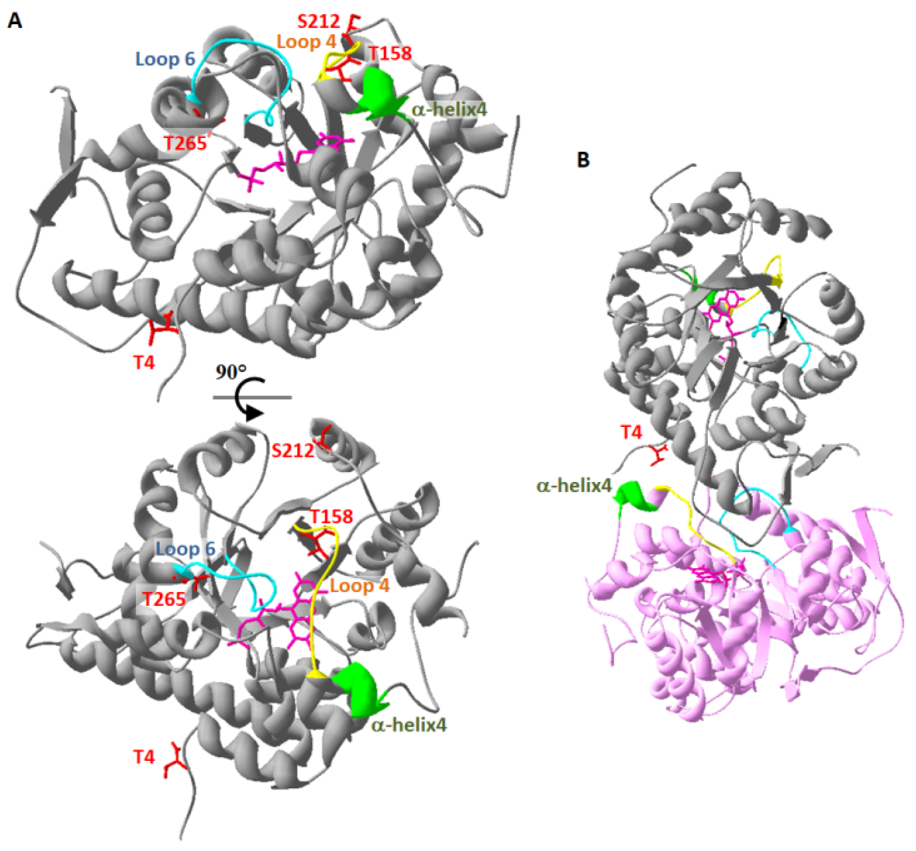
Phospho-mimetic GOX proteins exhibited different degrees of inhibition of their glycolate oxidase activity, except in one case, AtGOX1T212D, where the recombinant protein maintained GOX1WT activity (Table 2). This inhibition was seen to correlate with reduced amounts (or the absence) of FMN (Figure 1, Table 4) for GOXT4/5D and GOXT158/158D but this was not observed for GOXS212/213D and GOXT265/266D. When the phospho-mimetic mutation affected FMN content, the degree of inhibition was not constant since the mutation of T4/T5 to aspartate dramatically decreased GOX activity without changing the KM glycolate, while T158D and T159D mutations resulted in inactive recombinant GOX proteins (Table 2). These modifications in both Arabidopsis and maize GOX kinetics parameters could be attributed to the lack of FMN in these mutated proteins since its presence is essential for GOX activity (Table 4) [30]. Recently, the 3D structure of apo-GOX (lacking FMN) and holo-GOX from Nicotiana benthamiana revealed that loop4 (residues 157–165; 156–164 of AtGOX) and loop6 (residues 253–265; 252–264 of AtGOX) together with a loop situated between residues 28–33 (27–32 for AtGOX) formed a lid preventing the loss of FMN [30] (Figure 2A, Figure S1). Moreover, this lid had a strong interaction with a neighbouring GOX subunit of the tetrameric NbGOX, with α-helix4 (next to loop4) forming H-bonds with E3, T5 and N6 (E2, T4 and N5 of AtGOX) allowing a cooperative mechanism in FMN binding between GOX subunits of the same tetramer [30] (Figure 2B, Figure S1). Structural models of Arabidopsis thaliana GOX1 and GOX2 as well as ZmGO1 based on the structure of Spinacia oleracea GOX (PDB 1AL7) indicated that T4 formed H-bonds with R163, E165 and K169 of α-helix4 of a neighbouring GOX subunit.
When T4 was replaced by an aspartate in these models, only an H-bond with D163 of α-helix4 was present. Moreover, T158 is located close to a residue implicated in FMN-binding (T155) [31,32], the latter being also potentially phosphorylated [25]. Thus, T158 (present in loop4) and T4 (at the N-terminus) are located in essential domains for FMN binding; therefore, their mutation to aspartate, and probably their phosphorylation, may disturb FMN binding to holoGOX resulting in a less active or inactive enzyme. Furthermore, structural models indicated that when T158/159 was replaced by an aspartate, a new H-bond formed with Y129. This tyrosine residue normally formed an H-bond with FMN and when mutated to a phenylalanine (spinach GOXY129F) the resulting protein had only 3.5% of GOXWT activity and a kcat of only 0.74 s−1 instead of 20 s−1; however, FMN content was not affected [33]. Therefore, it is possible that phosphorylation of T158/159 could alter GOX activity even if FMN was retained in the GOX protein.
The mutation of T265/266 to aspartate decreased the glycolate-dependent kcat of AtGOX1/2T265D and ZmGO1T266D compared to the wild-type recombinant protein but only changed the KM glycolate of AtGOX1T265D (Table 2). As mentioned earlier, contrary to GOXT4D/T5D and GOXT158D/T159D, the FMN content of phospho-mimetic GOXT265D/T266D was comparable to recombinant GOXWT protein (Table 4) even though T265/T266 is located just at the end of loop6 (residues 253–265; 252–264 of AtGOX1/2) which is part of the “lid” structure involved in FMN loss in the pH sensor model [30]. Again, based on GOX structural models, T265 formed an H-bond with D285 of β-sheet7; however, when replaced by an aspartate this bond was broken and a new H-bond was formed with N253 of loop6 that is next to the active site H254 involved in proton abstraction during catalysis. We propose that GOXT265/T266D inhibited glycolate oxidase activity by bringing about a conformational change that interfered with catalysis via the displacement of H254.

3.2. Phospho-Site Mutations Suggest Differences between Photorespiratory GOX Proteins

Our studies to decipher photorespiratory GOX regulation by protein phosphorylation appeared to indicate that our different GOX enzymes (AtGOX1 versus AtGOX2 and AtGOX1/2 versus ZmGO1) did not always respond in a similar manner thus suggesting subtle differences between them even though they have a conserved photorespiratory role in planta. Both mutations at S212/S213 led to a similar decrease in the glycolate-dependent kcat of AtGOX2 and ZmGO1 while the kcat of AtGOX1 remained unaltered (Table 2). On the other hand, these mutations did not modify the KM glycolate of the different proteins (Table 2). Therefore, while AtGOX1 and AtGOX2 have been shown to have a redundant photorespiratory function [7] and similar kinetic properties ([34], Table 2), these observations suggest that they could have subtle regulatory and structural differences which could potentially explain reported differences in planta [11]. Furthermore, since S212/S213 was found to be important for AtGOX2 and ZmGO1 activity but not for AtGOX1 activity, we propose that the reduced kcat observed with GOXS212/213D does not reflect the actual consequences of a phosphorylated GOX at S212. It is indeed difficult to explain why AtGOX1 did not behave in a similar way to the other two GOX proteins since S212 is located in a loop between α-helixD and α-helixE that contains no known residues involved in glycolate oxidase activity. Our structural models showed that S212 was not involved in any H-bond formation; however, when replaced by an aspartate it could form a new H-bond with D54 in a loop between β-sheetA and β-sheetB of a neighbouring GOX subunit, but this was the case for all three proteins.
AtGOX1/2 T4 and ZmGO1 T5 mutations to valine were done to suppress any phosphorylation but also to mimic the sequence found in both AtHAOX1 and AtHAOX2, two enzymes that use more efficiently long-chain hydroxyl-acids [6], and HsHAOX2. As previously shown [8], AtGOX1 and AtGOX2 were less efficient using L-lactate as a substrate with both enzymes showing an 8-fold increase in KM although the kcat values were not significantly different when compared to glycolate (Table 2 and Table 3). However, contrary to the literature [8], both AtGOX1 and AtGOX2 were able use 2-hydroxy-octanoate as a substrate but again they were less efficient with lower kcat and increased KM values when compared to glycolate (Table 2 and Table 3). Interestingly, the C4-plant enzyme ZmGO1 had a similar kcat using either L-lactate or glycolate as a substrate while displaying only a 4-fold increase of KM and even though the kcat with 2-hydroxy-octanoate decreased in a similar manner to AtGOX proteins, its KM 2-hydroxy-octanoate was unaltered when compared to its KM glycolate (Table 2 and Table 3). Thus, even if the kinetics parameters of ZmGO1, AtGOX1 and AtGOX2 were similar for the glycolate oxidase reaction ([34], Table 2), ZmGO1 appeared to be able to use more efficiently both L-lactate and 2-hydroxy-octanoate (as seen from a comparison of kcat/KM ratios).
Differences between GOX proteins with respect to 2-hydroxy-octanoate were also observed when comparing the effect of T4/5 and T158/159 mutations to valine. Indeed AtGOX1T4V, AtGOX2T4V and ZmGO1T5V each exhibited a different alteration of KM 2-hydroxy-octanoate compared to their GOXWT counterparts (Table 3). This amino acid being important for GOX quaternary structure [30], its replacement by a valine may modify 2-hydroxy-octanoate specificity towards that of AtHAOX1 and AtHAOX2 due to possible subtle differences in 3D structure.
T158V and T159V mutations induced the same modifications of KM and kcat when using either of the three substrates tested thus indicating that the mutation affected the global enzymatic mechanism and it did not appear to be involved in determining substrate specificity (Table 2 and Table 3). Finally, AtGOX1/2T158V and ZmGO1T159v proteins were poorly active whatever the substrate used however for each protein the KM appeared to be lower than their GOXWT protein counterparts (Table 2 and Table 3). This inferred that T158 was involved in substrate binding as well as enzymatic activity and that when mutated to valine (as in Arabidopsis HAOX proteins) this somehow improved substrate binding. In conclusion to this part of the discussion, we can say that V4/5 and V157/158 containing GOX proteins were not transformed into HAOX enzymes as they were unable to use 2-hydroxy-octanoate more efficiently.

3.3. Discovering Conditions Inducing GOX Phosphorylation and Identifying GOX Kinases

To date, the only evidence of GOX protein phosphorylation has come from phosphoproteomics studies using mass spectroscopy analyses. From Table 1 it can be seen that only the peptide containing phosphorylated T158 has been reported more than once and although the accuracy of the methods in this domain have increased over the years, there are still a number of technical constraints and peptide identification is prone to error. It is therefore important to confirm GOX phosphorylation using other methods and/or confirm by mass spectroscopy using less complex samples instead of total protein extracts. Furthermore, it is necessary to identify conditions that bring about GOX phosphorylation so as to better understand its function. The T4 phospho-site of AtGOX1 and AtGOX2 was identified in leaves of plants subjected to oxygen depletion (5% of O2 for 3 h) (Table 1). In such low O2 conditions, photorespiration would be less important and GOX activity could be reduced via T4/T5 phosphorylation. Although the T158 phospho-site has been seen in several different phosphoproteome experiments, no significant differences in the quantity of this phosphopeptide were reported whatever the conditions tested (Table 1), even when comparing dark versus light and low versus high CO2 concentrations that are expected to modulate photorespiratory activity [28]. Thus, T158 phosphorylation may have a role that is not linked to photorespiration. Several studies have implicated GOX in response to pathogen attack [9,35,36], and therefore regulation of GOX activity, together with catalase, may be a way to modulate H2O2 production as part of plant defence signalling and this could involve protein phosphorylation. Arabidopsis phospho-site S212 was identified in a phosphoproteomic study of the triple kinase mutant snrk2.2/2.3/2.6 subjected either to ABA or dehydration treatments [26]. SnRK2.2, SnRK2.3 and SnRK2.6 (also known as OST1) are three kinases activated by ABA [37] and we have recently shown that another photorespiratory enzyme (SHMT1) responded to ABA and altered stomatal movements in response to salt stress [38]. Thus, GOX could be a target of leaf ABA signalling either in mesophyll cells or stomata. Indeed, the quantity of the TL(pS)WK phosphopeptide was shown to increase in response to either ABA or dehydration and this increase was absent in snrk2.2/2.3/2.6 seedlings [26].
Therefore, phosphoproteomics studies have had very limited success in providing insights into the eventual role of GOX phosphorylation. Since this post translational modification is a rapid and reversible response to environmental stimuli, experiments should be conducted to identify when GOX is differentially phosphorylated. With respect to GOX photorespiratory function, different conditions expected to modulate photorespiratory flux should be examined such as day/night cycle, variable CO2/O2, and stresses like heat, drought, salt and high light as well as pathogen attack. To simplify the detection of GOX phosphopeptides by mass spectroscopy, GOX should be specifically immunoprecipitated from soluble protein extracts at different times during the stress treatments or during the day/night cycle. In this way, it should be possible to identify conditions where GOX phosphorylation is modulated.
Of course, our in vitro data using recombinant phospho-mimetic GOX proteins only give an indication of what GOX phosphorylation might actually be doing with respect to enzyme activity. Indeed, GOX was produced in Escherichia coli as a recombinant phospho-mimetic protein and therefore we do not know whether this led to a non-physiological alteration of GOX structure (and activity). Since the modification was constitutive and present as soon as the protein was synthesized, it does not reflect a reversible phosphorylation of a protein. For instance, is the absence of FMN in GOXT4/5D and GOXT158/159D due to structural changes induced by the aspartate that inhibits FMN entry into the apo-protein to form the halo-protein, and therefore would phosphorylation lead to changes that favour FMN removal from the halo-protein? Furthermore, phospho-mimetic mutations do not always lead to the modifications in protein function brought about by an actual phosphorylation. Thus, to assess the real role of GOX phosphorylation, identification of the protein kinases (and also the phosphatases) involved would be crucial. Based on the SUBA database (http://suba.plantenergy.uwa.edu.au/), 33 protein kinases are predicted to be addressed to the peroxisome. Using machine learning methods to identify proteins carrying plant peroxisomal PST1 targeting signals, 11 protein kinases were identified as being addressed to the peroxisome [39]. More recently, a list of about 200 confirmed Arabidopsis peroxisomal proteins was compiled and four kinases and eight phosphatases or phosphatase subunits were identified [40]. A strategy to identify AtGOX1/2 kinases could be to retrieve knock-out mutant lines for predicted and/or verified peroxisomal protein kinases and compare AtGOX1/2 phosphorylation status by mass spectroscopy after AtGOX1/2 immunoprecipitation from soluble proteins extracted from wild-type and mutant plants treated to previously identified conditions that induce AtGOX1/2 phosphorylation.
In conclusion, photorespiratory GOX has the potential to be regulated by protein phosphorylation at several distinct sites. In general, phospho-mimetic recombinant GOX proteins exhibited reduced activities and this could be explained by predicted changes in structural interactions affecting key residues involved in FMN binding and catalysis. Phospho-mimetic GOX did not show any alteration in substrate specificity although C4-plant ZmGO1 did appear to have a relaxed substrate specificity when compared to C3-plant AtGOX1 and AtGOX2.

4. Materials and Methods

4.1. Plasmid Constructions and Site-Directed Mutagenesis

To produce recombinant proteins, previously made pET28a-AtGOX1, pET28a-AtGOX2 and pET28a-ZmGO1 expression plasmids [34] were used as templates to introduce point mutations using specific primers pairs (Table S1) and the QuikChange® II XL site-directed mutagenesis kit (Agilent®, Les Ulis, France), according to the manufacturer’s instructions. This strategy generated T4V, T4D, T158V, T158D, S212A, S212D, T265A, T265D (AtGOX1 and AtGOX2) and T5V, T5D, T159V, T159D, S213A, S213D, T266A, T266D (ZmGO1) mutated proteins. All constructions were subsequently verified by DNA sequencing using T7 and T7-term primers (Table S1).

4.2. Purification of Recombinant GOX Proteins, SDS-PAGE and Determination of FMN Content

GOX proteins were purified as described by Dellero et al. [34]. The purity of each recombinant GOX protein was checked by SDS-PAGE (10% acrylamide) stained with Coomassie Brilliant Blue [41]. Recombinant GOX proteins used to measure FMN content were purified without FMN in buffers and FMN levels were evaluated by spectrophotometry using the A280/A450 nm ratio.

4.3. GOX Activity Measurements

Enzyme activities were measured using 5 µg of purified recombinant GOX in 50 mM Tris-HCl, 0.1 mM FMN, pH 8.0 and different glycolate (0.05 to 10 mM), L-lactate (0.3 to 10 mM) and 2-hydroxy-octanoate (0.15 to 5 mM) concentrations by an enzyme-coupled reaction at 30 °C. H2O2 produced by GOX activity was quantified in the presence of 0.4 mM o-dianisidine and 2 U horseradish peroxidase by measuring the ∆A440nm using a Varian Cary 50 spectrophotometer. KM and kcat values were calculated using SigmaPlot 13.0 software, based on the curve fitting Michaelis–Menten equation: v0 = Vmax[S]/(KM + [S]).

4.4. Structural Analysis

AtGOX1, AtGOX2 and ZmGO1 3D-structures were designed based on the spinach GOX (UniProtKB: P05414) 3D-structure (PDB: 1al7.1) using the SWISS-MODEL server (https://swissmodel.expasy.org/) modelling service. Manipulation of 3D-structures was realized using Deepview (Swiss Pdb-viewer) (https://spdbv.vital-it.ch/) and included the compute H-bond formation and mutation tools.

Supplementary Materials

The following are available online at https://www.mdpi.com/2223-7747/9/1/27/s1, Figure S1: AtGOX1, AtGOX2, AtGOX3, AtHAOX1 and AtHAOX2 protein sequence alignments showing the conservation of phosphorylated residues and other important residues and structures, Figure S2: Amino acid sequence alignments of Lactococcus lactis, Aerococcus viridians, Nostoc and Chlamydomonas reinhardtii lactate oxidases, with glycolate oxidases and hydroxy-acid oxidases of human and plants, Figure S3: Purified recombinant His-tag proteins of wild-type AtGOX1, AtGOX2 and ZmGO1 and their phospho-site mutated forms on Coomassie-stained SDS-PAGE gels, Table S1: Primers for site-directed mutagenesis and DNA sequencing.

Author Contributions

Conceptualization, M.H. and M.J.; Methodology, M.H. and M.J.; Formal Analysis, M.H. and M.J.; Investigation, M.J., Y.L. and S.M.; Resources, M.H.; Data Curation, M.H. and M.J.; Writing & Editing, M.H. and M.J.; Supervision, M.H. and M.J.; Project Administration, M.H.; Funding Acquisition, M.H. and M.J. All authors have read and agreed to the published version of the manuscript.

Funding

This work was supported by a public grant overseen by the French National Research Agency (ANR) as part of the «Investissement d’Avenir» program, through the “Lidex-3P” project and a French State grant (ANR-10-LABX-0040-SPS) funded by the IDEX Paris-Saclay, ANR-11-IDEX-0003-02. This work was also supported by the ANR-14-CE19-0015 grant REGUL3P awarded to M.H., Y.L. was supported by a PhD fellowship from the China Scholarship Council.

Acknowledgments

We would like to thank Younès Dellero (INRA Rennes, France) who helped to initiate recombinant GOX studies in the team.

Conflicts of Interest

The authors declare no conflict of interest.

References

  1. Bauwe, H.; Hagemann, M.; Fernie, A.R. Photorespiration: Players, partners and origin. Trends Plant Sci. 2010, 15, 330–336. [Google Scholar] [CrossRef] [PubMed]
  2. South, P.F.; Cavanagh, A.P.; Liu, H.W.; Ort, D.R. Synthetic glycolate metabolism pathways stimulate crop growth and productivity in the field. Science 2019, 363, eaat9077. [Google Scholar] [CrossRef] [PubMed] [Green Version]
  3. Shen, B.R.; Wang, L.M.; Lin, X.L.; Yao, Z.; Xu, H.W.; Zhu, C.H.; Teng, H.Y.; Cui, L.L.; Liu, E.E.; Zhang, J.J.; et al. Engineering a New Chloroplastic photorespiratory bypass to increase photosynthetic efficiency and productivity in rice. Mol. Plant 2019, 12, 199–214. [Google Scholar] [CrossRef] [Green Version]
  4. Flügel, F.; Timm, S.; Arrivault, S.; Florian, A.; Stitt, M.; Fernie, A.R.; Bauwe, H. The photorespiratory metabolite 2-Phosphoglycolate regulates photosynthesis and starch accumulation in Arabidopsis. Plant Cell 2017, 29, 2537–2551. [Google Scholar] [CrossRef] [PubMed] [Green Version]
  5. Dellero, Y.; Jossier, M.; Schmitz, J.; Maurino, V.G.; Hodges, M. Photorespiratory glycolate-glyoxylate metabolism. J. Exp. Bot. 2016, 67, 3041–3052. [Google Scholar] [CrossRef] [Green Version]
  6. Esser, C.; Kuhn, A.; Groth, G.; Lercher, M.J.; Maurino, V.G. Plant and animal glycolate oxidases have a common eukaryotic ancestor and convergently duplicated to evolve long-chain 2-hydroxy acid oxidases. Mol. Biol. Evol. 2014, 31, 1089–1101. [Google Scholar] [CrossRef] [Green Version]
  7. Dellero, Y.; Jossier, M.; Glab, N.; Oury, C.; Tcherkez, G.; Hodges, M. Decreased glycolate oxidase activity leads to altered carbon allocation and leaf senescence after a transfer from high CO2 to ambient air in Arabidopsis thaliana. J. Exp. Bot. 2016, 67, 3149–3163. [Google Scholar] [CrossRef] [Green Version]
  8. Engqvist, M.K.M.; Schmitz, J.; Gertzmann, A.; Florian, A.; Jaspert, N.; Arif, M.; Balazadeh, S.; Mueller-Roeber, B.; Fernie, A.R.; Maurino, V.G. GLYCOLATE OXIDASE3, a glycolate oxidase homolog of yeast L-lactate cytochrome c oxidoreductase, supports L-lactate oxidation in roots of arabidopsis. Plant Physiol. 2015, 169, 1042–1061. [Google Scholar] [CrossRef] [Green Version]
  9. Rojas, C.M.; Senthil-Kumar, M.; Wang, K.; Ryu, C.-M.; Kaundal, A.; Mysore, K.S. Glycolate oxidase modulates reactive oxygen species-mediated signal transduction during nonhost resistance in Nicotiana benthamiana and Arabidopsis. Plant Cell 2012, 24, 336–352. [Google Scholar] [CrossRef] [Green Version]
  10. Saji, S.; Bathula, S.; Kubo, A.; Tamaoki, M.; Aono, M.; Sano, T.; Tobe, K.; Timm, S.; Bauwe, H.; Nakajima, N.; et al. Ozone-sensitive arabidopsis mutants with deficiencies in photorespiratory enzymes. Plant Cell Physiol. 2017, 58, 914–924. [Google Scholar] [CrossRef]
  11. Kerchev, P.; Waszczak, C.; Lewandowska, A.; Willems, P.; Shapiguzov, A.; Li, Z.; Alseekh, S.; Mühlenbock, P.; Hoeberichts, F.A.; Huang, J.; et al. Lack of GLYCOLATE OXIDASE1, but not GLYCOLATE OXIDASE2, attenuates the photorespiratory phenotype of CATALASE2-deficient Arabidopsis. Plant Physiol. 2016, 171, 1704–1719. [Google Scholar] [CrossRef]
  12. Yamaguchi, K.; Nishimura, M. Reduction to below threshold levels of glycolate oxidase activities in transgenic tobacco enhances photoinhibition during irradiation. Plant Cell Physiol. 2000, 41, 1397–1406. [Google Scholar] [CrossRef]
  13. Xu, H.; Zhang, J.; Zeng, J.; Jiang, L.; Liu, E.; Peng, C.; He, Z.; Peng, X. Inducible antisense suppression of glycolate oxidase reveals its strong regulation over photosynthesis in rice. J. Exp. Bot. 2009, 60, 1799–1809. [Google Scholar] [CrossRef] [Green Version]
  14. Lu, Y.; Li, Y.; Yang, Q.; Zhang, Z.; Chen, Y.; Zhang, S.; Peng, X.X. Suppression of glycolate oxidase causes glyoxylate accumulation that inhibits photosynthesis through deactivating Rubisco in rice. Physiol. Plant. 2014, 150, 463–476. [Google Scholar] [CrossRef]
  15. Zelitch, I.; Schultes, N.P.; Peterson, R.B.; Brown, P.; Brutnell, T.P. High glycolate oxidase activity is required for survival of maize in normal air. Plant Physiol. 2009, 149, 195–204. [Google Scholar] [CrossRef] [Green Version]
  16. Hodges, M.; Dellero, Y.; Keech, O.; Betti, M.; Raghavendra, A.S.; Sage, R.; Zhu, X.-G.; Allen, D.K.; Weber, A.P.M. Perspectives for a better understanding of the metabolic integration of photorespiration within a complex plant primary metabolism network. J. Exp. Bot. 2016, 67, 3015–3026. [Google Scholar] [CrossRef]
  17. Timm, S.; Florian, A.; Wittmiß, M.; Jahnke, K.; Hagemann, M.; Fernie, A.R.; Bauwe, H.; Rostock, D.; Germany, S.T. Serine acts as a metabolic signal for the transcriptional control of photorespiration-related genes. Plant Physiol. 2013, 162, 379–389. [Google Scholar] [CrossRef] [Green Version]
  18. Sandalio, L.M.; Gotor, C.; Romero, L.C.; Romero-Puertas, M.C. Multilevel regulation of peroxisomal proteome by post-translational modifications. Int. J. Mol. Sci. 2019, 20, 4881. [Google Scholar] [CrossRef] [Green Version]
  19. Ortega-Galisteo, A.P.; Rodríguez-Serrano, M.; Pazmiño, D.M.; Gupta, D.K.; Sandalio, L.M.; Romero-Puertas, M.C. S-Nitrosylated proteins in pea (Pisum sativum L.) leaf peroxisomes: Changes under abiotic stress. J. Exp. Bot. 2012, 63, 2089–2103. [Google Scholar] [CrossRef]
  20. Palmieri, M.C.; Lindermayr, C.; Bauwe, H.; Steinhauser, C.; Durner, J. Regulation of plant glycine decarboxylase by S-nitrosylation and glutathionylation. Plant Physiol. 2010, 152, 1514–1528. [Google Scholar] [CrossRef] [Green Version]
  21. Bartsch, O.; Mikkat, S.; Hagemann, M.; Bauwe, H. An autoinhibitory domain confers redox regulation to maize glycerate kinase. Plant Physiol. 2010, 153, 832–840. [Google Scholar] [CrossRef] [Green Version]
  22. Da Fonseca-Pereira, F.; Geigenberger, P.; Thormählen, I.; Souza, P.V.L.; Nesi, A.N.; Timm, S.; Fernie, A.R.; Araújo, W.L.; Daloso, D.M. Thioredoxin h2 contributes to the redox regulation of mitochondrial photorespiratory metabolism. Plant Cell Environ. 2019. [Google Scholar] [CrossRef]
  23. Reinholdt, O.; Schwab, S.; Zhang, Y.; Reichheld, J.; Alisdair, R. Redox-regulation of photorespiration through mitochondrial thioredoxin o1. Plant Physiol. 2019, 181, 442–457. [Google Scholar] [CrossRef] [Green Version]
  24. Hodges, M.; Jossier, M.; Boex-Fontvieille, E.; Tcherkez, G. Protein phosphorylation and photorespiration. Plant Biol. 2013, 15, 694–706. [Google Scholar] [CrossRef]
  25. Wu, X.N.; Rodriguez, C.S.; Pertl-Obermeyer, H.; Obermeyer, G.; Schulze, W.X. Sucrose-induced receptor kinase SIRK1 regulates a plasma membrane aquaporin in Arabidopsis. Mol. Cell. Proteom. 2013, 12, 2856–2873. [Google Scholar] [CrossRef] [Green Version]
  26. Umezawa, T.; Sugiyama, N.; Takahashi, F.; Anderson, J.C.; Ishihama, Y.; Peck, S.C.; Shinozaki, K. Genetics and phosphoproteomics reveal a protein phosphorylation network in the abscisic acid signaling pathway in Arabidopsis thaliana. Sci. Signal. 2013, 6, rs8. [Google Scholar] [CrossRef]
  27. Choudhary, M.K.; Nomura, Y.; Wang, L.; Nakagami, H.; Somers, D.E. Quantitative Circadian Phosphoproteomic analysis of Arabidopsis reveals extensive clock control of key components in physiological, metabolic, and signaling pathways. Mol. Cell. Proteom. 2015, 14, 2243–2260. [Google Scholar] [CrossRef] [Green Version]
  28. Abadie, C.; Mainguet, S.; Davanture, M.; Hodges, M.; Zivy, M.; Tcherkez, G. Concerted changes in the phosphoproteome and metabolome under different CO2/O2 gaseous conditions in Arabidopsis rosettes. Plant Cell Physiol. 2016, 57, 1544–1556. [Google Scholar]
  29. Aryal, U.K.; Krochko, J.E.; Ross, A.R.S. Identification of phosphoproteins in arabidopsis thaliana leaves using polyethylene glycol fractionation, immobilized metal-ion affinity chromatography, two-dimensional gel electrophoresis and mass spectrometry. J. Proteome Res. 2012, 11, 425–437. [Google Scholar] [CrossRef]
  30. Liu, Y.; Wu, W.; Chen, Z. Structures of glycolate oxidase from Nicotiana benthamiana reveal a conserved pH sensor affecting the binding of FMN. Biochem. Biophys. Res. Commun. 2018, 503, 3050–3056. [Google Scholar] [CrossRef]
  31. Lindqvist, Y.; Brändén, C.-I. The structure of glycolate oxidase from spinach. Proc. Natl. Acad. Sci. USA 1985, 82, 6855–6859. [Google Scholar] [CrossRef] [PubMed] [Green Version]
  32. Lindqvist, Y.; Brändén, C.I. The active site of spinach glycolate oxidase. J. Biol. Chem. 1989, 264, 3624–3628. [Google Scholar] [PubMed]
  33. Macheroux, P.; Kieweg, V.; Massey, V.; Söderlind, E.; Stenberg, K.; Lindqvist, Y. Role of tyrosine 129 in the active site of spinach glycolate oxidase. Eur. J. Biochem. 1993, 213, 1047–1054. [Google Scholar] [CrossRef] [PubMed] [Green Version]
  34. Dellero, Y.; Mauve, C.; Boex-Fontvieille, E.; Flesch, V.; Jossier, M.; Tcherkez, G.; Hodges, M. Experimental evidence for a hydride transfer mechanism in plant glycolate oxidase catalysis. J. Biol. Chem. 2015, 290, 1689–1698. [Google Scholar] [CrossRef] [PubMed] [Green Version]
  35. Xu, Y.-P.; Yang, J.; Cai, X.-Z. Glycolate oxidase gene family in Nicotiana benthamiana: Genome-wide identification and functional analyses in disease resistance. Sci. Rep. 2018, 8, 8615. [Google Scholar] [CrossRef] [Green Version]
  36. Chern, M.; Bai, W.; Chen, X.; Canlas, P.E.; Ronald, P.C. Reduced expression of glycolate oxidase leads to enhanced disease resistance in rice. PeerJ 2013, 1, e28. [Google Scholar] [CrossRef] [Green Version]
  37. Fujita, Y.; Nakashima, K.; Yoshida, T.; Katagiri, T.; Kidokoro, S.; Kanamori, N.; Umezawa, T.; Fujita, M.; Maruyama, K.; Ishiyama, K.; et al. Three SnRK2 protein kinases are the main positive regulators of abscisic acid signaling in response to water stress in arabidopsis. Plant Cell Physiol. 2009, 50, 2123–2132. [Google Scholar] [CrossRef] [Green Version]
  38. Liu, Y.; Mauve, C.; Lamothe-Sibold, M.; Guerard, F.; Glab, N.; Hodges, M.; Jossier, M. Photorespiratory serine hydroxymethyltransferase 1 activity impacts abiotic stress tolerance and stomatal closure. Plant Cell Environ. 2019, 42, 2567–2583. [Google Scholar] [CrossRef]
  39. Lingner, T.; Kataya, A.R.; Antonicelli, G.E.; Benichou, A.; Nilssen, K.; Chen, X.-Y.; Siemsen, T.; Morgenstern, B.; Meinicke, P.; Reumann, S. Identification of novel plant peroxisomal targeting signals by a combination of machine learning methods and in vivo subcellular targeting analyses. Plant Cell 2011, 23, 1556–1572. [Google Scholar] [CrossRef] [Green Version]
  40. Pan, R.; Hu, J. Proteomics of peroxisomes. Subcell. Biochem. 2018, 89, 3–45. [Google Scholar]
  41. Laemmli, U.K. Cleavage of structural proteins during the assembly of the head of bacteriophage T4. Nature 1970, 227, 680–685. [Google Scholar] [CrossRef] [PubMed]
Figure 1. Absorption spectra of recombinant AtGOX1, AtGOX2 and ZmGO1 and their T158/T159 phospho-site mutated forms.
Plants 09 00027 g001
Figure 2. Localisation of phosphorylated residues and important structures of AtGOX1. A structural model of AtGOX1 based on the 3D-structure of spinach GOX [31]. (A) AtGOX1 monomer showing phosphorylated residues T4, T158, S212 and T265 (red), loops 4 (yellow) and 6 (blue), α-helix4 (green) and the flavin mononucleotide (FMN) (pink). The bottom structure is rotated by 90° with respect to the top structure. (B) View of two AtGOX monomers (in grey and in pink) and the proximity between T4 (red) of a GOX subunit with α-helix4 (green) of a neighbouring GOX subunit.
Plants 09 00027 g002
Table 1. Phosphopeptides associated with AtGOX1 and AtGOX2.
Gene Name, LocusPhosphopeptidePeptide LocationSample Type, AgeTreatmentReferences
AtGOX1, At3g14420 orAtGOX2, At3g14415MEI(pT4)NVTEYDAIAK1–14/367LeavesOxygen depletionPhosPhAt 4.0 1
AIAL(pT155)VDTPRL151-161/367Seedlings,
11 days
Sucrose depletion [25]
AIALTVD(pT158)PRL151-161/367Seedling,
2 weeks
ABA and dehydration [26]
Seedlings,
10 days
Continuous light for 24 h[27]
RosetteVarying O2/CO2 conditions[28]
TL(pS212)WK210-214/367Seedlings,
2 weeks
ABA and dehydration [26]
QLDYVPA(pT265)ISALEEVVK258-273/367Cauline leaves, 2 months [29]
AtGOX1 At3g14420NHI(pT355)TEWDTPR352-362/367Seedlings,
10 days
Continuous light for 24 h[27]
The phosphorylated residue in each peptide is shown in bold with its position numbered according to AtGOX1. Peptide location shows the position of each phosphopeptide with respect to the number of residues in AtGOX1. 1 PhosPhAt 4.0: http://phosphat.mpimp-golm.mpg.de/.
Table 2. Effect of phospho-site mutations on glycolate-dependent kinetic parameters of recombinant AtGOX1, AtGOX2 and ZmGO1.
KMkcat KMkcat KMkcat
AtGOX1(µM)(s−1)AtGOX2(µM)(s−1)ZmGO1(µM)(s−1)
WT210 ± 9011.12 ± 3.08WT279 ± 3010.93 ± 3.48WT126 ± 3411.45 ± 1.86
T4V123 ± 359.59 ± 1.57T4V251 ± 7012.09 ± 1.50T5V135 ± 568.67 ± 2.32
T4D151 ± 440.61 ± 0.46 *T4D390 ± 2151.40 ± 0.42 *T5D157 ± 310.53 ± 0.22 *
T158V100 ± 19 *2.36 ± 0.47 *T158V133 ± 83 *2.49 ± 0.60 *T159V46 ± 20 *2.40 ± 0.35 *
T158Dno activityT158Dno activityT159Dno activity
S212A257 ± 3112.13 ± 2.15S212A249 ± 146.10 ± 0.64 *S213A89 ± 137.19 ± 0.46 *
S212D277 ± 2411.46 ± 2.31S212D325 ± 135.23 ± 0.41 *S213D129 ± 286.39 ± 1.45 *
T265A276 ± 11214.02 ± 2.65T265A237 ± 6711.33 ± 3.27T266A91 ± 1811.75 ± 2.94
T265D531 ± 50 *0.14 ± 0.06 *T265D388 ± 1592.06 ± 1.09 *T266D173 ± 650.62 ± 0.77 *
Mean values ± SD from three independent biological replicates. Statistical significance was determined by a Student’s t-test. Values in bold and marked by an asterisk were significantly different compared to the corresponding WT protein (p < 0.05).
Table 3. Effect of selected phospho-site mutations on L-lactate and 2-hydroxy-octanoate dependent kinetic parameters of recombinant AtGOX1, AtGOX2 and ZmGO1.
L-lactate2-hydroxy-octanoate
EnzymeKMkcatKMkcat
(µM)(s−1)(µM)(s−1)
AtGOX1WT1664 ± 2939.09 ± 1.37757 ± 825.95 ± 0.58
AtGOX1T4V1844 ± 9119.01 ± 3.59350 ± 221 *4.66 ± 1.56
AtGOX1T4D2377 ± 270 *0.30 ± 0.07 *1255 ± 8180.39 ± 0.17 *
AtGOX1T158V549 ± 160 *2.30 ± 0.56 *191 ± 51 *2.89 ± 0.89 *
AtGOX1T158Dno activityno activity
AtGOX2 WT2094 ± 7916.81 ± 1.54487 ± 993.78 ± 1.26
AtGOX2 T4V2119 ± 4536.65 ± 1.17951 ± 91 *4.05 ± 2.94
AtGOX2 T4D2019 ± 1690.67 ± 0.17 *1721 ± 677 *0.59 ± 0.30 *
AtGOX2 T158V439 ± 171 *1.71 ± 0.53 *501 ± 1162.46 ± 0.83
AtGOX2 T158Dno activityno activity
ZmGO1 WT495 ± 7510.85 ± 0.08136 ± 195.89 ± 0.14
ZmGO1 T4V488 ± 23311.99 ± 1.68168 ± 595.88 ± 1.59
ZmGO1 T4D719 ± 2330.57 ± 0.26 *414 ± 84 *0.53 ± 0.40 *
ZmGO1 T158V161 ± 29 *3.55 ± 0.53 *48 ± 36 *3.22 ± 0.39 *
ZmGO1 T158Dno activityno activity
Mean values ± SD from three independent biological replicates. Statistical significance was determined by a Student’s t-test. Values in bold and marked by an asterisk were significantly different compared to the corresponding WT protein (p < 0.05).
Table 4. A280/450nm ratios of recombinant AtGOX1, AtGOX2 and ZmGO1 proteins.
AtGOX1Ratio
280/450 nm
AtGOX2Ratio
280/450 nm
ZmGO1Ratio
280/450 nm
WT8.7 ± 2.1WT8.3 ± 0.7WT8.1 ± 1.8
T4V10.7 ± 4.1T4V6.9 ± 2.2T5V6.7 ± 3.0
T4D28.3 ± 6.1 *T4D36.5 ± 7.3 *T5D25.7 ± 6.2 *
T158V8.7 ± 2.3T158V8.2 ± 1.7T159V7.9 ± 1.1
T158D23.0 ± 8.3 *T158D20.5 ± 9.3 *T159D19.5 ± 7.1 *
S212A6.9 ± 3.3S212A12.4 ± 5.1S213A6.6 ± 3.5
S212D6.6 ± 4.0S212D10.4 ± 2.2S213D6.1 ± 1.7
T265A9 ± 3.3T265A9.6 ± 2.4T266A7.0 ± 0.1
T265D8.5 ± 2.5T265D11.8 ± 4.5T266D21.2 ± 12.1
Mean values ± SD from three independent experiments. Statistical significance was determined by a Student’s t-test. Values in bold and marked by an asterisk were significantly different compared to the corresponding WT protein (p < 0.05).