Article

Ursane Triterpenes and Norisoprenoids from Anchusa italica Retz. and Their Chemotaxonomic Significance

1
School of Pharmaceutical Sciences, South-Central Minzu University, Wuhan 430074, China
2
College of Life Sciences, South-Central Minzu University, Wuhan 430074, China
3
Qinghai Tibetan Medicine Research Institute, Xining 810016, China
4
Science and Technology Cooperation Base for Evaluation and Utilization of Traditional Medical Resources, South-Central Minzu University, Wuhan 430074, China
*
Authors to whom correspondence should be addressed.
Plants 2025, 14(9), 1385; https://doi.org/10.3390/plants14091385
Submission received: 4 March 2025 / Revised: 28 April 2025 / Accepted: 29 April 2025 / Published: 3 May 2025
(This article belongs to the Special Issue Isolation and Structure Elucidation of Plant Bioactive Compounds)

Abstract

:
A total of 31 compounds were isolated from the ethyl acetate and n-butanol fractions of Anchusa italica Retz., which contained one ursane triterpenoid, 2α,3β,19α-trihydroxy-23-formyl-urs-12-en-28,21β-olide (1), and five norisoprenoids: (2R,6R,9S)-9-hydroxy-4-megastigmen-3-one-2-O-β-D-glucopyranoside (3); (2R,6S,9S)-9-hydroxy-megastigman-4,7-dien-3-one-2-O-β-D-glucopyranoside (4); (+)-isololiolide β-D-glucopyranoside (5); (2S,8R)-loliolide β-D-glucopyranoside (6a); and (2R,8S)-loliolide β-D-glucopyranoside (6b). It also contained 25 known compounds (2 and 7–30). The chemical structures of the compounds, inclusive of their absolute configurations, were ascertained using spectroscopic methods such as NMR, HR-MS, and quantum chemical calculations (computational NMR and ECD), in combination with relevant literature data. Moreover, the chemotaxonomic significance of the isolated substances was discussed, with compounds 1, 2, and 7–13 potentially broadening the application of triterpenes as taxonomic markers for the classification of the genus Anchusa.

1. Introduction

Anchusa italica Retz. belongs to the genus Anchusa (Boraginaceae family) and is a perennial herbaceous plant that is naturally distributed in the Mediterranean region and tropical areas. In China, it is found in the northwestern and southwestern regions, such as Xinjiang, Gansu, and Sichuan [1]. As an important medicinal resource in the Uyghur medicine system, A. italica is recorded as “Gao Zi Wan” (meaning “tough”) and often used in the clinical treatment of cardiovascular and cerebrovascular diseases such as hypertension, palpitations, coughs, and depression [2]. Chemical investigations into A. italica have revealed that this plant contains abundant natural products, such as essential oils, triterpenoids, flavonoids, steroids, and alkaloids. From the perspective of pharmacological effects, the extracts and active compounds of A. italica exhibit a wide range of biological activities, including cardiovascular protection and anti-inflammatory, anti-bacterial, anti-oxidative, and neuroprotective effects [3]. To date, research has mainly focused on isolating and identifying total flavonoids and triterpenoid components, as well as exploring the mechanisms by which they protect myocardial cells [4]. Systematic analyses of their composition, activity, and contribution to plant taxonomy remain lacking.
To explore more compounds with biological activities and their contribution to the chemotaxonomic significance of this plant, we provide a certain basis for the development and utilization of A. italica. The whole plant powder was refluxed with 70% ethanol and then sequentially extracted with petroleum ether (PE), dichloromethane (CH2Cl2), ethyl acetate (EtOAc), and n-butanol (BuOH). The EtOAc and BuOH fractions were separated using chromatographic separation techniques: normal, reversed-phase silica gel column chromatography (CC), and HPLC. A total of thirty-one compounds (including nine triterpenes, twelve sesquiterpenes, four flavonoids, and six lignans) were identified, with six new compounds (1, 3, 4, 5, 6a, and 6b) as well as fourteen known compounds (15, 16, 18–25, and 27–30) first reported from A. italica (Figure 1). Compounds 5, 6a, and 6b are a differential isomer and a pair of stereoisomers, and in this study, the absolute configurations of these three compounds were identified for the first time using DP4+ analysis and ECD calculations. The chemotaxonomic significance of the isolated substances is also discussed.

2. Results

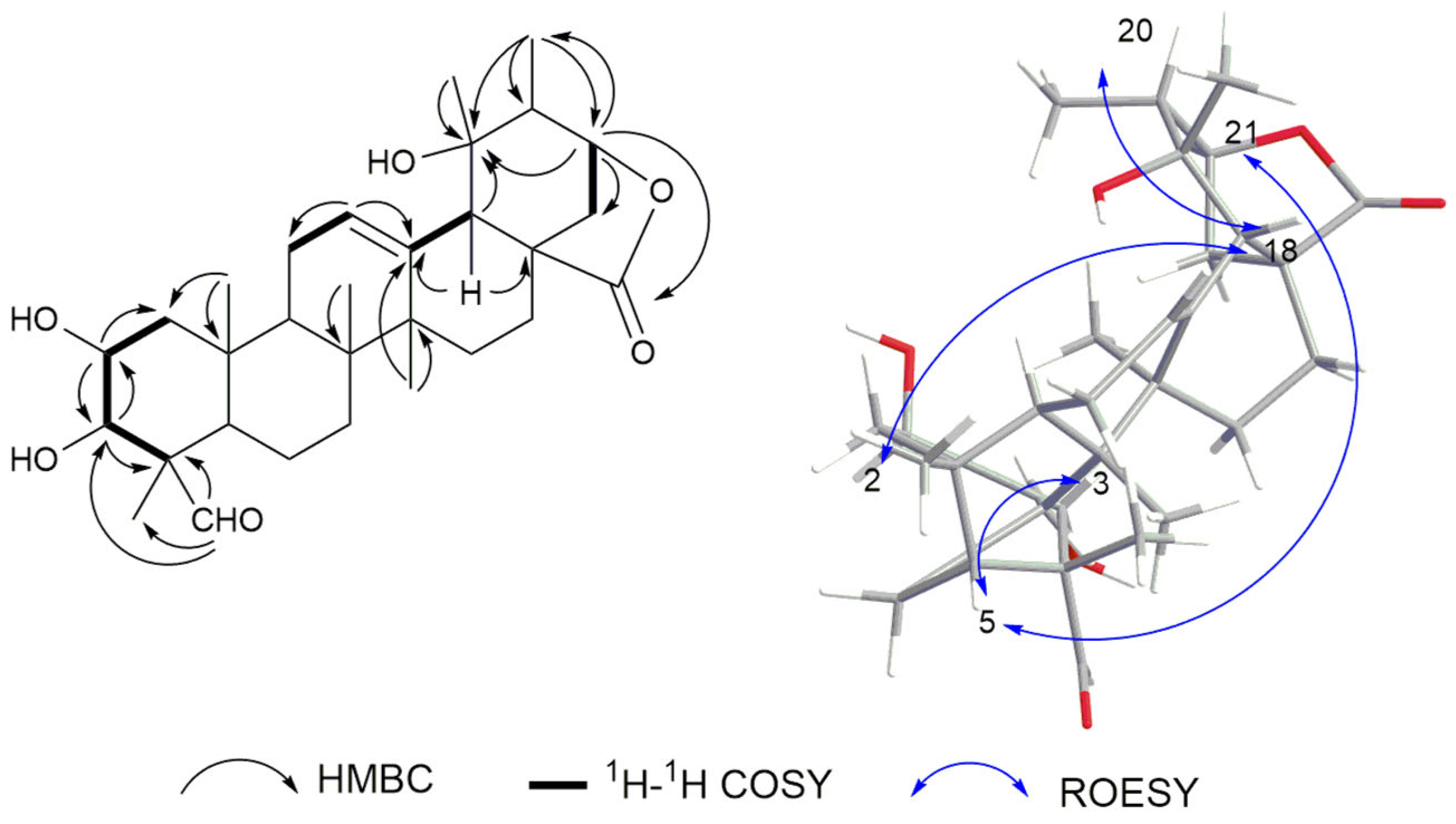
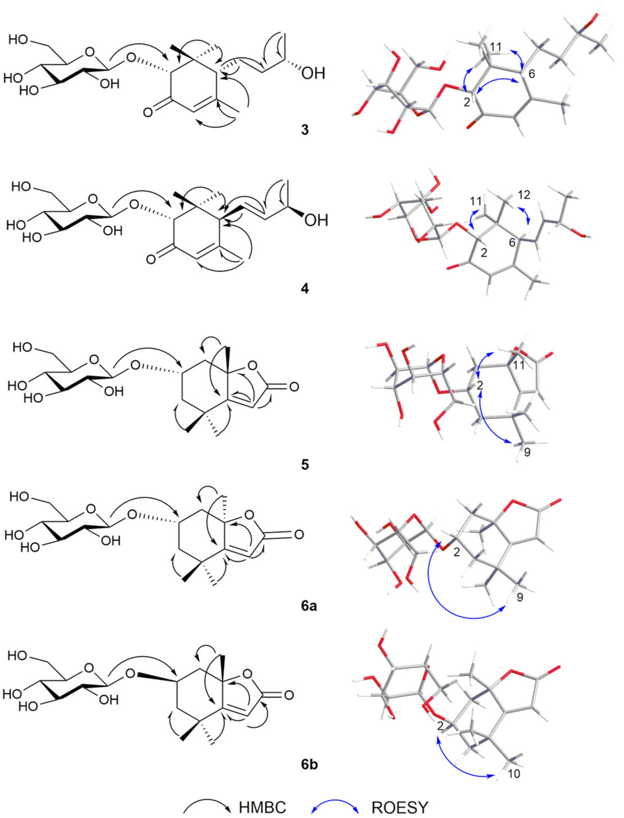
2.1. Structure Elucidation

Chemical investigation of the aerial parts of A. italica yielded six novel compounds (1, 3–5, 6a, and 6b) and 25 known analogues (2 and 730), including 2α,3β,19α-trihydroxy- 23-formyl-urs-12-en-28,21β-olide (1); (2R,6R,9S)-9-hydroxy-4-megastigmen-3-one-2-O- β-D-glucopyranoside (3); (2R,6S,9S)-9-hydroxy-megastigman-4,7-dien-3-one-2-O-β-D- glucopyranoside (4); (+)-isololiolide β-D-glucopyranoside (5); (2S,8R)-loliolide β-D- glucopyranoside (6a); (2R,8S)-loliolide β-D-glucopyranoside (6b); 2α,3β,21,24- tetrahydroxyoleanan-12-en-28-oic acid (2); niga-ichigoside F1 (7); niga-ichigoside F2 (8); pinfaensin (9); glucosyl tormentate (10); myrianthic acid (11); 24-epi-pinfaensic acid (12); hydroxyasiatic acid (13); (+)-vomifoliol (14) [3,4]; lippianoside E (15) [5]; 3-oxo-α- ionol-β-D-glucopyranoside (16) [6]; asysgangoside (17) [4]; (6S, 9R)-roseoside (18) [7]; sammangaoside B (19) [8]; (+)-isololiolide (20) [9]; tricin (21) [10]; 6-hydroxykaempferol 3-β-rutinoside (22) [11]; kaempferol 3-O-rutinoside (23) [12]; narcissin (24) [13]; (+)-syringaresinol (25) [14]; (+)-mediaresinol (26) [4]; dehydrodiconiferyl alcohol (27) [15]; vibruresinol (28) [16]; dehydrodiconiferyl alcohol 4-O-β-D-glucopyranoside (29) [17]; and syringaresinol-4′-O-β-D-glucopyranoside (30) [14].
Compound 1 was obtained as yellow amorphous powder with a molecular formula of C30H44O6 deduced from m/z 501.3211 [M + H]+, with an unsaturation degree of 9. The 1D NMR data (Table 1) revealed the presence of six tertiary methyl groups at [δH 0.90 (CH3-25), 0.95 (CH3-26), 1.05 (3H, d, J = 7 Hz, CH3-29), 1.21 (CH3-30), 1.26 (CH3-24), and 1.39 (CH3-27); δC 18.0, 17.4, 13.8, 28.4, 21.4, and 26.2, respectively]; three oxymethine protons at [δH 3.11 (1H, d, J = 9.5 Hz, H-3), 4.06 (1H, ddd, J = 5, 7, 10 Hz, H-2), and 4.34 (1H, d, J = 5.5 Hz, H-21); δC 82.6, 69.2, and 83.9]; one olefinic proton at [δH 5.53 (1H, t, J = 4 Hz, H-12); δC 131.3 and 137.7]; one aldehyde group at [δH 9.90 (1H, s, H-23); δC 208.5]; and one ester carbonyl group at (δC 185.4), suggesting that it is an ursane derivative. Its NMR data (Table 1) showed a strong similarity to 2α,3β,19α,23-tetrahydroxyurs-12-en-28, 21β-olide [3], except that the chemical shift of C-23 changed from δC 66.4 to δC 208.5 in compound 1, indicating that the C-23 changed from an oxymethylene to an aldehyde group.
The following correlations helped to confirm the presence of the ursolic acid skeleton: HMBC correlations of H-12 (δH 5.53) with C-11 and C-13 (δC 25.4 and 137.7); H-18 (δH 2.40) with C-13, C-17, and C-19 (δC 137.7, 45.8, and 74.3). Moreover, the following HMBC correlations helped to confirm the presence of the γ-lactone unit: correlations of H-21 (δH 4.35) with C-19, C-22, C-28, and C-30 (δC 74.3, 34.0, 185.4, and 28.4, respectively), together with the unsaturation degree of 9. Other correlations were also found, like H-23 (δH 9.90) with C-4 and C-24 (δC 55.3 and 21.4); CH3-25 (δH 0.90) with C-1 and C-10 (δC 47.5 and 39.3); CH3-27 (δH 1.39) with C-13 and C-14 (δC 137.7 and 42.7); and CH3-30 (δH 2.40) with C-19, C-20, and C-21 (δC 74.3, 45.3, and 83.9). Based on biosynthesis considerations, the natural ursane skeleton has an α-orientation for H-5 and a β-orientation for H-18 and CH3-25. The ROESY correlations of H-2 (δH 4.06) with H-18 (δH 2.40) and H-20 (δH 1.79), and H-5 (δH 1.15) with H-3 and H-21 (δH 3.11 and 4.34) suggested that H-2 is β-oriented, while H-3 and H-21 are α-oriented (Figure 2). Taking these data together, compound 1 was identified as 2α,3β,19α-trihydroxy-23-formyl-urs-12-en-28,21β-olide.
Compound 3 was obtained as a colorless oil, possessing a molecular formula of C19H32O8 deduced from m/z 389.2170 [M + H]+ as well as the unsaturation degree of 4. The 1D NMR data (Table 2 and Table 3) revealed the presence of four methyl groups at δH 2.03 (δC 24.6, CH3-10), 1.19 (δC 21.0, CH3-11), 1.19 (δC 19.8, CH3-13) and 0.88 (δC 24.5, CH3-12); one olefinic proton at [δH 5.81 (1H, d, J = 1.0 Hz, H-4) and δC 124.0]; a ketocarbonyl (δC 201.2) forming a conjugation with the olefinic bond; an isomeric proton at δH 4.20 (1H, s, H-2); and an oxygenated methyl proton at δH 3.87 (1H, m, H-9). Chemical shifts in the δH 3.23–3.90, along with an anomeric proton resonating at [δH 4.33 (1H, d, J = 8.0 Hz, H-1′), δC 102.0], were assigned as part of a sugar moiety and matched well with previously reported data as a β-glucopyranose. The 1H and 13C NMR data (Table 2 and Table 3) for 3 were similar to those for (2R,6R,9R)-2,9-dihydroxy-4- megastigmen-3-one [18]. Only one more glucose unit is present in 3, and the chemical shift value of C-2 was down-field shifted from δC 76.0 (in (2R,6R,9R)-2,9-dihydroxy- 4-megastigmen-3-one) to δC 77.2 in 3. HMBC correlations were analyzed from CH3-11 (δH 1.19) to C-1, C-2, C-6, and C-12 (δC 43.2, 77.2, 54.5, and 24.5, respectively); from H-10 (δH 2.03) to C-4, C-5, and C-6 (δC 124.0, 168.6, and 54.5); from H-7 (δH 2.11) and H-8 (δH 1.60) to C-6 (δC 54.5); and from H-13 (δH 1.19) to C-8 and C-9 (δC 38.2 and 75.4). Glucose was attributable to C-2 by HMBC correlations from δH 4.33 (H-1′) to δC 77.2 (C-2). The relative configuration of 3 was conducted to find correlations among H-2 (δH 4.20), H-6 (δH 2.10) and CH3-11 (δH 0.88), providing β-oriented configurations.
To determine the absolute configuration of the sugar molecule of compound 3, acid hydrolysis followed by chemical derivatization was performed to determine the D/L configuration of β-glucopyranose (Figures S59 and S64). The β-glucopyranose from the acid hydrolysis of compound 3 and the standard β-D glucopyranose were chemically reacted to give the final product. Their retention times were compared by HPLC analysis, which showed that the β-glucopyranose in compound 3 was identified as the D-form, as they showed the same retention time (tR = 20.2 min). To further verify the configuration of HO-9, two isomers (2R,6R,9S) and (2R,6R,9R) of 3 were subjected to NMR calculations with the DP4+ analysis. These results showed that (2R,6R,9S) of 3 (Figure S56) was the most likely structure for 3, with a 100.00% DP4+ probability based on all of the NMR data. The theoretical calculations of ECD spectra for 3 (2S,6S,9R) and the enantiomer (2R,6R,9S) were also performed using the TDDFT method. The calculated ECD spectra (2R,6R,9S) were in agreement with the experimental data (a positive peak at λ250 nm and a negative peak at λ300 nm), which indicated the absolute configuration (Figure 3). Taking these data together, compound 3 was identified as (2R,6R,9S)-9-hydroxy-4- megastigmen-3-one-2-O-β-D-glucopyranoside.
Compound 4 is a white powder, possessing a molecular formula of C19H30O8 deduced from m/z 387.20135 [M + H]+. The unsaturation degree is 5. The 1D NMR data (Table 2 and Table 3) suggested compound 4 might be the glucoside derivative at C-2 of the known compound 2,9-dihydroxy-megastigman-4,7-dien-3-one, because the chemical shift value of C-2 was down-field shifted from δC 75.5 in 2,9-dihydroxy-megastigman- 4,7-dien-3-one to δC 77.4 in 4 [19]. Glucose was attributable to C-2 by HMBC correlations from H-1′ (δH 4.36) to C-2 (δC 77.4). The β-glucopyranose in 4 was identified as the D-form, as they showed the same retention time (tR = 20.2 min) in HPLC analysis. The relative configuration was assigned by ROESY correlations of H-2 (δH 4.17) with H-8 (δH 5.77), H-9 (δH 4.40), and CH3-11 (δH 0.90), suggesting that H-2, H-9, and CH3-11 were on the same side of the molecule. The absolute configuration of 4 was determined via ECD calculations using the TDDFT method. The calculated ECD data for (2R,6S,9S) were in agreement with the experimental data of 4 (Figure 3), which indicated that compound 4 could be identified as (2R,6S,9S)-9-hydroxy-megastigman-4,7-dien-3-one-2-O-β-D- glucopyranoside.
Compound 5 was obtained as a colorless oil, possessing a molecular formula of C17H26O8 deduced from m/z 381.1519 [M + Na]+ and 13C NMR data, as well as an unsaturation degree of 5. The 1D NMR data (Table 2 and Table 3) revealed the presence of three methyl groups at [(δH 1.59 (CH3-11), 1.31 (CH3-10) and 1.28 (CH3-9); δC 25.4, 30.1, and 25.1)]; one olefinic proton at [δH 5.77 (1H, s, C-6), δC 113.4]; one ester carbonyl at (δC 173.8); and an isomeric proton at δH 4.27 (1H, m, H-2). Chemical shifts from δH 3.10 to 3.92, along with an anomeric proton resonating at [δH 4.42 (1H, d, J = 8.0 Hz, H-1′), δC 102.5], were assigned as part of a sugar moiety, which matched well with previously reported data, and subsequently confirmed as β-D-glucopyranose by HPLC (tR = 20.2 min, Figures S66 and S67). The 1H and 13C NMR data (Table 2 and Table 3) of 5 were similar to those of a known compound, (6S,7aS)-6-hydroxy-4,4,7a-trimethyl-6,7-dihydro-5H-1-benzofuran-2-one [20]. Only one more glucose unit appeared in 5, with the chemical shift value from δC 63.9 of (6S,7aS)-6-hydroxy-4,4,7a-trimethyl-6,7-dihydro-5H-1-benzofuran-2-one to δC 72.9 of 5. HMBC correlations were observed among H-9 (δH 1.28) with C-3, C-4, C-5, and C-10 (δC 48.9, 35.9, 183.8 and 30.1, respectively); H-11 (δH 1.59) with C-1, C-5, and C-8 (δC 45.6, 183.8, and 88.3); and H-6 (δH 5.77) to C-4, C-5, C-7, and C-8 (δC 35.9, 183.8, 173.8 and 88.3). Glucose was assigned at C-2 by the HMBC correlation (Figure 4) of H-1′ (δH 4.42) with C-2 (δC 72.9). The ROESY data of compound 5 indicated that H-2 was α-oriented by correlations of δH 4.27 (H-2) with δH 1.31 (CH3-9) and δH 1.59 (CH3-11). At last, the ECD analysis of 5 (2R,8R) and the enantiomer (2S,8S) were performed using the TDDFT method. The calculated ECD spectra for (2S,8S) were in agreement with the experimental spectra of 5 (Figure 5). Taking these data together, compound 5 was identified as (2S,8S)-isololiolide β-D-glucopyranoside.
Due to the similar 1D and part of 2D NMR data (HSQC, HMBC) of compounds 6a, 6b, and 5 (Table 2 and Table 3), the configuration of 6a and 6b was analyzed by ROESY experiment, and further verified by ECD calculations performed by the TDDFT method. ROESY results of stereoisomers 6a and 6b suggested that the H-2 (δH 4.28) of 6a was correlated with CH3-9 (δH 1.27), while that of 6b was correlated with CH3-10 (δH 1.45). At the same time, no correlations were found between H-2 (δH 4.28 and 4.30) and CH3-11 (δH 1.75 and 1.74) of both compounds. In addition, the calculated ECD spectra for (2S,8R) were in agreement with the experimental data of 6a, and the same spectra for (2R,8S) with the experimental data of 6b (Figure 5). In general, compound 6a was identified as (2S,8R)-loliolide β-D-glucopyranoside and compound 6b was identified as (2R,8S)-loliolide β-D-glucopyranoside.

2.2. The Protective Effects of Compounds 1–20 on Hypoxia/Reoxygenation-Injured Neonatal Rat Cardiomyocytes

An in vitro experimental model of hypoxia/reoxygenation (H/R) injury in neonatal rat cardiomyocytes was developed to evaluate the protective effects of isolated compounds 1–20. Neonatal rat cardiomyocytes were subjected to H/R injury for four hours and subsequently evaluated for the potential protective effects of compounds 1–20. The survival of H/R-treated neonatal rat cardiomyocytes was slightly increased after treatment with compounds 2, 4, 6b, 7, 9, 17, and 20 compared to the control group (H/R treatment only), suggesting that those compounds may have a protective effect on H/R-injured neonatal rat cardiomyocytes (Figure S65).

2.3. Chemotaxonomic Significance

In the present study, 31 compounds were identified from the aerial part of A. italica (Table 4). They were categorized as triterpenes (1, 2, and 7–13), norisoprenoids (3–6 and 14–20), flavonoids (21–24), and lignans (25–30). Among these compounds, three classes of compounds including norisoprenoids, flavonoids, and lignan derivatives were isolated from A. italica, which have been reported in other Anchusa species and even some species in the Boraginaceae family (Table 4). Compounds 18, 23, 24, 25, and 30 were reported in Borago officinalis L. [7], A. strigose Banks and Sol. [12], Heliotropium angiospermum [13], and Moltkia aurea Boiss. [14], leading to a close relationship from the perspective of chemical composition. Compounds 15, 16, 18–25, and 27–30 were found for the first time in A. italica, which may complement the results from existing investigations. In previous studies, triterpenes isolated from A. italica were rarely identified; thus, compounds 1, 2, and 7–13 might expand the use of triterpenes, especially ursane triterpenoids, as chemical taxonomic markers for the classification of the genus Anchusa.

3. Materials and Methods

3.1. General Experimental Procedures

NMR spectra were obtained on a Bruker AM-500 MHz spectrometer (Bruker, Karlsruher, Germany) using TMS as an internal reference. HR-ESI-MS was performed on a quadrupole ion trap high-resolution mass spectrometer (ThermoFisher Scientific, Waltham, MA, USA). CD spectra were obtained using a Chirascan detector (Applied Photophysics Limited Shanghai Representative Office, Shanghai, China). Optical rotation values were recorded on a Rudolph Auto pol IV polarimeter (Rudolph, Hackettstown, NJ, USA). UV full-wavelength scanning was conducted on a Shimadzu UV-2401 PC spectrophotometer (Shimadzu, Kyoto, Japan). CC was performed using silica gel (200–300 mesh, 300–400 mesh, Qingdao Haiyang Chemical Group Co., Qingdao, China). Thin-layer chromatography was performed on silica gel GF254 (Qingdao Haiyang Chemical Group Co., China). D101 macroporous resin was purchased from Tianjin Yunkai Company, China. MCI gel was purchased from Mitsubishi Chemical Group Co. (Mitsubishi Chemical Group, Toyko, Japan). Semi-preparative HPLC was performed on a QingBoHua HPLC system (QingBoHua Technology, Beijing, China) equipped with a binary high-pressure pump, a dual–wavelength UV detector, and a C18 column (250 mm × 10 mm, 5 μm, YMC Co., Ltd., Kyoto, Japan). Other chemical reagents were purchased from Sinopharm Chemical Reagent Co., Ltd., Shanghai, China.
The ECD calculations were carried out using Gaussian 09. Systematic conformational analyses for compounds 3, 4, 5, 6a, and 6b were performed via Confab using the MMFF94 molecular mechanics force field calculation. All of these conformers were further optimized using time-dependent density functional theory (TDDFT) at the B3LYP/6–311G (d, p) level in methanol with the IEFPCM model. Vibrational frequency analysis confirmed the stable structures. Based on the optimized structures, the ECD calculation was conducted using TDDFT with a total of 60 excited states. The ECD spectrum was simulated in SpecDis using a Gaussian function with half–bandwidths of 0.3 eV for compounds 3, 4, and 5.

3.2. Plant Material

The whole plant of A. italica was collected from Xinjiang Uygur Autonomous Region. The plant was picked and identified as A. italica by researcher Shi Leiling of the Xinjiang Institute of Traditional Chinese Medicine and Ethnomedicine. The specimen (A20220907) is stored in the Museum of Traditional Chinese Medicine, College of Traditional Chinese Medicine, Xinjiang Medical University.

3.3. Extraction and Isolation

The powder (2.5 kg) of A. italica was extracted in 70% ethanol under reflux (3 × 2 h each time). The ethanol extract was evaporated under reduced pressure to obtain the total extract (356 g). It was dissolved in water and then extracted three times sequentially with PE, CH2Cl2, EtOAc, and BuOH to provide the fractions accordingly.
The ethyl acetate soluble fraction (21 g) was subjected to CC (9.4 × 40 cm) over silica gel (200–300 mesh) and eluted with CH2Cl2/MeOH (100:0–70:30, v/v) to yield three fractions (Y1–3). Y1 (6 g) was subjected to CC (3 × 60 cm) over silica gel (300–400 mesh) and eluted with PE-EtOAc (85:15, v/v) to yield two fractions (Y11–12). Y11 (150 mg) was prepared by HPLC using acetonitrile (MeCN)/H2O (35:65, 210/254 nm) to provide compounds 20 (tR = 20 min, 3 mg), 21 (tR = 23 min, 2.6 mg), and 14 (tR = 27 min, 2.5 mg). Y12 (200 mg) was prepared by HPLC using MeCN/H2O (30:70, 210/254 nm) to provide compounds 15 (tR = 14 min, 3 mg), 25 (tR = 18 min, 13 mg), 26 (tR = 22 min, 3 mg), and 1 (tR = 29 min, 7 mg). Y2 (3.8 g) was subjected to CC (3 × 60 cm) over silica gel (300–400 mesh) and eluted with CH2Cl2/MeOH (50:1–10:1, v/v) to yield two fractions (Y21–22). Y21 (100 mg) was prepared by HPLC using MeCN/H2O (25:75, 210/254 nm) to provide compounds 27 (tR = 16 min, 1.8 mg) and 28 (tR = 20 min, 7 mg). Y22 (700 mg) was prepared by HPLC using MeCN/H2O (20:80, 210/254 nm) to provide compounds 7 (tR = 16 min, 134 mg), 8 (tR = 23 min, 86 mg), and 9 (tR = 29 min, 128 mg). Y3 (350 mg) was prepared by HPLC using MeCN/H2O (18:82, 210/254 nm) to provide compounds 10 (tR = 20 min, 12.6 mg), 11 (tR = 36 min, 23.4 mg), 23 (tR = 43 min, 3.5 mg), and 24 (tR = 45 min, 3.6 mg).
The BuOH soluble fraction (45.87 g) was subjected to CC (9.4 × 40 cm) over silica gel (200–300 mesh) and eluted with CH2Cl2/MeOH (100:0–60:40, v/v) to yield two fractions (Z1-Z2). Z1 (3.86 g) was subjected to CC (3 × 60 cm) over silica gel (300–400 mesh) and eluted with PE-EtOAc (75:25, v/v) to yield two fractions (Z11-Z12). Z11 (160 mg) was prepared by HPLC using MeCN/H2O (20:80, 210/254 nm) to provide compounds 3 (tR = 15.5 min, 2.1 mg), 16 (tR = 32 min, 1.0 mg), and 12 (tR = 38 min, 7.3 mg). Z12 (230 mg) was prepared by HPLC using MeCN/H2O (17:83, 210/254 nm) to provide compounds 22 (tR = 36 min, 4 mg), 29 (tR = 39 min, 2.3 mg), and 30 (tR = 45 min, 10.7 mg). Z2 (2.5 g) was subjected to CC (3×60 cm) over silica gel (300–400 mesh) and eluted with CH2Cl2/MeOH (70:30, v/v) to yield three fractions (Z21-Z23). Z21 (300 mg) was prepared by HPLC using MeCN/H2O (15:85, 210/254 nm) to provide compounds 13 (tR = 17 min, 3.4 mg), 4 (tR = 24 min, 0.8 mg), and 2 (tR = 29 min, 5.4 mg). Z22 (200 mg) was prepared by HPLC using MeCN/H2O (12:88, 210/254 nm) to provide compounds 17 (tR = 20 min, 23 mg), 18 (tR = 24 min, 6 mg), and 19 (tR = 36 min, 10.5 mg). Z23 (150 mg) was prepared by HPLC using MeCN/H2O (10:90, 210/254 nm) to provide compounds 5 (tR = 28 min, 1.3 mg), 6a (tR = 30 min, 0.8 mg), and 6b (tR = 32 min, 0.8 mg).
Spectroscopic Data
2α,3β,19α-trihydroxy-23-formyl-urs-12-en-28,21β-olide (1): yellow amorphous powder; α D 20 = +70.4° (c 0.03, MeOH); UV (MeOH) λmax 205 nm; 1H and 13C NMR data (CD3OD, 500 and 125 MHz), see Table 1; HR-ESI-MS m/z 501.3211 [M + H]+ (calcd for C30H45O6, 501.3200). Supplementary data were available at Figures S1–S10.
(2R,6R,9S)-9-hydroxy-4-megastigmen-3-one-2-O-β-D-glucopyranoside (3): colorless oil; α D 20 = +28.0° (c 0.05, MeOH); UV (MeOH) λmax 240 nm; 1H and 13C NMR data (CD3OD, 500 and 125 MHz), see Table 2 and Table 3; HR-ESI-MS m/z 389.2170 [M + H]+ (calcd for C19H33O8, 389.2160). Supplementary data were available at Figures S11–S19, S56, Tables S1–S8.
(2R,6S,9S)-9-hydroxy-megastigman-4,7-dien-3-one-2-O-β-D-glucopyranoside (4): α D 20 = −38.1° (c 0.05, MeOH); UV (MeOH) λmax 205 nm; 1H and 13C NMR data (CD3OD, 500 and 125 MHz), see Table 2 and Table 3; HR-ESI-MS m/z 387.2013 [M + H]+ (calcd for C19H31O8, 387.2017). Supplementary data were available at Figures S20–S28, S57–58, Tables S9–S32.
(+)-isololiolide β-D-glucopyranoside (5): α D 20 = +4.0° (c 0.02, MeOH); UV (MeOH) λmax 210 nm; 1H and 13C NMR data (CD3OD, 500 and 125 MHz), see Table 2 and Table 3; HR-ESI-MS m/z 381.1519 [M + Na]+ (calcd for C17H26O8Na, 381.1522). Supplementary data were available at Figures S29–S37, Tables S33–S34.
(2S,8R)-loliolide β-D-glucopyranoside (6a): α D 20 = −51.4° (c 0.05, MeOH); UV (MeOH) λmax 210 nm; 1H and 13C NMR data (CD3OD, 500 and 125 MHz), see Table 2 and Table 3; HR-ESI-MS m/z 381.1518 [M + Na]+ (calcd for C17H26O8Na, 381.1522). Supplementary data were available at Figures S38–S46, Tables S35–S36.
(2R,8S)-loliolide β-D-glucopyranoside (6b): α D 20 = +5.7° (c 0.03, MeOH); UV (MeOH) λmax 210 nm; 1H and 13C NMR data (CD3OD, 500 and 125 MHz), see Table 2 and Table 3; HR-ESI-MS m/z 381.1518 [M + H]+ (calcd for C17H26O8Na, 381.1522). Supplementary data were available at Figures S47–S55, Tables S35–S36.

3.4. Acid Hydrolysis of New Compounds

The new compounds 3, 4, 5, 6a, and 6b (1 mg each) were dissolved in 2 mL of CF3COOH (4 mol/L) solution and reacted at 95 °C for 3 h. The reaction was cooled to room temperature, the reaction solution was extracted with an equal volume (2 mL) of CH2Cl2 three times, and the aqueous layer after extraction was completely concentrated. A 1 mg amount of D-glucose (China Academy of Food and Drug Administration, Lot No. 110833) and the extracted aqueous phase were taken into four 25 mL round-bottomed flasks, and anhydrous pyridine (0.5 mL) and L-cysteine methyl ester hydrochloride (1.0 mg) were added, respectively; the reaction was carried out at 60 °C for 1 h. After cooling, 5 μL of o-toluene isothiocyanate was added to each reaction, and the reaction was carried out at 60 °C for 1 h. The solution was cooled to room temperature, the reaction solutions of the above four groups were extracted with an equal volume (2 mL) of CH2Cl2 three times, and the reaction solution was completely concentrated. The reaction solution of the above four groups was diluted 1-fold with pyridine, the samples (5 μL) were analyzed by HPLC (MeCN/H2O 25:75, 0.8 mL/min) with a detection wavelength of 250 nm, and the analytical column was a Waters chromatographic column (4.6 × 250 mm, 5 μm particle size). The acid hydrolysis products of compounds 3, 4, 5, 6a, and 6b showed peaks at tR 20.2 min; D-glucose showed peaks at tR 20.2 min (Supporting Information Figures S59–S64).

3.5. Quantum Chemical Calculations (Computational NMR and ECD)

The theoretical calculations were carried out using Gaussian 09. At first, all conformers were optimized at PM6. Room-temperature equilibrium populations were calculated according to the Boltzmann distribution law, based on which dominative conformers of the population over 1% were kept. The chosen conformers were further optimized at B3LYP/6-31G (d, p) in gas phase. Vibrational frequency analysis confirmed the stable structures. NMR calculations were carried out using the Gauge-Including Atomic Orbitals (GIAO) method at mPW1PW91/6-311 + G (2d, p) level in methanol simulated by the IEFPCM model. The TMS-corrected NMR chemical shift values were averaged according to the Boltzmann distribution and fitted to the experimental values by linear regression. The calculated 13C- and 1H-NMR chemical shift values of TMS in methanol were 49.2 and 4.9 ppm respectively. To confirm the conclusions of NMR calculations, DP4+ analysis was also performed.
N i N = g i e E i k B T g i e E i k B T
where N is the number of conformer i with energy and degeneracy at temperature T, and kB is Boltzmann constant.
ECD calculations were conducted at a B3LYP/6-311G (d, p) level in methanol with the IEFPCM model using time-dependent density functional theory (TDDFT). The rotatory strengths for 30 excited states were calculated. The ECD spectrum was simulated using the ECD/UV analysis tool on the Yinfo Cloud Platform (https://cloud.yinfotek.com/ (27 April 2025)) by overlapping Gaussian functions for each transition, where σ represents the width of the band at a height of 1/e, while ΔEi and Ri are the excitation energies and rotatory strengths for transition i, respectively. The spectra of the enantiomers were produced directly through mirror inversion about the horizontal axis.
Δ ε E = 1 2.241 × 10 39 × 1 2 π σ i A Δ E i R i E E i 2 σ 2

3.6. Cell Culture and Hypoxia/Reoxygenation

Primary cultured neonatal rat cardiomyocytes were cultured at 37 °C with 95% air and 5% CO2 in DMEM supplemented with 10% FBS, 100 μg/mL streptomycin, and 100 units/mL penicillin. When the cells had grown to 70–80% confluence, they were pretreated with compounds or vehicle (DMSO) for 4 h (a time point with better efficacy in cardiomyocyte protection). The cells were then exposed to an anaerobic medium (serum- and glucose-free) in an hypoxia incubator chamber (SANYO, IX51) with an anoxic mixture gas (95% N2 and 5% CO2) for 6 h at 37 °C followed by reoxygenation for 4 h with fresh culture medium (95% air and 5% CO2) to simulate H/R injury in isolated hearts.

4. Conclusions

The phytochemical investigation of ethanolic extracts of A. italica led to the isolation and characterization of 31 compounds, including a new ursane triterpene (1) and five new norisoprenoids (3, 4, 5, 6a, and 6b). The chemical structures of the new compounds (including absolute configurations) were fully confirmed through comprehensive analyses involving 1D and 2D NMR, HR-ESI-MS, acid hydrolysis, and computational methods for ECD calculation. The protective effects of compounds 1–20 against hypoxia/reoxygenation (H/R)-induced cardiomyocyte injury were tested. The chemical taxonomic significance of the triterpenes (1, 2, and 7–13) may expand the use of terpenoids as a chemotaxonomy marker for the classification of the genus Anchusa.

Supplementary Materials

The following supporting information can be downloaded at: https://www.mdpi.com/article/10.3390/plants14091385/s1, Figures S1–S10, HR-ESI-MS, NMR, CD, and UV data of 1; Figures S11–S19: HR-ESI-MS, NMR, CD, and UV data of 3; Figures S20–S28: HR-ESI-MS, NMR, CD, and UV data of 4; Figures S29–S37: HR-ESI-MS, NMR, CD, and UV data of 5; Figures S38–S55: HR-ESI-MS, NMR, CD, and UV data of 6; Figures S56–58: Computational NMR results of compounds 3 and 4; Figures S59–S64: Acid hydrolysis of compounds 3–5, 6a, and 6b; Figures S65: The ability of compounds 1–20 against H/R-induced neonatal rat cardiomyocytes injury. Figure S66: Plant photograph of Anchusa italica Retz.; Table S1–S36: Text data of computational NMR and ECD for the new compounds.

Author Contributions

Conceptualization, L.S., R.G. and J.L.; methodology, L.S., G.Y. and Y.Z.; formal analysis, M.L., B.H., Z.M. and X.H.; writing—original draft preparation, L.S.; writing—review and editing, L.S., R.G. and J.L.; supervision, R.G. and J.L.; funding acquisition, R.G. and J.L. All authors have read and agreed to the published version of the manuscript.

Funding

This work was sponsored by the Fundamental Research Funds for the Central Universities of “South-Central Minzu University” (CZQ 24023), the Fund for Key R&D Program Project of Hubei Province (2024BCA002, 2024BBB033), The Modern Transmission and Innovation Research Team of Traditional Chinese Medicine, South-Central Minzu University (KTZ20054).

Data Availability Statement

Data is contained within the article or Supplementary Materials.

Conflicts of Interest

The authors declare no conflicts of interest.

References

  1. Teng, L.; Li, M.; Ma, G. Protection of total flavonoids from Anchusa italica Retz. and fourcompounds on hypoxia-reoxygenation inducedinjury in myocardial cells and its mechanism. Chin. Pharmacol. Bull. 2021, 37, 409–416. [Google Scholar]
  2. Liu, Y.; Hu, B.; Wang, Y.; Bao, F.; Li, H.; Chen, L. Chemical constituents of Anchusa italica Retz. and their protective effects on cardiomyocytes injured by hypoxia/reoxygenation. Phytochem. Lett. 2020, 38, 155–160. [Google Scholar] [CrossRef]
  3. Hu, B.; Liu, Y.; Zheng, M.; Zhang, R.; Li, M.; Bao, F.; Li, H.; Chen, L. Triterpenoids from Anchusa italica and their protective effects on hypoxia/reoxygenation induced cardiomyocytes injury. Bioorg. Chem. 2020, 97, 103714. [Google Scholar] [CrossRef] [PubMed]
  4. Hou, Y.; Chen, K.; Deng, X.; Fu, Z.; Chen, D.; Wang, Q. Anti-complementary constituents of Anchusa italica. Nat. Prod. Res. 2017, 31, 2572–2574. [Google Scholar] [CrossRef] [PubMed]
  5. Zhang, Y.; Chen, Y.; Wang, S.; Dong, Y.; Wang, T.; Qu, L.; Li, N.; Wang, T. Bioactive constituents from the aerial parts of Lippia triphylla. Molecules 2015, 20, 21946–21959. [Google Scholar] [CrossRef] [PubMed]
  6. Sun, J.; Xia, F.; Wang, S.; Wang, K.; Chen, J.; Tu, P. Structural elucidation of two new megastigmane glycosides from the leaves of Aquilaria sinensis. Chin. J. Nat. Med. 2015, 13, 290–294. [Google Scholar] [PubMed]
  7. Samy, M.N.; Hamed, A.N.; Sugimoto, S.; Otsuka, H.; Kamel, M.S.; Matsunami, K. Officinalioside, a new lignan glucoside from Borago officinalis L. Nat. Prod. Res. 2016, 30, 967–972. [Google Scholar] [CrossRef] [PubMed]
  8. Tripetch, K.; Ryoji, K.; Phannipha, C.; Yoshikazu, H.; Kazuo, Y. Megastigmane and iridoid glucosides from Clerodendrum inerme. Phytochemistry 2001, 58, 333–336. [Google Scholar]
  9. Xu, W.; Wang, J.; Ju, B.; Lan, X.; Ying, X.; Stien, D. Seven compounds from Portulaca oleracea L. and their anticholinesterase activities. Nat. Prod. Res. 2022, 36, 2547–2553. [Google Scholar] [CrossRef] [PubMed]
  10. Stochmal, A.; Simonet, A.M.; Macias, F.A.; Oleszek, W. Alfalfa (Medicago sativa L.) flavonoids. 2. Tricin and chrysoeriol glycosides from aerial parts. J. Agric. Food Chem. 2001, 49, 5310–5314. [Google Scholar] [CrossRef] [PubMed]
  11. Qu, C.; Wang, L.; Lin, H.; Shang, E.; Tang, Y.; Yue, S.; Jin, Y.; Tao, W.; Li, S.; Hua, Y.; et al. Hierarchical identification of bioactive components in a medicinal herb by preparative high-performance liquid chromatography and selective knock-out strategy. J. Pharm. Biomed. Anal. 2017, 20, 206–216. [Google Scholar] [CrossRef] [PubMed]
  12. Yarmolinsky, L.; Budovsky, A.; Khalfin, B.; Yarmolinsky, L.; Ben-Shabat, S. Medicinal properties of Anchusa strigose and its active compounds. Molecules 2022, 25, 8239. [Google Scholar] [CrossRef] [PubMed]
  13. dos Santos, G.L.; de Araújo, D.I.; Raimundo e Silva, J.P.; do Nascimento, Y.M.; de Souza, T.A.; Opretzka, L.C.; Villarreal, C.F.; Abreu, L.S.; dos Santos Junior, F.M.; de Melo, J.I.; et al. Sucrose Diester of Aryldihydronaphthalene-Type Lignan with Anti-inflammatory Activity from Heliotropium angiospermum. Rev. Bras. Farmacogn. 2022, 32, 734–740. [Google Scholar] [CrossRef]
  14. Harput, U.S.; Nagatsu, A.; Saracoglu, I. Antioxidant and cytotoxic effects of Moltkia aurea Boiss. Rec. Nat. Prod. 2012, 6, 62–66. [Google Scholar]
  15. Hu, X.; Qin, N.; Xue, J.; Li, S.; Huang, X.; Sun, J.; Xu, F.; Li, Z.; Li, D.; Hua, H. Dehydrodiconiferyl alcohol from Silybum marianum (L.) Gaertn accelerates wound healing via inactivating NF-κB pathways in macrophages. J. Pharm. Pharmacol. 2020, 72, 305–317. [Google Scholar] [CrossRef] [PubMed]
  16. In, S.J.; Seo, K.H.; Song, N.Y.; Lee, D.S.; Kim, Y.C.; Baek, N.I. Lignans and neolignans from the stems of Vibrunum erosum and their neuroprotective and anti-inflammatory activity. Arch. Pharm. Res. 2015, 38, 26–34. [Google Scholar] [CrossRef] [PubMed]
  17. Giang, L.T.; Park, S.; Lee, S.; Seo, Y.; Van, K.; Tai, B.H.; Hang, N.T.M.; Thao, V.M.; Van, C.; Ban, N.K.; et al. Hepatoprotective Lignan glycosides from the leaves and stems of Symplocos cochinchinensis (Lour.) S. Moore. Chem. Biodivers. 2024, 21, 202400896. [Google Scholar] [CrossRef] [PubMed]
  18. Brigida, D.A.; Marina, D.G.; Antonio, F.; Pietro, M.; Palma, O.; Fabio, T. Structure elucidation and phytotoxicity of C13 nor-isoprenoids from Cestrum parqui. Phytochemistry 2004, 65, 497–505. [Google Scholar]
  19. Xu, W.; Yang, J.; Zhu, X.; Hu, Y.; Xu, S.; Li, Y.; Zhao, Y. Ionol derivatives from Euphorbia tirucalli. Rec. Nat. Prod. 2017, 11, 285–289. [Google Scholar]
  20. Vingre, S.M.G.; Costa, F.L.P. A theoretical study to the loliolide molecule and its isomers: A study by circular dichroism, QTAIM, and NMR theoretical methods. J. Mol. Model. 2021, 27, 116. [Google Scholar] [CrossRef] [PubMed]
Figure 1. Chemical structures of compounds 1–30 (* for the new compounds).
Plants 14 01385 g001
Figure 2. Key 1H-1H COSY (black bold), HMBC (black arrow) and ROESY (blue arrow) correlations for 1.
Plants 14 01385 g002
Figure 3. Experimental and calculated ECD spectra for 3 and 4.
Plants 14 01385 g003
Figure 4. Key HMBC (black arrow) and ROSEY (blue arrow) correlations for 3–5, 6a, and 6b.
Plants 14 01385 g004
Figure 5. Experimental and calculated ECD spectra for 5, 6a, and 6b.
Plants 14 01385 g005
Table 1. 1H and 13C-NMR data (500 and 125 MHz) of compounds 1 (CD3OD, δ in ppm, J in Hz).
PositionδCδHPositionδCδH
147.50.96, 1H, m
2.06, 1H, m
1629.91.23, 1H, m
1.81, 1H, m
269.24.06, 1H, ddd, 5, 7, 101745.8
382.63.11, 1H, d, 9.51857.02.40, 1H, s
455.3 1974.3
558.31.15, 1H, m2045.31.79, 1H, m
620.01.46, 1H, m
1.81, 1H, m
2183.94.34, 1H, d, 5.5
734.01.61, 2H, m2234.01.61, 1H, m
3.34, 1H, d, 9.0
841.9 23208.59.90, 1H, s
947.11.70, 1H, m2421.41.26, 3H, s
1039.3 2518.00.90, 3H, s
1125.41.99, 1H, m
2.08, 1H, m
2617.40.95, 3H, s
12131.35.53, 1H, t, 4.02726.21.39, 3H, s
13137.7 28185.4
1442.7 2913.81.05, 3H, d, 7.0
1529.31.08, 1H, m
1.89, 1H, m
3028.41.21, 3H, s
Table 2. 1H-NMR data (500 MHz) of compounds 3–6 (CD3OD, δ in ppm, J in Hz) 1.
Position34 56a6b
1 1.45, 1H, t, 12.0
2.65, 1H, dd, 2.0, 12.0
1.65, 1H, dd, 4.0, 14.0
2.65, 1H, dt, 2.5, 14.5
1.77, 1H, dd, 4.0, 14.0
2.67, 1H, dt, 2.5, 14.5
24.20, 1H, s4.17, 1H, s4.27, 1H, tt, 4.0, 12.04.28, 1H, m4.30, 1H, m
3 1.38, 1H, t, 12.0
2.18, 1H, dd, 2.0, 12.0
1.55, 1H, dd, 4.0, 14.0
2.20, 1H, dt, 2.5, 14.5
1.44, 1H, dd, 4.0, 14.0
2.21, 1H, dt, 2.5, 14.5
45.81, 1H, d, 1.05.91, 1H, d, 1.0
5
62.10, 1H, m2.77, 1H, d, 7.55.77, 1H, s5.74, 1H, s5.74, 1H, s
71.60, 1H, m
2.11, 1H, m
5.76, 1H, m
81.62, 2H, m5.77, 1H, m
93.87, 1H, m4.40, 1H, m1.31, 3H, s1.27, 3H, s1.28, 3H, s
102.03, 3H, d, 1.01.92, 3H, d, 10.01.28, 3H, s1.43, 3H, s1.45, 3H, s
110.88, 3H, s0.90, 3H, s1.59, 3H, s1.75, 3H, s1.74, 3H, s
121.19, 3H, s1.11, 3H, s
131.19, 3H, d, 6.01.30, 3H, d, 6.5
Glc
14.33, 1H, d, 8.04.36, 1H, d, 7.54.42, 1H, d, 8.04.37, 1H, d, 8.04.39, 1H, d, 8.0
23.13, 1H, m3.17, 1H, m3.12, 1H, m3.17, 1H, m3.17, 1H, m
33.33, 1H, m3.33, 1H, m3.29, 1H, m3.36, 1H, m3.35, 1H, m
43.24, 1H, m3.29, 1H, m3.24, 1H, m3.27, 1H, m3.27, 1H, m
53.24 13.22, 1H, m3.29 13.27, 1H, m3.27 1
63.65, 1H, m
3.83, 1H, m
3.67, 1H, m
3.83, 1H, m
3.63, 1H, m
3.89, 1H, m
3.64, 1H, m
3.85, 1H, m
3.65, 1H, m
3.86, 1H, m
1 Overlapped with other signals.
Table 3. 13C-NMR data (125 MHz) of compounds 3–6 (CD3OD, δ in ppm).
Position3456a6b
143.242.845.642.945.4
277.277.472.974.274.5
3201.2200.948.946.944.4
4124.0125.035.937.137.0
5168.6164.7183.8185.8185.7
654.558.6113.4113.1113.1
725.9128.3173.8174.4174.4
838.2138.588.388.988.9
975.477.030.130.931.0
1024.623.725.126.626.7
1121.025.525.427.127.0
1224.521.0
1319.821.0
Glc
1102.0102.5102.7102.9103.2
275.175.374.875.375.3
378.178.177.878.578.4
471.771.571.571.771.7
577.878.077.877.977.9
662.962.662.662.862.8
Table 4. A comparison of the compounds from Anchusa italica with other species in the genus Anchusa or Boraginaceae 1,2,3.
NumberCompoundTypeA. italica
12α,3β,19α-trihydroxy-23-formyl-urs-12-en-28,21β-olidetriterpene1, 2
22α,3β,21,24-tetrahydroxyoleanan-12-en-28-oic acidtriterpene1
3(2R,6R,9S)-9-Hydroxy-4-megastigmen-3-one-2-O-β-D-glucopyranosidenorisoprenoids1, 2
4(2R,6S,9S)-9-Hydroxy-megastigman-4,7-dien-3-one-2-O-β-D-glucopyranosidenorisoprenoids1, 2
5(+)-Isololiolide β-D-glucopyranosidenorisoprenoids1, 2
6a(2S,8R)-Loliolide β-D-glucopyranosidenorisoprenoids1, 2
6b(2R,8S)-Loliolide β-D-glucopyranosidenorisoprenoids1, 2
7Niga-ichigoside F 1triterpene1
8Niga-ichigoside F 2triterpene1
9Pinfaensintriterpene1,
10Glucosyl tormentatetriterpene1
1123-Hydroxytormentic acidtriterpene1
1224-epi-pinfaensic acidtriterpene1
1319α-Hydroxyasiatic acidtriterpene1
14(+)-Vomifoliolnorisoprenoids1
15Lippianoside Enorisoprenoids1, 3
163-oxo-α-ionol-β-D-glucopyranosidenorisoprenoids1, 3
17Asysgangosidenorisoprenoids1
18(6S,9R)-Roseosidenorisoprenoids1, 3Borago officinalis L.
19Sammangaoside Bnorisoprenoids1, 3
20(+)-Isololiolidenorisoprenoids1, 3
21Tricinflavone1, 3
226-Hydroxykaempferol 3-β-rutinosideflavonol1, 3
23Kaempferol 3-O-rutinosideflavonol1, 3Anchusa strigosa Banks & Sol.
24Narcissinflavonol1, 3Heliotropium angiospermum
25(+)-Syringaresinollignans1, 3Moltkia aurea Boiss.
26(+)-Mediaresinollignans1
27Dehydrodiconiferyl alcohollignans1, 3
28Vibruresinollignans1, 3
29Dehydrodiconiferyl alcohol 4-O-β-D-glucopyranosidelignans1, 3
30Syringaresinol-4′-O-β-D-glucopyranosidelignans1, 3Moltkia aurea Boiss.
1 Reported in this study; 2 New compound; 3 Compound reported from Anchusa italica for the first time.
Disclaimer/Publisher’s Note: The statements, opinions and data contained in all publications are solely those of the individual author(s) and contributor(s) and not of MDPI and/or the editor(s). MDPI and/or the editor(s) disclaim responsibility for any injury to people or property resulting from any ideas, methods, instructions or products referred to in the content.

