Article

Silencing of Putative Plasmodesmata-Associated Genes PDLP and SRC2 Reveals Their Differential Involvement during Plant Infection with Cucumber Mosaic Virus

by
Richita Saikia
1,2,3,
Athanasios Kaldis
1,
Carl Jonas Spetz
3,
Basanta Kumar Borah
2 and
Andreas Voloudakis
1,*
1
Laboratory of Plant Breeding and Biometry, Faculty of Crop Science, Agricultural University of Athens, 11855 Athens, Greece
2
Department of Agricultural Biotechnology, Assam Agricultural University, Jorhat 785013, Assam, India
3
Division of Biotechnology and Plant Health, Norwegian Institute of Bioeconomy Research, 1433 Ås, Norway
*
Author to whom correspondence should be addressed.
Plants 2025, 14(3), 495; https://doi.org/10.3390/plants14030495
Submission received: 30 October 2024 / Revised: 3 February 2025 / Accepted: 4 February 2025 / Published: 6 February 2025

Abstract

Plant viruses utilize a subset of host plasmodesmata-associated proteins to establish infection in plants. In the present study, we aimed to understand the role of two plant genes, one encoding a putative plasmodesma located protein (PDLP) and a homolog of soybean gene regulated by cold 2 protein (SRC2) during Cucumber mosaic virus (CMV) infection. Virus-induced gene silencing (VIGS) was used to silence PDLP and SRC2 genes in Nicotiana benthamiana and in two related solanaceous plants, N. tabacum and Capsicum chinense Jacq. (Bhut Jolokia). Up to 50% downregulation in the expression of the PDLP gene using the TRV2-PDLP VIGS construct was observed in N. benthamiana and N. tabacum while, using the same gene construct, 30% downregulation of the target mRNA was observed in C. chinense. Similarly, using the TRV2-SRC2 VIGS construct, a 60% downregulation of the SRC2 mRNA was observed in N. benthamiana, N. tabacum, and a 40% downregulation in C. chinense as confirmed by qRT-PCR analysis. Downregulation of the PDLP gene in N. benthamiana resulted in delayed symptom appearance up to 7–12 days post inoculation with reduced CMV accumulation compared to the control plants expressing TRV2-eGFP. In contrast, SRC2-silenced plants showed enhanced susceptibility to CMV infection compared to the control plants. Our data suggest that the PDLP gene might facilitate infection of CMV, thus being a susceptibility factor, while the SRC2 gene could play a role in resistance to CMV infection in N. benthamiana.

1. Introduction

To establish an infection, most plant viruses utilize specialized membrane-linked cytoplasmic channels known as plasmodesmata (PD) for intercellular trafficking of virus particles in the host [1]. They encode at least one dedicated protein termed movement protein (MP) that can interact with host proteins and modify the structure of PD [2,3]. Viral MPs can form tubules that assemble in PD and thereby manipulate the size exclusion limit of PD for the transport of viral complexes to the adjacent cells [4]. Viruses encoding such tubule-forming MPs belong to six major families: Badnaviridae, Bromoviridae, Caulimoviridae, Ilaviridae, Secoviridae, and Tospoviridae [5,6]. To date, only a few of the host proteins localized to PD have been shown to interact with viral MPs. These include proteins such as calreticulin [7], protein kinase PAPK1 [8], remorin [9], endoplasmic reticulum (ER)-embedded proteins including reticulons [10,11], plasmodesmata-associated class 1 reversibly glycosylated polypeptide [12], ankyrin-repeat containing protein (ANK) [13], synaptotagmin [14], and other plasmodesmata-located proteins (PDLPs) [5,15]. It is suggested that ANK together with β-glucanases could regulate the size exclusion limit of plasmodesmata [13]. ER-embedded proteins have been shown as positive regulators for viral movement and spread of cucumber mosaic virus (CMV) [10]. PDLPs have been shown as positive regulators for viral movement and spread of grapevine fan leaf virus (GFLV) and cauliflower mosaic virus (CaMV) [5]. Another viral protein of CaMV, named P6, colocalized with host PDLP1 and CaMV MP in the PD [16]. The study also showed that P6 physically interacts with a C2 calcium-dependent membrane-targeting protein, SRC2 (Soybean Response to Cold) from Arabidopsis, and this protein is capable of a close association with the tubule formation in the PD. Although the expression of pepper CaSRC2-1 gene was shown to be upregulated during bacterial and viral infection in chilli (Capsicum annum) [17], little is known about the exact role of SRC2 and PDLP genes during plant viral infection, particularly for broad host range viruses like CMV. CMV infects more than 1000 plant species [18,19]. These include some of the most valuable horticultural crops such as chilli, cucumber, okra, squash, and tomato [20]. CMV has a linear tripartite genome of positive-sense single-stranded (ss) RNA (RNA1/RNA2/RNA3) and belongs to the family Bromoviridae [21]. It is also known that the MP of CMV localizes to the PD like other viruses belonging to Bromoviridae family [22]; however, the exact role of host proteins during CMV infection is not well defined as of yet.
RNA interference (RNAi) or post-transcriptional gene silencing (PTGS) is a natural surveillance system adopted by plants that can regulate levels of endogenous or foreign transcripts by degrading them in a sequence-specific manner. The PTGS system becomes activated in response to the formation of double-stranded RNA (dsRNA), which involves sequence-specific recognition of target mRNA molecules based on sequence homology and subsequent degradation of these molecules by host ribonucleases and other proteins in the RNA induced silencing complex (RISC), thereby inhibiting their translation process [23]. The RNAi mechanism can be evoked using recombinant viral vectors that can efficiently deliver silencing inducer molecules to plant cell and study target gene function. This reverse genetic approach of studying plant genes using viral vectors is called virus-induced gene silencing (VIGS). Some of the recombinant viral vectors are derived from the genome of Potato virus X (PVX), Tobacco mosaic virus (TMV), and Tobacco rattle virus (TRV), of which TRV- and PVX- have been widely utilized [24,25]. The TRV-VIGS has been used to characterize genes in many plant species related to plant developmental processes, biotic and abiotic stress tolerance, symbiosis, metabolite synthesis, and plant evolution [25,26,27,28]. TRV-VIGS was first reported in N. benthamiana for silencing an endogenous gene, namely phytoene desaturase (PDS), and this vector system was further modified [29,30,31]. Since then, several disease resistance and susceptibility genes were identified against viral infection in plant species using the TRV-VIGS system [32,33,34].
In the present study, the role of two genes, one encoding putative plasmodesmata-localized protein PDLP and a protein homolog of SRC2 from Nicotiana benthamiana and N. tabacum were studied using the TRV-based VIGS system for their involvement in plant defense against or susceptibility to CMV.

2. Results

2.1. Homology Analysis of PDLP and SRC2 Genes in the Nicotiana and Capsicum Species

Using BLASTn and phylogenetic analysis with known PDLP genes from Arabidopsis thaliana, we selected a homolog of PDLP gene (XM_016610375.1) from N. tabacum which was identified in N. benthamiana (Niben101Scf03456g00018.1, 95.16% sequence identity with 99% query cover), C. annum (XM_016721975.2, 80.26% sequence identity with query cover 99%) and C. chinensis (MCIT02000006.1, 75.26% sequence identity with query cover 100%) (Figure 1A, Supplementary Figure S1). Similarly, a homolog of the SRC2 gene (XM_016630487.1) from N. tabacum was selected which was identified in N. benthamiana (DQ465395.1, 93.26% sequence identity with 99% query cover), C. annum (NM_001324570.1, 76.98% sequence identity with 100% query cover), and C. chinensis (AB442165.1, 76.68% sequence identity with query cover 94%) (Figure 1B, Supplementary Figure S2) by using BLASTn. The homologous sequences of PDLP and SRC2 were used to design primers for PCR analyses (RT-PCR and RT-qPCR) and for construction of the VIGS clones.

2.2. Relative Expression of PDLP and SRC2 Genes During CMV Infection in N. benthamiana

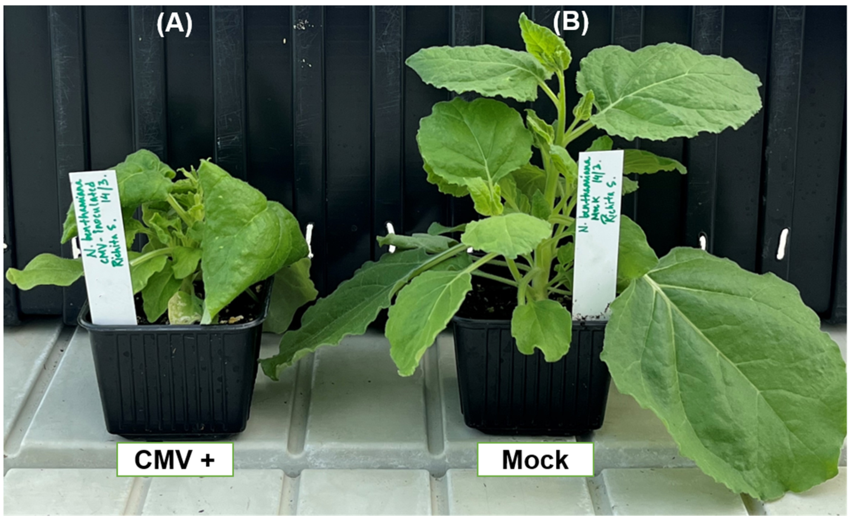
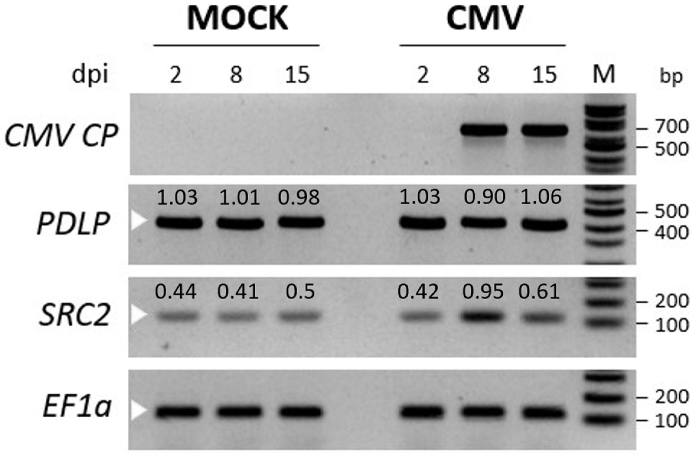
To investigate the expression pattern of PDLP and SRC2 genes, N. benthamiana leaves were inoculated with CMV, phenotyped, and sampled at 2, 8, and 15 days post inoculation (dpi). Typical downward leaf curling and mosaic symptoms were observed at 8 dpi, but the symptoms were more prominent at 14 dpi (Figure 2). Using semi-quantitative RT-PCR, the CMV CP was detected only at 8 and 15 dpi in the inoculated leaves, but not in the 2 dpi leaf samples (Figure 3).
No CMV-specific band was detected in the systemic leaves of the plants treated with the phosphate buffer (mock) (Figure 3). The expression levels of the PDLP and SRC2 genes were observed, and it was found that the PDLP gene expression was nearly the same between the mock and CMV inoculated plants, suggesting that the gene was constitutively expressed irrespective of the CMV infection. However, the expression of the SRC2 gene was upregulated upon CMV inoculation, as observed at 8 and 15 dpi compared to the mock (Figure 3). Therefore, to understand the functional role of these two genes during viral infection, we decided to silence the genes in N. benthamiana employing a TRV-based VIGS strategy.

2.3. VIGS of PDLP and SRC2 Genes in Tobacco and Bhut Jolokia Pepper

2.3.1. Designing of VIGS Constructs and Silencing of PDLP and SRC2 Genes in N. benthamiana, N. tabacum, and C. chinense

A conserved region of PDLP (383 bp) and SRC2 (364 bp) genes from tobacco, N. benthamiana, C. annum, and C. chinense was identified using multiple sequence alignment by Clustal Omega (Figures S1 and S2). The conserved regions of PDLP and SRC2 gene sequences were amplified using PCR with the respective primer pairs (Table 1) and cloned into the pTRV2 plasmid vector. Colony PCR of the transformed E. coli cells revealed positive bacterial colonies harboring the cloned constructs, pTRV2-NtPDLP and pTRV2-NtSRC2. Sanger sequencing of at least three positive clones from each gene fragment confirmed the presence of NtPDLP and NtSRC2 gene fragments in pTRV2 in sense orientation. Alignment of the sequenced PDLP and SRC2 gene fragments with N. tabacum PDLP (XM_016610375.1) and SRC2 (XM_016630487.1) revealed 100% nucleotide identity.
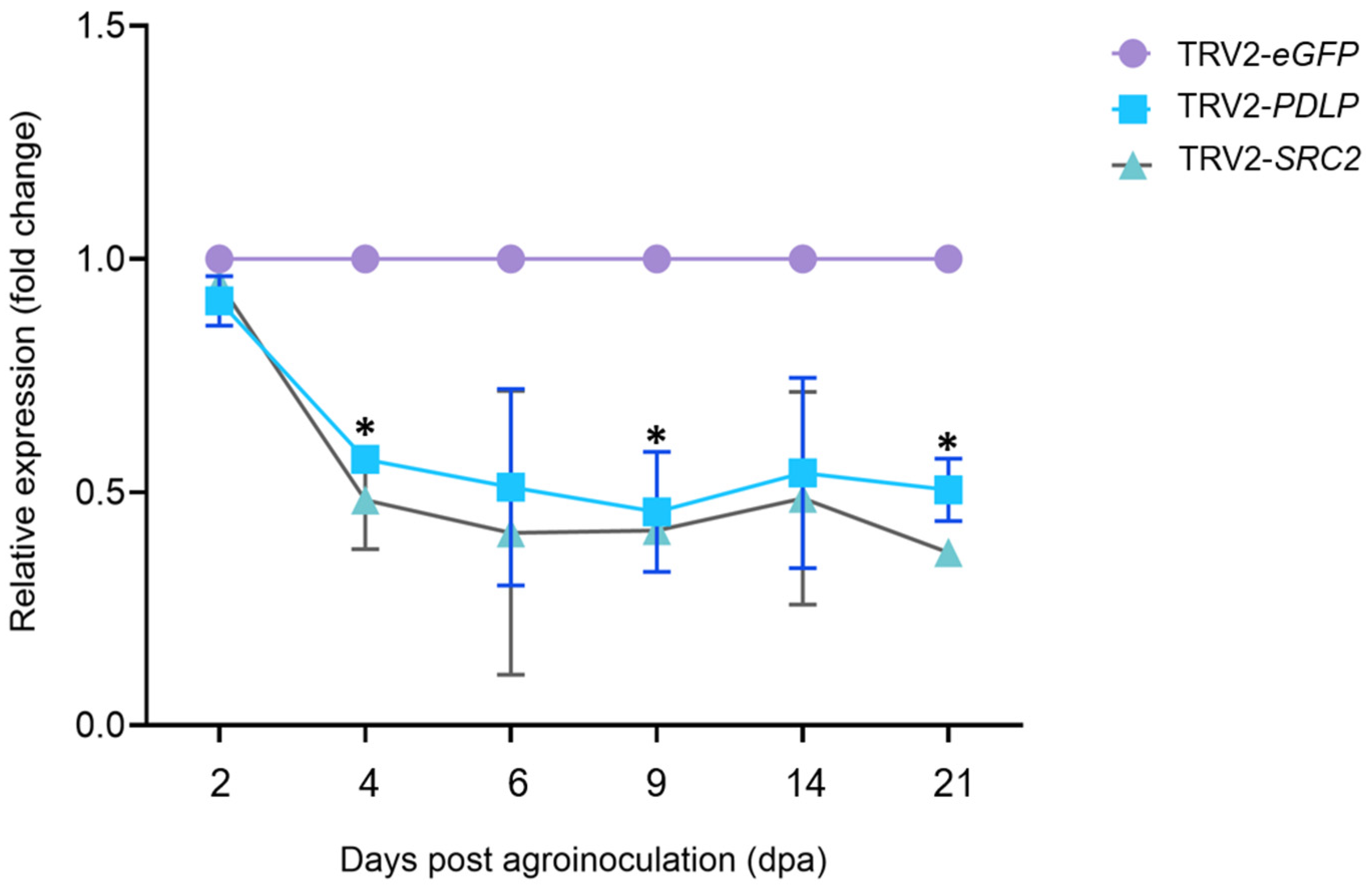
Transient expression of PDLP and SRC2 gene fragments cloned in the pTRV2 expression vector was carried out through agroinfiltration in N. benthamiana, N. tabacum, and C. chinense leaves (Supplementary Figure S3). The phytoene desaturase gene (PDS) from N. benthamiana cloned in pTRV2, which results in photobleaching effect upon gene silencing, was used as a positive control (Figure S4). The silencing experiment for PDLP and the SRC2 gene in N. benthamiana was repeated two times, while the silencing experiment for these genes in N. tabacum and C. chinense plants was performed once. The silencing efficiency of the target genes (PDLP and SRC2) was evaluated using RT-qPCR and compared between the silenced and the control plants. In the PDLP-silenced group, the expression level of the PDLP gene was 0.4- to 0.6-fold lesser than the GFP control with the highest downregulation at 9 dpa (Figure 4). Similarly, the expression of the SRC2 gene in the SRC2-silenced group was 0.3- to 0.5-fold lesser than the GFP control with highest downregulation observed at 21 dpa (Figure 4).
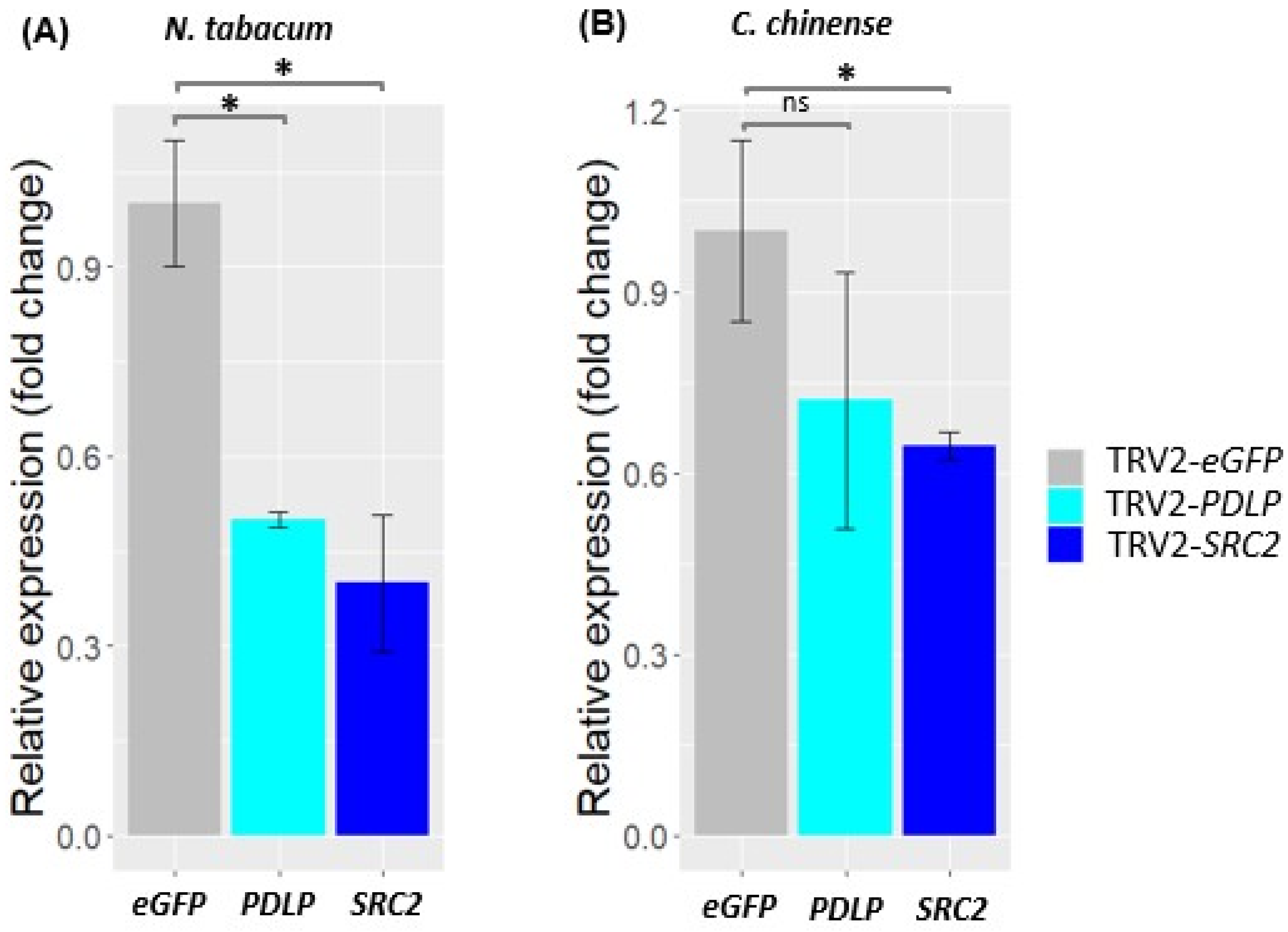
The silencing of PDLP and SRC2 genes in N. tabacum and C. chinense Bhut Jolokia plants was analyzed at 14 dpa (Figure 5). Using the same VIGS constructs, a considerable level of PDLP and SRC2 gene silencing was also observed in all species tested, despite having sequence polymorphisms among these species. The expression of the PDLP gene in N. tabacum and C. chinense was 0.5-fold and 0.7-fold lesser than the GFP control (a reduction in gene expression by 50% and 30%, respectively) (Figure 5). Similarly, in both N. tabacum and C. chinense Jacq., the expression of the SRC2 gene was downregulated to approximately 0.4-fold and 0.6-fold, respectively, in comparison to the GFP control (downregulation up to 60% and 40% in N. tabacum and C. chinense Jacq., respectively) (Figure 5).

2.3.2. Disease Development in PDLP- and SRC2-Silenced N. benthamiana Plants Upon CMV Infection

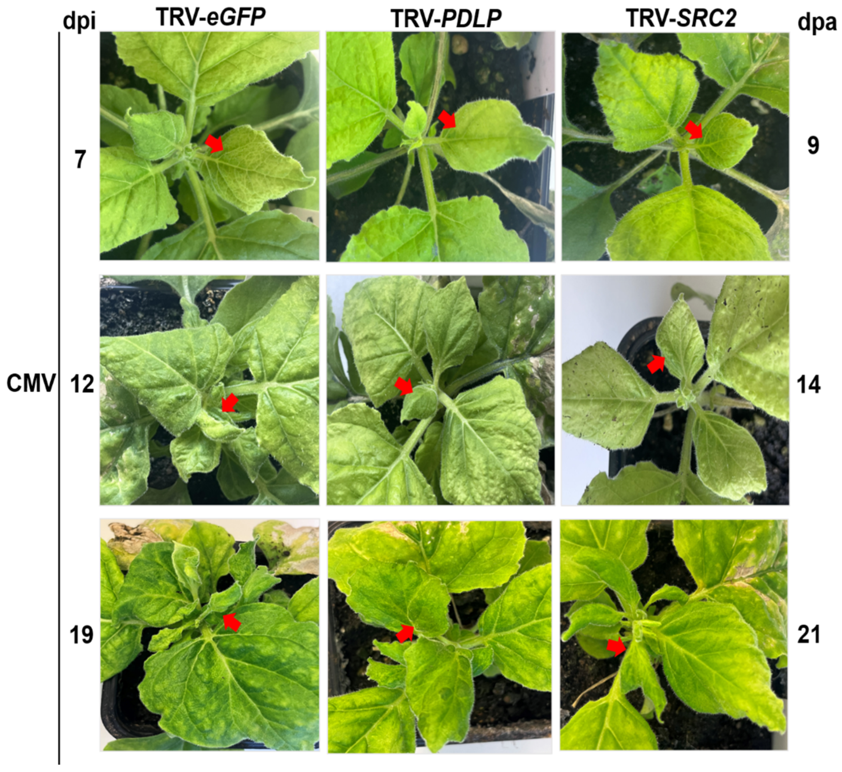
The PDLP- and SRC2-silenced N. benthamiana plants were visually observed upon CMV infection and disease symptoms were recorded at different time points, i.e., 7, 12, and 19 dpi (Figure 6 and Supplementary Figure S5). In the eGFP control plants, the typical mosaic symptom of CMV was observed at 7 dpi and the symptoms gradually intensified with severe downward leaf curling at 19 and 21 dpi. In contrast, the PDLP-silenced plants displayed reduced viral symptoms in comparison to the eGFP control. In the SRC2-silenced plants, leaf curling similar to or even greater than the eGFP control plants was observed at 19 and 21 dpi, indicating that silencing of SRC2 might have promoted CMV infection in N. benthamiana (Figure 6 and Supplementary Figure S5).

2.3.3. Relative Quantification of CMV Titer in PDLP- and SRC2-Silenced N. benthamiana Plants

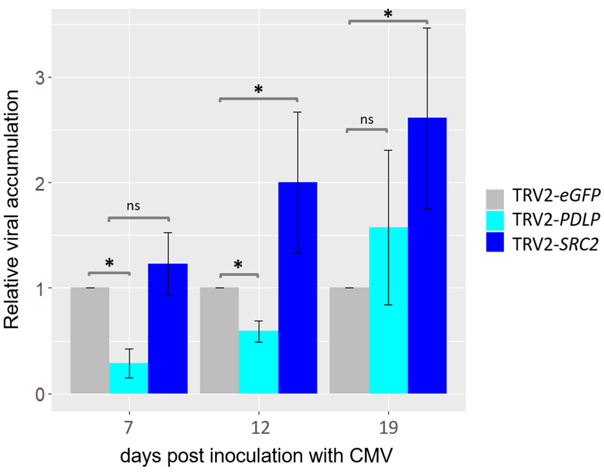
Systemic (uninoculated) leaves were collected at different time points upon CMV infection, and the viral titer was evaluated using RT-qPCR analyses. Significant reduction in the CMV titer was observed at 7 and 12 dpi but not at 19 dpi in the PDLP-silenced plants in comparison to the control plants (i.e., eGFP) by RT-qPCR analysis (Figure 7). Similarly, it was found that the level of CMV was significantly higher in the SRC2-silenced plants when compared to the eGFP control plants at 12 and 19 dpi by using RT-qPCR (Figure 7).

2.3.4. Callose Deposition in the PDLP- and SRC2-Silenced N. benthamiana Plants Upon CMV Infection

To strengthen the RT-PCR and RT-qPCR results, a callose deposition assay was conducted. Callose deposits initiated at 7 dpi and progressively increased, with higher callose deposits observed at 19 dpi in the eGFP control plants after CMV infection. In contrast, lesser callose deposits were observed in the PDLP-silenced plants at 7, 12, and 19 dpi. In the SRC2-silenced plants, callose deposition exhibited a near-equivalent level to that of the control group at 7, 12, and 19 dpi (Supplementary Figure S6).

2.3.5. Accumulation of Reactive Oxygen Species (ROS) in the PDLP- and SRC2-Silenced N. benthamiana Plants Upon CMV Infection

The level of hydrogen peroxide (H2O2), a ROS molecule, was assessed in the PDLP- and SRC2-silenced plants following CMV infection. Remarkably, elevated levels of H2O2 were detected in systemic leaves of the control and SRC2-silenced groups at 7, 12, and 19 dpi (Supplementary Figure S7). In contrast, the concentrations of H2O2 in PDLP-silenced plants were notably lower in comparison to the control and SRC2-silenced groups upon CMV infection at 7, 12, and 19 dpi (Supplementary Figure S7).

3. Discussion

It is known that plasmodesmata-associated proteins and plasmodesmata (PD) are crucial for cell-to-cell movement of plant viruses [35]. PDLPs and SRC2 are plasmodesmata-associated genes [5,16,35]. Some viruses recruit movement proteins that localize to the PD and interact with the PDLP protein to form a movement tubule facilitating, as a result, the transport of viral particles allowing systemic spread of the virus in the plant. The movement protein of CMV also localizes in the plasmodesmata [22]; however, the role of PDLPs in viral disease development is not well studied. The SRC2 protein is also a PD-associated membrane protein that colocalizes with Arabidopsis AtPDLP and interacts with the movement protein of cauliflower mosaic virus (CaMV) in planta [16]. The expression of pepper CaSRC2-1 gene was upregulated during bacterial and viral infection in Capsicum annum [17]. Little is known about the expression of the SRC2 gene of Nicotiana spp. during infection with CMV. Thus, these two genes, PDLP and SRC2, are promising candidates for promoting disease susceptibility or resistance to CMV in tobacco; therefore, they were functionally characterized in our study.
The expression of these two tobacco genes was transiently silenced utilizing a TRV-based VIGS strategy. The expression of PDLP upon CMV infection in N. benthamiana plants was consistent between mock and CMV inoculated samples, suggesting that the expression of PDLP is not significantly affected by CMV infection. On the other hand, the expression of SRC2 was slightly upregulated at 8 dpi upon CMV infection, corresponding with the initiation of CMV expression at the same time point (Figure 3). Similar observations were made in pepper (C. annum) and soybean (Glycine max) [17,36]. Takahashi and Shimosaka (1997) found that SRC2 expression in soybean was upregulated in response to cold stress and mechanical wounding [36]. Additionally, Kim et al. (2008) observed an induction of SRC2 gene expression in pepper during bacterial and viral infection [17].
Through TRV-based VIGS, an average of about 80% downregulation can be achieved in the endogenous transcript levels of the targeted gene compared to vector control plants [25]. The initiation of silencing generally starts within 2–3 weeks after agroinfiltration [25]. The efficiency of silencing may vary due to various factors such as the VIGS-vector infection, multiplication, and systemic spread, the level of viral-vector titer in the host, the homology between the target gene fragment in the viral-vector and the endogenous target gene mRNA, and the environmental conditions during plant growth [25]. The present study revealed significant downregulation in both PDLP and SRC2 at 4, 9, and 21 dpa (Figure 4). RT-qPCR analysis revealed a significant downregulation of the PDLP gene by 50% in N. tabacum, N. benthamiana, and 30% in C. chinense (Figure 4 and Figure 5), along with the downregulation of the SRC2 gene by 60% in N. tabacum, N. benthamiana, and 40% in C. chinense (Figure 3 and Figure 4). Similar observations were reported in the knockdown lines of PDLP-1, PDLP-2, and PDLP-3 triple mutants in Arabidopsis thaliana which was resistant to powdery mildew infection [37]. Kim et al. (2008) proposed that SRC2 from pepper (C. annuum) plays an essential role in inducing resistance to bacterial and viral pathogens by eliciting the hypersensitive response (HR) to restrict pathogen growth [17]. Liu et al. (2016) provided evidence that SRC2 recognizes INF1, an elicitor protein of Phytophthora capsici, and elicits HR in pepper [38]. They showed that silencing of the SRC2 gene increased susceptibility of pepper plants to P. capsici infection. In addition, the silencing experiment did not reveal any developmental changes such as alteration in plant height in the PDLP- and SRC2-silenced plants when compared to the control plants (TRV2-eGFP). It is noteworthy that this is the first report of a TRV-based silencing of plant endogenous genes in C. chinense Bhut Jolokia. No developmental alterations were observed in N. benthamiana and C. chinense in any VIGS experiment, except for N. tabacum where development changes such as growth inhibition and minor leaf crumbling were observed at 14 dpa (Supplementary Figure S4). Photobleaching, indicative of PDS gene silencing, started at 9 dpa and became even more prominent at 14 and 21 dpa (Supplementary Figure S4), suggesting that silencing effect commenced approximately 1–2 weeks post agroinfiltration. This experiment allowed us to determine the silencing effect of plant endogenous genes with the TRV-based VIGS strategy, which was demonstrated within 14 dpa.
The current investigation unveiled that silencing of the PDLP in N. benthamiana resulted in a notable reduction in the accumulation of the CMV titer in the systemic leaves. The PDLP gene family comprises a set of multiple genes, specifically PDLP1 to PDLP8 in Arabidopsis, with predicted molecular weights ranging from 30 to 35 kDa [39]. The downregulation of the PDLP gene resulted in delayed symptoms of CMV infection up to 12 dpi but not until 19 dpi, suggesting that targeting multiple PDLP genes, if present, of N. benthamiana could potentially lead to further delay in viral symptom development. Another possibility is that CMV might exploit other plasmodesmata proteins to facilitate transport at later stages of systemic infection. Therefore, it is important to understand the role of other PDLPs in N. benthamiana. It can be speculated that the reduced H2O2 accumulation observed in the PDLP-silenced plants (Figure S7) is likely a result of decreased CMV accumulation compared to the TRV2-eGFP-treated plants. It is known that in a CMV–plant compatible interaction, there is an increase in H2O2 accumulation [40] that agrees with our observations. The reduced leaf mottling and mosaic symptoms in the PDLP-silenced plants correlate nicely with the reduced CMV titer and ROS accumulation (Figure 7 and Figure S7). Plants produce callose deposits in the cell periphery as a defense mechanism in response to pathogen invasion. Previous studies have demonstrated that overexpression of PDLP1 and PDLP5 enhances callose deposition at PD [37,41], which is consistent with the low callose deposition in our PDLP-silenced plants. Lim et al. (2016) demonstrated that the overexpression of PDLP5 resulted in a reduction in the trafficking of azelaic acid (AzA) and glycerol-3-phosphate (G3P) through PD [42]. This impairment of PD trafficking was associated with a significant decline in Systemic Acquired Resistance (SAR) in plants. Similarly, Carella et al. (2015) observed that overexpressing of either PDLP1 or PDLP5 in Arabidopsis plants led to the loss of systemic movement of the crucial SAR protein, DEFECTIVE IN INDUCED RESISTANCE1 (DIR1), resulting in a compromised SAR [43]. The overexpression of PDLPs in A. thaliana correlates with an enhanced systemic spread of pathogens suggesting their role in promoting pathogen spread. We could hypothesize that PDLP silencing would augment SAR for the benefit of the plant and having negative effect for CMV. In addition, PDLPs are known to be involved in viral movement [5,6,15,16]. For example, for tubule-guided viral movement through plasmodesmata, the viral movement proteins (MPs) of GFLV and CaMV in Arabidopsis [5] and cowpea mosaic virus in N. benthamiana [15] interact with the host PDLPs. The interaction of viral MPs with host PDLPs results in the formation of movement tubules that allow cell-to-cell movement of plant viruses across adjacent cells [5]. CMV-MP also localizes in the plasmodesmata as it is a member of the ‘30K MP superfamily’ group [22]. This finding is consistent with the results obtained from our PDLP-silenced N. benthamiana plants, where a reduction in PDLP expression led to decreased CMV accumulation. While the exact function of PDLPs in virus movement is still under investigation, future research can be prioritized to understand the structural interactions between tubule-forming MPs (e.g., CMV) and PDLPs. Overall, these findings suggest that PDLP silencing of N. benthamiana led to decreased CMV accumulation in systemic tissue, suggesting that PDLPs are possibly involved in facilitating the systemic spread of CMV and as a result increase the viral titer, so they should be considered as susceptibility factors. Generating knockout/knockdown lines of PDLP genes in Bhut Jolokia and N. benthamiana using the CRISPR/Cas9 system could aid in developing virus-resistant plants against CMV.
In contrast, silencing of SRC2 resulted in increased susceptibility to CMV infection (Figure 7), indicating that SRC2 might be involved in the resistance to CMV in N. benthamiana. Kim et al. 2008 [17] reported that during both bacterial and viral infections in C. annum, the pepper encoding CaSRC2-1 (a single C2 domain-containing protein) gene was upregulated. Limited information exists regarding the expression patterns of the SRC2 gene in Nicotiana spp. during the infection with CMV (our data indicate a slight SRC2 upregulation upon CMV infection [Figure 3]). A recent study found that the silencing of GmSRC2 in soya resulted in a significant enhancement of disease symptoms and an increase in the biomass of Phytophthora sojae [44]. A structural characteristic identified in AtSRC2.2 is its C2 domain, which serves as a lipid-binding domain found in numerous eukaryotic proteins [16]. The C2 domain, first identified in Ca2+-dependent isoforms of protein kinase C, is integral to cellular signalling in plants and animals [17]. Plant proteins harboring C2 domains have been associated with facilitating the intercellular spread (cell-to-cell movement) of plant viruses [45]. An interesting characteristic of AtSRC2.2 is that its C2 domain shares 36% similarity with AtSYT4, a member of the synaptotagmin family, possessing two C2 domains (C2A and C2B) [16]. Not much is known about the involvement of AtSYT4 in virus movement, but there is another synaptotagmin, AtSYTA, which is involved in the movement of several viruses, such as cabbage leaf curl virus, tobacco mosaic virus, and squash leaf curl virus by interacting with their respective MPs [45]. It is noteworthy that AtSYTA is also associated with regulating freezing tolerance [46], a trait shared among SRC2 proteins. Several studies have suggested that the entry of Ca2+ is a key factor in initiating pattern-triggered immunity (PTI) in plant cells [44]. In the SRC2-silenced plants, a higher H2O2 accumulation was observed (Supplementary Figure S7) that could be the consequence of higher CMV load (Figure 7) in the compatible interaction with N. benthamiana agreeing with [40]. Therefore, further investigation is needed to understand the role of SRC2 in plant defense response. The notion that enhancing resistance to CMV in plants could be achieved by overexpressing the SRC2 gene is now a hypothesis to test.

4. Materials and Methods

4.1. Plant Materials and Growth Conditions

Nicotiana benthamiana, N. tabacum, and Capsicum chinense Jacq. (Bhut Jolokia) plants were grown in a greenhouse operating at 20 ± 3 °C and 70% humidity with a 16 h/8 h day/night cycle. Three- to four-week-old plants were selected for performing the VIGS experiment, and they were divided into four groups—TRV1 + TRV2-NtPDLP, TRV1 + TRV2-NtSRC2, TRV1 + TRV2-eGFP (negative control), and TRV1 + TRV2-NbPDS (positive control for silencing).

4.2. Development of TRV-Based Recombinant Constructs for VIGS

The TRV-based VIGS system, developed by Liu et al., 2002 [31], was used to generate the recombinant VIGS constructs for silencing of putative PDLP and SRC2 genes in Solanaceous plants including Nicotiana benthamiana, N. tabacum, and C. chinense. TRV is a bipartite ss (+) RNA virus comprising two genomes, RNA1 and RNA2. Both TRV-RNA1 and TRV-RNA2 genomes were previously cloned in the binary plasmid vectors pBIN19 and pCambia0390, respectively [31]. The pTRV-RNA2 plasmid vector was digested by the BamHI-HF restriction enzyme (New England Biolabs, USA) to obtain a linearized plasmid DNA. The digested product was subjected to gel electrophoresis for confirmation along with the quantification of the digested pTRV2 and was stored at −20 °C for further use.
For the integration of the PDLP and SRC2 gene fragments, the In-Fusion® HD seamless cloning system (Takara Bio Inc., Shiga, Japan) was employed. The nucleotide sequences corresponding to the putative PDLP (XM_016610375.1) and SRC2 (XM_016630487.1) genes of N. tabacum were employed as queries in the VIGS tool provided by the Sol Genomics Network (SGN) to identify the optimal fragment for gene silencing purposes. Primers were designed for the amplification of the fragments of PDLP and SRC2 gene using the primer3 tool (http://primer3.ut.ee/) (accessed on 15 February 2022). The designed primer had a 15-base extension at the 5′ end homologous to the linearized pTRV2 vector (Table 1). The amplification of PDLP and SRC2 gene fragments was performed by RT-PCR utilizing RNA from N. tabacum leaves as a template. The PCR-amplified product underwent confirmation and quantification through 1.5% agarose gel electrophoresis and was subsequently cloned into the linearized pTRV2 vector as per the manufacturer’s instructions (In-Fusion® HD cloning kit, Clontech, Mountain View, CA, USA). The recombinant plasmid constructs (pTRV2-NtPDLP and pTRV2-NtSRC2) were transformed into E. coli Stellar Competent Cells (Takara Bio Inc., Shiga, Japan), followed by a heat shock at 42 °C for 30 s. The bacterial cultures were spread over Luria–Bertani (LB) plates containing kanamycin (50 µg/mL) and were subjected to overnight incubation at 37 °C for colony development. Colony PCR was carried out to confirm the presence of recombinant plasmids in the transformed bacterial colonies, using specific primers (TRV2-1530F and TRV2-1809R) that bind on the TRV2 plasmid vector (Table 1). Two positive transformants for each of the recombinant plasmids were selected for plasmid isolation using NucleoSpin® Plasmid Kit (Machery-Nagel, Düren, Germany) and stored for further use. The confirmation of the successful integration of the NtPDLP and NtSRC2 gene fragment into the pTRV2 vector was validated through Sanger sequencing (Azenta Life Sciences UK, Essex, UK).
Table 1. List of primers used in this study.
Name of PrimerSequence (5′ to 3′) *Product Size (bp)Target SpeciesFunction
Nt_PDLP-INF-378F
Nt_PDLP-INF-760R
GCCTCCATGGGGATCGTGTACAAAGGCTGTGCTAA
GCTCGGTACCGGATCGTCTCAAGCCTATCACTAAAC
413N. tabacum
N. benthamiana
Cloning of PDLP gene fragment to pTRV2
Nt_SRC2-INF-125F
Nt_SRC2-INF-488R
GCCTCCATGGGGATCCATTAGATATCAAAGTTATTGC
GCTCGGTACCGGATCGGTTTTCCGCTCTCTGTTAT
394N. tabacum
N. benthamiana
Cloning of SRC2 gene fragment to pTRV2
eGFP-INF-286F
eGFP-INF-646R
GCCTCCATGGGGATCGAGCGCACCATCTTCTTCAA
GCTCGGTACCGGATCGCTTCT CGTTGGGGTCTTTG
391-Cloning of eGFP gene fragment to pTRV2
TRV2-1530F
TRV2-1809R
GTTTTTATGTTCAGGCGGTTC
TCAAGATCAGTCGAGAAT GTCA
280E. coli
A. tumefaciens agroinfiltrated plant species
Colony PCR,
Detection of TRV2
TRV1-257F
TRV1-491R
GCTGAGCAGAGGAGTCATTTC
ACCCATGAACCATGTTTTTGT
235E. coli
A. tumefaciens agroinfiltrated plant species
Detection of TRV1
Nb_PDLP-RT-F
Nb_PDLP-RT-R
GGGACTGTGTGAACTGTGTG
ACTCCTAACCCCACACCAAC
210N. tabacum
N. benthamiana
Gene expression analysis for PDLP
Nb_SRC2-RT-F
Nb_SRC2-RT-R
ATCCACCCGTACAACAACCT
AGCAGCCATCTCTCCAACAT
164N. tabacum
N. benthamiana
Gene expression analysis for SRC2
CMV_CP_RT-F
CMV_CP_RT-R
GAAGCTTGTTTCGCGCATTC
TCCGGATGCTGCATACTGAT
175CMVGene expression analysis for CMV CP
Nb_EF1a-F
Nb_EF1a-R
AGCTTTACCTCCCAAGTCATC
AGAACGCCTGTCAATCTTGG
116N. tabacum
N. benthamiana
Gene expression analysis for EF1a [47]
* The region that anneals to the TRV2 vector for the In-Fusion cloning is underlined.
To serve as a negative control in the VIGS assay, we introduced a 361 nt segment from the enhanced GFP (eGFP) gene (a gene that does not exist in plants) into the pTRV2 plasmid vector. The insert fragment was derived through PCR amplification using In-Fusion primers designed for this purpose, with a pBIN61-eGFP plasmid vector employed as the template. All subsequent steps for constructing the engineered pTRV2_eGFP plasmid were executed by the aforementioned procedures. The positive control in the VIGS assay involved the targeted silencing of a fragment of N. benthamiana phytoene desaturase gene (PDS) exhibiting a photobleaching phenotype. This control was employed to validate the efficiency of the gene silencing system.

4.3. Transformation and Mobilization of VIGS Constructs into Agrobacterium Tumefaciens (GV3101 Strain)

The recombinant plasmids (pTRV2-NtPDLP and pTRV2-NtSRC2) were transformed into Agrobacterium tumefaciens competent cells (GV3101 strain) using the freeze–thaw method with minor modifications [48]. The plasmids were added to the surface of frozen GV3101 competent cells and promptly placed in a heat block at 37 °C for 5 min. Following the heat treatment, the transformed GV3101 competent cells were mixed with LB and subjected to shaking at 28 °C for 4 h. The transformed bacterial cells were spread onto an LB agar plate containing 50 µg/mL kanamycin and 25 µg/mL rifampicin and incubated at 28 °C for 2–4 days for colony development. Positive colonies were further confirmed by colony PCR as mentioned earlier for E. coli. Similarly, the pTRV1 binary plasmid vector carrying the TRV-RNA1 genome was introduced into GV3101 competent cells.

4.4. Agrobacterium-Mediated Transient Expression and Virus Induced Gene Silencing

VIGS constructs were delivered to three- to four-week-old N. benthamiana, N. tabacum, and C. chinense plants via agroinfiltration. Primary and secondary cultures of the relevant Agrobacteria were cultured in LB supplemented with kanamycin (50 μg/mL) and rifampicin (25 µg/mL) at 28 °C in a shaker incubator overnight at 220 rpm. The secondary cultures were centrifuged at 3000× g for 10 min. The supernatant was discarded, and the resulting cell pellet was resuspended in a resuspension buffer (10 mM 2-(N-Morpholino) ethanesulfonic acid hydrate (MES-hydrate), pH 5.6 adjusted with KOH, 10 mM MgCl2, and 200 µM acetosyringone). The final bacterial OD600 was adjusted to 0.5. The agroinfiltration mixtures were obtained by mixing the bacteria harboring TRV1 and TRV2-PDLP/TRV2-SRC2/TRV2-eGFP constructs in a 1:1 (v/v) ratio. The TRV2-eGFP was used as the negative control, whereas the TRV2-PDS was used as a positive control for silencing. The final mixtures were kept in the dark at room temperature for 3 h before agroinoculation. For the VIGS study, the plants were categorized into four distinct groups (three biological replicates per group), TRV1 + TRV2-NtPDLP, TRV1 + TRV2-NtSRC2, TRV1 + TRV2-eGFP (negative control), and TRV1 + TRV2-NbPDS (positive control for silencing). Two matured leaves per plant were agroinoculated using a needleless syringe on the abaxial side of the leaf. Subsequently, the plants were maintained in a growth chamber, during which tissue sampling was performed at different time points (2, 4, 6, 9, 14, and 21 days post agro-inoculation), and were subjected to RNA analysis, CMV inoculation, and disease symptom observation.

4.5. Mechanical Inoculation of CMV in Agroinfiltrated N. benthamiana Plants

Sap inoculation of CMV in plants was performed using carborundum powder (Supplementary Figure S3). The inoculating sap was prepared by collecting sap material from previously CMV-infected plants and gently rubbed on the adaxial side of the leaf using carborundum powder. For CMV inoculation in the silenced plants, leaves were initially agroinfiltrated with the VIGS constructs, and two days later, sap inoculation with CMV was performed to allow sufficient time for silencing to be initiated. Systemic leaf samples were collected at different time points (7, 12, and 19 days post inoculation (dpi) with CMV) and RNA was isolated to assess the viral titer in the silenced plants via quantitative RT-PCR (RT-qPCR).

4.6. PCR and RT-qPCR Analysis

PCR was performed to detect the viral titer of CMV using gene-specific primers (Table 1). The total reaction volume (20 µL) consisted of 2 µL of the 10X PCR Buffer (ThermoFisher Scientific, Waltham, MA, USA), 0.5 µL of 10 mM dNTPs, 0.5 µL of 10 μM of forward and reverse primers each (Table 1), and 0.1 µL of AmpliTaq DNA polymerase (250 U/μL) (ThermoFisher Scientific, USA). PCR was carried out with initial denaturation temperature at 95 °C for 5 min followed by 30 cycles consisting of denaturation at 95 °C for 30 s, annealing at 58 °C for 30 s, and extension at 72 °C for 30 s, and then final extension for 7 min at 72 °C.
For the validation of gene silencing, RT-qPCR was performed by extracting RNA from the systemic leaves of the silenced plants at different time points with the Plant/Fungi Total RNA Purification kit (NORGEN Biotek Corporation, Canada) according to the manufacturer’s instructions. Three biological replicates were pooled for each experimental group. The investigation of pTRV2-NtPDLP and pTRV2-NtSRC2 silencing was performed by sampling tissues at 2, 4, 6, 9, 14, and 21 dpa. Tissue sampling for the detection of CMV CP (proxy for CMV titer) was performed at 7, 12, and 19 days post inoculation (dpi) with CMV. The RNA samples were quantified, and their purity was determined spectrophotometrically with NanoDrop™ 2000 Spectrophotometer (ThermoFisher Scientific, USA). cDNA was synthesized using 200 ng total RNA in a 20 μL reaction volume, using the SuperScript VILO cDNA synthesis kit (Invitrogen, Waltham, MA, USA) as per the manufacturer’s instructions. RNaseOUTTM Recombinant Ribonuclease Inhibitor safeguards against the degradation of target RNA due to ribonuclease contamination. The 5X VILOTM Reaction mix consists of random primers, MgCl2, and dNTPs in a buffer formulation optimized for RT-qPCR. The RT reaction was performed in a thermocycler (BioRad, Hercules, CA, USA) at 25 °C for 10 min, followed by incubation at 42 °C for 60 min, and terminated at 85 °C for 5 min. The RT products were used as a template for amplifying the PDLP and SRC2 genes using the designed gene-specific primers (Table 1). PCR amplification for the detection of NtPDLP, NtSRC2, and Elongation factor 1 alpha (EF1a) was performed in a FastGene ULTRA Cycler (NIPPON Genetics EUROPE, Düren, Germany) using the FirePol DNA Polymerase (Solis BioDyne, Tartu, Estonia) and specific primers (Table 1). The total reaction volume consisted of 10X Buffer B (Solis BioDyne, Estonia), 10 mM dNTPs, 10 μM of forward and reverse (Table 1) primers each, 25 mM MgCl2 and 0.2 μL (5 U/μL) Taq DNA polymerase (Solis BioDyne, Estonia). PCR for the PDLP and EF1a genes was carried out with initial denaturation temperature at 95 °C for 5 min followed by 35 cycles consisting of denaturation at 95 °C for 20 s, annealing at 58 °C for 20 s, and extension at 72 °C for 1 min, and then final extension continued for 7 min at 72 °C. PCR for the SRC2 gene was carried out with initial denaturation temperature at 95 °C for 5 min followed by 35 cycles consisting of denaturation at 95 °C for 20 s, annealing at 58 °C for 15 s and extension at 72 °C for 1 min, and then final extension continued for 7 min at 72 °C. Primer specificity was verified by melting curve analysis. To obtain a semi-quantitative estimation of the time course expression profiles of PDLP and SRC2 gene CMV infection, ImageJ software (v1.54m) (https://imagej.nih.gov/ij/) was employed to measure the band intensity of the gel image. In addition, band intensities were calculated for the housekeeping gene of mock and CMV-infected plants and used as control. The values for the target genes for mock and CMV-infected plants were normalized with the housekeeping gene (EF1a).
qPCR was performed in a CFX96TM Touch Real-Time PCR Detection System (Bio-Rad, USA) to quantify the expression levels of the target genes (pTRV2-PDLP and pTRV2-SRC2) compared to the control group (plants inoculated with pTRV2-eGFP) and to check the viral titer (CMV) in the silenced and control plants using SYBR Green qPCR mix (ThermoFisher Scientific, USA). Expression of the target genes was normalized with the housekeeping gene EF1a that was found more stable to F-box based on BestKeeper housekeeping gene analysis [49], agreeing with [47]. The fold change was calculated using the delta CT (2−ΔΔCt) method [50].
Statistical analyses were performed using Microsoft Office Excel (Microsoft Excel, Redmond, Washington, DC, USA). Student’s t-test was used for significance analysis between two treatments. The standard error is shown as the standard deviation between the biological replicates.

4.7. Histochemical Analysis

4.7.1. Staining for Callose Deposition in Leaf Tissues

Aniline blue staining facilitates the detection and quantification of cell wall depositions of callose. Callose deposition assay using Aniline blue staining was performed following the procedure described in [51]. A total of three biological replicates were taken for the callose deposition test. Two infected leaf samples were harvested and transferred to 50 mL polypropylene tubes containing a destaining solution (1:3 acetic acid/ethanol). The tubes were incubated overnight with mild agitation. The destained leaves were washed with 150 mM K2HPO4 (washing solution) for 30 min. The plant materials were incubated in an aniline blue staining solution (1.3 g of K2HPO4 with 5 mg aniline blue in 50 mL H2O) for at least 2 h in the polypropylene tubes wrapped in aluminium foil for light protection. The samples were embedded in 50% glycerol on a glass slide and the callose deposition was visualized using a fluorescent microscope equipped with a UV lamp. Glycerol ensures a prolonged observation time and reduces bubble formation. The callose deposits appear stained bright blue in color (Supplementary Figure S6).

4.7.2. Staining for H2O2 Detection in Leaf Tissues

The 3,3- diaminobenzidine (DAB) assay was performed following the procedure outlined in [52]. The DAB stain is mainly used to visualize the production of H2O2 in leaves and roots. For the detection of H2O2, 0.1 g of DAB was dissolved in 100 mL of ddH2O (pH adjusted to 3.8 using HCl). The DAB solution was freshly prepared before starting the experiment. The DAB assay was conducted using three biological replicates for each experimental group. Systemic leaves were harvested and treated with the DAB solution and incubated overnight in the dark with mild agitation (50 rpm). The DAB solution was discarded, leaves were rinsed with ddH2O to remove excessive staining solution and immersed in 96% ethanol. The samples were then incubated overnight to remove the chlorophyll. Leaves were washed with 75% ethanol and kept in 20% glycerol solution to prevent the leaves from breaking easily and enabling rehydration. Dark brown color precipitates indicated accumulation of H2O2. The images of the leaf samples were taken under a light microscope with a 5X objective lens (Supplementary Figure S7).

5. Conclusions

A better understanding of the functional roles of PDLP and SRC2 in the plant’s response to CMV infection is provided. This experimental study suggests that PDLP and SRC2 are potential susceptibility and resistance genes during CMV infection in Nicotiana benthamiana, respectively. The VIGS method can be applied for screening candidate genes involved in viral resistance or disease susceptibility in C. chinense cv Bhut Jolokia and other crop plants.

Supplementary Materials

The following supporting information can be downloaded at: https://www.mdpi.com/article/10.3390/plants14030495/s1, Figure S1: Multiple sequence alignment of PDLP genes of Nicotiana benthamiana (Niben101Scf03456g00018.1), Capsicum chinense (MCIT02000006.1), C. annum (XM_016721975.2), and N. tabacum (XM_016610375.1). Red-colored lines indicate gene fragment (383 bp) used as VIGS targeted region for silencing of the PDLP gene. Sequence alignment was performed using Clustal Omega; Figure S2: Multiple sequence alignment of SRC2 genes of Nicotiana benthamiana (DQ465395.1), Capsicum chinense (AB442165.1), C. annum (NM_001324570.1) and N. tabacum (XM_016630487.1). Red-colored lines indicate gene fragment (364 bp) used as VIGS targeted region for silencing of the SRC2 gene. Sequence alignment was performed using Clustal Omega; Figure S3: Illustrative depiction of the methodology involved in the agroinfiltration of the VIGS constructs, cucumber mosaic virus (CMV) inoculation, and gene expression analysis in the experimental plants; Figure S4: Phenotypic observations of VIGS-silenced Nicotiana benthamiana, Nicotiana tabacum, and Capsicum chinense plants (A) at 21 days post agroinoculation (dpa). (B) Phytoene desaturase (PDS) gene was used as a positive control. Silencing of PDLP and SRC2 genes did not alter the development in the N. benthamiana and C. chinense; in contrast, N. tabacum exhibited some leaf crumbling symptoms; Figure S5: Representative images of N. benthamiana plants upon CMV-inoculation at 19 dpi. dpi = days post CMV inoculation, CMV = cucumber mosaic virus, (-) = non-silenced plant with CMV infection, Mock = Phosphate buffer treated plant. TRV2-SRC2, TRV2-eGFP (control), and TRV2-PDLP are VIGS silenced plants observed upon CMV inoculation; Figure S6: Microscopic observation of callose deposits upon cucumber mosaic virus (CMV) in PDLP- and SRC2-silenced Nicotiana benthamiana plants. Callose deposition assay using Aniline blue staining was performed according to the procedure described by Schenk et al., 2015. The callose deposition was visualized using a fluorescent microscope with a UV lamp. White arrow indicates callose deposits. dpa = days post agroinoculation. dpi = days post infection. Figure S7: Macroscopic (A) and microscopic (B) detection of H2O2 by 3,3- diaminobenzidine (DAB) staining in VIGS-silenced Nicotiana benthamiana plants at different time-points after DAB staining. Control (eGFP) and SRC2-silenced plants exhibited elevated levels of H2O2 as compared to the PDLP-silenced group. Dark brown patches (indicated by green arrows) represent areas of reactive oxygen species (ROS) accumulation (see results of RT-qPCR in Figure 7), where darker shades indicate higher viral accumulation and lighter shades indicate lower viral accumulation. DAB assay was performed following the procedure outlined by Bach-Pages and Preston 2018. dpa = days post agroinoculation; dpi = days post CMV infection.

Author Contributions

R.S.: Writing—original draft, review, and editing, Methodology, Validation, Formal analysis, Data curation; A.K.: Writing—review, and editing, Methodology, Supervision, Data curation; C.J.S.: Writing—review, and editing, Supervision, Resources, Funding acquisition; B.K.B.: Writing—review, and editing, Supervision; A.V.: Conceptualization, Supervision, Project administration, Writing—review, and editing, Resources, Funding acquisition. All authors have read and agreed to the published version of the manuscript.

Funding

The funding to carry out the research in Norway was provided by the Norwegian Research Council Grant (287794). This research was partly funded by the Education, Audiovisual and Culture Executive Agency (EACEA) of the European Commission for the 3-year-long ERASMUS+ Capacity Building in Higher Education project (598797-EPP-1-2018-1-EL-EPPKA2-CBHE-JP) entitled ‘AdaptNET—Strengthening education, research and innovation for climate-smart crops in India’.

Data Availability Statement

All data supporting this study are available within the paper and within the Supplementary Materials published online.

Acknowledgments

The authors acknowledge Supriya Chakraborty (Jawaharlal Nehru University, New Delhi, India) for proving the visual VIGS marker. The authors also acknowledge Dag-Ragnar Blystad (Norwegian Institute of Bioeconomy Research, Norway) for providing the CMV isolate to carry out the inoculation experiment on the VIGS silenced plants.

Conflicts of Interest

The authors declare no conflicts of interest.

References

  1. Kumar, D.; Kumar, R.; Hyun, T.K.; Kim, J.-Y. Cell-to-Cell Movement of Viruses via Plasmodesmat. J. Plant Res. 2015, 128, 37–47. [Google Scholar] [CrossRef] [PubMed]
  2. Schoelz, J.E.; Harries, P.A.; Nelson, R.S. Intracellular Transport of Plant Viruses: Finding the Door out of the Cell. Mol. Plant 2011, 4, 813–831. [Google Scholar] [CrossRef]
  3. Lucas, W.J. Plant Viral Movement Proteins: Agents for Cell-to-Cell Trafficking of Viral Genomes. Virology 2006, 344, 169–184. [Google Scholar] [CrossRef]
  4. Heinlein, M. Plasmodesmata: Channels for Viruses on the Move. Methods Mol. Biol. 2015, 1217, 25–52. [Google Scholar] [CrossRef] [PubMed]
  5. Amari, K.; Boutant, E.; Hofmann, C.; Schmitt-Keichinger, C.; Fernandez-Calvino, L.; Didier, P.; Lerich, A.; Mutterer, J.; Thomas, C.L.; Heinlein, M.; et al. A Family of Plasmodesmal Proteins with Receptor-like Properties for Plant Viral Movement Proteins. PLoS Pathog. 2010, 6, e1001119. [Google Scholar] [CrossRef] [PubMed]
  6. Ritzenthaler, C.; Hofmann, C. Tubule-Guided Movement of Plant Viruses. Plant Cell Monogr. 2007, 7, 64–83. [Google Scholar] [CrossRef]
  7. Chen, M.-H.; Tian, G.-W.; Gafni, Y.; Citovsky, V. Effects of Calreticulin on Viral Cell-to-Cell Movement. Plant Physiol. 2005, 138, 1866–1876. [Google Scholar] [CrossRef]
  8. Lee, J.-Y.; Taoka, K.; Yoo, B.-C.; Ben-Nissan, G.; Kim, D.-J.; Lucas, W.J. Plasmodesmal-Associated Protein Kinase in Tobacco and Arabidopsis Recognizes a Subset of Non-Cell-Autonomous Proteins. Plant Cell 2005, 17, 2817–2831. [Google Scholar] [CrossRef] [PubMed]
  9. Raffaele, S.; Bayer, E.; Lafarge, D.; Cluzet, S.; German Retana, S.; Boubekeur, T.; Leborgne-Castel, N.; Carde, J.-P.; Lherminier, J.; Noirot, E.; et al. Remorin, a Solanaceae Protein Resident in Membrane Rafts and Plasmodesmata, Impairs Potato Virus X Movement. Plant Cell 2009, 21, 1541–1555. [Google Scholar] [CrossRef] [PubMed]
  10. Ham, B.; Wang, X.; Toscano-Morales, R.; Lin, J.; Lucas, W. Plasmodesmal Endoplasmic Reticulum Proteins Regulate Intercellular Trafficking of Cucumber Mosaic Virus in Arabidopsis. J. Exp. Bot. 2023, 74, 4401–4414. [Google Scholar] [CrossRef]
  11. Tilsner, J.; Kriechbaumer, V. Reticulons 3 and 6 Interact with Viral Movement Proteins. Mol. Plant Pathol. 2022, 23, 1807–1814. [Google Scholar] [CrossRef] [PubMed]
  12. Zavaliev, R.; Sagi, G.; Gera, A.; Epel, B.L. The Constitutive Expression of Arabidopsis Plasmodesmal-Associated Class 1 Reversibly Glycosylated Polypeptide Impairs Plant Development and Virus Spread. J. Exp. Bot. 2010, 61, 131–142. [Google Scholar] [CrossRef]
  13. Ueki, S.; Spektor, R.; Natale, D.M.; Citovsky, V. ANK, a Host Cytoplasmic Receptor for the Tobacco Mosaic Virus Cell-to-Cell Movement Protein, Facilitates Intercellular Transport through Plasmodesmata. PLoS Pathog. 2010, 6, e1001201. [Google Scholar] [CrossRef] [PubMed]
  14. Uchiyama, A.; Shimada-Beltran, H.; Levy, A.; Zheng, J.Y.; Javia, P.A.; Lazarowitz, S.G. The Arabidopsis Synaptotagmin SYTA Regulates the Cell-to-Cell Movement of Diverse Plant Viruses. Front. Plant Sci. 2014, 5, 584. [Google Scholar] [CrossRef]
  15. den Hollander, P.W.; Kieper, S.N.; Borst, J.W.; van Lent, J.W.M. The Role of Plasmodesma-Located Proteins in Tubule-Guided Virus Transport Is Limited to the Plasmodesmata. Arch. Virol. 2016, 161, 2431–2440. [Google Scholar] [CrossRef] [PubMed]
  16. Rodriguez, A.; Angel, C.A.; Lutz, L.; Leisner, S.M.; Nelson, R.S.; Schoelz, J.E. Association of the P6 Protein of Cauliflower Mosaic Virus with Plasmodesmata and Plasmodesmal Proteins. Plant Physiol. 2014, 166, 1345–1358. [Google Scholar] [CrossRef] [PubMed]
  17. Kim, Y.C.; Kim, S.Y.; Choi, D.; Ryu, C.M.; Park, J.M. Molecular Characterization of a Pepper C2 Domain-Containing SRC2 Protein Implicated in Resistance against Host and Non-Host Pathogens and Abiotic Stresses. Planta 2008, 227, 1169–1179. [Google Scholar] [CrossRef]
  18. Roossinck, M.J. Evolutionary History of Cucumber Mosaic Virus Deduced by Phylogenetic Analyses. J. Virol. 2002, 76, 3382–3387. [Google Scholar] [CrossRef]
  19. Edwardson, J.R.; Christie, R.G. CRC Handbook of Viruses Infecting Legumes; CRC Press: Boca Raton, FL, USA, 1991. [Google Scholar]
  20. Ashwathappa, K.V.; Krishna Reddy, M.; Venkataravanappa, V.; Madhavi Reddy, K.; Hemachandra Reddy, P.; Lakshminarayana Reddy, C.N. Genome Characterization and Host Range Studies of Cucumber Mosaic Virus Belonging to the Subgroup IB Infecting Chilli in India and Screening of Chilli Genotypes for Identification of Resistance. VirusDisease 2021, 32, 535–547. [Google Scholar] [CrossRef] [PubMed]
  21. Roossinck, M.J. Cucumber Mosaic Virus; a Model for RNA Virus Evolution. Mol. Plant Pathol. 2001, 2, 59–63. [Google Scholar] [CrossRef]
  22. Sáray, R.; Fábián, A.; Palkovics, L.; Salánki, K. The 28 Ser Amino Acid of Cucumber Mosaic Virus Movement Protein Has a Role in Symptom Formation and Plasmodesmata Localization. Viruses 2021, 13, 222. [Google Scholar] [CrossRef] [PubMed]
  23. Tenllado, F.; Llave, C.; Díaz-Ruíz, J.R. RNA Interference as a New Biotechnological Tool for the Control of Virus Diseases in Plants. Virus Res. 2004, 102, 85–96. [Google Scholar] [CrossRef] [PubMed]
  24. Lange, M.; Yellina, A.L.; Orashakova, S.; Becker, A. Virus-Induced Gene Silencing (VIGS) in Plants: An Overview of Target Species and the Virus-Derived Vector Systems. Methods Mol. Biol. 2013, 975, 1–14. [Google Scholar] [CrossRef] [PubMed]
  25. Senthil-Kumar, M.; Mysore, K.S. Tobacco Rattle Virus–Based Virus-Induced Gene Silencing in Nicotiana Benthamiana. Nat. Protoc. 2014, 9, 1549–1562. [Google Scholar] [CrossRef]
  26. Purkayastha, A.; Dasgupta, I. Virus-Induced Gene Silencing: A Versatile Tool for Discovery of Gene Functions in Plants. Plant Physiol. Biochem. 2009, 47, 967–976. [Google Scholar] [CrossRef]
  27. Becker, A.; Lange, M. VIGS--Genomics Goes Functional. Trends Plant Sci. 2010, 15, 1–4. [Google Scholar] [CrossRef] [PubMed]
  28. Shi, G.; Hao, M.; Tian, B.; Cao, G.; Wei, F.; Xie, Z. A Methodological Advance of Tobacco Rattle Virus-Induced Gene Silencing for Functional Genomics in Plants. Front. Plant Sci. 2021, 12, 671091. [Google Scholar] [CrossRef]
  29. Kumagai, M.H.; Donson, J.; Della-Cioppa, G.; Harvey, D.; Hanley, K.; Grill, L.K. Cytoplasmic Inhibition of Carotenoid Biosynthesis with Virus-Derived RNA. Proc. Natl. Acad. Sci. USA 1995, 92, 1679–1683. [Google Scholar] [CrossRef]
  30. Ratcliff, F.; Martin-Hernandez, A.M.; Baulcombe, D.C. Technical Advance. Tobacco Rattle Virus as a Vector for Analysis of Gene Function by Silencing. Plant J. 2001, 25, 237–245. [Google Scholar] [CrossRef]
  31. Liu, Y.; Schiff, M.; Dinesh-Kumar, S.P. Virus-Induced Gene Silencing in Tomato. Plant J. 2002, 31, 777–786. [Google Scholar] [CrossRef] [PubMed]
  32. Verlaan, M.G.; Hutton, S.F.; Ibrahem, R.M.; Kormelink, R.; Visser, R.G.F.; Scott, J.W.; Edwards, J.D.; Bai, Y. The Tomato Yellow Leaf Curl Virus Resistance Genes Ty-1 and Ty-3 Are Allelic and Code for DFDGD-Class RNA–Dependent RNA Polymerases. PLOS Genet. 2013, 9, e1003399. [Google Scholar] [CrossRef] [PubMed]
  33. Hayward, A.; Padmanabhan, M.; Dinesh-Kumar, S.P. Virus-Induced Gene Silencing in Nicotiana Benthamiana and Other Plant Species. Methods Mol. Biol. 2011, 678, 55–63. [Google Scholar] [CrossRef] [PubMed]
  34. Liu, H.; Fu, D.; Zhu, B.; Yan, H.; Shen, X.; Zuo, J.; Zhu, Y.; Luo, Y. Virus-Induced Gene Silencing in Eggplant (Solanum Melongena). J. Integr. Plant Biol. 2012, 54, 422–429. [Google Scholar] [CrossRef] [PubMed]
  35. Reagan, B.C.; Burch-Smith, T.M. Viruses Reveal the Secrets of Plasmodesmal Cell Biology. Mol. Plant-Microbe Interact. 2020, 33, 26–39. [Google Scholar] [CrossRef] [PubMed]
  36. Takahashi, R.; Shimosaka, E. CDNA Sequence Analysis and Expression of Two Cold-Regulated Genes in Soybean. Plant Sci. 1997, 123, 93–104. [Google Scholar] [CrossRef]
  37. Caillaud, M.-C.; Wirthmueller, L.; Sklenar, J.; Findlay, K.; Piquerez, S.J.M.; Jones, A.M.E.; Robatzek, S.; Jones, J.D.G.; Faulkner, C. The Plasmodesmal Protein PDLP1 Localises to Haustoria-Associated Membranes during Downy Mildew Infection and Regulates Callose Deposition. PLOS Pathog. 2014, 10, e1004496. [Google Scholar] [CrossRef]
  38. Liu, Z.; Liu, Y.; Shi, L.; Yang, S.; Shen, L.; Yu, H.; Wang, R.; Wen, J.; Tang, Q.; Hussain, A.; et al. SGT1 Is Required in PcINF1/SRC2-1 Induced Pepper Defense Response by Interacting with SRC2-1. Sci. Rep. 2016, 6, 21651. [Google Scholar] [CrossRef]
  39. Ye, Z.-W.; Chen, Q.-F.; Chye, M.-L. Arabidopsis Thaliana Acyl-CoA-Binding Protein ACBP6 Interacts with Plasmodesmata-Located Protein PDLP8. Plant Signal. Behav. 2017, 12, e1359365. [Google Scholar] [CrossRef] [PubMed]
  40. Hernández, J.A.; Gullner, G.; Clemente-Moreno, M.J.; Künstler, A.; Juhász, C.; Díaz-Vivancos, P.; Király, L. Oxidative Stress and Antioxidative Responses in Plant–Virus Interactions. Physiol. Mol. Plant Pathol. 2016, 94, 134–148. [Google Scholar] [CrossRef]
  41. Lee, J.Y.; Wang, X.; Cui, W.; Sager, R.; Modla, S.; Czymmek, K.; Zybaliov, B.; Van Wijk, K.; Zhang, C.; Lu, H.; et al. A Plasmodesmata-Localized Protein Mediates Crosstalk between Cell-to-Cell Communication and Innate Immunity in Arabidopsis. Plant Cell 2011, 23, 3353–3373. [Google Scholar] [CrossRef]
  42. Lim, G.-H.; Shine, M.B.; de Lorenzo, L.; Yu, K.; Cui, W.; Navarre, D.; Hunt, A.G.; Lee, J.-Y.; Kachroo, A.; Kachroo, P. Plasmodesmata Localizing Proteins Regulate Transport and Signaling during Systemic Acquired Immunity in Plants. Cell Host Microbe 2016, 19, 541–549. [Google Scholar] [CrossRef] [PubMed]
  43. Carella, P.; Isaacs, M.; Cameron, R.K. Plasmodesmata-Located Protein Overexpression Negatively Impacts the Manifestation of Systemic Acquired Resistance and the Long-Distance Movement of DEFECTIVE IN INDUCED RESISTANCE1 in Arabidopsis. Plant Biol. 2015, 17, 395–401. [Google Scholar] [CrossRef] [PubMed]
  44. Deng, S.; Zhang, Y.; Fang, X.; Gou, H.; Sun, R.; Xuan, H.; Wang, H.; Zhao, J.; Xing, H.; Guo, N. Overexpression of GmSRC2 Confers Resistance to Phytophthora Sojae in Soybean. Preprint 2023. [Google Scholar] [CrossRef]
  45. Lewis, J.D.; Lazarowitz, S.G. Arabidopsis Synaptotagmin SYTA Regulates Endocytosis and Virus Movement Protein Cell-to-Cell Transport. Proc. Natl. Acad. Sci. USA 2010, 107, 2491–2496. [Google Scholar] [CrossRef] [PubMed]
  46. Yamazaki, T.; Kawamura, Y.; Minami, A.; Uemura, M. Calcium-Dependent Freezing Tolerance in Arabidopsis Involves Membrane Resealing via Synaptotagmin SYT1. Plant Cell 2008, 20, 3389–3404. [Google Scholar] [CrossRef]
  47. Liu, D.; Shi, L.; Han, C.; Yu, J.; Li, D.; Zhang, Y. Validation of Reference Genes for Gene Expression Studies in Virus-Infected Nicotiana Benthamiana Using Quantitative Real-Time PCR. PLoS ONE 2012, 7, e46451. [Google Scholar] [CrossRef]
  48. Weigel, D.; Glazebrook, J. Transformation of Agrobacterium Using Electroporation. CSH Protoc. 2006, 2006. [Google Scholar] [CrossRef] [PubMed]
  49. Pfaffl, M.W.; Tichopad, A.; Prgomet, C.; Neuvians, T.P. Determination of Stable Housekeeping Genes, Differentially Regulated Target Genes and Sample Integrity: BestKeeper—Excel-Based Tool Using Pair-Wise Correlations. Biotechnol. Lett. 2004, 26, 509–515. [Google Scholar] [CrossRef] [PubMed]
  50. Schmittgen, T.D.; Livak, K.J. Analyzing Real-Time PCR Data by the Comparative C(T) Method. Nat. Protoc. 2008, 3, 1101–1108. [Google Scholar] [CrossRef]
  51. Schenk, S.T.; Schikora, A. Staining of Callose Depositions in Root and Leaf Tissues. Bio-protocol 2015, 5, e1429. [Google Scholar] [CrossRef]
  52. Bach-Pages, M.; Preston, G.M. Methods to Quantify Biotic-Induced Stress in Plants. Methods Mol. Biol. 2018, 1734, 241–255. [Google Scholar] [CrossRef]
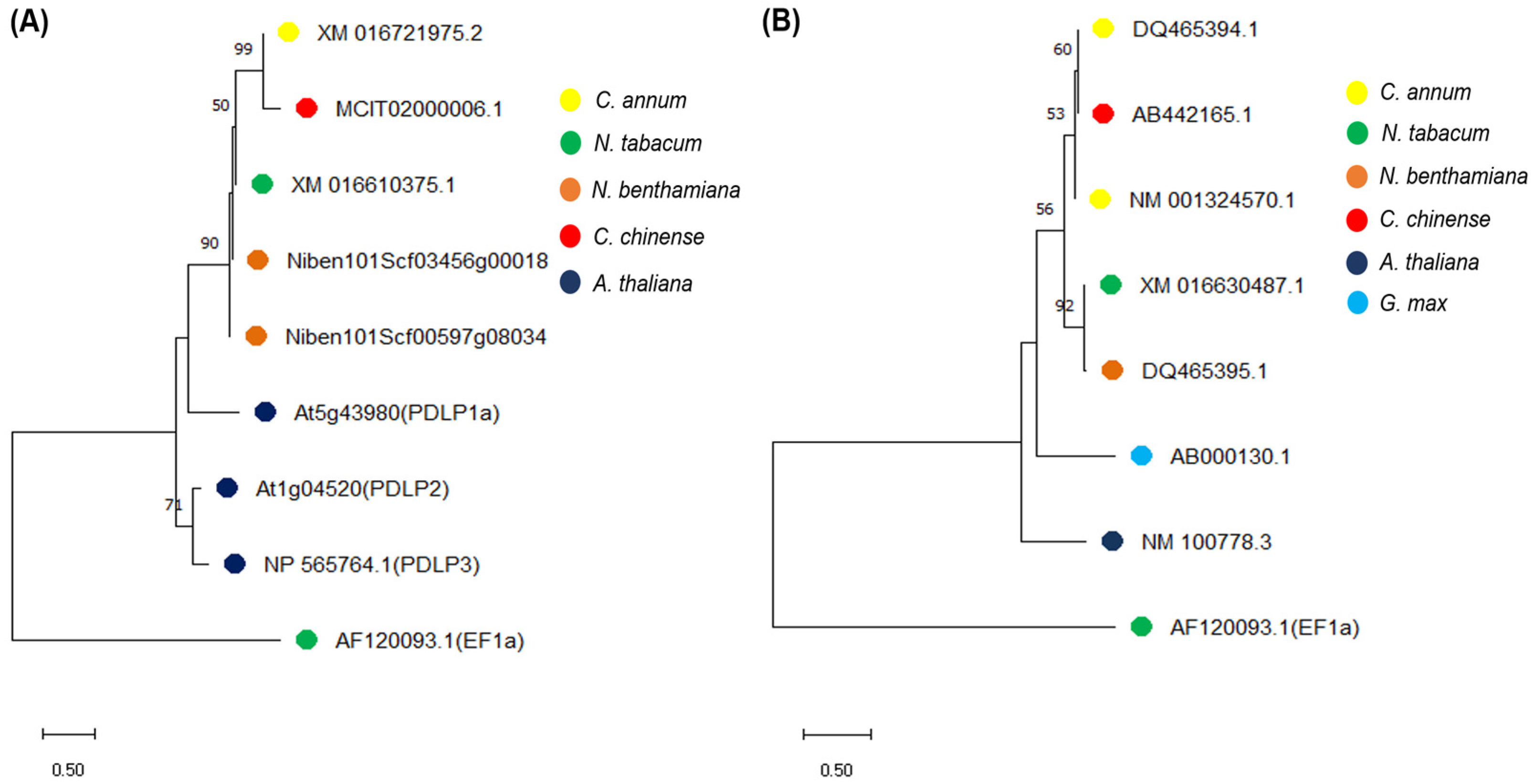
Figure 1. Phylogenetic analysis of (A) PDLP- and (B) SRC2-related genes from related plant species using the neighbor-joining (NJ) method. Each branch point is a representation of 10,000 bootstrap replicates. Bootstrap value higher than 50 is shown here. Nicotiana tabacum elongation factor 1-alpha gene sequence was used as an outgroup.
Plants 14 00495 g001
Figure 2. Phenotypic observation of Nicotiana benthamiana plants. (A) Cucumber mosaic virus (CMV)-infected plant and (B) mock (phosphate buffer)-treated plant at 14 days post treatment.
Plants 14 00495 g002
Figure 3. Time course expression profiles of PDLP and SRC2 genes upon cucumber mosaic virus (CMV) infection in Nicotiana benthamiana. A semi-quantitative RT-PCR analysis is shown. A fragment of CMV coat protein gene (CP, 657 bp) and N. benthamiana PDLP (413 bp) and SRC2 (123 bp) were amplified from cDNA obtained from systemic leaves at 2, 8, and 15 days post inoculation with CMV or phosphate buffer (mock). PCR was performed for 30 cycles and three biological replicates were pooled for each experimental group. The elongation factor 1 alpha (EF1a) gene fragment (116 bp) from N. benthamiana was used as an internal control. White color arrows indicate amplified target gene fragments. M indicates 100 bp ladder (New England BioLabs, NEB, Ipswich, MA, USA). The values above the bands represent the ratio of band intensity of normalized PDLP and SRC2 genes in mock and CMV-infected N. benthamiana plants.
Plants 14 00495 g003
Figure 4. Time course expression analysis of NtPDLP and NtSRC2 genes in silenced Nicotiana benthamiana plants. Relative quantification of NtPDLP and NtSRC2 gene expression in silenced and control (TRV2-eGFP) plants at different time points. In addition, 50% and 60% percent downregulations of PDLP and SRC2 genes were observed in N. benthamiana 14 days post agroinoculation. Purple color indicates control-treated (eGFP) group, blue color indicates pTRV2-NtPDLP-treated and green color indicates pTRV2-NtSRC2-treated plants. Asterisk (*) indicates significant differences between the control and silenced plants, p-value < 0.05 (Student’s t-test); dpa = days post agroinoculation.
Plants 14 00495 g004
Figure 5. Expression analysis of PDLP and SRC2 genes in silenced Nicotiana tabacum and Capsicum chinense plants at 14 days post agroinoculation (dpa). eGFP indicates the control-treated plants. Fifty and thirty percent downregulation of PDLP gene was observed in N. tabacum and C. chinense (Bhut Jolokia), respectively, whereas sixty and forty percent downregulation of SRC2 gene was observed in N. tabacum and C. chinense, respectively. Student’s t-test was performed for mean comparisons. Asterisk (*) indicates significant difference in gene expression level between PDLP-, SRC2-silenced plants, and control (eGFP) plants (p < 0.05, Student’s t-test). ns: non-significant.
Plants 14 00495 g005
Figure 6. Phenotypic observations of VIGS-silenced Nicotiana benthamiana plants at different time-points after cucumber mosaic virus (CMV) inoculation. Plants were first agroinfiltrated with VIGS constructs, followed by sap inoculation with CMV two days later to ensure the initiation of gene silencing. In the control (eGFP) plants, initial symptoms of CMV infection were observed at 7 days post infection (dpi). Subsequently, symptoms such as downward leaf curling and mosaic symptoms appeared more prominently at 12 and 19 dpi. The PDLP-silenced N. benthamiana plants showed reduced viral symptoms in comparison to the control and SRC2-silenced plants. Red arrows indicate typical disease symptoms. dpa = days post agroinoculation.
Plants 14 00495 g006
Figure 7. Relative abundance (RT-qPCR) of cucumber mosaic virus (CMV) titer in PDLP- and SRC2-silenced Nicotiana benthamiana plants at 7, 12, and 19 days post inoculation (dpi) with CMV. Plants agroinoculated with GV3101 strain carrying pTRV2-eGFP plasmids were used as control. The elongation factor 1 alpha (EF1a) gene fragment from N. benthamiana was used for internal control and normalization purposes. Student’s t-test was performed for mean comparisons. Asterisk (*) indicates significant difference in relative viral titer between PDLP-, SRC2-silenced plants, and control (eGFP) plants (p < 0.05, Student’s t-test). ns: non-significant.
Plants 14 00495 g007
Disclaimer/Publisher’s Note: The statements, opinions and data contained in all publications are solely those of the individual author(s) and contributor(s) and not of MDPI and/or the editor(s). MDPI and/or the editor(s) disclaim responsibility for any injury to people or property resulting from any ideas, methods, instructions or products referred to in the content.