Article

Pectate Lyase FvePL1 Is Required for Pollen Fertility and Mediates Drought Response in Woodland Strawberry

1
School of Life Sciences, Guizhou Normal University, Guiyang 550001, China
2
Key Laboratory of Plant Physiology and Development Regulation, Guizhou Normal University, Guiyang 550001, China
3
Key Laboratory of State Forestry Administration on Biodiversity Conservation in Mountainous Karst Area of Southwestern China, Guizhou Normal University, Guiyang 550001, China
*
Authors to whom correspondence should be addressed.
These authors contributed equally to this work.
Plants 2025, 14(23), 3583; https://doi.org/10.3390/plants14233583
Submission received: 15 October 2025 / Revised: 10 November 2025 / Accepted: 20 November 2025 / Published: 24 November 2025

Abstract

Successful fertilization is essential for fruit bearing and yield enhancement, relying on male gametophyte which facilitates sexual reproduction by transferring the sperm cell to the ovule. To accomplish this task, pectate lyase is secreted to lubricate the sperm cell towards the female partner by different strategies. However, the specific impact of strawberry PL in male sterility and achene development remained elusive. Here, we systematically investigated the functions of diploid strawberry Fragaria vesca pectate lyase 1 (FvePL1), determining its localization in the cell wall and membrane. In situ hybridization presented its maximum expression in the anther, particularly the endothecium, connective tissue, and septum. Analysis of RNAi mutants and green fluorescent protein (GFP)-tagged overexpression lines demonstrated that the failure of FvePL1 significantly inhibited the fruit set due to stunted achenes. In addition, the deficiency of FvePL1 expression resulted in a 68.29% reduction in the number of pollen grains, a 73.27% decrease in pollen viability, morphological alterations of exine and intine, impaired pollen tube, and the inability of the sperm nucleus to reach its target due to the delayed and incomplete tapetal degeneration. In addition, the suppression of FvePL1 resulted in a 65.02% increase in survival rate withholding irrigation for 30 days, conferring enhanced drought tolerance by negatively influencing cell wall structure. Therefore, this study identified FvePL1 as a crucial regulator of pollen development, fertilization, and achene maturation and abiotic stress. These findings provide a framework for advancing research on the development of the male gametophyte in strawberry and even yield optimization in Rosaceous crops.

1. Introduction

The emergence of fruits marks an evolutionary breakthrough, profoundly enhancing seed dispersal efficiency and initiating the adaptive radiation of angiosperms. A successful fruit set necessitates precise pollination via pollen transfer to the pistil. Upon successful fertilization, the ovary develops into a fruit, sheltering embryonic seeds destined for germination for the next generation of flowering plants [1]. However, successful fertilization requires normal male gametophyte, indispensable for orchestrating the biological functions of the elaborately structured pollen grain [2]. Pollen development within the anther locules of the stamen involves a multistage process requiring precise orchestration [3]. In addition, the delivery of sperm cells to the female gametophyte was facilitated by pollen tube extension from the grain’s apex through the transmitting tract’s extracellular matrix, culminating in stigmatic papillae penetration and subsequent access to maternal reproductive tissues [4]. During this process, the cell wall is crucial for directional expansion of pollen tube germination and the success of double fertilization [5].
Pectin, as a natural macromolecule in biomedical and drug delivery applications, is a principal macromolecular polysaccharide component of the growing pollen tube apex and has been reported to be indispensable for fertilization [6,7]. Its remodeling is governed by wall-modifying enzymes that balance pectin depolymerization and polysaccharide deposition, establishing a dynamic equilibrium of elastic, plastic, and rigid properties necessary for tube elongation [8,9]. Investigations of pectin participate into pollen cell walls have been elucidated in Arabidopsis [8]. Such architectural complexity involving pectin underscores the capacity of pollen tube to overcome the mechanical and biochemical barriers of pistil [10]. Pectate lyases (PLs) have emerged as critical regulators of pollen tube growth and fertilization in Arabidopsis, notwithstanding the significance of pectin modification to regulate cell wall stability and extensibility [11,12]. They are accountable for the degradation of pectin by cleaving the α-1,4-polygalactyronic acid chain and its activity is stringently regulated to ensure proper cell wall assembly and remodeling during diverse developmental processes [13]. Additionally, Arabidopsis PLs have been demonstrated to have maximal expression in the mature pollen grains and post-pollination pistils, underscoring their indispensable roles in pollen tube elongation and fertilization [14]. Recently, they have been proved to be indispensable for facilitating the loosening of the innermost layer of the pollen wall, which is crucial during the initial pollen hydration and pollen tube emergence loosening during the initial stages of pollen tube germination in Arabidopsis [15]. Moreover, PLs are secreted by the elongating pollen tube into the extracellular matrix, and enable modification of transmitting tract pectin networks, enhancing pollen tube penetration through pistil tissues to expedite pollen tube growth and to aid the sperm cell in reaching the ovule in Arabidopsis [15].
Strawberry serves as an excellent model for investigating fertilization-induced fruit development, owing to its unique fleshy fruit derived from a receptacle, an enlarged stem tip supporting hundreds of seed-bearing ovaries [3]. Fertilization triggers the production of fertilized achenes, with fruit size and yield demonstrating a positive correlation to seed number. The removal of fertilized ovules from the receptacle prevents the formation of fleshy fruit, underscoring that insufficient male gametophyte compromises fruit set and reduces yield. Generally, more achenes promoted larger and more bountiful fleshy fruits. Therefore, the regulatory mechanisms governing pollen development are requisite for enhancing fruit production [16]. Sixteen FvePL homologs have been identified in the diploid woodland strawberry Fragaria vesca [17]. Co-expression network analysis revealed that FvePLs had close association with genes involved in the development of vegetive and reproductive organs. In addition, histological observations demonstrated that FvePL1, 4 and 7 enhanced cortical cell division and expansion, thereby promoting fruit softening, also in cultivated strawberry [18]. It has been acknowledged that the decreased expression of FvePL1 (FvH4_2g19540) was linked to altered petal architectures, and significantly reduced the number of pollen grains [17]. Nevertheless, no experimental evidence has established its involvement in pollen tube morphogenesis via direct cell wall remodeling. Therefore, there is a necessity to comprehend how FvePL1 guides the pollen and fruit maturation at the cellular and subcellular levels.
In this work, we aimed to elucidate the function of FvePL1 in pollen tube growth, specifically testing its essential roles in cell wall remodeling and the biomechanical properties governing this process. Through an integrated approach combining physiological, genetic, and cytological analyses, we demonstrate that FvePL1 is critical for pollen fertility, achene development and enhanced drought tolerance. Our findings provide a molecular mechanism for male gametophyte development, offering a strategic target for enhancing strawberry fertility and yield through genetic breeding.

2. Results

2.1. Molecular Characterization of FvePL1 Which Was Maximumly Expressed During Anther Maturation and Fruit Transformation

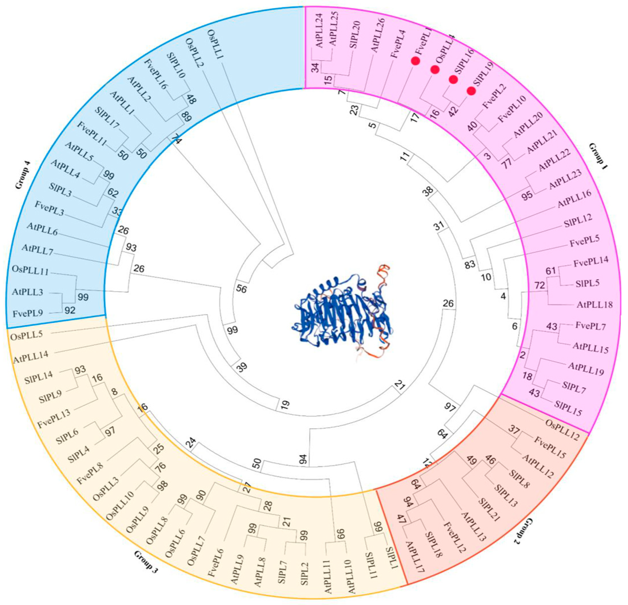
The coding sequence (CDS) of FvePL1 is 1341 bp long, encoding 446 amino acids (3 bp is a stop codon) with a molecular mass of 49.81 kD and an isoelectric point of 8.44. To identify proteins homologous to FvePL1, a phylogenetic tree was constructed within PL family members from other species, including Oryza sativa, Solanum lycopersicum, and Arabidopsis, revealing notable sequence similarity to OsPLL4, SlPLL16, and SlPLL19, suggesting conserved functional attributes among these orthologs (Figure A1). The expression profiles of FvePL1 across six major organs, anthers (at developmental stages of eight, ten and twelve) and mature pollens were detected [3]. The data showed that FvePL1 was widely expressed in multiple tissues. However, its transcripts were found the most in mature fruit, followed by floral tissues and leaf, with pronounced expression localized to anther stage 12 and mature pollen (Figure 1A,B).
Histological observation further corroborated the transcripts of FvePL1 in various tissues. Visible signatures of FvePL1 were observed in epidermal cells and mesophyll of leaves by in situ hybridization. Similarly, they were displayed in epidermal cell layer and parenchyma of stem, filament, and petal, aligning with critical vascular differentiation processes during vegetative organ maturation. Distinct hybrid signals of FvePL1 were also observed in mature fruit, and the enlarged image of mature fruit confirmed the strong staining in the epidermal cell layer and parenchyma of the cortex cells, suggesting its higher expression at the turning stage to regulate fruit firmness due to degrading pectin. By contrast, there was only weak expression of FvePL1 in the epidermal of green fruit. No discernable signals were found in anther stage 8 when microspore mother cells appeared and four locules became distinct, and stage 10 when tapetum degeneration was initiated. Conversely, prominent and intense hybridization signals of FvePL1 were noticed in the endothecium, connected tissues of anthers, epidermis and pollen, suggesting its potential roles in degenerating middle layers, thus facilitating the degradation of the primary cell wall and promotion of pollen penetration (Figure 1C).

2.2. FvePL1 Was Localized in Cell Wall, Cell Membrane and Cytoplasm

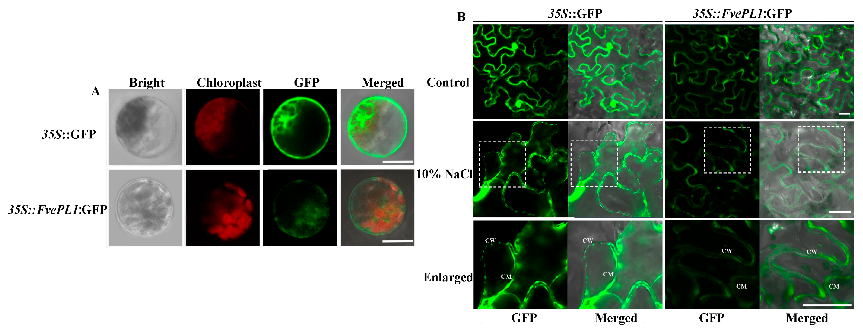
To explore the specific localization of FvePL1 in cells, the vector of 35S::FvePL1:GFP was introduced into Arabidopsis protoplasts for subcellular localization (Figure 2A). Microscopic visualization showed that robust green fluorescent signals, as the positive control of 35S::GFP-transformed protoplasts, were localized in the whole cell. Meanwhile, FvePL1 was predominantly presented in the cell membrane and cytoplasm. To further clarify whether FvePL1 was localized in the cell wall, 35S::FvePL1:GFP and control were individually transiently transformed into Nicotiana benthamiana leaves following plasmolysis induction via 10% NaCl treatment. While non-induced controls exhibited uniform fluorescence throughout the cellular compartment, plasmolyzed tissues expressing 35S::GFP displayed distinct GFP fluorescence localized to the cell wall (Figure 2B). When the cells expressed 35S::FvePL1:GFP after plasmolysis, the enlarged image shows that the strong green signals were found to be localized in the cell walls in plasmolyzed cells, implicating FvePL1 in dynamic cell wall modulation.

2.3. Reduction in Fvepl1 Resulted in Fruit Defects and Lower Seed Production

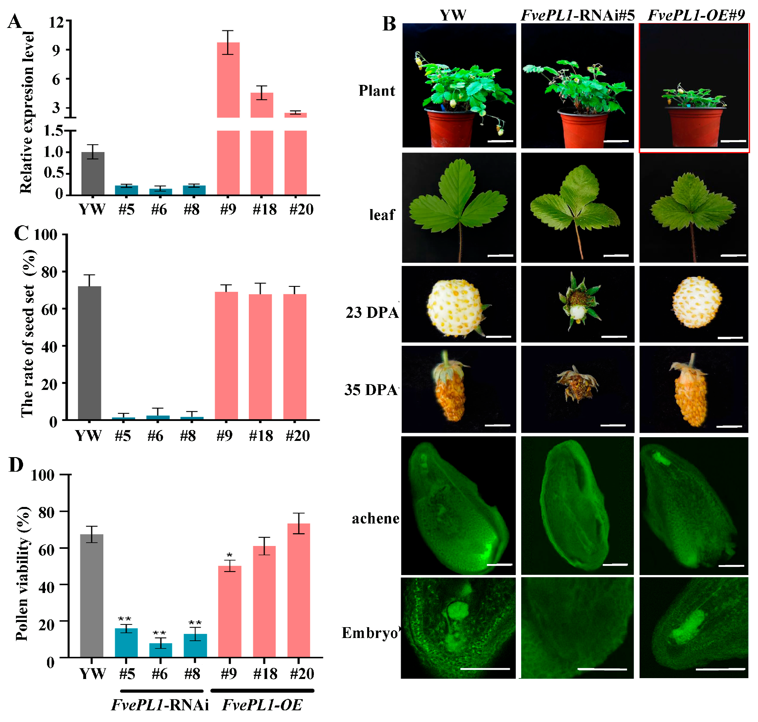
To analyze detailed functions of the FvePL1, an over-expression (OE) and RNAi vectors were constructed and transformed into YW5AF7 (YW) genetic background via agrobacterium-mediated method. After hygromycin resistance screening, transgenic seedlings were further determined using qRT-PCR to test the transcript levels of FvePL1 to identify the positive lines (Figure 3A). The upregulation of expression was found in FvePL1-OE #9, #18 and #20, which were a 9.18-fold, 4,82-fold and 2.89-fold increase compared to that of the YW, individually. By contrast, the expression levels were remarkably lower in FvePL1-RNAi#5, #6 and #8 compared to control. Thus, seedlings of FvePL1-OE#9 and FvePL1-RNAi#5 were further subjected to comprehensive phenotypic analysis compared to YW.
Morphological analysis of whole plants showed that FvePL1-OE displayed wrinkled leaves, smaller seedlings and stunted growth compared to YW and RNAi counterparts. Fruits of RNAi lines did not develop further after pollination at 23 and 35 days post-anthesis (DPA) observed using stereoscopic analysis (Figure 3B). Notably, FvePL1 downregulation directly correlated with impaired fruit set and seed development. With deep observation using confocal laser scanning microscopy, a normal embryo could be detected at the top of the ovule, and the central cells also differentiated normally to form endosperm in FvePL1-OE and YW plants, revealing marked differences (Figure 3B). However, the images show that a tubular structure full of dead cells and premature endosperm cellularization was present in the ovule of FvePL1-RNAi, resulting in the abortion of forming normal embryos and endosperm. No discernable difference was noticed in seed rate in YW and overexpression lines, which were both approximately 75%, whereas they were remarkably decreased in RNAi lines which were only 2%~4%, in correspondence with the reduction in fruit bear and the lower seed set attributed to the downregulation of FvePL1 (Figure 3C). In addition, pollen viability in the FvePL1-RNAi line were individually 19.24%, 12.76% and 18.67%, whereas the values in overexpression lines were 53.84%, 61.16% and 74.58%, resulting in a 73.27% reduction (Figure 3D).

2.4. FvePL1 Is Required for the Development of Exine and Intine of Pollen Grains

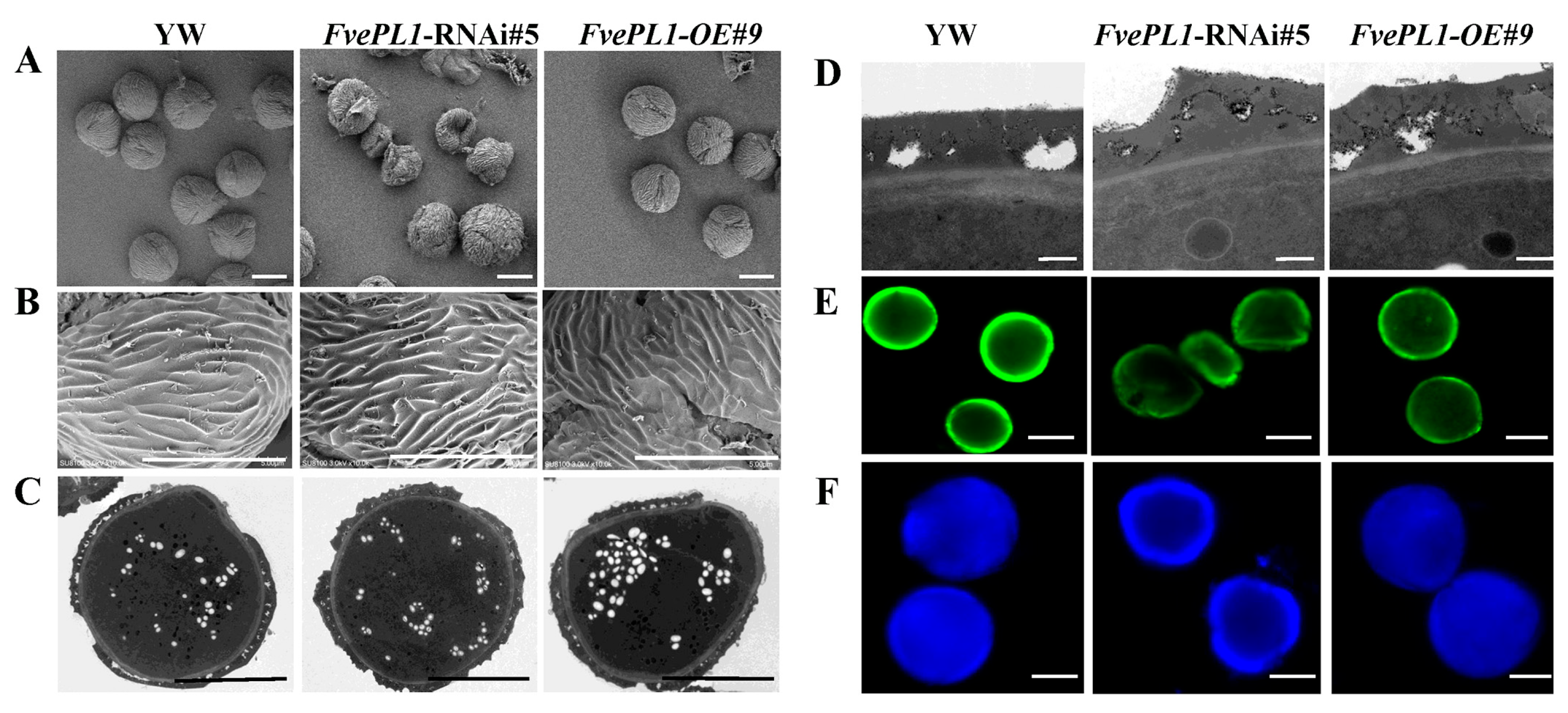
To assess the influence of FvePL1 on pollen cell walls, scanning electron microscopy (SEM) was utilized to investigate the ultrastructural changes in pollen grains and adjacent cell walls regulated by FvePL1 (Figure 4A). The pollen surface of YW or FvePL1-OE has multiple germination pores, accompanied by orderly arrays of raised striations on the surface of pollen grains (Figure 4B). In contrast, most FvePL1-RNAi pollen grains displayed irregular contours, deformed convex lines and depressed inward, resulting in abnormal aperture, and ultimately repressing pollen germination (Figure 4B).
To provide more detailed information on the internal morphology of pollen grains at the ultrastructural level, mature pollens from the new open flowers of three genotypes were fixed and processed to prepare for transmission electron microscopy (Figure 4C). The fine exine was composed of the baculum and the tectum. Except for more dispersed starch granules found after repressing FvePL1, no apparent abnormalities were observed in the cytoplasm. However, there was significantly different morphology of intine and exine regulated by FvePL1 through enlarging the cell wall structures (Figure 4D). First, the intine thickness of pollen became thicker in FvePL1-RNAi lines. Secondly, less white signals were present around the intine, indicating that fewer starch granules were accumulated in the exine after the deficiency of FvePL1. Thirdly, the surface of exine in FvePL1-RNAi lines, especially the structure of tectum, were much more overflowing furrow regions, whereas the smooth surface of exine was observed in YW. By focusing on the structural characteristics of the exine and intine layers, we identified the roles of FvePL1 in the modifications of the cell wall architecture.
Exine is mainly composed of sporopollenin and cellulose to protect the internal structure of pollen from damage. Intine with a pectocellulosic composition is thicker under the pollen apertures, from where the pollen tube emerges. Auramine O staining was used to test the functions of FvePL1 on the exine using confocal microscopy (Figure 4E). The images illustrate that strong and continuous green fluorescence was observed in the outline of YW and FvePL1-OE. On the contrary, the fluorescence of FvePL1-RNAi plants was discontinuous and weak in the outer wall boundary. We further used Calcofluor white to examine the intine influenced by FvePL1 (Figure 4F). Consistently, a hollow center and only weak blue fluorescence of the outline were noticed in pollen grains of FvePL1-RNAi, while substantial blue signals were found in YW and FvePL1-OE. The abnormal development of exine and intine demonstrated that FvePL1 dysregulated exine and intine organization, precipitating pollen developmental defects.

2.5. FvePL1-RNAi Lead to the Decreased Pollen Number Due to Degradation of Tapetum

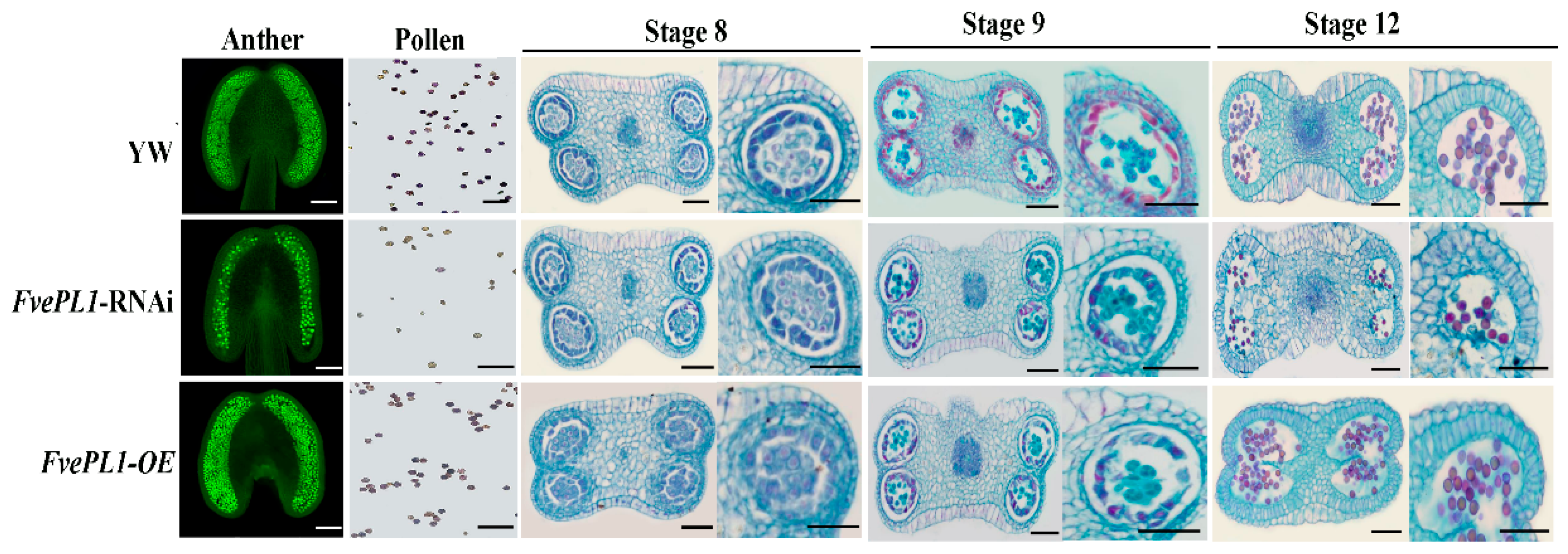
To determine the functional role of FvePL1 in pollen development, anthers at day 1 post-anthesis were harvested and analyzed via confocal microscopy. The images showed that FvePL1-OE exhibited pollen quantities equivalent to YW, whereas a statistically significant reduction in mature pollen grain of FvePL1-RNAi compared to other two genotypes (Figure 5). To elucidate the mechanism underlying the diminished pollen production, developmental dynamics across anther maturation were examined through paraffin sectioning. Cytological analysis demonstrated that all genotypes achieved normal anther dehiscence by stage 8, with no discernible differences in microsporogenesis initiation, including synchronized meiotic entry of tetrads or four-locule compartmentalization. Subsequent histological evaluations revealed progressive cellular differentiation: locular expansion facilitated tetrad release by stage 9, concurrent with middle layer degeneration, while tapetal cells in wild-type and FvePL1-OE lines underwent complete degradation by this phase. However, the failure degradation of tapetum in FvePL1-RNAi was noticed, resulting in no distinct separation at Stage 12. In addition, more round and uniformly larger pollen grains were formed in YW and FvePL1-OE. On the contrary, limited viable pollen grains were differentiated, suggesting the aborted pollen grains caused by the downregulation of FvePL1.

2.6. FvePL1 Is Essential for Pollen Germination

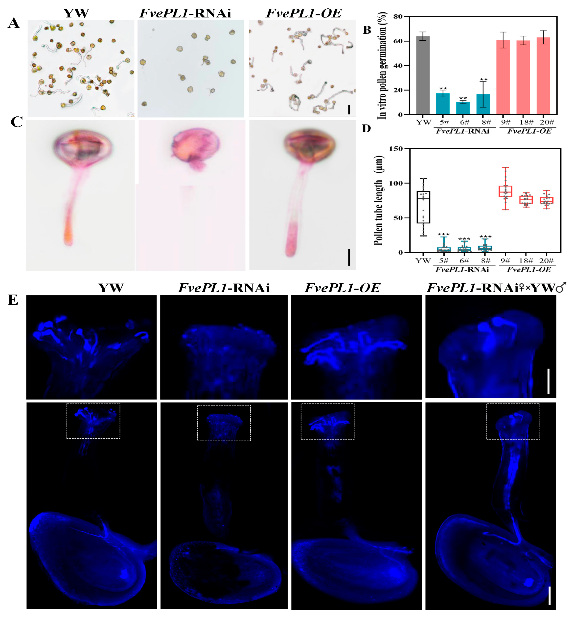
To assess the influence of FvePL1 on pollen germination, we observed pollen germination by an in vitro germination experiment (Figure 6A). The results showed that the most mature pollen grains of YW and FvePL1-OE transgenic plants uniformly germinated and an opaque pollen tube emerged from pollen grains. Differently, in addition to the smaller number of pollen grains in FvePL1-RNAi lines, barely any pollen tube could be observed. In addition, we found that the pollen germination rates of YW and FvePL1-OE transgenic lines were individually 61.47%, 60.98% and 66.79%. However, there was no more than 20% after disrupting the FvePL1 expression, which decreases by 68.29%, in accordance with the results of pollen viability (Figure 6B). Despite some pollen tube produced in FvePL1-RNAi lines, the tube length was terrifically shorter than 10 μm by staining observation (Figure 6C). No doubt, the pollen tube lengths in FvePL1-OE and YW were both longer than 40 μm, especially up to 95 μm in YW (Figure 6D). These findings collectively demonstrate that FvePL1 deficiency disrupts pollen germination and severely impairs tube elongation.
To further detect the in vivo germination of mature pollen, we collected pistils after pollination and stained pistils utilizing aniline blue (Figure 6E). The images showed obvious outlines and distinct blue fluorescence of the transmitting tract and ovary in FvePL1-OE, which were no apparent abnormalities from YW, indicating the male sperm cell could successfully move their target after pollination (Figure 6E). By highlighting their stigmas, clear long pegs of germination tubes were displayed, especially in the FvePL1-OE line. In contrast, only slight blue signals were found in FvePL1-RNAi lines, indicative of no obvious pollen observed on stigma. When further zooming in the top of pistils, barely no germination tube of FvePL1-RNAi was photographed, consistently with the results of in vitro germination experiments, corroborating that the decrease in FvePL1 reduced the pollen germination rate and inhibited pollen tube elongation. Notably, reciprocal pollination experiments confirmed retained female fertility in FvePL1-RNAi plants, as wild-type pollen exhibited normal germination and tube extension within mutant pistils, indicating the normal female fertility of FvePL1-RNAi which could receive pollen of YW normally. Based on in vitro and in vivo germination experiments, despite the normal development of stigma and female gametophyte, the reduction in FvePL1 resulted in abnormal pollen germination, significantly shortening the length of pollen tubes.

2.7. Downregulation of FvePL1 Improved Drought Tolerance

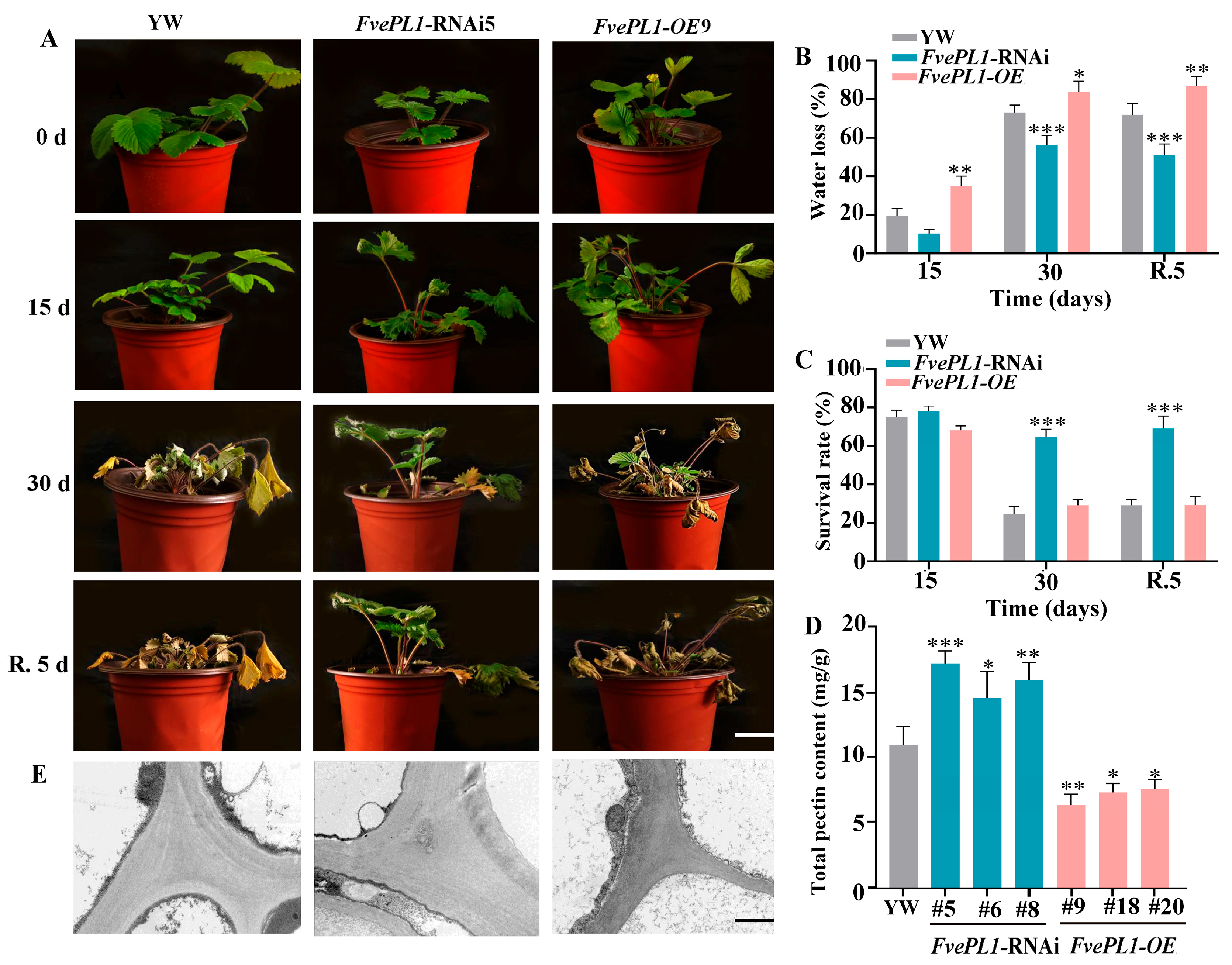
To elucidate the role of FvePL1 in drought stress, three genotypes underwent water deficit treatment. Phenotypic observation revealed that YW exhibited subtle leaf hyponasty, a hallmark of moderate water deficit at 15 days. In the same period, FvePL1-OE plants displayed pronounced drought-induced symptoms, including downward leaf curvature, whereas FvePL1-RNAi plants maintained significantly normal morphology compared to other counterparts under identical conditions. Prolonged drought to 30 days exacerbated overexpression of plant stress symptoms, manifesting severe stress symptoms, including leaf desiccation and whole-plant wilting. By contrast, RNAi plants exhibited markedly attenuated drought symptoms, corroborating drought tolerance after FvePL1 suppression. After 5 d recovery, plants of FvePL1-RNAi resumed normal growth (Figure 7A). Evaluations of water loss and survival rates indicated a pronounced enhancement in drought tolerance for FvePL1 RNAi plants relative to both YW and overexpression lines. This was particularly evidenced by a 55.04% increase in survival rate following a 30-day period of irrigation withholding (Figure 7B,C). Furthermore, in contrast, FvePL1-OE displayed a reduced drought tolerance and lower survival rate. In addition, the total pectin accumulation of FvePL1 RNAi was almost 1.52- and 2.47-fold than that in YW and FvePL1-OE (Figure 7D). Correspondingly, ultrastructural analysis of cell wall architecture revealed striking morphological divergence. The electron-dense material observed in the tricellular junction zone of FvePL1-RNAi was more intensive than other two genotypes (Figure 7E). By contrast, the adjacent cells of FvePL1-OE were separated, and the junction zone was degraded, loose and separated.

3. Discussion

In this study, we highlighted the intricate relationship between FvePL1 and male reproductive tissues. A noticeable gap in the current research is the exploration of FvePLs’ broader roles beyond pectin modification, potentially interacting with features of pollen grains and functions in reproductive plant tissues.

3.1. FvePL1 Implicated in Male Gametophyte Development Is Indispensable for Fruit Set

FvePL1, harboring a conserved pectin lyase domain, was localized to the cell wall and membrane. In addition, in situ hybridization revealed its predominant expression in the endothecium, connective, septum, epidermal cell layers and parenchyma of the cortex cells. Transgenic analyses demonstrated that FvePL1 deficiency caused severe fruiting failure, evidenced by defective and stunted achenes. By a comprehensive assessment of the entire biological process of male gametophyte, the downregulated FvePL1 led to the decreased number of pollen grains, less viability, morphological aberrations in exine and intine, impaired pollen germination germinate, defected formation of pollen tubes, and ultimately led to failed fertilization. All the data illustrated that FvePL1 played robust and vital roles in the male sterility of strawberry, aligning with most functions of PL-like in Arabidopsis [18,19], Brassica campestris [20], and Gossypium hirsutum [21]. Therefore, FvePL1 is required for developing achene and strawberry yield [18].

3.2. The Aborted Pollens Caused by the Reduction in Fvepl1 Initiated from the Incomplete and Delayed Degradation of Tapetum and Septum

To explore the mechanism underlying the function of FvePL1 on the male gametophyte development, our findings discovered that FvePL1 suppression disrupted the development of tapetum and pollen wall biogenesis, leading to male sterility defects. Cytological observation revealed conserved microsporogenesis stages across three genotypes. Tetrads were held and microspore mother cells tightly packed within the anther locules before entering meiosis. Interestingly, YW and overexpression lines exhibited synchronized locule expansion, allowing individual tetrads to separate from each other. In contrast, the incomplete and delayed middle layer degradation was noticed in the FvePL1-RNAi lines, suggesting that the failure of pollen development was initiated form tapetum and septum degradation. The tapetum has been recognized to supply nutrients and metabolic substances to aid pollen grain maturation. It undergoes programmed cell death (PCD) and secretes enzymes that degrade cellular organelles. This degradation process helps in the formation of sporopollenin which is essential for exine formation, and aids the release of mature pollen grains from the anther [22].
The incomplete and delayed degradation of tapetum and septum caused by the downregulation of FvePL1 can be described as follows: (1) FvePL1 contributed in disrupting the integrity of the cell wall of tapetum; (2) FvePL1 might participate in the supplied process of nutrients from tapetum; and (3) FvePL1 affected the pectin-mediated signaling cascades pathway that exhibited premature PCD of tapetum and disordered cell wall. Cell death has been hypothesized to be necessary to provide nutrients to growing pollen tubes but also to facilitate the pollen tube penetration [4]. These coordinated processes ensure synchronized pollen development, whereas dysregulation induces developmental discordance. Collectively, FvePL1 acts as a negative regulator of tapetal and septal degradation pathways, where its diminished activity disrupts reproductive efficiency and strawberry yield.

3.3. Different Regulatory Mechanisms of FvePL1 Influencing Pollen and Fruit Development

Despite its downregulation inducing severe male sterility defects, the overexpression of FvePL1 presented no discernable difference in pollen maturation phenotypes compared to YW. Integrated analysis of in vitro and in vivo germination indicated that FvePL1 deficiency selectively disrupted pollen germination and tube elongation, despite maintaining pistil integrity and female gametophyte functionality. This dysfunction likely arises from perturbed signaling pathways essential for pollen tube biogenesis, independent of post-germination pollen–pistil interactions [23]. In contrast to pollen defects, FvePL1-OE transgenic lines accelerated fruit softening relative to wild-type controls [17]. The divergent phenotypes caused by FvePL1 elicited a tissue-dependent regulatory molecular mechanism. The digested pectin could act as signaling molecules to feedback control of tapetum degradation and pollen maturation that is yet to be confirmed [24]. Considering that normal female fertility in the FvePL1-RNAi pollen pollinated lines, the fruit-initiated stage of FvePL1-OE plants paralleled wild-type progression. With the maturation of fleshy fruit, additional transcription factors might regulate the functions of FvePL1. For example, overexpression of FvePL4 promotes fruit softening due to the ABA-induced FvWRKY48 bound to the FvePL4 promoter via a W-box [25]. Thus, different transcriptional factors could influence FvePL1 in regulating pollen and fruit development.

3.4. FvePL1 Negatively Mediated Drought Response in Woodland Strawberry

Given that FvePL1 modulates pectin in pollen which constitutes a heterogeneous, branched, and highly hydrated polysaccharide network within the plant cell wall, it confers enhanced tolerance to drought and osmotic stress [26]. Consequently, the pectin-composed cell wall plays a direct role in the drought stress response [27]. Furthermore, its derivatives, including O-acetylated pectin, regulate growth and biotic/abiotic stress adaptation [28]. Functional analysis of FvePL1 in strawberry revealed that its overexpression elevated plant dwarfing and disrupted cell wall architecture, especially cell junction zones, thereby decreasing drought tolerance. Conversely, the failure of FvePL1 expression profoundly increased drought stress. This evidence suggested that pectate lyase modulates cell wall biomechanics and osmotic adjustment through targeted catalysis of alpha-1,4-glycosidic linkages in pectic polysaccharides [29]. Likewise, over-expression of the PMEI enhances drought tolerance of Glycyrrhiza uralensis [30]. Furthermore, previous studies showed that candidate genes related to cell wall remodeling enzymes (such as PL, PME, PMEI, and expansins) were related with plant response to drought tolerance and could provide an interesting point for developing molecular markers for breeding drought-resistant plants [31]. The findings offer a comprehensive understanding of the contribution of FvePL1 on pollen development and abiotic stress. FvePL1 might act in a direct or signal pathway, setting the stage for future research into its deeper mechanism. As climate unpredictability intensifies and challenges for pollen viability and seedling growth emerge, this research shows significance, as FvePL1 could regulate fertilization efficiency or response to stress.

4. Materials and Methods

4.1. Plant and Growth Conditions

The Fragaria vesca strain Yellow Wonder 5AF7 (YW5AF7) inbred line, derived from the seventh generation of selfing, served as the wild-type genotype in this study. RNA interference (RNAi) and overexpression constructs were both generated in YW5AF7. Seedlings were cultivated in a controlled growth chamber with a continuous 16 h light/8 h dark photoperiod at 22 °C, 65% relative humidity, and 350 μmol m−2 s−1 light intensity.

4.2. Phylogenetic Tree Construction

Protein sequences of FvePL1 were downloaded from the Genome Database for Rosaceae (GDR: www.rosaceae.org/ (accessed on 14 October 2025)) and the homology search was conducted using the amino acid sequences for FvePL1 from the UniProt database (https://www.uniprot.org/ (accessed on 14 October 2025)). Multiple alignment was performed using ClustalW version 2.0 (http://www.clustal.org/clustal2/ (accessed on 14 October 2025)) and the relative accession number list of PL gene family in Oryza sativa, Arabidopsis, Solanum lycopersicum, and F. vesca were listed in Table A1. A phylogenetic tree was constructed using the maximum likelihood (ML) method with 1000 bootstrap replicates via MEGA7.0 (http://www.megasoftware.net (accessed on 14 October 2025)).

4.3. Subcellular Location

The FvePL1 coding sequence was PCR-amplified and cloned into the XbaI site of the pM999 vector to generate CaMV 35S::FvePL1:GFP plasmid. Protoplasts were isolated from the mesophyll tissues of 4-week-old Arabidopsis leaves (Columbia-0 ecotype). A total of 100 μL (2 × 10⁴) protoplast was mixed with 10 μL (10~20 μg) recombinant or control plasmid, then 110 μL PEG solution was added for transfection. Transfected protoplasts were imaged using a ZEISS LSM 710 confocal microscope (Zeiss, Oberkochen, Germany) under a 488 nm blue light laser.

4.4. In Situ Hybridization

For cytological observation of FvePL1 expression, leaf, stem, filament, petal, anther (at developmental stages 8, 10 and 12) [3], green and mature fruits were collected and fixed in RNase-free FAA solution (4% formaldehyde (v/v), 10% acetic acid, and 50% ethanol), followed by dehydration through graded ethanol series, embedded in paraffin wax and sectioned (8 μm) using Leica RM2255 (Leica, Wetzlar, Germany). A gene-specific cDNA fragment of FvePL1 was amplified using ISH-F/R primer for in situ hybridization. Their PCR product was then cloned into the pGEM-T vector. Sense and antisense RNA probes were synthesized using SP6 and T7 RNA polymerase, respectively. In situ hybridization experiments were performed, including prehybridization, hybridization, and washing using a DIG RNA labeling Kit (Roche, Basel, Switzerland) applied to the tissue paraffin section [17]. Sides were photographed under a BX53 microscope (Olympus, Tokyo, Japan).

4.5. RNAi and Overexpression Constructs and Transgenic Strawberry Generation

For RNAi vectors, the partial coding sequences about 500 bp targeting the at 5′-end coding regions of FvePL1 were amplified, respectively, by RT-PCR and placed upstream and downstream of pDS1301 in forward directions using KpnI/BamHI and reverse directions using SpeI/SacI. To generate the FvePL1 overexpression, regions of FvePL1-GFP were amplified from pM999-FvePL1-GFP and cloned into pMDC32 binary vector at the digestion sites of KpnI and PacI which the GFP reporter fused at the C-terminus of the gene. The constructs were individually transformed into agrobacterium strain GV3101. F. vesca transformation and regeneration were performed using previously published protocols [16]. To confirm transgenic lines, seedlings of various genotypes were sampled. All primers used in this study are listed in Table A2.

4.6. Plasmolysis Assay

The young leaves of Nicotiana benthamiana were infected with the Agrobacterium strain GV3101, carrying pMDC32-FvePL1-GFP vector or empty vector pMDC32-GFP as control, individually. At three days after agroinfiltration, N. benthamiana leaves were infiltrated with 10% NaCl solution. Then, leaf sections were excised 10~15 min after injection and examined using confocal laser microscopy (Zeiss, Germany).

4.7. Seed Observation

Four-day post-anthesis (DPA) seeds were dissected from achenes and imaged using a Zeiss Stemi SV 6 microscope. For confocal analysis, seeds were fixed in 4% glutaraldehyde (12.5 mM cacodylate, pH 6.9), dehydrated through ethanol gradients (30~100%), cleared in benzyl benzoate–benzyl alcohol (2:1), and visualized on a confocal microscope (Zeiss, Germany) whereas 488 nm excitation was used to image autofluorescence of the seeds for observing fluorescent [16].

4.8. RNA Extraction and Quantitative RT-PCR

Total RNA was extracted using a Total RNA extraction Kit (Promega, Madison, WI, USA) and reverse transcribed to cDNA with PrimerScript RT reagent Kit (TaKaRa, Shiga, Japan). The qRT-PCR was performed on a BioRad CFX96 Real-time system with SYBR Green PCR MasterMix (Applied Biosystems, Thermo Scientific, Foster City, CA, USA) under the following conditions: 5 min at 95 °C, followed by 40 cycles of 10 s at 95 °C, 10 s at 55 °C and 30 s at 72 °C, as well as 10 min at 72 °C. The relative expression level was analyzed using a modified 2−ΔΔCT method. For all qRT-qPCRs, Fveactin (FvH4_4g24420) was used as the internal control. The qRT-PCR experiments were performed from three biological replicates (with three technical repeats).

4.9. Pollen Viability Assay

Pollen grains from newly opened flowers of six seedlings of each genotype were collected and then stained with 30 μL 0.3% 3-(4,5)-dimethylthiahiazo(-z-y1)-2,5-di-phenytetrazoliumromide (MTT) solution (0.3 g MTT dissolved in 100 mL phosphate-buffered saline) for 20~30 min to detect pollen viability. Stained samples were mounted on slides, and a cover glass was attached for observation with an Olympus BX-53 microscope.

4.10. Cell Wall Imaging Using SEM and TEM

For scanning electron microscopy (SEM) detection, matured pollen grains were collected directly from strawberry stamens and fixed in 2.5% glutaraldehyde. The fixed samples were rinsed three times with 0.1 M phosphate buffer, with each rinse lasting 15 min. For dehydration, the tissues were sequentially immersed in 30%–50%–70%–80%–90%–95%–100%–100% alcohol, spending 15 min in each, and then in isopentyl acetate for 15 min. Regarding drying, the samples were placed in a critical point dryer K850 for the drying process [32]. Subsequently, samples were immobilized on a specimen mount coated with modeling clay and rapidly frozen in liquid nitrogen for about 30 s under vacuum and coated with gold using Ion Sputter MC1000 (Hitachi, Tokyo, Japan) under scanning electron microscopy SU8100 (Hitachi, Tokyo, Japan) [33].
For transmission electron microscopy (TEM) observation, the above samples were fixed with 4% (w/v) formaldehyde and 2% (w/v) glutaraldehyde in 0.1 M PBS (pH 7.2) under vacuum (0.6 bar) at 4 °C for 1 h, during which the vacuum was slowly broken three times, then incubated in fresh fixative solution at 4 °C overnight. The samples were washed three times in phosphate buffer, postfixed for 2 h in 1% (w/v) osmium tetroxide in phosphate buffer at room temperature, rinsed five times for 5 min in PBS, and dehydrated in a graded ethanol series (30 to 100% v/v) with 1 h incubations in each bath. The samples were then subjected to embedding, polymerization, ultrathin sectioning, staining, and observation under a transmission electron microscopy HT7800 (Hitachi, Tokyo, Japan).

4.11. Calcofluor White and Auramine O Staining

Anthers were collected at 10:00 am on the first day post flowering and mounted on slides. After staining with 0.001% auramine O (1 g Auramine O dissolved in 1 mL phosphate-buffered saline) which was added into 1 mL 17% (m/v) sugar for 15 min, the images were observed at 488 nm excitation and 500 to 570 nm emission. The intine of dissected anthers was stained by calcofluor white and 10% KOH for 1 min with a cover slip and immediately analyzed using a confocal microscope. Calcofluor white was obtained using the excited images at 340~388 nm, and the emission was collected at 400 to 450 nm.

4.12. Pollen Germination Assays In Vitro and In Vivo

To observe the pollen tubes in vitro, pollen grains sampled from six seedlings of each genotype were incubated on a pollen germination medium (100 g/L sucrose, 0.02% boric acid, 0.5% agar) in an incubator at 22 °C for 5–6 h in the dark and finally observed under an Olympus BX53 microscope. We calculated the rate of germination according to germinated pollen/total number of pollens × 100% and measured the pollen tube length using ImageJ software (https://imagej.net/ij/ (accessed on 14 October 2025)). For in vivo pollen germination of F. vesca, pistils of wild-type emasculated flowers were hand-pollinated with pollen from three genotypes. Notably, the hybrid pistils of FvePL1-RNAi were also collected after crosspollination using pollen from YW. One hour after saturated pollination, pistils were collected, fixed in FAA reagent (10% formalin, 80% ethanol, 10% glacial acetic acid) and vacuumed for 1 h and fixed for 10~12 h, softened with 30% NaOH overnight and washed with ddH2O. After bleaching with 6% hydrogen peroxide for 0.5 h, pistils were mounted on a microscope slide using a drop of 0.1% decolorized aniline blue solution for at least four hours and carefully pressed with a cover slip to open the pistil longitudinally. Images were observed under the excited at 488 nm, and the emission at 500 to 570 nm.

4.13. Drought Stress Treatment, Water Loss and Total Pectin Assay

Ten seedlings of strawberries of different genotypes were placed in a controlled greenhouse maintained at 20~22 °C and 55~68% humidity. Drought treatment was applied by withholding irrigation for 30 days. Post-stress recovery protocols included rehydration via supplemental irrigation at five days under standard growth conditions. Leaves harvested from the soil-grown plants at the same corresponding time were weighed. Water loss was evaluated by determining the decrease in leaf weight. Twenty leaves from three seedlings were used as a biological repeat. Three biological replicates were used for each genotype. Meanwhile, survival rate was calculated by counting the healthy leaves. Determination of pectin content was adopted using the carbazole colorimetric method according to the previous study [15].

4.14. Statistical Analysis

All experimental results are presented as mean ± standard deviation (SD) (n ≥ 3). Data were analyzed and visualized using GraphPad Prism (GraphPad software Inc., version 8.0, Boston, MA, USA). The one-way analysis of variance (ANOVA) followed by Tukey’s test was employed to determine the statistical significance (*, p < 0.05; **, p < 0.01; ***, p < 0.001).

5. Conclusions

This study comprehensively demonstrated that the downregulation of FvePL1 resulted in the failures of seed and achene development, and established its essential role in pollen grain exine and intine formation, as well as pollen tube germination. We further identified that suppression of FvePL1-induced delayed and incomplete tapetal degeneration, resulting in impaired pollen maturation, reduced pollen number, and decreased viability. In addition, the deficiency of FvePL1-enhanced drought tolerance through regulating cell wall structure was determined. These findings provide a mechanistic basis of the function of FvePL1 in male fertility and stress adaptation, offering potential applications for improving crop yield and resilience under abiotic stress.

Author Contributions

Conceptualization, X.H. and H.Y.; methodology, Y.J.; software, Y.W.; validation, G.S., L.Z. and N.L.; formal analysis, N.L.; investigation, N.L.; writing—original draft preparation, N.L.; writing—review and editing, X.H.; visualization, X.H.; supervision, N.L.; project administration, G.S.; funding acquisition, H.Y. All authors have read and agreed to the published version of the manuscript.

Funding

The work was supported by the National Natural Science Foundation of China (32460717 and 32160699) and Natural Science Foundation of Guizhou Province [QKHJC-ZK [2023] ZD026].

Institutional Review Board Statement

Transgenic plant materials were contained in the School of life science of Guizhou Normal University. The generation and handling of transgenic Fragaria vesca plants in this research complied with all relevant institutional and national biosafety regulations. The work was approved by the Institutional Biosafety Committee (IBC) of Guizhou Normal University.

Data Availability Statement

The original contributions presented in this study are included in the article. Further inquiries can be directed to the corresponding author.

Conflicts of Interest

The authors declare no conflict of interest.

Abbreviations

The following abbreviations are used in this manuscript:
PLpectate lyase
YWYellow Wonder 5AF7
MTT3-(4,5)-dimethylthiahiazo(-z-y1)-2,5-di-phenytetrazoliumromide
GFPgreen fluorescent protein
GDRGenome database for Rosaceae
DPAday post-anthesis

Appendix A

Table A1. The list of primers used in this study.
NameForward Sequence (5′-3′)Reverse Sequence (5′-3′)Application
FvePL1ATGAGGTTAGCTAGCTCGAGGTTAACATTGACGACCAAACCGAmplification
pM999-FvePL1GCTCTAGAATGAGGTTAGCTAGCTCGAGGGCTCTAGAACATTGACGACCAAACCGGCASubcellular location
FvePL1-RNAiGGACTAGTGGTACCACGACGACTGGAATGAGCACGCGAGCTCGGATCCAGGCTTGCAGTCGTGGATGTGRNAi line
FvePL1-OECGGGGTACCATGAGGTTAGCTAGCTCGAGGCGAGCTCTTAACATTGACGACCAAACCGOverexpression line
q-FvePL1GGAAGTGCTGATCCTACCATTACTGTGGTCTGAACTCTATGTGTqRT-PCR
FveactinCCCAAGTAAGGATGCCCCCATGTTCGTTGGCAAGGGGAGCAAGACAGTTGGTAGqRT-PCR
FvePL1TCTCATGCGCAACAGGGAATGTTGCGGCCGAATCCAATACISH
HygACGTTGCAAGACCTGCCTGAAATCCAGTCAATGACCGCTGTTATGCPositive verification
Table A2. The accession number of PL gene family in different species.
Gene IDAccession NumberGene IDAccession Number
OsPLL1LOC_Os01g36620AtPLL1At3g09540
OsPLL2LOC_Os01g62000AtPLL2At3g55140
OsPLL3LOC_Os02g12300AtPLL3At5g09280
OsPLL4LOC_Os04g05050AtPLL4At4g22080
OsPLL5LOC_Os05g22800AtPLL5At4g22090
OsPLL6LOC_Os06g05209AtPLL6At1g11920
OsPLL7LOC_Os06g05260AtPLL7At1g30350
OsPLL8LOC_Os06g05272AtPLL8At1g14420
OsPLL9LOC_Os06g38510AtPLL9At3g02720
OsPLL10LOC_Os06g38520AtPLL10At3g01270
OsPLL11LOC_Os08g18970AtPLL11At5g15110
FvePL1FvH4_2g19540AtPLL12At5g04310
FvePL2FvH4_3g00620AtPLL13At3g54920
FvePL3FvH4_3g01680AtPLL14At5g55720
FvePL4FvH4_4g05760AtPLL15At5g63180
FvePL5FvH4_4g25110AtPLL16At1g67750
FvePL6FvH4_4g31900AtPLL17At3g53190
FvePL7FvH4_5g06720AtPLL18At3g27400
FvePL8FvH4_5g15430AtPLL19At4g24780
FvePL9FvH4_5g27090AtPLL20At3g07010
FvePL10FvH4_6g02350AtPLL21At5g48900
FvePL11FvH4_6g14680AtPLL22At3g24670
FvePL12FvH4_6g16670AtPLL23At4g13210
FvePL13FvH4_6g49230AtPLL24At3g24230
FvePL14FvH4_6g52190AtPLL25At4g13710
FvePL15FvH4_7g18400AtPLL26At1g04680
FvePL16FvH4_7g20920SlPL1Solyc01g010740
SlPL2Solyc02g067450SlPL12Solyc05g014000
SlPL3Solyc02g080910SlPL13Solyc05g055510
SlPL4Solyc02g087670SlPL14Solyc06g071020
SlPL5Solyc02g093580SlPL15Solyc06g071840
SlPL6Solyc03g058890SlPL16Solyc06g083580
SlPL7Solyc03g058910SlPL17Solyc09g005850
SlPL8Solyc03g071570SlPL18Solyc09g008380
SlPL9Solyc03g113150SlPL19Solyc09g061890
SlPL10Solyc04g010230SlPL20Solyc09g091430
SlPL11Solyc05g007080SlPL21Solyc11g008140
Figure A1. Phylogenetic analysis of FvePL1 homologs from Solanum lycopersicum, Oryza sativa, and Arabidopsis. The central micrograph depicted the three-dimensional protein structure of FvePL1. The red dots indicated the homologs to FvePL1.
Plants 14 03583 g0a1

References

  1. Kang, C.; Darwish, O.; Geretz, A.; Shahan, R.; Alkharouf, N.; Liu, Z. Genome-scale transcriptomic insights into early-stage fruit development in woodland strawberry Fragaria vesca. Plant Cell 2013, 25, 1960–1978. [Google Scholar] [CrossRef] [PubMed]
  2. Edlund, A.F.; Swanson, R.; Preuss, D. Pollen and stigma structure and function: The role of diversity in pollination. Plant Cell 2004, 16 (Suppl. S1), S84–S97. [Google Scholar] [CrossRef] [PubMed]
  3. Hollender, C.A.; Geretz, A.C.; Slovin, J.P.; Liu, Z. Flower and early fruit development in a diploid strawberry, Fragaria vesca. Planta 2012, 235, 1123–1139. [Google Scholar] [CrossRef] [PubMed]
  4. Burri, J.T.; Vogler, H.; Laubli, N.F.; Hu, C.; Grossniklaus, U.; Nelson, B.J. Feeling the force: How pollen tubes deal with obstacles. New Phytol. 2018, 220, 187–195. [Google Scholar] [CrossRef]
  5. Parre, E.; Geitmann, A. Pectin and the role of the physical properties of the cell wall in pollen tube growth of Solanum chacoense. Planta 2005, 220, 582–592. [Google Scholar] [CrossRef]
  6. Hou, S.; Shi, J.; Hao, L.; Wang, Z.; Liao, Y.; Gu, H.; Dong, J.; Dresselhaus, T.; Zhong, S.; Qu, L.J. VPS18-regulated vesicle trafficking controls the secretion of pectin and its modifying enzyme during pollen tube growth in Arabidopsis. Plant Cell 2021, 33, 3042–3056. [Google Scholar] [CrossRef]
  7. Xiang, T.; Yang, R.; Li, L.; Lin, H.; Kai, G. Research progress and application of pectin: A review. J. Food Sci. 2024, 89, 6985–7007. [Google Scholar] [CrossRef]
  8. Dardelle, F.; Lehner, A.; Ramdani, Y.; Bardor, M.; Lerouge, P.; Driouich, A.; Mollet, J.C. Biochemical and immunocytological characterizations of Arabidopsis pollen tube cell wall. Plant Physiol. 2010, 153, 1563–1576. [Google Scholar] [CrossRef]
  9. Liu, Y.; Yang, Y.; Yin, C.; Zhu, J.; Cao, J.; Guo, M.; Chen, G. ABA accelerates postharvest softening in Prunus domestica L. by regulating pectin demethylation and depolymerization via modulation of cell wall metabolism. Food Chem. 2025, 496, 146813. [Google Scholar] [CrossRef]
  10. Xu, Y.; Xu, L.; Zhang, M.; Wang, H.; Wang, Y.; Zhang, X.; Zhang, K.; Sui, Y.; Qian, J.; Jia, S.; et al. PbrBGAL6 promotes pollen tube growth by influencing apical pectin level in Pyrus bretschneideri. BMC Genom. 2025, 26, 321. [Google Scholar] [CrossRef]
  11. Rockel, N.; Wolf, S.; Kost, B.; Rausch, T.; Greiner, S. Elaborate spatial patterning of cell-wall PME and PMEI at the pollen tube tip involves PMEI endocytosis, and reflects the distribution of esterified and de-esterified pectins. Plant J. 2008, 53, 133–143. [Google Scholar] [CrossRef]
  12. Robil, J.M. The pollen tube’s secret to slick growth? A dab of pectate lyase-like enzyme. Plant Physiol. 2024, 194, 606–608. [Google Scholar] [CrossRef] [PubMed]
  13. Chebli, Y.; Geitmann, A. Cellular growth in plants requires regulation of cell wall biochemistry. Curr. Opin. Cell Biol. 2017, 44, 28–35. [Google Scholar] [CrossRef] [PubMed]
  14. Palusa, S.G.; Golovkin, M.; Shin, S.B.; Richardson, D.N.; Reddy, A.S.N. Organ-specific, developmental, hormonal and stress regulation of expression of putative pectate lyase genes in Arabidopsis. New Phytol. 2007, 174, 537–550. [Google Scholar] [CrossRef] [PubMed]
  15. Chebli, Y.; Geitmann, A. Pectate lyase-like lubricates the male gametophyte’s path toward its mating partner. Plant Physiol. 2023, 194, 9. [Google Scholar] [CrossRef]
  16. Guo, L.; Luo, X.; Li, M.; Joldersma, D.; Plunkert, M.; Liu, Z. Mechanism of fertilization-induced auxin synthesis in the endosperm for seed and fruit development. Nat. Commun. 2022, 13, 3985. [Google Scholar] [CrossRef]
  17. Huang, X.; Sun, G.; Wu, Z.; Jiang, Y.; Li, Q.; Yi, Y.; Yan, H. Genome-wide identification and expression analyses of the pectate lyase (PL) gene family in Fragaria vesca. BMC Genom. 2023, 24, 435. [Google Scholar] [CrossRef]
  18. Santiago-Domenech, N.; Jimenez-Bemudez, S.; Matas, A.J.; Rose, J.K.; Munoz-Blanco, J.; Mercado, J.A.; Quesada, M.A. Antisense inhibition of a pectate lyase gene supports a role for pectin depolymerization in strawberry fruit softening. J. Exp. Bot. 2008, 59, 2769–2779. [Google Scholar] [CrossRef]
  19. Jiang, J.; Yao, L.; Yu, Y.; Lv, M.; Miao, Y.; Cao, J. PECTATE LYASE-LIKE10 is associated with pollen wall development in Brassica campestris. J. Integr. Plant Biol. 2014, 56, 1095–1105. [Google Scholar] [CrossRef]
  20. Jiang, J.; Yao, L.; Yu, Y.; Liang, Y.; Jiang, J.; Ye, N.; Miao, Y.; Cao, J. PECTATE LYASE-LIKE 9 from Brassica campestris is associated with intine formation. Plant Sci. 2014, 229, 66–75. [Google Scholar] [CrossRef]
  21. Sun, H.; Hao, P.; Ma, Q.; Zhang, M.; Qin, Y.; Wei, H.; Su, J.; Wang, H.; Gu, L.; Wang, N.; et al. Genome-wide identification and expression analyses of the Pectate lyase (PEL) gene family in cotton (Gossypium hirsutum L.). BMC Genom. 2018, 19, 661. [Google Scholar] [CrossRef]
  22. Yin, W.Z.; Yang, H.X.; Wang, Y.T.; Feng, P.; Deng, Y.; Zhang, L.S.; He, G.H.; Wang, N. Oryza sativa PECTIN DEFECTIVE TAPETUM1 affects anther development through a pectin-mediated signaling pathway in rice. Plant Physiol. 2022, 189, 1570–1586. [Google Scholar] [CrossRef] [PubMed]
  23. Kim, Y.J.; Zhang, D.; Jung, K.H. Molecular basis of pollen germination in cereals. Trends Plant Sci. 2019, 24, 1126–1136. [Google Scholar] [CrossRef] [PubMed]
  24. Duan, Q.; Liu, M.J.; Kita, D.; Jordan, S.S.; Yeh, F.J.; Yvon, R.; Carpenter, H.; Federico, A.N.; Garcia-Valencia, L.E.; Eyles, S.J.; et al. FERONIA controls pectin- and nitric oxide-mediated male-female interaction. Nature 2020, 579, 561–566. [Google Scholar] [CrossRef] [PubMed]
  25. Zhang, W.W.; Zhao, S.Q.; Gu, S.; Cao, X.Y.; Zhang, Y.; Niu, J.F.; Liu, L.; Li, A.R.; Jia, W.S.; Qi, B.X.; et al. FvWRKY48 binds to the pectate lyase FvPLA promoter to control fruit softening in Fragaria vesca. Plant Physiol. 2022, 189, 1037–1049. [Google Scholar] [CrossRef]
  26. Cheng, M.; Meng, F.; Qi, H.; Mo, F.; Wang, P.; Chen, X.; Wang, A. Escaping drought: The pectin methylesterase inhibitor gene Slpmei27 can significantly change drought resistance in tomato. Plant Physiol. Biochem. 2022, 192, 207–217. [Google Scholar] [CrossRef]
  27. Zhu, J.; Lee, B.H.; Dellinger, M.; Cui, X.; Zhang, C.; Wu, S.; Nothnagel, E.A.; Zhu, J.K. A cellulose synthase-like protein is required for osmotic stress tolerance in Arabidopsis. Plant J. 2010, 63, 128–140. [Google Scholar] [CrossRef]
  28. Shahin, L.; Zhang, L.; Mohnen, D.; Urbanowicz, B.R. Insights into pectin O-acetylation in the plant cell wall: Structure, synthesis, and modification. Cell Surf. 2023, 9, 100099. [Google Scholar] [CrossRef]
  29. Calderone, S.; Mauri, N.; Manga-Robles, A.; Fornale, S.; Garcia-Mir, L.; Centeno, M.L.; Sanchez-Retuerta, C.; Ursache, R.; Acebes, J.L.; Campos, N.; et al. Diverging cell wall strategies for drought adaptation in two maize inbreds with contrasting lodging resistance. Plant Cell Environ. 2024, 47, 1747–1768. [Google Scholar] [CrossRef]
  30. Li, Y.; Zhang, W.; Huang, Y.; Cui, G.; Zhang, X. Exogenous silicon improved the cell wall stability by activating non-structural carbohydrates and structural carbohydrates metabolism in salt and drought stressed Glycyrrhiza uralensis stem. Int. J. Biol. Macromol. 2024, 283, 137817. [Google Scholar] [CrossRef]
  31. Schumacher, C.; Thümecke, S.; Schilling, F.; Köhl, K.A.-O.; Kopka, J.A.-O.; Sprenger, H.A.-O.; Hincha, D.A.-O.; Walther, D.A.-O.; Seddig, S.; Peters, R.; et al. Genome-wide approach to identify quantitative trait loci for drought tolerance in tetraploid potato (Solanum tuberosum L.). Int. J. Mol. Sci. 2021, 22, 6123. [Google Scholar] [CrossRef]
  32. Hoedemaekers, K.; Derksen, J.; Hoogstrate, S.W.; Wolters-Arts, M.; Oh, S.A.; Twell, D.; Mariani, C.; Rieu, I. BURSTING POLLEN is required to organize the pollen germination plaque and pollen tube tip in Arabidopsis thaliana. New Phytol. 2015, 206, 255–267. [Google Scholar] [CrossRef]
  33. Truskina, J.; Bruck, S.; Stintzi, A.; Boeuf, S.; Doll, N.M.; Fujita, S.; Geldner, N.; Schaller, A.; Ingram, G.C. A peptide-mediated, multilateral molecular dialogue for the coordination of pollen wall formation. Proc. Natl. Acad. Sci. USA 2022, 119, e2201446119. [Google Scholar] [CrossRef]
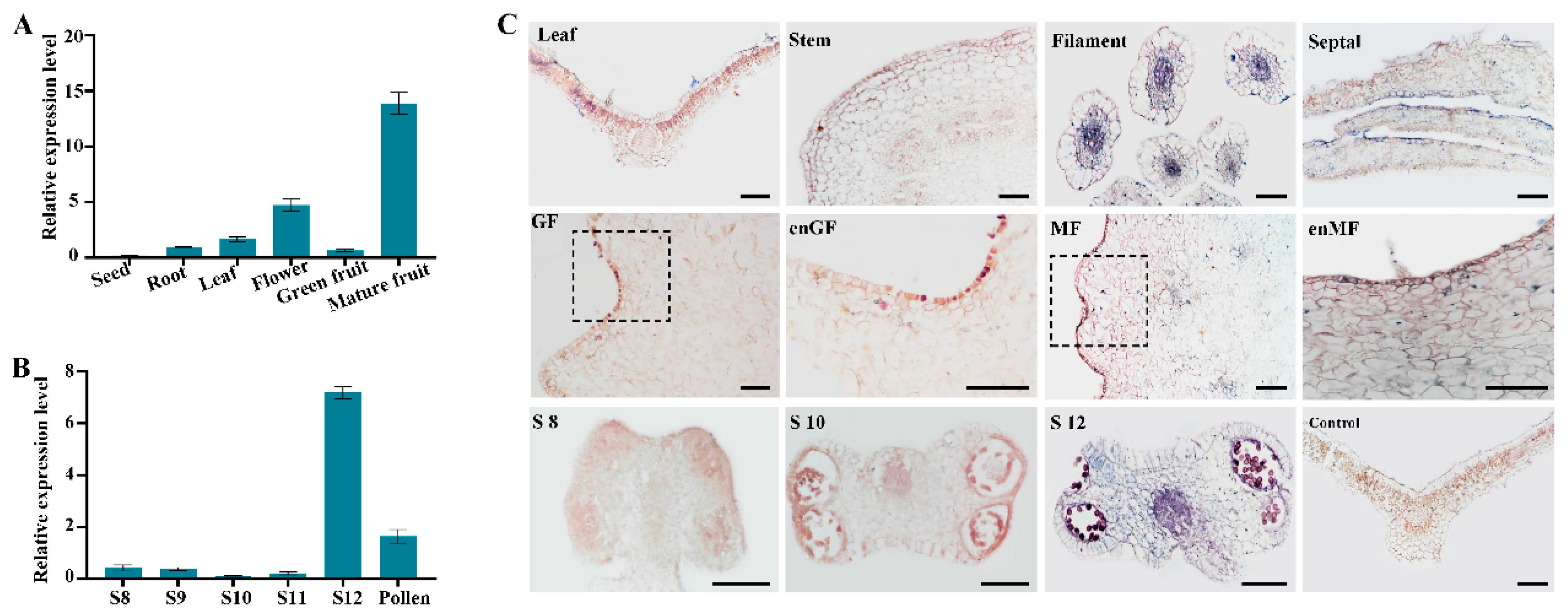
Figure 1. Characterization and spatiotemporal dynamics of FvePL1. (A) Tissue-specific and (B) developmental expression profiling of FvePL1 in anther ranging from different stages, including leaf, stem, filament, septal, S8 (developmental stage 8 of anther), S10 (developmental stage 10 of anther), S12 (developmental stage 12 of anther), green and mature fruits; (C) in situ hybridization analysis of FvePL1. GF, green fruit; MF, mature fruit; enGF and enMF, enlarged image of black box in green fruit and mature fruits, individually, bar = 100 μm.
Plants 14 03583 g001
Figure 2. Subcellular localization of FvePL1. (A) Fluorescence microscopy analysis in transiently expressed Arabidopsis protoplast cells. Overlay of the GFP fluorescence (green), chlorophyll autofluorescence (red), bright field, and the combined images, bar = 10 μm. (B) FvePL1:GFP subcellular localization in epidermal cells of N. benthamiana leaves under control conditions and plasmolysis (10% NaCl). The white boxes were enlarged as shown below. CW, cell wall; CM, cell membrane, bar = 20 μm.
Plants 14 03583 g002
Figure 3. Phenotypic characterization of FvePL1 transgenic strawberry plant. (A). Relative expression level of FvePL1, (B). Morphological comparisons of plants (bar = 5 cm), leaf (bar = 2 cm), fruits at 23 days post-anthesis (DPA), 35 DPA (bar = 0.5 cm), achene and embryo (bar = 100 μm), (C). Seed rate, (D). Pollen viability, in YW and transgenic lines. Statistical analysis was performed using one-way ANOVA using Tukey’s test (*, p < 0.05, **, p < 0.01).
Plants 14 03583 g003
Figure 4. Aberrant pollen cell wall development in FvePL1-deficient plants. (A). Scanning electron microscopy (SEM) images of mature pollen, (B). Surface enlarged morphology, (C). Transmission electron microscopy (TEM) observation of mature pollen, (D). The enlarged images of the pollen wall structure, (E). Cryosections of pollen grains stained by auramine O staining with a strong fluorescence of the exine, (F). Calcofluor white fluorescence of the inner cellulosic sublayer of the intine of three genotypes. Bar = 10 μm.
Plants 14 03583 g004
Figure 5. The number of pollen grains were impaired in FvePL1-RNAi lines. The laser confocal microscopy images of pollen grains in the anther of three genotypes on the first day post flowering. In addition, the cytological observation of the anther at stage 8, 9 and 12 in YW, FvePL1-RNAi and FvePL1-OE lines were detected and the images zoomed. Bar = 100 μm.
Plants 14 03583 g005
Figure 6. Pollen germination was significantly impaired in FvePL1-RNAi plants. (A). Morphological characterization of pollen germination, bar = 20 μm. (B). Comparison of pollen germination percentages in vitro. (C). Histological observation of germinated pollen tube, bar = 20 μm. (D). The length of pollen tube among overexpression, RNAi of FvePL1 and YW. The data were the average values of each of the three lines with comparison between multiple samples determined by one-way ANOVA using Tukey’s test (**, p < 0.01; ***, p < 0.001). (E). In vivo pollen-tube growth in pistils in 20 min after pollination of YW, FvePL1-RNAi, FvePL1-OE, and reciprocal pollination experiments and zoomed images. Bar = 50 μm.
Plants 14 03583 g006
Figure 7. Downregulation of FvePL1-enhanced drought tolerance. (A). Phenotypic assessments of three genotypes subjected to 15 d and 30 d drought stress, as well as R. 5 d (recovery 5 d) (bar = 5 cm). (B). Water loss rate, (C). Survival rate and (D) total pectin content in YW and FvePL1 transgenic plants. The asterisks represent the significant differences between YW and FvePL1 transgenic plants. Statistical analysis was performed using one-way ANOVA using Tukey’s test (*, p < 0.05; **, p < 0.01; ***, p < 0.001). (E). Ultrastructural analyses of cell corners of leaf epidermis were analyzed (bar = 2 μm).
Plants 14 03583 g007
Disclaimer/Publisher’s Note: The statements, opinions and data contained in all publications are solely those of the individual author(s) and contributor(s) and not of MDPI and/or the editor(s). MDPI and/or the editor(s) disclaim responsibility for any injury to people or property resulting from any ideas, methods, instructions or products referred to in the content.