Article

Ectopic Over-Expression of BjuAGL9-2 Promotes Flowering and Pale-Yellow Phenotype in Arabidopsis

1
College of Rural Revitalization, Fuyang Institute of Technology, Fuyang 236031, China
2
College of Agronomy and Biotechnology, Southwest University, Chongqing 400716, China
3
School of Life Advanced Agriculture Bioengineering, Yangtze Normal University, Chongqing 408100, China
*
Authors to whom correspondence should be addressed.
These authors contributed equally to this work.
Plants 2025, 14(22), 3502; https://doi.org/10.3390/plants14223502
Submission received: 25 September 2025 / Revised: 3 November 2025 / Accepted: 8 November 2025 / Published: 17 November 2025

Abstract

Brassica juncea is an important leafy vegetable, and flowering time is a key determinant of its yield and quality. In this study, one significantly up-regulated gene, BjuAGL9-2, was identified from RNA-Seq data. qRT-PCR analysis confirmed that BjuAGL9-2 expression was significantly elevated in reproductive organs and reproductive stages. Further five BjuAGL9-2 over-expression (OE) lines were subsequently generated, which showed an early-flowering and pale-yellow leaf phenotype compared to the wild type. qRT-PCR assays found that the mRNA of core floral integrator genes was changed in Arabidopsis OE lines. Yeast two-hybrid (Y2H) and bimolecular fluorescence complementation (BiFC) assays indicated that BjuAGL9-2 interacted with BjuTUA5, BjuZFP7, BjuGSTU5, and BjuMAPK16 in vivo. Sub-cellular localization assays showed that BjuAGL9-2 localizes in the nucleus, whereas its interacting partners localize in the cytoplasm. qRT-PCR assays further revealed that BjuTUA5 and BjuGSTU5 were up-regulated in flower buds, while BjuZFP7 and BjuMAPK16 were down-regulated. During vegetative stages, all four genes were up-regulated in B. juncea. As for BjuAGL9-2 interaction protein-encoding homolog genes, except AtGSTU5, the other three genes were up-regulated in Arabidopsis OE lines. Additionally, qRT-PCR analysis of chlorophyll biosynthesis-related genes showed that 19 of 27 genes were up-regulated, while 8 genes were down-regulated, in Arabidopsis OE lines. Collectively, these findings suggest that BjuAGL9-2 promotes flowering and contributes to the pale-yellow phenotype by regulating its interacting protein-coding genes, floral integrators, and chlorophyll biosynthesis genes.

1. Introduction

Brassica juncea (L.) Czern. is an important leafy cruciferous vegetable that is widely consumed in China, particularly as a deeply processed pickle. As a vegetative-harvested crop, delaying flowering to increase yield and quality is a major breeding objective in B. juncea. In Arabidopsis thatliana, the flowering time regulatory network has been extensively studied. Six major signaling pathways, the autonomous, photoperiod, vernalization, gibberellic acid, ambient temperature, and aging pathways, have been reported to play critical roles in flowering regulation [1]. Within these pathways, multiple transcription factor families, including bZIP, MADS-box, WRKY, and MYB participate in control of flowering [2,3]. As plants transition to the reproductive stage, signals from these six pathways converge on floral integrator genes SOC1 (suppressor of over-expression of CO1) and FT (flowering locus T). Then SOC1 and FT further transmit these signals to floral meristem identification genes LFY (leafy), AP1 (apetala 1), and FUL (fruitfull), thereby initiating flowering [1,4,5,6,7].
The MADS-box transcription factor family has been widely reported to participate in diverse processes of plant growth and development, particularly in inflorescence architecture and floral organ formation [8,9]. Arabidopsis thaliana contains 108 MADS-box members, while Brassica rapa. Zea mays, and Vitis vinifera harbor 160, 87, and 54 members, respectively [10,11,12,13]. Based on sequence characteristics, the MADS-box family can be classified into two major types. Type I genes typically contain a conserved MADS-box domain together with a highly C-terminal region [14]. Type II can be subdivided into MIKCc-type and MIKC*-type clade [15,16]. MICK-type genes usually harbor four conserved domains. The MADS-box domain functions as a DNA-binding region, recognizing and binding to the CArG-box motif [17,18]. The I (intervening) domain specifically binds to downstream targets [19]. The K (keratin-like) domain facilitates formation of higher-order MADS-box protein complexes [20,21]. However, the function of the C-terminal domain remains unclear.
ABCDE is a well-known model to explain floral organ determination at the genetic level [22]. In Arabidopsis, except Apetala2, all other genes belong to the MADS-box family [23]. AtAP1 (apetala1) is a member of the A-class genes and is required for floral meristem development and floral organ identity [24]. The B-class genes AtAP3 (apetala3) and PI (pistillata) control petal and stamen identity [25]. The C-class gene AG (agamous) is essential for reproductive organ identity and floral meristem determinacy [26]. The D-class genes STK (seedstick), SHP1 (shatterproof1), and SHP2 (shatterproof2) regulate ovule identity [27,28]. The E-class genes SEP1 (AGL2, sepallata1), SEP2 (AGL4, sepallata2), SEP3 (AGL9, sepallata3), and SEP4 (sepallata4) are required for sepal, petal, stamen, and carpel development [29,30,31,32,33].
In this study, BjuAGL9-2 was found to be significantly up-regulated in flower organs and reproductive stages. Transgenic over-expression BjuAGL9-2 Arabidopsis showed an early-flowering and pale-yellow phenotype. qRT-PCR assays found that the mRNA expression levels of AtFT, AtSOC1, AtCO (constans), AtFUL, AtSVP, and AtAP1 were down-regulated, but AtLFY and AtLC were up-regulated, in Arabidopsis OE lines compared to the wild type. Y2H and BiFC assays confirmed that BjuAGL9-2 interact with BjuTUA5, BjuZFP7, BjuGSTU5, and BjuMAPK16 in the nucleus. Sub-cellular location assays indicated that BjuAGL9-2 localized the nucleus, whereas its interacting partners were localized the cytoplasm. qRT-PCR analysis showed that BjuTUA5 and BjuGSTU5 were up-regulated in flower buds, while BjuZFP7 and BjuMAPK16 were down-regulated. During vegetative stages, all four genes were up-regulated in B. juncea. And in Arabidopsis OE lines, except AtGSTU5, the other three homologs of BjuAGL9-2 interaction protein-encoding genes, AtTUA5, AtZFP7, and AtMAPK16, were all up-regulated compared to the wild type. Further, qRT-PCR assays revealed that among 27 chlorophyll biosynthesis-related genes, 19 genes were up-regulated and 8 genes were down-regulated in Arabidopsis OE lines. Collectively, these findings suggest that BjuAGL9-2 promotes flowering and contributes to the pale-yellow phenotype by regulating the expression of its interacting protein-coding genes, floral integrators, and chlorophyll biosynthesis genes.

2. Results

2.1. BjuAGL9-2 Functions in Promoting Plant Flowering and Pale-Yellow Phenotype in A. thaliana
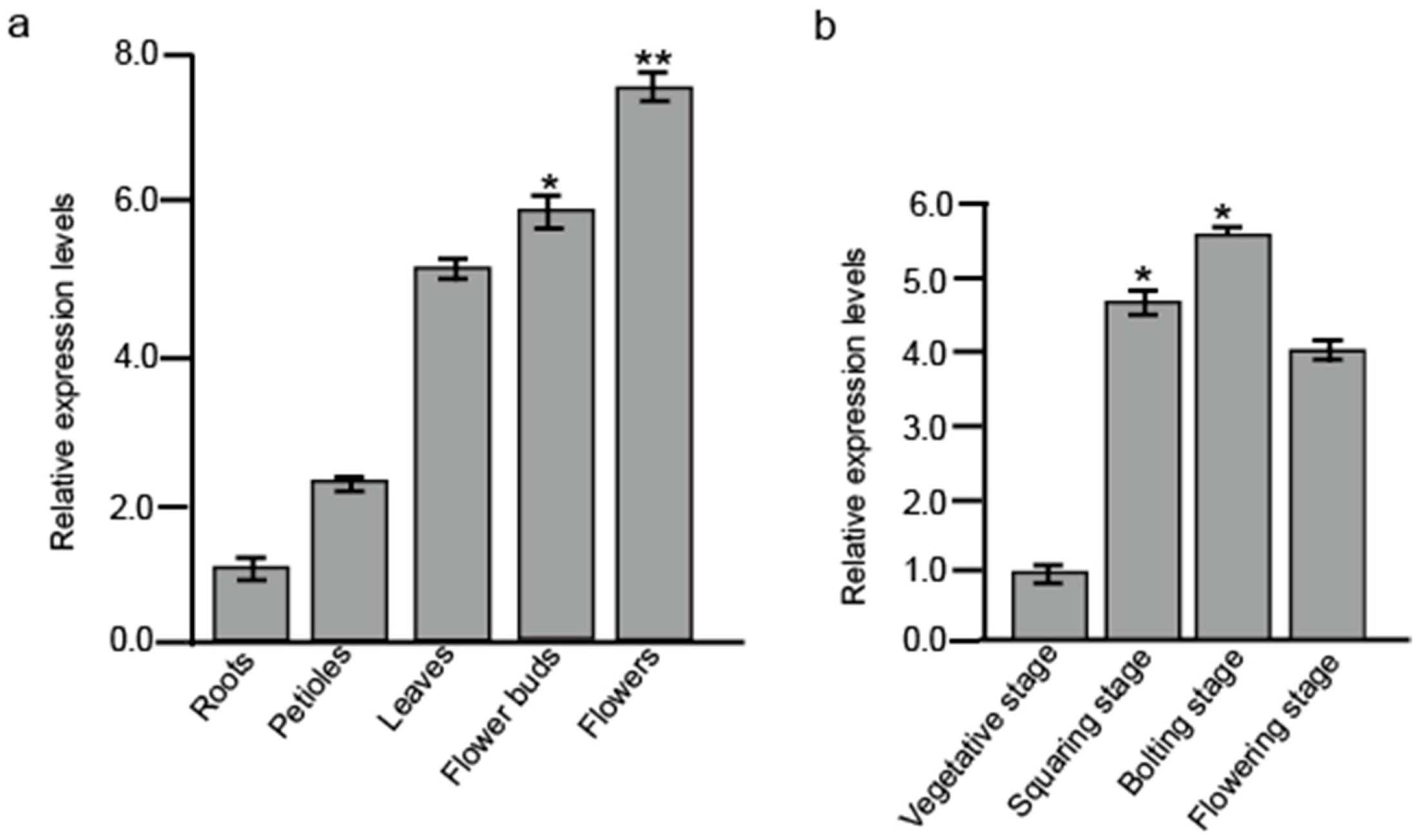
To compare gene expression profiles in B. juncea shoot tips at vegetative and flowering stages, RNA-Seq was recruited to analysis differentially expressed genes (DEGs). One gene named BjuAGL9-2 was found to be significantly up-regulated at the reproductive stage. qRT-PCR assays further confirmed that BjuAGL9-2 was up-regulated in petioles, leaves, flower buds, and flowers, with particularly strong expression in flower buds and flowers compared to roots (Figure 1a). Across different developmental stages, BjuAGL9-2 expression was elevated in reproductive stages, showing significant up-regulation at both squaring and bolting stages compared to the vegetative stage (Figure 1b).
Figure 1. BjuAGL9-2 expression levels in different tissues and at different development stages. (a) BjuAGL9-2 expression levels in different tissues. (b) BjuAGL9-2 expression levels at different development stages. * p < 0.05, ** p < 0.01.
Plants 14 03502 g001
To investigate the function of BjuAGL9-2 in plant growth and development, Arabidopsis OE plants were generated (Figure 2). qRT-PCR analysis indicated that BjuAGL9-2 transcript levels were elevated in all five OE lines (Figure 2c). Among them, three lines (OE3, OE4, and OE5) displayed strong induction, with approximately 10-fold, 20-fold, and 15-fold higher expression compared to the wild type, respectively (Figure 2c). Western blot analysis further confirmed the accumulation of GFP-BjuAGL9-2 protein in all five OE lines (Figure 2d). Phenotype observations reveal that 25 of OE1, 26 of OE2, 27 of OE3, 28 of OE4, and 27 of OE5 plants initiated flowering approximately 28 days after transfer to nutrient soil, whereas the wild type flowered at around 35 days. qRT-PCR assays indicated that the mRNA expression levels of AtFT, AtSOC1, AtCO, AtFUL, AtSVP, and AtAP1 were down-regulated, in which AtFT, AtSOC1, AtCO, AtFUL, and AtAP1 were significantly down-regulated, but the mRNA expression levels of AtLFY and AtLC were significantly up-regulated in Arabidopsis OE lines compared to the wild type (Figure 2e,f). In addition, all five transgenic lines showed a pale-yellow phenotype from the four-true-leaf stage to the flowering stage (Figure 2). These results indicate that the over-expression of BjuAGL9-2 promotes flowering and induces the pale-yellow phenotype.

2.2. BjuAGL9-2 Interaction Protein Screening

To detect the protein interaction network in regulating plant flowering, the bait vector pGBKT7-BjuAGL9-2 was used to screen potential candidates from a B. juncea bolting stage flower buds yeast library, which was constructed by using the Gateway cloning system. The length of PCR amplification fragments is approximately 900 bp, 800 bp, 800 bp, 800 bp, 800 bp, 800 bp, 500 bp, 800 bp, 800 bp, 800 bp, 500 bp, and 800 bp, respectively (Figure S1). Sequencing analysis identified several putative BjuAGL9-2-interacting proteins, including BjuANXAD1, BjuTUA5, BjuZFP7, BjuGSTU5, BjuVA09G18110, BjuWRKY11, BjuRHY1A, BjuVA01G03870, and BjuMAPK16 (Table 1).

2.3. Interactive Analyses Between BjuAGL9-2 and Screened Proteins

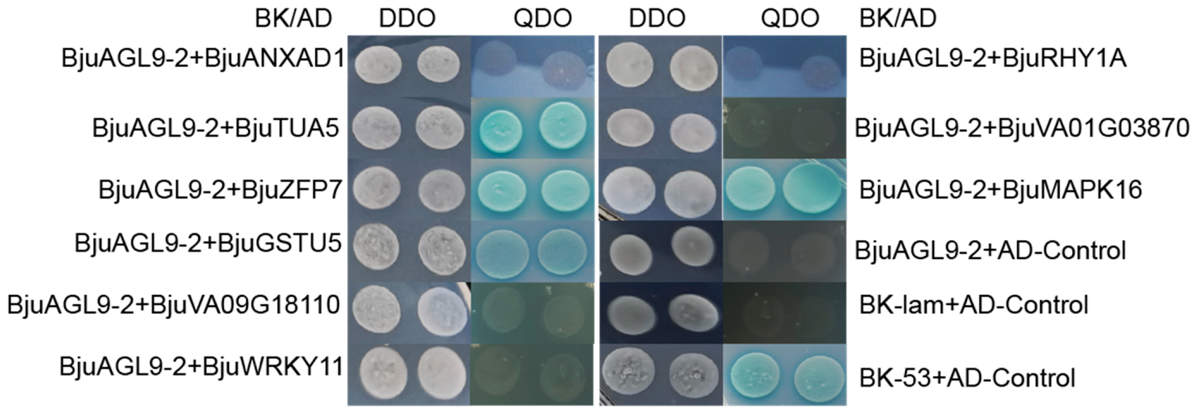
To confirm the interaction between BjuAGL9-2 and candidate proteins, which were identified from the yeast library, pGBKT7-BjuAGL9-2 and each of the prey constructs (pGADT7-BjuANXAD1, pGADT7-BjuTUA5, pGADT7-BjuZFP7, pGADT7-BjuGSTU5, pGADT7-BjuVA09G18110, pGADT7-BjuWRKY11, pGADT7-BjuRHY1A, pGADT7-BjuVA01G03870, and pGADT7-BjuMAPK 16) were co-transformed into Gold strain, pairwise. Growth and reporter activation on QDO medium demonstrated that BjuAGL9-2 interacts with BjuTUA5, BjuZFP7, BjuGSTU5, and BjuMAPK16 in vivo (Figure 3).
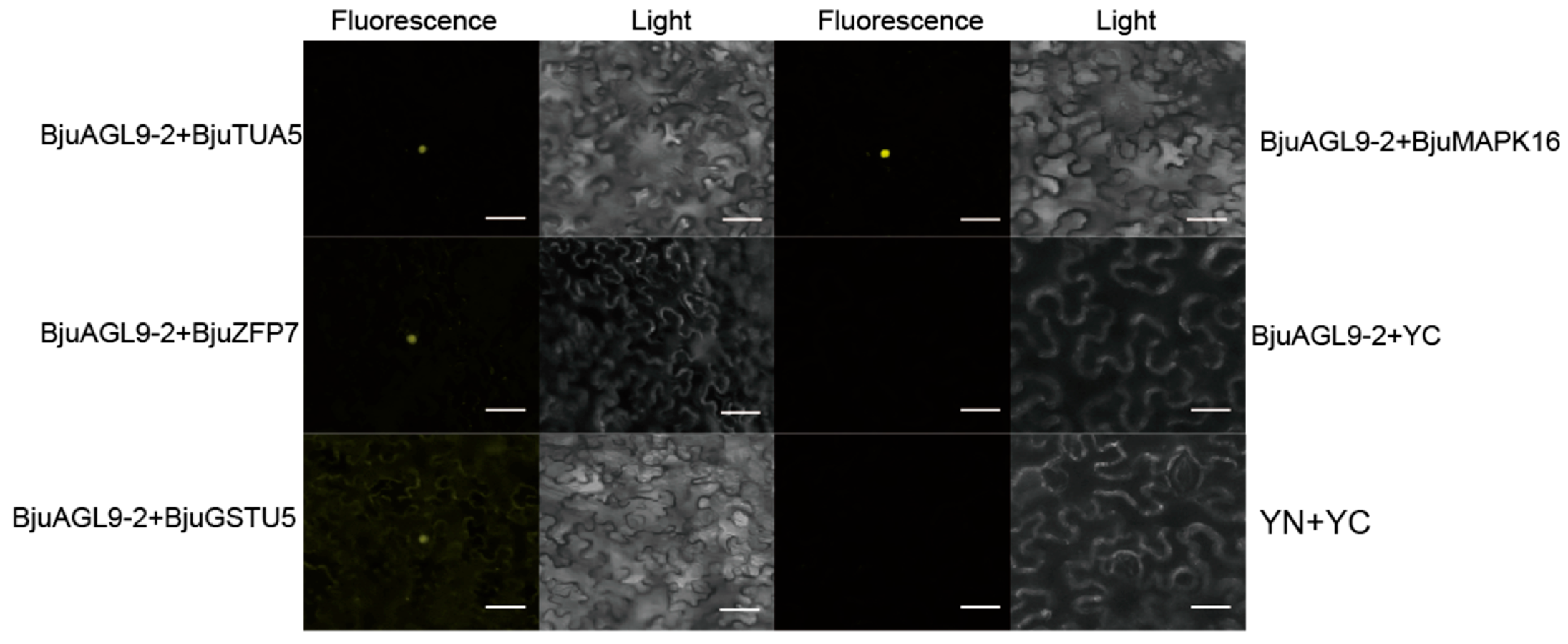
To further confirm the interaction between BjuAGL9-2 with its candidate partners (BjuTUA5, BjuZFP7, BjuGSTU5, and BjuMAPK16), BiFC assays were performed in Nicotiana benthamiana leaf cells. Split YFP complementation assays revealed strong yellow fluorescent signals in the nucleus when BjuAGL9-2 was co-infiltrated with each of the candidate proteins (Figure 4). These results confirmed that BjuAGL9-2 interacted with BjuTUA5, BjuZFP7, BjuGSTU5, and BjuMAPK16 in the nucleus (Figure 4).

2.4. Sub-Cellular Location Assays

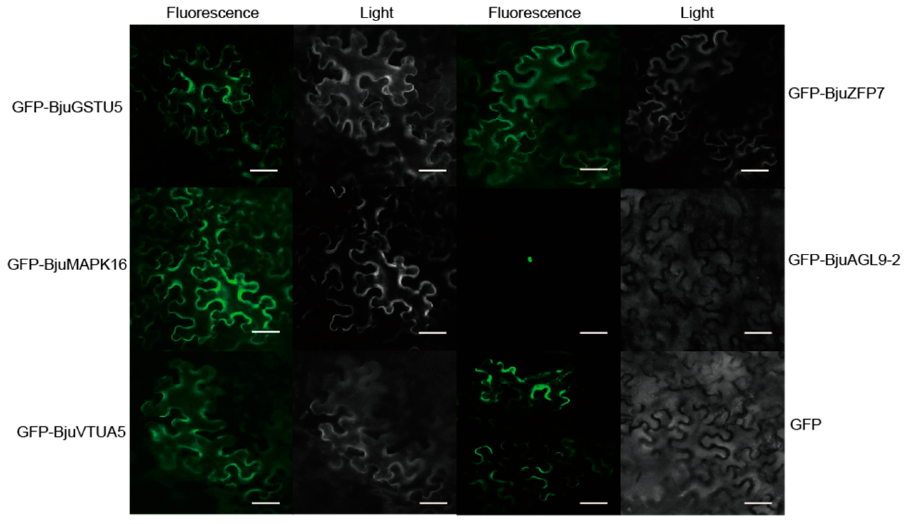
To analyze the sub-cellular location of BjuAGL9-2, BjuTUA5, BjuZFP7, BjuGSTU5, and BjuMAPK16, their coding sequences were fused to GFP and transiently expressed in Nicotiana benthamiana epidermal cells. Fluorescence microscopy revealed that GFP-BjuAGL9-2 localized predominantly to the nucleus, whereas GFP-BjuTUA5, GFP-BjuZFP7, GFP-BjuGSTU5, GFP-BjuMAPK16, and GFP localized to the cytoplasm (Figure 5).

2.5. Interactive Protein-Encoding Gene Expression Analysis in Different Tissues and at Different Development Stages in B. juncea

To examine the mRNA expression levels of genes encoding BjuAGL9-2-interacting proteins (BjuTUA5, BjuZFP7, BjuGSTU5, and BjuMAPK16) in different tissues, qRT-PCR analysis was conducted (Figure 6a). BjuTUA5 was up-regulated in petioles, leaves, flower buds, and flowers, with particularly strong expression in reproductive organs (flower buds and flowers), compared to roots. BjuZFP7 showed elevated expression in petioles, leaves, and flowers, with significant up-regulation in petioles and leaves but was significantly down-regulated in flower buds compared to roots. BjuGSTU5 displayed about 2-fold changes in petioles, leaves, and flowers but was specifically and significantly up-regulated in flower buds. BjuMAPK16 was up-regulated in petioles, leaves, and flowers, with a significant increase in leaves and flowers, but was down-regulated in flower buds compared to roots (Figure 6a).
Analysis of mRNA expression at different developmental stages revealed that BjuTUA5, BjuGSTU5, and BjuMAPK16 were generally up-regulated during reproductive stages. In particular, BjuTUA5 exhibited significant changes in expression across all reproductive stages, while BjuMAPK16 was markedly up-regulated at both the squaring and flowering stages (Figure 6b). In contrast, BjuZFP7 showed increased expression at both the squaring and flowering stages but was down-regulated at the bolting stage (Figure 6b). Regarding the impact of BjuAGL9-2 on the expression levels of homologs of interaction protein-encoding genes, qRT-PCR assays indicated that whereas AtGSTU5 was down-regulated, AtTUA5, AtZFP7, and AtMAPK16 were up-regulated in Arabidopsis OE lines compared to the wild type (Figure 6c).

2.6. Chlorophyll Synthesis Gene Expression Levels in Arabidopsis OE Lines

To investigate the effect of BjuAGL9-2 on chlorophyll biosynthesis, qRT-PCR assays were performed on wild-type and Arabidopsis OE lines (Table S1). For the first step of chlorophyll biosynthesis, all genes were up-regulated in OE lines except AtHEMA1, which was down-regulated. Specifically, AtHEMA3, and AtHEML were up-regulated compared to the wild type (Figure 7a). For the second step, AtHEMB1, AtHEMF1, and AtHEMGs were down-regulated, whereas AtHEMB2, AtHEMC, AtHEMD, AtHEMEs, and AtHEMF2 were up-regulated, in OE lines (Figure 7b). For the third step, multiple genes also showed altered expression. AtCHLs, AtCRD1, AtDVR, AtPORs, and AtCHLG were up-regulated, while AtCHIs and AtCAO were down-regulated, in Arabidopsis OE lines compared to the wild type (Figure 7c).

3. Discussion

In plants, flowering time is regulated by six major pathways, in which FT and FLC act as key integrators. In this study, BjuAGL9-2, a member of the MADS-box family, was identified as being up-regulated in reproductive organs and reproductive stages (Figure 1). To investigate BjuAGL9-2’s role in plant growth, BjuAGL9-2 OE lines were generated (Figure 2). Three Arabidopsis OE lines with significantly elevated expression levels of BjuAGL9-2 showed early-flowering compared to the wild type (Figure 2). As plant floral integrator genes, FT, SOC1, CO, FUL, AP1, and LFY function in promoting flowering, but FLC and SVP function in delaying flowering [1,4,5,6,7]. qRT-PCR assays indicated that the floral integrator genes FT, SOC1, CO, FUL, and AP1 were down-regulated, but LFY and FLC were significantly up-regulated, in Arabidopsis OE lines. This means that BjuAGL9-2 functions in regulates both positive and negative floral integrator genes. To further elucidate the molecular regulatory network of BjuAGL9-2 in flowering, Y2H and BiFC assays were performed. The results demonstrated that BjuTUA5, BjuZFP7, BjuGSTU5, and BjuMAPK16 interact with BjuAGL9-2 in the nucleus (Table 1, Figure 3 and Figure 4). Sub-cellular location assays confirmed that BjuAGL9-2 is localized in the nucleus, whereas its interacting proteins are localized in the cytoplasm (Figure 5). This means that the interacting proteins may be transported to nucleus for their interaction and function in prompt flowering.
BjuVA10G20230 was identified as TUA5 (tubulin alpha-5 chain), suggesting functions in pollen development, while AGL9 is highly expressed in stamens, indicating its involvement in stamens development [32,34,35]. In this study, qRT-PCR analysis showed that BjuTUA5 was up-regulated in reproductive organs (flower buds and flowers) and at reproductive stages, including the squaring and bolting stages (flower buds) and the flowering stage (flowers, Figure 6). In Arabidopsis OE lines, its homolog gene AtTUA5 was up-regulated compared to the wild type (Figure 6c). These findings suggest that AGL9-2 may function in stamen development by regulating AtTUA5 at both the transcription and translation levels. BjuVA08G27100 was identified as ZFP7 (zinc finger protein 7), which has been reported to be down-regulated by 90% in floral organs from stages 4 to 12 [36]. Consistent with these findings, in this study, BjuZFP7 expression was down-regulated in flower buds and at reproductive stages in B. juncea (Figure 6), further supporting its role in floral organ development. However, in Arabidopsis OE lines, the expression level of AtZFP7 was slightly up-regulated (Figure 6c). This means that BjuAGL9-2 functions by negatively regulating BjuZFP7 expression in flower organs but positively regulates BjuZFP7 expression in leaves. BjuVA03G025670 was identified as GSTU5 (glutathione S-transferase) and has been implicated in defense response [37]. qRT-PCR assays revealed that BjuGSTU5 was significantly up-regulated in flower buds and reproductive stages (squaring, bolting, and flowering stages, Figure 6). Similarly, BjuVA10G20860 was identified as MAPK16 (mitogen-activated protein kinase 16), which is also involved in defense response [38]. Its expression was significantly up-regulated in flowers and reproductive stages (squaring, bolting, and flowering, Figure 6). However, in Arabidopsis OE lines, the homologous gene AtGSTU5 was down-regulated, but AtMAPK16 was up-regulated compared to the wild type (Figure 6c). These results suggest that the interaction between BjuAGL9-2 and BjuVA03G025670/MAPK16 may play an opposite role in defense-related processes during reproductive development.
Chlorophyll content largely determines plant color, and its biosynthesis can be categorized into three steps [39]. In this study, Arabidopsis OE lines displayed a pale-yellow phenotype (Figure 2a,b). To explore the molecular basis of this phenotype, qRT-PCR assays were conducted to examine the expression of chlorophyll biosynthesis genes. Among the five genes in the first step, AtHEMA1 was down-regulated, while AtHEMA2, AtHEMA3, AtHEML1, and AtHEML2 were up-regulated in Arabidopsis OE lines compared to the wild type (Figure 7a). Previous studies have shown that HEMA3 and HEMA2 do not contribute to GluTR activity, whereas HEMA1 is the key enzyme for ALA synthesis in plants [40,41]. The down-regulation of AtHEMA1 observed in Arabidopsis OE lines is consistent with their pale-yellow phenotype (Figure 7a). Furthermore, HEML2 has been identified as a positive regulator of chlorophyll biosynthesis, with heml2 mutants showing reduced chlorophyll a/b content, while heml1 mutants do not display such a decrease [42]. In this study, the up-regulation of AtHEML1 and AtHEML2 in Arabidopsis OE lines suggests that BjuAGL9-2 may influence chlorophyll a/b accumulation through the induction of AtHEML1 expression, even though the repression of AtHEMA1 ultimately contributes to the pale-yellow phenotype.
HEMB1 and HEMB2 are involved in the second step of chlorophyll biosynthesis and have been reported to function in plant immunity and the heme biosynthetic pathway, respectively [43,44]. In Arabidopsis OE lines, AtHEMB1 and AtHEMB2 were slightly up-regulated, compared to the wild type (Figure 7b), suggesting that BjuAGL9-2 may positively regulate AtHEMB expression. HEMC, encoding porphobilinogen deaminase (PBGD), and HEMD, encoding uroporphyrinogen III synthase (UROS), both function in the early stages of chlorophyll and heme biosynthesis [45,46]. HEME genes, encoding UROD, catalyze the decarboxylation of uroporphyrinogen III to coproporphyrinogen III [47,48]. In this study, AtHEMC, AtHEMD, AtHEME1, and AtHEME2 were all up-regulated in Arabidopsis OE lines compared to the wild type (Figure 7b), indicating that BjuAGL9-2 positively regulates these genes during early chlorophyll and heme biosynthesis. HEMF genes are involved in leaf development, and mutants exhibit developmentally regulated and light-dependent leaf lesions [49]. In this study, only AtHEMF2 was significantly up-regulated, while AtHEMF1 was slightly down-regulated, in Arabidopsis OE lines compared to the wild type (Figure 7b). These results suggest that BjuAGL9-2 contributes to the yellowing phenotype primarily through the regulation of AtHEMF2. HEMG genes, encoding protoporphyrinogen oxidase, are associated with the lesion-mimic phenotype when mutated [50]. In this study, AtHEMG was slightly down-regulated, consistent with the absence of the lesion-mimic phenotype in Arabidopsis OE lines (Figure 7b). This indicates that BjuAGL9-2 does not regulate HEMG genes.
The third step of chlorophyll biosynthesis involves 12 genes (CHLH, CHLI1, CHLI2, CHLD, CHLM, CRD1, DVR, PORA, PORB, PORC, CHLG, and CAO) and seven enzymes (MgCh, MgPMT, MgPME, DVR, POR, CHLG, and CAO) [39]. The MgCh enzyme consists of three subunits and is encoded by CHLH, CHLI1, CHLI2, and CHLD, respectively [51,52,53,54]. In Arabidopsis OE lines, AtCHLH and AtCHLD were up-regulated, while AtCHLI1 and AtCHLI2 were down-regulated, compared to the wild type (Figure 7c). In Arabidopsis, mutations in CHLI subunits result in a yellow-green phenotype [55]. Thus, the down-regulation of AtCHLI1 and AtCHLI2 in Arabidopsis OE lines is consistent with the observed pale-yellow phenotype (Figure 7c). CHLM functions as a positive regulator of chlorophyll–protein complex formation [56]]. In this study, AtCHLM was significantly up-regulated in Arabidopsis OE lines, indicating that BjuAGL9-2 positively regulates chlorophyll–protein complexes (Figure 7c). CRD1 is also involved in chlorophyll synthesis, and its mutation leads to an elevated chlorophyll a/b ratio and a pale-green phenotype [57]. In this study, AtCRD1 was up-regulated in Arabidopsis OE lines (Figure 7c), suggesting that it may not contribute to the pale-yellow phenotype. DVR is required for chlorophyll synthesis, and loss-of-function mutants showed reduced chlorophyll contents and a pale-green phenotype [58]. In this study, AtDVR was only slightly up-regulated in Arabidopsis OE lines (Figure 7c), indicating that it does not contribute to the pale-yellow phenotype. Protochlorophyllide oxidoreductase (POR) is a key enzyme for chlorophyll synthesis that promotes the greening of etiolated plants. In Arabidopsis, there are three POR isoforms. The ectopic expression of PORA in the porB-1 porC-1 double-mutant rescues growth defects and chlorophyll deficiency, demonstrating that PORA is an essential component for bulk chlorophyll biosynthesis [59,60,61],,. In Arabidopsis OE lines, all three genes (AtPORA, AtPORB, and AtPORC) were up-regulated, suggesting that they are not responsible for the pale-yellow phenotype in Arabidopsis OE lines (Figure 7c). CHLG functions as a positive regulator of ALA synthesis, and antisense suppression of CHLG reduces ALA production [62]. In our study, AtCHLG was up-regulated in Arabidopsis OE lines, indicating that BjuAGL9-2 positively regulates CHLG expression (Figure 7c). Finally, CAO is required for chlorophyll b precursor biosynthesis [63]. qRT-PCR assays showed that AtCAO was down-regulated in OE lines (Figure 7c), suggesting that BjuAGL9-2 influences the chlorophyll b content through regulation of AtCAO. All those results suggesting that BjuAGL9-2 functions in the pale-yellow phenotype by regulating AtHEMA1, AtHEML1, AtHEMF2, AtCHLI1, AtCHLI2, and AtCAO in Arabidopsis OE lines.
Taken together, the results of this study demonstrate that BjuAGL9-2 promotes flowering and induces a pale-yellow phenotype in Arabidopsis. A qRT-PCR assay confirmed that the floral integrator genes FT, SOC1, CO, FUL, and AP1 were down-regulated, but LFY and FLC were significantly up-regulated, in Arabidopsis OE lines. BjuAGL9-2 is nuclear-localized, but its interacting proteins BjuTUA5, BjuZFP7, BjuGSTU5, and BjuMAPK16 reside in the cytoplasm. The mRNA expression levels of BjuAGL9-2, BjuTUA5, and BjuGSTU5 were up-regulated in flower buds, whereas BjuZFP7 and BjuMAPK16 were down-regulated. Notably, all four interacting genes were up-regulated during vegetative stages in B. juncea. In Arabidopsis OE lines, while AtGSTU5 was down-regulated, AtTUA5, AtZFP7, and AtMAPK16 were up-regulated, compared to the wild type. In addition, expression profiling of chlorophyll biosynthesis genes showed that 19 genes were up-regulated and 8 genes were down-regulated in Arabidopsis OE lines, providing a molecular explanation for the pale-yellow phenotype. Collectively, these findings suggest that BjuAGL9-2 integrates flowering and leaf color regulation into chlorophyll metabolism by modulating key transcriptional and protein interaction networks.

4. Materials and Methods

4.1. Plant Growth Conditions

B. juncea plants were grown under natural field conditions (18–23 °C; 8 h photoperiod/16 h dark). For tissue-specific expression analysis, total RNA was extracted from roots, petioles, leaves, flower buds, and flowers. For developmental stage-specific expression analysis, RNA was extracted from leaves at the vegetative stage (four-true-leaf stage), flower buds at the squaring (first flower buds observed with a diameter of 3 mm) and bolting stages (before the first flower was observed), and flowers (the first flower observed) at the flowering stage. Total RNA was extracted using the RNAprep Pure Plant Kit (Tiangen, Beijing, China), and 1 μg of total RNA was used for cDNA synthesis (Table S1). The ALL-In One 5*RT MasterMix (abm, Zhenjiang, China) kit was used for cDNA synthesis, and the synthesis program was 37 °C, 15 min, 60 °C, 10 min, 95 °C 3 min.

4.2. BjuAGL9-2 Over-Expression Line Screening and Phenotype Observation

The coding sequence of BjuAGL9-2 was cloned into the p1300-GFP vector under the control of the CaMV35S promoter. The transformation of Arabidopsis thaliana was performed using the Agrobacterium-mediated (GV3101 strain with a pJIC SA_Rep plasmid) floral dip method [64]. T0 transgenic plants were initially screened on hygromycin-containing medium, and genomic DNA from leaves was used to identify 21 lines of T1 generation, 15 lines of T2 generation, and 8 lines of T3 generation. For gene expression analysis, total RNA was isolated from leaves of Arabidopsis homozygous OE lines 3, 4, and 5, as well as wild-type plants. For phenotype observation, 30 seeds of each BjuAGL9-2 OE lines were sown on MS solid medium, left at 4 °C for 3 d, transferred to 23 °C under continuous white light (16 h light/8 h dark) until the growth of four true leaves, and then transferred to nutrient soil. Transcripts of BjuAGL9-2, the interaction protein-encoding genes, chlorophyll biosynthesis-related genes, the homologs of interacting protein-encoding genes (AtTUA5, AtZFP7, AtGSTU5, and AtMAPK16), and floral integrator genes were amplified using specific primers (Table S1). qRT-PCR was performed on the Roche LightCycler 480II (Roche, Basel, Switzerland), with 40 cycles, with Atactin and Bjuactin2 serving as the internal control. The reaction mixture was 2× qPCR mix 5 μL, cDNA 4.2 μL, primer-F: 0.8 μL, primer-R: 0.8 μL. And the PCR program was as follows: 40 cycles of 95 °C for 30 s; 95 °C for 10 s; 58 °C for 10 s; 72 °C for 10 s. Relative gene expression was calculated using the 2−ΔΔCt method, and each sample was analyzed in triplicate for both biological and technical replicates. For protein detection, total protein was extracted from transgenic plants using extraction buffer (50 mM Tris-HCl, pH 7.5; 100 mM NaCl; 1 mM EDTA, pH 8.0; 10% glycerol; 0.5% SDS). Western blot assays were performed using anti-GFP antibody (ABMART, Shanghai, China), with total protein from the A. thaliana wild type serving as the control. Then confirmed Arabidopsis OE lines were used for phenotype observation.

4.3. BjuAGL9-2 Interaction Protein Screening

The Matchmaker Gold Yeast two Hybrid System (Clontech, Tokyo, Japan) was used to screen proteins interacting with BjuAGL9-2. The bait plasmids pGBKT7-BjuAGL9-2 was co-transformed with a B. juncea bolting-stage flower buds yeast cDNA library (108 cfu/mL) into Saccharomyces cerevisiae Y2H Gold competent cells. Transforms were selected on SD/-His-Leu-Trp medium plates, and single colonies were subsequently transferred into liquid QDO (SD/-Ade/-His-Leu-Trp) medium for further selection. PCR amplification was performed on positive clones, and the resulting products containing candidate interaction protein-encoding genes were sequenced. The sequencing results were analyzed using BLAST (Braju tum V 2.0 cds) searches against the NCBI database and the Brassica database (http://brassicadb.cn/#/GeneSequence/, accessed in 2 November 2025) to identify potential interactors.

4.4. The Interaction Analysis Between BjuAGL9-2 and Screened Proteins

To confirm the interactions between BjuAGL9-2 and candidate protein identified from the library screen, the bait construct pGBKT7-BjuAGL9-2 and prey constructs (pGADT7-BjuANXAD1, pGADT7-BjuTUA5, pGADT7-BjuZFP7, pGADT7-BjuGSTU5, pGADT7-BjuVA09G18110, pGADT7-BjuWRKY11, pGADT7-BjuRHY1A, pGADT7-BjuVA01G03870, and pGADT7-BjuMAPK 16) were co-transformed pairwise into the Saccharomyces cerevisiae Y2H Gold strain. Self-activation of BjuAGL9-2 was tested on SD/-Trp medium, and bait toxicity as well as recombinant plasmid activation were examined on SD/-Leu-Trp medium plates at 30 °C. Negative controls consisted of Y2H Gold [pGADT7-control] × [pGBKT7-lam], while positive controls were Y2H Gold [pGADT7-control] × [pGBKT7-p53]. Protein–protein interaction assays were performed on DDO (SD/-Leu-Trp) and QDO (SD/-Ade/-His-Leu-Trp/ABA/X-α-gal) medium plates. The plates were incubated at 30 °C for 3–5 days before analysis.

4.5. Bimolecular Florescence (BiFC) Analysis and Confocal Microscopy

BiFC assays were performed to further confirm the interaction between BjuAGL9-2 and candidate proteins. Briefly, the coding sequence of BjuTUA5, BjuZFP7, BjuGSTU5, and BjuMAPK16 were cloned into the pEARLEYGATE201-YC vector, while BjuAGL9-2 was cloned into the pEARLEYGATE202-YN vector via LR recombination. Each construct was introduced into Agrobacterium tumefaciens strain GV3101. Agrobacterium cultures carrying the respective constructs were co-infiltrated into leaves of 4-week-old Nicotiana benthamiana, together with the silencing suppressor P19. After 48–60 h, yellow fluorescent protein (YFP) signals were detected using a confocal laser scanning inverted microscope (LSM510 Meta; Carl Zeiss, Jena, Germany).

4.6. Sub-Cellular Localization Assays

The sub-cellular location of BjuAGL9-2 and its interacting proteins was examined using GFP fusion constructs, in which GFP was fused to BjuAGL9-2 and its interacting protein N-terminal. Briefly, the coding sequences of BjuAGL9-2, BjuTUA5, BjuZFP7, BjuGSTU5, and BjuMAPK16 were cloned into the p1300-GFP vector. Each construct was transformed into Agrobacterium tumefaciens strain GV3101. Agrobacterium cultures carrying the constructs were co-infiltrated with the silencing suppressor P19 into leaves of 4-week-old Nicotiana benthamiana plants. After 48-60 h, green fluorescent protein (GFP) signals were observed using a fluorescence microscope.

Supplementary Materials

The following supporting information can be downloaded at: https://www.mdpi.com/article/10.3390/plants14223502/s1, Figure S1. The PCR results of genes which may produce proteins interacted with BjuAGL9-2. Figure S2. Bimolecular fluorescence complementation (BiFC) assays in tobacco leaf cells testing protein-protein interaction between BjuAGL9-2 with BjuTUA5, BjuZFP7, BjuGSTU5 and Bju-MAPK16. Table S1 Primer sequences used for Y2H, BiFC, qRT-PCR and sub-cellular location assays.

Author Contributions

Conceptualization, G.H.; method, K.R., and R.M.; writing—original draft preparation, J.Z. and M.S.; writing—review and editing, all authors. All authors have read and agreed to the published version of the manuscript.

Funding

This research was funded by the Chongqing Natural Science Foundation (CSTB2025NSCQ-GPX0023) under the Chongqing Seeds Industry Innovation and Breakthrough Project “Unveiling the List and Leading the Way” and the Project “Breeding and High Yield, Quality, Wide Suitable and Processing Specialized Varieties Tumorous Stem Breeding and Demonstration”.

Data Availability Statement

The original contributions presented in this study are included in the article/Supplementary Materials. Further inquiries can be directed to the corresponding authors.

Conflicts of Interest

The authors declare no conflicts of interest.

References

  1. Fornara, F.; De, M.A.; Coupland, G. Snapshot: Control of flowering in Arabidopsis. Cell 2010, 141, 5502. [Google Scholar] [CrossRef]
  2. Méndez-Vigo, B.; Martínez-Zapater, J.M.; Alonso-Blanco, C. The flowering repressor SVP underlies a novel Arabidopsis thaliana QTL interacting with the genetic background. Plos Genet. 2013, 9, e1003289. [Google Scholar] [CrossRef]
  3. Weigel, D. Natural variation in Arabidopsis: From molecular genetics to ecological genomics. Plant Physiol. 2011, 158, 2–22. [Google Scholar] [CrossRef]
  4. Andres, F.; Coupland, G. The genetic basis of flowering responses to seasonal cues. Nat. Rev. Genet. 2012, 13, 627–639. [Google Scholar] [CrossRef]
  5. Benlloch, R.; Berbel, A.; Serrano-Mislata, A.; Madueno, F. Floral initiation and inflorescence architecture: A comparative view. Ann. Bot. 2007, 100, 659–676. [Google Scholar] [CrossRef]
  6. Parcy, F. Flowering: A time for integration. Int. J. Dev. Biol. 2005, 49, 585–593. [Google Scholar] [CrossRef]
  7. Serrano-Mislata, A.; Goslin, K.; Zheng, B.B.; Rae, L.; Wellmer, F.; Graciet, E.; Madueno, F. Regulatory interplay between leafy, apetala 1/cauliflower and terminal flower 1: New insights into an old relationship. Plant Signal Behav. 2017, 12, e1370164. [Google Scholar] [CrossRef]
  8. Kater, M.M.; Dreni, L.; Colombo, L. Functional conservation of MADS-box factors controlling floral organ identity in rice and Arabidopsis. J. Exp. Bot. 2006, 57, 3433–3444. [Google Scholar] [CrossRef]
  9. Tanabe, Y.; Hasebe, M.; Sekimoto, H.; Nishiyama, T.; Kitani, M.; Henschel, K.; Munster, T.; Theissen, G.; Nozaki, H.; Ito, M. Characterization of MADS-box genes in charophycean green algae and its implication for the evolution of MADS-box genes. Proc. Natl. Acad. Sci. USA 2005, 102, 2436–2441. [Google Scholar] [CrossRef]
  10. Alvarez-Buylla, E.R.; Pelaz, S.; Liljegren, S.J.; Yanofsky, M.F. An ancestral MADS-box gene duplication occurred before the divergence of plants and animals. Proc. Natl. Acad. Sci. USA 2000, 97, 5328–5333. [Google Scholar] [CrossRef]
  11. Parenicova, L.; Folter, S.; Kieffer, M.; Horner, D.S.; Favalli, C.; Busscher, J.; Cook, H.E.; Ingram, R.M.; Kater, M.M.; Davies, B.; et al. Molecular and phylogenetic analyses of the complete MADS-box transcription factor family in Arabidopsis: New openings to the MADS world. Plant Cell 2003, 15, 1538–1551. [Google Scholar] [CrossRef] [PubMed]
  12. Duan, W.K.; Song, X.M.; Liu, T.K.; Huang, Z.N.; Ren, J.; Hou, X.L.; Li, Y. Genome-wide analysis of the MADS-box gene family in Brassica rapa (Chinese cabbage). Mollecular Genet. Genom. 2015, 290, 239–255. [Google Scholar] [CrossRef] [PubMed]
  13. Grimplet, J.; Martínez-Zapater, J.M.; Carmona, M.J. Structural and functional annotation of the MADS box transcription factor family in grapevine. BMC Genom. 2016, 17, 80. [Google Scholar] [CrossRef] [PubMed]
  14. Lai, X.; Daher, H.; Galien, A.; Hugouvieux, V.; Zubieta, C. Structural basis for plant MADS transcription factor oligomerization. Comput. Struct. Biotechnol. J. 2019, 17, 946–953. [Google Scholar] [CrossRef]
  15. Henschel, K.; Kofuji, R.; Hasebe, M.; Saedler, H.; Munster, T.; Theiben, G. Two ancient classes of MIKC-type MADS-box genes are present in the moss Physcomitrella patens. Mol. Biol. Evol. 2002, 19, 801–814. [Google Scholar] [CrossRef]
  16. Kaufmann, K.; Melzer, R.; Theissen, G. MIKC-type MADS-domain proteins: Structural modularity, protein interactions and network evolution in land plants. Gene 2005, 347, 183–198. [Google Scholar] [CrossRef]
  17. Riechmann, J.L.; Wang, M.; Meyerowitz, E.M. DNA-binding properties of Arabidopsis MADS domain homeotic proteins apetala1, apetala3, Ppistillata and agamous. Nucleic Acids Res. 1996, 24, 3134–3141. [Google Scholar] [CrossRef]
  18. Hayes, T.E.; Sengupta, P.; Cochran, B.H. The human c-fos serum response factor and the yeast factors GRM/PRTF have related DNA-binding specificities. Genes Dev. 1988, 2, 1713–1722. [Google Scholar] [CrossRef]
  19. Lai, X.L.; Vega-Leon, R.; Hugouvieux, V.; Blanc-Mathieu, R.; Wal, F.; Lucas, J.; Silva, C.S.; Jourdain, A.; Muino, J.M.; Nanao, M.H.; et al. The intervening domain is required for DNA-binding and functional identity of plant MADS transcription factors. Nat. Commun. 2021, 12, 4760. [Google Scholar] [CrossRef]
  20. Davies, B.; Egea-Cortines, M.; Silva, E.A.; Saedler, H.; Sommer, H. Multiple interactions amongst floral homeotic MADS box proteins. EMBO J. 1996, 15, 4330–4343. [Google Scholar] [CrossRef]
  21. Fan, H.Y.; Hu, Y.; Tudor, M.; Ma, H. Specific interactions between the K domains of AG and AGLs, members of the MADS domain family of DNA binding proteins. Plant J. 1997, 12, 999–1010. [Google Scholar] [CrossRef] [PubMed]
  22. Smaczniak, C.; Immink, R.G.H.; Angenent, G.C.; Kaufmann, K. Developmental and evolutionary diversity of plant MADS-domain factors: Insights from recent studies. Development 2012, 139, 3081–3098. [Google Scholar] [CrossRef] [PubMed]
  23. Jofuku, K.D.; Boer, B.G.; Montagu, M.V.; Okamuro, J.K. Control of Arabidopsis flower and seed development by the homeotic gene apetala2. Plant Cell 1994, 6, 1211–1225. [Google Scholar] [CrossRef] [PubMed]
  24. Chen, C.; Du, X.L. Leafy cotyledons: Connecting different stages of plant development. Front. Plant Sci. 2022, 13, 916831. [Google Scholar] [CrossRef]
  25. Krizek, B.A.; Meyerowitz, E.M. The Arabidopsis homeotic genes apetala3 and pistillata are sufficient to provide the B class organ identity function. Development 1996, 122, 11–22. [Google Scholar] [CrossRef]
  26. Pelayo, M.A.; Yamaguchi, N.; Ito, T. One factor, many systems: The floral homeotic protein agamous and its epigenetic regulatory mechanisms. Curr. Opin. Plant Biol. 2021, 61, 102009. [Google Scholar] [CrossRef]
  27. Favaro, R.; Pinyopich, A.; Battaglia, R.; Kooiker, M.; Borghi, L.; Ditta, G.; Yanofsky, M.F.; Kater, M.M.; Colombo, L. MADS-box protein complexes control carpel and ovule development in Arabidopsis. Plant Cell 2003, 15, 2603–2611. [Google Scholar] [CrossRef]
  28. Pinyopich, A.; Ditta, G.S.; Savidge, B.; Liljegren, S.J.; Baumann, E.; Wisman, E.; Yanofsky, M.F. Assessing the redundancy of MADS-box genes during carpel and ovule development. Nature 2003, 424, 85–88. [Google Scholar] [CrossRef]
  29. Zahn, L.M.; Kong, H.Z.; Leebens-Mack, J.H.; Kim, S.; Soltis, P.S.; Landherr, L.L.; Soltis, D.E.; DePamphilis, C.W.; Ma, H. The evolution of the sepallata subfamily of MADS-box genes. Genetics 2005, 169, 2209–2223. [Google Scholar] [CrossRef]
  30. Malcomber, S.T.; Kellogg, E.A. Sepallata gene diversification: Brave new whorls. Trends Plant Sci. 2005, 10, 435–472. [Google Scholar] [CrossRef]
  31. Mandel, M.A.; Yanofsky, M.F. The Arabidopsis AGL9 MADS box gene is expressed in young flower primordial. Sex Plant Repord. 1998, 11, 22–28. [Google Scholar] [CrossRef]
  32. Pelaz, S.; Ditta, G.S.; Baumann, E.; Wisman, E.; Yanofsky, M.F. B and C floral organ identity functions require sepallata MADS-box genes. Nature 2000, 405, 200–203. [Google Scholar] [CrossRef] [PubMed]
  33. Ditta, G.; Pinyopich, A.; Robles, P.; Pelaz, S.; Yanofsky, M.F. The SEP4 gene of Arabidopsis thaliana functions in floral organ and meristem identity. Curr. Biol. 2004, 21, 1935–1940. [Google Scholar] [CrossRef]
  34. Carpenter, J.L.; Ploense, S.E.; Snustad, D.P.; Silflow, C.D. Preferential expression of an alpha-tubulin gene of Arabidopsis in pollen. Plant Cell 1992, 4, 557–571. [Google Scholar] [CrossRef]
  35. Oakley, R.V.; Wang, Y.S.; Ramakrishna, W.; Harding, S.A.; Tsai, C.J. Differential expansion and expression of alpha- and beta-tubulin gene families in Populus. Plant Physiol. 2007, 145, 961–973. [Google Scholar] [CrossRef]
  36. Hu, T.Q.; Li, X.H.; Du, L.R.; Manuela, D.; Xu, M.L. Leafy and apetala1 down-regulated zinc finger protein 1 and 8 to release their repression on class B and C floral homeotic genes. Proc. Natl. Acad. Sci. USA 2023, 120, e2221181120. [Google Scholar] [CrossRef] [PubMed]
  37. Yang, Y.; Li, J.; Li, H.; Ding, Y.; Wu, W.; Qin, R.; Ni, J.; Wei, P.; Yang, J. OsGSTU5 and OsGSTU37 encoding glutathione reductases are required for cadmium tolerance in rice. Int. J. Environ. Sci. Technol. 2023, 20, 10253–10260. [Google Scholar] [CrossRef]
  38. Shi, J.; Zhang, L.; An, H.L.; Wu, C.G.; Guo, X.Q. GhMPK16, a novel stress-responsive group D MAPK gene from cotton, is involved in disease resistance and drought sensitivity. BMC Mol. Biol. 2011, 12, 22. [Google Scholar] [CrossRef]
  39. Zhao, M.H.; Li, X.; Zhang, X.X.; Zhang, H.; Zhao, X.Y. Mutation mechanism of leaf color in plants. Forests 2020, 11, 851. [Google Scholar] [CrossRef]
  40. Masuda, T.; Ohta, H.; Shioi, Y.; Tsuji, H.; Takamiya, K. Stimulation of glutamy 1-tRNA reductase activity by benzyladenine in greening cucumber cotyledons. Plant Cell Physiol. 1995, 36, 1237–1243. [Google Scholar]
  41. Nagai, S.; Koide, M.; Takahashi, S.; Kikuta, A.; Aono, M.; Sasaki-Sekimoto, Y.; Ohta, H.; Takamiya, K.; Masuda, T. Induction of isoforma of tetrapyrrole biosynthetic enzymes, AtHEMA2 and AtFC1, under stress conditions and their physiological functions in Arabidopsis. Plant Physiol. 2007, 144, 1039–1051. [Google Scholar] [CrossRef]
  42. Sinha, N.; Eirich, J.; Finkemeier, I.; Grimm, B. Glutamate 1-semialdehyde aminotransferase is connected to GluTR by GluTR-binding protein and contributes to the rate-limiting step of 5-aminolevulinic acid synthesis. Plant Cell 2022, 34, 4623–4640. [Google Scholar] [CrossRef] [PubMed]
  43. Wang, W.Q.; Tang, W.J.; Ma, T.T.; Niu, D.; Jin, J.B.; Wang, H.Y.; Lin, R.C. A pair of light signaling factors FHY3 and FAR1 regulates plant immunity by modulating chlorophyll biosynthesis. J. Integr. Plant Biol. 2016, 58, 91–103. [Google Scholar] [CrossRef] [PubMed]
  44. Tanaka, R.; Tanaka, A. Tetrapyrrole biosynthesis in higher plants. Annu. Rev. Plant Biol. 2007, 58, 321–346. [Google Scholar] [CrossRef] [PubMed]
  45. Quesada, V.; Sarmiento-Manus, R.; Gonzalez-Bayon, R.; Hricova, A.; Ponce, M.R.; Micol, J.L. Prophobilinogen deaminase deficiency alters vegetative and reproductive development and causes lesions in Arabidopsis. PLoS ONE 2013, 8, e53378. [Google Scholar] [CrossRef]
  46. Aizencang, G.I.; Bishop, D.F.; Forrest, D.; Astrin, K.H.; Desnick, R.J. Uroporphyrinogen III synthase: An alternative promoter controls erythroid-specific expression in the murine gene. J. Biol. Chem. 2000, 275, 2295–2304. [Google Scholar] [CrossRef]
  47. Shalygo, N.V.; Mock, H.P.; Averina, N.G.; Grimm, B. Photodynamic action of uroporphyrin and protochlorophyllide in greening barley leaves treated with cesium chloride. J. Photochem. Photobiol. B Biol. 1998, 42, 151–158. [Google Scholar] [CrossRef]
  48. Kiba, T.; Henriques, R.; Sakakibara, H.; Chua, N.H. Targeted degradation of pseudo-response regulator 5 by an SCFZTL complex regulated clock function and photomorphogenesis in Arabidopsis thalianan. Plant Cell 2007, 19, 2516–2530. [Google Scholar] [CrossRef]
  49. Narita, S.; Tanaka, R.; Ito, T.; Okada, K.; Taketani, S.; Inokuchi, H. Molecular cloning and characterization of a cDNA that encodes protoporphyrinogen oxidase of Arabidopsis thaliana. Gene 1996, 182, 169–175. [Google Scholar] [CrossRef]
  50. Molina, A.; Volrath, S.; Guyer, D.; Maleck, K.; Ryals, J.; Ward, E. Inhibition of protoporphyrinogen oxidase expression in Arabidopsis causes a lesion-mimic phenotype that induces systemic acquired resistance. Plant J. 1999, 17, 667–678. [Google Scholar] [CrossRef]
  51. Kruse, E.; Mock, H.P.; Grimm, B. Isolation and characterization of tobacco (Nicotiana tabacum) cDNA clones encoding proteins involved in magnesium chelation into protoporphyrin IX. Plant Mol. Biol. 1997, 35, 1053–1056. [Google Scholar] [CrossRef] [PubMed]
  52. Hiriart, J.B.; Lehto, K.; Tyystjarvi, E.; Junttila, T.; Aro, E.M. Suppression of a key gene involved in chlorophyll biosynthesis by means of virus-inducing gene silencing. Plant Mol. Biol. 2002, 50, 213–224. [Google Scholar] [CrossRef]
  53. Matsumoto, F.; Obayashi, T.; Sasaki, S.Y.; Ohta, H.; Takamiya, K.; Masuda, T. Gene expression profiling of the tetrapyrrole metabolic pathway in Arabidopsis with a mini-array system. Plant Physiol. 2004, 135, 2379–2391. [Google Scholar] [CrossRef] [PubMed]
  54. Pontier, D.; Albrieux, C.; Joyard, J.; Lagrange, T.; Block, M.A. Knockout of the magnesium protoporphyrin IX methyltransferase gene in Arabidopsis. J. Biol. Chem. 2007, 282, 2297–2304. [Google Scholar] [CrossRef] [PubMed]
  55. Mochizuki, N.; Brusslan, J.A.; Larkin, R.; Nagatani, A.; Chory, J. Arabidopsis genomes uncoupled 5 (GUN5) mutant reveals the involvement of Mg-chelatase H subunit in plastid-to-nucleus signal transduction. Proc. Natl. Acad. Sci. USA 2001, 98, 2053–2058. [Google Scholar] [CrossRef]
  56. Bang, W.Y.; Jeong, I.S.; Kim, D.W.; Im, C.H.; Ji, C.; Hwang, S.M.; Kim, S.W.; Son, Y.S.J.J.; Shiina, T.; Bahk, J.D. Role of Arabidopsis CHL27 protein for photosynthesis, chloroplast development and gene expression profiling. Plant Cell Physiol. 2008, 49, 1350–1363. [Google Scholar] [CrossRef]
  57. Xue, Y.J.; Dong, H.X.; Huang, H.R.; Li, S.P.; Shan, X.H.; Li, H.; Liu, H.K.; Xia, D.; Su, S.Z.; Yuan, Y.P. Mutation in Mg-protoporphyrin IX monomethyl ester (oxidative) cyclase gene ZmCRD1 causes chlorophyll-deficiency in maize. Front. Plant Sci. 2022, 13, 912215. [Google Scholar] [CrossRef]
  58. Nakanishi, H.; Nozue, H.; Suzuki, K.; Kaneko, Y.; Taguchi, G.; Hayashida, N. Characterization of the Arabidopsis thaliana mutant pcb2 which accumulates divinyl chlorophylls. Plant Cell Physiol. 2005, 46, 467–473. [Google Scholar] [CrossRef]
  59. Paddock, T.N.; Mason, M.E.; Lima, D.F.; Armstrong, G.A. Arabidopsis protochlorophyllide oxidoreductase A (PORA) restores bulk chlorophyll synthesis and normal development to a porbporc double mutant. Plant Mol. Biol. 2010, 72, 445–457. [Google Scholar] [CrossRef]
  60. Frick, G.; Su, Q.; Apel, K.; Armstrong, G.A. An Arabidopsis porbporc double mutant lacking light dependent NADPH: Protochlorophyllide oxidoreductases B and C is highly chlorophyll-deficient and developmentally arrested. Plant J. 2003, 35, 141–153. [Google Scholar] [CrossRef] [PubMed]
  61. Liang, M.T.; Gu, D.C.; Lie, Z.Y.; Yang, Y.Y.; Lu, L.X.; Dai, G.Y.; Peng, T.; Deng, L.; Zheng, F.; Liu, X.C. Regulation of chlorphyll biosynthesis by light-dependent acetylation of NADPH: protochlorophyll oxidoreductase A in Arabidopsis. Plant Sci. 2023, 330, 111641. [Google Scholar] [CrossRef] [PubMed]
  62. Tanaka, R.; Koshino, Y.; Sawa, S.; Ishiguro, S.; Okada, K.; Tanaka, A. Overexpression of chlorophyllide a oxygenase (CAO) enlarges the antenna size of photosystem II in Arabidopsis thaliana. Plant J. 2001, 26, 365–373. [Google Scholar] [CrossRef] [PubMed]
  63. Reinbothe, C.; Bartsch, S.; Eggink, L.L.; Hoober, K.; Brusslan, J.; Andrsde-Paz, R.; Monnet, J.; Reinbothe, S. A role for chlorophyllide a oxygenase in the regulated import and stabilization of light-harvesting chlorophyll a/b proteins. Proc. Natl. Acad. Sci. USA 2006, 103, 4777–4782. [Google Scholar] [CrossRef]
  64. Lian, X.P.; Zhang, H.C.; Zeng, J.; Wang, Y.K.; Bai, X.J.; Liu, Q.Y.; Zuo, T.H.; Zhang, Y.Z.; Converse, R.; Zhu, L.Q. C2H2-like zinc finger protein 1 cause pollen and pistil malformation through the auxin pathway. Plant Growth Regul. 2020, 90, 505–518. [Google Scholar] [CrossRef]
Figure 2. The phenotype observation of Arabidopsis OE lines. (a) The phenotype observation of Arabidopsis OE lines after transfer to nutrient soil after 28 days. (b) The third rosette leaf phenotype of Arabidopsis OE lines after transfer to nutrient soil after 28 days. (c) The mRNA expression levels of BjuAGL9-2 in OE lines. (d) The protein expression level in OE lines. (e,f) The mRNA expression levels of floral integrator genes in Arabidopsis OE lines. WT: wild type, OE1-OE5: Arabidopsis over-expression line 1 to line 5. * p < 0.05, ** p < 0.01, *** p < 0.001.
Plants 14 03502 g002
Figure 3. Interaction analysis between BjuAGL9-2 and screened proteins. Yeast strain Y2H Gold cells were co-transformed with the bait and prey transform plasmids and plated on SD/-Ade/-His-Leu-Trp/ABA/X-α-gal media.
Plants 14 03502 g003
Figure 4. Bimolecular fluorescence complementation (BiFC) assays in tobacco leaf cells testing protein–protein interaction between BjuAGL9-2 with BjuTUA5, BjuZFP7, BjuGSTU5, and BjuMAPK16. Fluorescence, yellow fluorescence signal; light, bright light field; bar = 100 µm.
Plants 14 03502 g004
Figure 5. BjuAGL9-2, BjuTUA5, BjuZFP7, BjuGSTU5, and BjuMAPK16 sub-cellular location assays. Light: the image under bright light field; fluorescence: the image under GFP fluorescence. Bar = 100 µm.
Plants 14 03502 g005
Figure 6. Transcription levels of genes producing interactive proteins in different tissues and at different developmental stages. (a) Transcription levels of genes producing interactive proteins in different tissues. (b) Interactive proteins encoding gene transcription levels at different developmental stages. (c) Transcription levels of homologs of interactive protein-encoding genes. OE3-OE5: Arabidopsis over-expression line 3 to line 5. * p < 0.05, *** p < 0.001.
Plants 14 03502 g006
Figure 7. Chlorophyll synthesis gene expression levels in Arabidopsis OE lines. (a) The first step of chlorophyll synthesis gene expression levels in Arabidopsis OE lines. (b) The second step of chlorophyll synthesis gene expression levels in Arabidopsis OE lines. (c) The third step of chlorophyll synthesis gene expression levels in Arabidopsis OE lines. OE3-OE5: Arabidopsis over-expression line 3 to line 5. * p < 0.05, ** p < 0.01, *** p < 0.001.
Plants 14 03502 g007
Table 1. Sequencing results of BjuAGL9-2 potential interacting proteins.
Gene IDGene NamesSimilarityFunction
BjuVA05G23270Annexin D1100%Abiotic stress, seedling development
BjuVA10G20230Tubulin alpha-5 chain (TUA5)99.19%Pollen development
BjuVA08G27100Zinc finger protein 7 (ZFP7)97.96%Trichome development
BjuVA03G25670Glutathione S-transferase U5 (GSTU5)100%Defense response
BjuVA09G18110Uncharacterized protein100%Unknown
BjuVA03G59720WRKY transcription factor 11 98.17%Abiotic stress responses
BjuVA08G03470E3 ubiquitin-protein ligase RHY1A100%Protein degradation
BjuVA01G03870Uncharacterized protein93.42%Unknown
BjuVA10G20860Mitogen-activated protein kinase 16 (MAPK16)100%Defense response
Disclaimer/Publisher’s Note: The statements, opinions and data contained in all publications are solely those of the individual author(s) and contributor(s) and not of MDPI and/or the editor(s). MDPI and/or the editor(s) disclaim responsibility for any injury to people or property resulting from any ideas, methods, instructions or products referred to in the content.