Article

Characterization and Biological Characteristics of Alternaria, Botryosphaeria, Pestalotiopsis, and Trichothecium Species Associated with Postharvest Loquat Fruit Rot in Yunnan, China

1
College of Agronomy and Life Sciences/Yunnan Urban Agricultural Engineering and Technological Research Center, Kunming University, Kunming 650214, China
2
State Key Laboratory of Desert and Oasis Ecology, Key Laboratory of Ecological Safety and Sustainable Development in Arid Lands, Xinjiang Institute of Ecology and Geography, Chinese Academy of Sciences, Urumqi 830011, China
3
University of Chinese Academy of Sciences, Beijing 100049, China
4
Kunming Edible Fungi Institute of All China Federation of Supply and Marketing Cooperatives, Kunming 650221, China
5
Agricultural Environment and Resources, Yunnan Academy of Agriculture Sciences, Kunming 650200, China
*
Authors to whom correspondence should be addressed.
These authors contributed equally to this work.
Plants 2025, 14(20), 3201; https://doi.org/10.3390/plants14203201
Submission received: 2 September 2025 / Revised: 27 September 2025 / Accepted: 1 October 2025 / Published: 18 October 2025

Abstract

Postharvest diseases caused by various fungal pathogens pose a significant threat to fruit quality, storage, and market value, making their identification and biological characterization essential for effective management strategies. This study examines the morphological and phylogenetic characteristics of Alternaria, Botryosphaeria, Pestalotiopsis, and Trichothecium species associated with loquat fruit rot in Yunnan, China. In May 2023, fruit rot of loquat in Yunnan, China, was classified into four types: ring rot, brown spot, black spot, and soft rot, with incidence rates of 4%, 6%, 6%, and 12%, respectively. Based on morphological features and molecular approaches, two strains of Botryosphaeria were identified as Botryosphaeria dothidea, which causes ring rot. Three strains of Trichothecium were identified as Trichothecium roseum, which is responsible for the brown spots. Three strains of Alternaria were identified as Alternaria alternata, which led to the appearance of black spots on the leaves. Similarly, two strains of Pestalotiopsis were identified as Pestalotiopsis kenyana, which causes soft rot. All identified species were verified to induce harvest loquat fruit rot by validating Koch’s postulates. This is the novel report of B. dothidea, T. roseum, and P. kenyana inducing postharvest fruit rot on loquat in Yunnan, China, and globally. It is also the first evidence that A. alternata causes postharvest fruit rot and gray leaf spot on loquat in Yunnan, China. The virulence differed among species, even within isolates of the same species. Additionally, the effect of temperature on the pathogenicity of A. alternata on loquat leaves was more than humidity. These findings enhance our understanding of the fungal pathogens affecting loquat fruit in the study area and highlight the importance of effective management strategies to minimize fruit rot. Further research is needed to investigate the ecological impacts of these species and potential control measures in agricultural practices.

1. Introduction

Loquat (Eriobotrya japonica Lindl.) is an evergreen, subtropical fruit tree originating from China, which is the largest producer with an annual yield surpassing 1 million tons [1,2]. Due to its delicious fruits and leaves, it is utilized in traditional Chinese medicine (TCM) to treat coughs and other respiratory disorders. The loquat has been cultivated for over 2100 years in China and has spread to the United States, Japan, Turkey, Spain, Pakistan, India, Brazil, Greece, and Cyprus [3]. It is primarily planted in China, mainly in Fujian, Sichuan, Chongqing, Yunnan, Zhejiang, Guangdong, Jiangsu, and other regions [4]. Among them, the planting area and yield in Yunnan Province are ranked fifth in China.
In the long history of cherishing loquat trees, fungal fruit diseases have caused severe economic losses. Nowadays, there are 17 species belonging to 12 genera, including Colletotrichum [5,6,7,8,9,10], Pestalotiopsis [7,11], Neopestalotiopsis [5,12,13], Alternaria [14], Botrytis [5], Diplodia [5], Penicillium [5], Rhizopus [5,15], Ceratobasidium [16], Geotrichum [17], and Diplocarpon [18], which have been reported to cause postharvest fruit rot of loquat worldwide; On the contrary, there are 15 species belonging to nine genera, including Colletotrichum [19,20,21,22], Pestalotiopsis [23,24], Alternaria [25,26,27], Fusicladium [28], Diplodia [29,30], Fusarium [31,32], Neofusicoccum [33], Diplocarpon [18], and Monilinia [34], which have been reported to be the cause of loquat fruit rot before harvest globally (Table 1). Among them, C. acutatum, P. eriobotryfolia, and A. tenuis were reported to cause postharvest fruit rot of loquat in Fujian province, China [7]; C. nymphaeae was reported to cause postharvest fruit rot of loquat in Sichuan province, China [10]; N. parvum was reported to cause before-harvest fruit rot of loquat in Chongqing, China [33]; C. scovillei was reported to cause postharvest fruit rot of loquat in Zhejiang province, China [9]; Ceratobasidium sp. was reported to cause postharvest fruit rot of loquat in Guangdong province, China [16]; C. acutatum was reported to cause postharvest fruit rot of loquat in Jiangsu province, China [8]; Alternaria sp. [27], C. nymphaeae, and C. eriobotryae [21] were reported to cause before-harvest fruit rot of loquat in Taiwan province, China; M. fructicola was reported to cause before-harvest fruit rot of loquat in Hubei province, China [34]. However, it remains unclear how many fungal pathogens can destroy postharvest loquat fruit in Yunnan Province, the fifth-largest producer of loquat fruit in China.
Traditionally, anamorphic fungal species are identified by morphological traits including colonies on natural substrates or culture media, reproductive structures (shape, size, and septation of conidia, glomerospores, sporangiospores, zoospores, or chlamydospores), sporulation patterns (conidiomata or naked conidia), conidiogenesis (conidiophores and conidiogenous cells), and hosts [35,36]. However, these characteristics vary with their growth stage and with environmental conditions such as different media and temperature, host, or natural substrate [37]. In recent years, multi-locus phylogeny combined with morphological characteristics has been proposed for distinguishing fungal species [38]. For example, Summerbell et al. (2011) proposed the application of the LSU to differentiate Trichothecium species [39], Woudenberg et al. (2013) proposed the application of a combination of the GAPDH, RPB2, and TEF1-α to differentiate Alternaria species [40], the multi-loci phylogeny of the ITS-, TUB-, and TEF-combined conidial characteristics was proposed to distinguish Pestalotiopsis species [41], and the phylogeny of the ITS and TEF1-α was proposed to distinguish Botryosphaeria species [42]. Hence, in the present study, fungal pathogens associated with symptomatic loquat fruits from Yongshan County, Zhaotong City, Yunnan Province, China, were identified using both morphological and multi-loci phylogeny, which is a fundamental task for the quick diagnosis, prevention, and control of postharvest loquat fruit rot.

2. Results

2.1. Natural Symptoms, Fungal Isolation

The disease symptoms and incidence rates of 200 loquat fruits harvested over two weeks were investigated in Yongshan County, Zhaotong City, Yunnan Province, China. The disease symptoms appeared two weeks after storage of loquat fruit at 14–30 °C with a relative humidity of 43–85%. Four types of diseases are involved in ring rot, brown spot, black spot, and soft rot, with incidence rates of 4%, 6%, 6%, and 12%, respectively (Figure 1). Early symptoms of ring rot on fruits appeared as dark brown ring spots, and later, the spots slowly coalesced to form large, irregularly marginated necrotic areas with gray mold at the center. However, the necrotic lesions did not become soft. The symptoms of the black spot appeared to be like those of the brown spot, initially a brown, round spot that gradually became significantly larger underwater. Finally, the disease spot of the black spot turned black and produced grayish-white mold at the center, while that of the brown spot remained bright brown. The symptom of soft rot appeared as a light brown, round spot, which subsequently became larger and softer.
A total of ten strains were isolated from symptomatic postharvest loquat fruits in Yongshan County, Zhaotong City, Yunnan Province, China. The ten strains were classed into four groups based on colony and conidial characteristics. The first group contained two strains, for instance, GJW41-13 and GJW42-17, isolated from ring rot; the second group contained three strains, GJW43-2, GJW43-10, and GJW43-17, from brown spot; the third group contained three strains, namely GJW44-5, GJW44-16, and GJW46-2, from black spot; and the fourth group contained two strains, namely GJW67-9 and GJW67-12, from soft rot.

2.2. Morphological and Molecular Identification of Potential Pathogenic Fungi

Colonies of GJW41-13 and GJW42-17 on PDA medium initially appeared white, with approximately circular or irregular margins at 27 °C. After three days of inoculation, the colonies GJW41-13 and 42-17 had diameters of 8.00cm and 9.00cm, respectively, and gradually became dark gray, with dense aerial mycelium. Their conidia were hyaline, subcylindrical, aseptate, and 17.5–24.1 × 5.7 to 7.2 μm, L/W = 2.7–4.1 (mean 21.0 × 6.6 μm, average L/W = 3.2, n = 30), like Botryosphaeria dothidea [(20-)23-27(-30) × 4-5(-6) (mean 26.2 × 5.4 μm)] [43]. Colonies of GJW43-2, GJW43-10, and GJW43-17 on PDA medium appeared light orange-red, with an orange powder and an irregularly rounded margin at 27 °C. After three days of inoculation, their colony diameters were 2.85 cm, 2.91 cm, and 2.54 cm, respectively. Their conidia were clusters, smooth, hyaline, thick-walled, ellipsoid to pyriform, 9.1–16.3 × 4.5–8.8 μm, L/W = 1.4–2.6 (mean 12.8 × 6.3 μm, average L/W = 2.1, n = 30), which was similar to Trichothecium roseum (10 to 18 × 7 to 9.5 μm) [44]. Colonies of GJW44-5, GJW44-16, and GJW46-2 on PDA medium appeared grayish-brown, white to gray with a flocculent texture on the front side, and a dark gray underside at 27 °C. After 3 days of inoculation, their colony diameters were 3.36 cm, 2.79 cm, and 3.58 cm, respectively. Their conidia were yellow-brown or black-brown, obclavate, subglobose, or ellipsoid, with 1–5 transverse septa and 1–3 longitudinal septa, 13.4–42.9 × 7.7–22.0 µm, L/W = 1.5–4.6 (mean 27.6 × 13.5 µm, average L/W = 2.8, n = 30), chain-typed, produced singly, separated, and pale brown conidiophores, which were similar to Alternaria alternata [26–30 × 5–9 µm with 4–7 transverse septa and a few or no longisepta] [45]. Colonies of GJW67-9 and GJW67-12 on the PDA medium were white and pale yellow on the reverse side at 27 °C. After three days of inoculation, the colony diameters were 3.33 cm and 3.28 cm, respectively. Pycnidia were black and scattered on the mycelial mats after 15–20 d. Conidia were fusoid, ellipsoid, straight or slightly curved, 4-septate, 20.9–27.6 × 4.0–5.4 µm, L/W = 4.5–6.9 (mean 24.3 × 4.6 µm, average L/W = 5.5, n = 20); three median cells were darker than other cells; the apical cell with 2–3 tubular appendages (mainly three) ranged from 6.6 to 16.3 µm, and the basal cell had one tubular appendage, ranging from 4.1 to 7.6 µm, which was similar to Pestalotiopsis kenyana [17.5–22 × 6–7 µm (mean 20 × 6.5 µm)] (Table 2) [46].
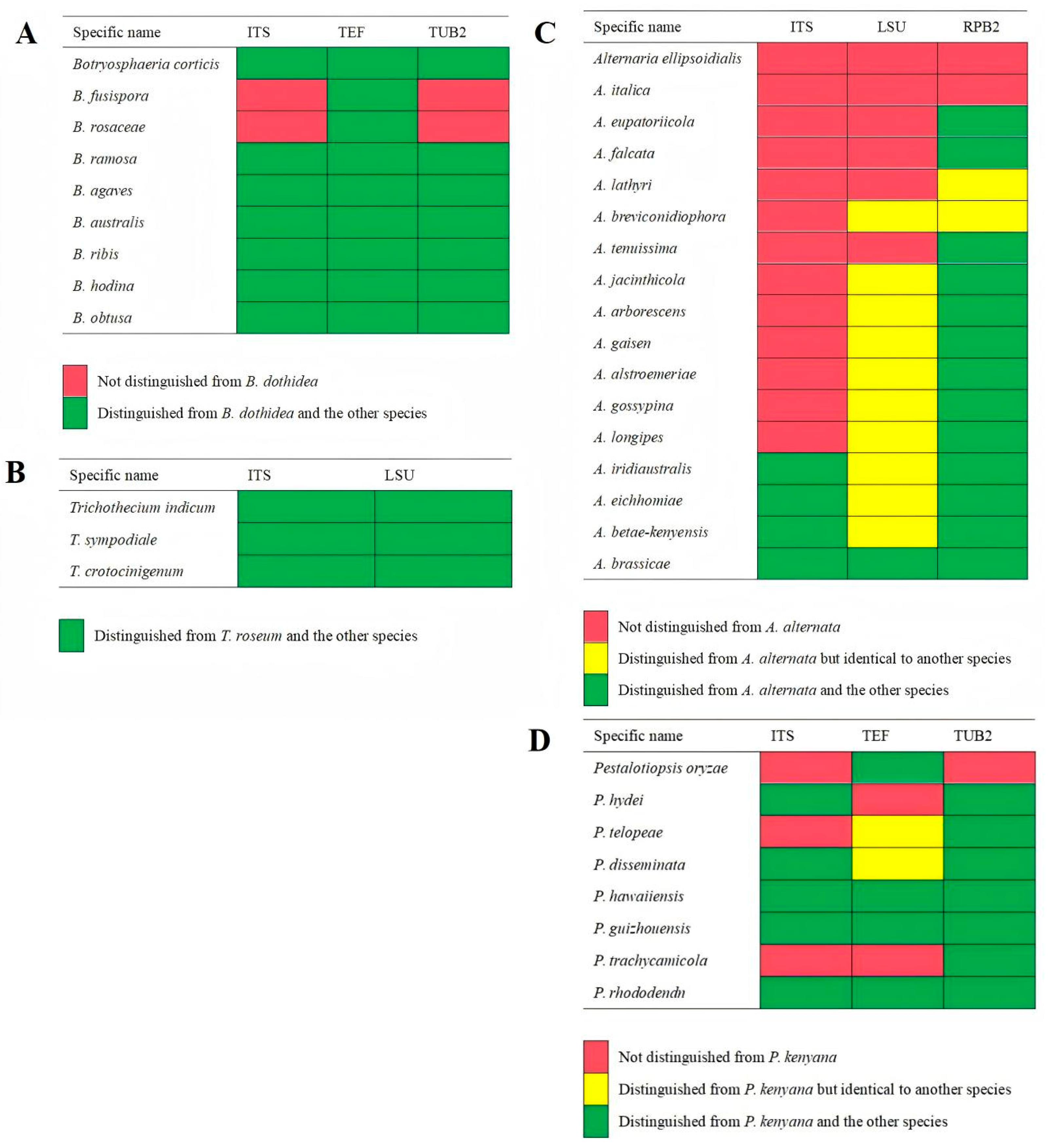
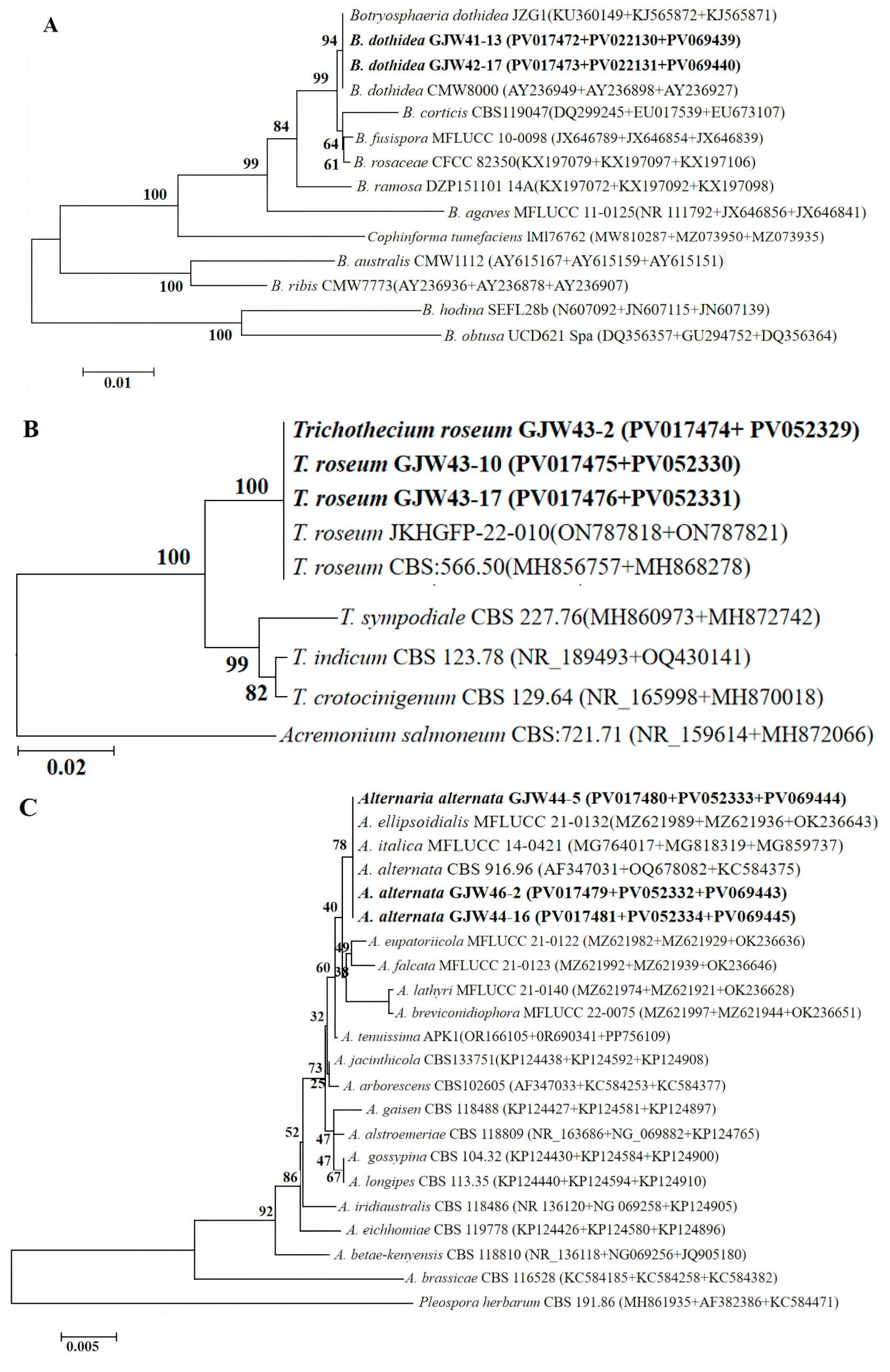
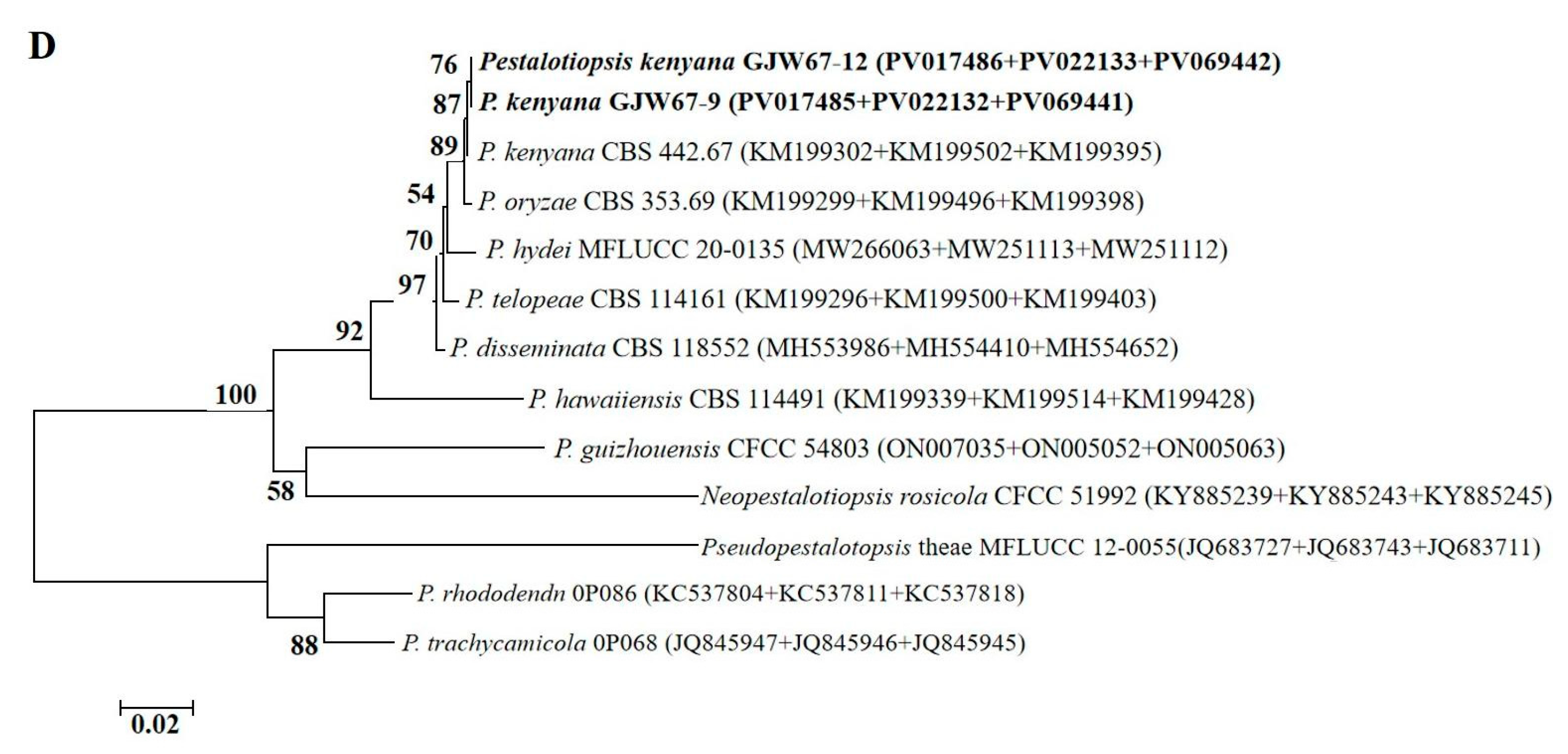
Preliminary morphological analysis of all the strains revealed that those belonging to groups I to IV were similar to B. dothidea, T. roseum, A. alternata, and P. kenyana, respectively. Thus, the strains from each morphotype were selected to confirm the classification results through single or multiple loci phylogenetic analyses. Firstly, the single-gene phylogenies are approximately consistent with multiple loci phylogenies; however, some of the genes sequenced in this study do not enable us to distinguish all of the species recognized here on their own, especially for the single-gene phylogenies of ITS within the genus Alternaria (Figure 2). Specially, the single-gene phylogenies of ITS and TUB2 cannot distinguish B. dothidea from B. fusispora and B. rosaceae; those of ITS and LSU fully distinguish T. roseum from T. indicum, T. sympodiale, and T. crotocinigenum; the ITS phylogeny cannot distinguish A. anternata from A. ellipsoidialis, A. italica, A. eupatoriicola, A. falcata, A. lathyri, A. breviconidiophora, A. tenuissima, A. jacinthicola, A. arborescens, A. gaisen, A. alstroemeriae, A. gossypina, and A. longipes; the LSU phylogeny cannot distinguish A. anternata from A. ellipsoidialis, A. italica, A. eupatoriicola, A. falcata, A. lathyri, and A. tenuissima; and neither can distinguish A. alstroemeriae from A. breviconidiophora, A. jacinthicola, A. arborescens, and A. gaisen, A. gossypina from A. longipes, A. iridiaustralis from A. eichhomiae, and A. betae-kenyensis; in contrast to that, the RPB2 phylogeny cannot distinguish A. alternata from A. ellipsoidialis and A. italica; neither can distinguish A. lathyri from A. breviconidiophora; the ITS phylogeny cannot distinguish P. kenyana from P. oryzae, P. telopeae and P. trachycamicola, and the TEF phylogeny cannot distinguish P. kenyana from P. hydei, nor can it distinguish P. telopeae from P. disseminate; in contrast that, the TUB2 phylogeny only cannot distinguish P. kenyana from P. oryzae. Secondly, the aligned concatenated sequences of 11 strains of Botryosphaeria, including 2 strains from loquat in the present study, and one strain of Cophinforma tumefaciens as the out-group, contained 1035 characters, including gaps (ITS: 1–430; TEF: 431–670; TUB2: 671–1035). For Trichothecium, the aligned combined sequences of five reference strains, three from this study and one strain of Acremonium salmoneum as the outgroup, comprised 917 characters, including gaps (ITS: 1–479; LSU: 480–917). For Alternaria, multigene sequence alignment of 18 reference strains, 2 strains from this study, and 1 strain of Pleospora herbarum as an outgroup included 1300 characters, including gaps (ITS: 1–547; LSU: 548–1019; RPB2: 1020–1300). For Pestalotiopsis, multigene sequence alignment of nine reference strains, two strains from this study, one strain of Pseudopestalotopsis theae, and one strain of Neopestalotiopsis rosicola as an outgroup included 1031 characters, including gaps (ITS: 1–496; TEF: 497–738; TUB2: 739–1031). The phylogram of Botryosphaeria shows that strains GJW41-13 and GJW42-17 (Group I) clustered with B. dothidea (JZG1 and CMW8000); strains GJW43-2, GJW43-10, and GJW43-17 (Group II) clustered with T. roseum (JKHGFP-22-010, CBS: 566.50); GJW44-5, GJW44-16, and GJW46-2 clustered with A. alternata (CBS 916.96), A. ellipsoidialis (MFLUCC 21-0132), and A. italica (MFLUCC 14-0421); and GJW67-9 and GJW67-12 (Group Ⅳ) clustered with P. kenyana (CBS 442.67) (Figure 3).

2.3. Pathogenicity on Loquat Fruits

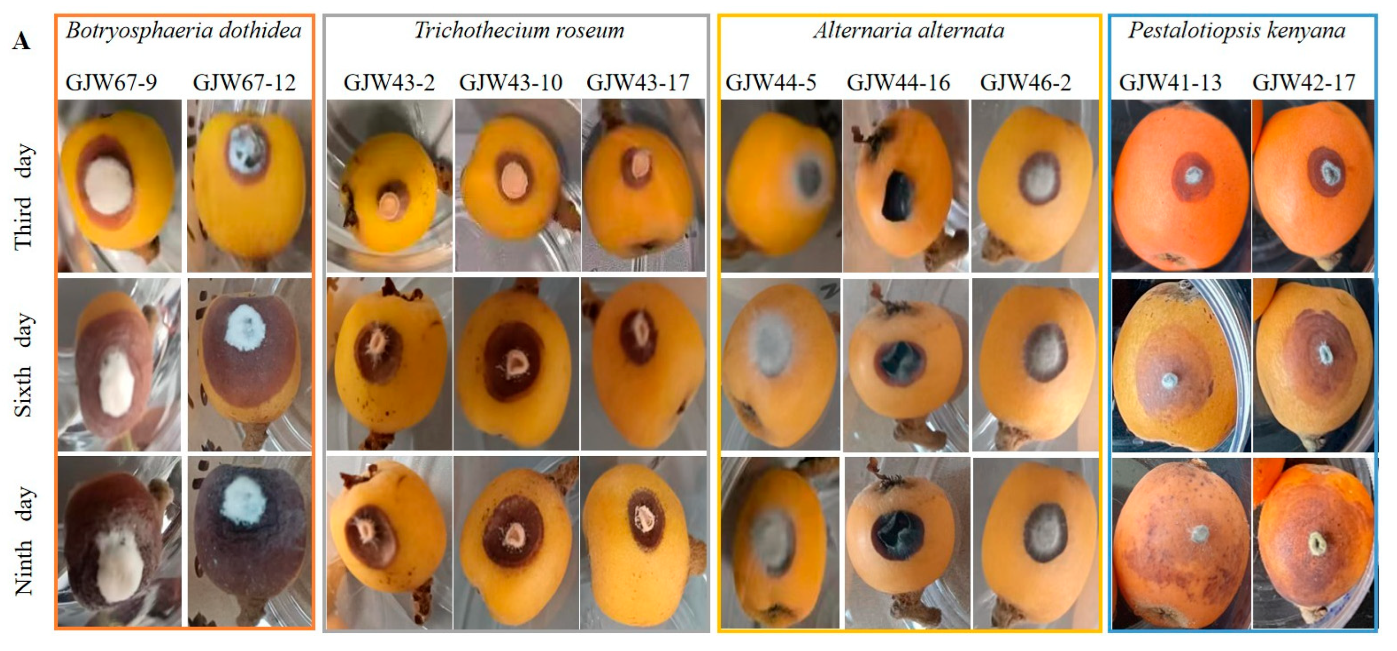
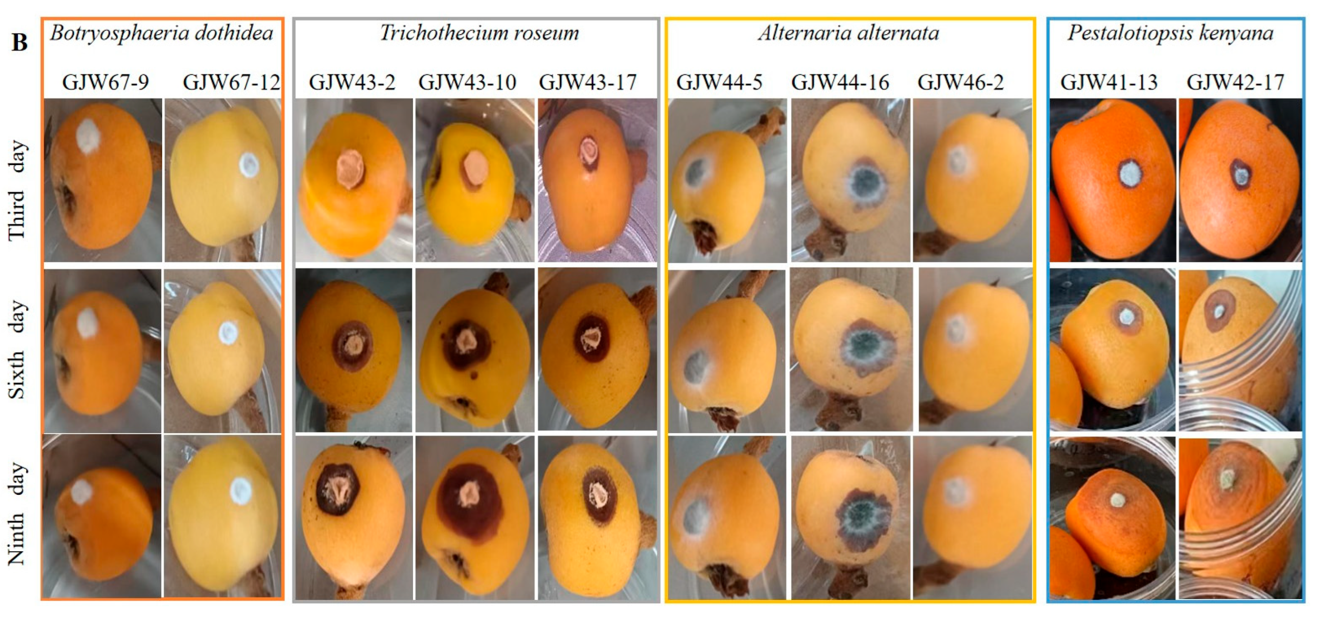
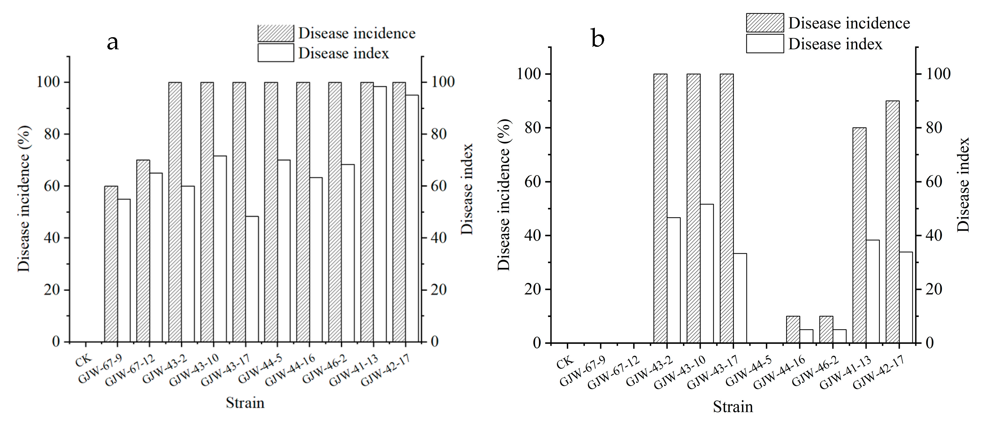
The pathogenicity tests on the loquat fruits showed that all the strains caused symptoms at the wounded site after two days of inoculation. Among them, GJW43-2, GJW43-10, GJW43-17 (T. roseum), GJE44-5, GJW44-16, GJW46-2 (A. alternata), and GJW41-13 and GJW42-17 (B. dothidea) produced symptoms on fruits at both the needle-wounded and non-wounded sites (Figure 4); however, the lesions, incidence rate, and disease index on the needle-wounded sites were more prominent than on the non-wounded sites (Figure 5). All the infected fruits developed symptoms similar to those observed in the market and stockroom. No symptoms appeared on either the control needle-wounded fruits or the non-wounded fruits. Moreover, the fungi were re-isolated from the inoculated fruits. They exhibited colonial and conidial features similar to those of the original strains, confirming that the ten strains in this study cause loquat ring rot, brown spot, black spot, and soft rot, respectively.
After nine days of incubation at 27 °C at a relative humidity of 60% in the light, disease incidence and disease index (severity) on loquats wound-inoculated with B. dothidea were 100% and 95–98.33, A. alternata 100% and 63.33–70, T. roseum 100% and 48.33–71.67, and P. kenyana 60–70% and 55.00–65.00 (Figure 6). After 9 days in the same conditions, these values on intact-inoculated loquats were as follows: B. dothidea 0, A. alternata 0–10% and 0–50, T. roseum 100% and 33.33–51.67, and P. kenyana 80–90% and 33.87–38.33 (Figure 6). In summary, the virulence of T. roseum and P. kenyana was more potent than that of B. dothidea and A. alternata. Additionally, the virulence of four pathogens varied, even though they belonged to the same species.
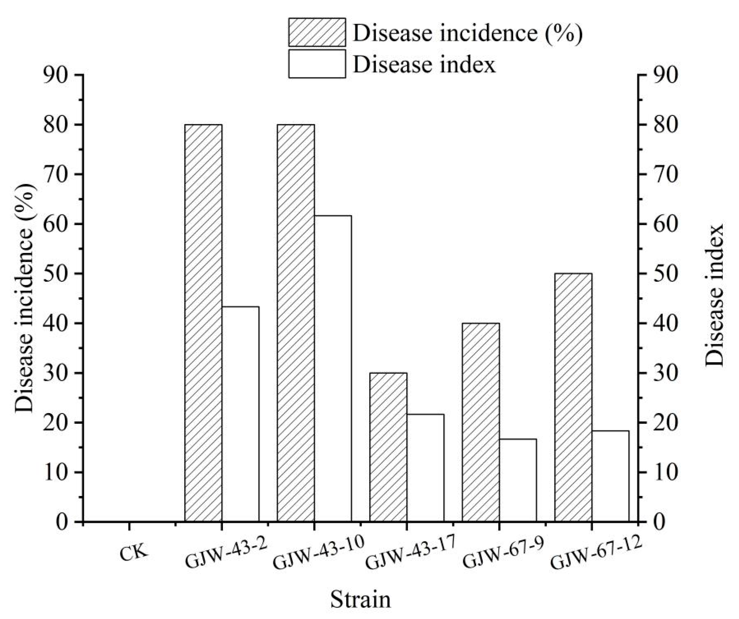
After nine days of inoculation at 27 °C at a relative humidity of 60% in the light, the disease incidence rate and disease index (severity) on loquats sprayed with the conidial suspension of B. dothidea were 40–50% and 16.67–18.33, respectively, and for T. roseum, they were 30–80% and 21.67–61.67, respectively. The virulence of T. roseum was more substantial than that of B. dothidea.
After 16 days of inoculation of whole plants with a 7 mm mycelial plug of B. dothidea, disease incidence on needle-wounded fruits by GJW41-13 and GJW42-17 was 90% and 85%, respectively. In contrast, the non-wounded fruits were 85% and 60%. The symptoms were similar to those of naturally infected fruits, but some diseased fruits were rotten.

2.4. Effect of Temperature and Humidity on Pathogenicity of Alternaria alternata on Loquat Leaves

After 5 days of inoculation at a relative humidity of 50% in the darkness, disease incidence rates on loquat leaves with mycelial plugs of GJW44-5, GJW44-16, and GJW46-2 at 15 °C, 25 °C, and 30 °C were 100%. However, the disease index (severity) of GJW44-5, GJW44-16, and GJW46-2 increased with increasing temperature, reaching 68, 70, and 82, respectively (Table 3).
Compared to temperature, the effect of humidity on pathogenicity was not noticeable. After 6 days of inoculation at 25 °C in darkness, disease incidence rates on loquat leaves with mycelial plugs of GJW44-5, GJW44-16, and GJW44-16 at relative humidity levels of 45%, 55%, 65%, 75%, 85%, and 95% were 90–100%. The disease index (severity) of GJW44-5, GJW44-16, and GJW44-16 at a suitable humidity of 85% was 44 (Table 4).

3. Discussion

Many studies have shown that a complex infection of multiple pathogens usually causes disease, and the dominant pathogens in different regions may vary [47,48,49,50]. Therefore, it is crucial to implement effective management strategies to identify symptoms and respond to pathogens accurately. For example, 38 out of 195 diseases of significant grain and oil crops in China are caused by at least two fungal pathogens; among them, 9 Fusarium species caused maize seedling blight, 15 Fusarium species caused maize root rot, and maize basal rot was caused by 18 species of Fusarium [47,48,49], 338 strains from peach brown rot in Beijing are M. fructicola, and 4 strains are M. fructigena, which indicated that the dominant pathogen of peach brown rot in Beijing is M. fructicola [50]. Morphological characteristics are essential but insufficient for the classification of fungal species at the level of the genus, so strains from postharvest loquat were preliminarily grouped based on colony and conidial morphology. They were identified by multilocus phylogenetic analysis. Seven strains were identified as B. dothidea, T. roseum, and P. kenyana. Of them, B. dothidea has been reported to be associated with pre-harvest or postharvest fruit rot of Chinese olive, plum, pomegranate, yellowhorn, jujube, and apple in China [51,52,53,54,55,56], T. roseum has been reported to be associated with postharvest fruit rot of purple passion, orange, tomato, apple, strawberry, Hami melon, peach (Prunus davidiana), and peppers (Capsicum spp.) [57,58], P. kenyana has been reported to be the agent of bayberry leaf blight in Zhejiang Province, leaf spot on Zanthoxylum schinifolium in Sichuan, and Rhododendron agastum in Guizhou, China [59,60,61]. The other three strains were morphologically similar to A. alternata but clustered with A. alternata, A. ellipsoidialis, and A. italica. In contrast with the size of A. alternata (13.4–42.9 × 7.7–22.0 µm), that of A. ellipsoidialis was 45–70 × 15–30 μm, and that of A. italica was 76–98 × 23–39 μm. Therefore, GJW44-5, GJW44-16, and GJW46-2 were identified as Alternaria alternata based on morphological characteristics and phylogenetic analyses. A. alternata has previously been reported as the causal agent of gray spots on pre-harvest loquat fruit in Greece and Palestine, as well as postharvest loquat fruit rot in Pakistan [14,25,26]. To the best of our knowledge, this is the first report of B. dothidea, T. roseum, and P. kenyana causing postharvest fruit rot on loquat in Yunnan, China, as well as worldwide. Additionally, this is the first report of A. alternata causing postharvest fruit rot on loquat in Yunnan, China.
On the basis of comparison of the single-loci phylogenies combined with the multi-loci phylogenies, ITS and LSU are proposed for the application of distinguishing different Trichothecium species; TEF should be applied prior to the application for distinguishing different Botryosphaeria species. The multi-loci phylogenies combined with morphological characteristics must be used to distinguish different Alternaria and Pestalotiopsis species, as none of the genes sequenced in this study can enable us to distinguish them.
The pathogenicity test on loquat fruits verified that all species produced symptoms at the wounded sites, and B. dothidea, T. roseum, and A. alternata produced symptoms on fruits at the intact sites by inoculation with a 7 mm mycelial plug. However, P. kenyana could produce symptoms on fruits at non-wounded sites by inoculation with conidial suspension, and B. dothidea could cause symptoms of fruit-handing on loquat trees. This finding indicates that wounding is conducive to infection and colonization by these strains. Prior research has shown that injury enhances the infectivity and pathogenicity of harmful fungi [22,62]. In addition, the virulence of T. roseum and P. kenyana was more potent than that of B. dothidea and A. alternata, which is consistent with the pathogenicity tests of Diplodia, Lasiodiplodia, and Neofusicoccum species causing the Botryosphaeria canker and the dieback of apple trees in Central Chile [63]. The effect of temperature on the pathogenicity of A. alternata on loquat leaves was more than humidity. Many studies have documented that all strains were more virulent on wounded plant tissues than on non-wounded ones [64,65], and virulence is varied among species, even strains, temperature, and humidity [66,67,68]. Therefore, the loquat fruits should be gently plucked, packed, loaded, and unloaded during harvest/handling, as well as stored and transported under 15 °C to reduce commercial risks. In the future, the immunogold labeling technology of fruit rot diagnosis and fungicide sensitivity screening will be developed based on these four pathogens.

4. Materials and Methods

4.1. Field Sampling and Isolation

In May 2023, the incidence of postharvest loquat fruit rot was investigated in Yongshan County, Zhaotong City, Yunnan Province, China. Ten diseased fruits were collected and transported to the laboratory for fungal isolation within three days, during which time the disease symptoms were recorded.
The diseased fruits were surface-sterilized by immersing them in 75% ethanol for 30 s, followed by a 1 min exposure to 2.5% sodium hypochlorite, and then rinsed five times with sterilized distilled water. Subsequently, tissue pieces (5 × 5 mm) were cut from the edge of necrotic lesions. Finally, the tissue pieces were on sterilized filter paper for 3–5 min to dry them and transferred onto rose bengal agar (RBA; 5.00 g peptone, 10.00 g glucose, 1.00 g potassium dihydrogen phosphate, 0.50 g magnesium sulfate, 0.03g rose bengal sodium salt, 0.10 g chloromycetin, 15.00 g agar per L) in Petri dishes at 25 °C for three days. Hyphal tips were picked and transported to new RBA or potato dextrose agar (PDA; 200.00 g potatoes, 20.00 g glucose, 0.10 g chloromycetin, 15.00 g agar/L) plates and incubated in the dark at 25 °C.

4.2. Pathogenicity Test

Inoculation of detached mature fruits was performed with a 7 mm mycelial plug. Ten isolates, including three strains of Alternaria sp., two strains of Botryosphaeria sp., two strains of Pestalotiopsis sp., and three strains of Trichothecium sp., were used to inoculate detached mature fruits of the loquat cultivar Big Five-pointed Star. Healthy mature loquat fruits were harvested from the fields of Xiluodu town, Yongshan County, Zhaotong City, Yunnan Province, China. They were inoculated as previously described by Nozawa et al. [11] with some modifications. Fruits were washed with sterilized water, disinfected with 75% ethanol for 1 min, and then air-dried. A 7 mm mycelial plug from the colony edge was placed upside down on wounded or non-wounded fruit surfaces; another 7 mm agar plug was placed upside down on wounded or non-wounded fruit surfaces as a control. Ten wounded fruits and ten non-wounded fruits were used per strain. All treatments have been maintained at 27 °C with a relative humidity of 60% in the light. All treatments have been kept at 27 °C with a relative humidity of 60% under normal light conditions. According to Koch’s postulates, all fungal strains used in the pathogenicity tests were re-isolated from artificially infected fruits to ensure their identity, as described above.
Inoculation of detached mature fruits was performed with conidial suspension. A 10 µL drop of conidial suspension (1 × 10⁶ conidia/mL) by harvesting conidia from two-week-old PDA grown in the dark at 25 °C was sprayed on each non-wounded fruit, and another 10 µL drop of 0.05% Tween-80 was sprayed on each non-wounded fruit as a control [69]. Ten wounded fruits and ten non-wounded fruits were used per strain of Pestalotiopsis and Trichothecium. The environmental conditions and re-isolation of all fungal strains and loquat fruits tested were the same as described above.
Inoculation of whole plants was performed with a 7 mm mycelial plug. Twenty healthy fruits on one healthy plant with a stem base of 10 cm growing on the campus of Kunming University were inoculated separately with the 7 mm mycelial plug of each strain of Botryosphaeria, respectively. Twenty healthy fruits on another healthy plant were inoculated with a 7 mm agar plug as a control. Then, all fruits were individually covered with moist, sterile cotton and placed in transparent polyethylene bags for five days to maintain high relative humidity and an outdoor temperature of 13 to 24 °C.
The incidence rate (IR) and disease index (DI) were evaluated. IR (diseased fruits/total number of fruits inoculated) × 100%. They were referring to assessing the severity of brown leaf spots of Italian ryegrass [70] with modification. In brief, the degree of loquat fruit infection by the fungi was divided into seven grades according to disease scores ranging from 0 to 6 (0: healthy fruits; 1: <1% of the fruit surface shows water-soaked spots; 2: 1–<5% of the fruit surface shows water-soaked spots; 3: 5–<10% of the fruit surface shows brown or black spots; 4: 10–<30% of the fruit surface shows brown or black spots; 5: 30–<70% of the fruit surface shows brown or black spots; 6: >70% of the fruit surface shows brown or black spots, or the fruit falls off). The DI was calculated using the following formula: DI = (∑disease grade × number of diseased fruits)/(total inoculated fruits × 6).
IR and DI of A. alternata on loquat leaves were also used to assess the severity of brown leaf spots of Italian ryegrass [70] with modification. In brief, the degree of loquat leaf infection by the fungi was divided into seven grades according to disease scores ranging from 0 to 5 (0: healthy leaves; 1: <1% of the leaf surface shows diseased spots; 2: 1–<5% of the leaf surface shows diseased spots; 3: 5–<10% of the leaf surface shows diseased spots; 4: 10–<15% of the fruit surface shows diseased spots; 5: <15% of the leaf surface shows diseased spots). The DI was calculated using the following formula: DI = (∑disease grade × number of diseased fruits)/(total inoculated fruits × 5).

4.3. Morphological Identification and Characterization

For descriptions of colony appearance, the isolated strains were incubated on PDA plates for 14 d in the dark at 25 °C. The conidiogenous cells, from which conidia were formed on RBA or PDA of fungal strains, were photographed and measured using an Olympus BX53 microscope (Olympus, Tokyo, Japan). Conidia (n = 20 or 30) of each species were randomly selected for morphological analysis.

4.4. Genomic DNA Extraction, Sequencing, and Phylogenetic Analysis

The genomic DNA was extracted from the mycelium cultivated on PDA media plates at 25 °C for 5–7 days using a Solarbio Fungi Genomic DNA Extraction Kit (Beijing Solarbio Science & Technology Co., Ltd., Beijing, China) according to the manufacturer’s protocol. The internal transcribed spacer (ITS) regions were first amplified and sequenced using the primers ITS1/ITS4 [71] for all pathogenic strains. The strains were initially classified at the genus level as Alternaria, Pestalotiopsis, and Trichothecium based on conidial morphology. They were identified as Botryosphaeria by BLAST analysis of ITS sequences in the nucleotide database. The LSU gene region was amplified and sequenced for the Alternaria strains using the primers LR0R and LR5. In contrast, the partial RNA polymerase II second-largest subunit (RPB2) was amplified and sequenced using the primers RPB2-5f2/fRPB2-7CR [72]. Translation elongation factor (TEF) using primers EF1-728F/EF1-986R [59] and partial beta-tubulin gene (TUB2) using primers T1/Bt2b [73] were then amplified and sequenced for strains of Botryosphaeria and Pestalotiopsis. The LSU gene region, using primers LR0R and LR5 [72], was amplified and sequenced for the Trichothecium strain.
The PCR reaction volume was followed by Doilom et al. [74]. The PCR reaction primers and PCR parameters for each fungal genus are listed in Table 5. The amplified PCR products were forwarded to Tsingke Biotechnology Co., Ltd., Beijing, China, a commercial sequencing service, for sequencing.
The sequence information was uploaded to GenBank. Using MEGA7.0, the sequences were aligned. Gaps were interpreted as missing data, and ambiguous areas were eliminated from the analysis. N-J phylogenetic trees were constructed with the Maximum Composite Likelihood method (ML). The names of the isolates from the present study are in bold in the trees. Maximum likelihood bootstrap support values are given at the nodes, respectively.

5. Conclusions

This research identified B. dothidea, T. roseum, A. alternata, and P. kenyana as the primary fungi causing ring rot, brown spot, black spot, and soft rot in loquat fruit in Yunnan, China. The varying virulence between species and strains highlights the complexity of postharvest fruit rot dynamics, indicating that closely related isolates exhibit different levels of pathogenicity. The virulence varied with species, even strains of the same species. The incidence rates of 4%, 6%, 6%, and 12% for the respective rot types underscore a significant concern for loquat production in the region. The findings contribute to the epidemiological understanding of these pathogens and serve as a reminder for postharvest management practices. Effective control measures are crucial in mitigating the impact of these diseases, which can result in substantial economic losses for farmers and compromise fruit quality. This study is the first to show that B. dothidea, T. roseum, and P. kenyana are involved in postharvest rot of loquats worldwide, and it is also the first evidence that A. alternata is responsible for the black spots of loquat fruits in Yunnan. The findings gained from this study are crucial for epidemiology and provide a cautionary tale for postharvest management of loquat in Yunnan, China. Further research is warranted to explore the ecological roles of these pathogens and develop targeted management strategies for agricultural practices. Understanding the interactions between these fungi and their environment will be crucial for formulating effective control measures and ensuring the sustainability of loquat cultivation, both in Yunnan specifically and globally.

Author Contributions

The contributions of J.-W.G. and C.-L.Y. are consistent. Conceptualization, J.-W.G. and L.Y.; methodology, J.-W.G. and C.-L.Y.; software, J.-W.G. and B.-Z.D.; validation, C.-L.Y., Y.B. and B.-Z.D.; formal analysis, S.-Y.Z. and R.-C.T.; data curation, J.-W.G. and C.-L.Y.; writing—original draft preparation, J.-W.G. and C.-L.Y.; writing—review and editing, J.-W.G., M.M. and L.Y.; visualization, M.Y., L.L., P.G., R.-C.T., J.Z. and C.-S.K.; supervision, J.-W.G.; project administration, J.-W.G. and L.Y. All authors have read and agreed to the published version of the manuscript.

Funding

The financial support was given by the Kunming University Talent Program (XJ20230077), the Yunnan Province Expert Workstation of Cai Peng (202405AF140079), the National Natural Science Foundation of China (No. 31860026), and the Major Science and Technology Special Plan of Yunnan Province (202202AE090035).

Data Availability Statement

The sequences of GJW41-13, GJW42-17, GJW43-2, GJW43-10, GJW43-17, GJW44-5, GJW44-16, GJW46-2, GJW67-9, and GJW67-12 (PV017472~PV017481, PV017485~PV017486 for ITS, PV069446~PV069448 for GAPDH, PV069450~PV069452 for TUB2), and the ACT gene of GJW200-1 (PV069453), GJW200-2 (PV069454), and GJW202-2 (PV069449) have been submitted to NCBI and opened to the public.

Acknowledgments

The authors would like to thank Ying-Xia Guang for providing the loquat fruit samples.

Conflicts of Interest

The authors declare no conflicts of interest.

References

  1. González-Domínguez, E.; Alves, A.; León, M.; Armengol, J. Characterization of Botryosphaeriaceae species associated with diseased loquat (Eriobotrya japonica) in Spain. Plant Pathol. 2017, 66, 77–89. [Google Scholar] [CrossRef]
  2. He, C.; Wang, L.; Li, Y.; Zhou, K.; Zhao, K.; Chen, D.; Li, J.; Song, H.; Tu, M. Loquat (Eriobotrya japonica) is a new natural host of tomato mosaic virus and citrus exocortis viroid. Plants 2024, 13, 1965. [Google Scholar] [CrossRef]
  3. Cai, J.; Chen, T.; Zhang, Z.; Li, B.; Qin, G.; Tian, S. Metabolic dynamics during loquat fruit ripening and postharvest technologies. Front. Plant Sci. 2019, 10, 619. [Google Scholar] [CrossRef]
  4. Luo, J.Q.; Zhang, Y.J.; Jing, L.H.; Wu, A.P.; Xia, M. Application of nutritional and functional value of Eriobotrya japonica. Farm Prod. Process. 2021, 4, 83–87. [Google Scholar]
  5. Palou, L.; Sánchez-Torres, P.; Montesinos-Herrero, C.; Taberner, V. Incidence and etiology of postharvest fungal diseases of loquat fruit (Eriobotrya japonica (Thunb.) Lindl. cv. ‘Algerie’) in Alacant province (Spain). Eur. J. Plant Pathol. 2016, 146, 847–860. [Google Scholar] [CrossRef]
  6. Takata, Y.; Komine, M.; Uchikawa, K.; Nozawa, S.; Watanabe, K. Pathogenicity of Colletotrichum species isolated from rotten fruit and asymptomatic flowers of loquat in Nagasaki Prefecture, Japan and characterization of C. nagasakiense Takata & Kyoko Watan. sp. nov. J. Gen. Plant Pathol. 2024, 90, 69–81. [Google Scholar] [CrossRef]
  7. Gu, H.; Liu, A.Y.; Chen, W.X.; Feng, S.J.; Shi, J.Y. Development and control of postharvest diseases of loquat fruit. Acta Hortic. 2007, 750, 437–443. [Google Scholar] [CrossRef]
  8. Cao, S.; Zheng, Y. Effect of 1-methylcyclopropene on anthracnose rot caused by Colletotrichum acutatum and disease resistance in loquat fruit. J. Sci. Food Agr. 2010, 90, 2289–2294. [Google Scholar] [CrossRef]
  9. Wu, J.; Wang, H.; Fang, L.; Xie, Y.; Wang, L. First report of Colletotrichum scovillei causing anthracnose fruit rot on postharvested Eriobotrya japonica in Zhejiang Province, China. Plant Dis. 2022, 106, 2752. [Google Scholar] [CrossRef]
  10. Wu, W.X.; Liu, Y.; Huang, X.Q.; Zhang, L. First report of anthracnose caused by Colletotrichum nymphaeae on loquat fruit in China. Plant Dis. 2018, 102, 243. [Google Scholar] [CrossRef]
  11. Nozawa, S.; Uchikawa, K.; Suga, Y.; Watanabe, K. Infection sources of Pestalotiopsis sensu lato related to loquat fruit rot in Nagasaki Prefecture, Japan. J. Gen. Plant Pathol. 2020, 86, 173–179. [Google Scholar] [CrossRef]
  12. Abbas, M.F.; Batool, S.; Khan, T.; Rashid, M. First report of Neopestalotiopsis clavispora causing postharvest fruit rot of loquat in Pakistan. J. Plant Pathol. 2022, 104, 459. [Google Scholar] [CrossRef]
  13. Palou, L.; Montesinos-Herreros, C.; Guardado, A.; Taberner, V. First report of Pestalotiopsis clavispora causing postharvest fruit rot of loquat in Spain. J. Plant Pathol. 2013, 95, S4-69. [Google Scholar]
  14. Bibi, H.; Haroon, U.; Farhana; Akbar, A.K.M.; Anar, M.; Batool, S.S.; Bilal, A.; Jabeen, H.; Ahmed, J.; Chaudhary, H.J.; et al. Impact of bacterial synthesized nanoparticles on quality attributes and postharvest disease control efficacy of apricot and loquat. J. Food Sci. 2023, 88, 16695. [Google Scholar] [CrossRef] [PubMed]
  15. Aslam, M.F.; Irshad, G.; Gondal, A.S.; Sajid, M.N.; Naz, F.; Karamat, M.Z.; Bashir, A.; Hyder, S.; Ahmed, R. First report of Rhizopus stolonifer causing postharvest fruit rot of loquat (Eriobotrya japonica) from Pakistan. Plant Dis. 2019, 103, 1410. [Google Scholar] [CrossRef]
  16. Li, S.N.; Jiang, S.C.; Zhang, W.M. First report of postharvest fruit rot of loquat (Rhaphiolepis loquata) caused by Ceratobasidium sp. in China. Plant Dis. 2024, 108, 533. [Google Scholar] [CrossRef]
  17. Hafeez, R.; Akhtar, N.; Shoaib, A.; Bashir, U.; Haider, M.S.; Awan, Z.A. First report of Geotrichum candidum from Pakistan causing postharvest sour rot in loquat (Eriobotrya japonica). J. Anim. Plant Sci. 2015, 25, 1737–1740. [Google Scholar]
  18. Gariglio, N.; Fonfría, M.A. Effect of fruit thinning on the mineral composition of loquat (“Eriobotrya japonica” Lindl.) fruit and its connection with purple spot. Span. J. Agric. Res. 2005, 4, 439–446. [Google Scholar] [CrossRef]
  19. Naz, F.; Abbas, M.F.; Rauf, C.A.; Tariq, A.; Mumtaz, F.A. First report of Colletotrichum gloeosporioides causing anthracnose on loquat in Pakistan. Plant Dis. 2017, 101, 1550. [Google Scholar] [CrossRef]
  20. Juárez-Vázquez, S.B.; Silva-Rojas, H.V.; Rebollar-Alviter, A.; Maidana-Ojeda, M.; Osnaya-González, M.; Fuentes-Aragón, D. Phylogenetic and morphological identification of Colletotrichum godetiae, a novel pathogen causing anthracnose on loquat fruits (Eriobotrya japonica). J. Plant Dis. Prot. 2019, 126, 593–598. [Google Scholar] [CrossRef]
  21. Damm, U.; Sun, Y.-C.; Huang, C.-J. Colletotrichum eriobotryae sp. nov. and C. nymphaeae, the anthracnose pathogens of loquat fruit in central Taiwan, and their sensitivity to azoxystrobin. Mycol. Prog. 2020, 19, 367–380. [Google Scholar] [CrossRef]
  22. Poti, T.; Kisaki, G.; Arita, K.; Akimitsu, K. Colletotrichum species associated with loquat anthracnose in Kagawa and Tokushima prefectures, Japan. J. Gen. Plant Pathol. 2024, 90, 241–253. [Google Scholar] [CrossRef]
  23. Perelló, A.E.; Larran, S. First report of Pestalotiopsis guepini on loquat in Argentina. Plant Dis. 1999, 83, 695. [Google Scholar] [CrossRef] [PubMed]
  24. Chen, Y.; Wang, W.-X.; Zhang, A.-F.; Yang, X.; Xu, Y.-L. First Report of Pestalotiopsis theae on loquat (Eriobotrya japonica) in Anhui Province of China. Plant Dis. 2013, 97, 558. [Google Scholar] [CrossRef]
  25. Tziros, G.T. Alternaria alternata causes leaf spot and fruit rot on loquat (Eriobotrya japonica) in Greece. Australas. Plant Dis. 2013, 8, 123–124. [Google Scholar] [CrossRef]
  26. Batta, Y. Control of Alternaria alternata on loquat (Eriobotrya japonica Lindl.) using detached fruits and leaf-disk assay. An-Najah Univ. J. Res. 2005, 19, 69–82. [Google Scholar] [CrossRef]
  27. Ko, Y.; Liu, C.W.; Chen, S.S.; Chen, C.Y.; Yao, K.S.; Maruthasalam, S.; Lin, C.H. First report of fruit rot of loquat caused by an Alternaria sp. in Taiwan. Plant Dis. 2010, 94, 481. [Google Scholar] [CrossRef]
  28. González-Domínguez, E.; Hinarejos, R.; Tuset, J.J. Identification and characterization of Fusicladium eriobotryae: Fungal pathogen causing mediterranean loquat scab. Acta Hortic. 2007, 750, 343–347. [Google Scholar] [CrossRef]
  29. Palou, L.; Taberner, V.; Montesinos-Herrero, C. First report of Diplodia seriata causing loquat fruit rot in Spain. Plant Dis. 2013, 97, 421. [Google Scholar] [CrossRef]
  30. Abbas, M.F.; Naz, F. First report of Diplodia seriata causing fruit rot of loquat in Pakistan. J. Plant Pathol. 2018, 100, 325. [Google Scholar] [CrossRef]
  31. Niazi, F.; Ali, M.; Haroon, U.; Farhana; Kamal, A.; Rashid, T.; Anwar, F.; Nawab, R.; Chaudhary, H.J.; Munis, M.F.H. Effect of green Fe2O3 nanoparticles in controlling Fusarium fruit rot disease of loquat in Pakistan. Brazil. J. Microb. 2023, 54, 1341–1350. [Google Scholar] [CrossRef] [PubMed]
  32. Abbas, M.F.; Naz, F.; Rauf, C.A.; Mehmood, N.; Shah, P.M.A.; Zhang, X.; Rosli, B.H.; Gleason, M.L. First report of Fusarium solani causing fruit rot of loquat (Eriobotrya japonica) in Pakistan. Plant Dis. 2017, 101, 839. [Google Scholar] [CrossRef]
  33. Zhai, L.; Zhang, M. First report of Neofusicoccum parvum causing fruit rot on Eriobotrya japonica in China. Plant Dis. 2019, 103, 2125. [Google Scholar] [CrossRef]
  34. Yin, L.; Zhang, S.; Du, J.; Wang, X.; Xu, W.; Luo, C. Monilinia fructicola on loquat: An old pathogen invading a new host. J. Integ. Agri. 2021, 20, 2009–2014. [Google Scholar] [CrossRef]
  35. Nwe, Z.M.; Htut, K.N.; Aung, S.L.L.; Gou, Y.-N.; Huang, C.-X.; Deng, J.-X. Two novel species and a new host record of Alternaria (Pleosporales, Pleosporaceae) from sunflower (Compositae) in Myanmar. MycoKeys 2024, 105, 337–354. [Google Scholar] [CrossRef]
  36. Hyde, K.D.; Saleh, A.; Aumentado, H.D.R.; Boekhout, T.; Bera, I.; Khyaju, S.; Bhunjun, C.S.; Chethana, K.W.T.; Phukhamsakda, C.; Doilom, M.; et al. Fungal numbers: Global needs for a realistic assessment. Fungal Divers. 2024, 128, 191–225. [Google Scholar] [CrossRef]
  37. Senanayake, I.C.; Rathnayake, A.R.; Marasinghe, D.S.; Calabon, M.S.; Gentekaki, E.; Lee, H.B.; Hurdeal, V.G.; Pem, D.; Dissanayake, L.S.; Wijesinche, S.N.; et al. Morphological approaches in studying fungi: Collection, examination, isolation, sporulation and preservation. Mycosphere 2020, 11, 2678–2754. [Google Scholar] [CrossRef]
  38. Li, J.F.; Jiang, H.B.; Jeewon, R.; Hongsanan, S.; Bhat, D.J.; Tang, S.M.; Lumyong, S.; Mortimer, P.E.; Xu, J.-C.; Camporesi, E.; et al. Alternaria: Update on species limits, evolution, multi-locus phylogeny, and classification. Stud. Fungi 2023, 8, 1. [Google Scholar] [CrossRef]
  39. Summerbell, R.C.; Gueidan, C.; Schroers, H.-J.; de Hoog, G.S.; Starink, M.; Rosete, Y.A.; Guarro, J.; Scott, J.A. Acremonium phylogenetic overview and revision of Gliomastix, Sarocladium, and Trichothecium. Stud. Mycol. 2011, 68, 139–162. [Google Scholar] [CrossRef]
  40. Woudenberg, J.H.C.; Groenewald, J.Z.; Binder, M.; Crous, P.W. Alternaria redefined. Stud. Mycol. 2013, 75, 171–212. [Google Scholar] [CrossRef]
  41. Maharachchikumbura, S.S.N.; Hyde, K.D.; Groenewald, J.Z.; Xu, J.; Crous, P.W. Pestalotiopsis revisited. Stud. Mycol. 2014, 79, 121–186. [Google Scholar] [CrossRef] [PubMed]
  42. Batista, E.; Lopes, A.; Alves, A. Botryosphaeriaceae species on forest trees in Portugal: Diversity, distribution and pathogenicity. Eur. J. Plant Pathol. 2020, 158, 693–720. [Google Scholar] [CrossRef]
  43. Chen, Y.-Y.; Dissanayake, A.J.; Liu, Z.-Y.; Liu, J.-K. Additions to karst fungi 4: Botryosphaeria spp. associated with woody hosts in Guizhou province, China including B. guttulata sp. nov. Phytotaxa 2020, 454, 186–202. [Google Scholar] [CrossRef]
  44. Götz, M.; Karbowy-Thongbai, B. First detection of Trichothecium roseum causing leaf spots on tomato in Germany. Plant Dis. 2023, 107, 1233. [Google Scholar] [CrossRef]
  45. Fonseca-Guerra, I.R.; Beltrán Pineda, M.E.; Benavides Rozo, M.E. Characterization of Alternaria alternata and Alternaria scrophulariae Brown Spot in Colombian quinoa (Chenopodium quinoa). J. Fungi 2023, 9, 947. [Google Scholar] [CrossRef]
  46. Li, W.-L.; Dissanayake, A.J.; Zhang, T.; Maharachchikumbura, S.S.N.; Liu, J.-K. Identification and pathogenicity of pestalotiod fungi associated with woody oil plants in Sichuan Province, China. J. Fungi 2022, 8, 1175. [Google Scholar] [CrossRef]
  47. Ni, Y.Q.Z.; Li, Y.; Liu, S. List of common fungal diseases of crops and their pathogens in China-the main grain and oil crops. J. Fung. Res. 2023, 21, 247–274. [Google Scholar]
  48. Wang, K.; Liu, F.; Cai, L. A name list of common agricultural phytopathogenic fungi in China. Mycosystema 2022, 41, 361–386. [Google Scholar]
  49. Han, S.L.; Wang, M.M.; Ma, Z.Y.; Raza, M.; Zhao, P.; Liang, J.M.; Wang, J.W.; Cai, L. Fusarium diversity associated with diseased cereals in China, with an updated phylogenomic assessment of the genus. Stud. Mycol. 2023, 104, 87–148. [Google Scholar] [CrossRef]
  50. Zhu, X.Q.; Chen, X.Y.; Guo, L.Y. Population structure of brown rot fungi on stone fruits in China. Plant Dis. 2011, 95, 1284–1291. [Google Scholar] [CrossRef]
  51. Lai, D.; Wang, D.; Shao, X.; Qin, J.; Zhuang, Q.; Xiao, W. First report of Botryosphaeria dothidea causing fruit rot on Chinese olive (Canarium album) in Guangdong Province of China. Plant Dis. 2024, 108, 1109. [Google Scholar] [CrossRef]
  52. Yuan, X.; Qi, Y.; Wu, Z.; Yang, C.; Cui, C. First report of Botryosphaeria dothidea causing postharvest fruit rot of plum in China. Plant Dis. 2024, 108, 1111. [Google Scholar] [CrossRef]
  53. Gu, C.-Y.; Yang, X.; Al-Attala, M.N.; Abid, M.; Phyo, S.S.M.; Zang, H.-Y.; Pan, R.; Chen, Y. First report of pomegranate fruit rot caused by Botryosphaeria dothidea in Anhui Province of China. Plant Dis. 2020, 104, 2736. [Google Scholar] [CrossRef]
  54. Liu, X.; Liu, H.X.; Han, X.L.; Zhang, Y.L.; Liang, Q.; Li, S.K.; Yang, K.Q. First report of Botryosphaeria dothidea causing fruit rot of rellowhorn (Xanthoceras sorbifolium) in China. Plant Dis. 2018, 102, 1662. [Google Scholar] [CrossRef]
  55. Ran, L.X.; Zhang, M.; Shen, H.M. First report of red rot of jujube fruit caused by Botryosphaeria dothidea in China. Plant Dis. 2018, 102, 1458. [Google Scholar] [CrossRef]
  56. Yu, C.; Diao, Y.; Lu, Q.; Zhao, J.; Cui, S.; Peng, C.; He, B.; Liang, Y.; Liu, H. Genome assembly and annotation of Botryosphaeria dothidea sdau11-99, a latent pathogen of apple fruit ring rot in China. Plant Dis. 2021, 105, 1555–1557. [Google Scholar] [CrossRef]
  57. Li, C.; Zhao, J.; Wang, J.; Wang, X.; Xiang, W.; Zhao, J. First report of Trichothecium roseum causing postharvest fruit rot on purple passion fruit in China. Plant Dis. 2022, 106, 3212. [Google Scholar] [CrossRef]
  58. Zhu, M.; Duan, X.; Cai, P.; Qiu, Z.; Li, Z. Genome sequence resource of Trichothecium roseum (ZM-Tr2021), the causal agent of postharvest pink rot. Plant Dis. 2023, 107, 205–209. [Google Scholar] [CrossRef]
  59. Liu, C.; Luo, F.; Zhu, T.; Han, S.; Li, S. Leaf spot disease caused by Pestalotiopsis kenyana on Zanthoxylum schinifolium in Sichuan Province, China. Plant Dis. 2021, 105, 3747. [Google Scholar] [CrossRef]
  60. Li, X.; Liu, L.; Li, H.; Lin, J.; Zuo, Y.; Peng, L.; Ding, H. Pestalotiopsis kenyana causes leaf spot disease on Rhododendron agastum in China. Crop Protect. 2024, 184, 106859. [Google Scholar] [CrossRef]
  61. Xun, W.; Wu, C.; Wu, X.; Bai, Q.; Sun, Y.; Shi, J.; Xie, D.; Jin, L. First report of bayberry leaf blight caused by Pestalotiopsis kenyana in Zhejiang Province, China. Plant Dis. 2023, 107, 2860. [Google Scholar] [CrossRef]
  62. Tan, Q.; Schnabel, G.; Chaisiri, C.; Yin, L.F.; Yin, W.X.; Luo, C.X. Colletotrichum species associated with peaches in China. J. Fungi 2022, 8, 313. [Google Scholar] [CrossRef]
  63. Díaz, G.A.; Valdez, A.; Halleen, F.; Ferrada, E.; Lolas, M.; Latorre, B. Characterization and pathogenicity of Diplodia, Lasiodiplodia, and Neofusicoccum species causing Botryosphaeria canker and dieback of apple trees in Central Chile. Plant Dis. 2022, 106, 925–937. [Google Scholar] [CrossRef] [PubMed]
  64. Imathiu, S.M.; Ray, R.V.; Back, M.; Hare, M.C.; Edwards, S.G. Fusarium langsethiae pathogenicity and aggressiveness towards oats and wheat in wounded and unwounded in vitro detached leaf assays. Eur. J. Plant Pathol. 2009, 124, 117–126. [Google Scholar] [CrossRef]
  65. Santos, M.C.; Viteri, L.O.; Araujo, S.H.; Mourão, D.C.; Câmara, M.P.; Amaral, A.G.; Oliveira, E.E.; Santos, G.R.d. Molecular characterization and pathogenicity of Colletotrichum on banana fruits: Wound effects on virulence and cross-infection. Microbiol. Res. 2025, 16, 4. [Google Scholar] [CrossRef]
  66. Davydenko, K.; Nowakowska, J.; Kaluski, T.; Gawlak, M.; Sadowska, K.; García, J.; Diez, J.; Okorski, A.; Oszako, T.A. Comparative study of the pathogenicity of Fusarium circinatum and other Fusarium species in Polish provenances of P. sylvestris L. Forests 2018, 9, 560. [Google Scholar] [CrossRef]
  67. López-Moral, A.; Lovera, M.; Antón-Domínguez, B.; Gámiz, A.M.; Michailides, T.J.; Arquero, O.; Trapero, A.; Agustí-Brisach, C. Effects of cultivar susceptibility, branch age, and temperature on infection by Botryosphaeriaceae and Diaporthe fungi on English walnut (Juglans regia). Plant Dis. 2022, 106, 2920–2926. [Google Scholar] [CrossRef]
  68. Qiu, J.; Liu, Z.; Xie, J.; Lan, B.; Shen, Z.; Shi, H.; Lin, F.; Shen, X.; Kou, Y. Dual impact of ambient humidity on the virulence of Magnaporthe oryzae and basal resistance in rice. Plant Cell Environ. 2022, 42, 3399–3411. [Google Scholar] [CrossRef]
  69. Guo, J.-W.; Yang, L.-F.; Liu, Y.-H.; Yang, J.; Wang, H.-F.; Liu, Y.-H.; Li, W.J. First report of pseudostem black spot caused by Pestalotiopsis microspora on tsao-ko in Yunnan, China. Plant Dis. 2016, 100, 1021. [Google Scholar] [CrossRef]
  70. Xue, L.; Liu, Y.; Zhou, S.; White, J.F.; Li, C. Characterization of Pyrenophora species causing brown leaf spot on Italian ryegrass (Lolium multiflorum) in Southwestern China. Plant Dis. 2020, 104, 1900–1907. [Google Scholar] [CrossRef]
  71. White, T.J.; Bruns, T.; Lee, S.; Taylor, J. Amplification and direct sequencing of fungal ribosomal RNA genes for phylogenetics. In PCR Protocols: A Guide to Methods and Applications; Innis, M.A., Gelfand, D.H., Sninsky, J.J., White, T.J., Eds.; Academic Press: San Diego, CA, USA, 1990; pp. 315–322. [Google Scholar]
  72. Vilgalys, R.; Hester, M. Rapid genetic identification and mapping of enzymatically amplified ribosomal DNA from several Cryptococcus species. J. Bacteriol. 1990, 172, 4238–4246. [Google Scholar] [CrossRef]
  73. Jiang, N.; Fan, X.L.; Tian, C.M. Identification and characterization of leaf inhabiting fungi from Castanea plantations in China. J. Fungi 2021, 7, 64. [Google Scholar] [CrossRef]
  74. Doilom, M.; Guo, J.-W.; Phookamsak, R.; Mortimer, P.E.; Karunarathna, S.C.; Dong, W.; Liao, C.-F.; Yan, K.; Pem, D.; Suwannarach, N.; et al. Screening of phosphate-solubilizing fungi from air and soil in Yunnan, China: Four novel species in Aspergillus, Gongronella, Penicillium, and Talaromyces. Front. Microbiol. 2020, 11, 585215. [Google Scholar] [CrossRef]
  75. Udayanga, D.; Castlebury, L.A.; Rossman, A.Y.; Hyde, K.D. Species limits in Diaporthe: Molecular reassessment of D. citri, D. cytosporella, D. foeniculina and D. rudis. Persoonia 2014, 32, 83–101. [Google Scholar] [CrossRef]
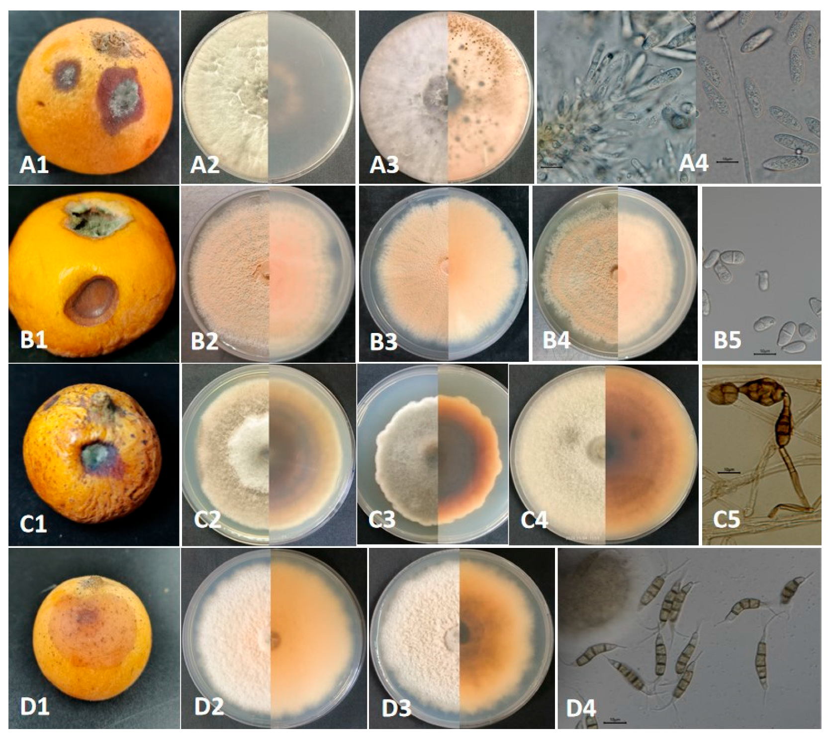
Figure 1. The naturally infected symptoms, colony features, and conidial characteristics of the Botryosphaeria, Trichothecium, Alternaria, and Pestalotiopsis species isolated from loquat fruit rot. (A1A4) correspond to Botryosphaeria dothidea; (B1B5) correspond to Trichothecium roseum; (C1C5) correspond to Alternaria alternata; (D1D4) correspond to Pestalotiopsis kenyana. Bars = 10 μm.
Plants 14 03201 g001
Figure 2. Comparison of gene ability to distinguish among different species. (A) Comparison of gene ability to distinguish Botryosphaeria dothidea from other Botryosphaeria species, (B) comparison of gene ability to distinguish Trichothecium roseum from other Trichothecium species, (C) comparison of gene ability to distinguish Alternaria alternata from other Alternaria species, (D) comparison of gene ability to distinguish Pestalotiopsis kenyana from other Pestalotiopsis species.
Plants 14 03201 g002
Figure 3. N-J phylogenetic trees of Botryosphaeria dothidea, Trichothecium roseum, Alternaria alternata, and Pestalotiopsis kenyana causing postharvest ring rot, brown spot, black spot, and soft rot of loquat in Yunnan, China. (A) B. dothidea based on ITS-, TEF-, and TUB2-sequenced data, (B) T. roseum based on ITS- and LSU-sequenced data, (C) A. alternata based on ITS-, LSU-, and RPB2-sequenced data, and (D) P. kenyana based on ITS-, TEF-, and TUB2-sequenced data. The strains from this study are indicated in bold. The scale bar shows the number of expected changes per site.
Plants 14 03201 g003aPlants 14 03201 g003b
Figure 4. Artificially infected fruits of loquat with wounded inoculation and non-wounded inoculation, with mycelial plug. (A) The symptoms of artificially infected fruits with wounded inoculation with the mycelial plugs of Botryosphaeria dothidea GJW67-9 and GJW67-12, Trichothecium roseum GJW43-2, GJW43-10, and GJW43-17, Alternaria alternata GJW44-5, GJW44-16, and GJW46-2, Pestalotiopsis kenyana GJW41-13 and GJW42-17, respectively. (B) The symptoms of artificially infected, non-wounded inoculated fruits with the mycelial plugs of B. dothidea GJW67-9 and GJW67-12, T. roseum GJW43-2, GJW43-10, and GJW43-17, A. alternata GJW44-5, GJW44-16, and GJW46-2, P. kenyana GJW41-13 and GJW42-17, respectively. All treatments were maintained at 27 °C with a relative humidity of 60% under normal light conditions.
Plants 14 03201 g004aPlants 14 03201 g004b
Figure 5. Incidence rate and disease index of postharvest fruit rot artificially infected with the mycelial plug of Botryosphaeria dothidea GJW67-9 and GJW67-12, Trichothecium roseum GJW43-2, GJW43-10, and GJW43-17, Alternaria alternata GJW44-5, GJW44-16, and GJW46-2, Pestalotiopsis kenyana GJW41-13 and GJW42-17, respectively. (a) All the treatments were inoculated on wounded sites; (b) all were inoculated on non-wounded sites.
Plants 14 03201 g005
Figure 6. Incidence rate and disease index of postharvest non-wounded fruit rot artificially infected with a conidial suspension of five strains of Botryosphaeria dothidea GJW67-9 and GJW67-12, and Trichothecium roseum GJW43-2, GJW43-10, and GJW43-17, respectively.
Plants 14 03201 g006
Table 1. List of fungi that cause loquat fruit decay.
No. Pathogens Period Location Authors and References
1Colletotrichum gloeosporioidesBefore-harvestTaxila and Wah Cantt, PakistanNaz, F. [19]
PostharvestAlacant Province, SpainPalou, L. [5]
PostharvestNagasaki Prefecture, JapanTakata, Y. [6]
2C. godetiaeBefore-harvestTetela, Morelos State, MexicoJuárez-Vázquez, S.B. [20]
3C. acutatumPostharvestFujian Province, ChinaGu, H. [7]
PostharvestJiangsu Province, ChinaCao, S. [8]
4C. fioriniaePostharvestNagasaki Prefecture, JapanTakata, Y. [6]
Before-harvestNagasaki Prefecture, JapanPoti, T. [22]
5C. scovilleiPostharvestZhejiang Province, ChinaWu, J. [9]
6C. eriobotryaeBefore-harvestTaiwan Province, ChinaDamm, U. [21]
7C. nymphaeaePostharvestNagasaki Prefecture, JapanTakata, Y. [6]
Before-harvestNagasaki Prefecture, JapanPoti, T. [22]
Before-harvestTaiwan Province, ChinaDamm, U. [21]
PostharvestSichuan Province, ChinaWu, W.X. [10]
8Pestalotiopsis eriobotryfoliaPostharvestFujian Province, ChinaGu, H. [7]
9P. sensuPostharvestAlacant Province, SpainNozawa, S. [11]
10P. guepiniBefore-harvestBuenos Aires, ArgentinaPerelló A.E. [23]
P. theaeBefore-harvestAnhui Province, ChinaChen, Y. [24]
11Neopestalotiopsis clavisporaPostharvestAlacant Province, SpainPalou, L. [5]
PostharvestPakistanAbbas, M.F. [12]
PostharvestSpainPalou, L. [13]
12Alternaria alternataBefore-harvestThessaloniki, GreeceTziros, G.T. [25]
Before-harvestPalestineBatta, Y. [26]
PostharvestIslamabad, PakistanBibi, H. [14]
13A. tenuissimaPostharvestFujian Province, ChinaGu, H. [7]
14Alternaria sp.Before-harvestTaiwan Province, ChinaKo, Y. [27]
15Fusicladium eriobotryaeBefore-harvestSpainGonzález-Domínguez, E. [28]
16Botrytis cinereaPostharvestAlacant Province, SpainPalou, L. [5]
17Diplodia seriataPostharvestAlacant Province, SpainPalou, L. [5]
Before-harvestAlicante Province, SpainPalou, L. [29]
Before-harvestPunjab and Khyber Paktoon Khawa, PakistanAbbas, M.F. [30]
18Penicillium expansumPostharvestAlacant Province, SpainPalou, L. [5]
19Rhizopus stoloniferPostharvestAlacant Province, SpainPalou, L. [5]
PostharvestRawalpindi and Swat, PakistanAslam, M.F. [15]
20Ceratobasidium sp.PostharvestGuangdong Province, ChinaLi, S.N. [16]
21Fusarium oxysporumBefore-harvestIslamabad, PakistanNiazi, F. [31]
22F. solaniBefore-harvestPunjab Province, PakistanAbbas, M.F [32]
23Geotrichum candidumPostharvestLahore, PakistanHafeez, R. [17]
24Neofusicoccum parvumBefore-harvestChongqing, ChinaZhai, L. [33]
25Diplocarpon mespiliBefore and PostharvestSpainGariglio, N. [18]
26Monilinia fructicolaBefore-harvestWuhan, Hubei Province, ChinaYin, L. [34]
Table 2. Morphological characteristics of the fungal species causing loquat postharvest fruit rot in China.
Morphotype Group Strain Name Colony Character on PDA Conidia Similar Species
Group Ⅰ
Botryosphaeria dothidea
GJW41-13
GJW42-17
Circular and initially white colonies gradually turned gray-green, with a dark green color on the back, and featured short and thick aerial hyphae with an irregular colony margin. Colony diameters were 8.0 cm and 9.0 cm after 3 d at 28 °C in the light, respectively.Hyaline and subcylindrical, aseptate, 17.5–24.1 × 5.7 to 7.2 μm, L/W = 2.7–4.1 (mean 21.0 × 6.6 μm, average L/W = 3.2, n = 30)Botryosphaeria dothidea [(20-)23-27(-30) × 4-5(-6) (mean 26.2 × 5.4 μm)]
Group Ⅱ
Trichothecium roseum
GJW43-2
GJW43-10
GJW43-17
Circular and initially white colonies gradually produced dense, pink, and circular structures (conidiophores and conidia) with a rough colony margin. Colony diameters were 2.7 cm, 2.7 cm, and 2.8 cm after 3 d at 28 °C in the light, respectively.Produced in clusters, smooth, hyaline, thick-walled, 1-septa, ellipsoid to pyriform, 9.1–16.3 × 4.5–8.8 μm, L/W = 1.4–2.6 (mean 12.8 × 6.3 μm, average L/W = 2.1, n = 30)Trichothecium roseum (10 to 18 × 7 to 9.5 μm)
Group Ⅲ
Alternaria alternata
GJW44-5
GJW44-16
GJW46-2
Initially a white colony and turned olive green to black 7 days post-incubation. Featured pale brown, thick, and cottony aerial hyphae, with a reserve of black surrounded by a light-brown circle. Colony diameters were 6.7 cm, 5.6 cm, and 7.2 cm after 3 d at 28 °C in the light, respectivelyProduce chained conidia singly, separated, and pale brown conidiophores. Conidia yellow-brown or black-brown, obclavate, subglobose, ellipsoid, with 1–5 transverse septa and 1–3 longitudinal septa, 13.4–42.9 × 7.7–22.0 µm, L/W = 1.5–4.6 (mean 27.6 × 13.5 µm, average L/W = 2.8, n = 30)Alternaria alternata [26–30 × 5–9 µm with 4–7 transverse septa and a few or no longisepta]
Group Ⅳ
Pestalotiopsis kenyana
GJW67-9
GJW67-12
Smooth-edged, dense, whitish, with sparse aerial mycelium on the surface, produced a yellowish or black oil-drop mass (conidiophores and conidia) and a yellowish reserve with an irregular margin—colony diameter was 3.3 cm after 4 d at 28 °C in the light.Fusoid, ellipsoid, straight or slightly curved, 4-septate, 20.9–27.6 × 4.0–5.4 μm, L/W = 4.5–6.9 (mean 24.3 × 4.6 μm, average L/W = 5.5, n = 20); three median cells were darker than other cells; apical cell with 2–3 tubular appendages (mainly three) ranging from 6.6 to 16.3 μm, basal cell with one tubular appendage ranging from 4.1 to 7.6 μm.Pestalotiopsis kenyana [17.5–22 × 6–7 µm (mean 20 × 6.5 µm)]
Note: L/W = length-to-width ratio.
Table 3. Disease incidence and disease index of Alternaria alternata GJW44-5, GJW44-16, and GJW46-2 at a relative humidity of 50%.
Temperature/℃ Alternaria alternata GJW44-5 A. alternata GJW44-16 A. alternata GJW46-2
Disease Incidence/% Disease Index Disease Incidence/% Disease Index Disease Incidence/% Disease Index
15100321004610062
25100421006210070
30100681007010082
Table 4. Disease incidence and disease index of Alternaria alternata GJW44-5, GJW44-16, and GJW46-2 at 25 °C.
Humidity/% Alternaria alternata GJW44-5 A. alternata GJW44-16 A. alternata GJW46-2
Disease Incidence/%Disease IndexDisease Incidence/%Disease IndexDisease Incidence/%Disease Index
45903290329036
5510034100409036
65100401004010038
75100441004010038
85100441004410044
9510044100429038
Table 5. PCR reaction primers (forward and reverse) and protocols for amplification of gene loci of each fungal genus.
Locus Primer Names and Primer Sequences PCR Protocols References
ITSITS1: TCC GTA GGT GAA CCT GCG G
ITS4: TCC TCC GCT TAT TGA TAT GC
(95 °C 30 s, 55 °C 50 s, 72 °C 1 min) × 39 cyclesWhite, T.J. [71]
Udayanga, D. [75]
LSULROR: ACC CGC TGA ACT TAA GC
LR5: ATC CTG AGG GAA ACT TC
(95 °C 1 min, 55 °C 2 min, 72 °C 90 s) × 35 cyclesVilgalys, R. [72]
Doilom, M. [74]
TEFEF1-728F: CAT CGA GAA GTT CGA GAA GG
EF1-986R: TAC TTG AAG GAA CCC TTA CC
(95 °C 30 s, 58 °C 50 s, 72 °C 1 min) × 39 cyclesVilgalys, R. [72]
Udayanga, D. [75]
TUBT1: AAC ATG CGT GAG ATT GTA AGT
Bt2b: ACC CTC AGT GTA GTG ACC CTT GGC
(95 °C 30 s, 58 °C 50 s, 72 °C 1 min) × 39 cyclesJiang, N. [73]
Udayanga, D. [75]
RPB2RPB2-5F2: GGG GWG AYC AGA AGA AGG C
fRPB2-7CR: CCC ATR GCT TGY TTR CCC AT
(95 °C 1 min, 52 °C 2 min, 72 °C 90 s) × 35 cyclesJiang, N. [73]
Doilom, M. [74]
Disclaimer/Publisher’s Note: The statements, opinions and data contained in all publications are solely those of the individual author(s) and contributor(s) and not of MDPI and/or the editor(s). MDPI and/or the editor(s) disclaim responsibility for any injury to people or property resulting from any ideas, methods, instructions or products referred to in the content.