Article

Cloning and Functional Analysis of ClVND1, a Member of the OsNAC7 Subfamily of the NAC Family in Chrysanthemum lavandulifolium

1
College of Landscape Architecture and Forestry, Qingdao Agricultural University, Qingdao 266109, China
2
Shandong Key Laboratory for Germplasm Innovation of Saline-Alkaline Tolerant Grasses and Trees, Qingdao Agricultural University, Qingdao 250000, China
*
Authors to whom correspondence should be addressed.
These authors contributed equally to this work.
Plants 2025, 14(18), 2925; https://doi.org/10.3390/plants14182925
Submission received: 1 August 2025 / Revised: 10 September 2025 / Accepted: 17 September 2025 / Published: 20 September 2025

Abstract

Chrysanthemum × morifolium is a commercially important flower worldwide. Chrysanthemum lavandulifolium is the main model plant for the research on Chrysanthemum. Enhancing stress resistance in C. lavandulifolium is highly significant for improving commercial chrysanthemum production. NAC transcription factors are key regulators of plant growth, development, and stress responses. In this study, we cloned ClVND1—a member of the OsNAC7 subfamily within the NAC transcription factor family—from Chrysanthemum lavandulifolium. The gene comprises a 1164 bp coding sequence (CDS) encoding a protein of 387 amino acids. Overexpression of ClVND1 promotes secondary cell wall thickening in the stems of transgenic Arabidopsis, stimulates lateral root growth, and consequently enhances tolerance to salt and low-temperature stress in seedlings. Phenotypic analysis showed that transgenic Arabidopsis exhibited reduced inflorescence elongation and plant height compared to wild-type controls, but an earlier flowering time. These findings suggest that ClVND1 enhances stress resistance by promoting lateral root development, while also suppressing inflorescence growth and accelerating flowering time.

1. Introduction

During growth and development, plants are frequently exposed to a range of abiotic and biotic stresses, including drought, high salinity, low temperature, and pathogen infection. These stresses typically induce cellular damage, adversely impacting plant growth and productivity [1]. Transcription factors play important roles in plant adaptation to the environment and resistance to stress. NAC transcription factors play a key role in regulating secondary wall synthesis and serve as major regulators of broad-spectrum stress resistance in plants [2,3]. Within the NAC family, the OsNAC7 subfamily is one of the most extensively studied groups and includes key members such as SND1, NST1, URP7, BRN1/2, and VND1VND7 [4,5,6,7,8,9,10,11,12,13]. Its primary functions involve the regulation of secondary cell wall formation in stems, roots, and anthers. They play a key role in regulating lignin biosynthesis, growth and development, as well as stress responses [11,14,15,16,17]. Within the OsNAC7 subfamily, NST1, NST2, and SND1 are classified as secondary wall-associated NAC transcription factors and function as master switches that activate the expression of genes involved in secondary cell wall biosynthesis [18]. Previous studies have demonstrated that the ClSND1 gene from Chrysanthemum nankingense promotes thickening of the secondary xylem and its cell walls in transgenic Nicotiana benthamiana, thereby conferring enhanced tolerance to salt and low-temperature stress in seedlings. Additionally, it suppresses plant growth and promotes early flowering [1,19,20]. Recent studies have revealed that NAC transcription factors are expressed throughout plant development and across various tissues, where they play crucial roles in lignin biosynthesis, growth regulation, and responses to both abiotic and biotic stresses [21]. A total of 102 NAC family members have been identified in pepper (Capsicum annuum) [22], 101 in tomato (Solanum lycopersicum) [23], 190 in maize (Zea mays) [24], 143 in grape (Vitis vinifera) [25], and 152 in soybean (Glycine max) [26]. Overexpression of the ZmNAC071 gene in Arabidopsis enhances plant stress tolerance, while the celery AgNAC63 gene is highly responsive to various abiotic stresses [27,28].
VND is a group of NAC family transcription factors that encode a specific NAC domain. Seven vascular-related VND genes have been found in A. thaliana. Zhou et al. investigated the roles of VND1VND5 genes in regulating secondary cell wall biosynthesis in Arabidopsis thaliana [29,30]. VND1VND5 expression was specifically observed in vessel elements, with VND4 and VND5 also detected in the secondary xylem vessels of the root hypocotyl [11]. Tan et al. demonstrated that VND1, VND2, and VND3 play distinct roles in the differentiation of xylem vessel elements within cotyledons. The VND subfamily has also been examined in banana (Musa), in addition to well-established models such as Arabidopsis and poplar. MusaVND1 encodes a highly conserved NAC domain and is a homolog of Arabidopsis VND1 [31]. MusaVND1 activates the expression of genes involved in the lignin and cellulose biosynthetic pathways. Transgenic banana plants overexpressing MusaVND1 exhibited increased lignin and cellulose content compared to wild-type controls [32]. Previous studies indicate that the five VND genes in Chrysanthemum nankingense participate in regulating root and stem development, as well as mediating responses to drought and salinity stress [33]. However, the functional role of VND genes in C. lavandulifolium has not yet been reported [34].

2. Results

2.1. Bioinformatic Characterization of the ClVND1 Gene

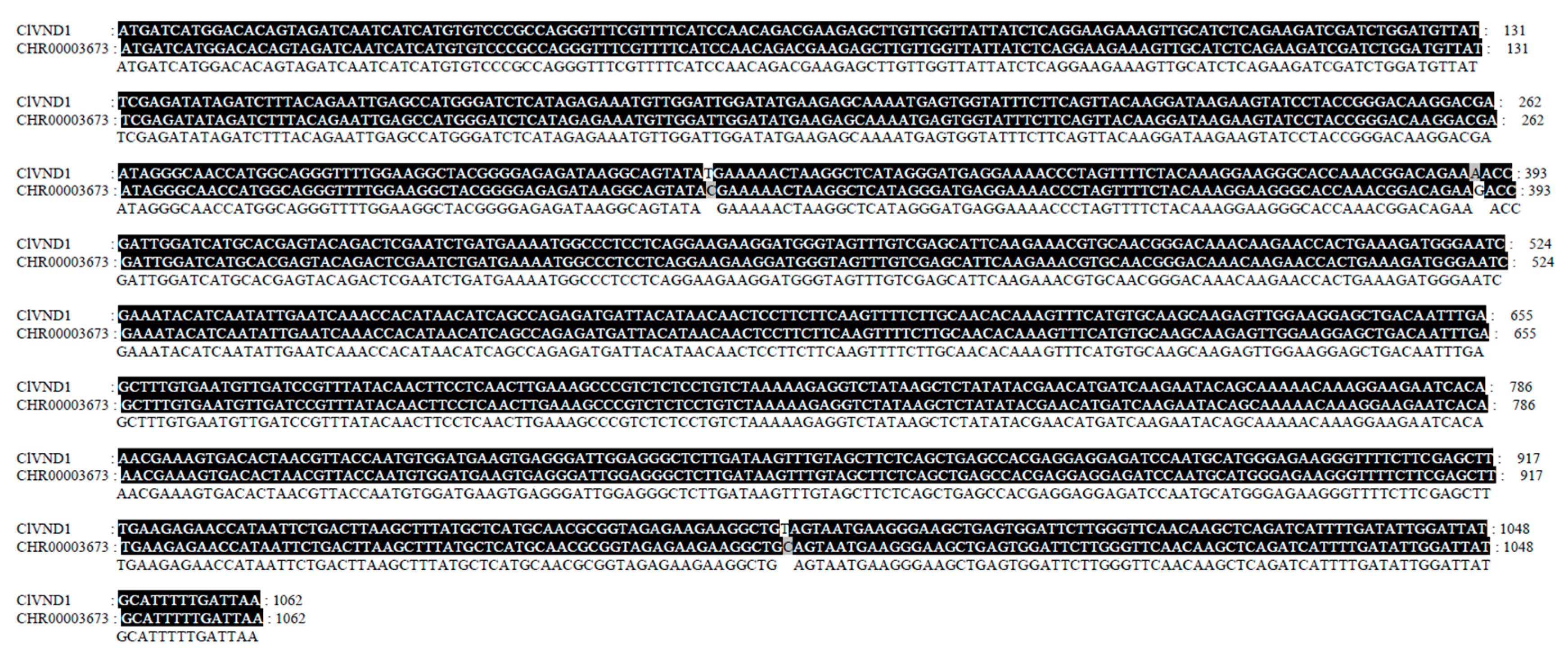
A gene designated ClVND1 was cloned from C. lavandulifolium. Its coding region comprises 1164 base pairs, which encode a polypeptide of 387 amino acids. The ClVND1 gene sequence was obtained from the NCBI database (accession no. PX275817). We found that the ClVND1 gene sequence shares high sequence similarity with Chrysanthemum nankingense CHR0003673-RA (Appendix A).
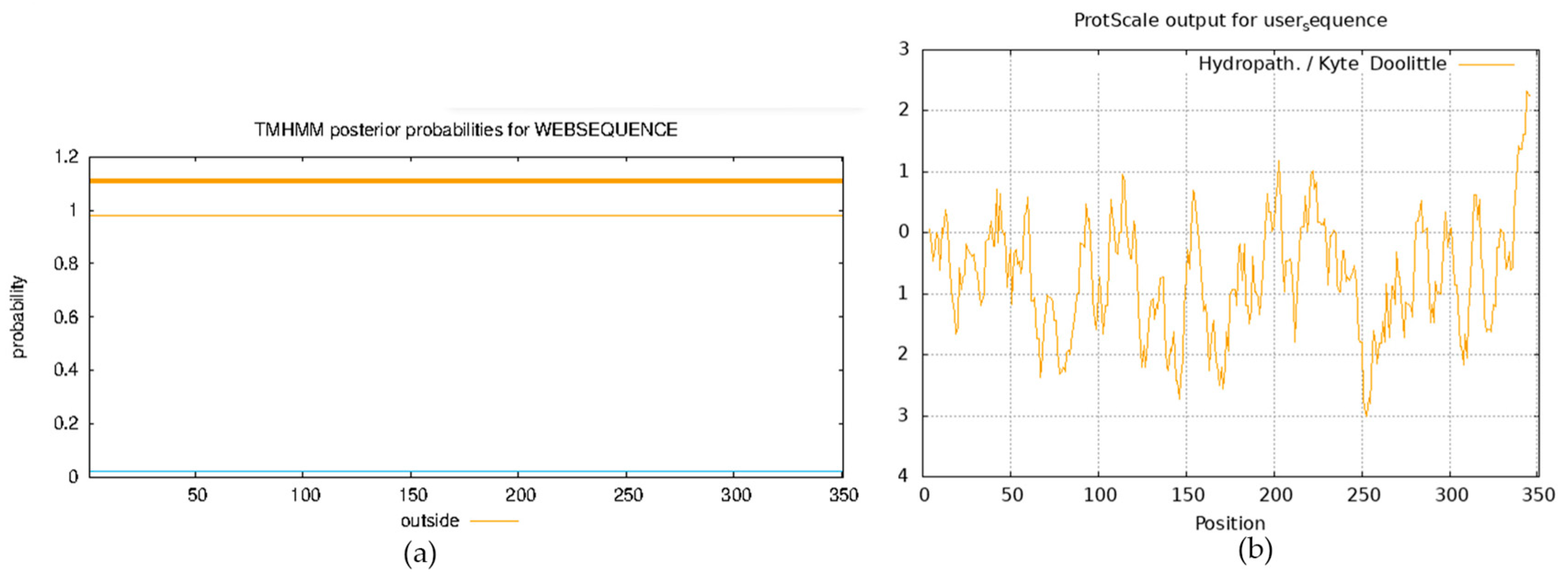
The physicochemical properties of the ClVND1-encoded protein were evaluated. The physicochemical analysis revealed that this protein is composed of 351 amino acid residues. Its theoretical isoelectric point (pI) was calculated to be 5.31, with a relative molecular mass of 40,897.95 Da. The molecular formula was predicted as C1814H2788N492O558S15 (total atoms: 5667), and its instability index was 69.15. The protein has an average hydrophobicity of −0.672 and an instability index (II) of 45.37, indicating that it is unstable. ProtScale analysis revealed that the ClVND1 protein possesses distinct hydrophobic and hydrophilic regions, and was overall determined to be hydrophilic. Analysis of the ClVND1 protein sequence predicted the absence of transmembrane domains, indicating a non-transmembrane structure (Appendix B).
The secondary structure of the ClVND1 protein was predicted to consist primarily of irregular coil (43.09%) and α-helix (46.62%), with extended strands accounting for the remaining 10.29% (Figure 1). The tertiary structure of the ClVND1 protein was predicted using Phyre2 and was found to be consistent with its secondary structure prediction (Figure 1). The function and domain architecture of the ClVND1 protein were predicted using the CD-Search online tool. Results indicated that the protein belongs to the NAC family (Figure 1). NAC transcription factors, which are unique to terrestrial plants, play pivotal roles in regulating plant growth and development, hormonal signal transduction, and stress responses. The ClVND1 gene has been implicated in mediating plant responses to adverse environmental stresses.
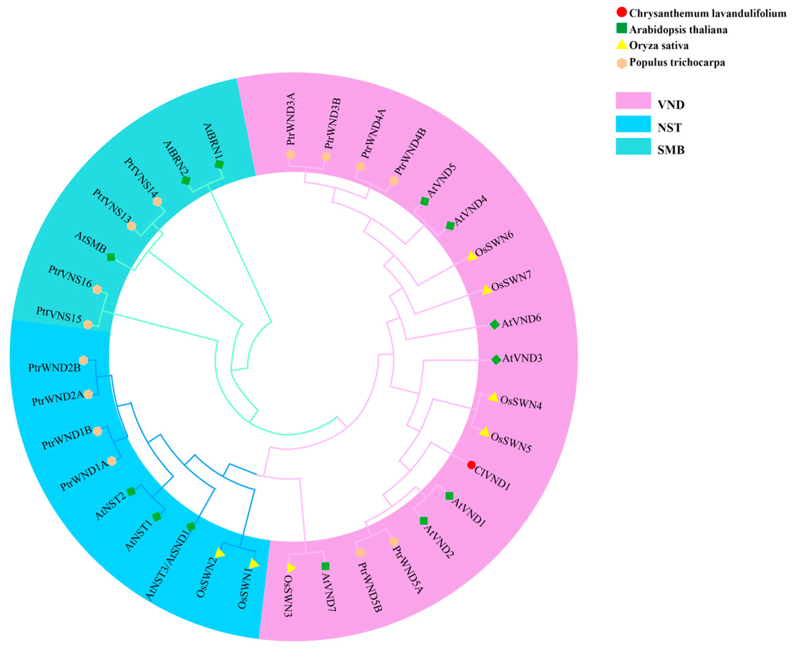
The evolutionary relationships among VNS proteins were inferred by constructing a maximum likelihood phylogenetic tree using amino acid sequences from diverse species. Phylogenetic analysis indicated that ClVND1 is most closely related to AtVND1, AtVND2, PtrWND5A, and PtrWND5B, and most distantly related to AtVND7 and OsSWN3 (Figure 2). ClVND1 was phylogenetically grouped within the same clade as homologs from Arabidopsis thaliana (AtVND1, AtVND2, AtVND3), Oryza sativa (OsSWN6, OsSWN7), and Populus trichocarpa (PtrWND5A, PtrWND5B). Based on these phylogenetic findings, ClVND1 can be classified as a member of the VND subfamily.

2.2. Analysis of Subcellular Localization in Nicotiana benthamiana

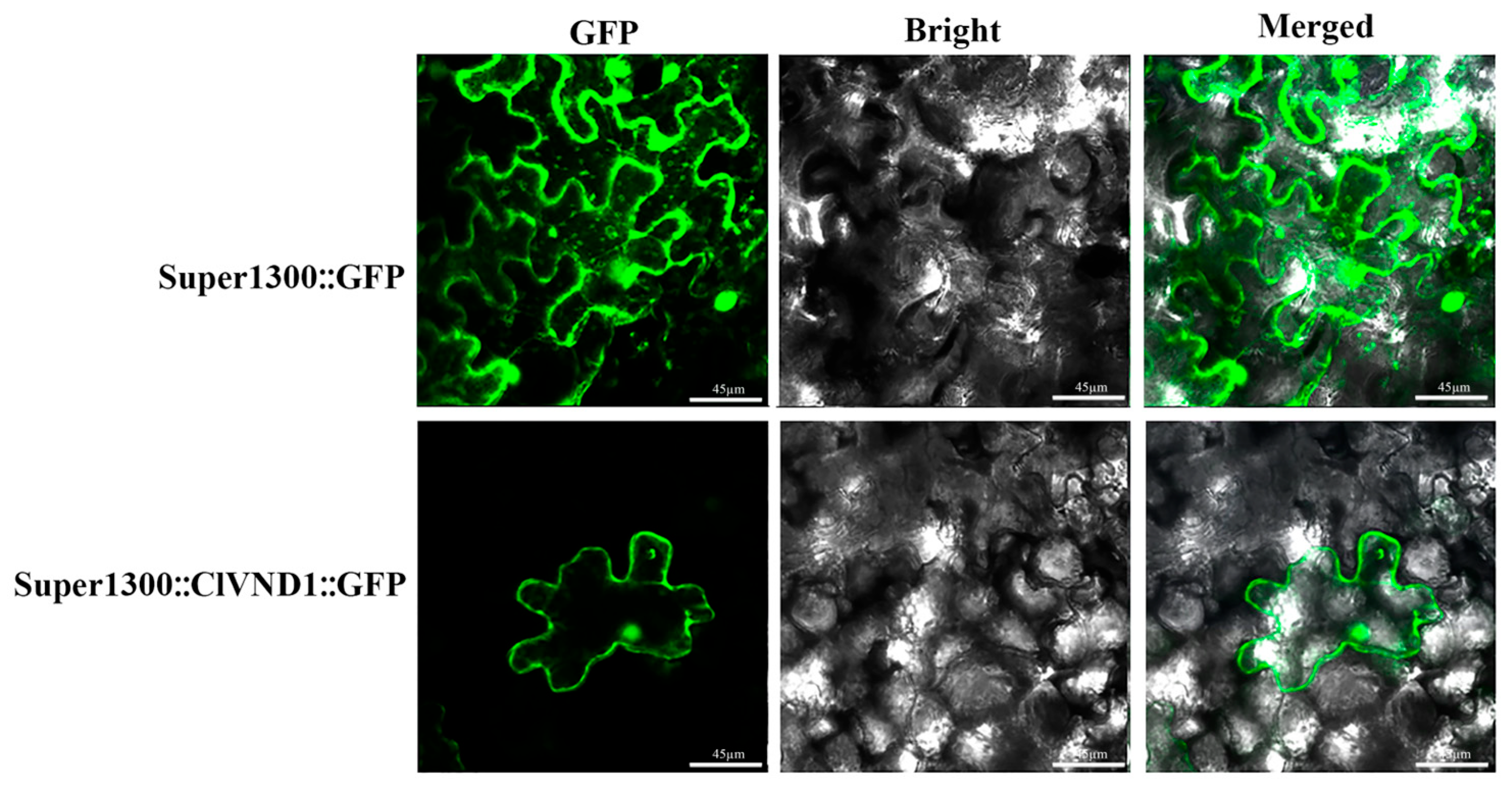
To determine the subcellular localization of the ClVND1 protein, fluorescence-based localization analysis was performed in transgenic Nicotiana benthamiana. The fluorescence signal in the leaves of the transgenic plants was observed, and it was found that the Super1300 empty transgenic N. benthamiana could recognize the fluorescence signal in the nucleus and cell membrane of the leaves. Fluorescence signals were detected in the nucleus and cell membrane of Super35S::ClVND1::GFP transgenic N. benthamiana leaves (Figure 3). These results indicate that ClVND1 is localized in the nucleus and cell membrane.

2.3. Identification of Positive Transgenic Lines and Validation by qRT-PCR

Genomic DNA was isolated from healthy wild-type (WT) and transgenic Arabidopsis lines to serve as a template for PCR with primers 1300-F/1300-R, using WT plants as a negative control. Amplification of a specific 1164 bp fragment was observed exclusively in the transgenic lines (Figure 4A). The absence of this band in the WT control confirms the successful genomic integration of the ClVND1 transgene, thereby validating the transgenic plants at the DNA level. To examine the expression level of ClVND1 in each transgenic line, cDNA reverse-transcribed from both wild-type (WT) and transgenic plants was used as the template for amplification with gene-specific primers ClVND1-F/ClVND1-R and the reference primers Actin8-F/Actin8-R. As shown in Figure 4B, compared to the WT, the expression levels in all OE-ClVND1 lines were significantly increased. Among them, the OE-1 line exhibited the highest expression level, which was 4.79 times that of the WT, while the OE-2 line showed the lowest expression level, which was 3.28 times that of the WT. Based on its highest expression level, the OE-1 line was selected for subsequent experiments.

2.4. Response of Transgenic A. thaliana to NaCl and Low-Temperature (4 °C) Stress Treatments

Under 4 °C conditions over a three-day period, the root upper part of ClVND1-transgenic A. thaliana displayed significantly enhanced growth relative to wild-type controls, suggesting improved cold stress tolerance. Under 200 mmol/L NaCl, CLVND1-overexpressing lines exhibited a developed substantially more of short lateral roots compared with wild-type plants (Figure 5).
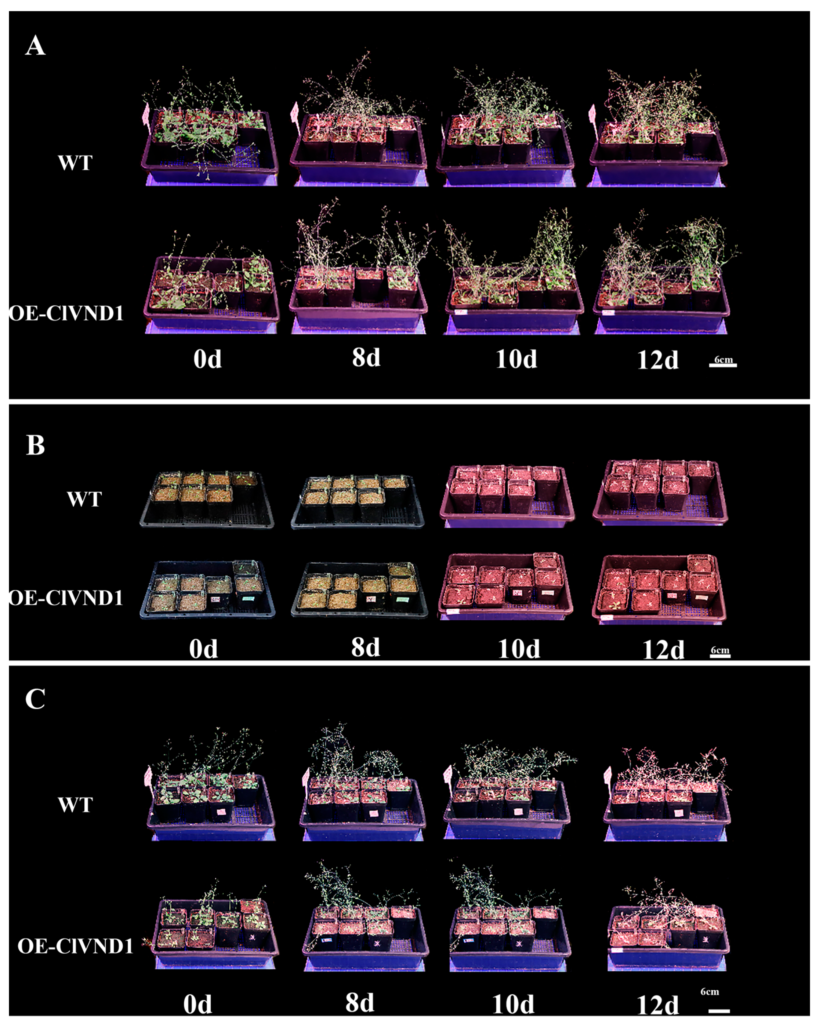
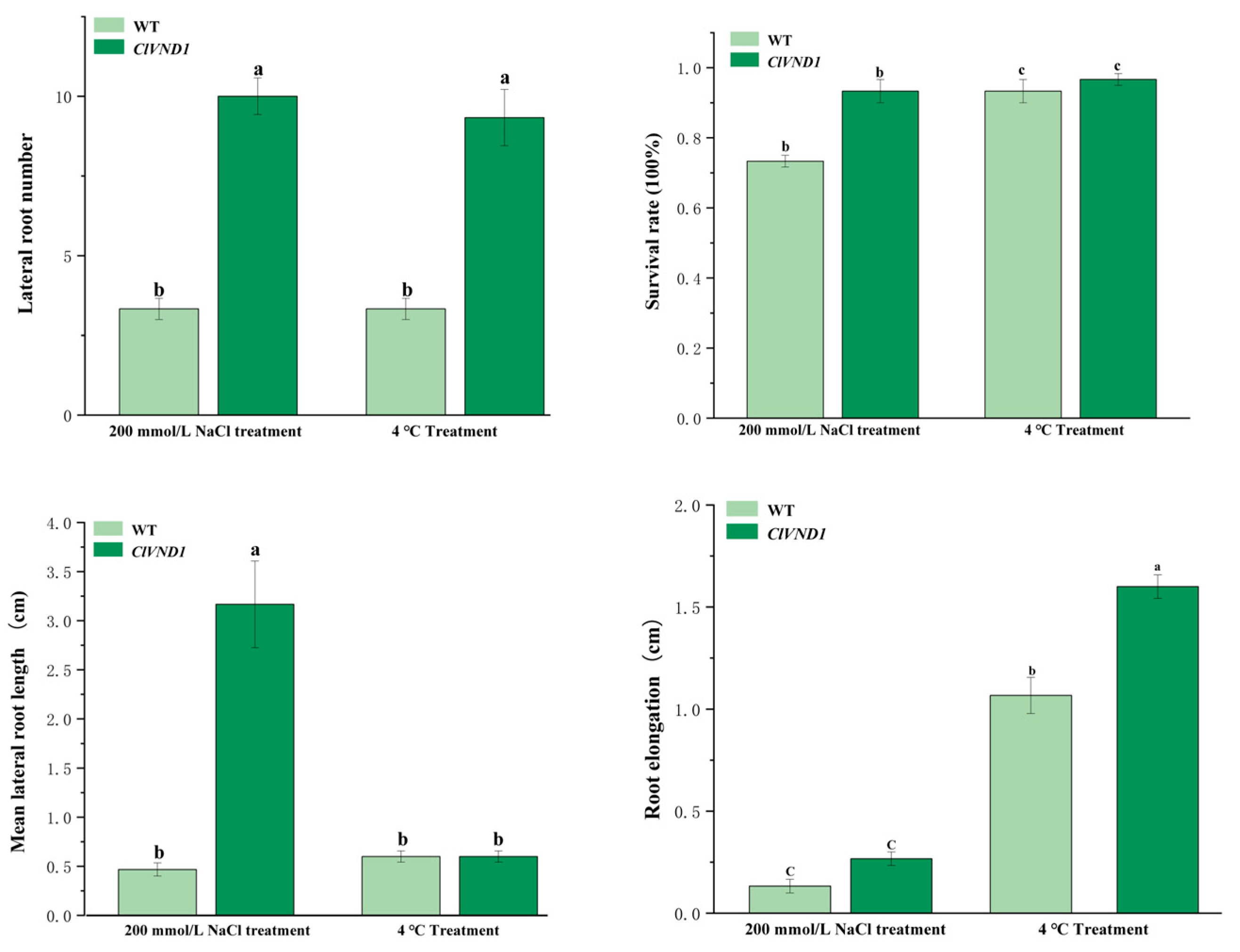
After phenotypic comparison between experimental and control groups, quantitative metrics were collected and subjected to statistical analysis (Figure 6). Under NaCl stress, ClVND1-overexpressing Arabidopsis thaliana exhibited enhanced lateral root development, showing significantly greater lateral root number and length compared to wild-type controls. Transgenic plants produced an average of 10.0 lateral roots versus 3.33 in wild-type plants (p < 0.05). The lateral root length in transgenic lines was approximately 100% greater than that of wild-type specimens, and their survival rate was 27% higher under stress conditions. Under 4 °C treatment, ClVND1-overexpressing transgenic Arabidopsis thaliana showed a pronounced increase in lateral root production relative to wild-type controls. The average lateral root length in transgenic plants was 13% of that in wild-type specimens, while lateral root extension reached only 50% of the wild-type level. Furthermore, the survival rate of transgenic lines was 4% of that observed in wild-type plants (Figure 7). The results demonstrated that ClVND1-overexpressing Arabidopsis thaliana exhibited enhanced lateral root growth under stress conditions compared to wild-type plants, indicating that the ClVND1 gene from Chrysanthemum lavandulifolium plays a crucial regulatory role in root development under abiotic stress.

2.5. Observation of the Growth and Development of Transgenic A. thaliana

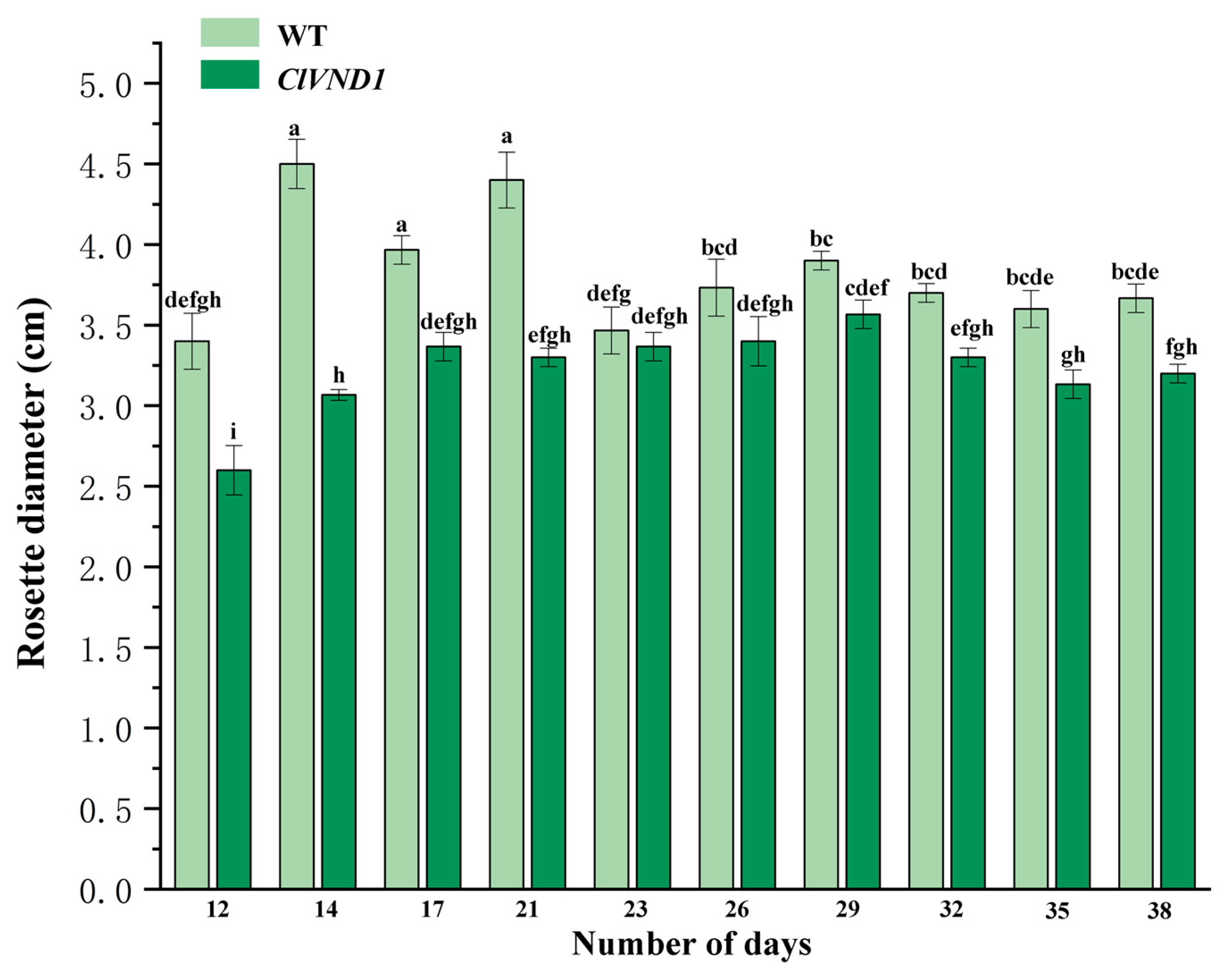
We transplanted transgenic and wild-type A. thaliana seedlings into 7 cm × 7 cm pots after they had developed two true leaves. The rosette diameter was measured and statistically analyzed, with the results presented in Figure 8. The results indicated that during the early growth stage, wild-type Arabidopsis thaliana exhibited a larger rosette diameter compared to transgenic lines, suggesting that the vegetative growth of the transgenic Arabidopsis thaliana was inhibited.
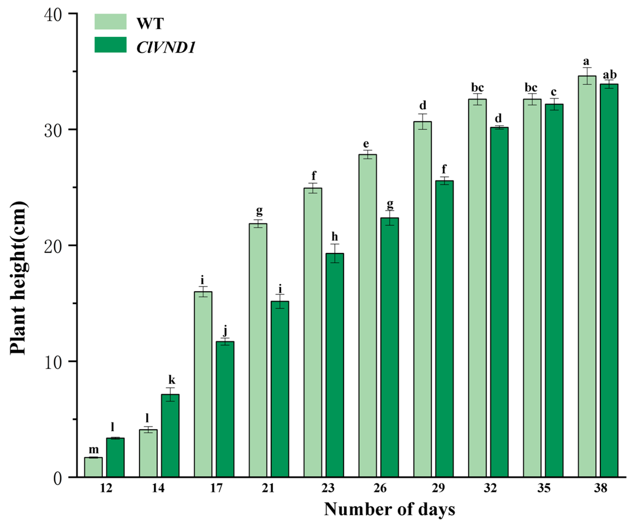
Analysis of the statistical results of Arabidopsis thaliana height (Figure 9) revealed that with the growth of Arabidopsis thaliana inflorescences, the elongation rate and height of wild-type Arabidopsis thaliana inflorescences were higher than those of transgenic Arabidopsis thaliana, indicating that the growth of transgenic Arabidopsis thaliana inflorescences was also inhibited. Before the 17th day, the plant height of the transgenic Arabidopsis thaliana was higher than that of the wild-type Arabidopsis thaliana. After the 17th day, the plant heights of both wild-type and transgenic plants showed an upward trend. However, the plant heights of transgenic Arabidopsis thaliana were all lower than those of wild-type plants, indicating that the growth and development of transgenic Arabidopsis thaliana were restricted. However, the flowering period of the transgenic materials was earlier than that of the control group. Wild-type Arabidopsis thaliana began to produce inflorescences 9 to 12 days after transplantation and started to flower on the 12th day. Arabidopsis thaliana transformed with the ClVND1 gene began to produce inflorescences 7 to 12 days after transplantation and started to flower on the 10th day. Based on the above experimental results, it is concluded that ClVND1 resists salt stress and low temperature stress by regulating the root system, while inhibiting the growth of inflorescences and promoting flowering.

2.6. Paraffin Section Analysis of Stem Segments in Wild-Type and Transgenic Arabidopsis thaliana

Paraffin sections of entire stem segments (3–4 cm in length) from 1300::ClVND1::GFP and 1300::GFP transgenic A. thaliana were prepared and examined. As shown in Figure 10, paraffin sections of stem tissues from 1300::ClVND1::GFP and 1300::GFP (control) transgenic A. thaliana were examined. Morphometric analysis revealed a significant increase in secondary cell wall thickness in ClVND1-overexpressing plants compared to the control. The mean secondary cell wall thickness was 11.631 μm in 1300::ClVND1::GFP transgenic lines versus 4.85 μm in 1300::GFP control lines, demonstrating that ClVND1 overexpression enhances secondary cell wall deposition (Figure 10).

3. Discussion

As an important ornamental flower, the exploration of drought-resistant related genes and the selection and breeding of low-temperature and salt-tolerant Chrysanthemum lavandulifolium through molecular biological methods provide new approaches for the growth and development of modern Chrysanthemums and molecular breeding for stress regulation. NAC family transcription factors have been demonstrated to confer broad-spectrum stress resistance in plants. The research results demonstrate that the ClVND1 gene enhances plant tolerance to both salt and cold stress. This finding aligns with previous reports on other members of the NAC transcription factor subfamily. The OsNAC7 subfamily is one of the most extensively studied subfamilies within the NAC transcription factor family, and includes key members such as SND1, NST1, URP7, BRN1/2 and VND1VND7. Previous studies have demonstrated that the ClSND1-like gene contributes to lignin biosynthesis in Chrysanthemum lavandulifolium and participates in the regulation of plant growth, salt tolerance, and osmotic stress response [35]. ClBRN1 overexpression significantly thickens secondary cell walls in Arabidopsis thaliana, thereby enhancing plant mechanical strength and stress resistance [36]. The ClNUM1 gene plays a role in the synthesis of secondary cell walls, thereby enhancing the resistance of Nicotiana benthamiana [37]. These findings are consistent with the aforementioned study. Furthermore, it was observed that transgenic lines exhibited a higher average number of lateral roots along with an earlier flowering time compared to wild-type plants. In 2019, Yang et al. employed an Arabidopsis-based overexpression vector to functionally characterize the soybean NAC gene GmNAC109 [38]. Sequence analysis revealed that GmNAC109 shares high homology with ATAF1, a regulator of plant responses to both biotic and abiotic stresses. Overexpression of GmNAC109 enhances abiotic stress tolerance and promotes lateral root formation. Furthermore, transgenic plants exhibited earlier flowering compared to wild-type plants. Zhang et al. overexpressed the CcNAC1 gene in jute (Corchorus capsularis) to investigate its biological function [39]. The relative expression level of the 3-ketoacyl-CoA synthase (KCS) gene was significantly up-regulated in transgenic plants, which also exhibited earlier flowering compared to wild-type controls [40]. This finding aligns with the results of the present study. Olsen et al. identified the NAC-preferred cis-elements CGT[GA] and their extensions TTNCGTA and TTGCGTGT, and revealed that NAC proteins bind these motifs as dimers. This discovery establishes the molecular foundation for elucidating ClVND1’s DNA-binding specificity [41].
Paraffin sections were made from stem segments of transgenic and wild-type Arabidopsis thaliana for observation, and it was found that the thickness of secondary cell walls in the stems of transgenic Arabidopsis thaliana with ClVND1 gene was significantly greater than that of the control group. Studies have shown that the eight genes (PTVNS01-08) homologous to VND in poplar and Arabidopsis thaliana play a significant role in xylem development and secondary wall formation. PtVNS7/PtrWND6 and PtVNS8/PtrWND6B, similar to Arabidopsis VND7, exert primary regulatory functions and play a significant role in vessel differentiation and secondary wall thickening [42,43]. It is consistent with the above results.
In this study, we cloned the ClVND1 gene from Chrysanthemum lavandulifolium, which encodes a protein of 351 amino acids. Bioinformatic analysis revealed that ClVND1 contains a conserved NAC domain and is predicted to be an unstable, non-transmembrane protein, confirming its classification within the NAC family. Subcellular localization indicate that the ClVND1 protein is localized to both the nucleus and the cell membrane. Although ClVND1 is predicted to be devoid of transmembrane helices, its transient expression consistently reveals dual localization at the plasma membrane and the nucleus. This pattern parallels the well-documented behaviors of NPR1, OsMADS25, and bZIP28—transcriptional regulators that likewise lack canonical transmembrane domains yet accomplish membrane-to-nucleus shuttling via distinct mechanisms: NPR1 through redox-controlled oligomerization and subsequent disulfide-mediated dissociation [44,45], OsMADS25 via N-terminal myristoylation and palmitoylation [46], and bZIP28 via proteolytic liberation of a soluble, NLS-bearing fragment [47]. Consequently, the nuclear–plasma membrane dual localization of ClVND1 is not anomalous; rather, it exemplifies a conserved paradigm in which non-transmembrane plant transcription factors or signaling proteins exploit lipid modification, redox sensitivity, or proteolytic processing to achieve signal-dependent subcellular relocalization.
ClVND1 is concurrently induced by low temperature and salt stress in Chrysanthemum lavandulifolium, positioning it as a pivotal candidate for enhancing stress tolerance in this species.

4. Materials and Methods

4.1. Experimental Materials

Aseptic seedlings of Chrysanthemum lavandulifolium were provided by the College of Landscape Architecture and Forestry at Qingdao Agricultural University and cultured on 1/2MS medium [36]. Genetic transformation of ClVND1 was conducted in Arabidopsis thaliana [36], and its subcellular localization was analyzed using Nicotiana benthamiana [36]. All plants were grown in an artificial climate chamber at Qingdao Agricultural University (Qingdao, China) under controlled conditions: temperature of 25 °C, relative humidity of 60%, light intensity of 2500 lx, and a photoperiod of 16 h light/8 h dark.

4.2. Test Method

4.2.1. Target Gene Fragment

Leaves of C. lavandulifolium with good growth were selected for total RNA extraction. The total RNA extraction kit used was the FastPure® Universal Plant Total RNA Isolation Kit (Vazyme, Nanjing, China). The reverse transcription kit used was the HiScript III 1st Strand cDNA Synthesis Kit (+gDNA wiper) (Vazyme, Nanjing, China). The full-length coding sequence of the target gene was designed according to the Chrysanthemum nankingense genome data (Table 1). The full-length coding sequence of the target gene was obtained by high-fidelity DNA polymerase 2 × Phanta Flash Master Mix (Dye Plus) (Vazyme, Nanjing, China). The PCR thermal cycler adopts the T Series Multi-Block Thermal Cycle (LonGene®, Hangzhou, China). The PCR amplification system was 25 µL and the reaction system was as follows: 2 × Phanta Max Master Mix: 12.5 µL; ClVND1-F: 1 µL; ClVND1-R: 1 µL; cDNA: 2 µL; ddH2O: 8.5 µL. The reaction procedure was predenaturation at 95 °C for 3 min. Denaturation at 95 °C for 15 s, annealing at 56 °C for 15 s, extension at 72 °C for 60 s, 35 cycles; Extend at 72 °C for 5 min (LonGene®PCR thermal cycler, Hangzhou, China). The PCR products were separated by agarose gel electrophoresis. the PCR products were recovered by bands with the same length as the target gene and sent to Sangong BioEngineering (Shanghai, China) Co., Ltd. for sequencing.

4.2.2. Bioinformatics Analysis

Based on the amino acid sequence of ClVND1, amino acid sequence multiple alignment was performed using MEGA11.0.13, and its sequence was analyzed in detail. DNAMAN version 8.0 (Lynnon Biosoft, Quebec, QC, Canada) was used to align the sequences.

4.2.3. Observation of the Subcellular Localization Signal

The Super1300::ClVND1::GFP overexpression vector was subsequently transformed into N. benthamiana by the Agrobacterium-mediated floral dip method, Select N. benthamiana leaves that have been infected for 4 weeks and place them in the dark for 2 to 3 days. The GFP signals in the lower epidermis of N. benthamiana leaves were observed by fluorescence microscope (Leica DM 2500, Wetzlar, Germany) and laser confocal microscope (Leica TCS SP5, Wetzlar, Germany). The overexpressed Super 1300-GFP were all retained in the laboratory [36].

4.2.4. Construction of the Super1300 Overexpression Vector

The design of primers containing restriction sites is shown in Table 1.
According to the CDS region of the ClVND1 gene and the multiple cloning sites of pSuper1300::GFP vector, primers containing PstI endonuclease and KpnI endonuclease restriction sites and without a termination codon were designed. The target fragment containing restriction site was obtained by PCR reaction and agarose gel recovery. The target fragment containing the restriction site and pSuper1300::GFP vector were double digested (37 °C, 2 h) with endonucleases PstI and KpnI (NEB, Beijing, China) to obtain the target fragment and linear vector with sticky ends. The recombinant plasmid was transformed into E. coli competent DH5α (WEIDI, Shanghai, China) and spread on a solid LB medium containing 50 mg/L kanamycin for resistance screening and identified by colony PCR. The resulting positive plaque was sent to a sequencing company for sequencing and plasmid recovery. The constructed pSuper1300::ClVND1::GFP expression vector was transformed into Agrobacterium tumefaciens GV3101 (Shanghai, Weidi Biology, Shanghai, China) by freeze-thawing and spread on the LB solid medium with 3 antibiotics (50 mg/L kanamycin, 50 mg/L gentamicin, 50 mg/L rifampicin) for resistance screening. The positive colonies was identified by colony PCR and transferred to the liquid LB culture of 3-antibiotics for propagation. After these positive strains were sent for sequencing in Sangong BioEngineering (Shanghai, China) Co., Ltd.

4.2.5. Identification of Transgenic Plants

To identify transgenic positive plants, we extracted total DNA from the leaves of healthy wild-type (WT) and transgenic Arabidopsis lines, which served as templates for PCR amplification using the 1300-F/1300-R primer pair. The wild-type plant was used as a negative control, allowing molecular identification of transgenic plants at the DNA level. Total DNA was extracted from healthy, well-growing A. thaliana leaves using the FlaPure Plant DNA Extraction Kit (Gensand Biotech, Beijing, China). The target gene was obtained by high-fidelity DNA polymerase 2 × Phanta Flash Master Mix (Dye Plus) (Vazyme, Nanjing, China). The PCR thermal cycler adopts the T Series Multi-Block Thermal Cycle (LonGene®, Hangzhou, China). The PCR amplification system was 25 µL and the reaction system was as follows: 2 × Phanta Max Master Mix: 12.5 µL; 1300-F: 1 µL; 1300-R: 1 µL; cDNA: 2 µL; ddH2O: 8.5 µL. The reaction procedure was predenaturation at 95 °C for 3 min. Denaturation at 95 °C for 15 s, annealing at 58.2 °C for 15 s, extension at 72 °C for 60 s, 35 cycles; Extend at 72 °C for 5 min (LonGene®PCR thermal cycler, Hangzhou, China). DNA fragments were separated by agarose gel electrophoresis and visualized using a DNA marker (GenStar, Beijing, China).

4.2.6. Quantitative Real-Time PCR Assay

RT-qPCR analysis was conducted on wild-type and transgenic plants. Tissue samples of A. thaliana were flash-frozen in liquid nitrogen, homogenized into a fine powder, and transferred to RNase-free 2 mL centrifuge tubes. Total RNA was extracted from healthy, well-growing A. thaliana leaves using the FastPure® Universal Plant Total RNA Isolation Kit (Vazyme, Nanjing, China). Subsequently, cDNA was synthesized from the extracted RNA using the HiScript III 1st Strand cDNA Synthesis Kit (+gDNA wiper) (Vazyme, Nanjing, China). RT-qPCR was performed on a StepONE Plus system (Applied Biosystems, Waltham, MA, USA) using TB Green® Premix Ex TaqTM II (Takara, Shiga Prefecture, Japan) and specific primers (Table 2). Three biological replicates were performed for each reaction, and the results were analyzed using the 2−ΔΔCT method. Data were visualized and statistically analyzed for significance using SPSS 27 and Origin 2022 software.

4.2.7. Transformation and Screening of A. thaliana

The Super1300::ClVND1::GFP overexpression vector was transformed into A. thaliana by the Agrobacterium-mediated floral dip method [48], after which the transgenic A. thaliana seeds were screened by a resistance screening medium (1/2MS + 30 g/L sucrose + 6 g/L agarose + 50 mg/L hygromycin) [36].

4.2.8. Stress Treatment and Phenotypic Observation

Abiotic stress treatment was carried out during the seedling stage of transgenic Arabidopsis thaliana, and 1/2MS medium was selected for subsequent experiments [36]. The transgenic Arabidopsis thaliana plants were subjected to salt stress at 4 °C and 200 mmol/L, and wild-type Arabidopsis thaliana grown under normal conditions was used as the control. Three days after processing, photos of Arabidopsis thaliana were taken and data were recorded.
Plants of similar growth vigor were selected from each treatment of Super1300::GFP and Super1300::ClVND1 lines for stress treatment. The salt treatment was conducted by applying a 200 mmol/L NaCl solution. Low temperature treatment using a low temperature incubator was performed at a temperature of ±4 °C with light for 12 h. The two treatments were cultured at ±25 °C, and a normal water supply was used for the control (CK). After 3 days, the transgenic plants of A. thaliana were photographed, and related data were collected.

4.2.9. Observation of the Growth and Development of Transgenic A. thaliana

Transgenic A. thaliana and the control group were transferred into a 7 cm × 7 cm flowerpot when two true leaves had grown. Under consistent growth conditions, rosette diameter, plant height, and flowering time were measured for both groups.

4.2.10. Paraffin Sectioning

Stem segments from the upper one-third of Arabidopsis thaliana were collected and fixed in FAA solution (70% alcohol:formalin:acetic acid 18:1:1) for 24 h. The samples were then softened in a 1:1 mixture of hydrogen peroxide and glacial acetic acid for 48 h. Subsequent processing included dehydration through an ethanol series, paraffin embedding, and sectioning at 10 μm thickness using a microtome. Finally, the samples were stained with safranine green. For each experiment, three biological replicates were prepared. The cell wall thickness of ten randomly selected cells per view was measured, and statistical analysis was performed.

4.2.11. Data Analysis

Three biological replicates and technical replicates were performed for treatments in this study. Excel2010 was used to organize the data, SPSS27 was used for statistical analysis, one-way ANOVA and least significant difference method were used to analyze the data (p < 0.05), and Origin2022 software was used to plot the data.

5. Conclusions

In summary, ClVND1 was cloned from C. lavandulifolium and identified as a gene of the OSNAC7 subfamily which encodes a nuclear protein and a cell membrane protein. This study found that the ClVND1 gene can regulate the growth and development of A. thaliana as well as improve its stress tolerance, shown by the enhanced tolerance of salt and low temperature, and increase the thickness of the secondary woody cell wall of its stem.

Author Contributions

Y.L. (Yueyue Liu): Conceived and designed the experiments, performed the experiments, analyzed the data, prepared figures and/or tables, and approved the final draft; C.M.: Conceived and designed the experiments, performed the experiments, analyzed the data, prepared figures and/or tables, and approved the final draft; H.Z.: Analyzed the data, prepared figures and/or tables, and approved the final draft.; Y.L. (Ying Liao): Analyzed the data, prepared figures and/or tables, and approved the final draft; Y.Z.: Analyzed the data, prepared figures and/or tables, and ap-proved the final draft; X.S.: Conceived and designed the experiments, authored or reviewed drafts of the article, and approved the final draft; H.W.: Conceived and designed the experiments, authored or reviewed drafts of the article, and approved the final draft. All authors have read and agreed to the published version of the manuscript.

Funding

This research was funded by National Natural Science Foundation of China (32101580); Qingdao Agricultural University PhD Start-up Fund (Grant Numbers: 6631120091, 6631122019); Herbaceous Germplasm Resources Survey and Collection Project of Forestry, Grassland Germplasm Resources Center of Shandong Province (6602423134) and Shandong Province Higher Education Institutions' "Youth Innovation Team Plan" (2022KJ166).

Data Availability Statement

The datasets generated during and/or analysed during the current study are available from the corresponding author on reasonable request.

Acknowledgments

We thank for Hai Wang and Xuebin Song of the Shandong Key Laboratory for Germplasm Innovation of Saline-alkaline Tolerant Grasses and Trees, Qingdao Agricultural University, for providing for help.

Conflicts of Interest

The authors have no relevant financial or non-financial interests to disclose.

Appendix A

Figure A1. Cloning and analysis of CDS sequences of the ClVND1 gene.
Plants 14 02925 g0a1

Appendix B

Figure A2. Bioinformatic Characterization of the ClVND1 gene in Chrysanthemum lavandulifolium. (a) Analysis of transmembrane domain architecture. (b) Evaluation of hydrophilic and hydrophobic characteristics.
Plants 14 02925 g0a2

References

  1. Sun, L.J.; Li, D.Y.; Zhang, H.J.; Song, F.M. The role of NAC transcription factors in Plant disease resistance and abiotic stress resistance. Genetics 2012, 34, 993–1002. [Google Scholar]
  2. Abe, H.; Yamaguchi-Shinozaki, K.; Urao, T.; Iwasaki, T.; Hosokawa, D.; Shinozaki, K. Role of Arabidopsis MYC and MYB homologs in drought- and abscisic acid-regulated gene expression. Plant Cell 1997, 9, 1859–1868. [Google Scholar]
  3. Mitsuda, N.; Seki, M.; Shinozaki, K.; Ohme-Takagi, M. The NAC transcription factors NST1 and NST2 of Arabidopsis regulate secondary wall thickenings and are required for anther dehiscence. Plant Cell 2005, 17, 2993–3006. [Google Scholar] [CrossRef]
  4. Zhong, R.Q.; Demura, T.; Ye, Z.-H. SND1, a NAC domain transcription factor, is a key regulator of secondary wall synthesis in fibers of Arabidopsis. Plant Cell 2006, 18, 3158–3170. [Google Scholar] [CrossRef]
  5. Ko, J.H.; Yang, S.H.; Park, A.H.; Lerouxel, O.; Han, K.H. AtNAC012, a member of the plant-specific NAC transcription factor family, negatively regulates xylary fiber development in Arabidopsis thaliana. Plant J. 2007, 50, 1035–1048. [Google Scholar] [CrossRef]
  6. Mitsuda, N.; Iwase, A.; Yamamoto, H.; Yoshida, M.; Seki, M.; Shinozaki, K.; Ohme-Takagi, M. NAC transcription factors, NST1 and NST3, are key regulators of the formation of secondary walls in woody tissues of Arabidopsis. Plant Cell 2007, 19, 270–280. [Google Scholar] [CrossRef]
  7. Soyano, T.; Thitamadee, S.; Machida, Y.; Chua, N.H. ASYMMETRIC LEAVES2-LIKE19/LATERAL ORGAN BOUNDARIES DOMAIN30 and ASL20/LBD18 regulate tracheary element differentiation in Arabidopsis. Plant Cell 2018, 20, 3359–3373. [Google Scholar] [CrossRef]
  8. Bennett, T.; Van Den Toorn, A.; Sanchez-Perez, G.F.; Campilho, A.; Willemsen, V.; Snel, B.; Scheres, B. SOMBRERO, BEARSKIN1, and BEARSKIN2 regulate root cap maturation in Arabidopsis. Plant Cell 2010, 22, 640–654. [Google Scholar] [CrossRef]
  9. Yamaguchi, M.; Ohtani, M.; Mitsuda, N.; Kubo, M.; Ohme-Takagi, M.; Fukuda, H.; Demura, T. VND-INTERACTING2, a NAC domain transcription factor, negatively regulates xylem vessel formation in Arabidopsis. Plant Cell 2010, 22, 1249–1263. [Google Scholar] [CrossRef]
  10. Fendrych, M.; Van Hautegem, T.; Van Durme, M.; Olvera-Carrillo, Y.; Huysmans, M.; Karimi, M.; Lippens, S.; Guérin, C.J.; Krebs, M.; Schumacher, K.; et al. Programmed cell death controlled by AtNAC033/SOMBRERO determines root cap organ size in Arabidopsis. Curr. Biol. 2014, 24, 931–940. [Google Scholar] [CrossRef]
  11. Zhou, J.L.; Zhong, R.Q.; Ye, Z.H. Arabidopsis NAC domain proteins, VND1 to VND5, are transcriptional regulators of secondary wall biosynthesis in vessels. PLoS ONE 2014, 9, e105726. [Google Scholar] [CrossRef]
  12. Endo, H.; Yamaguchi, M.; Tamura, T.; Nakano, Y.; Nishikubo, N.; Yoneda, A.; Kato, K.; Kubo, M.; Kajita, S.; Katayama, Y.; et al. Multiple classes of transcription factors regulate the expression of VASCULAR-RELATED NAC-DOMAIN7, a master switch of xylem vessel differentiation. Plant Cell Physiol. 2015, 56, 242–254. [Google Scholar] [CrossRef]
  13. Zhong, R.Q.; Ye, Z.H. The Arabidopsis NAC transcription factor NST2 functions together with SND1 and NST1 to regulate secondary wall biosynthesis in fibers of inflorescence stems. Plant Signal. Behav. 2015, 10, e989746. [Google Scholar] [CrossRef]
  14. Fang, S.; Shang, X.G.; Yao, Y.; Li, W.X.; Guo, W.Z. NST- and SND-subgroup NAC proteins coordinately act to regulate secondary cell wall formation in cotton. Plant Sci. 2012, 301, 110657. [Google Scholar] [CrossRef]
  15. Sun, L.; Liu, L.P.; Wang, Y.Z.; Yang, L.; Wang, M.J.; Liu, J.X. NAC103, a NAC family transcription factor, regulates ABA response during seed germination and seedling growth in Arabidopsis. Planta 2020, 252, 95. [Google Scholar] [CrossRef]
  16. Yang, J.H.; Lee, K.H.; Du, Q.; Yang, S.; Yuan, B.J.; Qi, L.Y.; Wang, H.Z. A membrane-associated NAC domain transcription factor XVP interacts with TDIF co-receptor and regulates vascular meristem activity. New Phytol. 2020, 226, 59–74. [Google Scholar] [CrossRef]
  17. Wen, J.; Wang, C.T.; Yang, Y.P. Research progress on the Regulation of Secondary Cell Wall Thickening in the xylem of plants. J. Southwest For. Univ. (Nat. Sci.) 2021, 41, 182–188. [Google Scholar]
  18. Zhong, R.Q.; Ye, Z.H. Complexity of the transcriptional network controlling secondary wall biosynthesis. Plant Sci. 2014, 229, 193–207. [Google Scholar] [CrossRef]
  19. Chen, X.; Lu, S.C.; Wang, Y.F.; Zhang, X.; Lv, B.; Luo, L.Q.; Xi, D.D.; Shen, J.B.; Ma, H.; Ming, F. OsNAC2 encoding a NAC transcription factor that affects plant height through mediating the gibberellic acid pathway in rice. Plant J. 2015, 82, 302–314. [Google Scholar] [CrossRef]
  20. Dong, F.; Huang, H.; Liu, J.; Zhang, M.; Zhou, Y.; Dai, S. Cloning and function analysis of ClNAC9 from Chrysanthemum lavandulifolium. Can. J. Plant Sci. 2018, 98, 1265–1279. [Google Scholar] [CrossRef]
  21. Liu, Q.; Zhang, G.Y.; Chen, S.Y. Structure and regulation of plant transcription factors. Chin. Sci. Bull. 2020, 45, 1465–1474. [Google Scholar]
  22. Huang, T.L.; Zhang, R.; He, Y.X.; Yang, R.Z.; Song, W.W.; Lai, Z.X.; Li, N.; Liu, S.C. Identification of NAC family members and expression analysis of their coding genes under NaCl stress in pepper. J. Plant Resour. Environ. 2023, 32, 12–24. [Google Scholar]
  23. Zhang, Y.L.; Zhang, C.; Chen, X.X.; Liu, A.X.; Sun, L.J. Identification and functional prediction of tomato NAC family genes. Anhui Agric. Sci. 2014, 20, 6549–6552+6561. [Google Scholar]
  24. Zhou, J.J.; Fang, Z.J.; Han, Q.J.; Pan, J.L.; Lian, Y.Y.; Gu, H.Y.; Sun, L.J. Identification and functional prediction of NAC family genes in maize. Genom. Appl. Biol. 2017, 36, 16. [Google Scholar]
  25. Sun, X.; Shangguan, L.F.; Fang, J.G.; Song, C.N.; Wang, C.; Mu, X. Bioinformatics analysis of NAC transcription factor family in grape. Genom. Appl. Biol. 2011, 30, 14. [Google Scholar]
  26. Wang, Y.; Bai, X. Bioinformatics analysis of soybean NAC gene family. Soybean Sci. 2014, 33, 325–333. [Google Scholar]
  27. He, L.; Bian, J.; Xu, J.Y.; Yang, K.J. Novel maize NAC transcriptional repressor ZmNAC071 confers enhanced sensitivity to ABA and osmotic stress by downregulating stress-responsive genes in transgenic Arabidopsis. J. Agric. Food Chem. 2019, 67, 890–8918. [Google Scholar] [CrossRef]
  28. Duan, A.Q.; Yang, X.L.; Feng, K.; Liu, J.X.; Xu, Z.S.; Xiong, A.S. Genome-wide analysis of NAC transcription factors and their response to abiotic stress in celery (Apium graveolens L.). Comput. Biol. Chem. 2020, 84, 107186. [Google Scholar] [CrossRef]
  29. Tamura, T.; Endo, H.; Suzuki, A.; Sato, Y.; Kato, K.; Ohtani, M.; Yamaguchi, M.; Demura, T. Affinity-based high-resolution analysis of DNA binding by VASCULAR-RELATED NAC-DOMAIN7 via fluorescence correlation spectroscopy. Plant J. Cell Mol. Biol. 2019, 100, 298–313. [Google Scholar] [CrossRef]
  30. Tan, T.T.; Endo, H.; Sano, R.; Kurata, T.; Yamaguchi, M.; Ohtani, M.; Demura, T. Transcription factors VND1-VND3 contribute to cotyledon xylem vessel formation. Plant Physiol. 2018, 176, 773–789. [Google Scholar] [CrossRef]
  31. Negi, S.; Tak, H.; Ganapathi, T.R. Xylem specific activation of 5′ upstream regulatory region of two NAC transcription factors (MusaVND6 and MusaVND7) in banana is regulated by SNBE-like sites. PLoS ONE 2018, 13, e0192852. [Google Scholar] [CrossRef]
  32. Negi, S.; Tak, H.; Ganapathi, T.R. Cloning and functional characterization of MusaVND1 using transgenic banana plants. Transgenic Res. 2015, 24, 571–585. [Google Scholar] [CrossRef]
  33. Wang, H.; Li, T.; Li, W.; Wang, W.; Zhao, H. Identification and analysis of Chrysanthemum nankingense NAC transcription factors and an expression analysis of OsNAC7 subfamily members. PeerJ 2021, 9, e11505. [Google Scholar] [CrossRef]
  34. Deng, Z.Y.; Luo, L.; Yu, C.; Zhang, Q.X.; Sui, Y.J. The research progress of ornamental plant NAC transcription factor. J. Plant Genet. Resour. 2024, 25, 737–750. [Google Scholar]
  35. Wang, H.; Li, T.; Zhou, Y.Y.; Wang, W.; Zhao, H.E. Cloning and expression analysis of Chrysanthemum lavandulifolium lignin-regulated Transcription factor ClSND1-like. Northwest Acta Bot. Sin. 2021, 41, 359–367. [Google Scholar]
  36. Li, Y.X.; He, W.T.; Liu, Y.Y.; Mei, C.D.; Wang, H.; Song, X.B. ClBRN1 from Chrysanthemum lavandulifolium enhances the stress resistance of transgenic Arabidopsis. PeerJ 2024, 12, e18620. [Google Scholar] [CrossRef]
  37. Zhang, W.X.; Wang, H.; Guo, Y.N.; Hao, X.Y.; Li, Y.X.; He, W.T.; Zhao, X.; Cai, S.Y.; Song, X.B. Functional Validation of Different Alternative Splicing Variants of the Chrysanthemum lavandulifolium ClNUM1 Gene in Tobacco. Curr. Issues Mol. Biol. 2024, 46, 5242–5256. [Google Scholar] [CrossRef]
  38. Yang, X.F.; Kim, M.Y.; Ha, J.M.; Lee, S.H. Overexpression of the Soybean NAC Gene GmNAC109 Increases Lateral Root Formation and Abiotic Stress Tolerance in Transgenic Arabidopsis Plants. Front. Plant Sci. 2019, 10, 1036. [Google Scholar] [CrossRef]
  39. Zhang, G.Y.; Huang, S.Q.; Zhang, C.; Li, D.F.; Wu, Y.B.; Deng, J.L.; Shan, S.L.; Qi, J.M. Overexpression of CcNAC1 gene promotes early flowering and enhances drought tolerance of jute (Corchorus capsularis L.). Protoplasma 2020, 258, 337–345. [Google Scholar] [CrossRef]
  40. Zhou, S.Z.; Luo, T.; Liu, L. Bioinformatics analysis of NAC gene family in moss. Genom. Appl. Biol. 2022, 41, 822–833. [Google Scholar]
  41. Olsen, A.N.; Ernst, H.A.; Leggio, L.L.; Skriver, K. DNA-binding specificity and molecular functions of NAC transcription factors. Plant Sci. 2005, 169, 785–797. [Google Scholar] [CrossRef]
  42. Ohtani, M.; Nishikubo, N.; Xu, B.; Yamaguchi, M.; Mitsuda, N.; Goué, N.; Shi, F.; Ohme-Takagi, M.; Demura, T. A NAC domain protein family contributing to the regulation of wood formation in poplar. Plant J. 2011, 67, 499–512. [Google Scholar] [CrossRef]
  43. Takata, N.; Awano, T.; Nakata, M.; Sano, Y.; Sakamoto, S.; Mitsuda, N.; Taniguchi, T. Populus NST/SND orthologs are key regulators of secondary cell wall formation in wood fibers, phloem fibers and xylem ray parenchyma cells. Tree Physiol. 2019, 39, 514–525. [Google Scholar] [CrossRef]
  44. Mou, Z.L.; Fan, W.H.; Dong, X.N. Inducers of Plant Systemic Acquired Resistance Regulate NPR1 Function through Redox Changes. Cell 2003, 113, 935–944. [Google Scholar] [CrossRef]
  45. Shi, Z.; Maximova, S.; Liu, Y.; Verica, J.; Guiltinan, M. Functional analysis of the Theobroma cacao NPR1 gene in arabidopsis. BMC Plant Biol. 2010, 10, 248. [Google Scholar] [CrossRef]
  46. Wu, J.Y.; Yang, S.Q.; Chen, N.N.; Jiang, Q.N.; Huang, L.L.; Qi, J.X.; Xu, G.H.; Shen, L.S.; Yu, H.; Fan, X.R.; et al. Nuclear translocation of OsMADS25 facilitated by OsNAR2.1 in response to nitrate signal promotes root growth by regulating the expression of OsMADS27 and OsARF7 in rice. Plant Commun. 2023, 4, 100642. [Google Scholar] [CrossRef]
  47. Liu, J.X.; Srivastava, R.; Che, P.; Howell, S. Salt stress responses in Arabidopsis utilize a signal transduction pathway related to endoplasmic reticulum stress signaling. Plant J. Cell Mol. Biol. 2007, 51, 897–909. [Google Scholar] [CrossRef]
  48. Qi, W.W.; Gong, X.L.; Wang, Y.; Wu, Y. Research Progress on the Application of Dipping Flower Method in Plant Genetic Transformation. Mod. Agric. Sci. Technol. 2014, 9–10+12. [Google Scholar]
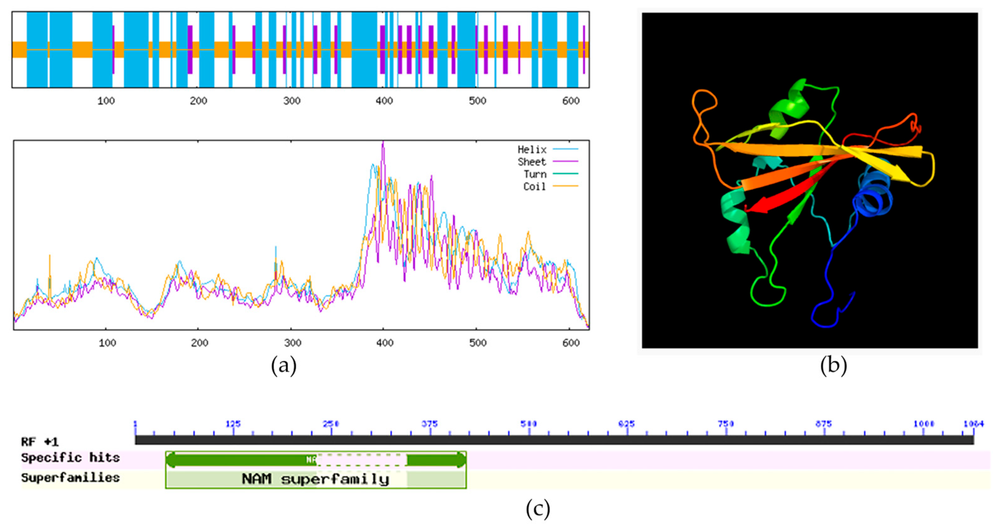
Figure 1. Bioinformatic Characterization of the ClVND1 gene in Chrysanthemum lavandulifolium. (a) Prediction of Protein Secondary Structure. (b) Prediction of Protein Tertiary Structure. The helical structures represent alpha-helices of the protein, the folded forms represent beta-sheets, and the loop regions do not possess a clearly defined regular structure, serving as flexible areas that connect different secondary structural domains. (c) Identification and Analysis of the Conserved Domain in the ClVND1 Gene.
Plants 14 02925 g001
Figure 2. Phylogenetic Analysis of ClVND1 among VNS Proteins from Divergent Taxa.
Plants 14 02925 g002
Figure 3. Analysis of Subcellular Localization in Nicotiana benthamiana. Green fluorescence typically refers to the light emitted by Green Fluorescent Protein (GFP).
Plants 14 02925 g003
Figure 4. (A) Identification of positive transgenic plants by PCR. WT: Wild-type; OE1, OE2, OE3: Transgenic lines. “M” denotes the super DNA marker. (B) Real-time PCR. WT: Wild-type; OE1, OE2, OE3: Transgenic lines. The different letters are significantly different (p < 0.05). Bars indicate standard errors (n = 3).
Plants 14 02925 g004
Figure 5. Growth of Arabidopsis thaliana under 200 mmol/L NaCl and 4 °C treatments for 3 days.
Plants 14 02925 g005
Figure 6. Phenotype of Arabidopsis under 4 °C and NaCl treatments. (A) Untreated A. thaliana. (B) A. thaliana under 4 °C low-temperature stress. (C) A. thaliana treated with 200 mmol/L NaCl.
Plants 14 02925 g006
Figure 7. Growth status statistics of A. thaliana at 4 °C and NaCl treatments for 3 days. The different letters are significantly different (p < 0.05). Bars indicate standard errors (n = 3).
Plants 14 02925 g007
Figure 8. Rosette Diameter of Transgenic A. thaliana. The different letters are significantly different (p < 0.05). Bars indicate standard errors (n = 3).
Plants 14 02925 g008
Figure 9. Growth height statistics of transgenic A. thaliana. The different letters are significantly different (p < 0.05). Bars indicate standard errors (n = 3).
Plants 14 02925 g009
Figure 10. Stem Anatomy of 1300::GFP and ClVND1-Transgenic A. thaliana. (A,B) Wild-type A. thaliana stem cross-sections. (C,D) ClVND1-transgenic A. thaliana stem cross-sections.
Plants 14 02925 g010
Table 1. The design of the primer that contains the enzyme tangent site.
Primer NameSequence (5′→3′)TM/°CPurpose
ClVND1-FgcTCTAGAatgatcatggacacagtaga57.29Amplification of the full-length VND1 sequence with restriction sites.
ClVND1-RaaCTGCAGatcaaaaatgcataatcca54.71
pSuper1300-Fgtgacgccatttcgccttttc57.69
pSuper1300-Rggtggtgcagatgaacttcagg58.69
Table 2. Primer sequences used for quantitative real-time PCR (qRT-PCR).
Primer NameSequence (5′→3′)TM/°CPurpose
ClVND1-FGTTTTGGAAGGCTACGGGGA59.96Real-time PCR Primers
ClVND1-RGTTTGTCCCGTTGCACGTTT60.18
Actin8-FTCAGCACTTTCCAGCAGATG55.22
Actin8-RCTGTGGACAATGCCTGGAC56.61
Disclaimer/Publisher’s Note: The statements, opinions and data contained in all publications are solely those of the individual author(s) and contributor(s) and not of MDPI and/or the editor(s). MDPI and/or the editor(s) disclaim responsibility for any injury to people or property resulting from any ideas, methods, instructions or products referred to in the content.