Article

The tae-miR164-TaNAC6A Module from Winter Wheat Could Enhance Cold Tolerance in Transgenic Arabidopsis thaliana

College of Life Science, Northeast Agricultural University, Harbin 150030, China
*
Author to whom correspondence should be addressed.
These authors contributed equally to this work.
Plants 2025, 14(18), 2849; https://doi.org/10.3390/plants14182849
Submission received: 30 July 2025 / Revised: 8 September 2025 / Accepted: 9 September 2025 / Published: 12 September 2025

Abstract

Cold stress impedes the growth and development of wheat (Triticum aestivum) and other crops, ultimately reducing both yields and quality. Research indicates that non-coding RNAs (ncRNAs) play a crucial role in regulating plant stress responses and resistance. In a previous study, we observed that the expression of tae-miR164 was inversely correlated with the expression of TaNAC6A in Dongnongdongmai 1 (Dn1), a winter wheat variety with high cold resistance, under cold-stress conditions. However, the molecular mechanism governing the cold responsiveness of the tae-miR164-TaNAC6A module was not fully understood. We found that tae-miR164 and TaNAC6A were both induced to express in opposite trends, and TaNAC6A was located in the nucleus. We also discovered that the expression of tae-miR164 and its target gene, TaNAC6A, was responsive to short-term freezing stress in transgenic Arabidopsis plants. Compared to wild-type (WT) Arabidopsis plants, OE-tae-miR164 plants showed decreased cold tolerance, whereas those overexpressing TaNAC6A demonstrated increased tolerance. On average, the OE-TaNAC6A and STTM-tea-miR164 plants exhibited fewer morphological abnormalities in response to cold stress, higher antioxidant enzyme activities and gene expression levels, lower levels of reactive oxygen species (ROS) and malondialdehyde (MDA), and higher expressions of AtDREB1, AtDREB2, and AtABI5 in the cold-signaling pathway. Thus, the biological functions of tae-miR164 and TaNAC6A were initially confirmed through heterologous expression strategies, and we have made the first attempt to elucidate its associated tae-miR164-TaNAC6A module of cold resistance. The findings of this research will support further investigations into the regulation of plant stress resistance by ncRNAs and will inform molecular module breeding strategies aimed at enhancing the cold tolerance of crop plants. Molecular module design breeding, as a significant breakthrough in modern biotechnology, is transforming traditional breeding models. Conventional hybrid breeding relies on empirical screening, which is time-consuming and subject to randomness. In contrast, molecular module breeding directly targets key genes and achieves precise regulation through technologies such as gene editing and synthetic biology.

1. Introduction

Temperature is well-known to affect plant growth and development, particularly exposure at low temperatures [1]. Most plants are able to adapt to gradually decreasing temperatures by altering their cold-responsive gene expression to acclimate their physiology and biochemistry [2]. Frost damage occurs at critical temperatures when ice nucleation begins, resulting in structural and membrane damage. Freezing-induced increases in membrane permeability result in solute loss, ionic imbalance, and organelle damage [3]. It is therefore critical to understand the molecular mechanism underlying cold-stress resistance in order to develop cold-tolerant crops.
Adaptation to cold stress entails a complex array of physiological and biochemical mechanisms. In most plants, cold acclimation typically involves the synthesis and accumulation of proline (Pro) and other amino acids, proteins, sugars, and antioxidants [4]. Cold stress disrupts plant photosynthesis and leads to the accumulation of excessive ROS, resulting in lipid peroxidation, increased membrane permeability, cytoplasmic leakage, elevated relative conductivity, and inhibited development [5,6]. In response, plants express antioxidant enzymes, such as superoxide dismutase (SOD), catalase (CAT), ascorbate peroxidase (APX), and peroxidase (POD), to neutralize ROS [7]. For instance, Guo et al. [8] observed that, in both rice varieties LJ25 (low-temperature stress resistance) and LJ11 (low-temperature stress sensitivity), cold stress prompted the accumulation of Pro, soluble protein, glutathione (GSH), SOD, and APX. However, an opposite trend in ROS and malondialdehyde (MDA) was observed in both varieties. This may reflect differences in the antioxidant capacity and membrane lipid peroxidation levels between the two rice varieties when coping with low-temperature stress.
In plants, miRNAs have been identified as playing a core regulatory role in the response to cold stress [9]. The low-temperature responsiveness of miRNAs was first observed in Arabidopsis (Arabidopsis thaliana) [9], and it was subsequently reported in poplar (Populus) [10], rice (Oryza sativa) [11], wheat [12], tomato (Solanum lycopersicum) [13], grape (Vitis vinifera) [14], and cotton (Gossypium hirsutum) [15]. In tomato, the overexpression of Sly-miR319d enhances cold tolerance by inhibiting the expression of GAMYB-like1 [16]. In transgenic Arabidopsis plants, the overexpression of tae-miR399 from wheat inhibited the expression of AtUBC24, which, in turn, inhibited the AtUBC24-mediated degradation of AtICE1, inducing the CBF signaling pathway, activating antioxidant defenses, and enhancing cold tolerance [17]. The highly conserved miR164 has been found to regulate not only plant development but also defense responses to environmental and biological stress signals [18,19]. In tomato, Sly-miR164A regulated the fruit ripening and quality by modulating the expressions of SlNAM2 and SlNAM3. Sly-miR164A was preferentially expressed during late fruit development, thereby regulating the fruit ripening and quality [20]. In wheat seedlings, the tae-miR164-TaNAC14 module regulated root growth and development as well as stress (drought and salt) tolerance. Specifically, TaNAC14 promoted root growth and development and enhanced drought tolerance, while tae-miR164 inhibited root development and decreased drought and salt tolerance by downregulating the expression of TaNAC14 [21].
NAC (NAM, ATAF, and CUC) transcription factors (TFs) are one of the largest TF families in plants and play important roles in their growth, development, and environmental stress response [22]. For instance, the overexpression of CaNAC064 from pepper (Piper nigrum) in transgenic Arabidopsis plants has been shown to reduce the MDA content, cold-injury index, and relative electrolytic leakage while increasing the antioxidant activity in response to cold stress [23]. Similarly, the overexpression of VvNAC17 from grape in transgenic Arabidopsis plants enhanced drought tolerance by upregulating the expressions of abscisic acid (ABA)- and stress-related genes [24]. Lastly, the overexpression of ZmSNAC13 of maize (Zea mays) in transgenic Arabidopsis plants improved the tolerance to both drought and salt stresses. In this case, the primary stress tolerance mechanism was found to involve the ABA pathway and the MAPK signaling cascade [25].
Wheat is one of the most important cereal crops. Notably, Dongnongdongmai 1 (Dn1), a hexaploid (2n = 6x = 42, AABBDD) wheat, was reported to be the first winter wheat variety capable of safely overwintering in Heilongjiang, China, exhibiting a greening rate of more than 85% [26]. This remarkable attribute makes Dn1 a promising research candidate to understand the molecular mechanism of cold-stress tolerance in wheat. In a previous study, we observed that the expression of tae-miR164 in Dn1 was negatively correlated with the expression of TaNAC6A (TraesCS5B02G054200, loc: chr5B (-) 59,165,237 -59170277) in the field (−25 °C). However, the regulatory mechanism underlying the tae-miR164-TaNAC6A module remained unclear. Therefore, we sought to reveal this molecular mechanism by characterizing the biological functions of the tae-miR164-TaNAC6A module in regulating cold resistance. This study clarifies the key molecular modules of cold resistance in Triticum aestivum and their mechanisms of action, promotes the exchange of superior cold-resistant genetic resources, facilitates the cultivation of cold-resistant crop varieties, breaks through breeding bottlenecks, and provides new impetus for the development of the Triticum aestivum seed industry.

2. Results

2.1. Analysis of Conserved Motifs and Gene Structures of tae-miR164 and TaNAC6A

Using RNAfold to predict the secondary structure, we found that pre-tae-miR164 exhibited a complex but stable stem–ring structure. According to WebLogo analysis, the mature miR164 sequence is highly conserved. In order to verify whether tae-miR164 regulates the cold-stress response, several cis-acting elements within the pre-tae-miR164 promoter were identified using PlantCare, namely, LRE, Me-JAR, GARE, LTR, ABRE, and an enhancer (Figure S1).
TaNAC6A consists of 306 amino acids, with a relative molecular weight of 33.50 kDa. TaNAC6A contains a typical conserved NAM domain and is predicted to be localized to the nucleus. The ProTaNAC6A promoter contains a variety of cis-acting elements, including LTR, SARE, ABRE, and DRE, among others (Figure S2).
Bioinformatics predictions consistently indicate that the transcription of tae-miR164 and TaNAC6A is induced by low temperature.

2.2. Expressions of tae-miR164 and TaNAC6A of Dn1 Under Cold Stress

RT-qPCR was conducted to assess the expression levels of tae-miR164 and TaNAC6A in the tillering nodes and leaves of Dn1 under cold stress.
In tillering nodes, the tae-miR164 expression peaked at 5 °C and was the lowest at −25 °C, decreasing with colder temperatures.
The TaNAC6A expression initially rose then fell, with a minimum at 5 °C and a maximum at −10 °C, showing a threefold increase. At −25 °C, the TaNAC6A expression was half that at −10 °C and 2.5 times higher than that at 5 °C (Figure 1A).
In leaves, the tae-miR164 expression peaked at 0 °C, initially rising then falling. The TaNAC6A expression increased with lower temperatures, peaking at −25 °C (Figure 1B).
Both the tae-miR164 and TaNAC6A levels were higher in leaves than in tillering nodes, with tae-miR164 negatively regulating TaNAC6A.
This suggests that TaNAC6A’s elevated expression may relate to winter wheat cold resistance (Dn1).

2.3. Subcellular Localization of the TaNAC6A Protein

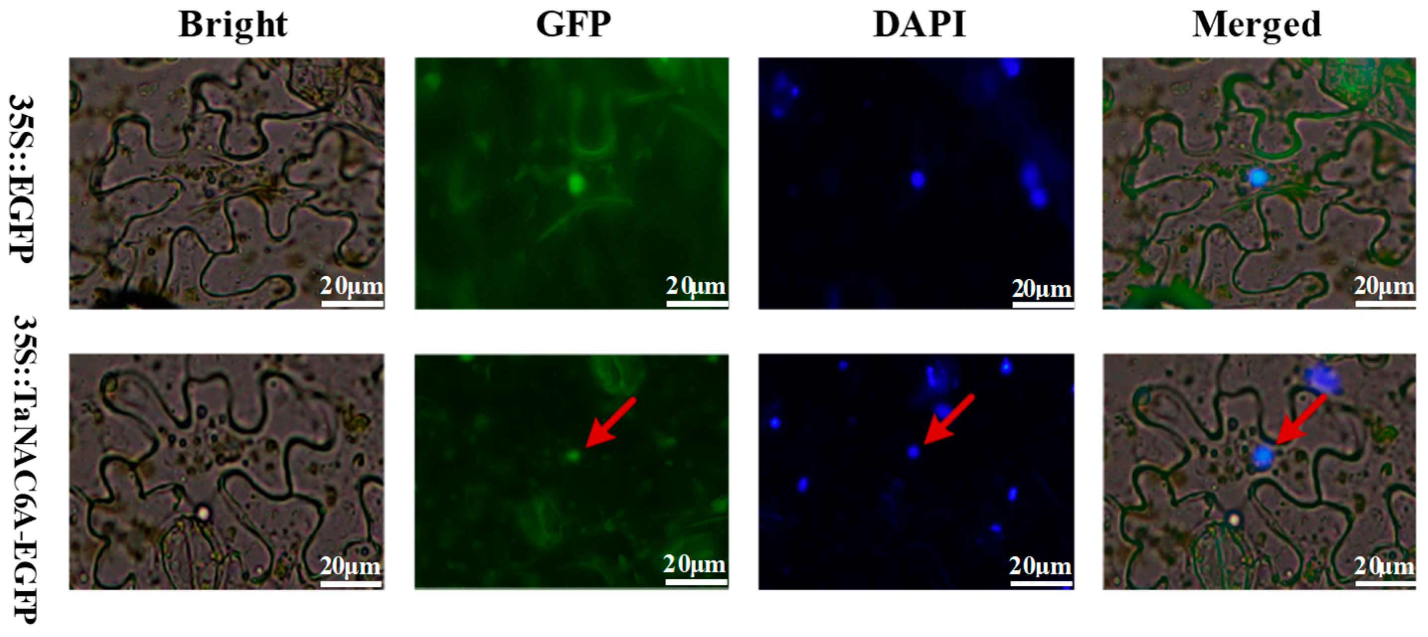
The pCAMBIA2300-TaNAC6A-EGFP fusion vector was developed for transient expression in tobacco to study the subcellular localization of TaNAC6A pr. A GFP signal was observed in the cell membranes, cytoplasm, and nuclei of tobacco leaves injected with Agrobacterium tumefaciens strain GV3101 carrying pCAMBIA2300-35S-EGFP. In contrast, green fluorescence was detected exclusively in the nuclei of tobacco leaves injected with Agrobacterium tumefaciens strain GV3101 carrying pCAMBIA2300-TaNAC6A-EGFP (Figure 2). This observation indicated that TaNAC6A is a protein located in the nucleus, and it may play a regulatory role.

2.4. Verification of Low-Temperature Responsiveness of Pre-tae-miR164 and TaNAC6A

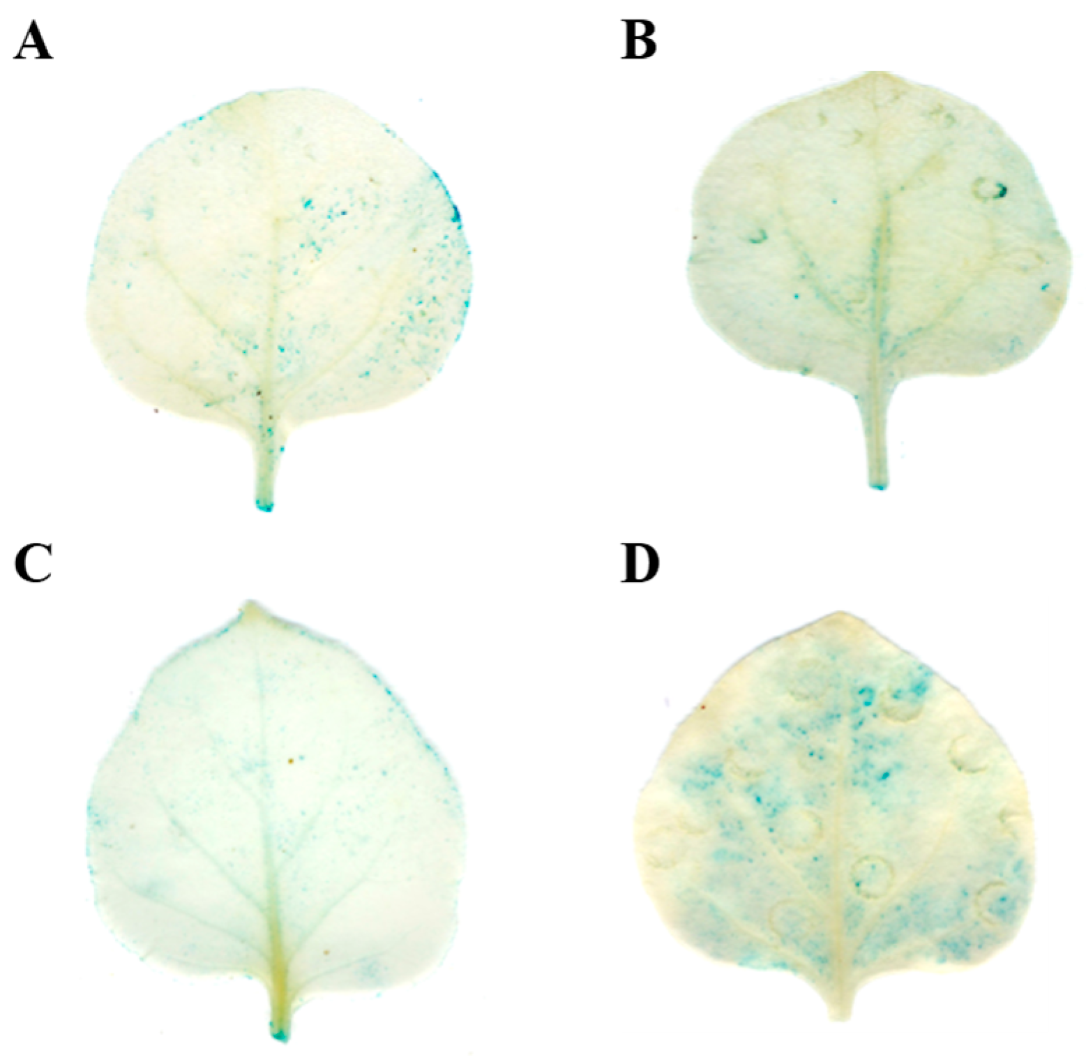
Biogenic analysis predicted the presence of low-temperature-responsive elements within the promoters of pre-tae-miR164 and TaNAC6A. These predictions were substantiated by examining the promoter activities of these two genes under low-temperature conditions. The promoters of tae-miR164 and TaNAC6A were capable of inducing GUS expression at normal temperatures (Figure 3A,B). However, after 4 h of exposure to 4 °C, the GUS staining appeared lighter in the PromiR164 tobacco leaves and more intense in the ProTaNAC6A tobacco leaves (Figure 3C,D). These outcomes indicate that tae-miR164 and TaNAC6A can respond to low temperature (4 °C), which might trigger the expression of key genes in the cold-signaling pathway.

2.5. Validation of the tae-miR164-TaNAC6A Regulatory Relationship

To verify the relationship between tae-miR164 and TaNAC6A, specific primers were designed at the 5′TUR of TaNAC6A mRNA (802 bp). PCR amplification using the 5′RACE technique yielded a single target band of 232 bp, corresponding to the size of the sequence after the first cleavage of TaNAC6A mRNA (Figure 4A). Sequencing of ten positive clones revealed that tae-miR164 cleaved the target mRNA at a single site, located between the 10th and 11th bases from the 5′ UTR of tae-miR164 (Figure 4B).
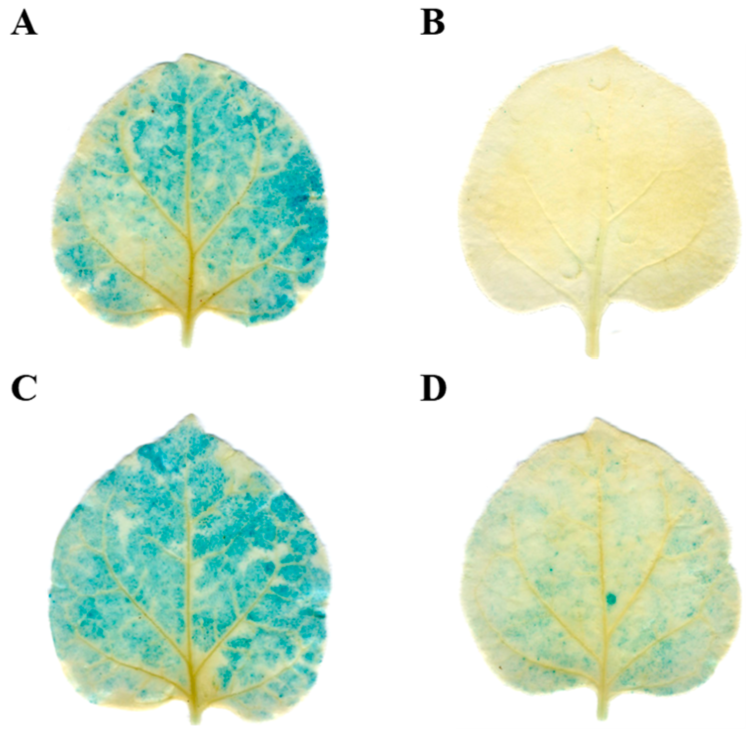
The relationship between tae-miR164 and TaNAC6A was further investigated using transient co-transformation in tobacco. Four types of Agrobacterium tumefaciens strain GV3101 were prepared: the first with the pBI121-GUS construct, the second with the pBI121-pre-tae-miR164 construct, the third with the pBI121-TaNAC6A construct, and the fourth consisting of a mixture containing both the pBI121-pre-tae-miR164 and pBI121-TaNAC6A constructs. These types were individually infiltrated into tobacco leaves. The GUS phenotype observed in leaves inoculated with the pBI121-GUS empty vector (Figure 5A) was comparable to that seen in leaves treated with the pBI121-TaNAC6A strain (Figure 5C). Conversely, leaves infiltrated with the pBI121-pre-tae-miR164 strain exhibited no GUS staining, as pre-tae-miR164 suppressed the GUS gene expression (Figure 5B). Furthermore, leaves co-transformed with pBI121-pre-tae-miR164 and pBI121-TaNAC6A displayed significantly reduced GUS-staining intensities (Figure 5D), suggesting that tae-miR164 negatively regulates the expression of TaNAC6A.
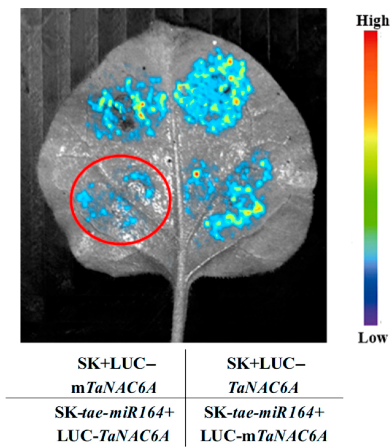
The interaction between tae-miR164 and TaNAC6A was validated once more using a dual-luciferase assay. Following the successful construction of the pGreenII-62-SK-pre-tae-miR164 expression vector, the pGreenII 0800-tae-miR164-TaNAC6A and pGreenII 0800-tae-miR164-mTaNAC6A (with a mutation in the TaNAC6A cleavage site by tae-miR164) expression vectors were also successfully constructed. The constructed vectors were introduced into the Agrobacterium tumefaciens strain GV3101 and subsequently expressed in tobacco leaves. The fluorescence intensity was observed to determine whether a targeting relationship exists between tae-miR164 and TaNAC6A. The fluorescence intensity was significantly reduced in tobacco leaves co-infiltrated with Agrobacterium tumefaciens strain GV3101 containing the SK-tae-miR164 plasmid and the LUC-TaNAC6A plasmid. However, the fluorescence intensity was enhanced in tobacco leaves co-infiltrated with the SK-tae-miR164 plasmid and the LUC-mTaNAC6A plasmid, which was introduced through an MRE (miRNA response element/miRNA binding site) mutation (Figure 6).
The three results collectively confirm that tae-miR164 targets and negatively regulates the expression of TaNAC6A. And it was found that the 5′ base pairing is indeed essential for MRE to exert its crucial function. tae-miR164 and TaNAC6A indeed exhibit an interaction relationship, and the tae-miR164-TaNAC6A module performed biological functions.

2.6. Phenotypic Changes in Transgenic Arabidopsis Plants

To investigate the functions of tae-miR164 and TaNAC6A in relation to cold stress, we constructed overexpression vectors for 35S-tae-miR164 and 35S-TaNAC6A and transformed them into Arabidopsis plants. From these transformations, we selected and obtained some OE-tae-miR164 and OE-TaNAC6A plants; among them, the OE-tae-miR164-3, OE-tae-miR164-7, OE-TaNAC6A-4, and OE-TaNAC6A-8 plants were high-expression lines. Additionally, we constructed the STTM-tae-miR164 (Short Targeted Tandem Motif knockout of tae-miR164) recombinant plasmid and transformed it into Arabidopsis plants. Subsequently, the third generation of genetically stable transgenic Arabidopsis plants, STTM-tae-miR164 plants, were selected; among them, the STTM-tae-miR164-1 plants and STTM-tae-miR164-2 plants were the lines in which tae-miR164 was most effectively knocked down. The nac6 mutant (SALK K135734C, TARI) originated from the Columbia accession (Col-0), with the mutation site located in the homologous gene AtNAC1 (AT1G56010) corresponding to TaNAC6A (Figure S3 and Table S5).
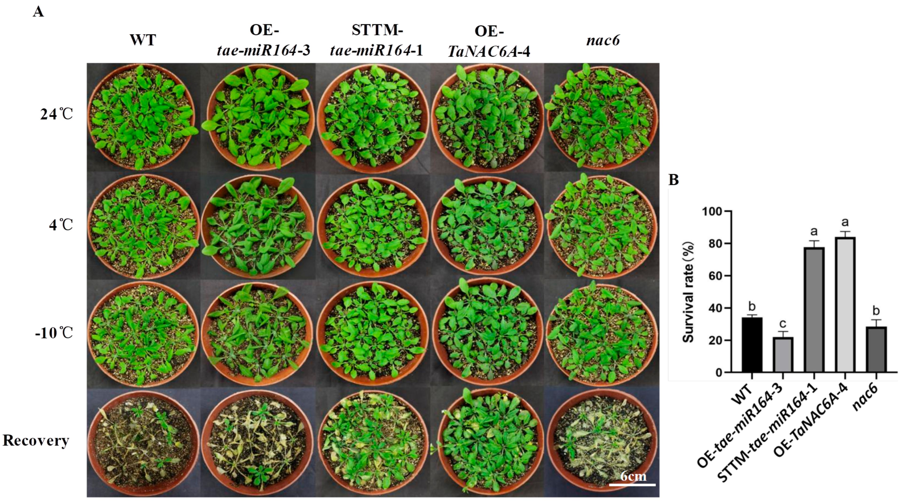
The phenotypes of five Arabidopsis plants (WT, OE-tae-miR164, OE-TaNAC6A, STTM-tae-miR164, and nac6) were observed under freezing stress (Figure 7A). No significant phenotypic differences were noted among the plants between 24 °C and 4 °C. However, at −10 °C, all plants displayed some degree of leaf curling or color deepening. Compared to the STTM-tae-miR164 and OE-TaNAC6A plants, the WT, OE-tae-miR164, and nac6 plants exhibited more pronounced color changes. Overall, the OE-tae-miR164 plants appeared to be the most affected by low temperature. Following a 7-day recovery period, both the STTM-tae-miR164 and OE-TaNAC6A plants returned to normal, whereas almost all the WT, OE-tae-miR164, and nac6 plants perished. The survival rates of the STTM-tae-miR164 and OE-TaNAC6A plants was about 80%, which were significantly higher than that of the WT, OE-tae-miR164, and nac6 plants (Figure 7B).
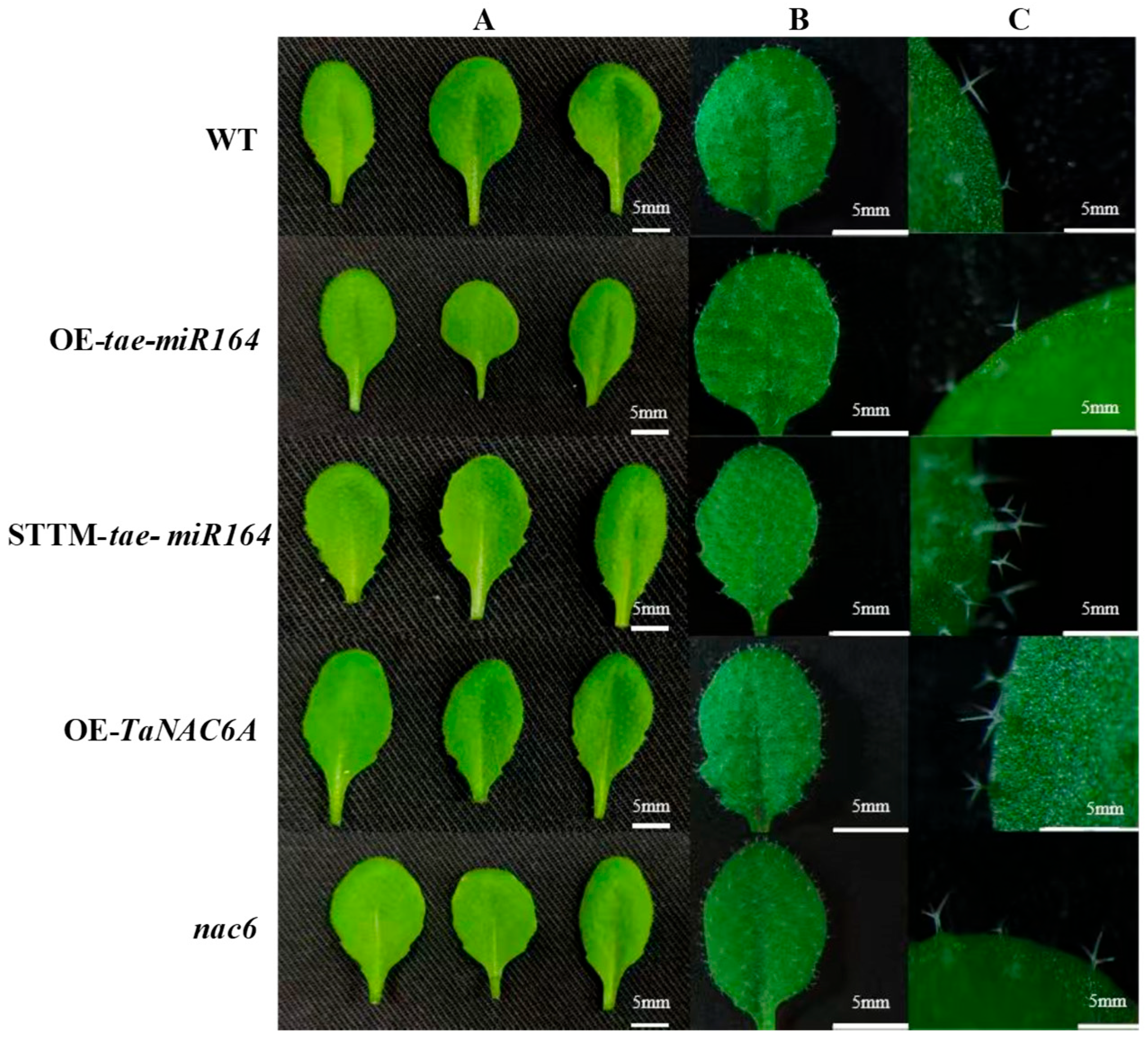
Direct observation of the leaves from various lines revealed distinct phenotypic differences (Figure 8A). Compared to the WT, OE-tae-miR164, and nac6 plants, the leaves of the STTM-tae-miR164 and OE-TaNAC6A plants exhibited increased serrations along the leaf margins. Additionally, the leaves of the OE-tae-miR164 plants displayed the lowest trichome densities. nac6 also had relatively low trichome densities, although they were not significantly different from those of the WT. Moreover, the trichomes of the WT and OE-tae-miR164 plants typically had two to three branches, whereas those of the STTM-tae-miR164 and OE-TaNAC6A plants generally had three to four branches (Figure 8).
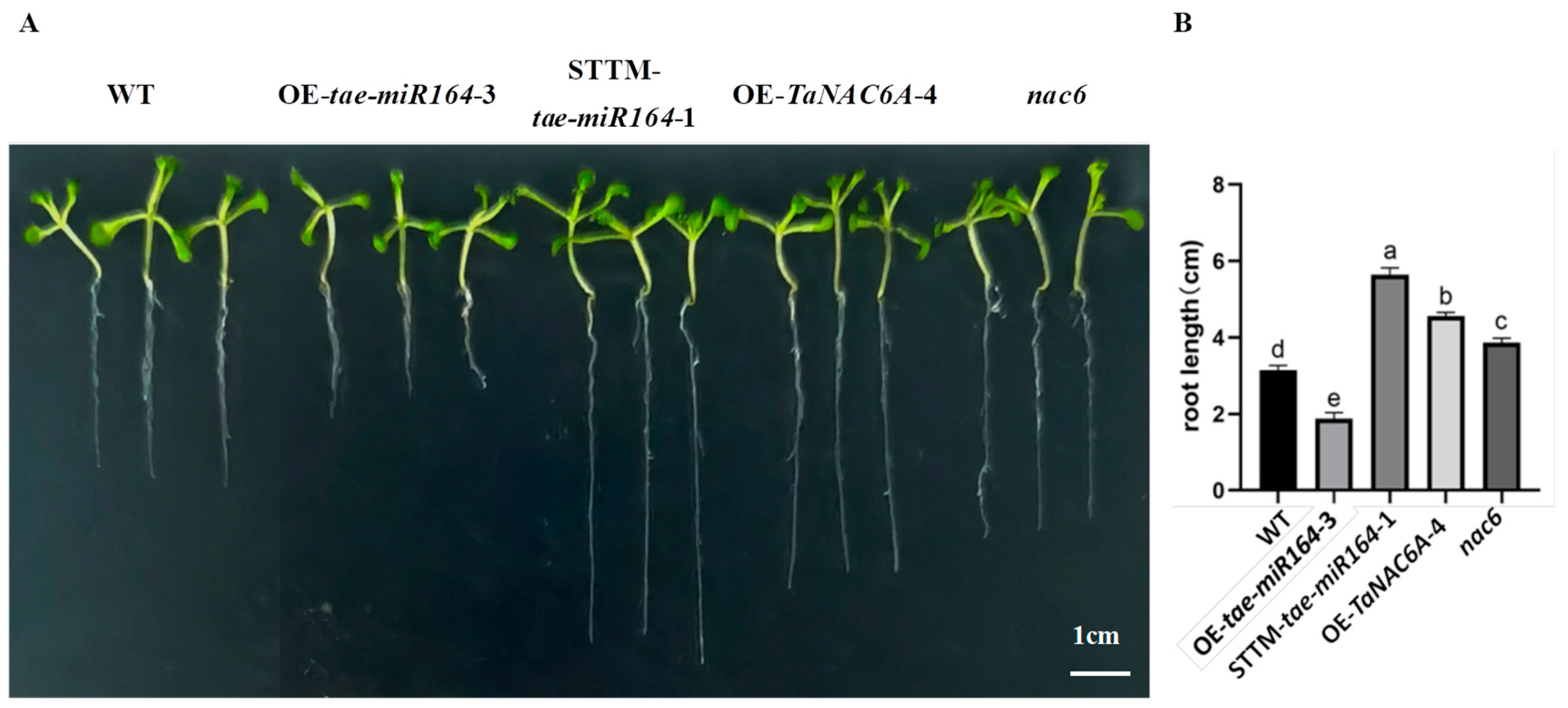
Under normal growth conditions, the primary roots of the OE-TaNAC6A plants were significantly longer than those of the WT and nac6 plants. Furthermore, the primary roots of the STTM-tae-miR164 plants were significantly longer than those of the WT and OE-tae-miR164 plants. Finally, the primary roots of the OE-tae-miR164 plants were much shorter, and they had fewer lateral roots compared to the other four types of plants (Figure 9).
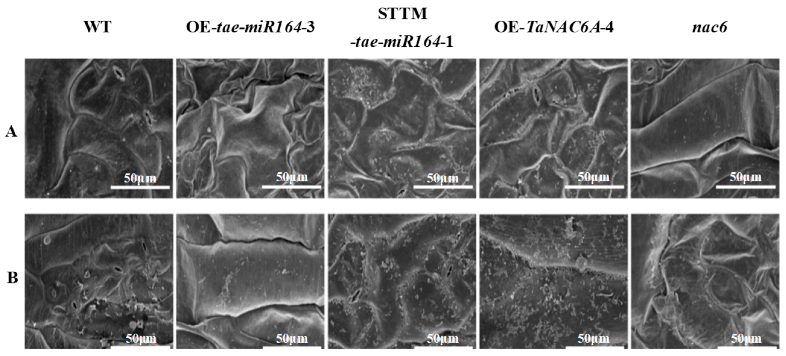
Many plants possess an outer epidermal layer composed of fatty acids or monohydric alcohols, known as the cuticle. This protective structure acts as a barrier against abiotic stressors, such as radiation, low temperature, and drought. In this study, we utilized scanning electron microscopy (SEM) to observe alterations in the cuticles of leaves cultivated under low temperature. At 24 °C, the OE-TaNAC6A and STTM-tae-miR164 plants exhibited thicker cuticles than those of the WT, OE-tae-miR164, and nac6 plants (Figure 10A). Following 3 days of exposure to 4 °C, the average cuticle thickness increased in all four transgenic Arabidopsis plants, mirroring the WT. Notably, the wax contents in the leaves of the OE-TaNAC6A and STTM-tae-miR164 plants were significantly greater than those of the other three Arabidopsis plants (Figure 10B).
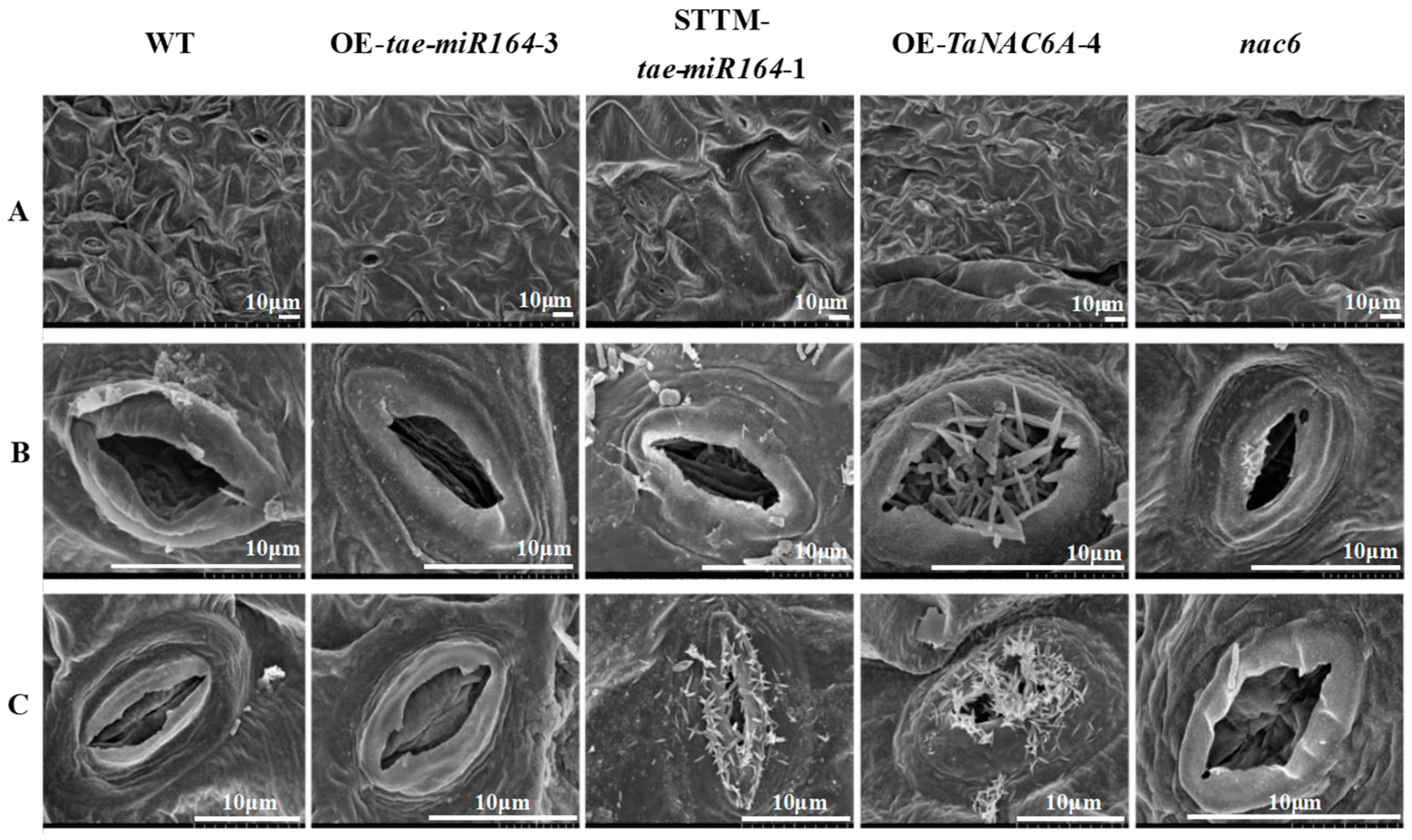
Stomata regulate the exchange of gases between the plant and the external environment. SEM observations indicated that the stomata of all the Arabidopsis plants were fully open at 24 °C. Following three days of exposure to 4 °C, the stomata of all lines tended to close to some degree. Notably, the stomata of the OE-TaNAC6A and STTM-tae-miR164 plants exhibited some degree of openness as well as wax buildup (Figure 11).
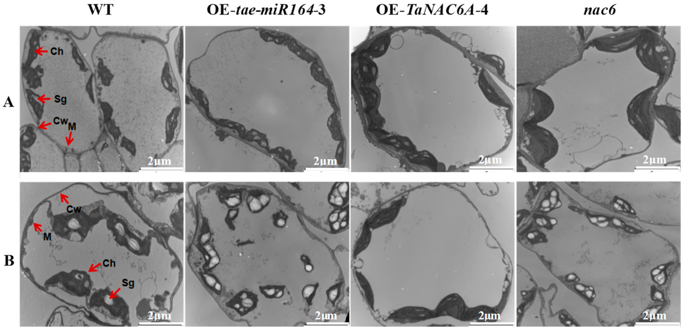
The subcellular structures of four different Arabidopsis plants were observed using transmission electron microscopy (TEM). At 24 °C, the mesophyll cells of the four plants exhibited well-defined structures with clear organelle contours, with no significant differences observed. After 3 days of exposure to 4 °C, the mesophyll cells of the WT plants experienced severe plasmolysis and displayed abnormal nuclear and mitochondrial morphologies. Furthermore, their chloroplasts were deformed and disintegrating, with ruptured chloroplast membranes migrating towards the centers of the mesophyll cells. These deformities were markedly more severe in the OE-tae-miR164 and nac6 plants, exhibiting notable increases in both the numbers and sizes of starch granules. In contrast, the OE-TaNAC6A plants showed no significant changes, apart from a slight tendency for chloroplasts to move away from the edges of the mesophyll cells. Additionally, these plants had significantly reduced numbers of starch granules (Figure 12).
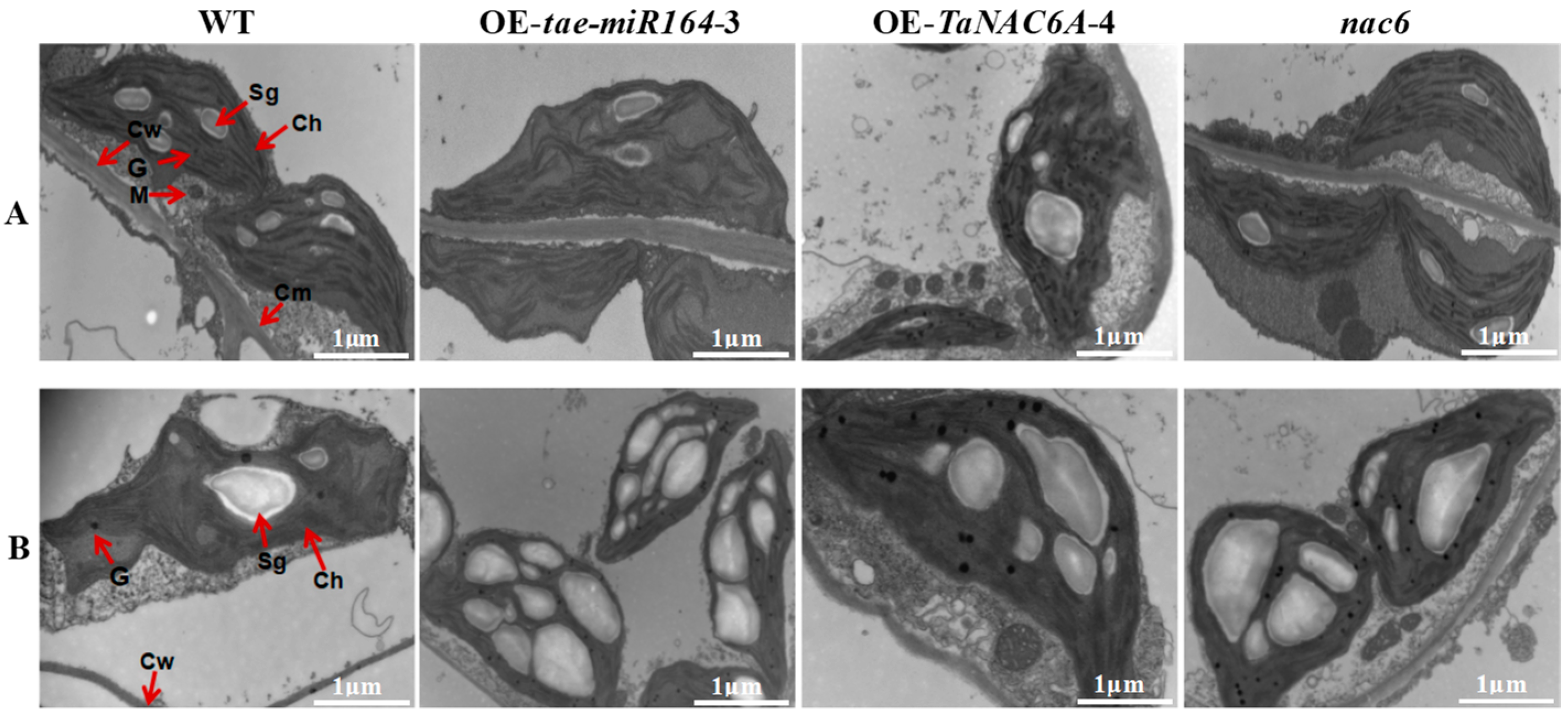
At 24 °C, the grana thylakoid structures in four plant genotypes were relatively well-ordered. Additionally, their mitochondria were round or oval and typically located either at junctions between chloroplasts and the plasma membrane or between two adjacent chloroplasts. After 3 days of exposure to 4 °C, the organelles in the WT plants began to disintegrate alongside vacuolization. Furthermore, the chloroplasts in the WT plants started to swell and deform, including the enlargement of starch granules. The number of grana decreased, and the thylakoid layers began to loosen and become disorganized, with some layers fusing together, resulting in blurred or missing thylakoid structures. However, in comparison to the WT plants, the OE-TaNAC6A plants retained normal chloroplast and mitochondrial structures even after being exposed to 4 °C for 3 days. This was evidenced by the absence of cell membrane disintegration and starch granule formation, indicating that the mesophyll cells could still maintain normal photosynthetic activity at this stage. In contrast, the cellular structures of the mesophyll cells in the OE-tae-miR164 and nac6 plants were significantly more damaged, with markedly increased numbers of starch granules. This suggested a substantial weakening in the ability to export photosynthetic products and an inability to perform normal photosynthesis. The numbers of starch granules increased in these plants, suggesting a weakened ability to export photosynthetic products (Figure 13).

2.7. Physiological Markers of Cold Resistance in Transgenic Arabidopsis Plants

The MDA content is one of the important indicators for evaluating the degree of peroxidation in the plant cell plasma membrane. The higher the MDA content, the greater the degree of membrane lipid peroxidation, and the more severe the cellular damage. Under normal conditions, plants contain relatively low Pro contents. However, when subjected to adverse environmental stresses, such as drought or cold, Pro accumulates in large amounts. The variation in conductivity can reflect the extent of cellular damage. Generally speaking, under cold stress, the lower the relative levels of electrical conductivity and MDA content, and the higher the Pro content, the less damage plant cells suffer, indicating stronger cold resistance in the plant.
Consequently, in order to evaluate whether the overexpression of tae-miR164 and TaNAC6A enhances cold tolerance in transgenic Arabidopsis plants, the electronic conductivities and the contents of MDA and Pro were measured.
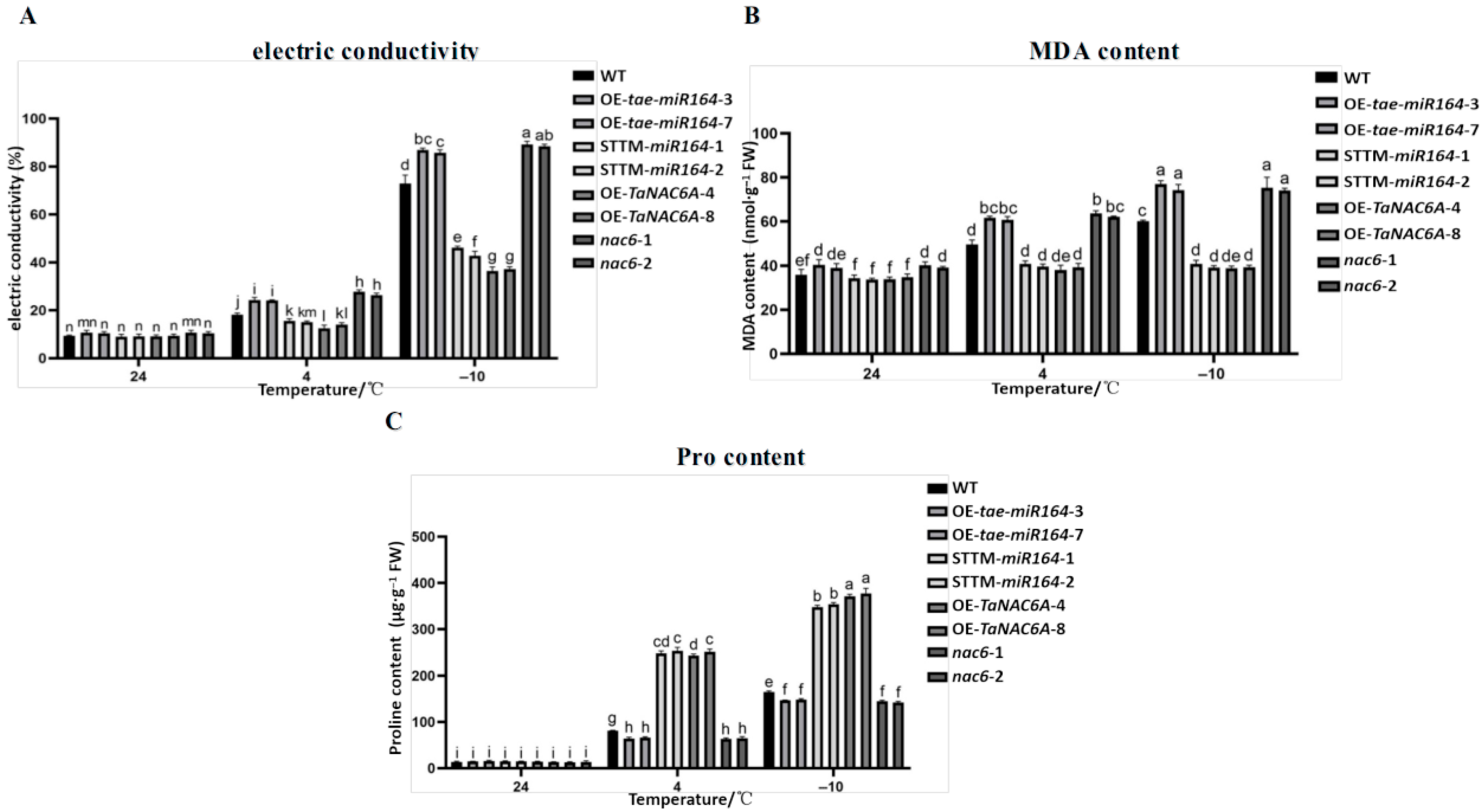
Overall, the electric conductivities of the five types of Arabidopsis plants tended to increase with decreasing temperature. At 4 °C and −10 °C, the electric conductivities of the STTM-tae-miR164 and OE-TaNAC6A plants were significantly lower than those of the WT, OE-tae-miR164, and nac6 plants (Figure 14A), indicating that the OE-tae-miR164 plants suffered more severe damage from cold stress. Conversely, the cold resistance of the transgenic Arabidopsis plants was improved, in which the expression level of TaNAC6A was relatively high.
At 4 °C and −10 °C, the MDA contents in the Arabidopsis plants significantly increased compared to those at 24 °C, indicating that the cell membranes suffered varying degrees of oxidative damage. The MDA contents in the STTM-tae-miR164 and OE-TaNAC6A plants were lower than those of the WT, OE-tae-miR164, and nac6 plants at 24 °C, 4 °C, and −10 °C, although the difference was most pronounced at −10 °C (Figure 14B). Furthermore, the Pro contents in the STTM-tae-miR164 and OE-TaNAC6A plants were higher than those of the WT, OE-tae-miR164, and nac6 plants at 4 °C and −10 °C (Figure 14C). It appears that the overexpression of tae-miR164 reduced the cold tolerance of the OE-tae-miR164 plants, whereas the overexpression of TaNAC6A enhanced the cold tolerance of the OE-TaNAC6A plants.

2.8. Cold-Induced ROS Production in Transgenic Arabidopsis Plants

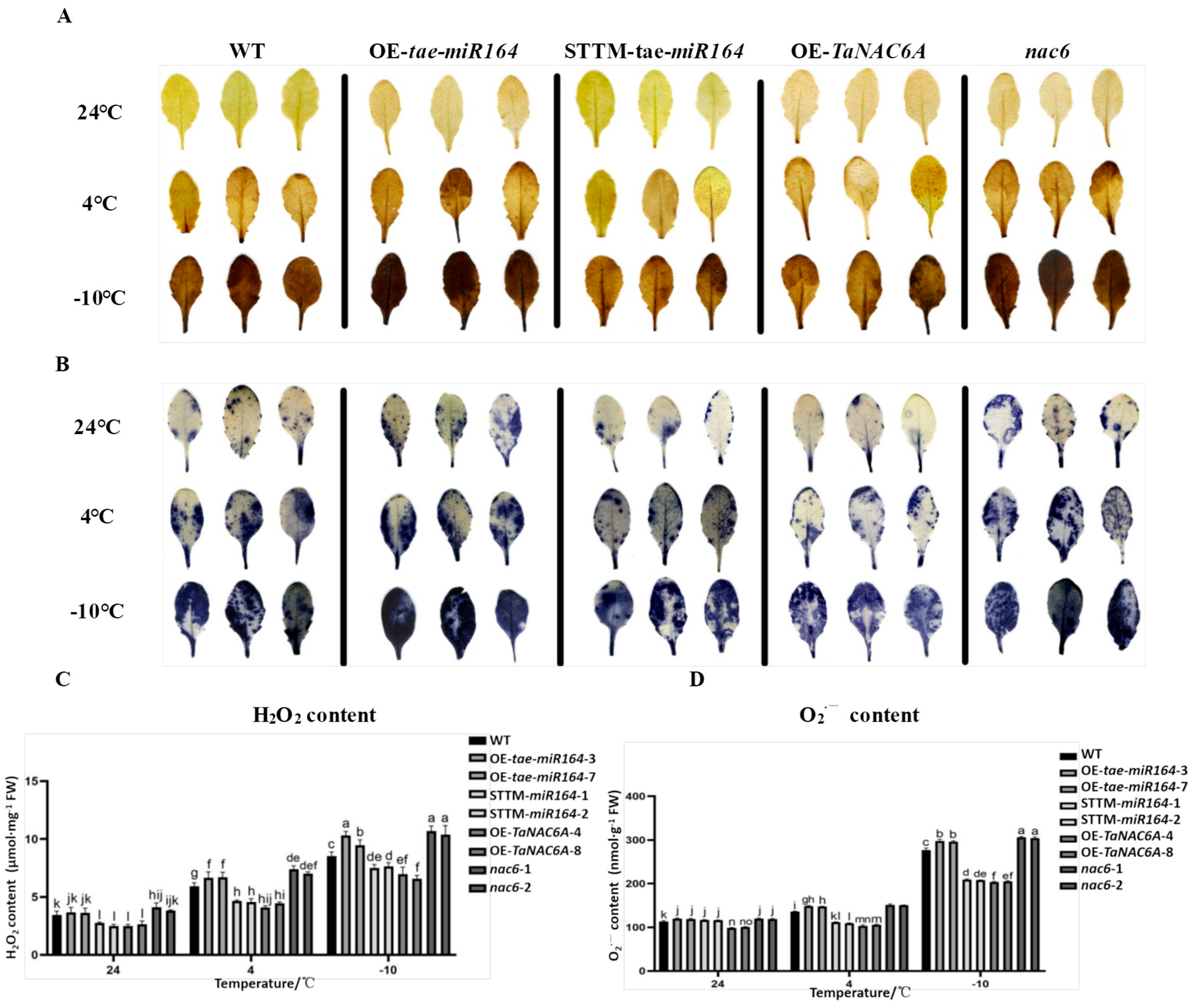
Cold stress frequently leads to the overproduction of reactive oxygen species (ROS), including hydrogen peroxide (H2O2) and superoxide radicals (O2·−), among others. To ascertain whether the overexpression of tae-miR164 and TaNAC6A influences the elimination of ROS, the levels of H2O2 and O2·− were quantified in cold-stressed Arabidopsis plants. The DAB and NBT chemical staining assays indicated that the WT plants exhibited more intense staining than both the OE-TaNAC6A and STTM-tae-miR164 plants at cold stress (4 °C and −10 °C). However, the OE-tae-miR164 and nac6 plants displayed darker staining than the WT plants (Figure 15A,B). The levels of H2O2 and O2·− in the WT plants gradually increased as the temperature decreased, with significantly higher levels at cold stress compared to those at 24 °C. No significant differences were observed in the levels of H2O2 and O2·− in the OE-TaTaNAC6A and STTM-tae-miR164 plants at 4 °C compared to at 24 °C. However, the levels of H2O2 and O2·− were higher at −10 °C than those at 24 °C. Furthermore, the levels of H2O2 and O2·− were lower in both the OE-TaTaNAC6A and STTM-tae-miR164 plants compared to those of the WT plants at both 4 °C and −10 °C. In contrast, the levels of H2O2 and O2·− in the OE-tae-miR164 and nac6 plants were higher than those in the WT plants (Figure 15C,D).

2.9. Changes in Antioxidant Enzyme Activities and Gene Expression Levels in Transgenic Arabidopsis Plants Under Cold Stress

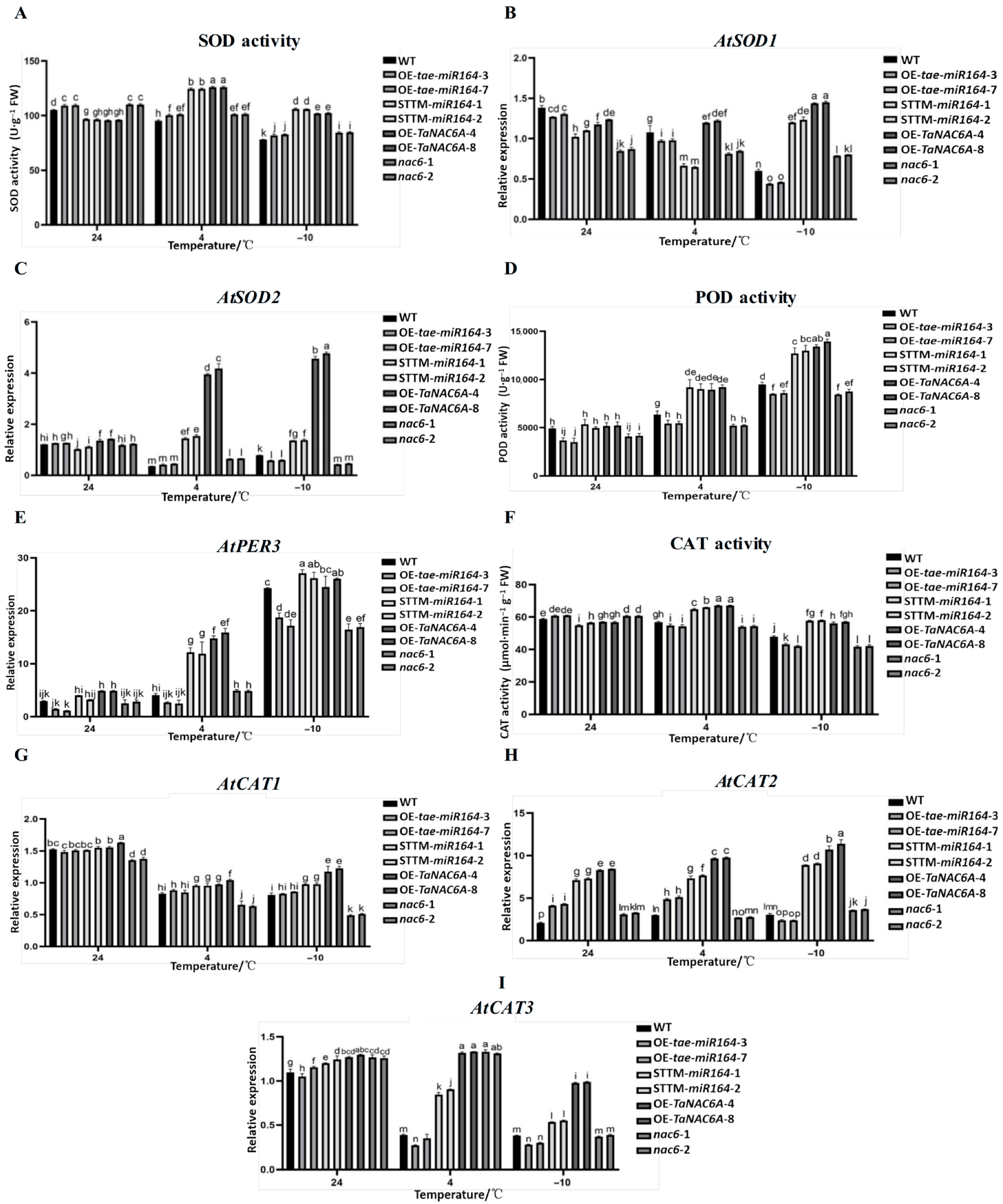
The SOD, POD, and CAT activities of the OE-TaNAC6A plants were the strongest under cold stress, followed by those of the STTM-tae-miR164 and WT plants, while the nac6 plants exhibited the lowest activity (Figure 16A,D,F). The RT-qPCR results for antioxidase-related genes (AtSOD1, AtSOD2, AtPER3, AtCAT1, AtCAT2, and AtCAT3) indicated that the expression levels of the OE-TaNAC6A and STTM-tae-miR164 plants were consistently higher than those of the WT and nac6 plants under the same low-temperature conditions (Figure 16B,C,E,G–I).

2.10. Alterations in the Expression of Cold-Signaling-Related Genes in OE-TaNAC6A Plants

Cold signaling is segregated into two major signaling pathways: ABA-independent and ABA-dependent pathways. In Arabidopsis, cold stress has been shown to induce the expression of ICE1, activating downstream DREB1/CBF members, which, in turn, induces an ABA-independent signaling pathway [27]. It is well-known that ABIs are key members in the ABA-dependent signaling pathway.
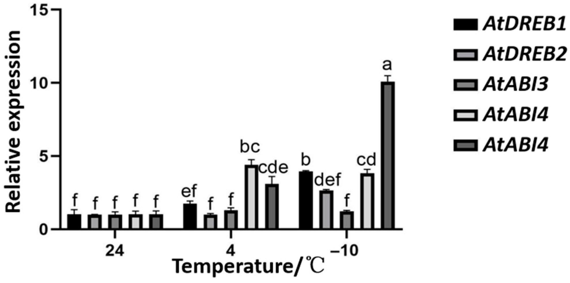
Therefore, we detected two genes (AtDREB1, AtDREB2) in the ABA-independent signaling pathway and three genes (AtABI3, AtABI4, and AtABI5) in the ABA-dependent signaling pathway by RT-qPCR. The results indicated that, as the temperature decreased, the expression levels of AtDREB1, AtDREB2, and AtABI5 in the OE-TaNAC6A plants with high TaNAC6A expression exhibited a gradual increase. At 4 °C, the expression levels of AtABI3 and AtABI4 were both higher than those at 24 °C and −10 °C (Figure 17).
These results indicate that the overexpression of TaNAC6A enhanced the cold-signaling pathways in the Arabidopsis plants.

3. Discussion

An increasing body of evidence indicates that miRNAs are crucial in regulating plant growth, development, and stress tolerance [28]. In a previous study, high-throughput RNA sequencing libraries were constructed using tillering nodes from Dn1 as samples, collected at temperatures of 5 °C and −10 °C. Thirty miRNA families were identified in the library from samples at 5 °C, while 24 miRNA families were identified in the library from samples at −10 °C. A total of 53 genes were predicted to be targets of the 30 miRNA families in the 5 °C library [29]. To fully leverage Dn1’s abundant genetic resources for cold resistance, it is essential to characterize the functions and regulatory mechanisms of the miRNAs associated with cold resistance. miR164 is one of the most conserved miRNAs and is involved in plant growth, development, and abiotic and biotic stress responses [21]. One miRNA molecule can control multiple target genes. Spychała et al. found that tae-miR164 targeted Lr46-RLK3, a rapid responsive gene to Puccinia triticina infection [30]. In Wang et al.’s research, TaMAPK4 was verified to be a target of tae-miR164, which played an important role in signaling during the wheat–Pst interaction [31].
Previous studies have also shown that tae-miR164 targets plant-specific NAC transcription factors from the NAM subfamily of wheat. Feng et al. revealed that TaNAC21/22, a NAC transcription factor gene, was the target of tae-miR164 and played an important role in regulating the resistance of host plants to stripe rust [32].
Specifically, miR164 negatively regulats the expression of its target NAC transcription factor (TF). We identified TaNAC6A mRNA as the target gene of tae-miR164 through psRNATarget analysis. Subsequently, this study confirmed that the tae-miR164-TaNAC6A module enhanced the cold resistance of Dn1, following a combination analysis of 5′RACE, tobacco transient co-transfection, and dual-luciferase assays.
Research into the stress-regulating functions of NAC transcription factors (TFs) has primarily focused on drought and salt stresses, with limited studies exploring signal transduction under low-temperature stress, particularly at sub-zero temperatures. Zhou et al. identified that the transient overexpression of TaNAC6A/6B/6D reduced the haustorium index of Yangmai158, and the stable transformation of TaNAC6A enhanced its resistance against Bgt, implying that TaNAC6s play important roles in basal resistance [33]. AtNAC1 in Arabidopsis is the homologous gene of TaNAC6A in wheat, which was analysized by WheatOmics. Rodríguez-García et al. identified that AtNAC1 activates the expression of peptidase-encoding AtCEPs in roots to limit the root hair growth of Arabidopsis plants [34]. Xie et al.’s study provided mechanistic insights into how AtNAC1 regulates root ground tissue maturation by coordinating with the SCR/SHR-CYCD6;1 module in Arabidopsis plants [35]. Xie et al. even discovered the AtNAC1 transduced auxin signal downstream of TIR1 to promote lateral root development [36]. Our study is the first to validate the biological functions of TaNAC6A in wheat and AtNAC1 in Arabidopsis plants in regulating plant cold tolerance.
Dong et al. discovered, in their research on tomato, that the miR164a-NAM3 module induces ethylene synthesis by directly regulating the expressions of SlACS1A, SlACS1B, SlAC01, and SlAC04, thereby conferring cold tolerance to transgenic tomato plants [37].
Li et al. treated wheat with phenanthrene (polycyclic aromatic hydrocarbons, PAHs) and found that phenanthrene stress (ubiquitous organic pollutants in the environment) accelerated the senescence and death of wheat roots while stimulating the emergence of new roots. Phenanthrene stress upregulated the expression of tae-miR164 and enhanced the silencing of TaNAC1, thereby inhibiting the occurrence of adventitious roots [38]. Our study found that the tae-miR164-TaNAC6A module can regulate cold tolerance in transgenic Arabidopsis plants, and we validated this at the phenotypic, physiological, and molecular levels.
Following recovery from exposure to −10 °C, the survival rates for the OE-TaNAC6A and STTM-tae-miR164 plants were 84% and 77.6%, respectively. In contrast, the survival rates for the OE-tae-miR164, nac6, and WT plants were 22%, 28.4%, and 34%, respectively. The OE-TaNAC6A plants exhibited a higher survival rate than the STTM-tae-miR164 plants, indicating significant cold tolerance. Additionally, under cold stress, the OE-tae-miR164 plants showed higher MDA contents and electrical conductivities and lower Pro contents compared to the other four lines.
In general, extremely low temperatures often result in the excessive production of ROS, the accumulation of MDA, and increased conductivity—biomarkers indicative of cell membrane damage [39]. Plants strictly regulate their ROS levels by recruiting enzymatic and non-enzymatic antioxidants [40,41]. The activities of SOD, CAT, and POD in wheat leaves increased after the Methyl jasmonate (MJ) pretreatment of the plants subjected to hardening temperature, as compared to the control [42]. In our study, at 4 °C and −10 °C, the leaves of the OE-tae-miR164 and nac6 plants exhibited deeper staining by DAB and NBT than those of the other three lines. Moreover, the H2O2 and O2·− contents of the OE-tae-miR164 and nac6 plants increased with decreasing temperature. In addition, markers of membrane permeability and membrane lipid peroxidation increased in the OE-tae-miR164 plants but decreased in the OE-TaNAC6A plants. The activities of SOD, POD, and CAT gradually decreased with decreasing temperature in the OE-tae-miR164 and nac6 plants. Finally, the expression levels of AtSOD1/2, AtPER3, and AtCAT1/2/3 were lower in the OE-tae-miR164 and nac6 plants than those in the other lines, consistent with the antioxidant activity assays.
DREB TFs regulate plant stress resistance. DREB1 and DREB2 regulate two different signaling pathways to resist stress caused by cold or dehydration [43]. ABA plays a crucial role in regulating plant growth during stress responses. By using molecular biology approaches, significant positive regulatory genes associated with ABA responses have been identified, including ABI3, ABI4, and ABI5 [44]. Here, we evaluated the differential expressions of five cold-stress-related genes in transgenic OE-TaNAC6A plants. The expression levels of AtDREB1/2 and AtABI3/4/5 were significantly altered by exposure to cold temperatures. Together, the results suggest that tae-miR164 negatively regulates cold tolerance while TaNAC6A promotes cold resistance.
The leaf morphology, size, and number are crucial for plant growth and development [45]. Compared to the WT plants, the OE-tae-miR164 plants exhibited smoother leaf margins, whereas the STTM-tae-miR164 plants showed increased serration. Notably, the trichome density in the OE-tae-miR164 plants was lower than that in the other lines. Most plant leaves possess a waxy cuticle, composed of fatty acids or monoalcohols synthesized by their epidermal cells [46], which serves as a barrier against abiotic stressors such as radiation, low temperatures, and drought. SEM observations revealed that both the OE-TaNAC6A and STTM-tae-miR164 plants had thicker, more waxy cuticles, even under normal temperature conditions. After exposure to 4 °C for three days, the stomata of all five lines tended to close, and their cuticles thickened. Nonetheless, the cuticles of the OE-TaNAC6A and STTM-tae-miR164 plants remained thicker than those of the other lines.
Exposure to low temperatures often results in plasma vesiculation, enlargement of chloroplasts and mitochondria, and the blurring or disappearance of nuclear and plasma membranes, which can sometimes lead to complete cellular disintegration [47]. In this study, TEM observations showed normal cell structures in all five lines at room temperature, with the OE-TaNC6A plants having larger starch granules. After three days of exposure to 4 °C, the WT, OE-tae-miR164, and nac6 plants displayed fusiform to circular chloroplast swelling, internal vacuolation, increases in osmophilic bodies, and expanded and/or disintegrated starch granules. In contrast, the OE-TaNC6A plants exhibited a relatively stable cell structure. Overall, the OE-tae-miR164 plants showed poor organellar stability in response to cold stress. Conversely, plants overexpressing TaNC6A demonstrated stable cellular structures in response to low temperature, indicating improved cold tolerance.
Given that wheat is the allohexaploid progenitor, it is well-known that its genome is vast and complex, resulting in slow progress in gene cloning and functional studies. Therefore, this study selected Arabidopsis plants for heterologous validation, which also was the potential limitation to the in-depth analysis of the molecular mechanism of the tae-miR164-TaNAC6A module in wheat.

4. Materials and Methods

4.1. Plant Materials, Growth Conditions, and Treatments

Seeds of winter wheat, Dn1 (with strong cold resistance), provided by our laboratory, were sown on 12 September 2020. Approximately 200 seeds were planted per row at 0.2 m intervals and a depth of 5 cm within a 2 m planting area. The seeds were covered with soil to promote natural germination, and the plants were managed using conventional practices. Under natural cooling conditions in the field, tillering nodes and leaves were sampled when the ten-day-average minimum temperature reached about 5 °C (on 10 October 2020), 0 °C (on 29 October 2020), −10 °C (on 25 November 2020), and −25 °C (on 3 January 2021) [48].
Arabidopsis seeds, ecotype Col-0, as wild type (WT), provided by our laboratory, Arabidopsis mutant seeds nac6 (SALK135734C, TAIR), purchased from The Arabidopsis Information Resource (TAIR), and the other three transgenic Arabidopsis seeds constructed by this study were cultivated under a light intensity of 120–150 μmolm−2 s−1, 24 °C, a 16/8 h day/night photoperiod, and a relative humidity of 60%. After 28 days of cultivating, the Arabidopsis seedlings were cold-acclimated at 4 °C for 3 days and subsequently exposed to −10 °C for 3 h. Leaves were sampled at three distinct time points: before cold acclimation, after 3 days of cold acclimation, and after 3 h of exposure to −10 °C. Samples were wrapped in tin foil, immediately frozen in liquid nitrogen, and stored at −80 °C for future use [49]. Following the cold treatment, plants were grown at room temperature for an additional 7 days to calculate the survival rate. Plants that were dry and yellow and unable to regenerate were considered dead.

4.2. Gene Expression Analysis

Frozen tissues of winter wheat and fresh leaves of Arabidopsis plants were homogenized using liquid nitrogen. Total RNA was isolated from the plants using the Ultrapure RNA Kit (CW0581M, CWBIO, Suzhou, China). The cDNA was then synthesized from the mRNA with the HiScript III 1st Strand cDNA Synthesis Kit (R312, Vazyme, Beijing, China). RT-qPCR was employed to quantify the gene expression, following our previously published method [50]. The mRNA levels of the target genes were quantified with the 2−∆∆CT method [51]. (Relevant RT-qPCR primers are listed in Table S1).

4.3. Bioinformatics Analysis

The amino acid sequence of TaNAC6A, along with the gene and promoter sequences of tae-miR164 and TaNAC6A, were obtained from wheatOmics (http://202.194.139.32/, 20 December 2024) The precursor and mature sequences of tae-miR164 were sourced from miRBase (https://www.mirbase.org/, 20 December 2024) and Dn1 miRNA libraries previously established by our lab. The base conservation in the mature miRNA was analyzed using WebLogo (http://weblogo.berkeley.edu/, accessed on 8 September 2025).
RNAfold (http://rna.tbi.univie.ac.at/cgi-bin/RNAWebSuite/RNAfold.cgi, 12 January 2025) was used to predict the stem–loop structure of the tae-miR164 precursor. ExPASy (http://expasy.org, 12 January 2025) was utilized to determine the physical and chemical properties of TaNAC6A, including the amino acid length, molecular weight, isoelectric point, and aliphatic index. PlantCare (http://bioinformatics.psb.ugent.be/webtools/plantcare/html, 12 January 2025) was employed to obtain the pre-tae-miR164 and TaNAC6A promoter sequences for cis element analysis. The subcellular localization prediction website (http://www.csbio.sjtu.edu.cn/bioinf/Cell-PLoc-2, 12 January 2025) was used to forecast the subcellular localization of TaNAC6A.

4.4. Subcellular Localization Analysis

The coding sequence (CDS) of TaNAC6A was constructed into the pCAMBIA2300 -EGFP vector. Tobacco leaves (seeded 28 days prior) were injected with Agrobacterium tumefaciens strain GV3101 carrying pCAMBIA2300-EGFP and pCAMBIA2300-TaNAC6A-EGFP. Seventy-two hours after the injection, the leaves were harvested to observe the EGFP signals under a fluorescence microscope (SZX10, Olympus, Tokyo, Japan).

4.5. Transient Expression Assay in Tobacco

To analyze the promoter activity of pre-tae-miR164 and TaNAC6A and verify the regulatory relationship between tae-miR164 and its target gene (TaNAC6A), a transient expression assay was performed on tobacco (Nicotiana tabacum). The pre-tae-miR164 and TaNAC6A promoter sequences (2000 bp) were constructed into the pBI121-GUS vector (relevant PCR primers are listed in Table S2). Tobacco leaves (seeded 28 days prior) were subsequently injected with Agrobacterium tumefaciens strain GV3101 carrying pBI121-pre-tae-miR164-GUS and pBI121-ProTaNAC6A-GUS. Seventy-two hours after the injection, the tobacco plants were exposed to 4 h at 4 °C. Subsequently, the leaves were harvested for histochemical staining.

4.6. 5′RACE Assay

The 5′RACE assay was conducted using a FirstChoice RLM-RACE Kit (Am1700, Thermo Fisher, Waltham, MA, USA), following the manufacturer’s instructions. (The 5′ primers, including 5′ RACE inner, and 5′ RACE outer, were provided by the kit; the 3′ primers are listed in Table S3).

4.7. Dual-Luciferase Assay

The pre-tae-miR164 sequence was constructed into the pGreenII-62-SK vector, and the upstream and downstream 100 bp sequences of the tae-miR164 recognition site mutation of TaNAC6A were constructed into the pGreenII0800-miRNA vector. (Relevant PCR primers are listed in Table S4.) Tobacco leaves, aged 28 days, were subsequently injected with Agrobacterium tumefaciens strain GV3101 (pSoup), carrying both control and target vectors at a 1:1 (v:v) ratio. The tobacco plants were subsequently cultured in the dark for 1 day and in the light for 2 days. The abaxial surfaces of the leaves, facing upwards, were sprayed with a reaction solution containing 1 mM of D-luciferin and then placed in a dark environment for 15 min. Following treatment, the leaves were observed using a chemiluminescence/fluorescence imaging analysis system (5200Multi, Tanon, Shanghai, China) to detect the luminescence.

4.8. Overexpression Vector Construction and Arabidopsis Transformation

The ORFs of TaNAC6A, pre-tae-miR164, and STTM-tae-miR164 were separately cloned into pCAMBIA2300, which was driven by the CaMV35S promoter and then transformed into the Agrobacterium tumefaciens strain GV3101. Particularly, the sequence was constructed using the mature tae-miR164 sequence as a template, with three bases (CTA) inserted between complementary bases at positions 9 and 10 at the 5′ end of tae-miR164. STTM-tae-miR164 forms a stem–loop structure following transcription, and the complementary portions of tae-miR164 and STTM-tae-miR164 exist in the form of single strands, binding complementarily with tae-miR164. The Agrobacterium tumefaciens strain GV3101 containing the target vector was individually transferred into Arabidopsis seedling ecotype Col-0 via the floral dip method. The positive transgenic lines were first screened on MS plates containing 50 mg·mL−1 kanamycin, and then the seedlings were detected by PCR.

4.9. Phenotyping and Physiological Index Assays

The seeds of Arabidopsis plants were sown on 1/2 MS solid medium, stratified for 2 days at 4 °C, and transferred to growth chambers (16 h day/8 h night at 24 °C). The length of the main root was measured by Image J 1.47 software. After 28-day-old Arabidopsis seedlings were cold-acclimated at 4 °C for 3 days, the morphologies of the leaves, stomata, and cellular ultrastructure were observed by a stereomicroscope (SZX10, Olympus, Tokyo, Japan), a scanning electron microscope (SU8010, Hitachi, Tokyo, Japan), and a transmission electron microscope (HT7800, Hitachi, Tokyo, Japan), respectively.
Leaf samples were collected after treatment. The electrical conductivity (relatively) was measured by a conductivity meter (DDS-307A, Leici, Shanghai, China). The relative H2O2 contents of the leaves were assessed by DAB staining, and the relative O2·− contents were assessed by NBT staining.
The contents of H2O2, O2·−, Pro, and MDA were measured using one-to-one corresponding commercial kits from Comin Biotechnology (H2O2-2-Y, SA-2-G, PRO-2-Y, MDA-2-Y, Coming, Suzou, China), following the manufacturer’s instructions. By using a visible spectrophotometer (721, Jinghua, Shanghai, China), the absorbance at 415 nm, 530 nm, 520 nm, and 532 nm was measured to calculate the contents of H2O2, O2·−, Pro, and MDA.
The activities of SOD, POD, and CAT were measured using the relevant commercial kits from Comin Biotechnology (SOD-2-W, POD-2-Y, CAT-2-W, Comin, Suzhou, China), following the manufacturer’s instructions. For the SOD, POD, or CAT activity determination, 0.1 g leaf samples was homogenized in 1 mL of PBS extraction buffer on ice. The absorbance at 560 nm, 470 nm, and 240 nm was measured to calculate the activities of SOD, POD, and CAT using a visible spectrophotometer (721, Jinghua, Shanghai, China). All experimental data were calculated according to the fresh weight.

4.10. Statistical Analysis

Three biological replicates were used for each experiment. Data are presented as the means of three biological replicates ± standard deviations (SDs), and data analyses were performed using IBM SPSS 26. All data were subjected to two-way analysis of variance (ANOVA). GraphPad Prism 8.0 was used for in-depth data analysis and for generating figures. A value of p < 0.05 was used to determine significance.

5. Conclusions

Taken together, our study suggests that the overexpression of tae-miR164 significantly downregulated the expression of TaNAC6A and reduced the cold tolerance of Arabidopsis plants. We identified that tae-miR164 and TaNAC6A might be involved in the low-temperature response. The subcellular location indicated that it might function as a transcription factor in cold-stress tolerance. We confirmed a physical interaction between tae-miR164 and TaNAC6A. Meanwhile, the results of the TaNAC6A and tae-miR164 overexpression in Arabidopsis plants indicated that the tae-miR164-TaNAC6A module played a key role in regulating the cold tolerance in the transgenic Arabidopsis plants in the epidermal hair development and leaf margin phenotype and in regulating the balance of ROS, as well as in promoting the expressions of AtDREB1, AtDREB2, and AtABI5. Conversely, the overexpression of TaNAC6A enhances the cold tolerance of Arabidopsis plants. Our research findings reveal the physiological and molecular mechanisms of the miR164-TaNAC6A module in winter wheat responding to cold stress, providing new insights for in-depth studies on the ncRNA regulation of plant stress resistance. This has significant guiding implications for the molecular module breeding of cold resistance in other crops in order to create crop varieties that better meet the needs of humans. But bottlenecks still exist in molecular module breeding technology, such as high costs and an incomplete understanding of the interaction mechanisms between module molecules.

Supplementary Materials

The following supporting information can be downloaded at https://www.mdpi.com/article/10.3390/plants14182849/s1, Figure S1. Bioinformatics analysis of tae-miR164 and its promoters. Figure S2. Bioinformatic analysis of TaNAC6A and the cis element in its promoter. Figure S3. Phylogenetic tree conducted by MEGA11 software. Table S1. Sequences of primers for qRT-PCR in wheat and Arabidopsis plants. Table S2. Sequences of primers for promoter amplification. Table S3. Sequences of primers for 5RACE. Table S4. Sequences of primers for dual-luciferase assay. Table S5. Sequences of primers for target gene clone.

Author Contributions

Q.X. designed the research; Z.D., X.Y., W.S. and Y.H. performed the experiments; Z.D. and W.S. analyzed the data; Q.X. and Z.D. wrote the manuscript; D.Z. and K.P. revised the manuscript. All authors have read and agreed to the published version of the manuscript.

Funding

This research was funded by the National Natural Science of China (31601236).

Data Availability Statement

The authors confirm that all data, tables, and figures in this manuscript are original.

Conflicts of Interest

The authors declare no conflicts of interest.

References

  1. Ding, Y.; Shi, Y.; Yang, S. Molecular Regulation of Plant Responses to Environmental Temperatures. Mol. Plant. 2020, 13, 544–564. [Google Scholar] [CrossRef]
  2. Soualiou, S.; Duan, F.; Li, X.; Zhou, W. Crop Production under Cold Stress: An Understanding of Plant Responses, Acclimation Processes, and Management Strategie. Plant Physiol. Biochem. 2022, 190, 47–61. [Google Scholar] [CrossRef] [PubMed]
  3. Thomashow, M.F. Plant Cold Acclimation: Freezing Tolerance Genes and Regulatory Mechanisms. Ann. Rev. Plant Physiol. Plant Mol. Biol. 1999, 50, 571–599. [Google Scholar] [CrossRef] [PubMed]
  4. John, R.; Anjum, N.A.; Sopory, S.K.; Akram, N.A.; Ashraf, M. Some Key Physiological and Molecular Processes of Cold Acclimation. Biol. Plantarum. 2016, 60, 603–618. [Google Scholar] [CrossRef]
  5. Zhou, J.; Wang, J.; Shi, K.; Xia, X.J.; Zhou, Y.H.; Yu, J.Q. Hydrogen Peroxide is Involved in the Cold Acclimation Induced Chilling Tolerance of Tomato Plants. Plant Physiol. Biochem. 2012, 60, 141–149. [Google Scholar] [CrossRef]
  6. Wang, Y.X.; Ya, H.U.; Chen, B.H.; Zhu, Y.F.; Dawuda, M.M.; Svetla, S. Physiological Mechanisms of Resistance to Cold Stress Associated with 10 Elite Apple Rootstocks. J. Integr. Agric. 2018, 17, 857–866. [Google Scholar] [CrossRef]
  7. Hussain, N.; Yasmeen, A.; Afzal Muhammad, A. Exogenously Applied Growth Promoters Modulate the Antioxidant Enzyme System to Improve the Cotton Productivity under Water Stress Conditions. Ital. J. Agron. 2020, 15, 165. [Google Scholar] [CrossRef]
  8. Guo, Z.; Cai, L.; Liu, C.; Chen, Z.; Guan, S.; Ma, W.; Pan, G. Low temperature Stress Affects Reactive Oxygen Species, Osmotic Adjustment Substances, and Antioxidants in Rice (Oryza sativa L.) at the Reproductive Stage. Sci. Rep. 2022, 12, 6224. [Google Scholar] [CrossRef]
  9. Sun, X.; Lin, L.; Sui, N. Regulation Mechanism of MicroRNA in Plant Response to Abiotic Stress and Breeding. Mol. Biol. Rep. 2019, 46, 1447–1457. [Google Scholar] [CrossRef]
  10. Lu, S.; Sun, Y.H.; Chiang, V.L. Stress-responsive microRNAs in Populus. Plant J. Cell Mol. Biol. 2008, 5, 131–151. [Google Scholar] [CrossRef] [PubMed]
  11. Barrera-Figueroa, B.E.; Gao, L.; Wu, Z.; Zhou, X.; Zhu, J.; Jin, H.; Liu, R.; Zhu, J.-K. High Throughput Sequencing Reveals Novel and Abiotic Stress-Regulated MicroRNAs in the Inflorescences of Rice. BMC Plant Biol. 2012, 12, 132. [Google Scholar] [CrossRef] [PubMed]
  12. Tang, Z.; Zhang, L.; Xu, C.; Yuan, S.; Zhang, F.; Zheng, Y.; Zhao, C. Uncovering Small RNA-mediated Responses to Cold Stress in a Wheat Thermosensitive Genic Male-Sterile Line by Deep Sequencing. Plant Physiol. 2012, 159, 721–738. [Google Scholar] [CrossRef]
  13. Cao, X.; Wu, Z.; Jiang, F.; Zhou, R.; Yang, Z. Identification of Chilling Stress-Responsive Tomato MicroRNAs and Their Target Genes by High-Throughput Sequencing and Degradome Analysis. BMC Genom. 2014, 15, 1130. [Google Scholar] [CrossRef]
  14. Wang, P.; Yang, Y.; Shi, H.; Wang, Y.; Ren, F. Small RNA and Degradome Deep Sequencing Reveal Respective Roles of Cold-Related MicroRNAs Across Chinese Wild Grapevine and Cultivated Grapevine. BMC Genom. 2019, 20, 740. [Google Scholar] [CrossRef]
  15. Wang, Q.; Liu, N.; Yang, X.; Tu, L.; Zhang, X. Small RNA-Mediated Responses to Low and High-Temperature Stresses in Cotton. Sci. Rep. 2016, 6, 35558. [Google Scholar] [CrossRef]
  16. Shi, X.; Jiang, F.; Wen, J.; Wu, Z. Overexpression of Solanum Habrochaites microRNA319d (sha-miR319d) Confers Chilling and Heat Stress Tolerance in Tomato (S. lycopersicum). BMC Plant Biol. 2019, 19, 214. [Google Scholar] [CrossRef]
  17. Peng, K.; Tian, Y.; Sun, X.; Song, C.; Ren, Z.; Bao, Y.; Xing, J.; Li, Y.; Xu, Q.; Yu, J.; et al. Tae-miR399-UBC24 Module Enhances Freezing Tolerance in Winter Wheat via a CBF Signaling Pathway. J. Agric. Food Chem. 2021, 69, 13398–13415. [Google Scholar] [CrossRef] [PubMed]
  18. Tsai, W.A.; Sung, P.H.; Kuo, Y.W.; Chen, M.-C.; Jeng, S.-T.; Lin, J.-S. Involvement of microRNA164 in Responses to Heat Stress in Arabidopsis. Plant Sci. 2023, 329, 111598. [Google Scholar] [CrossRef]
  19. Fang, Y.; Xie, K.; Xiong, L. Conserved miR164-targeted NAC Genes Negatively Regulate Drought Resistance in Rice. J. Exp. Bot. 2014, 65, 2119–2135. [Google Scholar] [CrossRef] [PubMed]
  20. Lin, D.; Zhu, X.; Qi, B.; Gao, Z.; Tian, P.; Li, Z.; Lin, Z.; Zhang, Y.; Huang, T. SlmiR164A Regulates Fruit Ripening and Quality by Controlling SlNAM2 and SlNAM3 in Tomato. Plant Biotechnol. J. 2022, 20, 1456–1469. [Google Scholar] [CrossRef]
  21. Qing, C.; Du, L.; Wen, M.; Ruo, N.; Bao, W.; Li, G.; Meng, M.; Xiang, L.; Hui, Z. The MiR164-TaNAC14 Module Regulates Root Development and Abiotic-stress Tolerance of Wheat Seedlings. J. Integr. Agric. 2023, 22, 981–998. [Google Scholar]
  22. Diao, P.; Chen, C.; Zhang, Y.; Meng, Q.; Lv, W.; Ma, N. The Role of NAC Transcription Tactor in Plant Cold Response. Plant Signal. Behav. 2020, 15, 1785668. [Google Scholar] [CrossRef]
  23. Hou, X.M.; Zhang, H.F.; Liu, S.Y.; Wang, X.-K.; Zhang, Y.-M.; Meng, Y.-C.; Luo, D.; Chen, R.-G. The NAC Transcription Factor CaNAC064 is a Regulator of Cold Stress Tolerance in Peppers. Plant Sci. 2020, 291, 110346. [Google Scholar] [CrossRef]
  24. Ju, Y.L.; Yue, X.F.; Min, Z.; Wang, X.-H.; Fang, Y.-L.; Zhang, J.-X. VvNAC17, a Novel Stress-responsive Grapevine (Vitis vinifera L.) NAC Transcription Factor, Increases Sensitivity to Abscisic Acid and Enhances Salinity, Freezing, and Drought Tolerance in Transgenic Arabidopsis. Plant Physiol. Biochem. 2020, 146, 98–111. [Google Scholar] [CrossRef]
  25. Luo, P.; Chen, Y.; Rong, K.; Lu, Y.; Wang, N.; Xu, Z.; Pang, B.; Zhou, D.; Weng, J.; Li, M.; et al. ZmSNAC13, a Maize NAC Transcription Factor Conferring Enhanced Resistance to Multiple Abiotic Stresses in Transgenic Arabidopsis. Plant Physiol. Biochem. 2022, 170, 160–170. [Google Scholar] [CrossRef] [PubMed]
  26. Liu, L.; Cang, J.; Yu, J.; Wang, X.; Huang, R.; Wang, J.; Lu, B. Effects of Exogenous Abscisic Acid on Carbohydrate Metabolism and the Expression Levels of Correlative Key Enzymes in Winter Wheat Under Low Temperature. Biosci. Biotechnol. Biochem. 2013, 77, 516–525. [Google Scholar] [CrossRef] [PubMed]
  27. Abhinandan, K.; Skori, L.; Stanic, M.; Hickerson, N.M.N.; Jamshed, M.; Samuel, M.A. Abiotic Stress Signaling in Wheat-An Inclusive Overview of Hormonal Interactions During Abiotic Stress Responses in Wheat. Front. Plant Sci. 2018, 9, 734. [Google Scholar] [CrossRef]
  28. Ma, X.; Zhao, F.; Zhou, B. The Characters of Non-Coding RNAs and Their Biological Roles in Plant Development and Abiotic Stress Response. Int. J. Mol. Sci. 2022, 23, 4124. [Google Scholar] [CrossRef]
  29. Huang, R.; Cang, J.; Yu, J.; Lu, B.; Liu, L.; Wang, J.; Guo, R.; Xu, C. Solexa Sequencing and Bioinformatics Analysis of Small RNA in Winter Wheat. Chin. Bull. Bot. 2014, 49, 8–18. [Google Scholar] [CrossRef]
  30. Spychała, J.; Tomkowiak, A.; Noweiska, A.; Bobrowska, R.; Bocianowski, J.; Sobiech, A.; Kwiatek, M.T. Diversity of Expression Patterns of Lr34, Lr67, and Candidate Genes towards Lr46 with Analysis of Associated miRNAs in Common Wheat Hybrids in Response to Puccinia triticina Fungus. Curr. Issues Mol. Biol. 2024, 46, 5511–5529. [Google Scholar] [CrossRef]
  31. Wang, B.; Song, N.; Zhang, Q.; Wang, N.; Kang, Z. TaMAPK4 Acts as a Positive Regulator in Defense of Wheat Stripe-Rust Infection. Front. Plant Sci. 2018, 15, 152. [Google Scholar] [CrossRef]
  32. Feng, H.; Duan, X.; Zhang, Q.; Li, X.; Wang, B.; Huang, L.; Wang, X.; Kang, Z. The Target Gene of tae-miR164, a Novel NAC Transcription Factor from the NAM Subfamily, Negatively Regulates Resistance of Wheat to Stripe rust. Mol. Plant Pathol. 2014, 15, 284–296. [Google Scholar] [CrossRef]
  33. Zhou, W.; Qian, C.; Li, R.; Zhou, S.; Zhang, R.; Xiao, J.; Wang, X.; Zhang, S.; Xing, L.; Cao, A. TaNAC6s are Involved in the Basal and Broad-spectrum Resistance to Powdery Mildew in Wheat. Plant Sci. 2018, 277, 218–228. [Google Scholar] [CrossRef]
  34. Rodríguez-García, D.R.; Rondón Guerrero, Y.D.C.; Ferrero, L.; Rossi, A.H.; Miglietta, E.A.; Aptekmann, A.A.; Marzol, E.; Pacheco, J.M.; Carignani, M.; Gabarain, V.B.; et al. Transcription Factor NAC1 Activates Expression of Peptidase-encoding AtCEPs in Roots to Limit Root Hair Growth. Plant Physiol. 2023, 194, 81–93. [Google Scholar] [CrossRef]
  35. Xie, C.; Li, C.; Wang, F.; Zhang, F.; Liu, J.; Wang, J.; Zhang, X.; Kong, X.; Ding, Z. NAC1 Regulates Root Ground Tissue Maturation by Coordinating with the SCR/SHR-CYCD6;1 Module in Arabidopsis. Mol. Plant 2023, 16, 709–725. [Google Scholar] [CrossRef]
  36. Xie, Q.; Frugis, G.; Colgan, D.; Chua, N.-H. Arabidopsis NAC1 Transduces Auxin Signal Downstream of TIR1 to Promote Lateral Root Development. Genes Dev. 2000, 14, 3024–3036. [Google Scholar] [CrossRef] [PubMed]
  37. Dong, Y.; Tang, M.; Huang, Z.; Song, J.; Xu, J.; Ahammed, G.J.; Yu, J.; Zhou, Y. The miR164a-NAM3 Module Confers Cold Tolerance by Inducing Ethylene Production in Tomato. Plant J. 2022, 111, 440–456. [Google Scholar] [CrossRef]
  38. Li, J.; Zhang, H.; Zhu, J.; Shen, Y.; Zeng, N.; Liu, S.; Wang, H.; Wang, J.; Zhan, X. Role of miR164 in the Growth of Wheat New Adventitious Roots Exposed to Phenanthrene. Environ. Pollut. 2021, 284, 117204. [Google Scholar] [CrossRef] [PubMed]
  39. Agarwal, P.; Mitra, M.; Banerjee, S.; Roy, S. MYB4 Transcription Factor, a Member of R2R3-subfamily of MYB Domain Protein, Regulates Cadmium Tolerance via Enhanced Protection Against Oxidative Damage and Increases Expression of PCS1 and MT1C in Arabidopsis. Plant Sci. 2020, 297, 110501. [Google Scholar] [CrossRef] [PubMed]
  40. Feng, S.; Wang, B.; Li, C.; Guo, H.; Bao, A.-K. Transcriptomic Analysis Provides Insight into the ROS Scavenging System and Regulatory Mechanisms in Atriplex canescens Response to Salinity. Int. J. Mol. Sci. 2022, 24, 242. [Google Scholar] [CrossRef]
  41. Hasanuzzaman, M.; Bhuyan, M.; Zulfiqar, F.; Raza, A.; Mohsin, S.M.; Al Mahmud, J.; Fujita, M.; Fotopoulos, V. Reactive Oxygen Species and Antioxidant Defense in Plants under Abiotic Stress: Revisiting the Crucial Role of a Universal Defense Regulator. Antioxidants 2020, 9, 681. [Google Scholar] [CrossRef]
  42. Repkina, N.; Ignatenko, A.; Holoptseva, E.; MiszalskI, Z.; Kaszycki, P.; Talanova, V. Exogenous Methyl Jasmonate Improves Cold Tolerance with Parallel Induction of Two Cold-Regulated (COR) Genes Expression in Triticum aestivum L. Plants 2021, 10, 1421. Plants 2021, 10, 1421. [Google Scholar] [CrossRef]
  43. Reeves, W.M.; Lynch, T.J.; Mobin, R.; Finkelstein, R.R. Direct Targets of the Transcription Factors ABA-Insensitive (ABI4 and ABI5) Reveal Synergistic Action by ABI4 and Several bZIP ABA Response Factors. Plant Mol. Biol. 2011, 75, 347–363. [Google Scholar] [CrossRef]
  44. Agarwal, P.K.; Agarwal, P.; Reddy, M.K.; Sopory, S.K. Role of DREB Transcription Factors in Abiotic and Biotic Stress Tolerance in Plants. Plant Cell Rep. 2006, 25, 1263–1274. [Google Scholar] [CrossRef]
  45. Arvidsson, S.; Pérez-Rodríguez, P.; Mueller-Roeber, B. A Growth Phenotyping Pipeline for Arabidopsis Thaliana Integrating Image Analysis and Rosette Area Modeling for Robust Quantification of Genotype Effects. New Phytol. 2011, 191, 895–907. [Google Scholar] [CrossRef] [PubMed]
  46. Riederer, M. Thermodynamics of the Water Permeability of Plant Cuticles: Characterization of the Polar Pathway. J. Exp. Bot. 2006, 57, 2937–2942. [Google Scholar] [CrossRef] [PubMed]
  47. Wang, Y.M.; Zhou, D.M.; Yuan, X.Y.; Zhang, X.H.; Li, Y. Modeling the Interaction and Toxicity of Cu Cd Mixture to Wheat Roots Affected by Humic Acids, in Terms of Cell Membrane Surface Characteristics. Chemosphere 2018, 199, 76–83. [Google Scholar] [CrossRef] [PubMed]
  48. Bao, Y.; Xing, J.; Liang, Y.; Ren, Z.; Fu, L.; Yu, J.; Wang, D.; Zhang, D.; Xu, Q.; Cang, J. Analysis of Overwintering Indexes of Winter Wheat in Alpine Regions and Establishment of A Cold Resistance Model. Field Crops Res. 2022, 275, 108347. [Google Scholar] [CrossRef]
  49. Tian, Y.; Peng, K.; Bao, Y.; Zhang, D.; Meng, J.; Wang, D.; Wang, X.; Cang, J. Glucose-6-phosphate Dehydrogenase and 6-phosphogluconate Dehydrogenase Genes of Winter Wheat Enhance the Cold Tolerance of Transgenic Arabidopsis. Plant Physiol. Biochem. 2021, 161, 86–97. [Google Scholar] [CrossRef]
  50. Peng, K.; Tian, Y.; Cang, J.; Yu, J. Overexpression of TaFBA-A10 from Winter Wheat Enhances Freezing Tolerance in Arabidopsis thaliana. J. Plant Growth Regul. 2022, 41, 314–326. [Google Scholar] [CrossRef]
  51. Livak, K.J.; Schmittgen, T.D. Analysis of Relative Gene Expression Data Using Real-time Quantative PCR and the 2−∆∆CT Method. Methods 2001, 25, 402–408. [Google Scholar] [CrossRef] [PubMed]
Figure 1. Expressions of tae-miR164 and TaNAC6A in response to cold stress. (A) Expressions of tae-miR164 and TaNAC6A in tillering nodes of Dn1. (B) Expressions of tae-miR164 and TaNAC6A in leaves of Dn1. Values represent the means ± SDs (n = 10). Different lowercase letters indicate significant differences between treatments (p < 0.05), determined by two-way ANOVA. The reference gene used is TaActin.
Plants 14 02849 g001
Figure 2. Subcellular localization of TaNAC6A protein. The Agrobacteria carrying 35 S:EGFP (control) and 35 S:TaNAC6A-EGFP vectors were transformed into the leaves of tobacco. The GFP signals of TaNAC6A-EGFP (bottom) and EGFP alone (top) were observed using a laser confocal microscope. DAPI was used as a nuclear marker in this experiment. Red arrow indicates the position of TaNAC6A-EGFP. Scale bars = 20 μm.
Plants 14 02849 g002
Figure 3. Analysis of promoter activity at different temperatures. GUS phenotype observed via histochemical staining. Leaves of tobacco separately injected with (A) 35S:Pro-miR164-GUS vector; (B) 35S:Pro-TaNAC6A-GUS vector; (C) 35S:Pro-miR164-GUS vector; (D) 35S:Pro-TaNAC6A-GUS vector after 4 h of exposure to 4 °C.
Plants 14 02849 g003
Figure 4. Interaction between tae-miR164 and its target (TaNAC6A). (A) PCR amplification products after 5′RACE; red box indicates the amplified band. M: Marker 2000. Lanes 1, 2, and 3: cleaved TaNAC6A fragments; (B) model of the cDNA structure and mRNA cleavage site of TaNAC6A, as determined by 5′ RACE. Red letters show tae-miR164 complementary sites with the nucleotide positions of TaNAC6A. The expanded regions show the RNA sequence of each complementary site from 5′ to 3′ and the miRNA sequence from 3′ to 5′. The red arrow indicates a cleavage site verified by 5′-RACE, with the frequency of cloned PCR products shown above the alignment.
Plants 14 02849 g004
Figure 5. GUS phenotypes observed through histochemical staining. (A) Leaf injected with pBI121-GUS empty vector; (B) leaf injected with pBI121-pre-tae-miR164 vector; (C) leaf injected with pBI121-TaNAC6A vector; (D) leaf injected with the mixture of pBI121-pre-tae-miR164 and pBI121-TaNAC6A vectors.
Plants 14 02849 g005
Figure 6. Dual-luciferase assay to verify the relationship between tae-miR164 and TaNAC6A. Dual-luciferase assay showing the activation of SK-driven luciferase reporter gene in tobacco leaves. Agrobacterium tumefaciens strain GV3101 carrying different combinations of constructs was infiltrated into different regions of a tobacco leaf. The portion marked with a red circle shows that the luciferase activity was significantly inhibited. Images of luciferase activities were taken three days after the infiltration.
Plants 14 02849 g006
Figure 7. Phenotypes and survival rates of Arabidopsis plants under freezing stress. (A) Plant phenotypes; scale bars = 6 cm; (B) survival rates Values represent the means ± SEs (n = 30). Different lowercase letters indicate significant differences between treatments (p < 0.05), determined by two-way ANOVA.
Plants 14 02849 g007
Figure 8. Changes in the leaf margins and trichomes of Arabidopsis plants. (A) Images of leaf margins from various lines; (B) increased trichome densities and (C) altered numbers of branches were associated with tae-miR164 deletions and TaNAC6A overexpression. Images were taken from the third leaves of 14-day-old seedlings. OE-tae-miR164 represents OE-tae-miR164-3 plants, STTM-tae-miR164 represents STTM-tae-miR164-1 plants, and OE-TaNAC6A represents OE-TaNAC6A-4 plants. Scale bars = 5 mm.
Plants 14 02849 g008
Figure 9. Root changes in Arabidopsis plants. (A) Lengths of primary roots at 10 days; scale bars = 1 cm; (B) statistical analysis of the lengths of the primary roots. Values represent the means ± SDs (n = 15). Different lowercase letters indicate significant differences between treatments (p < 0.05), determined by two-way ANOVA.
Plants 14 02849 g009
Figure 10. SEM micrographs of alterations in the cuticles of leaves of Arabidopsis plants. (A) OE-TaNAC6A and STTM-tae-miR164 plants exhibited thicker cuticles at 24 °C; (B) wax contents of OE-TaNAC6A and STTM-tae-miR164 plants were greater after 3 days of 4 °C cold stress. Scale bars = 50 μm.
Plants 14 02849 g010
Figure 11. SEM micrographs of stomatal morphology changes in Arabidopsis plants. (A,B) Stomata of all Arabidopsis plants were fully open at 24 °C; (C) stomata of OE-TaNAC6A and STTM-tae-miR164 plants still exhibited some degree of openness as well as wax buildup after 3 days of 4 °C cold stress. Scale bars = 10 μm.
Plants 14 02849 g011
Figure 12. TEM micrographs of subcellular structure changes in mesophyll cells of Arabidopsis plants. (A) Cells of all plants exhibited well-defined structures with clear organelle contours at 24 °C; (B) all cells suffered varying degrees of damage; however, OE-TaNAC6A plants showed no significant changes after 3 days of 4 °C cold stress. Ch: chloroplast. Cw: cell wall. M: mitochondrion. Sg: starch granule. Scale bars = 2 μm.
Plants 14 02849 g012
Figure 13. TEM micrographs of organellar morphologies undergoing changes in Arabidopsis plants. (A) Chloroplasts and mitochondria of all cells are normal at 24 °C; (B) chloroplasts and mitochondria of OE-TaNAC6A plants still remained normal after 3 days of 4 °C cold stress. Ch: chloroplast. Cw: cell wall. Cm: cell plasma membrane. M: mitochondrion. Sg: starch granule. G: osmiophilic body. Scale bars = 1 μm.
Plants 14 02849 g013
Figure 14. Physiological markers of cold tolerance in transgenic Arabidopsis plants. (A) Electric conductivity; (B) MDA content; (C) Pro content. Values represent the means ± SDs (n = 10). Different lowercase letters indicate significant differences between treatments (p < 0.05), determined by two-way ANOVA.
Plants 14 02849 g014
Figure 15. Changes in ROS levels in Arabidopsis plants under cold stress. (A) Leaves stained by DAB; (B) leaves stained by NBT; (C) H2O2 content; (D) O2·− content. Values represent the means ± SDs (n = 10). Different lowercase letters indicate significant differences between treatments (p < 0.05), determined by two-way ANOVA.
Plants 14 02849 g015
Figure 16. Changes in antioxidant enzyme activities and gene expression levels in Arabidopsis plants under cold stress. (A) SOD activity; (B) expression level of AtSOD1; (C) expression level of AtSOD2; (D) POD activity; (E) expression level of AtPER3; (F) CAT activity; (G) expression level of AtCAT1; (H) expression level of AtCAT2; (I) expression level of AtCAT3. Values represent the means ± SDs (n = 10). Different lowercase letters indicate significant differences between treatments (p < 0.05), determined by two-way ANOVA. The reference gene used is AtActin.
Plants 14 02849 g016
Figure 17. Relative expression levels of downstream target genes in Arabidopsis plants. Values represent the means ± SDs (n = 10). Different lowercase letters indicate significant differences between treatments (p < 0.05), determined by two-way ANOVA. The reference gene used is AtAction.
Plants 14 02849 g017
Disclaimer/Publisher’s Note: The statements, opinions and data contained in all publications are solely those of the individual author(s) and contributor(s) and not of MDPI and/or the editor(s). MDPI and/or the editor(s) disclaim responsibility for any injury to people or property resulting from any ideas, methods, instructions or products referred to in the content.