Article

Photochemical Responses of Parmotrema tinctorum and Usnea barbata to Light Variations in Cerrado Landscapes

by
Luciana Cristina Vitorino
1,*,
Márcio Rosa
2,
Bárbara Gonçalves Cruvinel
3,
Matheus Mendonça de Souza Marques
1,
Alex Marcelino Dos Santos
1 and
Layara Alexandre Bessa
3
1
Laboratory of Agricultural Microbiology, Instituto Federal Goiano, Rio Verde Campus, Rodovia Sul Goiana, Km 01, Rio Verde 75901-970, GO, Brazil
2
Fazenda Fontes do Saber, Rio Verde Campus, Rio Verde University (UniRV), Caixa Postal 104, Rio Verde 75901-970, GO, Brazil
3
Laboratory of Metabolism and Genetics of Biodiversity, Instituto Federal Goiano, Rio Verde Campus, Rodovia Sul Goiana, Km 01, Rio Verde 75901-970, GO, Brazil
*
Author to whom correspondence should be addressed.
Plants 2025, 14(17), 2802; https://doi.org/10.3390/plants14172802
Submission received: 13 August 2025 / Revised: 29 August 2025 / Accepted: 5 September 2025 / Published: 7 September 2025
(This article belongs to the Section Plant Response to Abiotic Stress and Climate Change)

Abstract

Epiphytic lichens are vital to tropical biodiversity, their distribution shaped by light. Parmotrema tinctorum and Usnea barbata, common in open Cerrado, endure high radiation, necessitating photoprotection. This study tested the hypothesis that the primary photochemistry of P. tinctorum and U. barbata responds differentially to light conditions across distinct landscapes of the Brazilian Savanna, to the height at which lichens were sampled, and to radiation levels from different components of the visible spectrum. Our results demonstrate that P. tinctorum and U. barbata possess efficient photoprotective mechanisms, such as energy dissipation as heat, which enable their survival in the dry and highly illuminated landscapes of the Brazilian Savanna. In particular, stressful environments such as Cerrado and Cerrado Ralo exhibited high DI0/RC values, leading to lower photochemical performance in lichen thalli. However, U. barbata showed greater resilience to light stress than P. tinctorum, likely due to the presence of antioxidant metabolites such as usnic acid. Lichens sampled at higher stem positions and exposed to elevated levels of photosynthetically active radiation (PAR) dissipated less energy as heat and exhibited lower photochemical performance, suggesting photosystem II (PSII) damage under these conditions. Conversely, when different components of the visible spectrum were analyzed separately, increasing light intensities reduced DI0/RC and enhanced Pi_ABS in the thalli, highlighting photodamage resistance in P. tinctorum and U. barbata. The ability of both species to adapt to high-light environments, combined with their physiological plasticity, supports their broad distribution in these tropical ecosystems.

1. Introduction

Epiphytic lichens are key components of biodiversity in tropical ecosystems. While generalist species tend to occur in open areas, stenoecious lichens (lichens with a narrow environmental tolerance) are restricted to the shaded interiors of forests [1]. Light is a major factor influencing lichen growth, survival, and distribution [2,3]. However, further research is needed to disentangle the specific effects of light intensity on lichen photobiont photosynthesis.
In open vegetation formations, lichens are exposed to high levels of radiation, experiencing repetitive and prolonged desiccation as part of their poikilohydric lifestyle [4]. However, upon rehydration, their metabolism can reactivate within minutes [5,6]. This remarkable resilience allows lichens to tolerate severe abiotic conditions, such as extreme temperatures and intense radiation [7,8,9], despite their well-documented sensitivity to pollution [10]. Thallus desiccation typically coincides with high solar exposure and lichens that colonize open vegetation areas must have efficient photoprotective mechanisms to prevent photodamage to the algal component [11].
Lichen symbiosis relies on an anatomical structure formed by the mycobiont, which can provide protection to the photobiont against excessive water loss, extreme temperatures, and high irradiance [12,13], conditions characteristic of the dry vegetation formations of the savanna. In these environments, the lichens Parmotrema tinctorum (Despr. ex Nyl.) Hale, and Usnea barbata (L.) Weber ex F.H. Wigg (both Parmeliaceae) are commonly found in open areas with sparse canopy cover, allowing significant light penetration. In dry climates, increased light exposure on lichen thalli exacerbates desiccation, and photosynthesis under severe desiccation can generate reactive oxygen species (ROS) [14,15,16]. Given these challenges, we investigated how light availability in different Cerrado landscapes influences the energy dissipation capacity and photodamage avoidance strategies of the foliose lichen P. tinctorum and the fruticose lichen U. barbata.
The amount of light in the environment can differentially affect foliose and fruticose lichens through its influence on humidity control [17]. P. tinctorum has thalli with broad lobes [18], with a flat surface, which receive direct radiation. This morphology promotes rapid hydration and dehydration, leading to abrupt fluctuations in metabolic activity. In U. barbata, however, the thalli exhibit a branched architecture with a cartilaginous central axis [19], which enhances resistance to desiccation. In this case, part of the thallus is not exposed to direct sunlight. In this structure, diffuse branches and filamentous lateral projections (fibrils) partially shade the thallus. We focused our study on these lichens in the Cerrado biome due to its critical ecological importance. The Cerrado is recognized as the most biodiverse savanna on the planet, harboring a vast number of endemic species of both flora and fauna. Furthermore, it functions as Brazil’s “cradle of waters,” giving rise to rivers that feed the main hydrographic basins of both Brazil and South America [20,21]. Consequently, its conservation is vital for maintaining regional and global ecological balance [22].
A study by Gauslaa and Goward [23] identified patterns in the vertical and horizontal distribution of Parmeliaceae on tree stems in temperate forests, likely influenced by light availability at different canopy heights. Based on this, we hypothesize that the primary photochemistry of P. tinctorum and U. barbata responds differentially to light conditions across distinct landscapes, the height at which lichens are sampled (which determines light exposure), and radiation intensity from different components of the visible spectrum.
Excess radiation can induce ROS formation, leading to damage to the photosynthetic apparatus, particularly PSII [24,25,26], and in severe cases, even lichen mortality. This occurs when absorbed excitation energy cannot be effectively used for electron transport and must instead be dissipated as heat or fluorescence [27,28]. PSII damage, as well as the species’ ability to dissipate excess energy, can be assessed through primary photochemical parameters measured via chlorophyll a fluorescence [29].
Chlorolichens are generally regarded as more resistant to photochemical damage than cyanolichens [30]. In tropical regions, lichens are predominantly associated with chlorophyte photobionts of the orders Trebouxiales and Trentepohliales, with Trebouxia representing the most frequently occurring genus [31]. However, stress tolerance and the physiological potential of lichen photobionts adapted to savanna conditions remain poorly understood. This study investigates the physiological responses of P. tinctorum and U. barbata to varying light intensities characteristic of the Brazilian Cerrado. By incorporating measurements of PSII activity, we aim to enhance understanding of the regulatory mechanisms underlying their photoprotective strategies.

2. Results

The models indicated that random effects associated with trunk height and lichen species influenced various chlorophyll a fluorescence parameters (ABS/RC, TR0/RC, ET0/RC, DI0/RC, PHIDO, PIABS, PHIPO, PSI0, and PHIE0). However, the simplest model, which considered only Landscape + Species, provided the best explanation for all primary photochemistry variables. The second-best model included Landscape + Species + Height (Table 1).
Based on the F-test results, the Landscape + Species model had a significantly greater effect than three other models in explaining ABS/RC. For ET0/RC, this model outperformed most of the evaluated models (five in total). Regarding DI0/RC and PHIDO, the effect of Landscape + Species was superior to that of three other models. For PIABS, it outperformed only one model, which included Landscape + Species + Height + PPFD + PFDUV + PFDG + PFDR + PFDFR + IRR + PAR. In the case of PHIPO, this model performed better than two other models, while for PSI0, it surpassed four models. Finally, for PHIE0, the Landscape + Species model was superior to three other models.
The landscape affected the amount of energy absorbed per reaction center (ABS/RC), with thalli sampled in Cerradão showing a more balanced energy absorption among reaction centers (4.34), with mean values similar to those observed in Mata de Galeria and transitional forest thalli (4.85 and 4.88, respectively) (Figure 1a). The TR0/RC mean values indicated that excited electrons were being transferred more slowly through the electron transport chain in thalli sampled in Cerrado Ralo (3.59), suggesting inefficiency or photosynthetic stress in the algal component (Figure 1b). However, electron transport, as indicated by ET0/RC mean values, was more efficient in thalli sampled in this environment and in Cerrado areas (0.38 and 0.48, respectively) (Figure 1c). DI0/RC data, however, were consistent with ABS/RC observations, showing that despite the efficient electron transport in thalli from Cerrado Ralo and Cerrado, high amounts of energy were dissipated as heat (7.39 and 10.93, respectively) (Figure 1d). Similarly, the high PHIDO values observed in thalli from Cerrado Ralo and Cerrado (0.62 and 0.46, respectively) indicate the activation of excess energy dissipation mechanisms (Figure 1e).
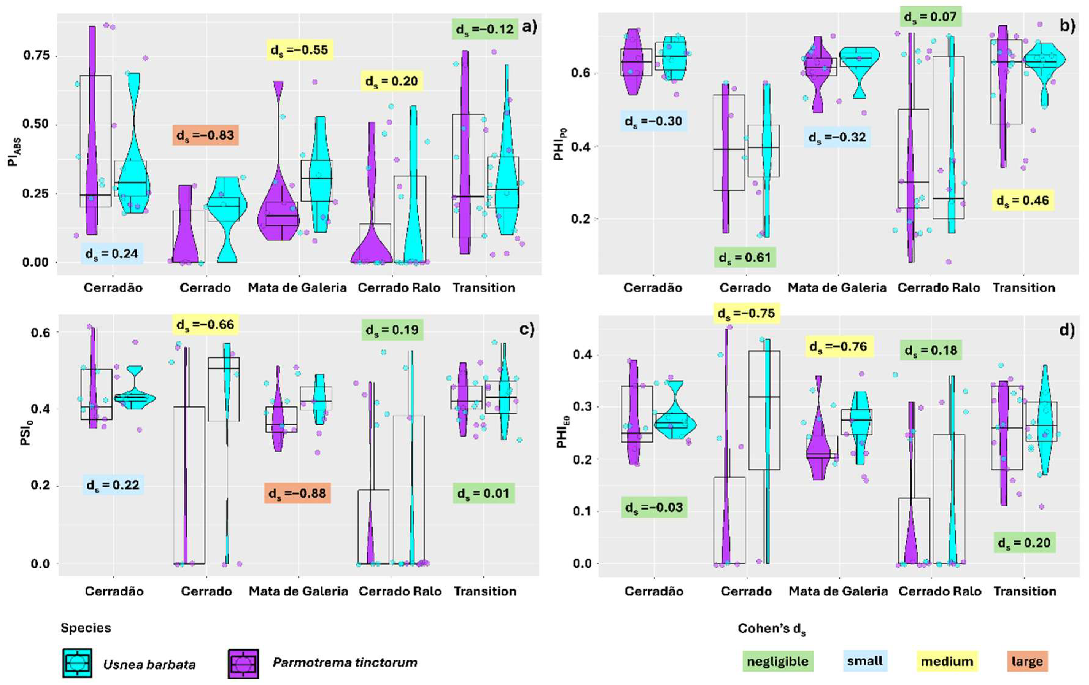
Photosynthetic performance variables were also affected by the landscape, responding consistently with the previously analyzed energy transport variables. Photosynthetic performance (PiABS) was lower in thalli sampled in Cerrado and Cerrado Ralo (0.124 and 0.128, respectively) compared to those in the other landscapes of the park (0.24, 0.31, and 0.38 for Gallery Forest, Transition Forest, and Cerradão, respectively) (Figure 2a). Similarly, thalli sampled in Cerrado and Cerrado Ralo exhibited a lower maximum quantum yield of primary photochemistry (PHIPO) (0.38 and 0.37, respectively) than those from the other landscapes (Figure 2b). The probability of a trapped exciton moving an electron through the electron transport chain beyond quinone (PSI0) and the quantum yield of electron transport (PHIE0) were also similarly affected by the landscape, with lower values observed in Cerrado and Cerrado Ralo (0.26 and 0.14 for PSI0, and 0.17 and 0.09 for PHIE0, respectively) (Figure 2c,d).
When evaluating the effect of lichen species within the different landscapes analyzed, we found that for ABS/RC, the effect size was moderate only in Gallery Forest, where U. barbata (4.60) exhibited lower mean ABS/RC values than P. tinctorum (5.01). In the other areas, the effect size was negligible or small (Figure 3a). For TR0/RC, the effect was negligible only when comparing species sampled in Transition Forest. In Cerradão, Gallery Forest, and Cerrado Ralo, the effect size was moderate, with U. barbata showing lower mean values in Cerrado Ralo and Gallery Forest (2.84 and 3.53, respectively) compared to P. tinctorum (3.00 and 3.67). Similarly, U. barbata exhibited lower TR0/RC means in thalli sampled in Cerrado (2.37 and 3.30 for U. barbata and P. tinctorum, respectively), with a large effect size. However, in the Cerradão landscape, P. tinctorum showed lower TR0/RC mean values (2.63) than U. barbata (2.85) (Figure 3b).
The ET0/RC values were also influenced by species, with P. tinctorum showing higher means than U. barbata across all landscapes evaluated. However, while the effect size was negligible in the Transition Forest and Cerrado Ralo, it was considered large in the Cerradão and Gallery Forest (Figure 3c). For DI0/RC, the differences between species were negligible in Cerradão, Cerrado Ralo, and Transition Forest. In Cerrado and Gallery Forest, the effect size was small, with U. barbata showing lower mean energy dissipation as heat (5.88 and 1.76, respectively) compared to P. tinctorum (8.40 and 1.96, respectively) (Figure 3d). Differences in PHIDO means were negligible between species in Cerrado Ralo and small in Cerradão and Gallery Forest. These results corroborate previous findings, with U. barbata exhibiting lower energy dissipation means (0.35 and 0.38, respectively) than P. tinctorum (0.37 and 0.39, respectively) (Figure 3e).
Photosynthetic performance (PiABS) of the thalli was also species-dependent, with the effect size being negligible only for thalli sampled in Transition Forest. In Cerradão, the effect size was small, with U. barbata showing a higher average performance (0.44) than P. tinctorum (0.37). In the Gallery Forest and Cerrado Ralo landscapes, the results were similar, with U. barbata exhibiting better photochemical performance (0.30 and 0.11, respectively) compared to P. tinctorum (0.23 and 0.15, respectively); however, the effect size was medium. In the Cerrado area, the effect size was large, and the results were consistent in the comparison between the species (0.18 and 0.09, respectively, for U. barbata and P. tinctorum) (Figure 4a).
The PHIPO data were also differentially affected by lichen species, although the differences were negligible in the Cerrado and Cerrado Ralo areas. In the Cerradão and Gallery Forest areas, however, the effect size was small, with U. barbata thalli showing better quantum yield (0.64 and 0.62, respectively) than P. tinctorum (0.63 and 0.61, respectively). In the Transition Forest, however, the effect size was moderate, with U. barbata (0.62) showing a higher average yield than P. tinctorum (0.58) (Figure 4b). For the PSI0 data, the effect size was negligible in thalli sampled from the Cerrado Ralo and Transition Forest. In the Cerradão landscape, however, the effect was small, with the highest averages found in U. barbata (0.46 and 0.43 for U. barbata and P. tinctorum, respectively). In the Cerrado area, the effect size was moderate, but the results were similar (0.40 and 0.18 for U. barbata and P. tinctorum, respectively). Higher averages for U. barbata (0.43) compared to P. tinctorum (0.38) were also found in the Gallery Forest landscape, though the effect size was large (Figure 4c).
The electron transport quantum yield (PHIE0) was also affected by species in the different landscapes evaluated. However, the effect size was negligible for samples from Cerradão, Cerrado Ralo, and Transition Forest. In the Cerrado and Gallery Forest landscapes, the effect size was moderate, with U. barbata showing higher average yields (0.40 and 0.27, respectively) compared to P. tinctorum (0.23 and 0.18) (Figure 4d).
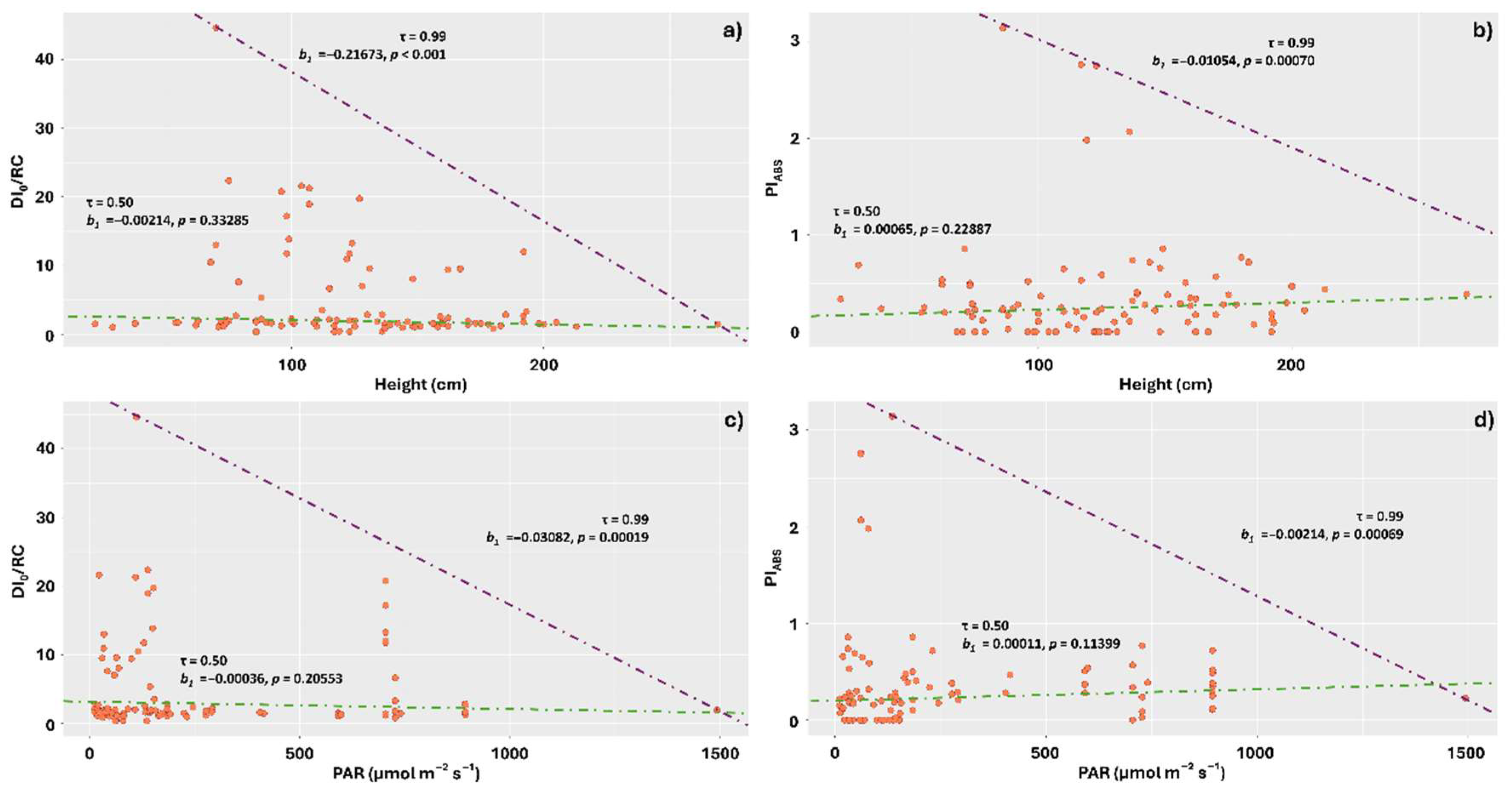
When we evaluated the effect of lichen sampling height on tree trunks, PAR, and different components of the visible spectrum on DI0/RC and PiABS, the regression at the 0.99 quantile indicated a negative effect of increasing lichen sampling height on energy dissipation in the form of heat, with thalli sampled on trunks between heights of 70 and 130 cm tending to show the highest average dissipation (Figure 5a). Similarly, this quantile showed a negative effect of increasing height on the photosynthetic performance index (PiABS), with the highest performance concentrated in the height range of 60 to 180 cm (Figure 5b).
At quantile 0.99, PAR also affected DI0/RC and PiABS, with the greatest dissipation and performances observed under lower amounts of PAR (respectively, between 0 and 200 µmol m−2 s−1 for DI0/RC, and between 0 and 300 µmol m−2 s−1 for PiABS) (Figure 5c,d).
At quantile 0.99, increasing PPFD on the stems negatively affected energy dissipation in the form of heat, with the highest average dissipations concentrated in the range between 30 and 500 µmol m−2 s−1 (Figure 6a). The effects of PPFD on PiABS, however, were contrasting for quantiles 0.50 and 0.99. At 0.50, the relationship was positive, with increases in PPFD enhancing photochemical performance, consistent with the observations for DI0/RC. At 0.99, however, the relationship was negative, with increases in PPFD leading to decreased performance in the stems. The highest performances were observed in the range between 0 and 120 µmol m−2 s−1 (Figure 6b).
Similar results were observed for the PFDUV component. At quantile 0.99, increases in PFDUV led to reductions in DI0/RC, while at quantile 0.50, increases positively affected photosynthetic performance. At quantile 0.99, however, higher PFDUV values negatively affected performance, with the highest indices observed in the range between 1 and 4 µmol m−2 s−1 for this component (Figure 6c,d).
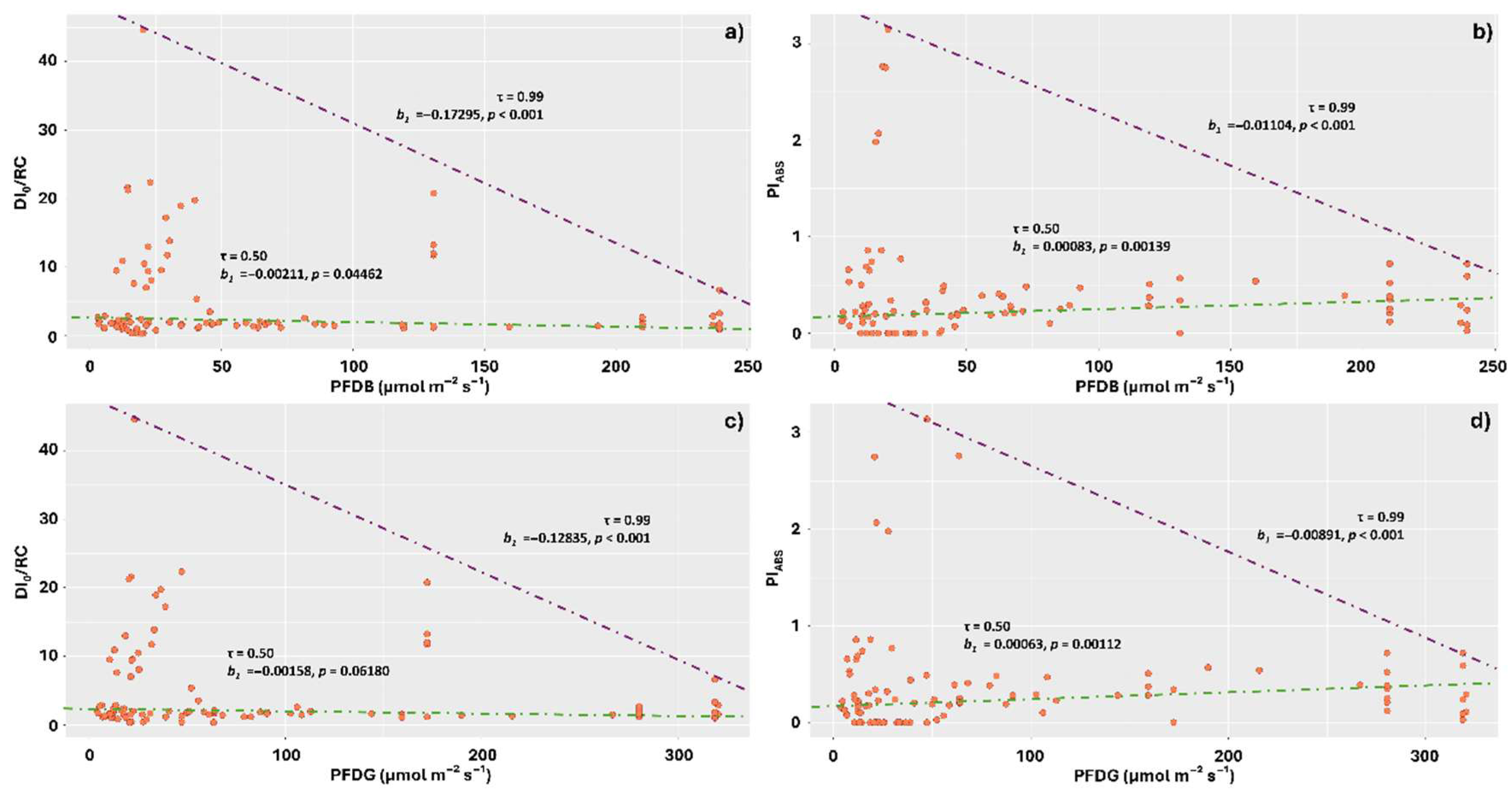
The means of PFDB and PFDG affected the DI0/RC values in a similar way, with negative relationships observed in the quantile triangle from 0.50 to 0.99. Thus, increases in these visible light components negatively affected energy dissipation, indicating photochemical stress in the stems where the incidence of these light components was lower. The highest energy dissipation in the form of heat was observed in the ranges of 0 to 50 µmol m−2 s−1 for PFDB and 0 to 60 µmol m−2 s−1 for PFDG (Figure 7a,c).
The PiABS responses were also similar for PFDB and PFDG, with contrasting responses observed between the 0.50 and 0.99 quantiles. In the first quantile, PiABS means tended to increase in response to increases in PFDB and PFDG values, which corroborates the results observed for DI0/RC. At the 0.99 quantile, however, photosynthetic performance decreased in response to increased values of the aforementioned components (Figure 7b,d).
The DI0/RC averages also responded negatively to the increase in the amounts of the red component of visible light, indicating higher stress levels under lower incidences of PFDR, with the highest averages concentrated in the range of 0 to 70 µmol m−2 s−1 (Figure 8a). PiABS, however, exhibited a response that corroborates this result at the 0.50 quantile level, with the lowest performances observed at the lowest incidences of PFDR. At the 0.99 quantile, however, the response was opposite, with PiABS values decreasing as the amount of this component increased (Figure 8b).
The responses to far-red light were similar to those observed for PFDR, with DI0/RC averages tending to decrease in response to increasing PFDFR at the 0.99 quantile (Figure 8c), with the greatest dissipations concentrated in the ranges of 10 to 70 µmol m−2 s−1. The results observed for PiABS at the 0.50 quantile corroborate those observed for DI0/RC; however, at the 0.99 quantile, they show a reduction in photochemical performance as the incident levels of PFDFR increased (Figure 8d).
Irradiance significantly affected the DI0/RC averages only at the 0.99 quantile, with lower IRR values leading to greater dissipation, with these being concentrated in the range of 0 to 50 W m−2 (Figure 9a). PiABS values, on the other hand, exhibited contrasting responses for the different quantiles. At 0.50, the best performances were observed under higher irradiances; however, at 0.99, the highest PiABS averages were observed under lower IRRs (Figure 9b).

3. Discussion

3.1. High Levels of Energy Dissipation Observed in Thalli Sampled from the Cerrado and Cerrado Ralo Landscapes Resulted in Lower Photochemical Performance

In the Cerrado, thalli were exposed to excessive light conditions (54,130 lm m−2), which can cause photoinhibition, burns, and dehydration [32]. These high light intensities led to the activation of algal protective mechanisms, including energy dissipation in the form of heat. Osyczka and Myśliwa-Kurdziel [1] suggest that the functional plasticity of the thylakoid membranes in photobionts plays a significant role in the dispersal abilities of lichens in particular habitats. U. barbata and P. tinctorum are abundant in Cerrado areas, and our findings support the idea that these species possess efficient adaptive and photoprotective mechanisms, which ensure their resilience and help prevent photodamage.
The photobionts of U. barbata and P. tinctorum are green algae of the genus Trebouxia [33,34,35,36], which are nonmotile coccoid algae with characteristic pyrenoids and lobed chloroplasts [37]. Studies show that the photochemical efficiency of these algae can respond differentially to environmental changes, such as increasing temperatures. For example, Trebouxia asymmetrica did not alter its Fv/Fm values when acclimated to different temperatures (17 and 20 °C). In contrast, Trebouxia sp. ‘arnoldoi’ and T. cretacea were more sensitive to temperature changes, with a decrease in Fv/Fm when exposed to 17 °C [38]. Thus, the photochemical and photosynthetic performance of thalli can be influenced by the symbiotic Trebouxia species, as these algae exhibit varying sensitivities to fluctuations in temperature and irradiance in their growth environment [39].
In this study, we examined the sensitivity of the photobionts of U. barbata and P. tinctorum to high light levels in the open Cerrado physiognomy. High irradiation levels can induce oxidative stress in photobionts. Muhetaer et al. [40] demonstrated a significant correlation between increased PAR and elevated activities of antioxidant enzymes, as well as higher H2O2 content, in the algae Phormidium ambiguum and Microcystis aeruginosa. Non-photochemical quenching (NPQ) in the chloroplast helps to dissipate excess light energy from PSII, constituting one of the mechanisms that protect photobionts against oxidative stress [41,42]. However, despite the protective role of NPQ, inevitable damage to PSII leads to a reduction in linear electron transport, which in turn limits downstream carbon assimilation.
In the Cerrado Ralo landscape, where irradiation levels are high (47,666 lm m−2), we also observed the activation of photoprotective mechanisms in the thalli. These mechanisms acted by increasing energy dissipation and decreasing chlorophyll a fluorescence performance. This is because the canopy in this plant formation is discontinuous, with many open areas that allow light to pass through and directly impact the thalli. Increases in NPQ or energy dissipation via active reaction centers (DI0/RC) are efficient mechanisms to alleviate excitation pressure in PSII, reducing damage to the photosynthetic apparatus [43]. However, this comes at the cost of overall photosynthetic performance (PiABS) [27]. Several studies highlight the negative effects of high light exposure on PiABS [44,45].
As expected for species exposed to excessive light, the high light intensity in the thalli of U. barbata and P. tinctorum in the Cerrado and Cerrado Ralo areas led to an increase in the average ABS/RC. This was due to a reduction in antenna size (fewer light-harvesting pigments—chlorophylls and carotenoids—per reaction center), which subsequently decreased ET0/RC. With reduced QA, which cannot be oxidized efficiently, the energy is not used effectively in photochemical reactions and electron transport but instead dissipates as heat (resulting in increased DI0/RC) [27,44,46].

3.2. U. barbata Thalli Exhibited Superior Photochemical Performance Compared to P. tinctorum Across All Sampled Landscapes

Studies suggest that Trebouxia photobionts possess mechanisms to repair PSII damage and resynthesize damaged proteins, primarily through the stimulation of cyclic electron transport (CET) in PSI. Beckett et al. [47] suggested that CET contributes to the photoprotection of lichenized algae under light stress. CET promotes ATP synthesis and facilitates the rapid recovery of PSII [43,48]. Although both U. barbata and P. tinctorum may activate these pathways, U. barbata exhibits additional protective mechanisms that enhance its resilience across different landscapes. One key factor contributing to U. barbata’s higher resistance is its production of antioxidant metabolites, particularly usnic acid, which protects against oxidative stress caused by excessive light [49]. As the dominant secondary metabolite in Usnea, usnic acid has strong antioxidant and cytoprotective properties [50]. Moreover, Usnea species retain little water [51], yet their photosynthetic pathways reactivate almost immediately upon exposure to humid air, demonstrating high plasticity in response to water deficits [52].
The morphological structure of usnic hair lichens further supports photoprotection. Their light-colored, semi-transparent cortex allows light penetration even when dry, exposing desiccated chlorophylls to oxidative stress. The yellow-green pigmentation, a combination of chlorophyll and usnic acid, helps filter solar radiation by reflectance, reducing heat stress [53,54,55] and prolonging photosynthesis after hydration [52]. Usnic acid accumulates in the cortical layer of Usnea lichens, acting as a natural light filter that regulates solar irradiance reaching the algal layer and protects the photobiont from excessive radiation [56]. This ability to synthesize usnic acid contributes to Usnea’s physiological plasticity. For instance, U. barbata contains approximately 1.5% usnic acid [57], which enhances light reflection and functions as a flexible solar radiation screen [58]. Phenolic acids also contribute to energy dissipation and protection against free radicals [49], acting in concert with the branched architecture of the Usnea thallus to minimize direct exposure of the photobiont to intense radiation. Engel [59] suggests that in addition to usnic acid, the metabolites atranorin, chloroatra-norin, barbatolic acid, lobaric acid, and salazinic acid, present in the thalli of lichens of this genus, have protective action against UVB radiation.
Gauslaa and Goward [23] suggest that under drought conditions, the cortical parenchyma of usnic lichens shrinks, leading to the collapse of photobiont cells. As a result, air fills the cortex, making it partially transparent. These morphological changes cause the cortical window to close partially, increasing chlorophyll reflectance and enhancing thallus photoprotection. The photoprotection responses observed in U. barbata across different environments can thus be attributed to the combined effects of high light incidence and low water availability. In contrast, under high humidity and shaded conditions, the cortex expands, and the photobiont cells regain turgidity, rendering the cortex more transparent. This shift optimizes photosynthesis by enhancing light capture.
Despite differences between U. barbata and P. tinctorum, species of Parmotrema are also considered resilient. Its widespread distribution across tropical phytophysiognomies highlights its ability to withstand low humidity and high light conditions. Studies show that in Usnea thalli, but especially in foliose lichens such as those of the genus Parmotrema [60], atranorin, a secondary metabolite belonging to the depsid class, contributes to the photoprotection of the thallus. Atranorin can prevent water from entering the spaces between hyphae in the cortex. The air-filled cavities with white atranorin crystals reflect excess light, playing a significant photoprotective role for the symbiotic green algae [54]. Despite this, the fruticose growth form of U. barbata, characterized by the presence of a cartilaginous central axis and a denser medulla [19,61], together with the production of high concentrations of cortical usnic acid, likely provides a more effective photoprotective effect than the atranorin deposited in the foliose cortex of P. tinctorum.
The more pronounced photoinhibition of photosynthesis in P. tinctorum appears to be a long-term regulatory mechanism for PSII, reducing metabolism and enhancing resistance to prolonged desiccation [4]. Barták et al. [62] suggest that lichens such as Xanthoria elegans tolerate high light stress effectively due to photoprotective mechanisms activated during photoinhibition. These mechanisms include photoinhibitory quenching (qIₜ), which involves structural modifications in the photosynthetic apparatus and constitutes a major component of NPQ. Similarly, Rautenberger and Hurd [63] observed that the combined action of photoinhibitory quenching and PSII reaction center quenching in response to light stress is a key mechanism enabling the chlorophyte Ulva rigida to thrive and form green tides in coastal ecosystems.
Although P. tinctorum exhibited lower PiABS levels, its capacity for heat dissipation was comparable to that of U. barbata, suggesting an efficient NPQ mechanism. Miyake et al. [64] suggested that, in desiccated thalli of P. tinctorum, the dissipation of excess light energy is characterized by a rapid fluorescence decay with a time constant of 27 ps in the far-red region. This quenching mechanism exhibits extremely high efficiency and is likely associated with the formation of a rapid quenching state in the peripheral-antenna system of PSII during desiccation. On the other hand, Adams et al. [65] demonstrated that green algal photobionts from lichens inhabiting arid environments accumulate zeaxanthin in response to increasing sunlight intensity, confirming the xanthophyll cycle-dependent nature of NPQ in symbiotic green algae. Their findings also indicated that substantial light exposure, beyond desiccation alone, is required to induce zeaxanthin accumulation in these photobionts. Therefore, the specific contribution of the xanthophyll cycle to fluorescence quenching in P. tinctorum remains an open question.

3.3. Lichens Sampled Higher on the Stem and Exposed to Higher PAR Levels Exhibited Lower Energy Dissipation as Heat and Reduced Photochemical Performance

High radiation levels induce photochemical damage, necessitating mechanisms to eliminate reactive oxygen species (ROS). If these mechanisms are compromised by other environmental stressors, excessive ROS accumulation can damage the photosynthetic apparatus, particularly PSII [66,67]. Although NPQ is only one of a host of mechanisms that protect against high light, our findings indicate that lichens sampled at higher positions on the stem—where they were more exposed to radiation in the open Cerrado and Cerrado Ralo landscapes—exhibited insufficient dissipation mechanisms, leading to photoinhibition and reduced photosynthetic capacity in photobiont cells. Mkhize et al. [68] suggest that lichens growing in fluctuating light conditions generally exhibit higher levels of NPQ compared to those exposed to constant full sunlight. If the lichens located further down the stem are subjected to more variable light conditions than those higher up, this could explain their elevated NPQ values. However, in the evaluated species, this damage appears to be reversible as long as the stress is not prolonged [11]. Additionally, contrasting performance responses at the 0.50 and 0.99 quantile levels suggest that photoprotective mechanisms are only activated beyond certain radiation thresholds, ultimately reducing PiABS.

3.4. Increasing Intensities of the Different Spectrum Components Reduced DI0/RC and Increased PiABS

Interestingly, when evaluating the spectral components separately, we observed a positive photochemical response of the thalli to individual increases in each component’s intensity. This is because, in general, a reduction in DI0/RC indicates that less energy is being dissipated per reaction center, meaning that more energy is effectively used for electron transport, which is favorable for photosynthetic efficiency. This suggests good acclimation of the lichen species to the prevailing environmental conditions (e.g., [69]). Conversely, an increase in PiABS indicates that the photosynthetic apparatus is more efficient and functionally active, with a greater capacity to convert absorbed light into chemical energy. This reflects a well-performing photosynthetic system adapted to environmental conditions [70]. This finding highlights the photochemical resistance of P. tinctorum, and especially U. barbata, to increased light levels, a resilience likely ensured by their ability to effectively quench excess energy under stress.
Numerous studies have demonstrated highly efficient NPQ mechanisms in lichens under high light intensity [62]. Barták et al. [71] show an increase in NPQ with desiccation of lichen thalli. In lichens that have dried out, NPQ is characterized by rapidly decaying fluorescence components around 690 and 740 nm. Notably, fluorescence quenched under dry conditions recovers within minutes after rewetting, suggesting that most quenching results from a reversible change in the state of antenna proteins rather than a reduction in protein content [72]. Beckett et al. [11] demonstrated that in desiccated thalli, the formation of ROS can lead to photoinhibition and photo-oxidative stress, thereby reducing the carbon fixation capacity of photobionts. Tolerance can be achieved by minimizing ROS production, through the synthesis of antioxidant pigments, thermal dissipation of excess absorbed light energy, scavenging of ROS after formation, or repairing ROS-induced damage. Evidence indicates the presence of two distinct pathways for dissipating excess light energy in dry thalli of P. tinctorum: one characterized by a rapid fluorescence decay with a time constant of 27 ps in the far-red region, absent in wet thalli, and another involving accelerated fluorescence decay in the 685–700 nm spectral range [64].
Field measurements during the dry season revealed that among the 11 macrolichen species evaluated, only the foliose chlorolichen P. tinctorum remained metabolically active, exhibiting slight carbon assimilation even under desiccation. This finding highlights its remarkable resistance to extreme environments [73]. Studies indicate that in Trebouxia algae exposed to desiccation and high light, variable chlorophyll fluorescence is lost, signifying a suspension of charge separation in PSII. Simultaneously, basal fluorescence (F0) is strongly quenched, which has been interpreted as evidence of high photoprotective non-radiative dissipation (NRD) [74]. This mechanism likely explains the observed resistance of P. tinctorum and U. barbata to increasing spectral components. Understanding these adaptive physiological strategies provides essential insights into the survival and distribution of lichens in the stressful landscapes of the savanna, forming a strong foundation for future research in this field.

4. Materials and Methods

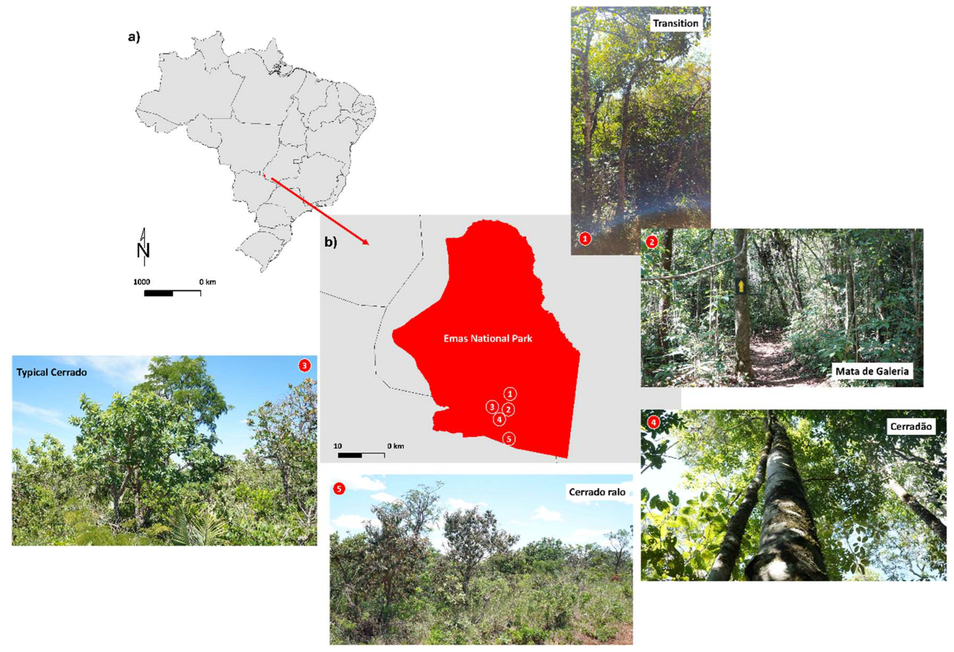
The foliose lichen Parmotrema tinctorum and the fruticose lichen Usnea barbata were evaluated in this study. Samples were observed in situ at their natural occurrence sites, which included different landscape types within the Emas National Park, Goiás, Brazil (Figure 10a). These landscapes were classified as Cerradão, Cerrado, Cerrado Ralo, Mata de Galeria, and Mata de Transição (Transition) based on the characteristics of the resident vegetation, following Lopes-Assad [75], and considering local light availability.
  • Cerradão (18°15′05.2″ S; 52°53′12.2″ W)—Mean illuminance: 13,023 lm m−2. Characterized by dense forest vegetation with structural features intermediate between savanna and tropical forest. Dominated by a continuous tree stratum, emergent trees, and a relatively open understory.
  • Typical Cerrado (18°19′42″ S; 52°52′51″ W)—Mean illuminance: 54,130 lm m−2. An open savanna formation with predominantly herbaceous and grassy vegetation, interspersed with a few shrubs and sparse trees.
  • Mata de Galeria (Gallery Forest) (18°15′33.6″ S; 52°53′13.7″ W)—Mean illuminance: 26,967 lm m−2. Located along the banks of the Formoso River, this vegetation type is dense, with multiple layers of trees, shrubs, and epiphytes.
  • Cerrado Ralo (18°15′33.4″ S; 52°53′21.6″ W)—Mean illuminance: 47,666 lm m−2. Slightly more open than the typical Cerrado, with a predominance of herbaceous vegetation, sparse shrubs, and widely spaced trees.
  • Mata de Transição (Transition) (18°13′44.1″ S; 52°52′40.8″ W)—Mean illuminance: 17,010 lm m−2. Represents an intermediate stage between open areas (campo sujo) and forested formations (cerradão). The vegetation is mixed, consisting of trees, shrubs, and grasses. The canopy is discontinuous, with sparse trees and a well-developed herbaceous-shrub layer (Figure 10b).
At each location, lichen sampling was conducted within a physically demarcated 200 m2 area.
The average illuminance (lm m−2) levels in each landscape were determined from measurements taken at five randomly selected points within the sampling area. Data were collected using a Li-Cor LI-180 spectroradiometer on 5–7 November. Physiological parameters and incident light quality on the lichen thalli were recorded between 10:00 and 11:00 AM. Measurements were taken in situ, with lichens remaining attached to tree trunks while the spectroradiometer was positioned accordingly. Based on these measurements, light intensity across landscapes was classified as low (<900 lm m−2—equivalent to <16.65 µmol m−2 s−1), medium (900–23,000 lm m−2—equivalent to 16.65–425.50 µmol m−2 s−1), high (23,000–50,000 lm m−2—equivalent to 425.50–925.00 µmol m−2 s−1), or very high (>50,000 lm m−2—equivalent to >925.00 µmol m−2 s−1), following an adaptation of the classification proposed by Nobel et al. [76].

4.1. Chlorophyll a Fluorescence in Thalli

Transient OJIP fluorescence of chlorophyll a was measured using a portable fluorometer (FluorPen FP 100, Photon Systems Instruments, Drásov, Czech Republic). Prior to measurement, all lichen thalli were dark-adapted for 30 min to ensure complete oxidation of the photosynthetic electron transport system. Subsequently, they were exposed to a 3000 µmol m−2 s−1 blue light pulse, and fluorescence kinetics were recorded. The minimum fluorescence (F0) was measured at 50 μs when all PSII reaction centers were open, defining the O step. This was followed by the J step (at 2 ms), the I step (at 30 ms), and the maximum fluorescence (Fm), corresponding to the P step when all PSII reaction centers were closed. These fluorescence parameters were used to estimate various PSII bioenergetic indices, following Strasser et al. [77], including: ABS/RC—specific light absorption flux per reaction center; TR0/RC—energy flux captured per reaction center at t = 0; ET0/RC—electron transport flux per reaction center; DI0/RC—specific energy dissipation flux at the antenna chlorophyll level; PHIPO—primary photochemical maximum quantum yield; PSI0—probability, at t = 0, of a trapped exciton moving an electron through the electron transport chain after Quinone; PHIE0—electron transport quantum yield after dark adaptation (30 min); PHIDO—photosynthetic quenching involving oxygen dissipation; and PiABS—photosynthetic performance index, integrating energy transfer processes from initial absorption to PQ reduction.

4.2. Light Measurement

The different types of light incident on the stems were measured using a spectroradiometer model LI-180, Li-Cor. For this purpose, the equipment was positioned attached to the tree trunk, in the position where the lichen occurred. The equipment was moved towards the sun, in order to capture the direct incident light. The spectral composition of the light was recorded in wavelength intervals of 1 nm (bandwidth of 12 nm). The information was recorded for the interval 380 to 780 nm and the components of the visible spectrum classified in the bands: Photosynthetic Photon Flux Density (PPFD), which refers to the number of photons that promote photosynthesis (in the range of 400 to 700 nm) and that reach a surface; Photon Flux Density Ultraviolet (PFDUV), which refers to the photon flux that is in the ultraviolet (UV) band, with wavelengths below 400 nm. Photon Flux Density Blue (PFDB), which measures the amount of photons in the blue light range (usually between 400 and 500 nm). Photon Flux Density Green (PFDG), which refers to the photon flux in the green light range (between 500 and 600 nm). Photon Flux Density Red (PFDR), which measures the amount of photons in the red range (usually between 600 and 700 nm). Photon Flux Density Far-Red (PFDFR), which refers to the photon flux in the far-red light range (usually between 700 and 800 nm) and Irradiance (IRR), which refers to the total power of electromagnetic radiation (including visible and non-visible light) that reaches a surface. It is a measure of the total energy delivered by radiation, regardless of its usefulness for photosynthesis.
Information on photosynthetically active radiation (PAR) incident on the stems was obtained using a PAR Sensor (APG-SQ-316, Apogee, North Logan, UT, USA). This sensor consists of a bar with 6 sensors, self-powered with an output of 0 to 800 mV. The sensor was previously calibrated for use with sunlight and subsequently positioned attached to the tree trunk, at the height of the lichen to be sampled. PAR information was collected by positioning the sensor towards the sun, in order to capture the direct incident light.

4.3. Position of Lichen on the Trunk

The position of each lichen on the tree trunk was recorded based on its height (cm) above the ground. Height measurements were obtained using a millimeter tape, extended from the ground to the lichen’s location, with or without the aid of a ladder. The evaluated height range spanned from 0.22 to 269 cm.

4.4. Experimental Design and Statistical Analyses

In each landscape, an average of 20 lichen specimens per species were evaluated. Samples were randomly collected along a 30 m transect extending toward the center of the landscape. The transect started 3 m from the edge of each study area to minimize edge effects. We employed Generalized Linear Mixed Models (GLMMs) to assess the effects of Landscape, Species, lichen height on the stem, and light spectrum characteristics on chlorophyll a fluorescence. Prior to model construction, we tested for multicollinearity among the variables using the “Car” package [78] and excluded those with a variance inflation factor (VIF) >5. The explanatory variables included Landscape, Species, Height, Photosynthetic Photon Flux Density (PPFD), Photon Flux Density in the UV range (PFDUV), Green (PFDG), Red (PFDR), Far-red (PFDFR), Infrared Radiation (IRR), and Photosynthetically Active Radiation (PAR). Chlorophyll a fluorescence parameters were analyzed separately, with Species and Height included as random factors, while the remaining explanatory variables were treated as fixed factors.
We utilized the MCMCglmm package [79] to perform the analyses in a Bayesian framework with the Markov Chain Monte Carlo algorithm. A total of 80,000 iterations with 20,000 burn-in chains and a Gaussian distribution were used. The Akaike information criteria (AIC) were used to select the best model: the model with the smallest AICc (the AIC corrected for sample size and the number of parameters), which was considered to be the most plausible for the explanation of the observed patterns [80]. The Delta AICci (ΔAICci, where i represents each model) was calculated as the difference between the AICc for the ith model and the smallest AICc observed. We also determined Akaike’s weight (wAICc), which represents the relative contribution of the ith model to the explanation of the observed pattern, given a set of competing models. The models with ΔAICc < 2 were all considered equally plausible as explanations of the observed pattern [81]. At the end of the analysis, the significance of the different models was compared to the simplest evaluated model (Landscape + Species).
The data were subjected to a normality test and subsequently analyzed using one-way ANOVA to assess the effect of Landscape on chlorophyll a fluorescence parameters. Significant differences between sampling sites were determined using Tukey’s test (p < 0.05). Additionally, the effect size of Species within each sampled Landscape was quantified using Cohen’s d, calculated with the cohensD function from the “effsize” package [82]. The d values were classified as small (≤0.2), medium (>0.2 to ≤0.5), or large (>0.5), following Sullivan and Feinn [83]. The relationships between visible light spectrum components, PAR, and lichen height on the trunk with fluorescence parameters were tested using quantile regression [84], implemented via the com.reg function from the “quantreg” package [85]. All analyses were performed in R, version 4.4.1 [86].

5. Conclusions

The results of this study demonstrate that P. tinctorum and U. barbata possess efficient photoprotective mechanisms, including energy dissipation as heat, which enable their survival in high-light environments such as the dry landscapes of the Brazilian Savanna. In particular, the challenging physiognomies of Cerrado and Cerrado Ralo, where high DI0/RC levels led to reduced photochemical performance, were highlighted. However, U. barbata exhibited greater resilience to light stress than P. tinctorum, likely due to the presence of antioxidant metabolites such as usnic acid. Additionally, lichens sampled at higher stem positions and under elevated PAR levels dissipated less energy as heat and exhibited lower photochemical performance, indicating damage to PSII under these conditions. Conversely, when different spectral components were analyzed separately, increased light intensities reduced DI0/RC and enhanced PiABS, suggesting photodamage resistance in P. tinctorum and U. barbata. The ability of both species to adapt to high-light conditions, coupled with their physiological plasticity, supports their widespread distribution in these tropical ecosystems. As a future perspective, we propose conducting molecular and metabolomic studies to characterize the antioxidant compounds and identify the regulatory pathways involved in the photoprotection mechanisms of U. barbata and P. tinctorum. Additionally, we recommend investigating these species across different seasons and microhabitats to better understand their physiological plasticity and adaptive limits in response to varying light intensities.

Author Contributions

Conceptualization, L.C.V. and L.A.B.; methodology, M.R.; formal analysis, B.G.C., M.M.d.S.M. and A.M.D.S.; investigation, B.G.C. and M.M.d.S.M.; resources, L.C.V.; writing—original draft preparation, L.C.V.; writing—review and editing, L.A.B.; visualization, L.C.V.; supervision, L.A.B.; project administration, L.A.B.; funding acquisition, L.C.V. and M.R. All authors have read and agreed to the published version of the manuscript.

Funding

This research received no external funding.

Data Availability Statement

All the data relevant to this manuscript are available on request from the corresponding author.

Acknowledgments

The authors would like to thank the Coordination for the Improvement of Higher Education Personnel (CAPES), the Foundation for Research Support of the State of Goiás (FAPEG), and the IFGoiano, Rio Verde campus for the infrastructure and the students involved in the study.

Conflicts of Interest

The authors declare no conflicts of interest.

References

  1. Osyczka, P.; Myśliwa-Kurdziel, B. The pattern of photosynthetic response and adaptation to changing light conditions in lichens is linked to their ecological range. Photosynth. Res. 2023, 157, 21–35. [Google Scholar] [CrossRef]
  2. Paoli, L.; Pisani, T.; Munzi, S.; Gaggi, C.; Loppi, S. Influence of sun irradiance and water availability on lichen photosynthetic pigments during a Mediterranean. Biologia 2010, 65, 776–783. [Google Scholar] [CrossRef]
  3. Piccotto, M.; Tretiach, M. Photosynthesis in chlorolichens: The influence of the habitat light regime. J. Plant Res. 2010, 123, 763–775. [Google Scholar] [CrossRef]
  4. Gasulla, F.; Del Campo, E.M.; Casano, L.M.; Guéra, A. Advances in understanding of desiccation tolerance of lichens and lichen-forming algae. Plants 2021, 10, 807. [Google Scholar] [CrossRef]
  5. Aubert, S.; Juge, C.; Boisson, A.M.; Gout, E.; Bligny, R. Metabolic processes sustaining the reviviscence of lichen Xanthoria elegans (Link) in high mountain environments. Planta 2007, 226, 1287–1297. [Google Scholar] [CrossRef]
  6. Lange, O.L.; Leisner, J.M.; Bilger, W. Chlorophyll fluorescence characteristics of the cyanobacterial lichen Peltigera rufescens under field conditions: II. Diel and annual distribution of metabolic activity and possible mechanisms to avoid photoinhibition. Flora 1999, 194, 413–430. [Google Scholar] [CrossRef]
  7. Boruah, T.; Dulal, K.; Das, P.N. Ecology of lichen. In Chemistry, Biology and Pharmacology of Lichen; Das, A.K., Sharma, A., Kathuria, D., Ansari, M.J., Bhardwaj, G., Eds.; Wiley: Hoboken, NJ, USA, 2024; pp. 49–69. [Google Scholar] [CrossRef]
  8. Morillas, L.; Roales, J.; Cruz, C.; Munzi, S. Resilience of epiphytic lichens to combined effects of increasing nitrogen and solar radiation. J. Fungi 2021, 7, 333. [Google Scholar] [CrossRef]
  9. Armstrong, R.A. Adaptation of lichens to extreme conditions. In Plant Adaptation Strategies in Changing Environment; Shukla, V., Kumar, S., Kumar, N., Eds.; Springer: Singapore, 2017; pp. 1–27. [Google Scholar] [CrossRef]
  10. Mikhaylov, A. Lichens as indicators of atmospheric pollution in urban ecosystems. Isr. J. Ecol. Evol. 2020, 67, 60–68. [Google Scholar] [CrossRef]
  11. Beckett, R.P.; Minibayeva, F.; Solhaug, K.A.; Roach, T. Photoprotection in lichens: Adaptations of photobionts to high light. Lichenologist 2021, 53, 21–33. [Google Scholar] [CrossRef]
  12. Veres, K.; Csintalan, Z.; Laufer, Z.; Engel, R.; Szabó, K.; Farkas, E. Photoprotection and high-light acclimation in semi-arid grassland lichens–a cooperation between algal and fungal partners. Symbiosis 2022, 86, 33–48. [Google Scholar] [CrossRef]
  13. Meeßen, J.; Sánchez, F.J.; Brandt, A.; Balzer, E.M.; de la Torre, R.; Sancho, L.G.; de Vera, J.P.; Ott, S. Extremotolerance and resistance of lichens: Comparative studies on five species used in astrobiological research I. Morphological and anatomical characteristics. Orig. Life Evol. Biosph. 2013, 43, 283–303. [Google Scholar] [CrossRef]
  14. Sachdev, S.; Ansari, S.A.; Ansari, M.I.; Fujita, M.; Hasanuzzaman, M. Abiotic stress and reactive oxygen species: Generation, signaling, and defense mechanisms. Antioxidants 2021, 10, 277. [Google Scholar] [CrossRef]
  15. Schneider, J.R.; Caverzan, A.; Chavarria, G. Water deficit stress, ROS involvement, and plant performance. Arch. Agron. Soil Sci. 2019, 65, 1160–1181. [Google Scholar] [CrossRef]
  16. Foyer, C.H. Reactive oxygen species, oxidative signaling and the regulation of photosynthesis. Environ. Exp. Bot. 2018, 154, 134–142. [Google Scholar] [CrossRef]
  17. Huiskes, A.H.L.; Gremmen, N.J.M.; Francke, J.W. Morphological effects on the water balance of Antarctic foliose and fruticose lichens. Antarct. Sci. 1997, 9, 36–42. [Google Scholar] [CrossRef]
  18. Spielmann, A.A.; Marcelli, M.P. Parmotrema sl (Parmeliaceae, lichenized Ascomycota) from Serra Geral slopes in central Rio Grande do Sul State, Brazil. Hoehnea 2009, 36, 551–595. [Google Scholar] [CrossRef]
  19. Popovici, V.; Bucur, L.; Gîrd, C.E.; Calcan, S.I.; Cucolea, E.I.; Costache, T.; Rambu, D.; Ungureanu-Iuga, M.; Oroian, M.; Mironeasa, S.; et al. Advances in the characterization of Usnea barbata (L.) Weber ex FH Wigg from Călimani Mountains, Romania. Appl. Sci. 2022, 12, 4234. [Google Scholar] [CrossRef]
  20. Souza, C.L.F.; de Oliveira, R.B.; Mustafé, D.N.; Nunes, K.A.C.; de Morais, E.M.B. O cerrado como o “berço das águas”: Potencialidades para a educação geográfica. Rev. Cerrados 2019, 17, 86–113. [Google Scholar] [CrossRef]
  21. Klink, C.A.; Machado, R.B. Conservation of the Brazilian cerrado. Conserv. Biol. 2005, 19, 707–713. [Google Scholar] [CrossRef]
  22. Colli, G.R.; Vieira, C.R.; Dianese, J.C. Biodiversity and conservation of the Cerrado: Recent advances and old challenges. Biodivers. Conserv. 2020, 29, 1465–1475. [Google Scholar] [CrossRef]
  23. Gauslaa, Y.; Goward, T. Sunscreening pigments shape the horizontal distribution of pendent hair lichens in the lower canopy of unmanaged coniferous forests. Lichenologist 2023, 55, 81–89. [Google Scholar] [CrossRef]
  24. Sharma, N.; Nagar, S.; Thakur, M.; Suriyakumar, P.; Kataria, S.; Shanker, A.K.; Landi, M.; Anand, A. Photosystems under high light stress: Throwing light on mechanism and adaptation. Photosynthetica 2023, 61, 250. [Google Scholar] [CrossRef]
  25. Kataria, S.; Jajoo, A.; Guruprasad, K.N. Impact of increasing Ultraviolet-B (UV-B) radiation on photosynthetic processes. J. Photochem. Photobiol. B Biol. 2014, 137, 55–66. [Google Scholar] [CrossRef]
  26. Shao, N.; Krieger-Liszkay, A.; Schroda, M.; Beck, C.F. A reporter system for the individual detection of hydrogen peroxide and singlet oxygen: Its use for the assay of reactive oxygen species produced in vivo. Plant J. 2007, 50, 475–487. [Google Scholar] [CrossRef]
  27. Janeeshma, E.; Johnson, R.; Amritha, M.; Noble, L.; Aswathi, K.R.; Telesiński, A.; Kalaji, H.M.; Auriga, A.; Puthur, J.T. Modulations in chlorophyll a fluorescence based on intensity and spectral variations of light. Int. J. Mol. Sci. 2022, 23, 5599. [Google Scholar] [CrossRef]
  28. Bassi, R.; Dall’Osto, L. Dissipation of light energy absorbed in excess: The molecular mechanisms. Annu. Rev. Plant Biol. 2021, 72, 47–76. [Google Scholar] [CrossRef]
  29. Zavafer, A.; Mancilla, C. Concepts of photochemical damage of Photosystem II and the role of excessive excitation. J. Photochem. Photobiol. C Photochem. Rev. 2021, 47, 100421. [Google Scholar] [CrossRef]
  30. Phinney, N.H.; Solhaug, K.A.; Gauslaa, Y. Rapid resurrection of chlorolichens in humid air: Specific thallus mass drives rehydration and reactivation kinetics. Environ. Exp. Bot. 2018, 148, 184–191. [Google Scholar] [CrossRef]
  31. Muggia, L.; Vancurova, L.; Škaloud, P.; Peksa, O.; Wedin, M.; Grube, M. The symbiotic playground of lichen thalli–a highly flexible photobiont association in rock-inhabiting lichens. FEMS Microbiol. Ecol. 2013, 85, 313–323. [Google Scholar] [CrossRef] [PubMed]
  32. Bianchi, E.; Paoli, L.; Colzi, I.; Coppi, A.; Gonnelli, C.; Lazzaro, L.; Loppi, S.; Papini, A.; Vannini, A.; Benesperi, R. High-light stress in wet and dry thalli of the endangered Mediterranean lichen Seirophora villosa (Ach.) Frödén: Does size matter? Mycol. Prog. 2019, 18, 463–470. [Google Scholar] [CrossRef]
  33. Fatima, S.; APA; RKC; Sharma, B.; Gaikwad, S.; Mohan, A.; Sequeira, S. Deciphering the symbiosis of endemic Usnea ghattensis and their photobiont Trebouxia sp. through molecular tools from the northern Western Ghats, India. Microb. Biosyst. 2021, 6, 30–42. [Google Scholar] [CrossRef]
  34. Ohmura, Y.; Takeshita, S.; Kawachi, M. Photobiont diversity within populations of a vegetatively reproducing lichen, Parmotrema tinctorum, can be generated by photobiont switching. Symbiosis 2019, 77, 59–72. [Google Scholar] [CrossRef]
  35. Rafat, A.; Ridgway, H.J.; Cruickshank, R.H.; Buckley, H.L. Isolation and co-culturing of symbionts in the genus Usnea. Symbiosis 2015, 66, 123–132. [Google Scholar] [CrossRef]
  36. Ohmura, Y.; Kawachi, M.; Kasai, F.; Watanabe, M.M.; Takeshita, S. Genetic combinations of symbionts in a vegetatively reproducing lichen, Parmotrema tinctorum, based on ITS rDNA sequences. Bryologist 2006, 109, 43–59. [Google Scholar] [CrossRef]
  37. Muggia, L.; Candotto-Carniel, F.; Grube, M. The lichen photobiont Trebouxia: Towards an appreciation of species diversity and molecular studies. In Algal and Cyanobacteria Symbioses; Grube, M., Seckbach, J., Muggia, L., Eds.; World Scientific: Singapore, 2017; pp. 111–146. [Google Scholar] [CrossRef]
  38. Molins, A.; Chiva, S.; Calatayud, Á.; Marco, F.; García-Breijo, F.; Reig-Armiñana, J.; Carrasco, P.; Moya, P. Multidisciplinary approach to describe Trebouxia diversity within lichenized fungi Buellia zoharyi from the Canary Islands. Symbiosis 2020, 82, 19–34. [Google Scholar] [CrossRef]
  39. Casano, L.M.; del Campo, E.M.; García-Breijo, F.J.; Reig-Armiñana, J.; Gasulla, F.; Del Hoyo, A.; Guéra, A.; Barreno, E. Two Trebouxia algae with different physiological performances are ever-present in lichen thalli of Ramalina farinacea. Co-existence versus competition? Environ. Microbiol. 2011, 13, 806–818. [Google Scholar] [CrossRef]
  40. Muhetaer, G.; Jayasanka, S.M.; Fujino, T. Oxidative stress and antioxidant responses of Phormidium ambiguum and Microcystis aeruginosa under diurnally varying light conditions. Microorganisms 2020, 8, 890. [Google Scholar] [CrossRef] [PubMed]
  41. Han, L.J.; Fan, D.Y.; Wang, X.P.; Xu, C.Y.; Xia, X.L.; Chow, W.S. The protective role of non-photochemical quenching in PSII photosusceptibility: A case study in the field. Plant Cell Physiol. 2023, 64, 43–54. [Google Scholar] [CrossRef]
  42. Murchie, E.H.; Ruban, A.V. Dynamic non-photochemical quenching in plants: From molecular mechanism to productivity. Plant J. 2020, 101, 885–896. [Google Scholar] [CrossRef]
  43. Mlinarić, S.; Begović, L.; Katanić, Z.; Galić, V. Changes of chlorophyll a fluorescence parameters influenced by light and temperature Stress. In Chlorophyll a Fluorescence Measurements in Croatia—First Twenty Years; Hrvoje, L., Marija, V.V., Zvonimir, Z., Eds.; Instituto Agrícola: Osijek, Croatia, 2023; pp. 29–42. [Google Scholar]
  44. Kalaji, H.; Rastogi, A.; Živčák, M.; Brestic, M.; Daszkowska-Golec, A.; Sitko, K.; Alsharafa, K.; Lotfi, R.; Stypiński, P.; Samborska, I. Prompt chlorophyll fluorescence as a tool for crop phenotyping: An example of barley landraces exposed to various abiotic stress factors. Photosynthetica 2018, 56, 953–961. [Google Scholar] [CrossRef]
  45. Bayat, L.; Arab, M.; Aliniaeifard, S.; Seif, M.; Lastochkina, O.; Li, T. Effects of growth under different light spectra on the subsequent high light tolerance in rose plants. AoB Plants 2018, 10, ply052. [Google Scholar] [CrossRef]
  46. Markulj Kulundžić, A.; Viljevac Vuletić, M.; Matoša Kočar, M.; Antunović Dunić, J.; Varga, I.; Sudarić, A.; Cesar, V.; Zdunić, Z. Effect of elevated temperature and excess light on photosynthetic efficiency, pigments, and proteins in the field-grown sunflower during afternoon. Horticulturae 2022, 8, 392. [Google Scholar] [CrossRef]
  47. Beckett, R.P.; Roach, T.; Minibayeva, F.; Werth, S. Alternative electron transport pathways contribute to tolerance to high light stress in lichenized algae. Physiol. Plant. 2023, 175, e13904. [Google Scholar] [CrossRef]
  48. Allakhverdiev, S.I.; Kreslavski, V.D.; Klimov, V.V.; Los, D.A.; Carpentier, R.; Mohanty, P. Heat stress: An overview of molecular responses in photosynthesis. Photosynth. Res. 2008, 98, 541–550. [Google Scholar] [CrossRef]
  49. Popovici, V.; Bucur, L.; Popescu, A.; Schröder, V.; Costache, T.; Rambu, D.; Cucolea, I.E.; Gird, C.E.; Caraiane, A.; Gherghel, D.; et al. Antioxidant and cytotoxic activities of Usnea barbata (L.) FH Wigg. dry extracts in different solvents. Plants 2021, 10, 909. [Google Scholar] [CrossRef]
  50. White, P.A.S.; Oliveira, R.C.M.; Oliveira, A.P.; Serafini, M.R.; Araújo, A.A.S.; Gelain, D.P.; Moreira, J.C.F.; Almeida, J.R.G.S.; Quintans, J.S.S.; Quintans-Junior, L.J.; et al. Antioxidant activity and mechanisms of action of natural compounds isolated from lichens: A systematic review. Molecules 2014, 19, 14496–14527. [Google Scholar] [CrossRef]
  51. Eriksson, A.; Gauslaa, Y.; Palmqvist, K.; Ekström, M.; Esseen, P.-A. Morphology drives water storage traits in the globally widespread lichen genus Usnea. Fungal Ecol. 2018, 35, 51–61. [Google Scholar] [CrossRef]
  52. Phinney, N.H.; Asplund, J.; Gauslaa, Y. The lichen cushion: A functional perspective of color and size of a dominant growth form on glacier forelands. Fungal Biol. 2022, 126, 375–384. [Google Scholar] [CrossRef] [PubMed]
  53. Galanty, A.; Popiół, J.; Paczkowska-Walendowska, M.; Studzińska-Sroka, E.; Paśko, P.; Cielecka-Piontek, J.; Podolak, I. (+)-Usnic acid as a promising candidate for a safe and stable topical photoprotective agent. Molecules 2021, 26, 5224. [Google Scholar] [CrossRef]
  54. Solhaug, K.A.; Larsson, P.; Gauslaa, Y. Light screening in lichen cortices can be quantified by chlorophyll fluorescence techniques for both reflecting and absorbing pigments. Planta 2010, 231, 1003–1011. [Google Scholar] [CrossRef] [PubMed]
  55. Kershaw, K.A. Studies on lichen-dominated systems. XII. The ecological significance of thallus color. Can. J. Bot. 1975, 53, 660–667. [Google Scholar] [CrossRef]
  56. Bhattacharyya, S.; Deep, P.R.; Singh, S.; Nayak, B. Lichen secondary metabolites and its biological activity. Am. J. PharmTech Res. 2016, 6, 1–17. [Google Scholar] [CrossRef]
  57. Rojas, J.O.H.N.; Londoño, C.E.S.A.R.; Ciro, Y. The health benefits of natural skin UVA photoprotective compounds found in botanical sources. Int. J. Pharm. Pharm. Sci. 2016, 8, 13–23. [Google Scholar]
  58. Thakur, M.; Chander, H. Potential of lichens: A review of bioactive compounds with biological activities. Biol. Forum–Int. J. 2021, 13, 39–47. [Google Scholar]
  59. Engel, K.; Schmidt, U.; Reuter, J.; Weckesser, S.; Simon-Haarhaus, B.; Schempp, C.M. Usnea barbata extract prevents ultraviolet-B induced prostaglandin E2 synthesis and COX-2 expression in HaCaT keratinocytes. J. Photochem. Photobiol. B 2007, 89, 9–14. [Google Scholar] [CrossRef]
  60. Harikrishnan, A.; Veena, V.; Lakshmi, B.; Shanmugavalli, R.; Theres, S.; Prashantha, C.N.; Shah, T.; Oshin, K.; Togam, R.; Nandi, S. Atranorin, an antimicrobial metabolite from lichen Parmotrema rampoddense exhibited in vitro anti-breast cancer activity through interaction with Akt activity. J. Biomol. Struct. Dyn. 2021, 39, 1248–1258. [Google Scholar] [CrossRef]
  61. Passo, A.; Rodriguez, J.M.; Chiapella, J. New records of Antarctic lichens. N. Z. J. Bot. 2015, 53, 216–223. [Google Scholar] [CrossRef]
  62. Barták, M.; Hájek, J.; Halıcı, M.G.; Bednaříková, M.; Casanova-Katny, A.; Váczi, P.; Puhovkin, A.; Misha, K.B.; Giordano, D. Resistance of primary photosynthesis to photoinhibition in Antarctic lichen Xanthoria elegans: Photoprotective mechanisms activated during a short period of high light stress. Plants 2023, 12, 2259. [Google Scholar] [CrossRef] [PubMed]
  63. Rautenberger, R.; Hurd, C.L. Photoprotection by photoinhibitory and PSII-reaction centre quenching controls growth of Ulva rigida (Chlorophyta) and is a pre-requisite for green tide formation. Planta 2024, 259, 111. [Google Scholar] [CrossRef]
  64. Miyake, H.; Komura, M.; Itoh, S.; Kosugi, M.; Kashino, Y.; Satoh, K.; Shibata, Y. Multiple dissipation components of excess light energy in dry lichen revealed by ultrafast fluorescence study at 5 K. Photosynth. Res. 2011, 110, 39–48. [Google Scholar] [CrossRef]
  65. Adams, W.W.; Demmig-Adams, B.; Lange, O.L. Carotenoid composition and metabolism in green and blue-green algal lichens in the field. Oecologia 1993, 94, 576–584. [Google Scholar] [CrossRef]
  66. Sachdev, S.; Ansari, S.A.; Ansari, M.I. Photosynthetic apparatus: Major site of oxidative damage. In Reactive Oxygen Species in Plants: The Right Balance; Sachdev, S., Ansari, S.A., Ansari, M.I., Eds.; Springer Nature: Singapore, 2023; pp. 75–92. [Google Scholar] [CrossRef]
  67. Krieger-Liszkay, A.; Shimakawa, G. Regulation of the generation of reactive oxygen species during photosynthetic electron transport. Biochem. Soc. Trans. 2022, 50, 1025–1034. [Google Scholar] [CrossRef] [PubMed]
  68. Mkhize, K.G.W.; Minibayeva, F.V.; Beckett, R.P. Adaptations of photosynthesis in sun and shade in populations of some Afromontane lichens. Lichenologist 2022, 54, 319–329. [Google Scholar] [CrossRef]
  69. Marečková, M.; Barták, M. Short-term responses of primary processes in PS II to low temperature are sensitively indicated by fast chlorophyll fluorescence kinetics in Antarctic lichen Dermatocarpon polyphyllizum. Czech Polar Rep. 2017, 7, 74–82. [Google Scholar] [CrossRef]
  70. Katanić, Z.; Atić, L.; Ferhatović, D.; Cesar, V.; Lepeduš, H. PSII photochemistry in vegetative buds and needles of Norway spruce (Picea abies L. Karst.) probed by OJIP chlorophyll a fluorescence measurement. Acta Biol. Hung. 2012, 63, 218–230. [Google Scholar] [CrossRef]
  71. Barták, M.; Hájek, J.; Orekhova, A.; Villagra, J.; Marín, C.; Palfner, G.; Casanova-Katny, A. Inhibition of primary photosynthesis in desiccating Antarctic lichens differing in their photobionts, thallus morphology, and spectral properties. Microorganisms 2021, 9, 818. [Google Scholar] [CrossRef]
  72. Dall’Osto, L.; Caffarri, S.; Bassi, R. A mechanism of nonphotochemical energy dissipation, independent from PsbS, revealed by a conformational change in the antenna protein CP26. Plant Cell 2005, 17, 1217–1232. [Google Scholar] [CrossRef]
  73. Boonpeng, C.; Pischom, M.; Butrid, P.; Noikrad, S.; Boonpragob, K. Laboratory and field measurements of water relations, photosynthetic parameters, and hydration traits in macrolichens in a tropical lower montane rainforest in Thailand. J. Plant Res. 2024, 137, 641–658. [Google Scholar] [CrossRef] [PubMed]
  74. Wieners, P.C.; Mudimu, O.; Bilger, W. Desiccation-induced non-radiative dissipation in isolated green lichen algae. Photosynth. Res. 2012, 113, 239–247. [Google Scholar] [CrossRef]
  75. Lopes-Assad, M.L. Produtos de Extrativismo do Cerrado: Preservando a caixa d’água do Brasil. Observatório de Conhecimento e Inovação em Bioeconomia; Fundação Getúlio Vargas—FGV-EESP: São Paulo, Brasil, 2023. [Google Scholar]
  76. Nobel, P.S.; Zaragoza, L.J.; Smith, W.K. Relation between mesophyll surface area, photosynthetic rate, and illumination level during development for leaves of Plectranthus parviflorus Henckel. Plant Physiol. 1975, 55, 1067–1070. [Google Scholar] [CrossRef]
  77. Strasser, R.J.; Tsimilli-Michael, M.; Srivastava, A. Chlorophyll fluorescence: A signature of photosynthesis. In Chlorophyll a Fluorescence: A Signature of Photosynthesis; Papageorgiou, G.C., Govindjee, Eds.; Springer: Dordrecht, The Netherlands, 2004; pp. 321–362. [Google Scholar] [CrossRef]
  78. Fox, J.; Weisberg, S.; Price, B.; Adler, D.; Bates, D.; Baud-Bovy, G.; Bolker, B.; Ellison, S.; Firth, D.; Friendly, M.; et al. R-Core. Package Car. 2019. Available online: https://cran.r-project.org/web/packages/car/index.html (accessed on 1 January 2020).
  79. Hadfield, J.D. MCMC methods for multi-response generalized linear mixed models: The MCMCglmm R package. J. Stat. Softw. 2010, 33, 1–22. [Google Scholar] [CrossRef]
  80. Burnham, K.P.; Anderson, D.R. Model Selection and Multimodel Inference, 2nd ed.; Springer: New York, NY, USA, 2002. [Google Scholar] [CrossRef]
  81. Zuur, A.; Ieno, E.N.; Walker, N.; Saveliev, A.A.; Smith, G.M. Mixed Effects Models and Extensions in Ecology with R; Springer Science & Business Media: Berlin/Heidelberg, Germany, 2009. [Google Scholar] [CrossRef]
  82. Pham, V.T.K.; Rediers, H.; Ghequire, M.G.K.; Nguyen, H.H.; De Mot, R.; Vanderleyden, J.; Spaepen, S. The plant growth-promoting effect of the nitrogen-fixing endophyte Pseudomonas stutzeri A15. Arch. Microbiol. 2017, 199, 513–517. [Google Scholar] [CrossRef] [PubMed]
  83. Sullivan, G.M.; Feinn, R. Using effect size—Or why the P value is not enough. J. Grad. Med. Educ. 2012, 4, 279–282. [Google Scholar] [CrossRef] [PubMed]
  84. Cade, B.S.; Noon, B.R. A gentle introduction to quantile regression for ecologists. Front. Ecol. Environ. 2003, 1, 412–420. [Google Scholar] [CrossRef]
  85. Koenker, R.; Chernozhukov, V.; He, X.; Peng, L. Handbook of Quantile Regression; CRC Press: Boca Raton, FL, USA, 2017. [Google Scholar] [CrossRef]
  86. R Core Team. R: A Language and Environment for Statistical Computing; R Foundation for Statistical Computing: Vienna, Austria, 2024; Available online: https://www.R-project.org/ (accessed on 27 July 2024).
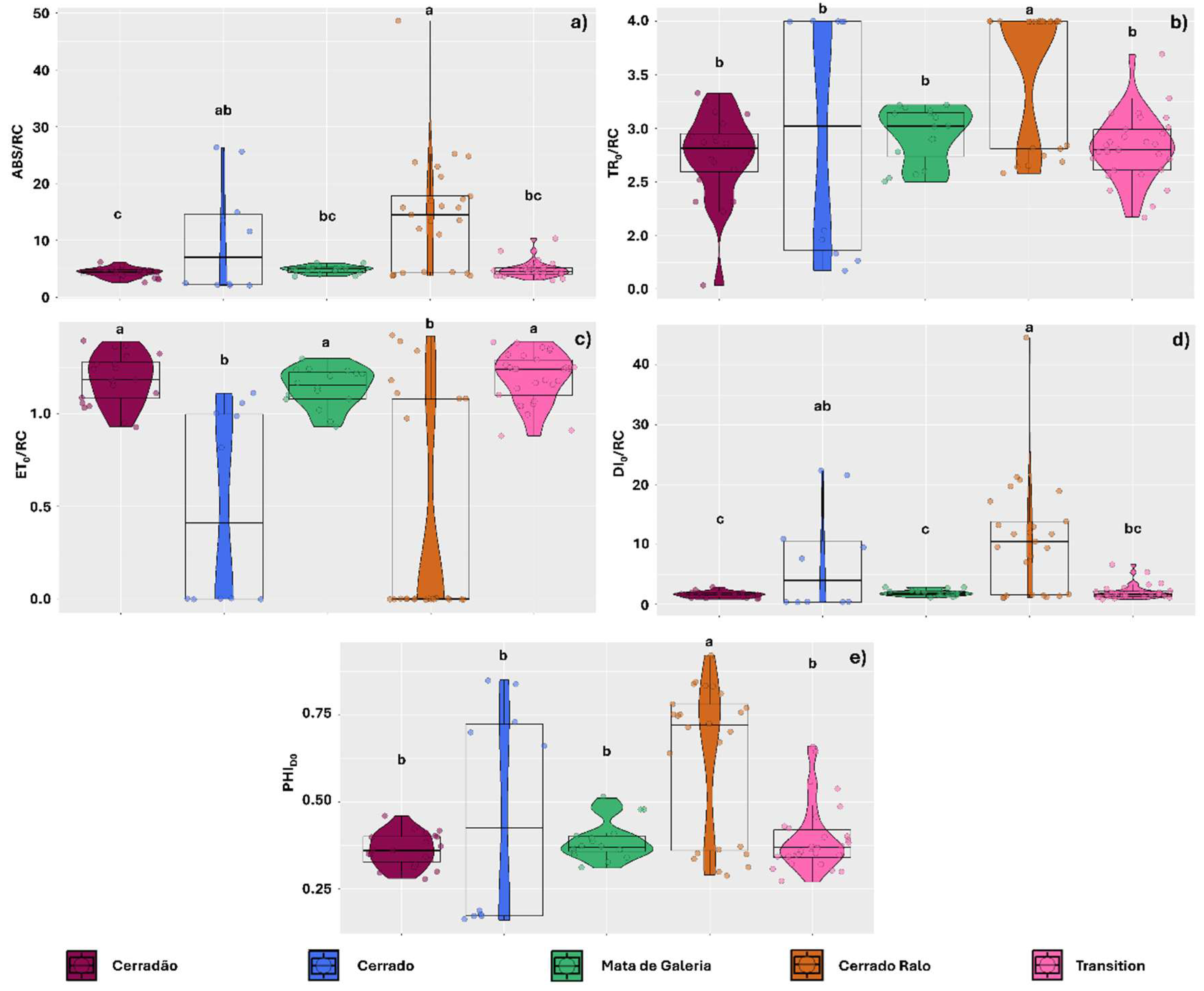
Figure 1. Chlorophyll a fluorescence parameters observed in thalli of the lichens Parmotrema tinctorum and Usnea barbata sampled across different landscapes in Emas National Park, Brazil. The parameters include: specific light absorption flux per reaction center (ABS/RC) (a); energy flux captured per reaction center at t = 0 (TR0/RC) (b); electron transport flux per reaction center (ET0/RC) (c); specific energy dissipation flux at the level of the antenna complex chlorophylls (DI0/RC) (d); and photosynthetic quenching associated with oxygen dissipation (PHIDO) (e). Violin plots depict data dispersion, with individual points representing observations. Boxplot central lines indicate medians, and letters above the boxplots denote means that do not differ significantly according to Tukey’s test (p < 0.05).
Plants 14 02802 g001
Figure 2. Chlorophyll a fluorescence parameters observed in thalli of the lichens Parmotrema tinctorum and Usnea barbata sampled across different landscapes in Emas National Park, Brazil. Parameters include photosynthetic performance index (PiABS) (a); maximum quantum yield of primary photochemistry (PHIPO) (b); probability of a trapped exciton transferring an electron beyond quinone in the electron transport chain (PSI0) (c); and quantum yield of electron transport (PHIE0) (d). Violin plots depict data dispersion, with individual points representing observations. Boxplot central lines indicate medians, and letters above the boxplots denote means that do not differ significantly according to Tukey’s test (p < 0.05).
Plants 14 02802 g002
Figure 3. Effect size (Cohen’s ds index) for chlorophyll a fluorescence parameters comparing thalli of the lichens Parmotrema tinctorum and Usnea barbata sampled across different landscapes in Emas National Park, Brazil. Parameters include: specific light absorption flux per reaction center (ABS/RC) (a); energy flux captured per reaction center at t = 0 (TR0/RC) (b); electron transport flux per reaction center (ET0/RC) (c); specific energy dissipation flux at the level of the chlorophylls in the antenna complex (DI0/RC) (d); and photosynthetic quenching involving oxygen dissipation (PHIDO) (e). The points associated with the violin plots represent data dispersion. The central line in the boxplots indicates the median, while the values and colors above the boxplots illustrate the Cohen’s ds effect size.
Plants 14 02802 g003
Figure 4. Effect size (Cohen’s ds index) for chlorophyll a fluorescence parameters comparing thalli of the lichens Parmotrema tinctorum and Usnea barbata sampled across different landscapes in Emas National Park, Brazil. Parameters include photosynthetic performance index (PiABS) (a); primary photochemical maximum quantum yield (PHIPO) (b); probability of a trapped exciton moving an electron through the electron transport chain after Quinone (PSI0) (c); and electron transport quantum yield (PHIE0) (d). The points associated with the violin plots represent data dispersion. The central line in the boxplots indicates the median, while the values and colors above the boxplots illustrate the Cohen’s ds effect size.
Plants 14 02802 g004
Figure 5. Quantile regression analysis of the relationships between chlorophyll a fluorescence parameters DI0/RC and PiABS with the sampling height of Parmotrema tinctorum and Usnea barbata lichens on tree trunks and photosynthetically active radiation (PAR) observed in different landscapes of Emas National Park, Brazil. Specific energy dissipation flux at the antenna complex chlorophylls level (DI0/RC) and photosynthetic performance index (PiABS) as a function of sampling height (a,b); DI0/RC and PiABS as a function of PAR (c,d). The figures show triangle-shaped envelopes corresponding to 0.50 (green line) and 0.99 (purple line) quantile fits.
Plants 14 02802 g005
Figure 6. Quantile regression analysis of the relationships between chlorophyll a fluorescence parameters DI0/RC and PiABS with the components of the visible spectrum: photosynthetic photon flux density (PPFD) and ultraviolet photon flux density (PFDUV), incident on thalli of Parmotrema tinctorum and Usnea barbata lichens sampled in different landscapes of Emas National Park, Brazil. Specific energy dissipation flux at the level of the antenna complex chlorophylls (DI0/RC) and photosynthetic performance index (PiABS) as a function of PPFD (a,b); DI0/RC and PiABS as a function of PFDUV (c,d). The figures show triangle-shaped envelopes corresponding to 0.50 (green line) and 0.99 (purple line) quantile fits.
Plants 14 02802 g006
Figure 7. Quantile regression analysis of the relationships between chlorophyll a fluorescence parameters DI0/RC and PiABS with the visible spectrum components: blue photon flux density (PFDB) and green photon flux density (PFDG), incident on thalli of Parmotrema tinctorum and Usnea barbata lichens sampled in different landscapes of Emas National Park, Brazil. Specific energy dissipation flux at the level of the antenna complex chlorophylls (DI0/RC) and photosynthetic performance index (PiABS) as a function of PFDB (a,b); DI0/RC and PiABS as a function of PFDG (c,d). The figures show triangle-shaped envelopes corresponding to 0.50 (green line) and 0.99 (purple line) quantile fits.
Plants 14 02802 g007
Figure 8. Quantile regression for the relationships between chlorophyll a fluorescence parameters (DI0/RC and PiABS) and the visible spectrum components: photon flux density red (PFDR) and photon flux density far-red (PFDFR), incident on thalli of the lichens Parmotrema tinctorum and Usnea barbata sampled in different landscapes of the Emas National Park, Brazil. Specific energy dissipation flux at the level of the antenna complex chlorophylls (DI0/RC) and photosynthetic performance index (PiABS) as a function of PFDR (a,b); DI0/RC and PiABS as a function of PFDFR (c,d). The figures show triangle-shaped envelopes corresponding to 0.50 (green line) and 0.99 (purple line) quantile fits.
Plants 14 02802 g008
Figure 9. Quantile regression for the relationships between chlorophyll a fluorescence parameters (DI0/RC and PiABS) and irradiance (IRR), incident on thalli of the lichens Parmotrema tinctorum and Usnea barbata sampled in different landscapes of the Emas National Park, Brazil. Specific energy dissipation flux at the level of the antenna complex chlorophylls (DI0/RC) and photosynthetic performance index (PiABS) as a function of IRR (a,b).
Plants 14 02802 g009
Figure 10. (a) Location of Emas National Park in the state of Goiás, Brazil. (b) Distribution of the different sampled landscapes within the park. The photographs depict the characteristic vegetation formations of each landscape type.
Plants 14 02802 g010
Table 1. Models used to test the effect of the number of landscape types, species, lichen height on the tree trunk, PAR, PPFD, PFDUV, PFDG, PFDR, PFDFR, and IRR on different chlorophyll a fluorescence parameters (ABS/RC, TR0/RC, ET0/RC, DI0/RC, PHIDO, PIABS, PHIPO, PSI0, and PHIE0).
ABS/RC
Model∆AICcwAICKAICPr (>F)
Landscape + Species0.00.66197651.47-
Landscape + Species + Height1.40.33678652.860.4332
Landscape + Species + Height + PAR12.60.00129653.470.3686
Landscape + Species + Height + PFDUV + PFDG + PFDR + PFDFR + IRR + PAR20.1<0.00114648.870.0201 *
Landscape + Species + Height + PFDG + PFDR + PFDFR + IRR + PAR20.3<0.00113647.270.0127 *
Landscape + Species + Height + PFDFR + IRR + PAR21.0<0.00111651.280.0846
Landscape + Species + Height + + IRR + PAR21.1<0.00110655.070.4936
Landscape + Species + Height + PFDR + PFDFR + IRR + PAR21.1<0.00112648.670.0252 *
Landscape + Species + Height + PPFD + PFDUV + PFDG + PFDR + PFDFR + IRR + PAR26.3<0.00115648.980.0178 *
TR0/RC
Model∆AICcwAICKAICPr (>F)
Landscape + Species0.00.747169.25-
Landscape + Species + Height2.00.278171.250.9820
Landscape + Species + Height + PAR18.1<0.0019171.920.4151
Landscape + Species + Height + + IRR + PAR31.2<0.00110173.270.5754
Landscape + Species + Height + PFDFR + IRR + PAR40.6<0.00111174.570.6126
Landscape + Species + Height + PFDR + PFDFR + IRR + PAR47.3<0.00112174.030.3897
Landscape + Species + Height + PFDG + PFDR + PFDFR + IRR + PAR52.6<0.00115174.110.3074
Landscape + Species + Height + PFDUV + PFDG + PFDR + PFDFR + IRR + PAR56.0<0.00113174.290.2556
Landscape + Species + Height + PPFD + PFDUV + PFDG + PFDR + PFDFR + IRR + PAR64.2<0.00114171.470.0876
ET0/RC
Model∆AICcwAICKAICPr (>F)
Landscape + Species0.00.75781.903-
Landscape + Species + Height2.00.29883.8510.8206
Landscape + Species + Height + PAR16.3<0.001981.2060.0955
Landscape + Species + Height + + IRR + PAR30.2<0.0011082.5160.1456
Landscape + Species + Height + PFDFR + IRR + PAR35.7<0.0011178.4550.0221 *
Landscape + Species + Height + PFDR + PFDFR + IRR + PAR42<0.0011276.3900.0083 **
Landscape + Species + Height + PFDG + PFDR + PFDFR + IRR + PAR48.5<0.0011576.8070.0089 **
Landscape + Species + Height + PFDUV + PFDG + PFDR + PFDFR + IRR + PAR53.9<0.0011378.2130.0134 *
Landscape + Species + Height + PPFD + PFDUV + PFDG + PFDR + PFDFR + IRR + PAR63.7<0.0011475.9260.0049 **
DI0/RC
Model∆AICcwAICKAICPr (>F)
Landscape + Species0.00.66197639.23-
Landscape + Species + Height1.30.33678640.560.4131
Landscape + Species + Height + PAR12.60.00129641.300.3802
Landscape + Species + Height + PFDUV + PFDG + PFDR + PFDFR + IRR + PAR20.1<0.00114636.630.0201 *
Landscape + Species + Height + PFDG + PFDR + PFDFR + IRR + PAR20.3<0.00113634.920.0121 *
Landscape + Species + Height + PFDFR + IRR + PAR21<0.00111638.790.0765
Landscape + Species + Height + + IRR + PAR21.1<0.00110642.940.5147
Landscape + Species + Height + PFDR + PFDFR + IRR + PAR21.1<0.00112636.220.0232 *
Landscape + Species + Height + PPFD + PFDUV + PFDG + PFDR + PFDFR + IRR + PAR26.3<0.00115637.080.0200 *
PHIDO
Model∆AICcwAICKAICPr (>F)
Landscape + Species0.00.71774.815-
Landscape + Species + Height1.80.29875.5160.6780
Landscape + Species + Height + PAR20.5<0.001974.8150.4793
Landscape + Species + Height + + IRR + PAR36.4<0.0011073.0760.6297
Landscape + Species + Height + PFDFR + IRR + PAR45.2<0.0011175.1740.2122
Landscape + Species + Height + PFDR + PFDFR + IRR + PAR52.6<0.0011277.8740.0615
Landscape + Species + Height + PFDG + PFDR + PFDFR + IRR + PAR57.9<0.0011380.5150.0189 *
Landscape + Species + Height + PFDUV + PFDG + PFDR + PFDFR + IRR + PAR65.3<0.0011478.6530.0322 *
Landscape + Species + Height + PPFD + PFDUV + PFDG + PFDR + PFDFR + IRR + PAR77.4<0.0011580.0070.0167 *
PIABS
Model∆AICcwAICKAICPr (>F)
Landscape + Species0.00.767140.86-
Landscape + Species + Height1.80.288142.870.9989
Landscape + Species + Height + PAR19.2<0.0019144.380.7888
Landscape + Species + Height + + IRR + PAR32.9<0.00110146.120.8639
Landscape + Species + Height + PFDFR + IRR + PAR41.2<0.00111145.940.5712
Landscape + Species + Height + PFDR + PFDFR + IRR + PAR49.8<0.00112147.120.5872
Landscape + Species + Height + PFDG + PFDR + PFDFR + IRR + PAR52.1<0.00115143.560.1576
Landscape + Species + Height + PFDUV + PFDG + PFDR + PFDFR + IRR + PAR56.4<0.00113144.510.1698
Landscape + Species + Height + PPFD + PFDUV + PFDG + PFDR + PFDFR + IRR + PAR64.1<0.00114140.690.0491 *
PHIPO
Model∆AICcwAICKAICPr (>F)
Landscape + Species0.00.71776.859-
Landscape + Species + Height1.80.29875.0260.6831
Landscape + Species + Height + PAR20.6<0.001974.3050.4852
Landscape + Species + Height + + IRR + PAR36.5<0.0011072.5910.6298
Landscape + Species + Height + PFDFR + IRR + PAR45.2<0.0011174.7000.2113
Landscape + Species + Height + PFDR + PFDFR + IRR + PAR52.5<0.0011277.4600.0598
Landscape + Species + Height + PFDG + PFDR + PFDFR + IRR + PAR57.7<0.0011380.2460.0174 *
Landscape + Species + Height + PFDUV + PFDG + PFDR + PFDFR + IRR + PAR65.1<0.0011478.3920.0297 *
Landscape + Species + Height + PPFD + PFDUV + PFDG + PFDR + PFDFR + IRR + PAR77.1<0.0011579.8420.0149 *
PSI0
Model∆AICcwAICKAICPr (>F)
Landscape + Species0.00.73790.765-
Landscape + Species + Height2.00.27888.7650.9987
Landscape + Species + Height + PAR18.8<0.001990.2860.1720
Landscape + Species + Height + + IRR + PAR34.1<0.0011089.3390.2057
Landscape + Species + Height + PFDFR + IRR + PAR43.2<0.0011191.3040.0737
Landscape + Species + Height + PFDR + PFDFR + IRR + PAR51.0<0.0011293.5650.0253 *
Landscape + Species + Height + PFDG + PFDR + PFDFR + IRR + PAR58.8<0.0011393.6520.0211 *
Landscape + Species + Height + PFDUV + PFDG + PFDR + PFDFR + IRR + PAR65.2<0.0011493.0200.0228 *
Landscape + Species + Height + PPFD + PFDUV + PFDG + PFDR + PFDFR + IRR + PAR75.0<0.0011597.1460.0042 **
PHIE0
Model∆AICcwAICKAICPr (>F)
Landscape + Species0.00.747143.35-
Landscape + Species + Height1.60.278141.350.9899
Landscape + Species + Height + PAR20.3<0.0019142.00.2610
Landscape + Species + Height + + IRR + PAR36.6<0.00110140.590.3541
Landscape + Species + Height + PFDFR + IRR + PAR46.3<0.00111142.540.1257
Landscape + Species + Height + PFDR + PFDFR + IRR + PAR55.1<0.00112144.260.0530
Landscape + Species + Height + PFDG + PFDR + PFDFR + IRR + PAR62.7<0.00113145.130.0321 *
Landscape + Species + Height + PFDUV + PFDG + PFDR + PFDFR + IRR + PAR70.1<0.00114143.970.0411 *
Landscape + Species + Height + PPFD + PFDUV + PFDG + PFDR + PFDFR + IRR + PAR80.8<0.00115147.750.0089 **
The models with Δ AICc < 2.0 are in bold type. AIC = Akaike value; AICc = AIC corrected by sample size and number of parameters in the model; wAIC = Akaike weight; K = number of parameters. Pr is the probability of the model in relation to the simplest model (Landscape + Species), obtained by the F-test. * = significant effect at 0.05 and ** = significant effect at 0.01.
Disclaimer/Publisher’s Note: The statements, opinions and data contained in all publications are solely those of the individual author(s) and contributor(s) and not of MDPI and/or the editor(s). MDPI and/or the editor(s) disclaim responsibility for any injury to people or property resulting from any ideas, methods, instructions or products referred to in the content.