Article

Patterns of Genetic and Clonal Diversity in Myriophyllum spicatum in Streams and Reservoirs of Republic of Korea

1
Nakdonggang National Institute of Biological Resources, Sangju 37242, Republic of Korea
2
Southeast Sea Fisheries Research Institute, National Institute of Fisheries Science, Namhae 53085, Republic of Korea
*
Author to whom correspondence should be addressed.
Plants 2025, 14(17), 2648; https://doi.org/10.3390/plants14172648
Submission received: 17 June 2025 / Revised: 19 August 2025 / Accepted: 19 August 2025 / Published: 26 August 2025
(This article belongs to the Special Issue Plant Genetic Diversity and Molecular Evolution)

Abstract

Myriophyllum spicatum is a globally distributed aquatic plant capable of sexual and clonal reproduction. Despite its ecological importance and biochemical potential, studies on its genetic and clonal structure in freshwater systems throughout South Korea remain limited. We investigated the genetic and clonal diversity of M. spicatum using 30 newly developed microsatellite markers across 120 individuals from six freshwater systems in South Korea. Overall, 148 alleles were identified, with an average polymorphism information content value of 0.530. Clonal diversity differed among populations, with the genotypes to individuals (G/N) ratio ranging from 0.200 to 1.000. Bottlenecks and clonal dominance were observed in riverine populations. High genetic differentiation (mean FST = 0.556) indicated limited gene flow, and STRUCTURE analysis revealed six distinct genetic clusters. No significant correlation was found between genetic and geographic distance, suggesting possible seed dispersal by waterfowl, particularly between adjacent populations. Genetic structure was shaped by habitat type, disturbance intensity, and reproductive strategy. Stable reservoir habitats favored sexual reproduction and higher genetic diversity, whereas disturbed river systems showed clonal dominance and reduced variation. These findings provide essential genetic insights for conservation planning and sustainable management of aquatic plant resources.

1. Introduction

Organisms naturally inhabit fragmented environments and adapt to the associated environmental conditions. Depending on the species, habitat characteristics influence various reproductive strategies [1,2,3,4,5]. The mode of reproduction largely determines gene transmission across time and space, making it a key factor in shaping population genetic structure [6,7,8,9].
Plants employ diverse reproductive strategies, including sexual and clonal reproduction, which significantly impact genetic diversity within populations. The balance between these two modes can vary widely depending on habitat conditions and genetic factors [10]. This balance, in turn, directly influences the genetic structure of natural populations. Clonal reproduction is expected to result in lower genetic diversity because of reduced gene flow, as it produces genetically identical offspring [11,12,13]. However, it can enhance survival by facilitating the rapid expansion of individuals and increasing reproductive success under certain conditions [14,15,16].
In freshwater ecosystems, submerged plants are crucial as primary producers and contribute to environmental stability [17,18,19]. Aquatic environments experience frequent disturbances and rapid changes, placing selective pressure on plants to adapt their growth and reproductive strategies [20,21,22]. Many aquatic plants have evolved the ability to reproduce both sexually and clonally to cope with these environmental challenges [23]. When sexual reproduction is limited because of ecological constraints, clonal reproduction becomes the dominant strategy, often favoring traits that enhance survival and environmental adaptability [24,25,26,27,28]. Clonal reproduction is particularly prevalent in wet, nutrient-poor, cold, or shaded environments [25]. Given these conditions, it is a common reproductive strategy among aquatic plants [29,30,31,32].
Myriophyllum spicatum, a native species in South Korea [33], is a perennial aquatic plant widely distributed across Europe and Asia, capable of thriving in diverse environmental conditions. This species prefers alkaline waters and is often found in disturbed areas affected by eutrophication [34,35,36,37]. In Europe, the United States, and South America, M. spicatum has been designated as an invasive species because of its aggressive reproduction and rapid spread [38,39,40,41]. It has also been identified as a dominant species in certain lakes [42,43]. The reproductive biology of M. spicatum contributes to its dominance. The flower stems emerge above the water surface, displaying seasonal dominance in distribution patterns. Male flowers are positioned at the top of the stem, whereas female flowers grow below during the early summer breeding season [44]. Simultaneously, auto-fragmentation occurs as flowers bloom, further enhancing its ability to spread. Seed germination occurs at water temperatures above 10 °C, providing a competitive advantage over other submerged species [45]. Although M. spicatum serves as a habitat for juvenile aquatic organisms, its excessive growth can lead to negative ecological impacts, such as the formation of dense canopies that limit light penetration and nutrient availability [46,47].
Myriophyllum spicatum is widely distributed in rivers and reservoirs across South Korea, particularly in slow-flowing waters [48]. Its extracts have radical scavenging properties and inhibitory effects on cyanobacterial growth [49]. Moreover, recent studies report additional physiological effects, such as anti-inflammatory activity [50]. The synthesis of its active secondary metabolites is strongly influenced by genetic and environmental factors [51,52,53,54,55,56,57]. While the genetic variation of M. spicatum has been analyzed in several countries [58,59,60,61,62,63,64], population-level genetic data for this species remain scarce in South Korea. Thus, considering its ecological importance [50] and biochemical potential [51], examining its genetic variation under different environmental conditions is essential.
In this study, we developed microsatellite markers to investigate the genetic variation and structure of M. spicatum populations inhabiting two contrasting habitat types: streams (lotic) and reservoirs (lentic) systems. Our work aims to increase our understanding of how hydrological conditions influence the genetic architecture of submerged macrophytes and to inform future strategies for the sustainable management of native aquatic plant resources.

2. Results

2.1. Variation in Microsatellite Loci

A total of 30 microsatellite loci were successfully amplified and analyzed across all sampled individuals (Table 1). The polymorphism information content (PIC) values ranged from 0.141 to 0.828, with a mean of 0.530, indicating generally high levels of polymorphism. Four loci (MyspMS19, MyspMS26, MyspMS28, and MyspMS56) exhibited PIC values below 0.3. Across all loci, 148 alleles were detected, ranging from 2 to 9 per locus, with a mean of 4.9. Unique and rare alleles were identified in 25 loci, each containing 1–5 such alleles; the total number of unique and rare alleles was 62. The mean observed heterozygosity (Ho) was 0.443, whereas the mean expected heterozygosity (He) was 0.577. This indicates that observed heterozygosity was generally lower than expected. Despite this, the mean inbreeding coefficient (FIS) was −0.566, suggesting an overall excess of heterozygotes. Additionally, six loci exhibited extreme FIS values ranging from −1 to 1, reflecting variation in inbreeding patterns or potential effects of population structure. Genetic differentiation among loci was high, with a mean F-statistic (FST) of 0.556. Locus-specific FST values ranged from 0.231 to 0.986, indicating considerable variability in genetic differentiation among loci.

2.2. Genetic and Clonal Diversity

Genetic and clonal diversity were analyzed across six populations using 30 microsatellite loci. Among the 148 alleles detected, 62 were unique and rare. The GS population exhibited the highest number of unique alleles, with 15 alleles found across 12 loci. In contrast, the NJ population had only four unique alleles, each from a different locus. Additionally, the KS population had the highest number of rare alleles, totaling seven, whereas no rare alleles (<0.05) were observed in the GS population. However, the NJ population showed the highest number of fixed (1.000) alleles, in 17 loci, whereas the KS population had the fewest, with only 4 fixed loci (Figure 1).
The percentage of polymorphic loci (P0.95) ranged from 40.0% in the NJ population to 76.7% in the KS population, with a mean of 61.7% (Table 2). The effective number of alleles per locus (Ae/L) varied from 1.32 (NJ) to 1.75 (KS), with an average of 1.56. Observed heterozygosity (Ho) ranged from 0.220 (NJ) to 0.618 (GS), with a mean of 0.443, whereas expected heterozygosity (He) ranged from 0.157 (NJ) to 0.348 (KS), with a mean of 0.272. These results indicate that the expected heterozygosity was generally lower than observed across populations.
A total of 67 distinct multilocus genotypes (MLGs) were identified from 120 individuals across all populations. The number of genets per population ranged from 4 to 20, with an average of 11.2, and each genotype included a mean of 2.8 ramets (Figure S1). The ratio of genets to sampled individuals (G/N) ranged from 0.200 in the BS population to 1.000 in the KS population. The number of ramets per genotype varied from 2 to 17. Simpson’s diversity index (1–D) ranged from 0.284 (BS) to 1.000 (KS), with a mean value of 0.752. Genotypic evenness (E) ranged from 0.379 (BS) to 1.000 (KS), with an average of 0.818.
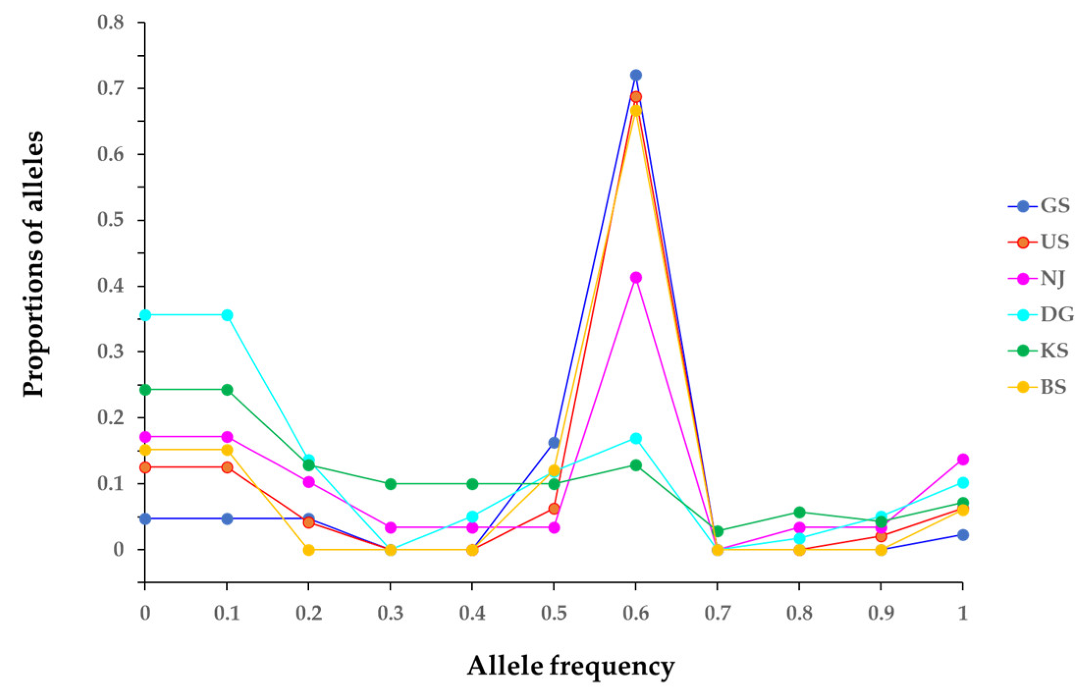
Furthermore, at the species level, expected heterozygosity (He) was higher than the observed heterozygosity (Ho), indicating the possibility of a genetic bottleneck occurring in some populations. The FIS of each population showed a phenomenon of heterozygosity excess. The bottleneck test from the infinite allele model (IAM), stepwise mutation model (SMM), and two-phase model (TPM) revealed strong, significant results for the GS, US, and BS populations—caused by the bottleneck phenomenon (Table 3). Furthermore, although the IAM model suggested a potential bottleneck in the NJ and KS populations, it was not statistically significant in the other models. These results indicate a mode-shift in allele frequency distributions (Figure 2). Notably, several populations exhibited a reversed L-shaped distribution pattern, which is consistent with signatures of a genetic bottleneck.

2.3. Genetic Differentiation and Gene Flow

Analysis of 30 microsatellite loci revealed that FIS values were predominantly negative across loci, indicating a general excess of heterozygotes among populations (Table 4). Overall locus-level FIS values ranged from −1.000 to 1.000 (Table 1), whereas within-population FIS values varied between −0.200 and −0.911 (Table 3), reflecting variation in the reproductive mode or clonal propagation.
Pairwise FST values ranged from 0.161 (between KS and DG) to 0.694 (between BS and NJ), with corresponding Nm estimates ranging from 0.110 to 1.300, respectively (Table 5). The lowest genetic differentiation and the highest estimated gene flow were observed between the geographically proximate KS and DG populations, whereas the greatest differentiation occurred between BS and US.
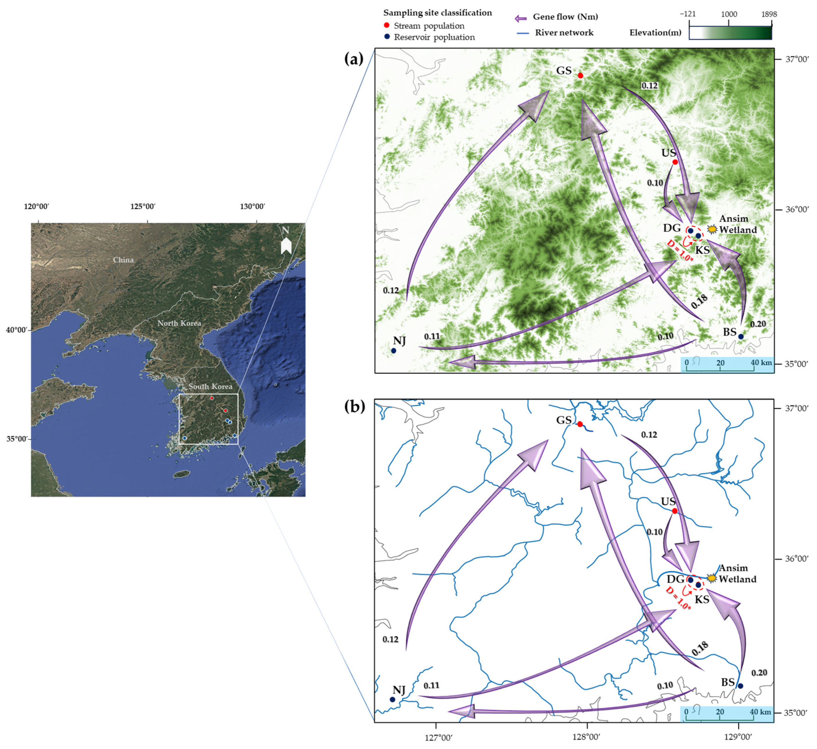
To examine the directionality of gene flow among the six sampling sites, we conducted a directional migration analysis (Figure 3). Most relative migration estimates were low (Nm < 0.2), and although some pairwise directions showed statistically significant values (e.g., BS KS, NJ KS), the magnitude of gene flow remained weak, indicating limited genetic connectivity among populations. Despite all six populations being located in hydrologically unconnected and independent waterbodies, a notably strong and statistically significant unidirectional gene flow was detected from DG to KS (Jost’s D = 1.00, p < 0.05). A Mantel test examining the relationship between genetic and geographic distances among the six populations revealed no significant correlation (r = 0.420, p > 0.05).

2.4. Spatial and Genetic Structure

Bayesian clustering analysis implemented in STRUCTURE identified six genetically distinct clusters (K = 6) among the populations, as determined by the ΔK method (Figure 4). Increasing values of K resulted in clearer resolution of the genetic subdivision, with each population corresponding to a discrete genetic cluster, indicating complete genetic separation among them.
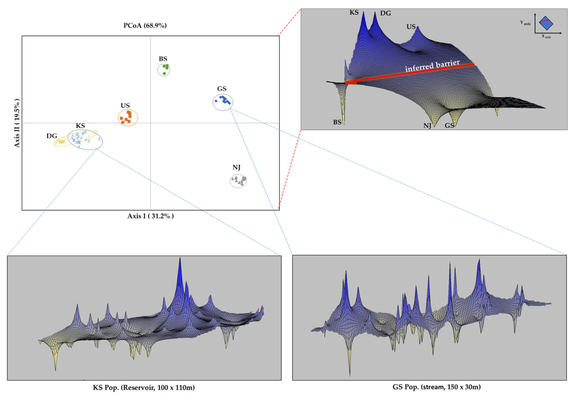
Principal coordinate analysis (PCoA) based on MLG data accounted for 68.9% of the total genetic variation across the three axes (Figure 5). Axis I, which explained 31.2% of the variation, separated the US, DG, and KS populations from the GS, NJ, and BS populations. The DG and KS populations were positioned proximally, whereas NJ exhibited strong divergence relative to the other populations.
Spatial genetic structure analyses identified a pronounced genetic discontinuity between the KS–DG–US and NJ–GS clusters, suggesting the presence of an inferred genetic barrier. The BS population was genetically differentiated from all other populations and formed an isolated unit in both ordination and clustering analyses. The analyzed spatial genetic patterns in two ecologically distinct populations—GS (stream) and KS (reservoir)—exhibited the most contrasting spatial structures. In the KS population (reservoir), intra-population genetic similarity was highest near the reservoir center and declined progressively toward the margins. Spatial autocorrelation was significantly positive within 20 m, with genetic similarity persisting up to a 60 m distance. Conversely, the GS population (stream) displayed a directional spatial structure that may reflect downstream clonal spread (R → L), as supported by the spatial autocorrelation analyses and field observations (Figure 6). Positive spatial autocorrelation was observed within 15 m and remained detectable up to 140 m.

3. Discussion

3.1. Variation in Microsatellite Loci

Microsatellite markers are powerful tools for detecting genetic variations and identifying diverse alleles, making them essential for population genetic studies [65,66]. In this study, the selected loci exhibited a wide range of PIC, from 0.141 to 0.828 (Table 2), reflecting their capacities to reveal genetic diversity. For robust analysis, we prioritized markers with PIC values above 0.5 to reduce potential bias caused by excessive allele variability. Furthermore, loci with PIC values below 0.3 were also retained based on comprehensive consideration of the clonal reproductive strategy of species and supporting evidence from previous studies [67,68]. Although these loci exhibited limited polymorphism, they harbored private alleles unique to specific populations, providing valuable insights into population differentiation and regional specificity (Figure 1).
The observed mean FIS value (−0.566) and excessive heterozygosity suggest that many loci deviated from the Hardy–Weinberg equilibrium toward heterozygote excess, likely due to clonal reproduction or outcrossing. Extremely high FIS values (1.000) in specific populations support clonal propagation. The broad range of population-level FIS values (−1.000 to 1.000) indicates variations in reproductive strategies across sites, with evidence of localized inbreeding. These results reflect the species’ mixed reproductive system and the influence of habitat conditions on genetic structure. This pattern and high FST values (mean FST = 0.556) indicate strong genetic differentiation among populations. Therefore, the microsatellite markers developed in this study are effective for detecting population structure and inferring the genetic origin of individuals.
Considering that M. spicatum is an aquatic species widely distributed worldwide, its gene flow is likely more limited than that of terrestrial plants. In species such as M. spicatum with sexual and clonal reproduction, the reproductive mode and environmental disturbance can significantly shape population structure. Hence, instead of relying solely on PIC values or heterozygosity levels, we selected markers through an integrated approach that considered allele frequency distributions and population-specific dynamics, and the validity of the developed microsatellite markers was confirmed. The subsequent sections provide a detailed discussion of the genetic analysis outcomes.

3.2. Genetic and Clonal Diversity

We analyzed the genetic diversity and clonal propagation of six M. spicatum populations using 30 microsatellite loci (Table 2). In our study, the species-level genetic diversity across all loci (expected heterozygosity, He = 0.577) was substantially lower than that reported by Cao et al. [62] (total He = 0.973) and Wu et al. [60] (Nei’s genotypic diversity [He], Overall D = 0.995).
Our results may reflect variations in environmental factors such as habitat area, population size, natural selection pressure, and disturbance intensity, as well as variations in the specificity of the developed markers. These findings suggest that the South Korean populations examined in this study may experience more constrained genetic variation than those reported in previous studies. Although the number of study sites was limited, the observed patterns were generally consistent with those reported in previous research.
Aquatic plants primarily propagate clonally, and many exhibit high clonal diversity [69], which can accumulate through occasional seed recruitment or be maintained through clonal growth in relatively stable environments [70]. Such patterns are often associated with habitats such as ponds, lakes, or areas with minimal water flow [71,72]. M. spicatum is an anemophilous species [73]; however, it may fail to produce seeds in flowing waters [74]. Thus, under high-flow conditions, sexual reproduction can be limited [71].
In our study, genetic and clonal diversity differed markedly between stream (GS and US) and reservoir (NJ, DG, KS, and BS) populations (Table 2). In stream habitats, allelic richness and expected heterozygosity were relatively low, and both GS and US were dominated by a single MLG. This MLG showed a downstream-oriented spatial signal, consistent with clonal spread (Figure S2). These results align with patterns previously reported [31,61]. Such a pattern may be associated with hydrological infrastructure (e.g., dams, sluice gates, and other water control structures), which could influence the balance between sexual and clonal reproduction.
Both GS and US are in watersheds subject to recurrent summer monsoon flooding and flow regulation. According to the national government-operated water quality monitoring [75] and flood forecasting systems in South Korea [76], the sites where the two stream populations are located are classified as flood-prone areas with existing flow regulation infrastructure. In South Korea, the monsoon and typhoon season typically occurs between June and September [77], which often overlaps with the reproductive period of M. spicatum [33]. Extreme rainfall events during this time could, therefore, substantially disrupt reproduction. Additionally, nearby agricultural activities and site management practices may further alter local hydrological and ecological conditions [76,77]. The clonal structure of GS and US, combined with heterozygosity excess under IAM, SMM, and TPM (Table 3), is consistent with recent bottleneck events potentially associated with environmental disturbances, as strong significance was detected across all three bottleneck detection models.
Water quality monitoring data for the two stream populations (March to September, 2016–2023; Table S3) indicate that mean total phosphorus and total nitrogen concentrations were within commonly eutrophic ranges [75], reaffirming the strong capacity of M. spicatum to survive, establish, and reproduce even under flow-affected river conditions [78].
On the contrary, the KS population showed the highest clonal diversity (G/N = 1.00), with every sampled individual having a unique MLG. This value exceeds the clonal diversity reported by Cao et al. [62] (mean G/N = 0.62). The discrepancy may be attributable to differences in site scale and sampling methodology: we analyzed a single population within one site, using a minimum spacing of 5 m between samples, whereas Cao et al. sampled multiple subpopulations within a single lake at approximately 1 m intervals. Consequently, our approach may have detected a broader range of diverse MLGs.
Two hypotheses might explain this pattern: First, high population density in a stable environment may have promoted competition, suppressing clonal growth and favoring sexual reproduction [79,80]. The large, deep reservoir habitat may also enable size variation that enhances individual persistence under low-resource conditions or limited light availability [81,82]. Second, the observed diversity may reflect the long-term accumulation and persistence of MLGs maintained by clonal propagation and occasional sexual recruitment [15,65,83]. However, the KS population, located in a long-standing reservoir, experienced anthropogenic modifications between 2014 and 2022, owing to lake park development. Satellite imagery of the reservoir revealed that the entire water body was drained as a result of construction activities (imagery date: September 2015 to April 2016; Figure S2), suggesting that the pre-development population size may have been larger than it is at present. If the lake bed has not been fully restructured, a broader range of genotypes—undetected in our sampling—could remain in deeper sediment layers.
By contrast, the BS population, which showed the lowest clonal diversity (G/N = 0.20), was located in a manmade pond established near a river in 2010. The initial establishment of M. spicatum may have resulted from intentional planting or recolonization from nearby sources (imagery date: September 2009 to April 2016; Figure S2). However, continued human interventions—such as plant removal and hydrological management—appear to have favored the selective persistence of a few stress-tolerant MLGs. BS exhibited the lowest clonal diversity and the strong dominance of a few clones. Among the four detected ramets, one MLG accounted for more than 60% of individuals (Figure S1). In disturbed environments, specific MLGs may prevail, owing to their greater adaptability [84,85,86,87,88,89,90,91,92]. Nonetheless, if only a small number of individuals retain their sexual capacity, long-term fitness could be compromised by processes such as Muller’s ratchet [93,94,95].
The contrasting results of the KS and BS populations, despite both originating from the same habitat type, suggest that the divergent patterns observed between stream and reservoir populations are driven by regional-scale ecological and anthropogenic processes rather than by solely site-specific factors. This highlights the need for broader-scale surveys in future research.

3.3. Genetic Differentiation and Gene Flow

Building on these differences in genetic and clonal diversity, we next examined the extent of genetic differentiation and patterns of gene flow among the six M. spicatum populations. High genetic differentiation in this species has previously been linked with clonal reproduction and may be further reinforced by geographic isolation, which limits gene flow [23,59,96]. Except in cases where populations were sampled as subpopulations within the same watershed (ΦST = 10% [62]), among-population differentiation is generally high in M. spicatum, as reported by Wu et al. (ΦPT = 0.521) [60] and Thum et al. (ΦST = 58%) [63]. Consistent with these findings, our analysis revealed high variations within populations and genetic differentiation among populations, with 57% (FST = 0.569) of the total genetic variance explained among populations.
Wu et al. [60] highlighted the role of geographic barriers, together with climatic conditions, on shaping genetic differentiation. Consistent with this, although directional gene flow was significant in a few pairwise comparisons, estimated Nm values were consistently low (all < 0.2), indicating that gene flow among populations was limited, despite the directional signal (Figure 3). Notably, despite the much smaller spatial extent of our sampling area, we observed a similar pattern, indicating that geographic barriers can emerge even across relatively narrow areas.
Genetic differentiation between the geographically unconnected DG and KS populations was the lowest (FST = 0.161, Nm = 1.300), suggesting the possibility of ongoing gene flow between them (Table 5). This pattern may reflect ecological connectivity through natural gene dispersal processes and anthropogenic introduction pathways associated with reservoir management of aquatic vegetation. Aquatic macrophytes are often transplanted for purposes such as water purification or landscape enhancement; however, mechanical removal activities may inadvertently disperse vegetative fragments to new locations.
The trajectory of seed dispersal can provide insights into the vector species, their life histories, and the timing of ingestion and excretion [97]. M. spicatum seeds are consumed and dispersed by various waterbirds [60,98,99], and highly mobile vectors such as birds and fish can transport seeds across hundreds of kilometers [100,101]. In this context, we explored ecological factors: for DG and KS, the proximity of both populations to the Ansim Wetland, a migratory bird sanctuary in the Geumho River basin (linear distance, <8 km) [102], raises the possibility of opportunistic gene flow by bird-mediated dispersal (Figure 3).
Despite the potential for biotic dispersal, the limited extent of gene flow observed in this study may suggest that M. spicatum has relatively low palatability or ecological value as a food source for birds and fish [35,46,58,103]. When gene flow or genetic differentiation is confined to localized areas, unique alleles within each population may contribute to the observed genetic structuring, as further supported by the PCoA and STRUCTURE analysis results (Figure 4 and Figure 5). These findings imply that gene flow in M. spicatum primarily occurs over short distances, with local environmental selection pressures further reinforcing genetic differentiation. Consequently, the genetic connectivity observed between DG and KS populations cannot be fully explained by natural dispersal mechanisms alone and may involve indirect anthropogenic influences.

3.4. Spatial and Genetic Structure

The pronounced genetic differentiation observed among six populations of M. spicatum implies limited gene flow [59,63,64,104,105]. Genetic barriers were observed between the NJ–GS and KS–DG–US population groups, while the BS population consistently showed patterns of genetic isolation even in PCoA (Figure 5). Because the studied populations are not hydrologically or geographically connected, the observed genetic patterns are likely shaped by localized environmental factors and restricted dispersal.
Spatial genetic structure analyses further revealed contrasting patterns between KS (reservoir) and GS (stream). KS exhibited a gradual genetic gradient, whereas GS showed directional clonal spread aligned with downstream flow (Figure 5 and Figure 6). These contrasts are broadly consistent with the results of previous studies on spatial genetic structure and dispersal in aquatic macrophytes [23,106,107,108,109,110], supporting the idea that physical habitat characteristics strongly influence local genetic structure. Collectively, our findings indicate that the spatial genetic structure of M. spicatum in South Korea results from a complex interplay of reproductive mode, habitat-specific conditions, and anthropogenic disturbances.
Although this study contrasted populations by habitat type, populations distributed along a continuous watershed may display different, connectivity-driven gene flow regimes. Moreover, environmentally mediated shifts in reproductive mode may lead to population-specific differences in seed germination and establishment. Together, these factors could generate diverse patterns of genetic diversity and population structure. Future research should integrate population genetic analyses with ecological and physiological assessments linked to life history and reproductive strategies.

3.5. Implications for Conservation and Sustainable Management

Long-term monitoring of the origin of collected individuals and historical habitat changes is essential to improve the efficiency of future conservation and resource management [111]. Populations experiencing substantial environmental fluctuations, such as stream-type habitats, may serve as important areas for the utilization of specific genotypes and for ecological monitoring. In South Korea, for example, stream populations such as GS and US and the reservoir populations NJ and BS illustrate this pattern, whereas reservoir population KS maintained comparatively rich genotypic diversity, despite a history of anthropogenic modification, indicating their potential value as key resources for the conservation of M. spicatum diversity.
This contrast is consistent with that in previous studies, indicating that clonal reproduction can lead to different outcomes in population structure and genetic diversity depending on habitat conditions [8]. Particularly, You et al. [112] demonstrated that continuous environmental fluctuations within habitats can promote and reinforce the dominance of specific clones within populations. Although not all environmental factors can be controlled, the presence or absence of planned watershed management may either support the conservation of aquatic plant resources or exert negative impacts. Incorporating such habitat-specific genetic structures into aquatic plant management could contribute to resource conservation and effective management strategies not only in South Korea but also in other regions facing similar hydrological gradients [113].

4. Materials and Methods

4.1. Sample Preparation
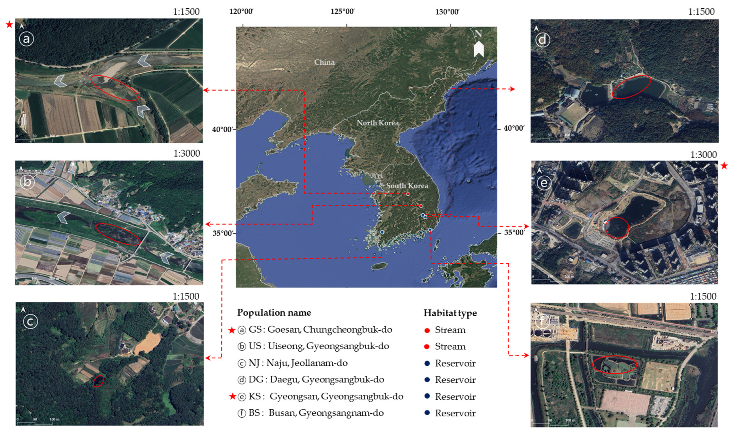
For the genetic analysis, a total of 120 individuals of M. spicatum were collected from six populations in 2023 (Figure 6 and Table S2). Each population was represented by 20 individuals, selected to reflect variation in habitat types and disturbance regimes. The stream populations (GS and US) are classified as local rivers managed by municipal governments. Site-specific information on hydrological management and usage was compiled through field observations, historical changes traced via satellite imagery, and documentation provided by the respective local authorities [76,77,114]. All individuals sampled within each population were separated by at least 5 m to avoid collecting the same clone. Prior to sampling, the taxonomic identity of M. spicatum was confirmed by a professional plant taxonomist through cross-validation.
The six sampling sites were strategically selected to reflect spatial and ecological heterogeneity, including differences in habitat types (e.g., streams and reservoirs) and disturbance intensity (Figure 6). To assess potential sources and patterns of habitat disturbance, satellite imagery was obtained for each site and its surrounding landscape. These images were extracted from Google Satellite basemaps and visualized using QGIS version 3.34 (accessed June 2025).
To examine the formation of spatial genetic structure within populations, we selected the GS and KS populations, which exhibited relatively intact clonal patches with visible spatial distribution patterns. Twenty individuals were sampled from each site within defined areas of 150 × 30 m and 100 × 110 m for GS and KS populations, respectively. A minimum distance of 5 m was maintained between individuals to reduce the likelihood of resampling identical genets. The geographic location of each sampled individual was georeferenced, and these coordinates were used for subsequent spatial genetic analyses. When the geographic coordinates of sampled individuals could not be recorded directly in the field, high-resolution satellite imagery was used to match the sampling locations with field photographs and secure accurate spatial coordinates.
Figure 6. Geographic distribution of six M. spicatum sampling sites in South Korea. The map illustrates the national distribution of stream (red) and reservoir (blue) populations, while panels (ⓐ–ⓕ) show high-resolution satellite images of the sampling habitats (scale 1:1500 and 1:3000). Sampling sites marked with red asterisks () were included in spatial genetic structure analyses.
Plants 14 02648 g006

4.2. Microsatellite Marker Development and Polymerase Chain Reaction (PCR) Amplification

Microsatellite regions were identified using a genome assembly and screened using the Microsatellite (MISA) tool (https://webblast.ipk-gatersleben.de/misa/, accessed on 23 August 2023). Screening parameters were set to detect a minimum of 10 repeats for di-nucleotide motifs, and at least 4 repeats for tri-, tetra-, penta-, and hexa-nucleotide motifs. Primers were designed using Primer3 (https://github.com/primer3-org/primer3, accessed on 23 August 2023) with the following criteria: an amplicon size of 100–300 bp, primer length of 20–24 bp, GC content between 40 and 60%, and melting temperature of 58 °C. Primer specificity was confirmed using SnapGene (GSL Biotech, Chicago, IL, USA) to ensure that each primer pair uniquely bound to the target sequence.
A total of 100 primer pairs were initially designed. PCR conditions were optimized, and each primer was tested on 6 individuals per population to assess polymorphism. Thirty polymorphic and reproducibly amplified loci were selected for further analysis of the total samples (Table S1). The final set of 30 validated microsatellite loci was submitted to GenBank, and accession numbers (PV231646–PV231675) are provided.
Microsatellite loci amplification was performed using the Mastercycler® Pro Gene Amplifier (Eppendorf, Hamburg, Germany). PCR was conducted at a total volume of 20 μL, using H-Star Taq DNA Polymerase (Biofact, Daejeon, Republic of Korea). Fluorescent labeling was performed following the protocol described by Schuelke [115], using four fluorescent dyes (6-FAM, VIC, NED, PET). Each reaction mixture contained a microsatellite-specific forward primer (0.4 μM), a reverse primer synthesized with an M13 tail sequence (5′-TGTAAAACGACGGCCAGT, 0.8 μM), and a fluorescently labeled M13 primer (0.4 μM). The PCR cycling conditions consisted of initial denaturation at 94 °C for 5 min, followed by 30 cycles of denaturation at 94 °C for 30 s, annealing at 58 °C for 45 s, and extension at 72 °C for 45 s. This was followed by 12 cycles at 94 °C for 30 s, 53 °C for 45 s, and 72 °C for 45 s. A final extension step was performed at 72 °C for 10 min. PCR products were mixed with GeneScan™ 500 ROX Size Standard Ladder (Applied Biosystems, Foster City, CA, USA) and HiDi™ formamide, denatured at 95 °C for 2 min, and then cooled to 4 °C. Allele sizing was performed using an ABI 3730xl DNA Analyzer (Applied Biosystems), and genotyping was conducted using the GeneMarker® software ver. 2.6.7 (SoftGenetics, State College, PA, USA). To visualize the distribution of common and population-specific alleles across 30 microsatellite loci, a heatmap was generated using the Heatmapper web tool (http://www.heatmapper.ca/expression/ accessed on 7 May 2025), based on allele frequency data from each sampling site.

4.3. Genetic and Clonal Diversity Analysis

The MICROCHECKER software ver. 2.2.3 [116] was used to examine the presence or absence of scoring errors in the microsatellite loci. The genetic analyses were performed based on the frequency of the aforementioned marker bands, following the categorization method of Steeves et al. [117]. PIC values were estimated using Cervus ver. 3.0.7 [118], whereas F-statistics were derived from Nei’s method implemented in FSTAT ver. 2.9.4 [119].
Genetic diversity was analyzed in the percentage of polymorphic loci (P0.95), the mean number of alleles per locus (A), the effective number of alleles per locus (Ae), and the observed (Ho) and expected (He) heterozygosity, using the Popgene software v1.32 [120]. Clonal diversity was analyzed by counting the number of ramets and genotypes. The ratio of different MLGs (G/N) to the total number of sampled individuals was used to estimate genotypic diversity. Additionally, the richness index (D), Simpson’s unbiased diversity index (1 − D), and genotypic evenness (E) were manually calculated based on MLG frequencies, following Simpson (1949) [121] and Pielou (1969) [122]. Wilcoxon’s signed-rank test was conducted using the BOTTLENECK software ver. 1.2.02 [123], under the assumptions of the IAM, SMM, and TPM, to identify populations that had recently experienced a decline in effective size. The genetic bottleneck test was conducted by comparing one- and two-tailed tests to determine significance and minimize the risk of distorted results. Significance was assessed using sign tests and Wilcoxon one-tailed tests, with estimation based on 1000 replications. Mode-shift indicator graphs were also generated to visualize allele frequency distributions across populations.

4.4. Genetic Differentiation and Gene Flow

To determine the genetic differentiation in each population, Wright’s F-statistics (FIS, FIT, FST; [124]) were calculated using FSTAT ver. 2.9.4 [119]. FIS values were estimated for polymorphic loci, applying the 0.95 criterion, and their significance was tested through permutation tests with 180,000 randomizations. To assess population differentiation, the statistical significance of FST and FIT was evaluated by calculating the 95% confidence intervals, and gene flow (expected as Nm) was estimated from the FST value using GenAIEx ver. 6.503 [125]. To examine the correlation between genetic differentiation and geographic distance among populations, Mantel tests [126] were conducted using the IBD software (isolation by distance) ver. 1.52 [127]. To assess directional gene flow and detect asymmetric migration among populations, we employed the DivMigrate online application (https://popgen.shinyapps.io/DivMigrate-online/ accessed on 7 May 2025), developed as part of the diveRsity R package ver. 1.9.90 [128]. The analysis was based on Gst estimates, with 1000 bootstrap replicates used to test the significance of asymmetric migration between population pairs. Additionally, Jost’s D was calculated as an alternative measure of genetic flow.

4.5. Spatial and Genetic Structure

To investigate the genetic structure of each population, we employed STRUCTURE ver. 2.3.4 [129], which identified discrete genetic clusters and estimated the proportion of individual genotypes assigned to each cluster. Simulations were run 10 times for each ΔK value (1–10), with 1,000,000 MCMC iterations following a burn-in of 100,000, using the admixture model and assuming correlated allele frequencies [130,131]. The optimal K value was visualized using the Structure Selector web tool [132].
Pairwise genetic distances among individuals were calculated, and Principal Coordinate Analysis (PCoA) was performed using GenAlEx ver. 6.503 [125] to generate relative spatial coordinates. The spatial positions of sampled individuals and the distributional extent of each population were standardized using the vegan package ver. 2.6.4 [133] in R ver. 4.3.2 [134]. These coordinates were subsequently scaled to reflect the actual sampling area accurately. The scaled coordinates and the genetic distance matrix were used in the Alleles in Space (AIS) software ver. 1.0 [135] to infer and visualize fine-scale spatial genetic structure. For the overall analysis across six populations, the XY coordinate space was divided into a 150 × 150 grid with a uniform weighting of 1. To assess within-population spatial genetic structure, grid settings were adjusted to match the actual sampling area for each habitat: a 100 × 110 grid for the KS population (reservoir) and a 150 × 30 grid for the GS population (stream).

5. Conclusions

Although M. spicatum is globally distributed, its ecological traits and genetic composition vary across regions in response to environmental heterogeneity, as shown by genetic studies from temperate regions of China, North America, and Europe. In this study, our analysis of six South Korean populations revealed a more nuanced and complex genetic structure.
The results further confirm that reproductive strategies of M. spicatum populations shift with environmental variability and highlight the value of long-term monitoring for understanding habitat-specific genetic structures. The balance between sexual reproduction and clonal propagation differed among habitats, offering evolutionary evidence of the species’ strong persistence and establishment capacities. In more variable environments, clonal propagation is critical for population survival, whereas in more stable habitats, higher rates of sexual reproduction likely support genetic diversity. Such dynamics may contribute to a more restricted spatial genetic structure than previously anticipated.
In South Korea, considerable resources are allocated annually to water quality management [111,136]. However, climate-driven changes such as unseasonal algal blooms are exerting increasing pressure on freshwater ecosystems [137]. Our findings underscore the importance of national initiatives aimed at mitigating environmental degradation and promoting ecosystem restoration. Although M. spicatum has been linked to ecological challenges in specific regions, its functional role in aquatic systems may ultimately depend on local management strategies.
The 30 microsatellite markers developed and applied in this study demonstrated strong discriminatory power among populations, underscoring their utility for long-term genetic monitoring and comparative studies in other regions. By providing novel insights into the genetic and clonal characteristics of M. spicatum populations in South Korea, recently re-evaluated in terms of their resource value, we hope this work will serve as a valuable resource for future research and management strategies.

Supplementary Materials

The following supporting information can be downloaded at: https://www.mdpi.com/article/10.3390/plants14172648/s1, Table S1: Amplification information primer sequences and characteristics of 30 microsatellite loci developed from Myriophyllum spicatum; Table S2: Summary of population information for six M. spicatum sampling sites in South Korea; Table S3: Annual water quality metrics (March–September means, 2016–2023) for the GS and US sites where M. spicatum was sampled; Figure S1: UPGMA tree based on Nei’s genetic distance, calculated for clonal and genetic relationships among six M. spicatum populations; Figure S2: Satellite imagery showing land use changes at two M. spicatum sampling sites in South Korea.

Author Contributions

Conceptualization, investigation, methodology, and formal analysis, E.-H.K., K.-R.K. and J.-N.Y.; Writing—original draft, E.-H.K. and J.-N.Y.; Funding acquisition and writing—review and editing, M.-H.L., J.G. and J.-N.Y. All authors have read and agreed to the published version of the manuscript.

Funding

This research was funded by the Korea Environment Industry & Technology Institute, Ministry of Environment of the Republic of Korea (grant number 2021003420003), to contribute to multi-ministerial national biological research resources.

Data Availability Statement

The original contributions presented in this study are included in the article. Further inquiries can be directed to the corresponding author.

Conflicts of Interest

The authors declare no conflicts of interest.

References

  1. Young, A.; Boyle, T.; Brown, T. The population genetic consequences of habitat fragmentation for plants. Trends Ecol. Evol. 1996, 11, 413–418. [Google Scholar] [CrossRef]
  2. Booy, G.; Hendriks, R.J.J.; Smulders, M.J.M.; van Groenendael, J.M.; Vosman, B. Genetic diversity and the survival of populations. Plant Biol. 2000, 2, 379–395. [Google Scholar] [CrossRef]
  3. Hussner, A.; Heidbüchel, P.; Coetzee, J.; Gross, E.M. From introduction to nuisance growth: A review of traits of alien aquatic plants which contribute to their invasiveness. Hydrobiologia 2021, 848, 2119–2151. [Google Scholar] [CrossRef]
  4. Adomako, M.O.; Zhang, Q.; Yu, F.H. Genotypic differences in response to different patterns of clonal fragmentation in the aquatic macrophyte Pistia stratiotes. J. Plant Ecol. 2022, 15, 1199–1212. [Google Scholar] [CrossRef]
  5. Zhang, H.; Chase, J.M.; Liao, J. Habitat amount modulates biodiversity responses to fragmentation. Nat. Ecol. Evol. 2024, 8, 1437–1447. [Google Scholar] [CrossRef] [PubMed]
  6. Sakai, A.K.; Allendorf, F.W.; Holt, J.S.; Lodge, D.M.; Molofsky, J.; With, K.A.; Baughman, S.; Cabin, R.J.; Cohen, J.E.; Ellstrand, N.C.; et al. The population biology of invasive species. Annu. Rev. Ecol. Syst. 2001, 32, 305–332. [Google Scholar] [CrossRef]
  7. Eckert, C.G.; Lui, K.; Bronson, K.; Corradini, P.; Bruneau, A. Population genetic consequences of extreme variation in sexual and clonal reproduction in an aquatic plant. Mol. Ecol. 2003, 12, 331–344. [Google Scholar] [CrossRef] [PubMed]
  8. Barrett, L.G.; Thrall, P.H.; Burdon, J.J.; Linde, C.C. Life history determines genetic structure and evolutionary potential of host–parasite interactions. Trends Ecol. Evol. 2008, 23, 678–685. [Google Scholar] [CrossRef]
  9. Stange, M.; Barrett, R.D.H.; Hendry, A.P. The importance of genomic variation for biodiversity, ecosystems and people. Nat. Rev. Genet. 2021, 22, 89–105. [Google Scholar] [CrossRef]
  10. Eckert, C.G.; Dorken, M.E.; Barrett, S.C. Ecological and evolutionary consequences of sexual and clonal reproduction in aquatic plants. Aquat. Bot. 2016, 135, 46–61. [Google Scholar] [CrossRef]
  11. Ashton, P.J.; Mitchell, D.S. Aquatic plants: Patterns and modes of invasion, attributes of invading species and assessment of control programs. In Biological Invasions: A Global Perspective; Drake, J.A., Mooney, H.A., Di Castri, F., Groves, R.H., Kruger, F.J., Rejmânek, M., Williamson, M.H., Eds.; John Wiley and Sons, Ltd.: London, UK, 1989; pp. 111–154. [Google Scholar]
  12. Starfinger, U.; Stöcklin, J. Seed, pollen, and clonal dispersal and their role in structuring plants populations. Prog. Bot. 1996, 57, 336–355. [Google Scholar]
  13. Piquot, Y.; Petit, D.; Valero, M.; Cuguen, J.; de Laguerie, P.; Vernet, P. Variation in sexual and asexual reproduction among young and old populations of the perennial macrophyte Dendroseris. Oikos 1998, 82, 139–148. [Google Scholar] [CrossRef]
  14. Silvertown, J. The evolutionary maintenance of sexual reproduction: Evidence from the ecological distribution of asexual reproduction in clonal plants. Int. J. Plant Sci. 2008, 169, 157–168. [Google Scholar] [CrossRef]
  15. Vallejo–Marín, M.; Dorken, M.E.; Barrett, S.C.H. The ecological and evolutionary consequences of clonality for plant mating. Annu. Rev. Ecol. Evol. Syst. 2010, 41, 193–213. [Google Scholar] [CrossRef]
  16. da Cunha, N.L.; Fischer, E.; Lorenz–Lemke, A.P.; Barrett, S.C.H. Floral variation and environmental heterogeneity in a tristylous clonal aquatic of the Pantanal wetlands of Brazil. Ann. Bot. 2014, 114, 1637–1649. [Google Scholar] [CrossRef]
  17. Wetzel, R.G. A comparative study of the primary production of higher aquatic plants, periphyton, and phytoplankton in a large, shallow lake. Int. Rev. Gesamt Hydrobiol. Hydrogr. 1964, 49, 1–61. [Google Scholar] [CrossRef]
  18. Jiang, H.S.; Zhang, Y.; Yin, L.; Li, W.; Jin, Q.; Fu, W.; Zhang, T.; Huang, W. Diurnal changes in photosynthesis by six submerged macrophytes measured using fluorescence. Aquat. Bot. 2018, 149, 33–39. [Google Scholar] [CrossRef]
  19. Yu, C.; Yang, L.; Leah, N.; Wei, L.; Ling, X.; Hong, S.J. The analysis of leaf traits of eight Ottelia populations and their potential ecosystem functions in karst freshwaters in China. Front. Plant Sci. 2019, 9, 1938. [Google Scholar] [CrossRef] [PubMed]
  20. Crawford, D.J.; Stuessy, T.F.; Silva, M. Allozyme divergence and the evolution of Dendroseris (Compositae: Lactuceae) on the Juan Fernandez Islands. Syst. Bot. 1987, 12, 435–443. [Google Scholar] [CrossRef]
  21. Barrett, S.C.H.; Eckert, C.G.; Husband, B.C. Evolutionary processes in aquatic plant populations. Aquat. Bot. 1993, 44, 105–145. [Google Scholar] [CrossRef]
  22. Santamaría, L. Why are most aquatic plants widely distributed? Dispersal, clonal growth and small–scale heterogeneity in a stressful environment. Acta Oecol. 2002, 23, 137–154. [Google Scholar] [CrossRef]
  23. Junk, W.J.; Piedade, M.T.F. Plant life in the floodplain with special reference to herbaceous plants. In The Central Amazon Floodplain: Ecology of a Pulsing System; Springer Nature: Berlin/Heidelberg, Germany, 1997; pp. 147–185. [Google Scholar]
  24. Sculthorpe, C.D. The Biology of Aquatic Vascular Plants; Edward Arnold: London, UK, 1967; p. 610. [Google Scholar]
  25. van Groenendael, J.M.; Klimeš, L.; Klimešová, J.; Hendriks, R.J.J. Comparative ecology of clonal plants. Philos. Trans. R. Soc. Lond. B Biol. Sci. 1996, 351, 1331–1339. [Google Scholar]
  26. Yakimowski, S.B.; Barrett, S.C.H. Clonal genetic structure and diversity in populations of an aquatic plant with combined vs. separate sexes. Mol. Ecol. 2014, 23, 2914–2928. [Google Scholar] [CrossRef]
  27. Klimešová, J.; Martínková, J.; Ottaviani, G. Belowground plant functional ecology: Towards an integrated perspective. Funct. Ecol. 2018, 32, 2115–2126. [Google Scholar] [CrossRef]
  28. Franklin, S.; Alpert, P.; Salguero–Gómez, R.; Janovský, Z.; Herben, T.; Klimešová, J.; Douhovnikoff, V. Next–gen plant clonal ecology. Perspect. Plant Ecol. Evol. Syst. 2021, 49, 125601. [Google Scholar] [CrossRef]
  29. Bowes, G. Aquatic plant photosynthesis: Strategies that enhance carbon gain. In Plant Life in Aquatic and Amphibious Habitats; Crawford, R.M.M., Ed.; Blackwell Scientific Publications: Oxford, UK, 1987; pp. 79–98. [Google Scholar]
  30. Gavrilescu, M. Water, Soil, and Plants Interactions in a Threatened Environment. Water 2021, 13, 2746. [Google Scholar] [CrossRef]
  31. Wu, Z.; Wang, Z.; Xie, D.; Wang, H.; Zhao, A.; Wang, Y.; Hanling, W.; xinwei, X.; Tao, L.; Zhao, J. Effects of highland environments on clonal diversity in aquatic plants: An interspecific comparison study on the Qinghai–Tibetan Plateau. Front. Plant Sci. 2022, 13, 1040282. [Google Scholar] [CrossRef]
  32. Higgisson, W.; Broadhurst, L.; Shams, F.; Gruber, B.; Dyer, F. Reproductive strategies and population genetic structure in two dryland river floodplain plants, Marsilea drummondii and Eleocharis acuta. Genes 2022, 13, 1506. [Google Scholar] [CrossRef]
  33. Korea National Arboretum. Myriophyllum spicatum L. Available online: http://www.nature.go.kr/kbi/plant/pilbk/selectPlantPilbkDtl.do?plantPilbkNo=26182 (accessed on 1 April 2025).
  34. Patten, B.C., Jr. Notes on the biology of Myriophyllum spicatum L. in a New Jersey lake. Bull. Torrey Bot. Club. 1956, 83, 5–18. [Google Scholar] [CrossRef]
  35. Aiken, S.G.; Newroth, P.R.; Wile, I. The biology of Canadian Weeds.: 34. Myriophyllum spicatum L. Can. J. Plant Sci. 1979, 59, 201–215. [Google Scholar] [CrossRef]
  36. Ortiz, J.E.; Marcarelli, A.M.; Juneau, K.J.; Huckins, C.J. Invasive Myriophyllum spicatum and nutrients interact to influence algal assemblages. Aquat. Bot. 2019, 156, 1–9. [Google Scholar] [CrossRef]
  37. Lin, Z.; Zhong, C.; Yu, G.; Fu, Y.; Guan, B.; Liu, Z.; Yu, J. Effects of sediments phosphorus inactivation on the life strategies of Myriophyllum spicatum: Implications for lake restoration. Water 2021, 13, 2112. [Google Scholar] [CrossRef]
  38. Glisson, W.J.; Larkin, D.J. Hybrid watermilfoil (Myriophyllum spicatum × Myriophyllum sibiricum) exhibits traits associated with greater invasiveness than its introduced and native parental taxa. Biol. Invasions 2021, 23, 2417–2433. [Google Scholar] [CrossRef]
  39. Gassmann, A.J.; Levy, A.; Tran, T.; Futuyma, D.J. Adaptations of an insect to a novel host plant: A phylogenetic approach. Funct. Ecol. 2006, 20, 478–485. [Google Scholar] [CrossRef]
  40. Beck, K.G.; Zimmerman, K.; Schardt, J.D.; Stone, J.; Lukens, R.R.; Reichard, S.; Randall, J.; Cangelosi, A.A.; Cooper, D.; Thompson, J.P. Invasive species defined in a policy context: Recommendations from the Federal Invasive Species Advisory Committee. Invasive Plant Sci. Manag. 2008, 1, 414–421. [Google Scholar] [CrossRef]
  41. Hussner, A.; Stiers, I.; Verhofstad, M.J.J.M.; Bakker, E.S.; Grutters, B.M.C.; Haury, J.; van Valkenburg, J.L.C.H.; Brundu, G.; Newman, J.; Clayton, J.S.; et al. Management and control methods of invasive alien freshwater aquatic plants: A review. Aquat. Bot. 2017, 136, 112–137. [Google Scholar] [CrossRef]
  42. Li, F.; Qin, Y.; Zhu, L.; Xie, Y.; Liang, S.; Hu, C.; Chen, X.; Deng, Z. Effects of fragment size and sediment heterogeneity on the colonization and growth of Myriophyllum spicatum. Ecol. Eng. 2016, 95, 457–462. [Google Scholar] [CrossRef]
  43. Fan, X.R.; Njeri, H.K.; Pu, Y.H.; La, Q.; Li, W.; Li, X.L.; Chen, Y.Y. Contrasting relationships between genetic diversity and species diversity in conserved and disturbed submerged macrophyte communities of Honghu Lake, a typical freshwater lake of Yangtze River Basin. Glob. Ecol. Conserv. 2021, 31, 01873. [Google Scholar] [CrossRef]
  44. Ahn, K.; Lim, J.; Lee, Y.; Choi, T.; Lee, K.; Im, M.; Go, Y.; Suh, J.; Shin, Y.; Kim, M. Vegetation classification and distributional pattern in Damyang riverine wetland. J. Environ. Impact Assess. 2016, 25, 89–102. [Google Scholar] [CrossRef]
  45. Xiao, C.; Wang, X.F.; Xia, J.; Li, G.H. The effect of temperature, water level and burial depth on seed germination of Myriophyllum spicatum and Potamogeton malaianus. Aquat. Bot. 2010, 92, 28–32. [Google Scholar] [CrossRef]
  46. Lillie, R.A.; Budd, J. Habititat architecture of Myriophyllum spicatum L. as an index to habitat quality for fish and macroinvertebrates. J. Freshw. Ecol. 1992, 7, 113–125. [Google Scholar] [CrossRef]
  47. Madsen, J.D. Methods for Management of Nonindigenous Aquatic Plants. In Assessment and Management of Plant Invasions; Luken, J.O., Thieret, J.W., Eds.; Springer Series on Environmental Management; Springer: New York, NY, USA, 1997; pp. 145–171. [Google Scholar]
  48. National Institute of Biological Resources. Available online: https://species.nibr.go.kr (accessed on 14 February 2025).
  49. Jeong, S.; Yang, D.; Joo, S.; Park, S. Allelopathic inhibition effects of Myriophyllum spicatum on growths of bloom–forming cyanobacteria and other phytoplankton species in coexistence experiments. J. Plant Biol. 2021, 64, 501–510. [Google Scholar] [CrossRef]
  50. Kim, C.H.; Lee, Y.K.; Kim, M.J.; Choi, J.S.; Hwang, B.S.; Cho, P.Y.; Kim, Y.J.; Jeong, Y.T. Evaluation antioxidant and anti–inflammatory activity of ethanolic extracts of Myriophyllum spicatum L. in lipopolysaccharide–stimulated RAW 264.7 cells. Korean J. Plant Res. 2023, 36, 15–25. [Google Scholar]
  51. Pichersky, E.; Gang, D.R. Genetics and biochemistry of secondary metabolites in plants: An evolutionary perspective. Trends Plant Sci. 2000, 5, 439–445. [Google Scholar] [CrossRef] [PubMed]
  52. Wink, M. Evolution of secondary metabolites from an ecological and molecular phylogenetic perspective. Phytochemistry 2003, 64, 3–19. [Google Scholar] [CrossRef]
  53. Christensen, K.B.; Petersen, R.K.; Petersen, S.; Kristiansen, K.; Christensen, L.P. Activation of PPARγ by metabolites from the flowers of purple coneflower (Echinacea purpurea). J. Nat. Prod. 2009, 72, 933–937. [Google Scholar] [CrossRef]
  54. Kwon, S.T.; Kamiya, J. Genetic variation of cytochrome P450 genes in garlic cultivars. Korean J. Plant Res. 2011, 24, 584–590. [Google Scholar] [CrossRef]
  55. Govindaraj, M.; Vetriventhan, M.; Srinivasan, M. Importance of genetic diversity assessment in crop plants and its recent advances: An overview of its analytical perspectives. Genet. Res. Int. 2015, 431–487. [Google Scholar] [CrossRef]
  56. Bhandari, H.R.; Bhanu, A.N.; Srivastava, K.; Singh, M.N.; Shreya, H.A. Assessment of genetic diversity in crop plants—An overview. Adv. Plants Agric. Res. 2017, 7, 279–286. [Google Scholar]
  57. Ribeiro, M.M.; Diamantino, T.; Domingues, J.; Montanari, Í., Jr.; Alves, M.N.; Gonçalves, J.C. Stevia rebaudiana germplasm characterization using microsatellite markers and steviol glycosides quantification by HPLC. Mol. Biol. Rep. 2021, 48, 2573–2582. [Google Scholar] [CrossRef]
  58. LaRue, E.A.; Grimm, D.; Thum, R.A. Laboratory crosses and genetic analysis of natural populations demonstrate sexual viability of invasive hybrid watermilfoils (Myriophyllum spicatum × M. sibiricum). Aquat. Bot. 2013, 109, 49–53. [Google Scholar] [CrossRef]
  59. Wu, Z.; Yu, D.; Wang, Z.; Li, X.; Xu, X. Great influence of geographic isolation on the genetic differentiation of Myriophyllum spicatum under a steep environmental gradient. Sci. Rep. 2015, 5, 15618. [Google Scholar] [CrossRef] [PubMed]
  60. Wu, Z.; Yu, D.; Li, X.; Xu, X. Influence of geography and environment on patterns of genetic differentiation in a widespread submerged macrophyte, Eurasian watermilfoil (Myriophyllum spicatum L., Haloragaceae). Ecol. Evol. 2016, 6, 460–468. [Google Scholar] [CrossRef]
  61. Tóth, V.R.; Endre, G.; Kovács, S.; Présing, M.; Horváth, H. Morphological and genetic variability of Myriophyllum spicatum in different shallow water bodies of Hungary. Wetlands 2017, 37, 351–362. [Google Scholar] [CrossRef]
  62. Cao, Q.; Hu, F.; Liu, N. Local–scale patterns of genetic variation in coexisting floating–leaved Nymphoides peltata and submerged Myriophyllum spicatum in Donghu Lake. J. Oceanol. Limnol. 2020, 38, 1825–1834. [Google Scholar] [CrossRef]
  63. Thum, R.A.; Chorak, G.M.; Newman, R.M.; Eltawely, J.A.; Latimore, J.; Elgin, E.; Parks, S. Genetic diversity and differentiation in populations of invasive Eurasian (Myriophyllum spicatum) and hybrid (Myriophyllum spicatum × Myriophyllum sibiricum) watermilfoil. Invasive Plant Sci. Manag. 2020, 13, 59–67. [Google Scholar] [CrossRef]
  64. Wu, Z.; Wang, Z.; Xie, D.; Zhang, J.; Cai, P.; Li, X.; Zhao, J. Extensive Sympatry and Frequent Hybridization of Ecologically Divergent Aquatic Plants on the Qinghai–Tibetan Plateau. Front. Plant Sci. 2022, 13, 851151. [Google Scholar] [CrossRef]
  65. Schlötterer, C. Evolutionary dynamics of microsatellite DNA. Chromosoma 2000, 109, 365–371. [Google Scholar] [CrossRef]
  66. Hearne, C.M.; Ghosh, S.; Todd, J.A. Microsatellites for linkage analysis of genetic traits. Trends Genet. 1992, 8, 288–294. [Google Scholar] [CrossRef] [PubMed]
  67. Morgante, M.; Olivieri, A.M. PCR-amplified microsatellites as markers in plant genetics. Plant J. 1993, 3, 175–182. [Google Scholar] [CrossRef] [PubMed]
  68. Bonin, A.; Bellemain, E.; Bronken Eidesen, P.; Pompanon, F.; Brochmann, C.; Taberlet, P. How to track and assess genotyping errors in population genetics studies. Mol. Ecol. 2004, 13, 3261–3273. [Google Scholar] [CrossRef]
  69. Ellstrand, N.C.; Roose, M.L. Patterns of genotypic diversity in clonal plant species. Am. J. Bot. 1987, 74, 123–131. [Google Scholar] [CrossRef]
  70. Hidding, B.; Meirmans, P.G.; Klaassen, M.; de Boer, T.; Ouborg, N.J.; Wagemaker, C.A.M.; Nolet, B.A. The effect of herbivores on genotypic diversity in a clonal aquatic plant. Oikos 2014, 123, 1112–1120. [Google Scholar] [CrossRef]
  71. Barrat–Segretain, M.H. Strategies of reproduction, dispersion, and competition in river plants: A review. Vegetatio 1996, 123, 13–37. [Google Scholar] [CrossRef]
  72. Dawson, H.F. Water flow and the vegetation of running waters. In Vegetation of Inland Waters, 1st ed.; Symoens, J.J., Ed.; Kluwer Academic Publishers: Dordrecht, The Netherlands, 1988; pp. 283–309. [Google Scholar]
  73. Cao, Q.J.; Mei, F.F.; Wang, L. Population genetic structure in six sympatric and widespread aquatic plants inhabiting diverse lake environments in China. Ecol. Evol. 2017, 7, 5713–5723. [Google Scholar] [CrossRef]
  74. Wani, A.A.; Arshid, S. Assessment of seed quality parameters and effect of physical and chemical treatments on seed germination of Myriophyllum spicatum L. Comunic. Sci. 2013, 4, 1–11. [Google Scholar]
  75. Ministry of Environment, Republic of Korea, National Institute of Environmental Research. Water Environment Information System. Available online: https://water.nier.go.kr/web/waterMeasure?pMENU_NO=2 (accessed on 1 April 2025).
  76. Ministry of Environment, Korea. Flood Hazard Map Information. Available online: https://floodmap.go.kr/fldara/fldaraList.do (accessed on 10 August 2025).
  77. Korea Meteorological Administration (KMA). Korea Meteorological Administration (KMA). Rainy Season Data and Typhoon Data. Available online: https://data.kma.go.kr/climate/rainySeason/selectRainySeasonList.do (accessed on 10 August 2025).
  78. Son, D.; Cho, K.H.; Lee, E.J. The potential habitats of two submerged macrophytes, Myriophyllum spicatum and Hydrilla verticillata in the river ecosystems, South Korea. Knowl. Manag. Aquat. Ecosyst. 2017, 418, 58. [Google Scholar] [CrossRef]
  79. Obeso, J.R. The costs of reproduction in plants. New Phytol. 2002, 155, 321–348. [Google Scholar] [CrossRef]
  80. Kleunen, M.V.; Fischer, M.; Schmid, B. Effects of intraspecific competition on size variation and reproductive allocation in a clonal plant. Oikos 2001, 94, 515–524. [Google Scholar] [CrossRef]
  81. Tobiessen, P.; Snow, P.D. Temperature and light effects on the growth of Potamogeton crispus in Collins Lake, New York State. Can. J. Bot. 1984, 62, 2822–2826. [Google Scholar] [CrossRef]
  82. Strand, J.A.; Weisner, S.E. Morphological plastic responses to water depth and wave exposure in an aquatic plant (Myriophyllum spicatum). J. Ecol. 2001, 89, 166–175. [Google Scholar] [CrossRef]
  83. Eriksson, O. Dynamics of genets in clonal plants. Trends Ecol. Evol. 1993, 8, 313–316. [Google Scholar] [CrossRef]
  84. Bazzaz, F.A. Plants in Changing Environments: Linking Physiological, Population, and Community Ecology, 1st ed.; Cambridge University Press: Cambridge, UK, 1996. [Google Scholar]
  85. Stueffer, J.F.; de Kroon, H.; During, H.J. Exploitation of environmental heterogeneity by spatial division of labour in a clonal plant. Funct. Ecol. 1996, 10, 328–334. [Google Scholar] [CrossRef]
  86. Klimešová, J.; Dolezal, J.; Prach, K.; Kosnar, J. Clonal growth forms in Arctic plants and their habitat preferences: A study from Petuniabukta, Spitsbergen. Polish Polar Res. 2012, 33, 421–442. [Google Scholar] [CrossRef]
  87. Klimešová, J.; Ottaviani, G.; Charles–Dominique, T.; Campetella, G.; Canullo, R.; Chelli, S.; Janovský, Z.; Lubbe, F.C.; Martínková, J.; Herben, T. Incorporating clonality into the plant ecology research agenda. Trends Plant Sci. 2021, 26, 1236–1247. [Google Scholar] [CrossRef]
  88. Demetrio, G.R.; de Freitas Coelho, F. What are the consequences of clonal integration for floral traits and reproductive investment of a broadly distributed aquatic plant? Flora 2023, 303, 152292. [Google Scholar] [CrossRef]
  89. Hutchings, M.J.; Wijesinghe, D.K. Patchy habitats, division of labour and growth dividends in clonal plants. Trends Ecol. Evol. 1997, 12, 390–394. [Google Scholar] [CrossRef] [PubMed]
  90. De Kroon, H.; Huber, H.; Stuefer, J.F.; Van Groenendael, J.M. A modular concept of phenotypic plasticity in plants. New Phytol. 2005, 166, 73–82. [Google Scholar] [CrossRef]
  91. Fazlioglu, F.; Bonser, S.P. Phenotypic plasticity and specialization in clonal versus non–clonal plants: A data synthesis. Acta Oecol. 2016, 77, 193–200. [Google Scholar] [CrossRef]
  92. Timpane–Padgham, B.L.; Beechie, T.; Klinger, T. A systematic review of ecological attributes that confer resilience to climate change in environmental restoration. PLoS ONE 2017, 12, e0173812. [Google Scholar] [CrossRef]
  93. Muller, H.J. The relation of recombination to mutational advance. Mutat. Res.—Fund. Mol. M. 1964, 1, 2–9. [Google Scholar] [CrossRef]
  94. Felsenstein, J. The evolutionary advantage of recombination. Genetics 1974, 78, 737–756. [Google Scholar] [CrossRef] [PubMed]
  95. Lynch, M.; Latta, L.; Hicks, J.; Giorgianni, M. Mutation, selection, and the maintenance of life-history variation in a natural population. Evolution 1998, 52, 727–733. [Google Scholar] [CrossRef] [PubMed]
  96. Ivey, C.T.; Richards, J.H. Genotypic diversity and clonal structure of everglades sawgrass, Cladium jamaicense (Cyperaceae). Int. J. Pl. Sci. 2001, 162, 1327–1335. [Google Scholar] [CrossRef]
  97. Cousens, R.D.; Hill, J.; French, K.; Bishop, I.D. Towards Better Prediction of Seed Dispersal by Animals. Funct. Ecol. 2010, 24, 1163–1170. [Google Scholar] [CrossRef]
  98. Figuerola, J.; Green, A.J. Dispersal of aquatic organisms by waterbirds: A review of past research and priorities for future studies. Freshw. Biol. 2002, 47, 483–494. [Google Scholar] [CrossRef]
  99. Green, A.J.; Figuerola, J.; Sánchez, M.I. Implications of waterbird ecology for the dispersal of aquatic organisms. Acta Oecol. 2002, 23, 177–189. [Google Scholar] [CrossRef]
  100. Clausen, P.; Nolet, B.A.; Fox, A.D.; Klaassen, M. Long–distance endozoochorous dispersal of submerged macrophyte seeds by migratory waterbirds in northern Europe—A critical review of possibilities and limitations. Acta Oecol. 2002, 23, 191–203. [Google Scholar] [CrossRef]
  101. Reynolds, C.; Cumming, G.S. Seed traits and bird species influence the dispersal parameters of wetland plants. Freshw. Biol. 2016, 61, 1157–1170. [Google Scholar] [CrossRef]
  102. Yoon, J.; Park, H.C.; Park, C. Distribution and diversity of birds in the Ansim Wetland area of Daegu, a wildlife protection area. Korean J. Ornithol. 2023, 30, 125–135. [Google Scholar] [CrossRef]
  103. Willson, M.F.; Traveset, A. The Ecology of Seed Dispersal. In Seeds: The Ecology of Regeneration in Plant Communities; CABI: Wallingford, UK, 2000; pp. 85–110. [Google Scholar]
  104. Guastello, P.R.; Thum, R.A. Mesocosm and field evaluation of Eurasian and hybrid watermilfoil response to endothall in Jefferson Slough, Montana. J. Aquat. Plant Manag. 2018, 56, 63–67. [Google Scholar]
  105. Engloner, A.I.; Németh, K.; Kós, P.B.; Meglécz, E.; Bereczki, J. Genetic Diversity of the Submerged Macrophyte Ceratophyllum demersum Depends on Habitat Hydrology and Habitat Fragmentation. Front. Plant Sci. 2023, 14, 1277916. [Google Scholar] [CrossRef]
  106. Nazareno, A.G.; Knowles, L.L.; Dick, C.W.; Lohmann, L.G. By Animal, Water, or Wind: Can Dispersal Mode Predict Genetic Connectivity in Riverine Plant Species? Front. Plant Sci. 2021, 12, 626405. [Google Scholar] [CrossRef] [PubMed]
  107. Madsen, J.D.; Smith, D.H. Vegetative spread of eurasian watermilfoil colonies. J. Aquatic Plant Manag. 1997, 35, 63–68. [Google Scholar]
  108. De Meester, L.; Gómez, A.; Okamura, B.; Schwenk, K. The Monopolization Hypothesis and the Dispersal–Gene Flow Paradox in Aquatic Organisms. Acta Oecol. 2002, 23, 121–135. [Google Scholar] [CrossRef]
  109. Hussner, A. Long–Term Macrophyte Mapping Documents a Continuously Shift from Native to Non–Native Aquatic Plant Dominance in the Thermally Abnormal River Erft (North Rhine–Westphalia, Germany). Limnologica 2014, 48, 39–45. [Google Scholar] [CrossRef]
  110. Heidbüchel, P.; Hussner, A. Falling into Pieces: In Situ Fragmentation Rates of Submerged Aquatic Plants and the Influence of Discharge in Lowland Streams. Aquat. Bot. 2020, 160, 103164. [Google Scholar] [CrossRef]
  111. Thum, R.A. Genetic variation and aquatic plant management: Key concepts and practical implications. J. Aquat. Plant Manag. 2018, 56, 101–106. [Google Scholar]
  112. You, W.H.; Yu, D.; Liu, C.H.; Xie, D.; Xiong, W. Clonal integration facilitates invasiveness of the alien aquatic plant Myriophyllum aquaticum L. under heterogeneous water availability. Hydrobiologia 2013, 718, 27–39. [Google Scholar] [CrossRef]
  113. Bernos, T.A.; Jeffries, K.M.; Mandrak, N.E. Aquatic invasive species specialists’ perceptions on the importance of genetic tools and concepts to inform management. Biol. Invasions 2022, 24, 1863–1879. [Google Scholar] [CrossRef]
  114. National Assembly Budget Office (NABO). Evaluation of the Ecological Stream Restoration Project; NABO: Seoul, Republic of Korea, 2015; Available online: https://www.nabo.go.kr (accessed on 1 August 2025).
  115. Schuelke, M. An economic method for the fluorescent labeling of PCR fragments. Nat. Biotechnol. 2000, 18, 233–234. [Google Scholar] [CrossRef]
  116. Van Oosterhout, C.; Hutchinson, W.F.; Wills, D.P.; Shipley, P. MICRO-CHECKER: Software for identifying and correcting genotyping errors in microsatellite data. Mol. Ecol. Notes 2004, 4, 535–538. [Google Scholar] [CrossRef]
  117. Steeves, T.E.; Hale, M.L.; Gemmell, N.J. Development of polymorphic microsatellite markers for the New Zealand black stilt (Himantopus novaezelandiae) and cross-amplification in the pied stilt (Himantopus himantopus leucocephalus). Mol. Ecol. Res. 2008, 8, 1105–1107. [Google Scholar] [CrossRef]
  118. Kalinowski, S.T.; Taper, M.L.; Marshall, T.C. Revising how the computer program CERVUS accommodates genotyping error increases success in paternity assignment. Mol. Ecol. 2007, 16, 1099–1110. [Google Scholar] [CrossRef] [PubMed]
  119. Goudet, J. FSTAT (version 1.2): A computer program to calculate F–statistics. J. Hered. 1995, 86, 485–486. [Google Scholar] [CrossRef]
  120. Yeh, F.C.; Yang, R.C.; Boyle, T. POPGENE Version 1.32: Microsoft Windows–Based Freeware for Population Genetic Analysis, Quick User Guide; Center for International Forestry Research, University of Alberta: Edmonton, AB, Canada, 1999; pp. 1–29. [Google Scholar]
  121. Simpson, E.H. Measurement of diversity. Nature 1949, 163, 688. [Google Scholar] [CrossRef]
  122. Pielou, E.C. An Introduction to Mathematical Ecology; Wiley–Interscience: New York, NY, USA, 1969. [Google Scholar]
  123. Cristescu, R.; Sherwin, W.B.; Handasyde, K.; Cahill, V.; Cooper, D.W. Detecting bottlenecks using BOTTLENECK 1.2. 02 in wild populations: The importance of the microsatellite structure. Conserv. Genet. 2010, 11, 1043–1049. [Google Scholar] [CrossRef]
  124. Wright, S. The interpretation of population structure by F–statistics with special regard to systems of mating. Evolution 1965, 19, 395–420. [Google Scholar] [CrossRef]
  125. Peakall, R.; Smouse, P.E. GENALEX 6: Genetic Analysis in Excel. Population Genetic Software for Teaching and Research. Mol. Ecol. Notes 2006, 6, 288–295. [Google Scholar] [CrossRef]
  126. Mantel, N. The detection of disease clustering and a generalized regression approach. Cancer Res. 1967, 27, 209–220. [Google Scholar] [PubMed]
  127. Bohonak, A.J. IBD (isolation by distance): A program for analyses of isolation by distance. J. Hered. 2002, 93, 153–154. [Google Scholar] [CrossRef] [PubMed]
  128. Sundqvist, L.; Keenan, K.; Zackrisson, M.; Prodöhl, P.; Kleinhans, D. Directional genetic differentiation and relative migration. Ecol. Evol. 2016, 6, 3461–3475. [Google Scholar] [CrossRef] [PubMed]
  129. Pritchard, J.K.; Wen, W.; Falush, D. Documentation for STRUCTURE Software: Version 2; University of Chicago: Chicago, IL, USA, 2003. [Google Scholar]
  130. Evanno, G.; Regnaut, S.; Goudet, J. Detecting the number of clusters of individuals using the software structure: A simulation study. Mol. Ecol. 2005, 14, 2611–2620. [Google Scholar] [CrossRef] [PubMed]
  131. Falush, D.; Stephens, M.; Pritchard, J.K. Inference of population structure using multilocus genotype data: Linked loci and correlated allele frequencies. Genetics 2003, 164, 1567–1587. [Google Scholar] [CrossRef]
  132. Li, Y.L.; Liu, J.X. Structure Selector: A Web-Based Software to Select and Visualize the Optimal Number of Clusters Using Multiple Methods. Mol. Ecol. Resour. 2018, 18, 176–177. [Google Scholar] [CrossRef]
  133. Oksanen, J.; Kindt, R.; Legendre, P.; O’Hara, B.; Stevens, M.H.H.; Oksanen, M.J.; Suggests, M. The Vegan Package. Community Ecol. Package 2007, 10, 719. [Google Scholar]
  134. R Development Core Team. R: A Language and Environment for Statistical Computing; R Foundation for Statistical Computing: Vienna, Austria, 2020; Available online: https://www.r-project.org/ (accessed on 20 January 2025).
  135. Miller, M.P. Alleles in Space (AIS): Computer Software for the Joint Analysis of Interindividual Spatial and Genetic Information. J. Hered. 2005, 96, 722–724. [Google Scholar] [CrossRef]
  136. Ministry of Environment. 2025 Budget and Fund Management Plan. 2024. Available online: https://www.me.go.kr (accessed on 4 April 2025). (In Korean).
  137. Lee, M.; Kim, H.; Lee, J.Y.; Yang, J.E.; Lim, C. A shift towards integrated and adaptive water management in South Korea: Building resilience against climate change. Water Resour. Manag. 2022, 36, 1611–1625. [Google Scholar] [CrossRef]
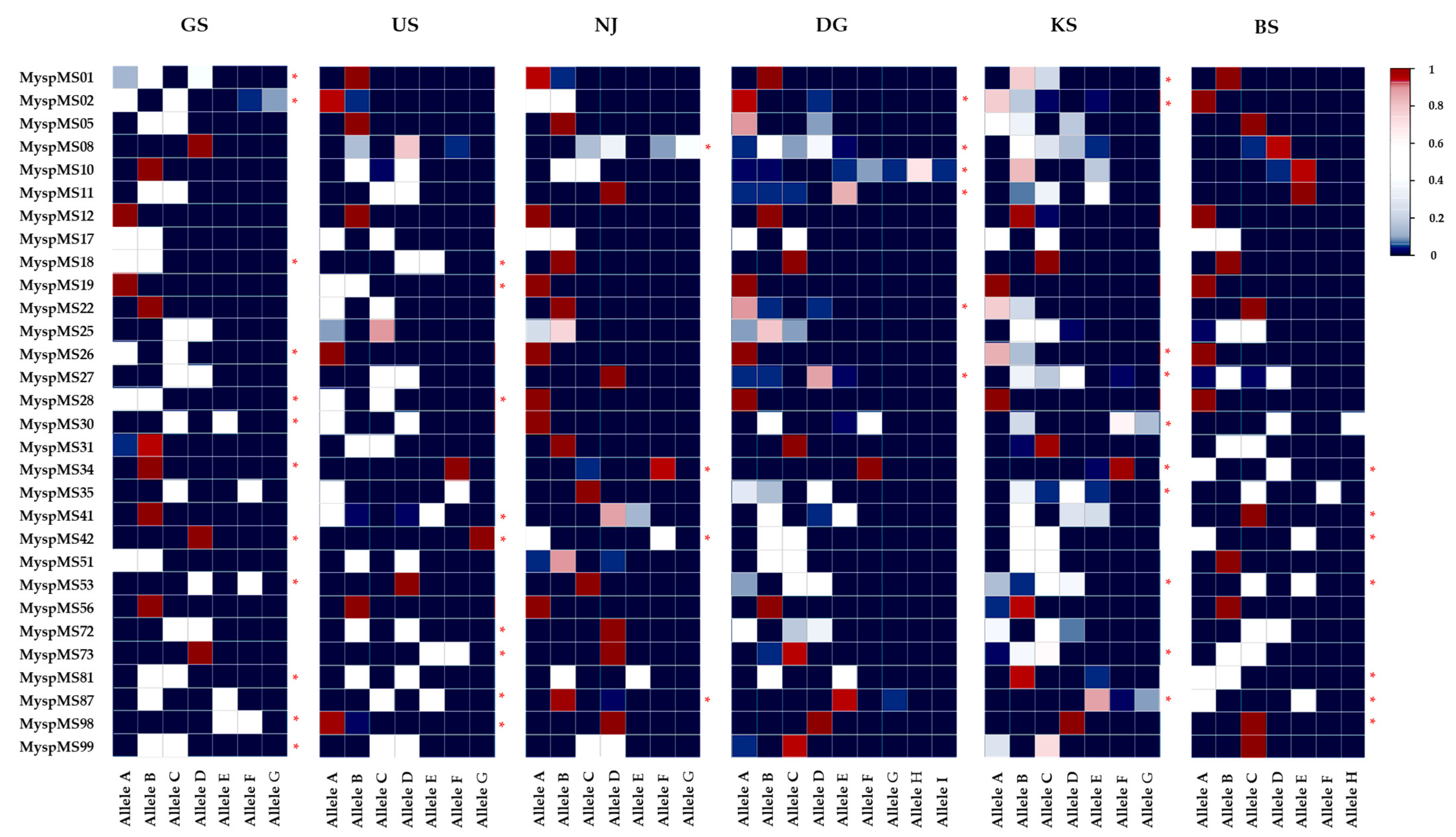
Figure 1. Heatmap of common and unique allele frequency distributions across six Myriophyllum spicatum populations (two streams: GS and US, and four reservoirs: NJ, DG, KS, and BS) based on 30 microsatellite loci. Each cell color represents the relative frequency of a particular allele (blue = 0, white ≈ 0.5, red = 1). * indicates loci containing population-specific unique (rare < 0.05) alleles.
Plants 14 02648 g001
Figure 2. Deviation from an L-shaped allele frequency distribution as evidence of a genetic bottleneck. The analysis is based on 30 microsatellite loci in six populations of M. spicatum.
Plants 14 02648 g002
Figure 3. Geographic barriers and directional gene flow of six Myriophyllum spicatum populations in South Korea: Elevation (a) and River network (b) maps. Jost’s D; asterisks (*) indicate significance at p < 0.05.
Plants 14 02648 g003
Figure 4. Classification of individuals from six populations of M. spicatum into several assumed clusters (ΔK = 6) identified using the STRUCTURE method.
Plants 14 02648 g004
Figure 5. Comparison of spatial genetic structure among six populations of M. spicatum using principal coordinate analysis (PCoA) and interpolated genetic landscapes. Bottom panels show surfaces for the KS (reservoir) and GS (stream) populations, based on actual sampling area and pairwise genetic distances, illustrating spatial genetic structure.
Plants 14 02648 g005
Table 1. Assessment of genetic variations of 30 microsatellite loci developed from M. spicatum. PIC: polymorphism information content; A: number of alleles; Ae/L: effective number of alleles, for all loci; Unique allele: alleles with frequency >0.05 were classified as common and those <0.05 as rare; Ho: observed heterozygosity; He: expected heterozygosity; FIS: inbreeding coefficient; FST: genetic differentiation.
GeneBank Accession No.LocusPICAAe/LUnique AlleleHoHeFISFST
CommonRare
PV231646MyspMS010.40941.96200.2500.449−0.4180.650
PV231647MyspMS020.36771.88310.4330.392−0.5260.314
PV231648MyspMS050.59242.75000.2250.652−0.0210.702
PV231649MyspMS080.54873.73200.2920.5880.2890.341
PV231650MyspMS100.67392.77410.3920.708−0.2130.589
PV231651MyspMS110.65853.66100.4920.709−0.6130.614
PV231652MyspMS120.38131.96010.0080.50400.986
PV231653MyspMS170.55532.63001.0000.625−10.231
PV231654MyspMS180.64452.90300.3330.694−10.792
PV231655MyspMS190.14121.13100.1670.153−10.500
PV231656MyspMS220.59042.57100.2580.663−0.4800.770
PV231657MyspMS250.56943.02000.4920.640−0.1840.394
PV231658MyspMS260.18531.30200.2170.198−0.7090.401
PV231659MyspMS270.48662.25020.6580.538−0.6170.278
PV231660MyspMS280.27231.34200.3330.292−10.474
PV231661MyspMS300.82085.57300.7250.840−0.6860.534
PV231662MyspMS310.38732.02100.3420.508−0.7830.665
PV231663MyspMS340.49461.67410.1920.530−0.7760.825
PV231664MyspMS350.72564.46100.7000.763−0.5650.459
PV231665MyspMS410.72853.46200.4920.766−0.4990.616
PV231666MyspMS420.82875.60400.6250.847−0.870.648
PV231667MyspMS510.48442.29000.6250.521−0.7130.339
PV231668MyspMS530.61762.97300.6000.676−0.5930.488
PV231669MyspMS560.24721.46000.0000.28910.951
PV231670MyspMS720.59243.00100.7750.648−0.7360.351
PV231671MyspMS730.69163.46210.4830.735−0.8110.678
PV231672MyspMS810.57752.15300.8330.611−0.9270.332
PV231673MyspMS870.56672.29220.5580.614−0.7850.536
PV231674MyspMS980.64562.17410.1750.682−0.9090.885
PV231675MyspMS990.44142.04100.6080.478−0.8230.342
Mean0.5304.92.681.730.330.4430.577−0.5660.556
Overall0.55414880.4652100.4430.577−0.6100.569
Table 2. Genetic and clonal diversity in six populations of M. spicatum analyzed by 30 microsatellite loci. N: number of samples; P (0.95): percentage of polymorphic loci; Ae/L: effective number of alleles per locus; Ho: observed heterozygosity, He: expected heterozygosity, G(R): number of multilocus genotypes (MLGs) per population (range of genets that have a ramet); G/N: ratio of genotype; D: Simpson’s diversity index (1–D); E: evenness of genotype.
PopulationNGeneticClonal
P (0.95)AAe/LHoHeG(R)G/NDE
GS2066.7531.670.6180.32711 (2–9)0.5500.7630.839
US2070.0561.640.6080.3248 (2–12)0.4000.5670.648
NJ2040.0461.320.2200.15712 (2–8)0.6000.9010.982
DG2066.7691.520.3520.25212 (2–8)0.6000.9481.000
KS2076.7741.750.4270.34820 (0)1.0001.0001.000
BS2050.0481.450.4320.2254 (2–17)0.2000.2840.379
Mean2061.757.71.560.4430.27211.20.5580.7520.818
Total1201001482.800.4430.57767 (53)0.5580.9590.987
Table 3. Results of the Wilcoxon signed-rank test for bottleneck detection (one- and two-tailed analyses) under three models: infinite allele model (IAM), two-phase mutation model (TPM), and stepwise mutation model (SMM). FIS = coefficient of inbreeding, calculated from 30 microsatellite loci. Significance levels are denoted as follows: p < 0.05 *, p < 0.001 ***.
PopulationHeIAMSMMTPMFIS
GS0.3270.250 ***0.278 ***0.299 ***−0.888
US0.3240.256 ***0.285 ***0.306 ***−0.870
NJ0.1570.261 *0.2890.314−0.382
DG0.2520.3530.3960.431−0.372
KS0.3480.331 *0.3690.406−0.200
BS0.2250.258 ***0.284 ***0.308 ***−0.911
Table 4. Fixation index (FIS) values for 30 microsatellite loci across six populations of Myriophyllum spicatum, along with overall estimates of FIS, FIT, and FST based on F-statistics. Significance levels are denoted as follows: p < 0.05 *, p < 0.01 **, p < 0.001 ***.
LocusGSUSNJDGKSBS
MyspMS01−0.570 ***-−0.027-−0.267-
MyspMS02−0.498 **−0.027 ***−1.000 ***−0.027−0.200-
MyspMS05−0.727 **--1.0000.220-
MyspMS08-0.7140.861−0.376 **−0.0161.000
MyspMS10-−0.905 ***−1.000 ***0.4110.8351.000
MyspMS11−1.000 ***−1.000 ***-0.645−0.553 **-
MyspMS12----0.000-
MyspMS17−1.000 ***−1.000 ***−1.000 ***−1.000 ***−1.000 ***−1.000 ***
MyspMS18−1.000 ***−1.000 ***----
MyspMS19-−1.000 ***----
MyspMS22-−1.000 ***-0.479−0.267-
MyspMS25−1.000 ***1.0001.0000.719−0.615 **−0.905 ***
MyspMS26−1.000 ***---−0.152-
MyspMS27−1.000 ***−1.000 ***-0.791−0.376 *−0.818 ***
MyspMS28−1.000 ***−1.000 ***----
MyspMS30−1.000 ***−1.000 ***-−0.905 ***0.370−1.000 ***
MyspMS311.000−1.000 ***--0.000−1.000 ***
MyspMS34--−0.027-0.000−1.000 ***
MyspMS35−1.000 ***−1.000 ***-−0.520 ***0.493−1.000 ***
MyspMS41-−0.818 ***0.782−0.744 ***−0.473 **-
MyspMS42--−1.000 ***−1.000 ***−0.484 *−1.000 ***
MyspMS51−1.000 ***−1.000 ***−0.056−1.000 **−0.100-
MyspMS53−0.900 ***--−0.520−0.122−1.000 ***
MyspMS56----1.000-
MyspMS72−0.727 **−1.000 ***-−0.510−0.524 **−1.000 ***
MyspMS73-−1.000 ***-−0.027 ***−0.587 **−1.000 ***
MyspMS81−1.000 ***−1.000 ***−1.000 ***−0.810−0.027−1.000 ***Overall
MyspMS87−1.000 ***−1.000 ***0.000−0.027−0.092−0.900 ***Nm = 0.190
MyspMS98−1.000 ***0.000----FIS = −0.610
MyspMS99−1.000 ***−1.000 ***−1.000 ***−0.027−0.357-FIT = 0.305
All −0.888 ***−0.870 ***−0.382 ***−0.372 ***−0.200 ***−0.911 ***FST = 0.569
Table 5. Correlation coefficients between all of the combinations of pairwise differentiation (FST; below) and gene flow (Nm; top). Values based on FST values.
Pop.GSUSNJDGKSBS
GS-0.2190.1670.1610.2290.189
US0.533-0.1340.1110.1530.110
NJ0.6000.652-0.2290.3150.175
DG0.6080.6920.522-1.3000.169
KS0.5210.6200.4420.161-0.215
BS0.5690.6940.5880.5970.538-
Disclaimer/Publisher’s Note: The statements, opinions and data contained in all publications are solely those of the individual author(s) and contributor(s) and not of MDPI and/or the editor(s). MDPI and/or the editor(s) disclaim responsibility for any injury to people or property resulting from any ideas, methods, instructions or products referred to in the content.