Article

Interactions Between the Transcription Factor BOL/DRNL/ESR2 and the Jasmonate Pathway

by
Beatriz E. Ruiz-Cortés
1,
Yolanda Durán-Medina
1,
C. Cecilia Ramos-Tamayo
1,2,
Herenia Guerrero-Largo
1,
Ma. Isabel Cristina Elizarraraz-Anaya
1,
Omar Fabián Hernández-Zepeda
3,
Enrique Ramírez-Chávez
1,
Michiel Lammers
4,
Ruud A. de Maagd
4,
Jorge Molina-Torres
1,
Stefan de Folter
2 and
Nayelli Marsch-Martínez
1,*
1
Irapuato Unit, Biotechnology and Biochemistry Department, Cinvestav, Irapuato C.P. 36824, Guanajuato, Mexico
2
Advanced Genomics Unit, Cinvestav, Irapuato C.P. 36824, Guanajuato, Mexico
3
Genómica Alimentaria, Universidad de la Ciénega del Estado de Michoacán de Ocampo, Sahuayo C.P. 59103, Michoacán, Mexico
4
Wageningen University & Research, P.O. Box 16, 6700 AA Wageningen, The Netherlands
*
Author to whom correspondence should be addressed.
Plants 2025, 14(12), 1757; https://doi.org/10.3390/plants14121757
Submission received: 7 May 2025 / Revised: 29 May 2025 / Accepted: 5 June 2025 / Published: 8 June 2025

Abstract

:
BOL/DRNL/ESR2, an AP2/ERF transcription factor, regulates early organ development in Arabidopsis (Arabidopsis thaliana). Its loss of function causes flower organ defects, while its overexpression induces green callus formation in roots without the addition of hormones. Jasmonates, plant hormones known as major players in stress responses, also regulate some aspects of organ development (e.g., stamen development and plant and organ growth). Here, we studied the interaction between BOL and the JA pathway. We found that exogenous application of methyl jasmonate (MeJA) partially rescued the stamen phenotypes in bol-cr mutants, linking BOL and JA-mediated stamen development. Moreover, MeJA treatments in wild-type plants partially mimicked some bol-D mutant phenotypes like reduced rosette and root size, while JA inhibition restored wild-type leaf curvature, suggesting an alteration in JA homeostasis in the gain-of-function mutant. BOL overexpression caused increased JA levels, whereas bol loss-of-function plants had reduced levels. Furthermore, inducible BOL activity led to downregulation of a JA-responsive marker. Finally, JA biosynthesis inhibition affected BOL-induced root callus formation and led to an expansion of the BOL expression domain in roots. Our findings indicate that BOL modulates parts of the JA pathway and that feedback from the JA pathway appears to affect expression of the transcription factor.

1. Introduction

In plants, the regulation of developmental processes involves transcriptional regulators and plant hormones, among other things [1]. One such transcriptional regulator is BOLITA/DÖRNROSCHEN-LIKE/ENHANCER OF SHOOT REGENERATION 2 (BOL/DRNL/ESR2), a gene that codes for a 306-amino-acid protein that belongs to the AP2/ERF family of transcription factors [2]. It is expressed at the earliest stages of the developing aerial organs, and at later stages, BOL expression is still detectable in developing petals, stamens, and lateral regions of the developing gynoecium [2,3,4,5]. The overexpression of BOL in Arabidopsis (Arabidopsis thaliana) causes severe shoot and root growth inhibition. Moreover, it induces the formation of green calli in roots, which can form a new plant when they are separated from this organ. In tobacco, BOL overexpression causes the formation of ectopic floral organs, stunting, and curvature in the leaves [2]. Moreover, BOL/ESR2 enhances shoot regeneration [4]. The loss of BOL/ESR2/DRNL function has been characterized in the drnl-2 mutant [5], which presents cotyledon fusions [6] and defects in the development of all floral organs, although the defects are more severe in the gynoecium and stamens [3,5,7]. Gynoecia in drnl-2 plants are slightly longer than those in wild-type plants and frequently present asymmetric valves, or lack one valve [3,7]. The number and size of stamens of the drnl-2 mutant are reduced compared to wild-type plants, and some of them are converted into filamentous structures [5]. While in Arabidopsis, the loss of BOL/DRNL function causes a partially penetrant phenotype of fused cotyledons [6], in tomato, the loss of function of the only ortholog of BOL, called LEAFLESS (LFS), produces pin-like shoots with active shoot apical meristems that are unable to form cotyledons or leaves. LFS expression was found in leaf primordia and axillary buds, consistent with its ortholog in Arabidopsis [8].
As a transcription factor, BOL/DRNL/ESR2 is expected to regulate the expression of multiple target genes associated with diverse processes. It has been reported to regulate CUP-SHAPED COTYLEDON 1 (CUC1), a gene involved in meristem specification [4]. This was found through a microarray expression analysis using an ESR2/BOL inducible line, 35S::ESR2-ER. The authors showed that the phenotypes of ESR2/BOL-induced expression decreased in the absence of CUC1 [4]. Moreover, BOL/DRNL regulates family members of the transcriptional regulators SHORT INTERNODES/STYLISH (SHI/STY), which activate auxin biosynthesis [9]. Furthermore, BOL and its homolog DÖRNROSCHEN (DRN) control axillary meristem initiation through their interaction with the transcription factor REVOLUTA (REV), directly regulating the transcription of the SHOOT MERISTEMLESS (STM) gene [9]. Both microarray analyses [4] and RNA-seq studies [10] have identified genes such as ARABIDOPSIS HISTIDINE PHOSPHOTRANSFER PROTEIN 6 (AHP6) as candidate targets acting downstream of DRNL/BOL. Recently, this transcription factor was found to interact with the Auxin Response Factor MONOPTEROS (MP) and to directly activate AHP6 and CYTOKININ OXIDASE 6 (CKX6) [11]. Moreover, BOL also activates ISOPENTENYLTRANSFERASE 5 (IPT5) [12]. Other high-throughput analyses have found more candidate targets [10] in the context of founder cell expression [13]. As more candidate target genes are found, it seems that BOL/DRNL/ESR2 may act as a general coordinator of several processes.
Differential gene expression analyses between a BOL gain-of-function mutant and wild-type plants showed that many different processes were altered in the mutant, including hormonal pathways, and one of these was the jasmonate pathway [2,14]. This opened the question about a possible relation between the transcription factor and this hormonal pathway. Jasmonic acid (JA) and its derivatives, usually known as jasmonates (JAs), are lipid-derived signaling molecules synthesized from α-linolenic acid via the lipoxygenase pathway [15,16,17,18,19,20,21,22,23,24]. Upon stress or developmental signals, JAs accumulate and are perceived by the CORONATINE INSENSITIVE1 (COI1) receptor, triggering degradation of JASMONATE-ZIM DOMAIN (JAZ) repressor proteins, which release transcription factors like MYC2. These transcription factors activate jasmonate-responsive genes. Among them, MYC2 activates JAZ genes, causing a negative feedback loop in the JA signaling cascade [16,21,25,26,27,28].
Jasmonates have been extensively studied as signaling molecules that participate in the plant response to biotic and abiotic stresses, but they also play an important role as regulators of diverse development processes [28]. One of the functions attributed to jasmonates is as promoters of reproductive organ development. Mutants in JA biosynthesis and perception genes, such as aos, opr3, or coi1, exhibit male infertility. Their stamens show delayed anther dehiscence, their filaments do not elongate enough to reach the level of the stigma, and pollen viability is severely reduced [24,28,29]. Jasmonates have also been found to be regulators of plant growth. Moreover, increased levels of JA in the plant reduced its growth by 50% due to an inhibition of mitosis that caused a decrease in cell number [30]. Furthermore, the exogenous application of methyl jasmonate (MeJA) reduced the growth of the main root by 50% [31] due to the inhibition of the cell cycle [32].
Recently, it has been found that JAs play a significant role in promoting root regeneration. Treating plants with MeJA promoted division in normally inactive quiescent center cells and sped up the replacement of columella stem cells when they were damaged [32], while treatment with a JA receptor inhibitor greatly reduced root formation [33]. Similarly, JAs act as signals for root regeneration from leaf explants [34].
Interestingly, in in vitro plant cultures, JAs have been found to both inhibit but also to promote organogenesis and organ growth, depending on their concentration, tissue, species, and combination with other plant growth regulators [35].
Given the enrichment in JA genes among the differentially expressed genes in the bol-D activation tagging mutant and the similarities of some of the reported effects of JA and BOL phenotypes, we sought to explore the relationship between this transcription factor and the jasmonate pathway in the context of plant development.

2. Results

2.1. Gene-Edited bol-cr Mutant Alleles Show Stamen, Gynoecium, and Cotyledon Defects

The BOL loss-of-function drnl-2 mutant allele [5] was generated in the Ler ecotype background and contains a missense mutation (A93V) in the AP2 domain. We sought to generate a new BOL mutant allele in the Col-0 ecotype to facilitate analyses of crosses to marker and mutant lines, many of which have been obtained in this ecotype. We also aimed to generate a new allele in which the whole AP2 domain was removed, to facilitate genotyping. For this, we used the CRISPR-Cas9 system to create a deletion in the AP2 domain of BOL in Col-0 plants. We designed two guide RNAs (gRNAs) that targeted the borders of the AP2 DNA binding domain in BOL (Figure 1a). We obtained 50 transformants in the T0 generation. T1 and T2 progeny plants were genotyped by PCR, and we found two independently edited homozygous mutant plants, named bol-cr1 and bol-cr2. Sequencing showed that bol-cr1 and bol-cr2 contain a deletion of 224 bp between the two gRNAs, resulting in the complete removal of the AP2 domain and a shift in the reading frame in the sequence remaining after the deletion. If translated, these transcripts would result in chimeric, shorter proteins (107 amino acids for bol-cr1 and 99 amino acids for bol-cr2), where the first 52 (bol-cr1) and 53 (bol-cr2) amino acids would be preserved, but the following amino acids would be different from the original BOL sequence (Figure 1a,b and Figure S1). We then allowed the mutations to segregate from the original T-DNA and confirmed, by PCR, that both mutant lines did not contain the transgene with the CRISPR-Cas9 machinery anymore.
bol-cr1 and bol-cr2 plants exhibited the same visible phenotype as the drnl-2 mutant phenotype. All the mutants (drnl-2, bol-cr1, and bol-cr2) presented longer roots than their wild-type backgrounds (Ler and Col-0) at 14 days after germination (Figure 1c). Both CRISPR-Cas9 mutants presented cotyledon fusion in about 4% of the plants (Figure 1d,e; n = 100), which is comparable to the drnl-2 mutant [6]. They also exhibited the range of altered gynoecia phenotypes previously reported for the drnl-2 mutant, including wild-type-like gynoecia, gynoecia with reduced valve sizes, and gynoecia with a single valve, as described in [3] and not observed in wild-type plants (Figure 1f,h). The stamens in the flowers of both BOL CRISPR-Cas9 mutants also showed altered phenotypes, similar to those reported for the drnl-2 mutant [5]. Stamen phenotypes ranged from long stamens that were the same or almost the same size and shape as the wild type to shorter stamens with smaller anthers than the wild type and stamens that did not develop anthers and only consisted of a filament (Figure 1g,i). Moreover, bol-cr mutants presented the characteristic phenotype of fusion between organs, especially between the gynoecium and anthers, in approximately 35% of the flowers (Figure 1j, arrows in Figure 1f). Since the phenotypes were comparable to the drnl-2 mutant phenotype, we concluded that the bol-cr mutants were suitable to study the loss of BOL function. As both bol-cr mutants presented the same phenotype, we used the bol-cr2 allele for further experiments.

2.2. Exogenous Application of MeJA to Bol Loss-of-Function Mutants Partially Recovers the Wild-Type Stamen Phenotype

To evaluate whether the anther phenotypes of bol loss-of-function mutant flowers could be due, at least partially, to a deficiency of jasmonates, we analyzed the effect of MeJA applications on bol-cr2 mutant flowers.
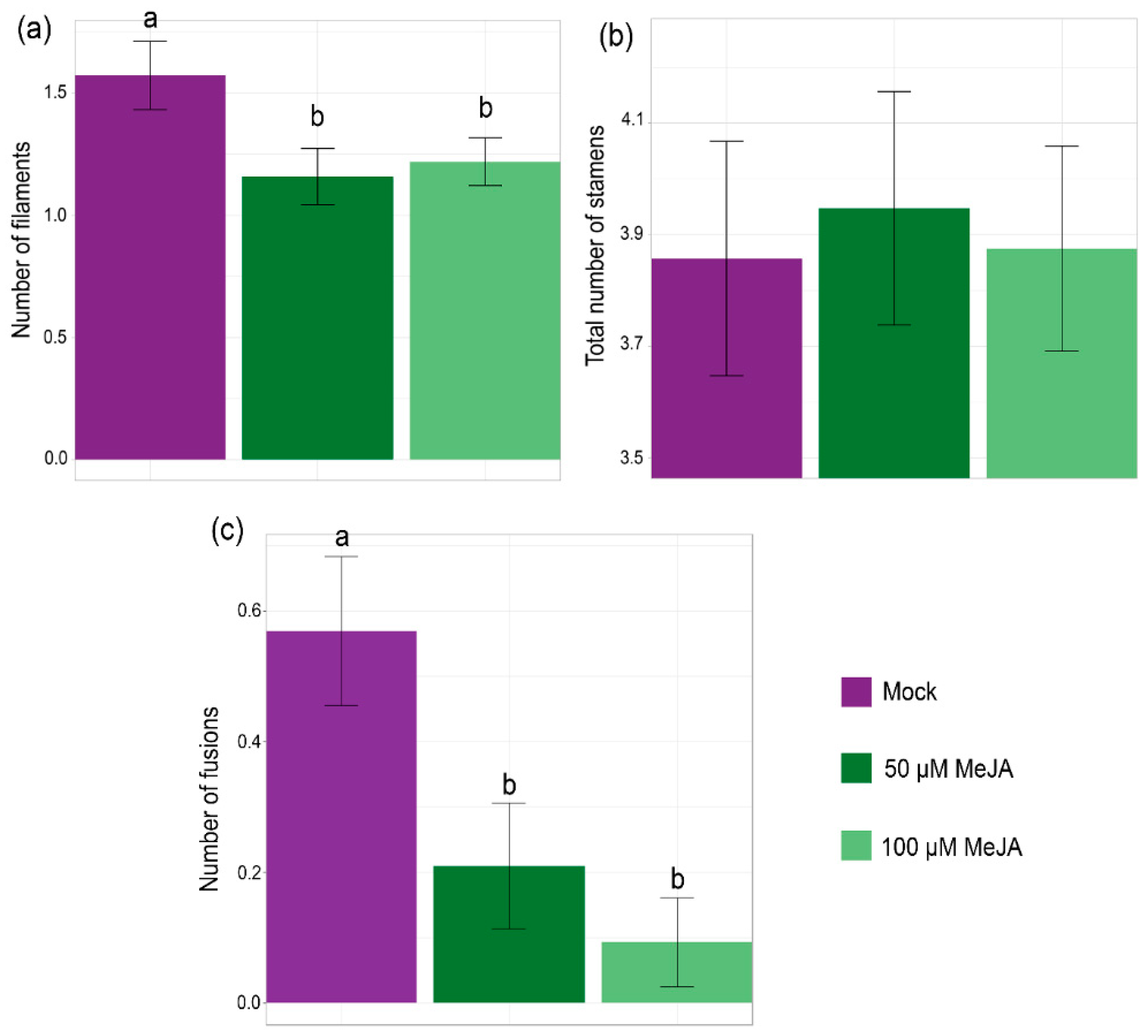
As the mutant presented variable stamen phenotypes, we counted the three different types of previously observed staminoid structures in control flowers and flowers that were treated with 50 and 100 μM MeJA: long stamens, short stamens, and filaments.
We observed a reduction in the number of filaments in the flowers treated with MeJA (Figure 2a) while the total number of all staminoid structures remained unchanged (Figure 2b). Surprisingly, we also observed fewer fusions between organs in the flowers of the plants that were treated with MeJA in comparison to control flowers (Figure 2c). Therefore, the exogenous MeJA application could partially recover the wild-type stamen phenotype in the bol-cr2 mutant. This suggested that the floral organ defect phenotype could be, at least in part, due to an alteration in the JA pathway.

2.3. MeJA Application or Inhibition Mimic or Partially Restore Bol-D Phenotypes

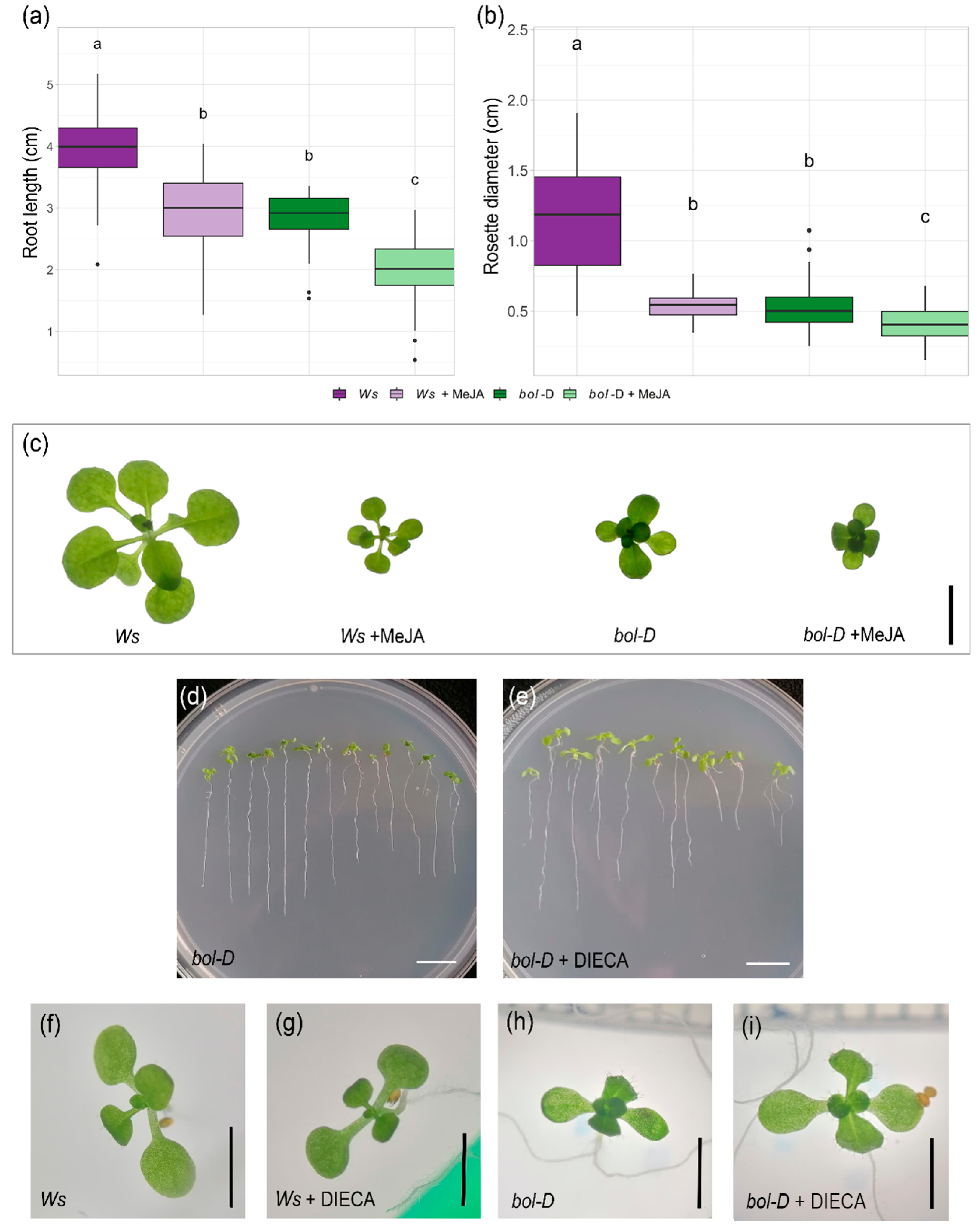
The activation-tagged gain-of-function mutant bol-D exhibits both reduced root growth and plant size [2]. Since JA applications also cause root growth inhibition [31,36] and rosette size reduction [30,32], we compared wild-type plants (Wassilewskija, Ws, the background ecotype of the bol-D mutant) treated with MeJA to bol-D plants.
We observed that, 9 days after germination (dag), root length and rosette diameter of wild-type plants treated with 50 µM MeJA and bol-D plants were equivalent (Figure 3a,b). This could suggest that the reduction in the size of the leaves and root length in bol-D plants could be due, at least partially, to an increase in the levels of jasmonates in the plant or another alteration in the pathway.
Despite the reduced size of the wild-type plants upon MeJA application, the characteristic phenotype of bol-D plants, which is curvature of the leaves, was not observed; rather, wild-type plants simply reduced their size (Figure 3c).
To test the opposite conditions, we used a previously reported JA biosynthesis inhibitor, sodium diethylditiocarbamate (DIECA) [37]. Interestingly, when bol-D plants were grown in the presence of DIECA, the curvature of the leaves was reduced, suggesting that this phenotype might result from an alteration in the internal jasmonate balance, which cannot be phenocopied by exogenous JA application (Figure 3d–i). The phenotype may also be the result of the combination of alterations in different processes caused by the gain of BOL function, including JA homeostasis.

2.4. Changes in BOL Expression Cause Differences in Jasmonic Acid Accumulation

The previous experiments suggested that some of the morphological phenotypes displayed by bol-cr and bol-D might be, at least partially, due to alterations in jasmonate homeostasis. Therefore, we quantified the level of jasmonic acid in different genetic backgrounds: (i) the Arabidopsis gain-of-function mutant bol-D; (ii) a BOL inducible line (35S::ESR2-ER), in which BOL is fused to a β-estradiol receptor fragment, and the application of β-estradiol (β-est) promotes its entrance to the nucleus [4,38]; (iii) a 35S::BOL overexpressor line in tobacco; and (iv) Arabidopsis bol-cr2 inflorescences. These were compared to their respective wild-type counterparts. For the inducible line, JA content was also compared to non-induced control plants, and it was evaluated two and four days after induction.
Interestingly, BOL gain-of-function and overexpressor plants were found to contain more jasmonic acid than wild-type plants. Arabidopsis bol-D and tobacco 35S::BOL plants presented a 2-fold and 6-fold increase, respectively, in JA content by mg of fresh weight than their corresponding wild-type plants (Figure 4a,b). In the inducible BOL line 35S::ESR2-ER, a higher JA content was detected 4 days after induction compared to non-induced plants (Figure 4c). Conversely, when comparing the JA content in inflorescences of wild-type and bol-cr2 mutant plants, bol-cr2 presented a reduction, containing less jasmonic acid than wild-type Col-0 plants (Figure 4d).
These results suggest that the jasmonate level is altered when BOL expression or function is altered.

2.5. BOL Affects the Expression of the JA Response Marker pJAZ10::GUSPlus

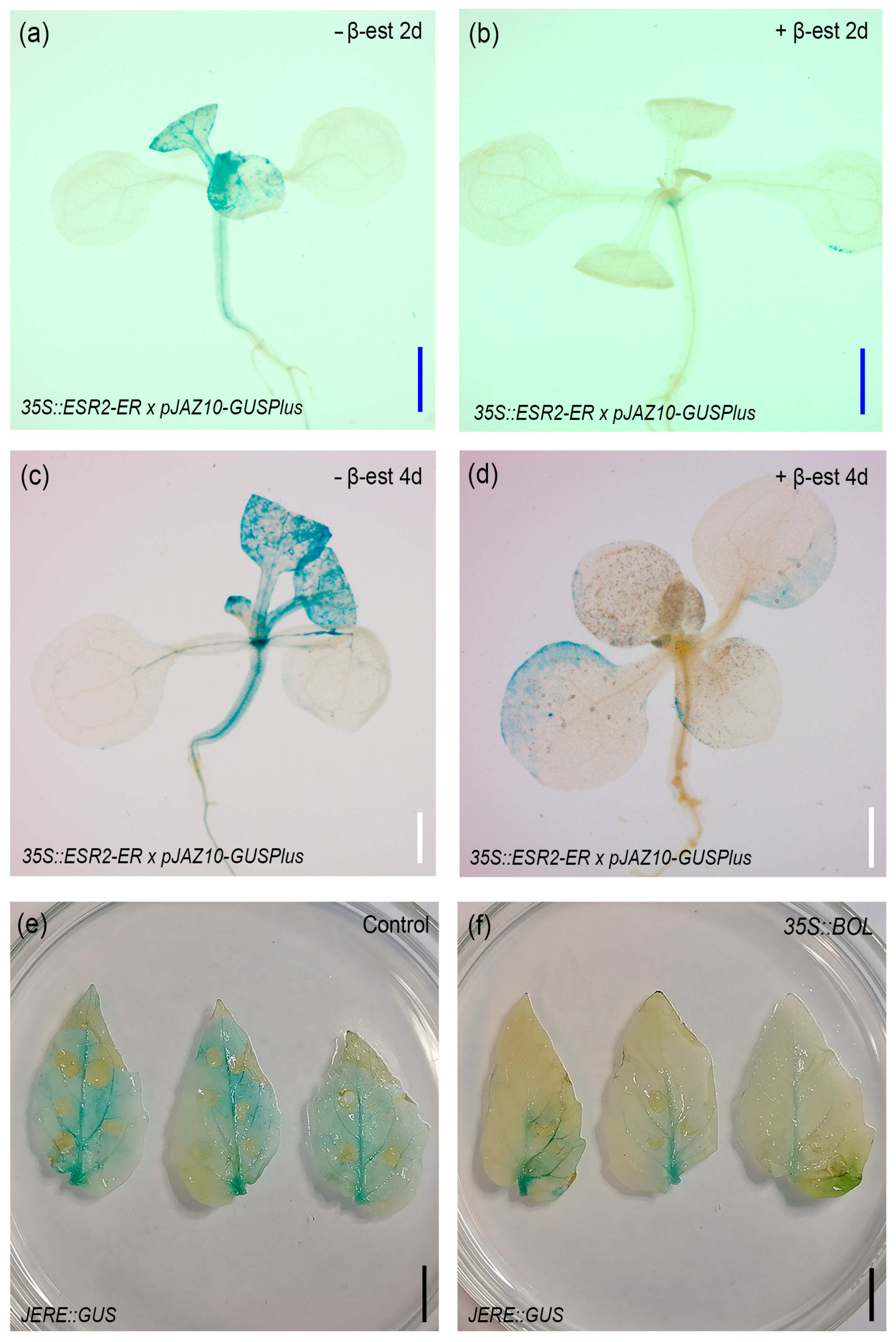
After observing the changes in JA content, we explored the effect of increased BOL activity in the transcriptional response to JA. For this, we analyzed the expression of the JA response marker pJAZ10::GUSPlus [39] in the inducible 35S::ESR2-ER background. We transferred 9-day-old seedlings to a β-estradiol medium (inducing medium) and analyzed the expression of the response marker from 1 to 4 days after induction. In normal conditions, expression was detected in the hypocotyl and meristematic region of the seedlings, and patchy expression was observed in the cotyledons and developing leaves (Figure S2a,c,e,g). At 2 and 4 days after BOL induction, we observed a reduction in expression in the hypocotyl and meristematic region (Figure 5b,d and Figure S2d,h) compared with those plants that were not induced (Figure 5a,c and Figure S2c,g). This expression pattern was consistently observed across the 1 to 4 days of analyses (Figure S2). We also evaluated the expression of the tomato JA response marker 4xJERE::GUS [40] after the transient expression of a 35S::BOL construct. We observed that the leaves that were infiltrated with the mock solution presented GUS expression in almost all the leaf (Figure 5c), but the ones infiltrated with the 35S::BOL construct presented a reduced expression of the marker (Figure 5d). These results suggest that the transient or inducible increased expression of BOL can cause a reduction in the response to JA, at least in the analyzed times and tissues.

2.6. Jasmonate Inhibition Impairs the Formation of BOL-Induced Green Calli

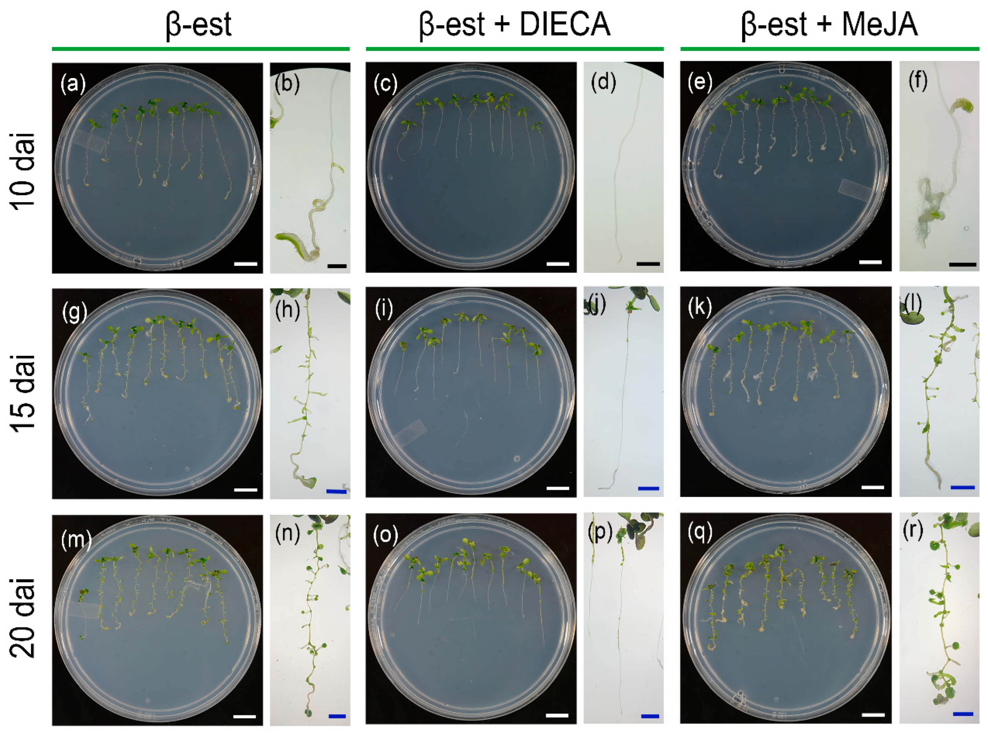
One of the characteristic phenotypes of BOL increased activity is the spontaneous formation of green calli in roots without the addition of any hormones. We asked whether the application of JA or the JA biosynthesis inhibitor DIECA would inhibit or exacerbate this phenomenon. For this, we analyzed the formation of green calli in inducible 35S::ESR2-ER plants cultivated in medium supplemented with MeJA or DIECA. We observed that regions with green calli were visible 10 days after induction, mostly at the tip of the root, in control plants and those treated with MeJA (Figure 6a,b,e,f and Figure S3a,c). However, these regions were less evident in the plants that were treated with JA synthesis inhibitor (Figure 6c,d and Figure S3b). Further in time, 15 and 20 days after germination, the green calli areas were observed along the entire root in the mock and MeJA-treated induced plants (Figure 6g,h,k–n,q,r and Figure S3d,f,g,i). In contrast, the plants treated with DIECA only presented small green calli areas in the differentiated part of the root, at the top of the root regions located close to the hypocotyl (Figure 6i,j and Figure S3e,h). These results suggest that the inhibition of JA biosynthesis reduced the ability of BOL/ESR2 to promote callus formation in roots, and they may indicate that JA is required for the formation of green calli induced by BOL/ESR2 activation.

2.7. The Inhibition of JA Biosynthesis Alters the Pattern of BOL Expression

To explore whether JAs could also play a role in the indirect regulation of BOL expression, we analyzed the expression of the BOL reporter line pBOL::GUS [2] in plants treated with and without the JA biosynthesis inhibitor.
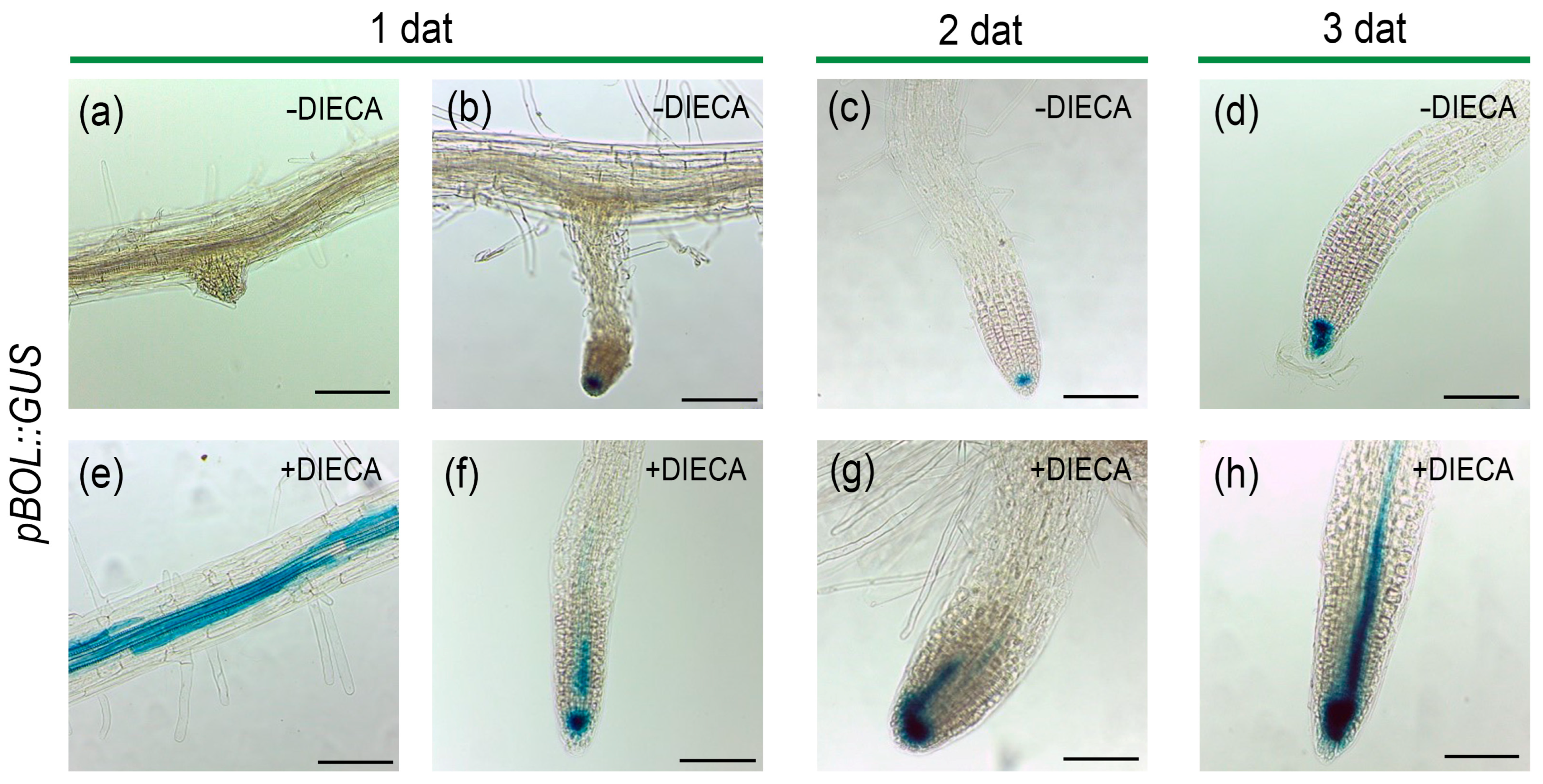
As previously reported, in control seedlings we observed the expression of BOL in leaf primordia [2,4]. We also observed sporadic expression in meristematic regions of the main and lateral roots (Figure 7a–d). Interestingly, when plants were treated with DIECA, we observed that the BOL expression domain expanded in the roots. From the only sporadic expression at the root tips, the expression was now observed in all roots and had expanded to the region above the root tip.
The expansion in the expression domain was larger as the plants were exposed to the JA inhibitor for longer times (Figure 7f–h). We also observed the ectopic expression of BOL in epidermal cells of the differentiated zone of the roots (Figure 7e). These changes suggest that JA appears to restrict BOL expression.

3. Discussion

The main question of this study was to explore the connections between the BOL/ESR2/DRNL transcription factor and the jasmonate pathway. The results of this exploration indicate that BOL can modulate the jasmonate pathway and that JAs restrict BOL expression.
The phenotypes of loss-of-function mutants include defects in floral organs, particularly stamens and gynoecia [3,5,7]. JA plays an important role in stamen development, as mutants of some of the JA biosynthesis enzymes present male sterility in Arabidopsis due to deficient filament elongation, non-viable pollen, and delayed anther dehiscence [22,29,41,42,43]. Defects in floral organs and sterility in JA biosynthetic gene mutants have also been reported in other species such as rice, tomato, or maize, highlighting the importance of JA in floral organ development [44]. Our results showed that an exogenous MeJA application could partially recover the wild-type phenotype in the bol-cr2 mutant, suggesting that a part of the BOL gene function in floral organ development involves achieving or maintaining proper JA homeostasis. One of the possible reasons why only partial recovery of the phenotype could be achieved and not a complete recovery is that BOL may be coordinating different processes simultaneously, including different hormonal pathways, not only jasmonates. Floral development also involves other hormones, such as auxins and gibberellins [45,46,47]. Another possibility is that, in addition to affecting the jasmonate content, the loss-of-function mutation also affects the response to this phytohormone so that, despite the increased content, the mutant becomes unable to respond in the same way as a wild-type plant. Surprisingly, we were only expecting partial recovery of organ formation, but a reduction in organ fusion was also observed. To date, we have not found information about a possible role of jasmonates in preventing organ fusion, and this is an interesting observation to further explore.
JA application or inhibition in wild-type or dominant mutants, and JA content quantification, further revealed a connection between BOL and this hormonal pathway. When we treated wild-type plants with MeJA, we observed a reduction in rosette size, which resembled the size of bol-D rosettes. JAs affect the cell cycle, and they inhibit cell growth in leaves [32]. Jasmonates affect cellular proliferation and expansion, and they could play a role in the synchronization of the development of different tissues in a new organ at specific stages. We noticed that the addition of exogenous jasmonates to wild-type plants caused a reduction in rosette size without leading to the leaf curvature characteristic of bol-D plants. However, when bol-D plants were treated with a jasmonate inhibitor, DIECA, leaf curvature was reduced, leading to flatter leaves, more similar to wild-type leaves. Therefore, even when the MeJA application could not replicate the curved leaf phenotype in wild-type plants, the inhibition of JA biosynthesis recovered the wild-type phenotype. This could suggest that the phenotype of the bol-D mutant could be caused, at least partially, by an excess of jasmonates in the plant, either one of the active molecules of the jasmonate pathway or a modified jasmonate molecule that is only produced in high levels in the bol-D mutant, possibly due to upregulation of JA biosynthesis genes. Another scenario is that jasmonates within the bol-D mutant plant could have a different distribution than in wild-type plants. Moreover, the leaf curvature phenotype may require a combination of changes in other pathways or genes, together with JAs, which may occur in the bol-D mutant but not in wild-type plants. Exploring this in greater detail through additional research will be essential to elucidate BOL function and can provide insights into the mechanisms by which it affects leaf shape.
Another phenotype that was recovered by the inhibition of JA biosynthesis was the spontaneous formation of green calli in roots, a conspicuous phenotype produced by BOL induction. Callus formation was severely reduced when plants were treated with DIECA, suggesting that jasmonates may act downstream of BOL or are required for the BOL-promoted callus formation phenotype. This finding was unexpected because these calli appear to develop from lateral roots, and calli have been found to have lateral root primordia identity [48,49]. JAs are known to inhibit lateral root formation [50,51]. However, the inhibition of JA biosynthesis by DIECA severely reduced the formation of calli. Therefore, the role of JA and the effect of DIECA on callus formation in induced 35S::ESR2-ER plants may not be due to an effect on lateral root formation, but rather in the conversion to green callus, possibly. In this work, we did not explore other phenotypes that may have been recovered by JA biosynthesis inhibition, and it will be interesting to explore further.
BOL seems to affect both JA levels and response. The phenotypes obtained after the JA and inhibitor treatments suggested that some of the changes in the development of BOL gain- and loss-of-function mutants, and inducible line, could be caused by an altered level of jasmonates. When JA levels were analyzed, we observed that all BOL overexpressor plants (35S::BOL in tobacco, bol-D and 35S::ESR2-ER in Arabidopsis) contained more jasmonic acid than wild-type plants, in contrast to the bol-cr2 mutant, in which JA levels decreased compared to wild-type plants. This indicated that BOL is able to modulate JA levels. Curiously, when BOL was induced in JA reporter lines, they showed reduced expression. This effect might be caused by a compensation where the increased JA accumulation by BOL induction activates JAZ genes as a part of a negative feedback loop in JA signaling, resulting in the reduced JA response observed despite the high levels of the hormone. Furthermore, the timing and tissue type in which these effects are exerted may be different. Future experiments will be essential to evaluate the JA response and accumulation upon BOL activation in different tissues and stages to better understand the temporal and spatial dynamics of this effect or regulation.
At this moment, these experiments do not reveal whether the influence of BOL on the jasmonate pathway is direct, with BOL binding and regulating the expression of JA genes, or indirect, through the regulation of other genes that in turn affect the pathway. It will be interesting to explore this further. It could be indirect, since a clear increase in JA content in inducible BOL (35S::ESR2-ER) occurred four days after induction. Recently, we found that BOL/ESR2 causes an increase in cytokinin content [12], and cytokinin has been reported to promote JA accumulation [52]. This could also be happening in Arabidopsis, but we cannot discard the possibility of direct regulation of BOL upon JA-related genes. Nevertheless, regardless of whether the regulation is direct or indirect, it may not be involved in rapid responses to wounding or stress, since these stimuli do not appear to activate BOL transcription.
However, JAs play a “double role” in the plant as chemical signals that trigger a rapid plant response in defense to herbivory [53,54] and wounding [55] and as developmental signals. In this regard, JA genes are also developmentally regulated. For instance, the transcription factor AGAMOUS (AG) directly regulates the expression of the JA biosynthetic gene DEFECTIVE IN ANTHER DEHISCENCE 1 (DAD1), ensuring proper JA levels during stamen maturation, including elongation of the filaments, pollen maturation, and anther dehiscence [47]. As in this example, we speculate that the role of BOL in the JA pathway may be related to developmental regulation.
Curiously, the interaction between the BOL and the JA pathways appears to include a feedback loop, as JAs appear to negatively affect BOL expression. The BOL expression pattern has been reported to be restricted to aerial developing young tissues and organs [2,3,4,5]. We have observed that this pattern is constant and remains unchanged in several different conditions. Interestingly, the expression pattern clearly changed when JA biosynthesis was inhibited. This was evident in the root. Though BOL expression is constant in very young developing aerial organs, it is also sporadically expressed in the tips of the main and the young lateral roots. This expression occurs only in very few roots, is not constant, and is very low [2]. The presence of low counts of BOL transcripts in RNA-seq experiments [56] and the longer root phenotype in drnl-2 and bol-cr loss-of-function mutants suggest that the gene could be expressed at very low or transient levels in specific regions of the roots, though this has not yet been thoroughly investigated.
The effect of JA inhibition on BOL expression in the root was striking. When the reporter line was transferred to a medium supplemented with the JA biosynthesis inhibitor, expression was then observed in all the analyzed roots and expanded to reach the inner tissues above the tip of the root, and even some mature root regions, where it was not observed before. This suggests that normal levels of JA, either in a cell or non-cell autonomous way, could be repressing BOL expression in the root. This may point to a feedback loop in the BOL–JA interaction; as BOL seems to modulate JA signaling and accumulation, JA, in turn, restricts BOL expression. It will be interesting to determine whether this effect is reflecting a repressing regulatory role of the JA pathway in BOL expression, and if so, whether this regulation is directly exerted by transcription factors downstream of the pathway (such as MYC2) or indirectly by affecting other factors. If it were a direct consequence of reduced jasmonate content within the plant, it could be a feedback loop, whereby, to restore the normal level of jasmonates, BOL gene expression is promoted. However, more experiments are needed to corroborate these speculations. When searching for elements that can regulate BOL and LFS expression, both BOL and LFS promoters were found to include GCC boxes [8], which are well-characterized cis-regulatory elements involved in the transcriptional regulation of jasmonate-responsive genes through their interaction with ERF transcription factors [57]. This could suggest that BOL expression may be directly modulated by jasmonates, but more research is required to test whether this is the case.
With the results presented in this work, we propose that BOL modulates the jasmonate pathway in a developmental context. Future experiments will be crucial to determining whether the regulation of the JA pathway by BOL is direct or indirect, identifying the specific JA pathway genes or intermediary genes targeted by BOL, and shedding light on the biological relevance of this regulation. Moreover, the putative regulatory JA–BOL feedback loop should be further studied to determine the spatiotemporal dynamics of this regulation, which will provide deeper insights into the mechanisms underlying the BOL and JA pathway interaction and its biological role.

4. Materials and Methods

4.1. Plant Materials

The lines used in this study were: Columbia (Col-0), Landsberg erecta (Ler), and Wassiliewskija (Ws) wild-type ecotypes; bol-D [2], drnl-2 [5], and bol-cr1 and 2 (generated in this work) mutants; pJAZ10::GUSPlus (JA response marker line) [39]; pBOL-GUS (BOL marker line) [2]; the inducible overexpression BOL line 35S::ESR2-ER for Arabidopsis thaliana [4,38]; 35S-BOL (BOL overexpressor line) and wild-type tobacco (Nicotiana tabacum cv SR1) [2]; and the jasmonate response marker line, 4xJERE::GUS in tomato (cv MT), where the tomato jasmonic acid response element (AGACCGCC) is fused to the β-glucuronidase gene [40].

4.2. Plant Growth

Arabidopsis seeds were sown in soil (peat moss, perlite, vermiculite 3:1:1) and were kept at 4 °C for 2 days before being transferred to a growth chamber at 22 °C under long day conditions. Two weeks after germination, plants were transferred to a greenhouse with natural light and temperature conditions. Tobacco and tomato seeds were grown in soil (peat moss, leaf soil, lama soil, perlite, and vermiculite 3:2:1:1:1) in a growth room at 24 °C under long day conditions (16 h of light/8 h of dark).
For Arabidopsis in vitro analyses, seeds were disinfected with chlorine gas in a vacuum chamber for 5–6 h and were sown in medium containing 0.5× MS (Caisson Labs, TX, USA), 1% sucrose, and 1% agar. Plates were kept for 2 days at 4 °C before being transferred to a growth chamber at 22 °C under long day conditions. Plates were incubated vertically for root measurement and calli evaluation and horizontally for rosette measurement.

4.3. Generation of a CRISPR-Cas9 Induced New Allele of BOL

Guide RNAs (gRNAs) were designed with the bioinformatic tool CRISPR-P [58]. Guides flanking the region coding for the AP2 domain in BOL, with the best score and without off-targets, were selected.
The vector containing the Cas9/gRNA cassette was generated using the Golden Gate system vectors [59,60]. gRNAs were inserted individually into level 1 vectors pICH47751 (gRNA1) and pICH47761 (gRNA2). Both gRNA constructs, NOSp::NPT2 OCST (selection marker; pICH47732) and pRPS5A:Cas9-NOSt (pICH47742) were inserted into the level 2 binary vector pAGM4723.
For the synthesis of the RNA fragments using vector pICH86966::AtU6p::sgRNA_PDS as a template, the PCR conditions were: 94 °C—5 min; 94 °C—15 s, 60 °C—30 s, 68 °C—1 min, 30 cycles; 68 °C—5 min. Purification of PCR fragments was performed with “DNA Clean & ConcentratorTM-5 Kit” (Zymo Research, CA, USA). Level 1 reaction conditions were: 37 °C—3 min, 16 °C—4 min, 20 cycles; 50 °C—5 min; 80 °C—10 min. Level 2 reaction conditions were: 37 °C—8 min, 16 °C—8 min, 7 cycles; 37 °C—10 min; 80 °C—15 min. Plasmid extraction was performed with “ZyppyTM Plasmid Miniprep Kit” (Zymo Research, Irvine, CA, USA). The BsaI and BpiI enzymes (BioLabs, San Diego, CA, USA) were used for digestion of level 1 and level 2 vectors, respectively. The T4 DNA ligase (Promega, Madison, WI, USA) enzyme was used for ligation. The final vector was corroborated by colony PCR, digestion with BamHI, EcoRI (Fermentas, Waltham, MA, USA), PstI (Thermo SCIENTIFIC, Waltham, MA, USA), AfiII (BioLabs, MA, USA) enzymes, and, finally, by sequencing.
The binary vector with the final construct was transformed in A. tumefaciens GV3101, and Col-0 plants were transformed by floral dip [61]. T0 transformant plants were selected with kanamycin. T1 and T2 transformants were genotyped by PCR and finally by sequencing. All the primers used are listed in Table S1.

4.4. GUS Expression Analysis

GUS staining was performed at 37 °C overnight with a standard X-gluc solution [3]. Images were taken with a Leica ICC50 HD optical microscope (Leica Microsystems, Wetzlar, Germany).

4.5. Phenotypic Analyses and Documentation

Images of floral organs of Col-0, bol-cr1 and bol-cr2 were taken using a Keyence VHX-7000 digital microscope (Keyence Corporation, Osaka, Japan). Pictures of in vitro-grown plants were taken with a Zeiss Stemi 2000-C stereoscope (Carl Zeiss AG, Jena, Germany) and a Nikon D7100 camera, (Nikon Corporation, Tokyo, Japan) and they were analyzed with ImageJ 1.54g software.

4.6. BOL Transient Expression Assays

Agrobacterium tumefaciens (GV2260) harboring a pMDC204 vector with the 35S::BOL construct was grown in 5–10 mL of LB medium supplemented with kanamycin (50 mg/L), rifampicin (25 mg/L), and ampicillin (50 mg/L) at 28 °C for 2 days until saturation (OD > 1). The culture was centrifuged at 4000 rpm for 10 min at room temperature, and the collected cells were resuspended in 10 mL of infiltration medium (10 mM MgCl2, 10 mM MES pH = 5.6, 100 mM acetosyringone). The bacteria were left in slight agitation at room temperature for at least 3 h. The cell suspension was injected into the abaxial side of young leaves of 4xJERE-GUS tomato plants with a 3 mL syringe without needle. Four to five injections were made in leaves of the same developmental stage of five different 8-week-old plants. Infiltrated leaves were collected 4 days after infiltration to evaluate GUS activity. Infiltration medium without bacteria was used as a mock treatment.

4.7. MeJA and DIECA Treatments

Before the experiment, new inflorescences emerging above cauline leaves of 6-week-old plants were identified and marked. They were allowed to grow for 8 days, and then all their open flowers were removed. The remaining closed flower buds were sprayed with a MeJA solution (50 or 100 μM MeJA, 0.01% Silwet L-77 in distilled water) in the morning and in the afternoon of that day. Starting one day after the spraying, 25 open flowers were collected each day, and the rest of the open flowers were removed. Staminoid structures of the collected flowers were analyzed. This procedure was performed for seven consecutive days to evaluate buds that were at different stages of development at the moment of treatment.
For the in vitro experiments, plants were grown in Petri dishes with MS and agar medium (as previously described) supplemented with either 50 µM MeJA or 250 µM DIECA.

4.8. Jasmonic Acid Quantification

Jasmonic acid was extracted from 250 mg of fresh tissue and analyzed according to [62,63,64,65,66] with some modifications. A total of 1 mL of ethyl acetate and 10 μL of 0.1 mg/mL (±)-Dihydrojasmonic acid (internal standard; Cat: CDS022683, Sigma-Aldrich, St. Louis, MO, USA) were added to each sample and mixed in a vortex. The samples were agitated for 30 min, and the supernatant was mixed with the previous one. The solvent was completely evaporated with nitrogen gas, and 100 μL of N′N′ di-isopropyl-ethylamine, 100 μL of chloroform, and 10 μL of PFB-Br (2,3,4,5,6-pentafluorobenzyl bromide, Sigma-Aldrich) were added to each sample for derivatization. The samples were incubated at 60 °C for 30 min. The solvents were completely evaporated with nitrogen gas, and the samples were resuspended in 200 μL of methanol, HPLC grade. A total of 1 µL of each sample was injected to analyze JA using GC-EIMS (Gas Chromatography, Agilent Technologies 7890A-Electron Ionization Mass Spectrometry, Agilent Technologies 5975C, Santa Clara, CA, USA) in SIM mode for 141, 181, 390 m/z. The separation was performed in a DB-1 MS ultra-inert capillary column (60 m length × 0.25 mm internal diameter and 0.25 μm film thickness; Agilent Technologies). The gas chromatograph was run under the following program: initial temperature 150 °C for 3 min, then a ramp at 4 °C per min up to 260 °C; this temperature was kept constant for 25 min. The quantification of the JA was performed using the internal standard of dihydrojasmonic added to each sample, as indicated above. An average of the areas of the internal standard was obtained for all the samples, obtaining a correction factor by dividing the area of the internal standard by the area of each sample and multiplying it by the correction factor obtained for each sample. The average area of the internal standard corresponds to a concentration of 0.005 µg/µL.

4.9. Statistical Analysis

Statistical analyses and graphs were made using RStudio (Version 4.2.0, RStudio Team, Boston, MA, USA). For parametric variables, ANOVA or Student’s t-tests were used, while the Kruskal–Wallis test was employed for nonparametric variables. When significant differences were found, LSD and Dunn’s post hoc tests were applied.

Supplementary Materials

The following supporting information can be downloaded at: https://www.mdpi.com/article/10.3390/plants14121757/s1; Figure S1. Genotyping of bol-cr mutants by PCR using genomic DNA; Figure S2. JA reporter line expression changes upon BOL induction; Figure S3. Effect of jasmonate inhibition or exogenous application in BOL-induced green calli formation; Table S1. Primers used for the generation of CRISPR-Cas9-induced BOL alleles bol-cr1 and bol-cr2.

Author Contributions

Conceptualization: B.E.R.-C., Y.D.-M. and N.M.-M.; methodology: N.M.-M., S.d.F., J.M.-T., B.E.R.-C., Y.D.-M., C.C.R.-T., M.L., R.A.d.M. and O.F.H.-Z.; formal analysis and investigation: B.E.R.-C., Y.D.-M., C.C.R.-T., M.I.C.E.-A., H.G.-L., O.F.H.-Z. and E.R.-C.; writing—original draft preparation: B.E.R.-C.; writing—review and editing: B.E.R.-C. and N.M.-M.; funding acquisition: N.M.-M.; resources: J.M.-T., S.d.F., R.A.d.M. and N.M.-M.; supervision: N.M.-M. All authors have read and agreed to the published version of the manuscript.

Funding

Work at the N.M.-M. lab was funded by project CF-2023-G-219 from the Mexican Secretary of Science, Humanities, Technology, and Innovation (SECIHTI, before Conahcyt, and earlier CONACyT), which also provided Ph.D. fellowships to B.E.R.-C. (707345), YDM (262981), C.C.R.-T. (858918), H.G.-L. (1008711), and O.F.H.-Z. (301808). O.F.H.-Z. worked at the laboratory of Martin Heil, which was funded by former CONACyT projects 258119 and 251102. The authors have no relevant financial or non-financial interests to disclose.

Data Availability Statement

Data are contained within the article and Supplementary Materials.

Acknowledgments

The authors thank Edward E. Farmer for the kind gift of pJAZ10::GUSPlus and Lazaro Peres for 4xJERE::GUS seeds. We also thank Juan Carlos Zavala for technical help, Juan Carlos Ochoa for administrative support, and David Díaz Ramírez for help with data analysis and technical advice.

Conflicts of Interest

The authors declare no conflicts of interest.

References

  1. Traas, J. Organogenesis at the Shoot Apical Meristem. Plants 2019, 8, 6. [Google Scholar] [CrossRef]
  2. Marsch-Martinez, N.; Greco, R.; Becker, J.D.; Dixit, S.; Bergervoet, J.H.W.; Karaba, A.; De Folter, S.; Pereira, A. BOLITA, an Arabidopsis AP2/ERF-like Transcription Factor That Affects Cell Expansion and Proliferation/Differentiation Pathways. Plant Mol. Biol. 2006, 62, 825–843. [Google Scholar] [CrossRef]
  3. Durán-Medina, Y.; Serwatowska, J.; Reyes-Olalde, J.I.; De Folter, S.; Marsch-Martínez, N. The AP2/ERF Transcription Factor DRNL Modulates Gynoecium Development and Affects Its Response to Cytokinin. Front. Plant Sci. 2017, 8, 1841. [Google Scholar] [CrossRef] [PubMed]
  4. Ikeda, Y.; Banno, H.; Niu, Q.W.; Howell, S.H.; Chua, N.H. The Enhancer of SHOOT REGENERATION 2 Gene in Arabidopsis Regulates CUP-SHAPED COTYLEDON 1 at the Transcriptional Level and Controls Cotyledon Development. Plant Cell Physiol. 2006, 47, 1443–1456. [Google Scholar] [CrossRef]
  5. Nag, A.; Yang, Y.; Jack, T. DORNRÖSCHEN-LIKE, an AP2 Gene, Is Necessary for Stamen Emergence in Arabidopsis. Plant Mol. Biol. 2007, 65, 219–232. [Google Scholar] [CrossRef] [PubMed]
  6. Chandler, J.W.; Cole, M.; Flier, A.; Grewe, B.; Werr, W. The AP2 Transcription Factors DORNRÖSCHEN and DORNRÖSCHEN-LIKE Redundantly Control Arabidopsis Embryo Patterning via Interaction with PHAVOLUTA. Development 2007, 134, 1653–1662. [Google Scholar] [CrossRef] [PubMed]
  7. Chandler, J.W.; Jacobs, B.; Cole, M.; Comelli, P.; Werr, W. DORNRÖSCHEN-LIKE Expression Marks Arabidopsis Floral Organ Founder Cells and Precedes Auxin Response Maxima. Plant Mol. Biol. 2011, 76, 171–185. [Google Scholar] [CrossRef]
  8. Capua, Y.; Eshed, Y. Coordination of Auxin-Triggered Leaf Initiation by Tomato LEAFLESS. Proc. Natl. Acad. Sci. USA 2017, 114, 3246–3251. [Google Scholar] [CrossRef]
  9. Zhang, C.; Wang, J.; Wenkel, S.; Chandler, J.W.; Werr, W.; Jiao, Y. Spatiotemporal Control of Axillary Meristem Formation by Interacting Transcriptional Regulators. Development 2018, 145, dev158352. [Google Scholar] [CrossRef]
  10. Frerichs, A.; Thoma, R.; Abdallah, A.T.; Frommolt, P.; Werr, W.; Chandler, J.W. The Founder-Cell Transcriptome in the Arabidopsis Apetala1 Cauliflower Inflorescence Meristem. BMC Genom. 2016, 17, 855. [Google Scholar] [CrossRef]
  11. Dai, Y.; Luo, L.; Zhao, Z. Genetic Robustness Control of Auxin Output in Priming Organ Initiation. Proc. Natl. Acad. Sci. USA 2023, 120, e2221606120. [Google Scholar] [CrossRef] [PubMed]
  12. Durán-Medina, Y.; Díaz-Ramírez, D.; Herrera-Ubaldo, H.; Di Marzo, M.; Cruz-Valderrama, J.E.; Guerrero-Largo, H.; Ruiz-Cortés, B.E.; Gómez-Felipe, A.; Reyes-Olalde, J.I.; Colombo, L.; et al. The Transcription Factor ENHANCER OF SHOOT REGENERATION 2 Orchestrates Cytokinin Dynamics Leading to Developmental Reprograming and Green Callus Formation. Plant Physiol. 2025, 198, kiaf182. [Google Scholar] [CrossRef]
  13. Comelli, P.; Glowa, D.; Chandler, J.W.; Werr, W. Founder-Cell-Specific Transcription of the DORNRÖSCHEN-LIKE Promoter and Integration of the Auxin Response. J. Exp. Bot. 2016, 67, 143–155. [Google Scholar] [CrossRef]
  14. Pauwels, L.; Inzé, D.; Goossens, A. Jasmonate-Inducible Gene: What Does It Mean? Trends Plant Sci. 2009, 14, 87–91. [Google Scholar] [CrossRef]
  15. Creelman, R.A.; Mullet, J.E. Jasmonic Acid Distribution and Action in Plants: Regulation during Development and Response to Biotic and Abiotic Stress. Proc. Natl. Acad. Sci. USA 1995, 92, 4114–4119. [Google Scholar] [CrossRef]
  16. Wasternack, C. Jasmonates: An Update on Biosynthesis, Signal Transduction and Action in Plant Stress Response, Growth and Development. Ann. Bot. 2007, 100, 681–697. [Google Scholar] [CrossRef] [PubMed]
  17. Bell, E.; Creelman, R.A.; Mullet, J.E.; Zenk, M.H. A Chloroplast Lipoxygenase Is Required for Wound-Induced Jasmonic Acid Accumulation InArabidopsis Communicated By. Plant Biol. 1995, 92, 8675–8679. [Google Scholar]
  18. Fonseca, S.; Chini, A.; Hamberg, M.; Adie, B.; Porzel, A.; Kramell, R.; Miersch, O.; Wasternack, C.; Solano, R. (+)-7-Iso-Jasmonoyl-L-Isoleucine Is the Endogenous Bioactive Jasmonate. Nat. Chem. Biol. 2009, 5, 344–350. [Google Scholar] [CrossRef]
  19. Gfeller, A.; Dubugnon, L.; Liechti, R.; Farmer, E.E. Jasmonate Biochemical Pathway. Sci. Signal 2010, 3, cm3. [Google Scholar] [CrossRef]
  20. Hamberg’, M.; Fahlstadius, P. Allene Oxide Cyclase: A New Enzyme in Plant Lipid Metabolism. Arch. Biochem. Biophys. 1990, 276, 5–526. [Google Scholar] [CrossRef]
  21. Kombrink, E. Chemical and Genetic Exploration of Jasmonate Biosynthesis and Signaling Paths. Planta 2012, 236, 1351–1366. [Google Scholar] [CrossRef] [PubMed]
  22. Park, J.H.; Halitschke, R.; Kim, H.B.; Baldwin, I.T.; Feldmann, K.A.; Feyereisen, R. A Knock-out Mutation in Allene Oxide Synthase Results in Male Sterility and Defective Wound Signal Transduction in Arabidopsis Due to a Block in Jasmonic Acid Biosynthesis. Plant J. 2002, 31, 1–12. [Google Scholar] [CrossRef]
  23. Schaller, A.; Stintzi, A. Enzymes in Jasmonate Biosynthesis—Structure, Function, Regulation. Phytochemistry 2009, 70, 1532–1538. [Google Scholar] [CrossRef]
  24. Yan, Y.; Borrego, E.; Kolomiets, M.V. Jasmonate Biosynthesis, Perception and Function in Plant Development and Stress Responses. In Lipid Metabolism; InTech: London, UK, 2013. [Google Scholar]
  25. Thines, B.; Katsir, L.; Melotto, M.; Niu, Y.; Mandaokar, A.; Liu, G.; Nomura, K.; He, S.Y.; Howe, G.A.; Browse, J. JAZ Repressor Proteins Are Targets of the SCFCOI1 Complex during Jasmonate Signalling. Nature 2007, 448, 661–665. [Google Scholar] [CrossRef]
  26. Fonseca, S.; Chico, J.M.; Solano, R. The Jasmonate Pathway: The Ligand, the Receptor and the Core Signalling Module. Curr. Opin. Plant Biol. 2009, 12, 539–547. [Google Scholar] [CrossRef]
  27. Kazan, K.; Manners, J.M. MYC2: The Master in Action. Mol. Plant 2013, 6, 686–703. [Google Scholar] [CrossRef] [PubMed]
  28. Wasternack, C.; Hause, B. Jasmonates: Biosynthesis, Perception, Signal Transduction and Action in Plant Stress Response, Growth and Development. An Update to the 2007 Review in Annals of Botany. Ann. Bot. 2013, 111, 1021–1058. [Google Scholar] [CrossRef]
  29. Qi, T.; Huang, H.; Song, S.; Xie, D. Regulation of Jasmonate-Mediated Stamen Development and Seed Production by a BHLH-MYB Complex in Arabidopsis. Plant Cell 2015, 27, 1620–1633. [Google Scholar] [CrossRef] [PubMed]
  30. Zhang, Y.; Turner, J.G. Wound-Induced Endogenous Jasmonates Stunt Plant Growth by Inhibiting Mitosis. PLoS ONE 2008, 3, e3699. [Google Scholar] [CrossRef]
  31. Staswick, P.E.; Sut, W.; Howell, S.H. Methyl Jasmonate Inhibition of Root Growth and Induction of a Leaf Protein Are Decreased in an Arabidopsis Thaliana Mutant. Proc. Natl. Acad. Sci. USA 1992, 89, 6837–6840. [Google Scholar] [CrossRef]
  32. Noir, S.; Bömer, M.; Takahashi, N.; Ishida, T.; Tsui, T.L.; Balbi, V.; Shanahan, H.; Sugimoto, K.; Devoto, A. Jasmonate Controls Leaf Growth by Repressing Cell Proliferation and the Onset of Endoreduplication While Maintaining a Potential Stand-by Mode. Plant Physiol. 2013, 161, 1930–1951. [Google Scholar] [CrossRef] [PubMed]
  33. Zhou, W.; Lozano-Torres, J.L.; Blilou, I.; Zhang, X.; Zhai, Q.; Smant, G.; Li, C.; Scheres, B. A Jasmonate Signaling Network Activates Root Stem Cells and Promotes Regeneration. Cell 2019, 177, 942–956.e14. [Google Scholar] [CrossRef]
  34. Zhang, G.; Zhao, F.; Chen, L.; Pan, Y.; Sun, L.; Bao, N.; Zhang, T.; Cui, C.X.; Qiu, Z.; Zhang, Y.; et al. Jasmonate-Mediated Wound Signalling Promotes Plant Regeneration. Nat. Plants 2019, 5, 491–497. [Google Scholar] [CrossRef]
  35. Kamińska, M. Role and Activity of Jasmonates in Plants under in Vitro Conditions. Plant Cell Tissue Organ. Cult. 2021, 146, 425–447. [Google Scholar] [CrossRef]
  36. Yan, Y.; Stolz, S.; Chételat, A.; Reymond, P.; Pagni, M.; Dubugnon, L.; Farmer, E.E. A Downstream Mediator in the Growth Repression Limb of the Jasmonate Pathway. Plant Cell 2007, 19, 2470–2483. [Google Scholar] [CrossRef]
  37. Farmer, E.E.; Caldelari, D.; Pearce, C.; Walker-Simmons, M.K.; Ryan, C.A. Diethyldithiocarbamic Acid Lnhibits the Octadecanoid Signaling Pathway for the Wound Lnduction of Proteinase Lnhibitors in Tomato Leaves. Plant Physiol. 1994, 106, 337–342. [Google Scholar] [CrossRef]
  38. Eklund, D.M.; Cierlik, I.; Ståldal, V.; Claes, A.R.; Vestman, D.; Chandler, J.; Sundberg, E. Expression of Arabidopsis SHORT INTERNODES/STYLISH Family Genes in Auxin Biosynthesis Zones of Aerial Organs Is Dependent on a GCC Box-like Regulatory Element. Plant Physiol. 2011, 157, 2069–2080. [Google Scholar] [CrossRef] [PubMed]
  39. Acosta, I.F.; Gasperini, D.; Chételat, A.; Stolz, S.; Santuari, L.; Farmer, E.E. Role of NINJA in Root Jasmonate Signaling. Proc. Natl. Acad. Sci. USA 2013, 110, 15473–15478. [Google Scholar] [CrossRef] [PubMed]
  40. Rushton, P.J.; Reinstädler, A.; Lipka, V.; Lippok, B.; Somssich, I.E. Synthetic Plant Promoters Containing Defined Regulatory Elements Provide Novel Insights into Pathogen- and Wound-Induced Signaling. Plant Cell 2002, 14, 749–762. [Google Scholar] [CrossRef]
  41. Sanders, P.M.; Lee, P.Y.; Biesgen, C.; Boone, J.D.; Beals, T.P.; Weiler, E.W.; Goldberg, R.B. The Arabidopsis DELAYED DEHISCENCE1 Gene Encodes an Enzyme in the Jasmonic Acid Synthesis Pathway. Plant Cell 2000, 12, 1041–1061. [Google Scholar] [CrossRef]
  42. Ishiguro, S.; Kawai-Oda, A.; Ueda, J.; Nishida, I.; Okada, K. The DEFECTIVE IN ANTHER DEHISCENCE1 Gene Encodes a Novel Phospholipase A1 Catalyzing the Initial Step of Jasmonic Acid Biosynthesis, Which Synchronizes Pollen Maturation, Anther Dehiscence, and Flower Opening in Arabidopsis. Plant Cell 2001, 13, 2191–2209. [Google Scholar] [CrossRef] [PubMed]
  43. Acosta, I.F.; Przybyl, M. Jasmonate Signaling during Arabidopsis Stamen Maturation. Plant Cell Physiol. 2019, 60, 2648–2659. [Google Scholar] [CrossRef]
  44. Nguyen, H.T.; To, H.T.M.; Lebrun, M.; Bellafiore, S.; Champion, A. Jasmonates—The Master Regulator of Rice Development, Adaptation and Defense. Plants 2019, 8, 339. [Google Scholar] [CrossRef]
  45. Reeves, P.H.; Ellis, C.M.; Ploense, S.E.; Wu, M.F.; Yadav, V.; Tholl, D.; Chételat, A.; Haupt, I.; Kennerley, B.J.; Hodgens, C.; et al. A Regulatory Network for Coordinated Flower Maturation. PLoS Genet. 2012, 8, e1002506. [Google Scholar] [CrossRef] [PubMed]
  46. Song, S.; Qi, T.; Huang, H.; Xie, D. Regulation of Stamen Development by Coordinated Actions of Jasmonate, Auxin, and Gibberellin in Arabidopsis. Mol. Plant 2013, 6, 1065–1073. [Google Scholar] [CrossRef] [PubMed]
  47. Marciniak, K.; Przedniczek, K. Comprehensive Insight into Gibberellin- and Jasmonate-Mediated Stamen Development. Genes 2019, 10, 811. [Google Scholar] [CrossRef]
  48. Atta, R.; Laurens, L.; Boucheron-Dubuisson, E.; Guivarc’h, A.; Carnero, E.; Giraudat-Pautot, V.; Rech, P.; Chriqui, D. Pluripotency of Arabidopsis Xylem Pericycle Underlies Shoot Regeneration from Root and Hypocotyl Explants Grown in Vitro. Plant J. 2009, 57, 626–644. [Google Scholar] [CrossRef]
  49. Sugimoto, K.; Jiao, Y.; Meyerowitz, E.M. Arabidopsis Regeneration from Multiple Tissues Occurs via a Root Development Pathway. Dev. Cell 2010, 18, 463–471. [Google Scholar] [CrossRef]
  50. Ishimaru, Y.; Hayashi, K.; Suzuki, T.; Fukaki, H.; Prusinska, J.; Meester, C.; Quareshy, M.; Egoshi, S.; Matsuura, H.; Takahashi, K.; et al. Jasmonic Acid Inhibits Auxin-Induced Lateral Rooting Independently of the CORONATINE INSENSITIVE1 Receptor. Plant Physiol. 2018, 177, 1704–1716. [Google Scholar] [CrossRef]
  51. Lakehal, A.; Bellini, C. Control of Adventitious Root Formation: Insights into Synergistic and Antagonistic Hormonal Interactions. Physiol. Plant 2019, 165, 90–100. [Google Scholar] [CrossRef]
  52. Uyehara, A.N.; Del Valle-Echevarria, A.R.; Hunter, C.T.; Nelissen, H.; Demuynck, K.; Cahill, J.F.; Gorman, Z.; Jander, G.; Muszynski, M.G. Cytokinin Promotes Jasmonic Acid Accumulation in the Control of Maize Leaf Growth. Plants 2023, 12, 3014. [Google Scholar] [CrossRef] [PubMed]
  53. Campos, M.L.; Kang, J.H.; Howe, G.A. Jasmonate-Triggered Plant Immunity. J. Chem. Ecol. 2014, 40, 657–675. [Google Scholar] [CrossRef]
  54. Kloth, K.J.; Dicke, M. Rapid Systemic Responses to Herbivory. Curr. Opin. Plant Biol. 2022, 68, 102242. [Google Scholar] [CrossRef] [PubMed]
  55. Chauvin, A.; Caldelari, D.; Wolfender, J.L.; Farmer, E.E. Four 13-Lipoxygenases Contribute to Rapid Jasmonate Synthesis in Wounded Arabidopsis Thaliana Leaves: A Role for Lipoxygenase 6 in Responses to Long-Distance Wound Signals. New Phytol. 2013, 197, 566–575. [Google Scholar] [CrossRef]
  56. Zhang, H.; Zhang, F.; Yu, Y.; Feng, L.; Jia, J.; Liu, B.; Li, B.; Guo, H.; Zhai, J. A Comprehensive Online Database for Exploring ∼20,000 Public Arabidopsis RNA-Seq Libraries. Mol. Plant 2020, 13, 1231–1233. [Google Scholar] [CrossRef]
  57. Brown, R.L.; Kazan, K.; McGrath, K.C.; Maclean, D.J.; Manners, J.M. A Role for the GCC-Box in Jasmonate-Mediated Activation of the PDF1.2 Gene of Arabidopsis. Plant Physiol. 2003, 132, 1020–1032. [Google Scholar] [CrossRef]
  58. Lei, Y.; Lu, L.; Liu, H.Y.; Li, S.; Xing, F.; Chen, L.L. CRISPR-P: A Web Tool for Synthetic Single-Guide RNA Design of CRISPR-System in Plants. Mol. Plant 2014, 7, 1494–1496. [Google Scholar] [CrossRef] [PubMed]
  59. Werner, S.; Engler, C.; Weber, E.; Gruetzner, R.; Marillonnet, S. Fast Track Assembly of Multigene Constructs Using Golden Gate Cloning and the MoClo System. Bioeng. Bugs 2012, 3, 38–43. [Google Scholar] [CrossRef]
  60. Brooks, C.; Nekrasov, V.; Lipppman, Z.B.; Van Eck, J. Efficient Gene Editing in Tomato in the First Generation Using the Clustered Regularly Interspaced Short Palindromic Repeats/CRISPR-Associated9 System. Plant Physiol. 2014, 166, 1292–1297. [Google Scholar] [CrossRef]
  61. Clough, S.J.; Bent, A.F. Floral Dip: A Simplified Method for Agrobacterium-Mediated Transformation of Arabidopsis Thaliana. Plant J. 1998, 16, 735–743. [Google Scholar] [CrossRef]
  62. Hopke, J.; Donath, J.; Blechert, S.; Boland, W. Herbivore-Induced Volatiles: The Emission of Acyclic Homoterpenes from Leaves of Phaseolus Lunatus and Zea Mays Can Be Triggered by a β-Glucosidase and Jasmonic Acid. FEBS Lett. 1994, 352, 146–150. [Google Scholar] [CrossRef] [PubMed]
  63. Heil, M.; Koch, T.; Hilpert, A.; Fiala, B.; Boland, W.; Linsenmair, K.E. Extrafloral Nectar Production of the Ant-Associated Plant, Macaranga Tanarius, Is an Induced, Indirect, Defensive Response Elicited by Jasmonic Acid. Proc. Natl. Acad. Sci. USA 2000, 98, 1083–1088. [Google Scholar] [CrossRef] [PubMed]
  64. Koch, T.; Krumm, T.; Jung, V.; Rgen Engelberth, J.; Boland, W. Differential Induction of Plant Volatile Biosynthesis in the Lima Bean by Early and Late Intermediates of the Octadecanoid-Signaling Pathway. Plant Physiol. 1999, 121, 153–162. [Google Scholar] [CrossRef] [PubMed]
  65. Heil, M.; Ibarra-Laclette, E.; Adame-Álvarez, R.M.; Martínez, O.; Ramirez-Chávez, E.; Molina-Torres, J.; Herrera-Estrella, L. How Plants Sense Wounds: Damaged-Self Recognition Is Based on Plant-Derived Elicitors and Induces Octadecanoid Signaling. PLoS ONE 2012, 7, e30537. [Google Scholar] [CrossRef]
  66. López-García, C.M.; Ávila-Hernández, C.A.; Quintana-Rodríguez, E.; Aguilar-Hernández, V.; Lozoya-Pérez, N.E.; Rojas-Raya, M.A.; Molina-Torres, J.; Araujo-León, J.A.; Brito-Argáez, L.; González-Sánchez, A.A.; et al. Extracellular Self- and Non-Self DNA Involved in Damage Recognition in the Mistletoe Parasitism of Mesquite Trees. Int. J. Mol. Sci. 2024, 25, 457. [Google Scholar] [CrossRef]
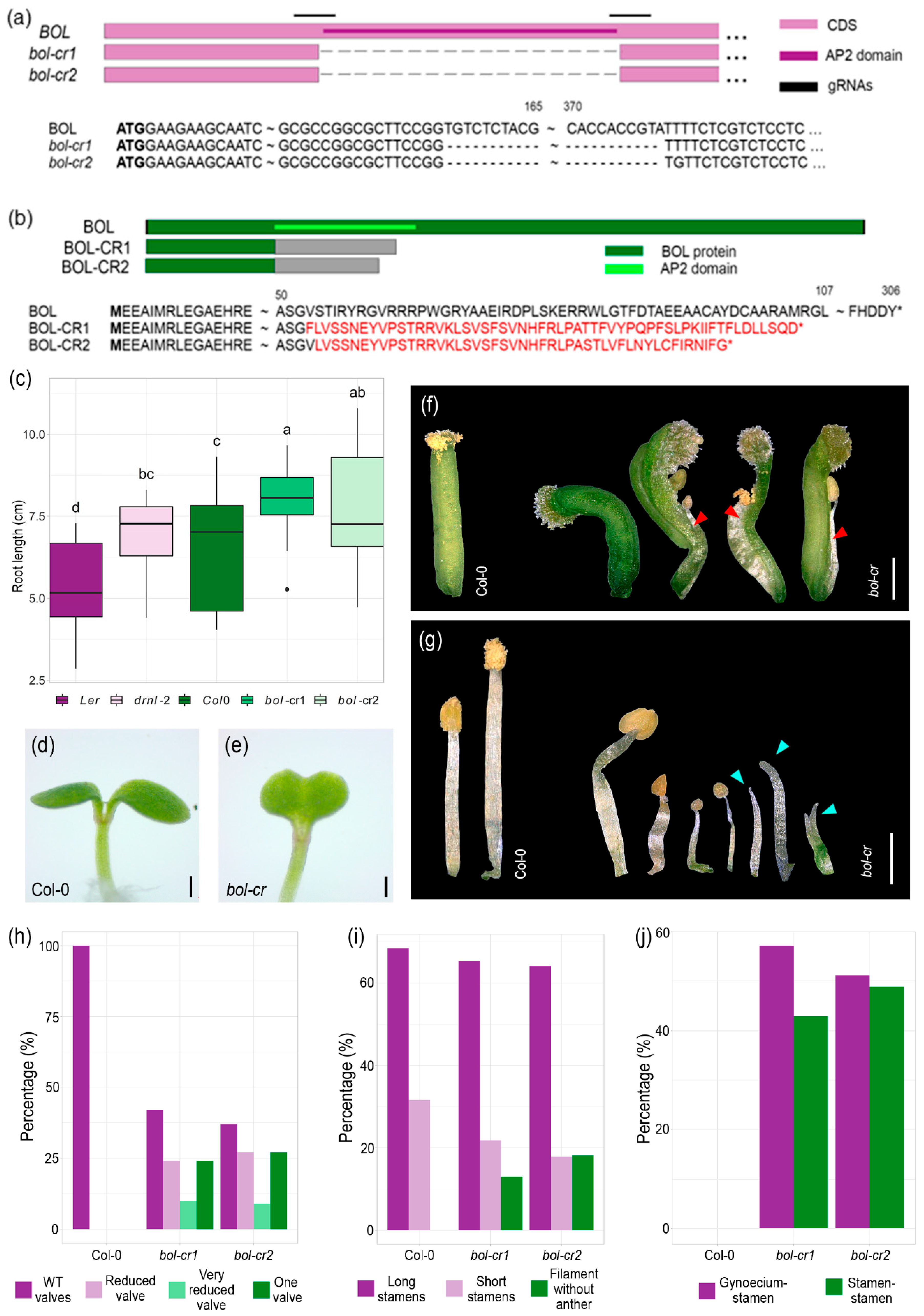
Figure 1. Genomic and protein structure of mutations generated by genomic editing and edited mutant phenotypes. (a,b) Genomic (a) and protein structure (b) of the CRISPR-Cas9-generated BOL mutations. Letters in red represent amino acids that are different from the original BOL protein. Asterisks represent stop codons. The dark green region represents the BOL protein, the light green region the AP2 domain within the protein, and the gray sections represent a region that, if translated, would produce a sequence different from the original BOL sequence. (c) Comparison of root length between 14-day-old wild-type and edited bol mutant (bol-cr) plants. ANOVA p-value = 3.58 × 10⁻⁶. Letters represent different statistical groups (Least Significant Difference, LSD test). (d) Representative 2-day-old Col-0 seedling phenotype. (e) Representative 2-day-old bol-cr mutant seedling phenotype showing fusion between cotyledons. (f) Representative gynoecium phenotypes of Col-0 wild-type and bol-cr mutants. The images show phenotypes observed in both bol-cr mutant lines. Red arrows indicate fusions between stamen and gynoecium. (g) Representative stamen phenotypes of Col-0 wild-type and bol-cr mutants. The images show phenotypes observed in both mutant lines. Blue arrows indicate filaments (stamens without anthers). (h) Frequencies of different gynoecium phenotypes of Col-0 and bol-cr mutants. (i) Frequencies of stamen phenotypes of Col-0 and bol-cr mutants. (j) Frequencies of type of fusion in flower organs of Col-0 and bol-cr mutants. Scale bars = 200 µm (d,e); 500 µm (f,g).
Plants 14 01757 g001
Figure 2. MeJA exogenous application to bol loss-of-function mutant flowers. (a) Number of filaments (stamens without anthers) per bol-cr2 flower, mock or treated with 50 and 100 µM MeJA. n = 20–25 flowers, Kruskal–Wallis p-value = 0.042. Letters represent different statistical groups (Dunn’s test). (b) Total number of stamens (long, short, and filaments) per bol-cr2 flower, mock or treated with 50 and 100 µM MeJA. n = 20–25 flowers, Kruskal–Wallis p-value = 0.965. (c) Number of floral organ fusions per bol-cr2 flower mock or treated with 50 and 100 µM MeJA. n = 20–25 flowers, Kruskal–Wallis p-value = 0.0003. Letters represent different statistical groups (Dunn’s test).
Plants 14 01757 g002
Figure 3. MeJA application or inhibition in wild-type and bol-D plants. (a) Root length of 9-day-old bol-D and Ws plants treated with 50 µM MeJA. (b) Rosette diameter of 9-day-old bol-D and Ws plants treated with 50 µM MeJA. (c) Rosette phenotype of 9-day-old bol-D and Ws plants treated with 50 µM MeJA. (d) 6-day-old bol-D plants. (e) 6-day-old bol-D plants germinated in 250 µM DIECA. (fi) Rosette phenotype of 6-day-old bol-D and Ws plants treated with 250 µM DIECA. Scale bar = 0.5 cm (ce); 0.25 cm (fi). In (a,b), ANOVA p-value = 2 × 10⁻¹⁶. Letters represent different statistical groups (LSD test).
Plants 14 01757 g003
Figure 4. Jasmonic acid content analyses in BOL gain and loss-of-function mutants and BOL constitutive and inducible overexpression lines. Quantification of jasmonic acid in (a) Ws wild-type and bol-D plants (Student’s t-test, p-value = 0.0004) and (b) wild-type and 35S::BOL tobacco (Student’s t-test, p-value = 0.002), (c) wild-type Col-0 and 35S::ESR2-ER plants two and four days after induction (ANOVA p-value = 0.0007; letters represent different statistical groups; LSD test), and (d) wild-type Col-0 and bol-cr2 inflorescences (Student’s t-test, p-value = 9.07 × 10⁻⁷, asterisks denote significance levels: ** p < 0.01, *** p < 0.001).
Plants 14 01757 g004
Figure 5. JA reporter line expression after BOL induction or transient expression. (ad) Expression of the Arabidopsis JA reporter pJAZ10::GUSPlus in the 35S::ESR2-ER BOL inducible background, transferred to mock (a,c) or induction media supplemented with β-estradiol (β-est) (b,d), 2 (a,b) and 4 (c,d) days after transference. (e,f) Expression of the tomato JA reporter 4xJERE::GUS after agroinfiltration with control (e) or 35S::BOL construct (f), evaluated 4 days after agroinfiltration. Scale bars = blue 1 mm (a,b), white 0.5 mm (c,d), black 1 cm (e,f).
Plants 14 01757 g005
Figure 6. Effect of jasmonate inhibition or exogenous application in BOL-induced green calli formation. Induced 35S::ESR2-ER plants in medium supplemented with only 10 µM β-estradiol (β-est) (a,b,g,h,m,n), in medium supplemented with 10 µM β-est and 250 µM DIECA (c,d,i,j,o,p), or in medium supplemented with 10 µM β-est and 50 µM MeJA (e,f,k,l,q,r), 10 (af), 15 (gl), and 20 (mr) days after induction (dai). Scale bars = white 1 cm, black 1 mm, blue 2.5 mm. MeJA, Methyl Jasmonate; DIECA, sodium diethyldithiocarbamate (JA biosynthesis inhibitor).
Plants 14 01757 g006
Figure 7. BOL expression in plants treated with a JA synthesis inhibitor. BOL expression in pBOL::GUS plants in mock medium, showing sporadic expression at the root tips (ad) and plants in medium supplemented with 250 µM DIECA (eh), one (a,b,e,f), two (c,g), and three (d,h) days after treatment (dat), where GUS staining is observed in all roots analyzed and expands to the region above the root tip and differentiated tissue. Scale bars = 0.1 mm.
Plants 14 01757 g007
Disclaimer/Publisher’s Note: The statements, opinions and data contained in all publications are solely those of the individual author(s) and contributor(s) and not of MDPI and/or the editor(s). MDPI and/or the editor(s) disclaim responsibility for any injury to people or property resulting from any ideas, methods, instructions or products referred to in the content.