Article

Genome Assembly and Structural Variation Analysis of Luffa acutangula Provide Insights on Flowering Time and Ridge Development

1
Institute of Agricultural Science Research of Jiangmen, Jiangmen 529060, China
2
College of Horticulture, South China Agricultural University, Guangzhou 510642, China
3
Dongguan Agricultural Scientific Research Center, Dongguan 523086, China
*
Authors to whom correspondence should be addressed.
These authors contributed equally to this work.
Plants 2024, 13(13), 1828; https://doi.org/10.3390/plants13131828
Submission received: 29 March 2024 / Revised: 21 June 2024 / Accepted: 21 June 2024 / Published: 3 July 2024
(This article belongs to the Special Issue Molecular Biology of Plant Growth and Development)

Abstract

:
Luffa spp. is an important worldwide cultivated vegetable and medicinal plant from the Cucurbitaceae family. In this study, we report a high-quality chromosome-level genome of the high-generation inbred line SG261 of Luffa acutangula. The genomic sequence was determined by PacBio long reads, Hi-C sequencing reads, and 10× Genomics sequencing, with an assembly size of 739.82 Mb, contig N50 of 18.38 Mb, and scaffold N50 of 56.08 Mb. The genome of L. acutangula SG261 was predicted to contain 27,312 protein-coding genes and 72.56% repetitive sequences, of which long terminal repeats (LTRs) were an important form of repetitive sequences, accounting for 67.84% of the genome. Phylogenetic analysis reveals that L. acutangula evolved later than Luffa cylindrica, and Luffa is closely related to Momodica charantia. Comparing the genome of L. acutangula SG261 and L. cylindrica with PacBio data, 67,128 high-quality structural variations (SVs) and 55,978 presence-absence variations (PAVs) were identified in SG261, resulting in 2424 and 1094 genes with variation in the CDS region, respectively, and there are 287 identical genes affected by two different structural variation analyses. In addition, we found that the transcription factor FY (FLOWERING LOCUS Y) families had a large expansion in L. acutangula SG261 (flowering in the morning) compared to L. cylindrica (flowering in the afternoon), which may result in the early flowering time in L. acutangula SG261. This study provides valuable reference for the breeding of and pan-genome research into Luffa species.

1. Introduction

Luffa spp. (2n = 26), belonging to the Cucurbitaceae family, includes nine species, namely L. cylindrica (L. aegyptiaca), L. acutangula, L. quinquefilia, L. operculata, L. saccata, L. sepium, L. graveolens, L. echinata, and L. astorii [1]. There are two domesticated types: the angled loofah (ridge gourd), Luffa acutangula L. Roxb., and the smooth loofah (smooth gourd), Luffa cylindrica L. Rome., which are annual and largely monoecious [2,3]. Luffa is native to India and widely cultivated in tropical and subtropical areas, such as China, Malaysia, India, Korea, Thailand, Central America, and Africa [4,5]. As a vegetable plant, it is rich in nutrition and contains many secondary metabolites which can be used as medicinal components, including saponins, alkaloids, flavonoids, anthraquinones, and steroids. Its isolated compounds possess broad pharmacological activities such as antidiabetic, hepatoprotective, antiulcer, anticancer, immunomodulatory, and antihyperlipidemic properties [6].
L. acutangula and L. cylindrica are two vegetable species commonly found in South and Southeast Asia. L. acutangula is widely grown; however, L. cylindrica is considered an underutilized crop [7]. There are obvious phenotypic differences between L. cylindrica and L. acutangula. There is a profound variation in fruit size, length, shape, and color [7]. The smooth gourd fruit is smooth and cylindrical, and the ridge gourd fruit has a tapering neck with some prominent longitudinal ridges [8]. In addition, the two species can also be distinguished according to color and flowering time: L. cylindrica has bright yellow flowers that bloom in the early morning (4–8 a.m.), while L. acutangula has pale cream flowers that bloom in the late afternoon (5–8 p.m.) [2,8]. It is very meaningful to study the difference in flowering time between these two species, which can contribute to exploring the different flowering mechanisms in these two species.
The process of introgressing desired traits acquired from interspecific hybridization into elite cultivars can be made more efficient using molecular breeding approaches. With the development of sequencing technology, genome assembly has also developed rapidly. The genome of L. cylindrica was sequenced and assembled for the first time using next-generation sequencing (NGS) technology (small insert (220 bp) library) in 2017, and a genome of 885.01 Mb was obtained [9]. Subsequently, three genome assemblies of L. cylindrica have been completed utilizing the PacBio long read single-molecule real-time (SMRT) sequencing platform [10,11,12], while only one genome assembly of L. acutangula has been completed [10].
To explore genetic variability and diversity that exist within a species, structural variation (SV) studies have been recently reported for some important crop plants, including corn, soybean, and rice [13]. Structural variations (SVs), including insertion, deletion, tandem repeat, inversion, translocation, copy number variations (CNVs) with a length of more than 50 bp, and chimerism variations present a more complicated situation [14]. Compared with single-nucleotide polymorphisms (SNPs), SVs occupy a larger proportion in the variation base and have significant impact on variations in the genome [15]. Numerous studies have indicated that SVs play a critical role in genome evolution and genetic control of agronomic traits such as flowering time, fruit size, and stress resistance [13,16] and gradually have become an increasingly important research field. Likewise, presence/absence variations (PAVs) also can contribute to trait variation [17].
The first attempt to perform a genome survey sequencing of Luffa cylindrica using next-generation sequencing (NGS) technology was carried out by An et al. [9]. Recently, Zhang et al. (2020) reported a de novo assembly of the L. cylindrica genome, utilizing the Pacific Biosciences (PacBio) sequencing platform [12]. Pootakham et al. previously assembled acutangula and cylindrica genomes and investigated alternative splicing events in Luffa [10], but little research has been conducted on the structural variability of L. acutangula and L. cylindrica. The narrow genetic and genomic resources obviously limited the breeding improvement of Luffa. In this study, we de novo assembled the genome of the high-generation inbred line L. acutangula SG261 by combining the PacBio long reads, Hi-C data, and 10× Genomics short reads. Genome annotation, comparative genome analysis, and structure variation identification were conducted based on the assembled genome sequences. PacBio long-read sequencing data were used to identify SVs between L. acutangula and L. cylindrica. The present study provides a valuable genetic resource for deciphering the genome evolution of Luffa species and is positioned to serve as the reference genome guiding the molecular breeding of Luffa crops.

2. Materials and Methods

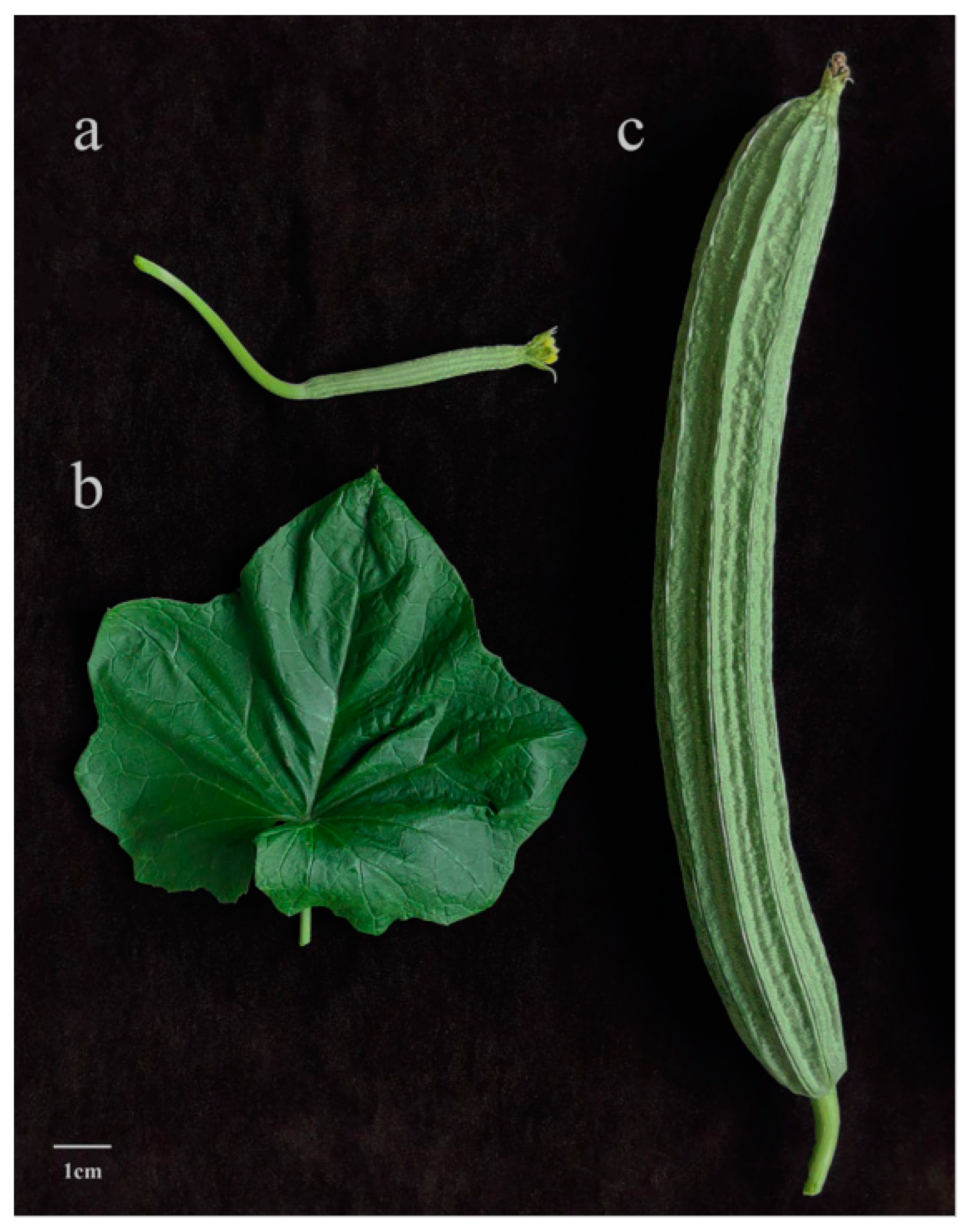
2.1. Plant Materials

The highly inbred line SG261 of L. acutangula was used in this study. This line has distinctive ridges and large flowers (blooming at night) (Figure 1). The seedlings were given adequate sunlight, nutrients, and water for normal growth in a greenhouse at Jiangmen Institute of Agricultural Sciences, in Jiangmen, Guangdong, China.

2.2. Section Observation

The ridges of young (the day after pollination) and mature fruits (eighth day after pollination) of L. acutangula and L. cylindrica were sampled to make paraffin sections by Henan Honglin Educational Instrument Co., Ltd. (Henan, China). After the material was taken, it was fixed, rinsed, dehydrated, permeabilized, embedded, sectioned, spread, baked, and dehydrated. Then, the sections were stained with plant tissue staining solution Safranin O-fast green and observed on an Olympus microscope [18].

2.3. Genome Sequencing

Young fresh leaves of SG261 were collected for genomic DNA extraction and sequencing. Total DNA was extracted using the cetyltrimethylammonium bromide (CTAB) method to construct PacBio and short-read libraries. The short-read libraries with an insertion size of 350 bp were prepared using VAHTS Universal DNA library preparation kit (Vazyme, Nanjing, China) by the Beijing Genomics Institute (BGI), and the insertion fragments of the library were detected using the Agilent Bioanalyzer 2100 (Agilent Technologies, Santa Clara, CA, USA). The library was sequenced on the MGI-SEQ 2000 sequencing platform to produce pair-end sequence data (2 × 150 bp) [19]. For the PacBio library, high-molecular-weight DNA was extracted as above. About 30 ug of high-molecular-weight DNA was used to prepare template library of 30–40 kb using the BluePippin Size Selection system (Sage Science, Beverly, MA, USA). The library was then prepared by adding specific splice sequences to both ends of the fragmented DNA. The library was sequenced on the PacBio Sequel II sequencing platform using standard sequencing for continuous long reads (CLRs) to generate long reads by SMRT sequencing chips [20].
To obtain Hi-C sequencing data, the chopped young leaves of SG61 were vacuum-fixed with 2% fresh formaldehyde in NIB buffer for 45 min, and glycine was added to a concentration of 0.375 M. The reaction was carried out for 5 min, and then liquid nitrogen was added and ground to powder. Then, it was filtered through a layer of Miracloth membrane. Isolated cells were lysed, and proteins were broken down using proteinase K at 65 degrees centigrade. DNA was purified by QIAamp DNA Mini Kit (Qiagen), and then dotted flat-end junctions were removed using Dynabeads® MyOne™ Streptavidin C1 (Thermofisher). Then, sequencing library was prepared using the NEBNext® Ultra™ II DNA library Prep Kit and sequenced on the BGI MGI-SEQ 2000 platform (San Diego, CA, USA) using the 150 PE mode.

2.4. RNA-Seq Library Construction and Sequencing

Fresh, disease-free, big flower buds before flowering were selected. The mixed flower buds from 5 individual plants were used for RNA extraction and transcriptome sequencing in three biological replicates. Total RNA was extracted using Trizol reagent (Invitrogen, Waltham, MA, USA), and the RNA purity and integrity were assessed by NanoDrop 2000 spectrophotometer (NanoDrop Technologies, Wilmington, DE, USA) and by Agilent 2100 Bioanalyzer (Agilent Technologies, CA, USA). After passing the quality inspection, the sequencing library was constructed using the VAHTS Universal V6 RNA-seq Library Kit for MGI (Vazyme, Nanjing, China) and quantified by Qubit 3.0 Fluorometer (Life Technologies, Carlsbad, CA, USA). The library was then assessed for size and quality by the Agilent 2100 Bioanalyzer (Agilent Technologies, CA, USA) and sequenced on the MGI-SEQ 2000 platform.

2.5. De Novo Sequencing and Genome Assembly

The raw BGI short reads were cleaned using SOAPnuke version 2.1.0 [21], with parameters “-lowQual = 20, -nRate = 0.005, -qualRate = 0.5”. The genome size and heterozygosity of SG261 were estimated by GCE version 1.0.2 [22] using k-mer frequency distribution generated from cleaned BGI short reads, with the following parameters: “-m 1 -D 8 -b 0 -H 1”. PacBio long reads were filtered (retaining the top 72 Gb longest reads, ~100 × genomic coverage) and then employed for correction using MECAT2 version 2.1 [23] and then trimmed and assembled using CANU version 2.0 [24], with parameters “GenomeSize = 879,830,000”. Bowtie2 version 2.4.5 [25] was used to map the 10× BGI data to the assembly, and Pilon version 1.23 software [26] was used to polish the assembly.
After assembling the genome contigs of SG261, Hi-C data were used to assign genome assembly to chromosome level. Hi-C clean reads were aligned onto the assembled genome by Juicer v1.5.7 [27], and then software 3D-DNA v180114 [28] was used to preliminarily cluster and orient the data. Then, the assembly genome was adjusted, reset, and aggregated by JuiceBox v1.11.08 [29] to improve the quality of chromosome assembly. The chromosome assembly genome was mapped to a published L. acutangula genome [10] using minimap2 v2.24 [30], and the final genome was obtained by reverse complementation and direction adjustment according to the mapped results.
To evaluate the assembly results, BWA-mem v0.7.17 [31] was used to map the Hi-C data to the assembled genome, and a Hi-C contact map was generated by HiCExplorer v3.5.3 software [32] with a 100 kb window and a threshold of −1.5 to 5. Finally, BUSCO v3.0.2 [33] was used to estimate the integrity of the assembly genome based on the Embryophyta_odb9 database (n = 1440).

2.6. Repetitive Element Annotation and Gene Prediction

Transposable elements (TEs) were annotated by EDTA v1.9.6 [34]. Taking the results of EDTA as a library, RepeatMasker v4.1.2.p1 [35] was used to annotate incomplete repetitive elements. Tandem Repeats Finder (TRF) v4.09 [36] was used to annotate the tandem repeats (TRs) with default parameters.
Three data sets from the RepeatMasker library (RMlib), expressed sequence tags (EST), and peptides were used for protein-coding gene prediction using the MAKER v3.31.10 pipeline [37]. To improve the quality of gene annotation, four cycles of annotation were conducted using MAKER. For expressed sequence tags, raw transcriptome data from flower buds of L. acutangula SG261 were cleaned using SOAPnuke v2.1.0, and the clean data were mapped to the assembly genome by HISAT2 v2.2.1 [38]. StringTie v2.2.1 [39] was used to reconstruct genome annotations and integrate annotations using the merge function and finally extract the transcription tag by gffread v0.12.1 [40]. The peptides produced were predicted by TransDecoder v5.5.0 (https://github.com/TransDecoder (accessed on 16 October 2022)) based on expressed sequence tags obtained from the prediction above. For functional annotation, we compared all predicted genes to the NT database, NR database, the Swissprot database, and the KEGG database using BLAST v2.11 [41], diamond v2.0.9 [42], and Interproscan v5.52-86 software [43], respectively.
Finally, a circular genome visualization map was constructed by Circos v0.69-8 [44] software to show the results of genome assembly and genome annotation.

2.7. Comparative Genome Analysis

Phylogenetic analysis was conducted using three Luffa genomes (L. acutangula SG261, L. acutangula [10], and L. cylindrica [11]) and another 10 species of Cucurbitaceae (Benincasa hispida [45], Citrullus lanatus [46], Cucumis melo [47], Cucumis sativus [48], Cucurbita argyrosperma [49], Cucurbita maxima and Cucurbita moschata [50], Cucurbita pepo [51], Lagenaria siceraria [52], and Momordica charantia [53]). The data were downloaded from Cucurbit Genomics Database (http://cucurbitgenomics.org/ (accessed on 20 October 2022)) and NCBI (https://www.ncbi.nlm.nih.gov/ (accessed on 20 October 2022)). OrthoFinder v2.5.4 [54] was used to identify orthologous groups for these species. The protein sequences of all single-copy genes were identified, and multiple sequence alignment was performed by MAFFT v7.487 [55] and trimAI v5.7.5 [56]. RAxML v8.2.12 [57] was used to construct a phylogenetic tree for each group of single-copy proteins, and a coalescent-based phylogenetic tree was constructed using ASTRAL v5.7.5 [58].
The software minimap2 v2.21 was used to compare the SG261 assembly genome with other Luffa genomes (L. acutangula [10] and L. cylindrica [11]). The alignment results of mapq60 were retained, and the PAFR v0.0.2 software was used to visualize the alignment results and obtain the synteny dot plots.

2.8. Structural Variants Analysis

Short reads were used to identify SNP and INDEL between SG261 genome and L. cylindrica genome (downloaded from NCBI, SRR10818295). Short reads were mapped to the assembled genome using BWA v0.7.17 with parameters “-R ‘@ rg\tid: lae518\TPU: lae518\TSM: lae518’”. Sambamba v0.8.0 [59] was used to mark and filter PCR duplication, and then SNP and InDel were called using bcftools v1.13 software [60]. Finally, densities of SNPs and InDels were calculated with a window size of 400 kb and displayed on the Circos plot.
Long reads were used to identify structural variants (SVs) between the L. acutangula SG261 genome and the L. cylindrica genome [11]. Minimap2 v2.21 was used to map the PacBio long reads of SG261 to the L. cylindrica genome with parameter “--MD -ax map-pb”, and samtools v1.13 [61] was used for sorting and indexing. Then, SVs were called using Sniffles v1.0.12 [62] with parameter “-l 50 -t 45”. The long reads of the L. cylindrica genome were downloaded from NCBI (SRP239503). Then, the long reads were mapped to SG261 genome, and SVs were called using the same method above.
We then filtered the SVs using the SG261 genome as a reference genome according to a previous study [13]. First, we removed imprecise SVs and SVs on scaffolds. Then, SVs longer than 100 kb in length and genotype of “0/0” were removed. Finally, we identified regions prone to false SVs and removed SVs that intersected with them. To identify these regions, PaSS [63] was used to simulate long reads based on the L. acutangula genome [10]. PacBio long reads of SG261 and long reads simulated by L. acutangula were mapped to the SG261 genome using minimap2, and SVs were called using Sniffles. SVs larger than 100 kb and imprecise SVs were then removed to obtain the final vcf file. Bedtools v2.30.0 [64] was used to extract the SVs between L. acutangula SG261 and L. cylindrica, which were demonstrated to intersect with the 2 vcf files. In addition, according to the genome annotation file, the genes affected by SVs were identified with Bedtools.
We identified PAVs between L. acutangula SG261 and the L. cylindrica genome using a sliding window method [17]. We divided the L. cylindrica genome into 500 bp windows with 100 bp as the step size and then mapped it with the assembled genome using BWA to identify PAV. According to the genome annotation file, the genes affected by PAVs were identified with Bedtools.

2.9. GO Enrichment and Functional Annotation of Genes with SVs and PAVs

Genes with SVs and PAVs were submitted to the NT database, NR database, and Swissprot database for functional annotation using BLASTN v2.11 and diamond v2.0.9 software, respectively. In addition, we used SG261 genome as the background and SVs and PAV-associated genes as foreground for GO enrichment. Interproscan v5.52-86 was used to annotate the SG261 genome genes with parameters “-f TSV -iprlookup -goterms -pa”. ClusterProfiler v4.0.0 [65] was used to enrich SV-related genes and PAV-related genes. p < 0.05 was a significant enrichment.

2.10. Analysis of Expansion and Contraction for Flowering-Time-Related Genes

According to the known flowering-related genes [66], we searched the annotation results of SV-related genes and found that there were 16 genes that might be related to flowering time. Then, we performed gene family expansion and contraction for some flowering-related gene families, including FT [67] and CO-like gene families [68], as well as unidentified FY (FLOWERING LOCUS Y) and EFM (EARLY FLOWERING MYB PROTEIN) families and some MYB [69], AP2/ERF [70], bZIP [71], NAC [72], and WAKY transcription factor families. We downloaded the sequences of these families in cucumber based on previous gene family analysis, and the unidentified FY and EFM were retrieved according to the NCBI database. The heatmap was obtained using TBtools [73].

3. Results and Discussion

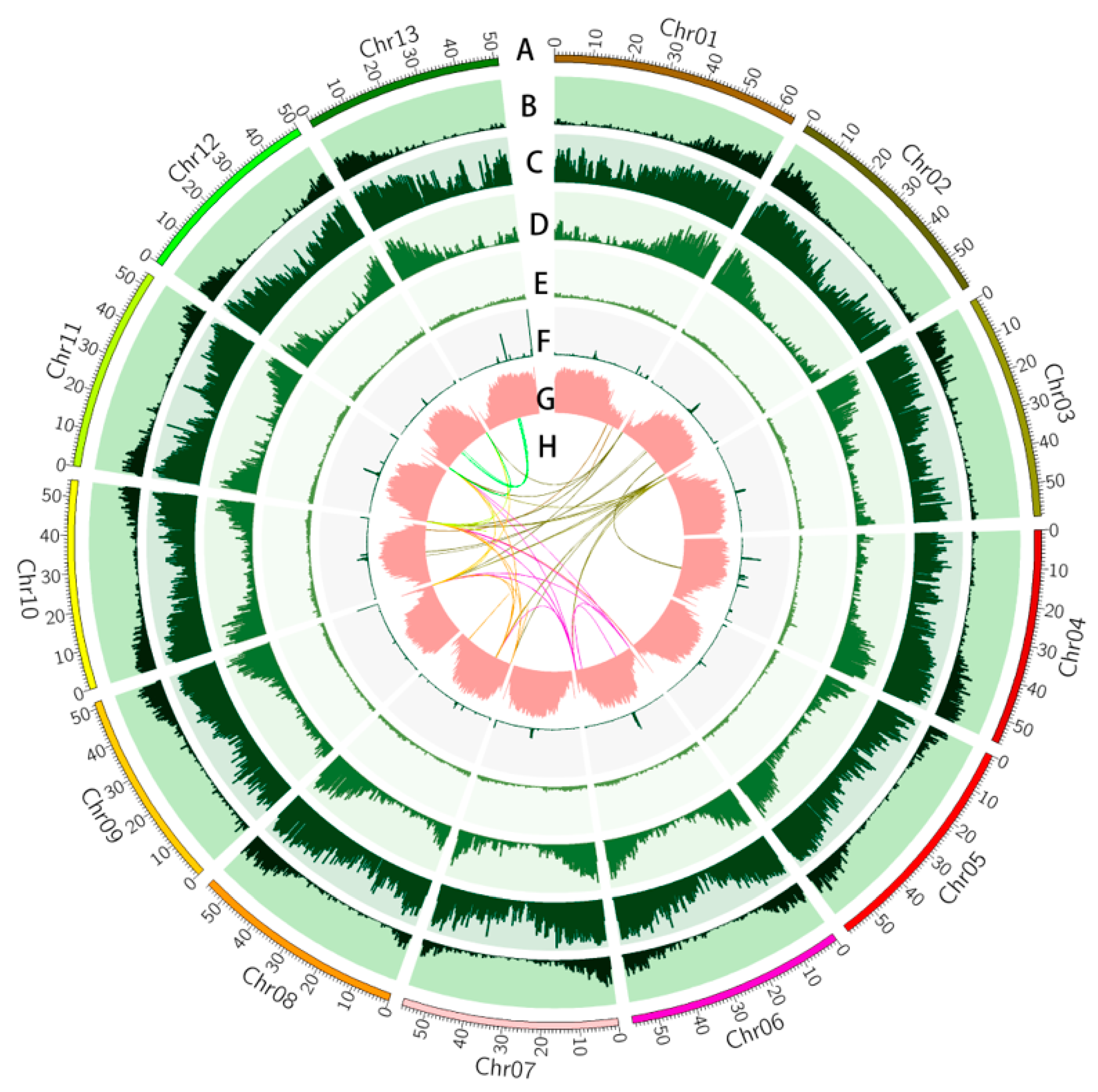
3.1. Genome Sequencing and Assembly

A high-quality chromosome-level genome of the inbred line SG261 was assembled in the present study. Firstly, 74.96 Gb clean reads were obtained by the MGI-SEQ 2000 sequencing system (Table S1). Based on 17-mer frequency, the estimated genome size was 879.83 Mb with a heterozygosity rate of 0.49% and 73.00% repetitive sequences (Figure S1, Table S2). Secondly, a total of 142.75 Gb in PacBio clean reads was generated and used for contig assembly (Table S1). The assembled contig genome was composed of 283 contigs with a total length of 739.31 Mb, of which contig N50 was 18.38 Mb in length, with an average length of 2.61 Mb (Table 1 and Table S4). Thirdly, 70.24 Gb of Hi-C data was obtained by the BGI MGI-SEQ 2000 sequencing system for assembly (Table S1). Based on Hi-C data, the contig sequences were anchor-corrected to obtain a scaffold-level genome. The 283 contigs were anchored to 31 scaffolds, including 13 pseudo-chromosomes and 18 scaffolds, of which the shortest was 50.01 Mb (chr12) and the longest was 64.19 Mb (chr01) (Table S5). The final pseudo-chromosomal level genome had a total length of 739.82 Mb, and the scaffold N50 was 56.08 Mb (Table 1). Finally, the Hi-C data were mapped to the assembled genome, and the Hi-C correlation heatmap obtained by HiCExplorer showed that the Hi-C-assisted assembly was high quality (Figure S2).
We also aligned short reads to the assembled genome, and 99.51% of the data could be mapped to the assembled genome, indicating that the assembled genome contained most of the genome information. The completeness of the assembled genome was 91.25%, evaluated by BUSCO (Table S3), which indicated that the SG261 genome assembly was complete and could be used for subsequent analysis. Compared with other Luffa genomes [10,11,12], the assembled genome of SG261 had the highest N50. In addition, we found that the SG261 genome and another L. acutangula genome are larger than the three L. cylindrica genomes, which confirmed that the genome of L. acutangula is larger than that of L. cylindrica (Table 2).

3.2. Genome Annotation

EDTA was used to annotate the complete repetitive sequences of the assembled genome, and RepeatMasker was used to annotate the incomplete repetitive sequences based on the results of EDTA. The results showed that 536.83 Mb (72.56% of the assembled genome) of the sequences was annotated as repetitive sequences, including 67.84% of the retrotransposon long terminal repeats (LTRs) and 4.72% of the DNA type transposon elements (Figure 2). Copia and Gypsy elements are the main elements in LTRs, 276,809 and 261,064 in number, covering 178.81 Mb and 227.38 Mb, accounting for 24.17% and 30.73% of the genome, respectively (Table S6). In addition, we also annotated the tandem repeats (TRs) using TRF, which are enriched at centromeric regions (Figure 2). The accumulation of repetitive elements, especially LTRs, may be the reason for the difference in k-mer size and assembly size of the genome [74] and also lead to the large genome of Luffa [10]. Comparing several Luffa genomes, it was found that although the genome of L. acutangula was generally larger than that of L. cylindrica, the length of non-repetitive sequences was less than that of L. cylindrica (Table S6). Therefore, a large number of repetitive sequences may lead to the larger genome of Luffa as compared to other Cucurbitaceae plants.
A total of 27,312 protein-coding genes were annotated in the assembled genome, with an average transcript length of 1305.67 bp (Table 2 and Table S6). Compared with other Luffa genomes [10,11,12], there are fewer protein-coding genes in SG261 than other Luffa genomes (Table 2 and Table S6). In addition, we functionally annotated all protein-coding genes, and a total of 25,356 genes were annotated, accounting for 92.84% of all protein-coding genes (Table S7).

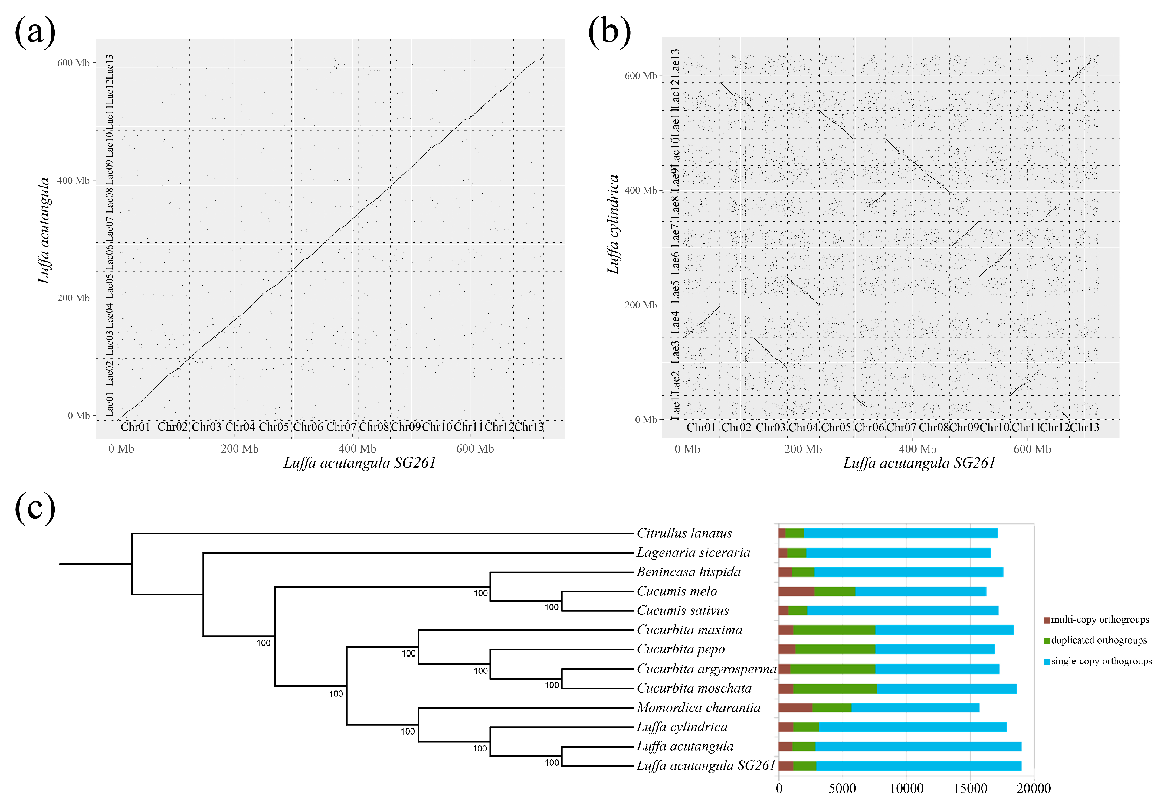
3.3. Synteny and Phylogenetic Analysis

Synteny analysis indicated that the SG261 assembled genome showed good linearity with the published L. acutangula genome [10] (Figure 3a). The SG261 assembled genome showed relatively low linearity with the L. cylindrica genome [11] with many mismatches of chromosomes. Among them, chromosomes 1, 9, 10, 11, and 13 of SG261 corresponded to chromosomes 4, 7, 6, 2, and 13 of the L. cylindrica genome, respectively, with same genome directions; chromosomes 2, 3, 4, 5, 7, and 8 of SG261 corresponded to chromosomes 12, 3, 5, 11, 10, and 9 of the L. cylindrica genome, respectively, with some reverse complementary strands (Figure 3b). In addition, we also found that the SG261 assembled genome has chromosome translocation between chromosomes 6 and 12 (chromosomes 1 and 8 of L. cylindrica), and there were small segments of chromosome inversion in chromosomes 7, 8, and 11 of the SG261 genome (chromosomes 10, 9, and 2 of L. cylindrica) (Figure 3b).
Three Luffa genomes and 10 other Cucurbitaceae genomes were used for phylogenetic analysis, and the homologous gene clusters of 13 species were identified (Figure 3c). Through homologous gene cluster analysis, we found that the number of single-copy and two-copy genes in the three Luffa genomes were similar (Figure 3c, Table S8). Phylogenetic analysis indicated that the three Luffa genomes were clustered to a branch, and the evolution time of L. acutangula was later than that of L. cylindrica, which confirmed the close relationship of the three Luffa genomes. Bitter gourd (M. charantia) was the closest cucurbit vegetable to the Luffa species, followed by Cucurbita, Cucumis, Benincasa, Lagenaria, and Citrullus, which confirmed the results in the previous study [10,75].

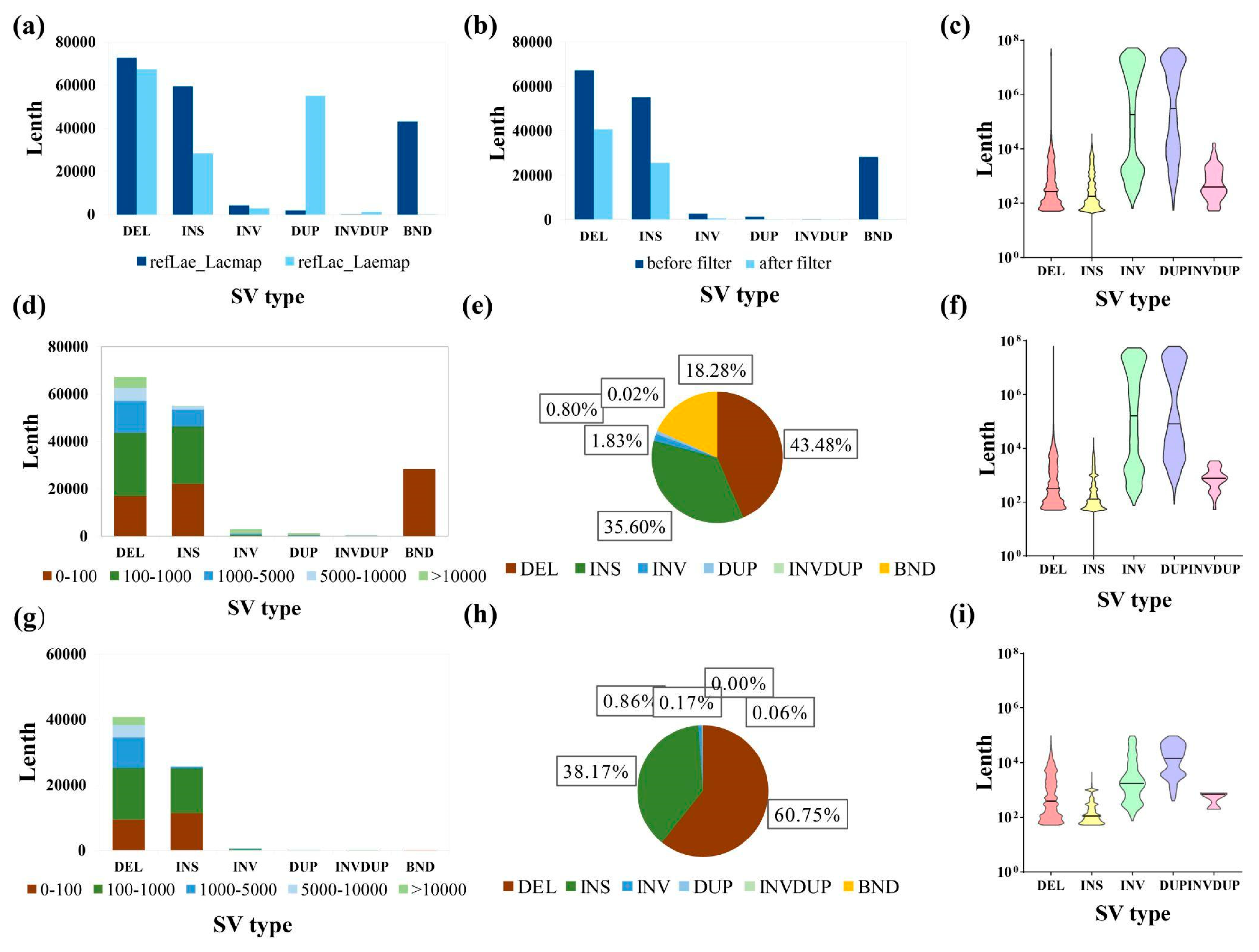
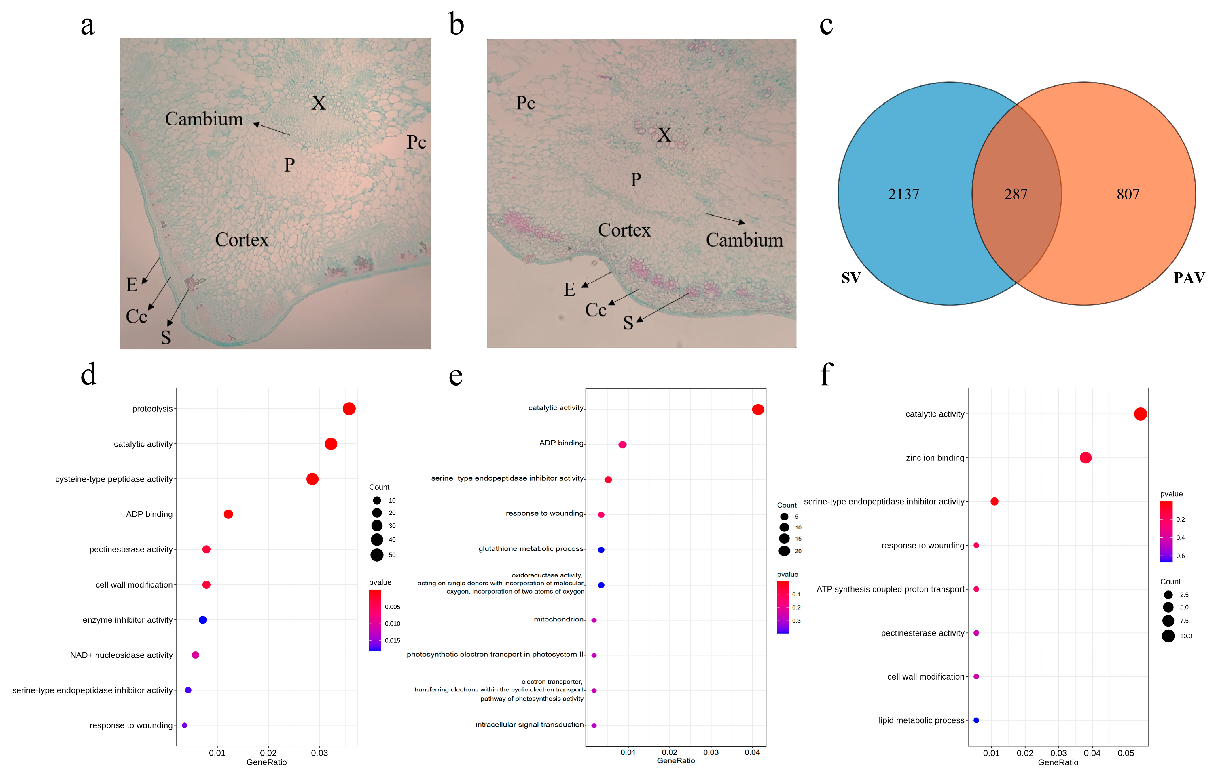
3.4. Variation between Luffa acutangula SG261 and Luffa cylindrica

Generally, genomic variation can be mainly divided into single-nucleotide polymorphisms (SNPs), small insertion or deletions (InDels, ≤50 bp), and structural variations (SVs, >50 bp) [76]. To explore the differences between L. acutangula and L. cylindrica, we used short-read data and long-read data to identify variants. Taking the L. acutangula SG261 genome as the reference genome, 11,505,354 variants were identified by the short reads mapped from the L. cylindrica genome, including 11,017,265 SNPs and 488,089 InDels, with an average of 14.89 SNPs and 0.66 InDels per kb. Interestingly, the density distribution of SNPs and InDels shared the same genomic regions with the gene density distribution (Figure 2). The density of SNPs and InDels was lower in the regions with dense repetitive elements but higher in the regions with high gene density. SNPs and InDels have the same density distribution in the genomes of pak choi, Chinese cabbage, and oilseed yellow sarson [17]. Most SNPs and InDels in the gene region come from the intergenic region, and the gene sequence is relatively conservative [77]. The high variation density of chromosome arm regions may be caused by the variation in intergenic regions.
Structural variations were identified using the PacBio long reads of L. acutangula SG261 and L. cylindrica. Analysis of SVs between the genomes of SG261 and L. cylindrica produced 154,594 SVs identified with SG261 as the reference genome and 181,460 SVs identified with L. cylindrica as the reference genome. Among the different types of SVs, there are more DUP and INVDUP (complex chimeric variants with both inversions and duplications on the chromosome) SVs identified in L. acutangula SG261 and more DEL, INS, INV, and BND SVs in L. cylindrica (Figure 4a, Tables S9 and S10). The length and number of different types of SVs and their distribution in the two genomes were the same (Figure 4c,f). We mainly focused on the SVs identified with L. acutangula SG261 as the reference genome. A total of 67,128 SVs were obtained after filtering (Table S11). The DEL and INS were the main types of variation in SVs, accounting for 43.8% and 35.6% of the total SVs before filtering and 60.75% and 38.17% of the total SVs after filtering (Figure 4d–h). The lengths of DEL and INS variants were mostly between 100 and 1000 bp (Figure 4f–i). For INV and DUP variants, the SVs before filtering were mainly distributed in the two ranges (1000–5000 bp and >10,000 bp). The distribution of SVs after filtering was similar to that of before filtering (Figure 4d,f,g,i and Table S12). Finally, we extracted 2424 genes with SVs in CDS regions based on the genome annotation results and the SV data.
Presence/absence variations (PAVs) were important variant types that differed between L. acutangula SG261 and L. cylindrica. We identified 55,978 PAVs in the L. acutangula SG261 genome, covering 43.38 Mb bases with 1094 genes, and 67,178 PAVs were identified in the L. cylindrica genome, covering 56.37 Mb bases with 1108 genes (Table S13). Analysis of the length and number distribution of PAVs found that the PAVs with a length of 500 bp were the most abundant (Figure S3). The longest PAVs in SG261 and L. cylindrica were 53,600 bp and 28,900 bp, respectively (Table S13).

3.5. Functional Annotation and GO Enrichment of SV and PAV Genes

In order to explore whether structural variation affects the phenotypic difference between L. acutangula and L. cylindrica, we performed functional annotation and GO enrichment for 2424 SV genes and 1094 PAV genes. We aligned SV and PAV genes to NT, NR, and Swissprot databases for functional annotation. A total of 2054 genes can be annotated in the 2424 SV genes. A total of 870 genes can be annotated in the 1094 PAV genes.
We carried out GO enrichment for 2424 SV genes, which were enriched for proteolysis (GO:0006508), catalytic activity (GO:0003824), cysteine-type peptidase activity (GO:0008234), ADP binding (GO:0043531), pectinesterase activity (GO:0030599), and cell wall modification (GO:0042545), as well as others (Figure 5d). The PAV genes were enriched for catalytic activity (GO:0003824), ADP binding (GO:0043531), serine-type endopeptidase inhibitor activity (GO:0004867), and response to wounding (GO:0009611), as well as others (Figure 5e). Subsequently, we obtained 287 genes containing both SVs and PAVs using TBtools (Figure 5c). These genes are enriched for catalytic activity (GO:0003824), zinc ion binding (GO:0008270), serine-type endopeptidase inhibitor activity (GO:0004867), response to wounding (GO:0009611), and pectinesterase activity (GO:0030599), among others (Figure 5f).

3.6. Structural Variation Genes Involved in Ridge Development

Ridges are the most important phenotype in L. acutangula in contrast to the absence of ridges in L. cylindrica. Compared with L. cylindrica, the cortical cells in L. acutangula grow faster longitudinally, resulting in longer and larger cortical cells. In addition, there are more cortical cells in L. acutangula than L. cylindrica at the ridge position (Figure 5a,b).
GO enrichment of SV- and PAV-related genes enriched for pectinesterase activity and cell wall modification can be seen in Figure 5c–f. For example, the DEL (49,297,508–49,311,124 bp) of chromosome 9 affected the pectinesterase PPME1-like gene (Lac_SG261_V1bChr09g017340.1) and the pectinesterase 63 gene (Lac_SG261_V1bChr09g017350.1); the DEL (50,615,779–50,616,960 bp) of chromosome 10 leads to the deletion of a large fragment of the pectinesterase/pectinesterase inhibitor 7 gene (Lac_SG261_V1bChr10g019340.1) and so on (Table S14). Pectin gels are capable of large changes in hydration and stiffness, which can alter the behavior of cells and tissues. For example, increasing the stiffness of cell wall pectin gel may result in decreased cell growth or if stiffened enough may cause cell–cell separation by gel fracture [78]. Variation in pectinesterase may lead to variation in cell growth, thus affecting the growth of fruits. Therefore, these genes affected by SVs and PAVs may play a role in the development of the ridges of L. acutangula.

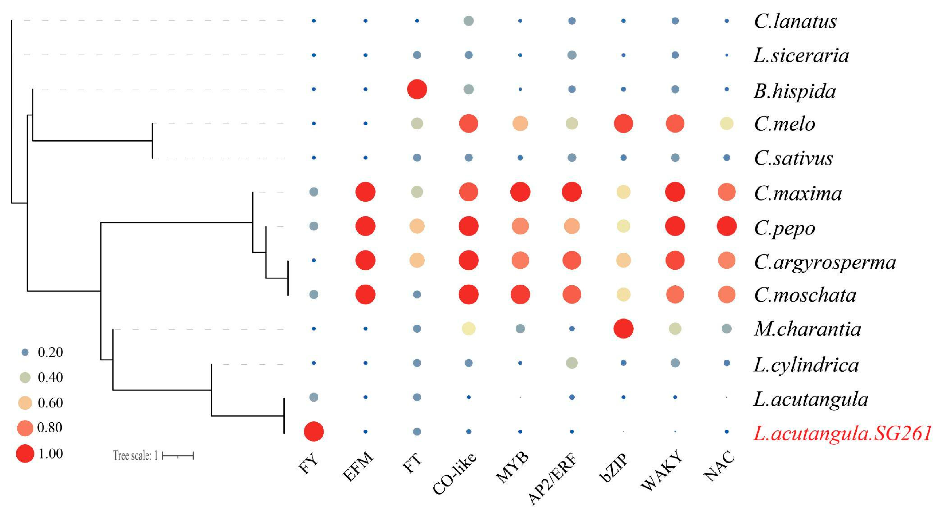
3.7. Analysis of Flowering-Time-Related Genes

Flowering is an important process that determines the success of plant reproduction. Plants must accurately combine internal and environmental signals to initiate the flowering process [66]. Flowering time has a very important influence on plant fitness and yield [79]. There are different flowering times between L. acutangula and L. cylindrica. The regulatory pathway of plant flowering has a relatively formed framework, and the flowering regulatory pathway map has been integrated in previous studies [66]. Compared to SNPs, SVs can cause large-scale perturbations of cis-regulatory regions and are therefore more likely to quantitatively change gene expression and phenotypes [13]. Based on the flowering regulatory pathway map and functional annotation of SV genes, we found 17 genes with SVs that may affect the flowering of Luffa (Table S15). These 17 genes are mainly affected by DEL-, INS-, and DUP-type SVs, among which a 78 bp insertion on chromosome 1 leads to an insertion of the PIE1 (Lac_SG261_V1bChr01g011540.1) gene; a 17,284 bp DEL of SG261 on chromosome 2 leads to the deletion of three adjacent FLOWERING LOCUS T (FT) genes (Lac_SG261_V1bChr02g021160.1—Lac_SG261_V1bChr02g021180.1) (Figure 6b, Table S15), the FT receives feedback from the upstream regulatory mechanism and has great influence on flowering [80]; a DUP on chromosome 8 leads to four duplications of three FLOWERING LOCUS Y (FY) genes (Lac_SG261_V1bChr08g012820.1, Lac_SG261_V1bChr08g012830.1, and Lac_SG261_V1bChr08g012880.1) (Figure 6a, Table S15), and the FY gene mutant blooms later in both long-day and short-day conditions [81,82]. In addition, some transcription factors may affect flowering time (Table S15). These genes with SVs may affect the flowering time in Luffa.
To explore the quantitative changes in flowering-related gene families in evolution, we performed expansion and contraction analysis of some gene families and genes. We downloaded these gene families and their protein sequences in cucumber, constructed homologous gene clusters of all genes based on protein sequences using Orthofinder v2.5.4, and checked the expansion and contraction of gene families according to the gene clusters (Figure 7). There are more FY genes in SG261 than other species, which further verified the authenticity of DUP variation at the position of FY genes. However, there was no significant difference in the number of genes in some flowering-related gene families, such as CO-like, FT, and EFM gene families, between SG261 and L. cylindrica (Table S16).

4. Conclusions

In this study, we reported a chromosome-scale reference genome assembly of L. acutangula. Our ribbed loofah genome enriches the loofah gene pool, and the sequenced varieties in this study are the best varieties selected and bred by our organization, which lays the data foundation for the subsequent research of our organization. In the structural variation analysis, the two ribbed loofah genomes allowed us to exclude the interference of intraspecific variation to a certain extent. The evolution time of L. acutangula was later than that of L. cylindrica, and there were some chromosomal translocations and large sequence inversions between L. acutangula and L. cylindrica. A total of 2424 and 1094 genes were affected by SVs and PAVs, respectively, in which 17 FT and FY genes may be related to the differences in flowering time observed in these Luffa species. This high-quality genome assembly and structural variation information will greatly facilitate the research of the molecular mechanisms of agronomic traits and provide valuable insights for pan-genome research and the molecular breeding of Luffa.

Supplementary Materials

The following supporting information can be downloaded at: https://www.mdpi.com/article/10.3390/plants13131828/s1, Figure S1. Distribution of K-mer frequencies for survey data. k-mer = 17. Figure S2. Hi-C heatmap of the genome assembly. Figure S3. Distribution of PAV between Lac and Lcy. Lac was L. acutangula SG261, Lcy was L. cylindrica. Table S1. Summary of sequencing data. Table S2. Statistical of 17-kmer genome estimation. Table S3. Quality assessment of the assembled genome of L. acutangula SG261 BUSCO. Table S4. Length details of genome assembly. Table S5. Chromosome length of the genome assembly of L. acutangula SG261. Table S6. Summary of repetitive sequences in the L. acutangula SG261 genome. Table S7. Functional annotations of L. acutangula SG261 mRNA genes. Table S8. Summary statistics of the gene families of 13 Cucurbitaceae plant species. Table S9. Statistical of SVs between L. acutangula SG261 and L. cylindrica with L. cylindrica as reference genome. Table S10. Statistical of SVs between L. acutangula SG261 and L. cylindrica with L. acutangula SG261 as reference genome. Table S11. Quantity statistics for filtering details of SVs with L. acutangula SG261 as reference genome. Table S12. Statistical of filtered SVs between L. acutangula SG261 and L. cylindrica with L. acutangula SG261 as reference genome. Table S13. Statistics for PAV analysis of L. acutangular SG261 and L. cylindrica genome. Table S14. List of genes enriched for pectinesterase activity (GO:0030599) and cell wall modification (GO:0042545). Table S15. Summary statistics of flowering-related gene. Table S16. Statistics of gene expansion and contraction in some gene families.

Author Contributions

S.C. (Shenglong Chen) and C.C. designed the experiments; S.F., A.H. and T.Z. performed the experiments and analyzed the data; S.C. (Shijun Chen), Z.Y. and C.C. provided advice and assistance during experiments and data analysis; S.F., C.C. and A.H. wrote the manuscript; all authors read and approved the final manuscript. All authors have read and agreed to the published version of the manuscript.

Funding

This work was funded by the Innovative Scientific Research Team and Leading Talent Project of Jiangmen in 2018. This work was also supported by the project “Creation of Vegetable Germplasm Resources and Molecular Marker Development in Aerospace Breeding (Dongguan)”.

Data Availability Statement

Genome assemblies, raw genome and transcriptome sequencing reads have been deposited in the National Center for Biotechnology Information BioProject database under the accession PRJNA1042866.

Conflicts of Interest

The authors declare no conflict of interest.

References

  1. Filipowicz, N.; Schaefer, H.; Renner, S.S. Revisiting Luffa (Cucurbitaceae) 25 Years after C. Heiser: Species Boundaries and Application of Names Tested with Plastid and Nuclear DNA Sequences. Syst. Bot. 2014, 39, 205–215. [Google Scholar] [CrossRef]
  2. Kalloo, G. Loofah. In Genetic Improvement of Vegetable Crops; Elsevier: Amsterdam, The Netherlands, 1993; pp. 265–266. ISBN 9780080408262. [Google Scholar]
  3. Prakash, K.; Pandey, A.; Radhamani, J.; Bisht, I.S. Morphological Variability in Cultivated and Wild Species of Luffa (Cucurbitaceae) from India. Genet. Resour. Crop Evol. 2013, 60, 2319–2329. [Google Scholar] [CrossRef]
  4. Islam, S.; Munshi, A.D.; Mandal, B.; Kumar, R.; Behera, T.K. Genetics of Resistance in Luffa cylindrica Roem. against Tomato Leaf Curl New Delhi Virus. Euphytica 2010, 174, 83–89. [Google Scholar] [CrossRef]
  5. Rabei, S.; Rizk, R.M.; Khedr, A.-H.A. Keys for and Morphological Character Variation in Some Egyptian Cultivars of Cucurbitaceae. Genet. Resour. Crop Evol. 2013, 60, 1353–1364. [Google Scholar] [CrossRef]
  6. Shendge, P.N.; Belemkar, S. Therapeutic Potential of Luffa acutangula: A Review on Its Traditional Uses, Phytochemistry, Pharmacology and Toxicological Aspects. Front. Pharmacol. 2018, 9, 1177. [Google Scholar]
  7. Kumari, S.A.S.M.; Nakandala, N.D.U.S.; Nawanjana, P.W.I.; Rathnayake, R.M.S.K.; Senavirathna, H.M.T.N.; Senevirathna, R.W.K.M.; Wijesundara, W.M.D.A.; Ranaweera, L.T.; Mannanayake, M.A.D.K.; Weebadde, C.K.; et al. The Establishment of the Species-Delimits and Varietal-Identities of the Cultivated Germplasm of Luffa acutangula and Luffa aegyptiaca in Sri Lanka Using Morphometric, Organoleptic and Phylogenetic Approaches. PLoS ONE 2019, 14, e0215176. [Google Scholar]
  8. Cui, J.; Cheng, J.; Wang, G.; Tang, X.; Wu, Z.; Lin, M.; Li, L.; Hu, K. QTL Analysis of Three Flower-Related Traits Based on an Interspecific Genetic Map of Luffa. Euphytica 2015, 202, 45–54. [Google Scholar]
  9. An, J.; Yin, M.; Zhang, Q.; Gong, D.; Jia, X.; Guan, Y.; Hu, J. Genome Survey Sequencing of Luffa cylindrica L. and Microsatellite High Resolution Melting (SSR-HRM) Analysis for Genetic Relationship of Luffa Genotypes. Int. J. Mol. Sci. 2017, 18, 1942. [Google Scholar] [CrossRef]
  10. Pootakham, W.; Sonthirod, C.; Naktang, C.; Nawae, W.; Yoocha, T.; Kongkachana, W.; Sangsrakru, D.; Jomchai, N.; U-Thoomporn, S.; Sheedy, J.R.; et al. De Novo Assemblies of Luffa acutangula and Luffa cylindrica Genomes Reveal an Expansion Associated with Substantial Accumulation of Transposable Elements. Mol. Ecol. Resour. 2021, 21, 212–225. [Google Scholar]
  11. Wu, H.; Zhao, G.; Gong, H.; Li, J.; Luo, C.; He, X.; Luo, S.; Zheng, X.; Liu, X.; Guo, J.; et al. A High-Quality Sponge Gourd (Luffa cylindrica) Genome. Hortic. Res. 2020, 7, 128. [Google Scholar] [CrossRef]
  12. Zhang, T.; Ren, X.; Zhang, Z.; Ming, Y.; Yang, Z.; Hu, J.; Li, S.; Wang, Y.; Sun, S.; Sun, K.; et al. Long-Read Sequencing and de Novo Assembly of the Luffa cylindrica (L.) Roem. Genome. Mol. Ecol. Resour. 2020, 20, 511–519. [Google Scholar] [CrossRef] [PubMed]
  13. Alonge, M.; Wang, X.; Benoit, M.; Soyk, S.; Pereira, L.; Zhang, L.; Suresh, H.; Ramakrishnan, S.; Maumus, F.; Ciren, D.; et al. Major Impacts of Widespread Structural Variation on Gene Expression and Crop Improvement in Tomato. Cell 2020, 182, 145–161.e23. [Google Scholar] [CrossRef] [PubMed]
  14. Mills, R.E.; Walter, K.; Stewart, C.; Handsaker, R.E.; Chen, K.; Alkan, C.; Abyzov, A.; Yoon, S.C.; Ye, K.; Cheetham, R.K.; et al. Mapping Copy Number Variation by Population-Scale Genome Sequencing. Nature 2011, 470, 59–65. [Google Scholar] [CrossRef] [PubMed]
  15. Audano, P.A.; Sulovari, A.; Graves-Lindsay, T.A.; Cantsilieris, S.; Sorensen, M.; Welch, A.E.; Dougherty, M.L.; Nelson, B.J.; Shah, A.; Dutcher, S.K.; et al. Characterizing the Major Structural Variant Alleles of the Human Genome. Cell 2019, 176, 663–675.e19. [Google Scholar] [CrossRef]
  16. Li, H.; Wang, S.; Chai, S.; Yang, Z.; Zhang, Q.; Xin, H.; Xu, Y.; Lin, S.; Chen, X.; Yao, Z.; et al. Graph-Based Pan-Genome Reveals Structural and Sequence Variations Related to Agronomic Traits and Domestication in Cucumber. Nat. Commun. 2022, 13, 682. [Google Scholar] [CrossRef] [PubMed]
  17. Li, P.; Su, T.; Zhao, X.; Wang, W.; Zhang, D.; Yu, Y.; Bayer, P.E.; Edwards, D.; Yu, S.; Zhang, F. Assembly of the Non-heading Pak Choi Genome and Comparison with the Genomes of Heading Chinese Cabbage and the Oilseed Yellow Sarson. Plant Biotechnol. J. 2021, 19, 966–976. [Google Scholar] [CrossRef]
  18. Fischer, A.H.; Jacobson, K.A.; Rose, J.; Zeller, R. Paraffin embedding tissue samples for sectioning. CSH Protoc. 2008, 2008, prot4989. [Google Scholar] [CrossRef] [PubMed]
  19. Jeon, S.A.; Park, J.L.; Park, S.-J.; Kim, J.H.; Goh, S.-H.; Han, J.-Y.; Kim, S.-Y. Comparison between MGI and Illumina sequencing platforms for whole genome sequencing. Genes Genom. 2021, 43, 713–724. [Google Scholar] [CrossRef]
  20. Weirather, J.L.; de Cesare, M.; Wang, Y.; Piazza, P.; Sebastiano, V.; Wang, X.J.; Au, K.F. Comprehensive comparison of Pacific Biosciences and Oxford Nanopore Technologies and their applications to transcriptome analysis. F1000Research 2017, 6, 100. [Google Scholar] [CrossRef]
  21. Chen, Y.; Chen, Y.; Shi, C.; Huang, Z.; Zhang, Y.; Li, S.; Li, Y.; Ye, J.; Yu, C.; Li, Z.; et al. SOAPnuke: A MapReduce Acceleration-Supported Software for Integrated Quality Control and Preprocessing of High-Throughput Sequencing Data. Gigascience 2018, 7, gix120. [Google Scholar] [CrossRef]
  22. Liu, B.; Shi, Y.; Yuan, J.; Hu, X.; Zhang, H.; Li, N.; Li, Z.; Chen, Y.; Mu, D.; Fan, W. Estimation of Genomic Characteristics by Analyzing K-Mer Frequency in de Novo Genome Projects. arXiv 2013, arXiv:1308.2012. [Google Scholar] [CrossRef]
  23. Xiao, C.-L.; Chen, Y.; Xie, S.-Q.; Chen, K.-N.; Wang, Y.; Han, Y.; Luo, F.; Xie, Z. MECAT: Fast Mapping, Error Correction, and de Novo Assembly for Single-Molecule Sequencing Reads. Nat. Methods 2017, 14, 1072–1074. [Google Scholar] [CrossRef] [PubMed]
  24. Koren, S.; Walenz, B.P.; Berlin, K.; Miller, J.R.; Bergman, N.H.; Phillippy, A.M. Canu: Scalable and Accurate Long-Read Assembly via Adaptive -Mer Weighting and Repeat Separation. Genome Res. 2017, 27, 722–736. [Google Scholar] [CrossRef] [PubMed]
  25. Langmead, B.; Salzberg, S.L. Fast Gapped-Read Alignment with Bowtie 2. Nat. Methods 2012, 9, 357–359. [Google Scholar] [CrossRef]
  26. Walker, B.J.; Abeel, T.; Shea, T.; Priest, M.; Abouelliel, A.; Sakthikumar, S.; Cuomo, C.A.; Zeng, Q.; Wortman, J.; Young, S.K.; et al. Pilon: An Integrated Tool for Comprehensive Microbial Variant Detection and Genome Assembly Improvement. PLoS ONE 2014, 9, e112963. [Google Scholar] [CrossRef] [PubMed]
  27. Durand, N.C.; Shamim, M.S.; Machol, I.; Rao, S.S.P.; Huntley, M.H.; Lander, E.S.; Aiden, E.L. Juicer Provides a One-Click System for Analyzing Loop-Resolution Hi-C Experiments. Cell Syst. 2016, 3, 95–98. [Google Scholar] [CrossRef]
  28. Dudchenko, O.; Batra, S.S.; Omer, A.D.; Nyquist, S.K.; Hoeger, M.; Durand, N.C.; Shamim, M.S.; Machol, I.; Lander, E.S.; Aiden, A.P.; et al. De Novo Assembly of the Genome Using Hi-C Yields Chromosome-Length Scaffolds. Science 2017, 356, 92–95. [Google Scholar] [CrossRef]
  29. Durand, N.C.; Robinson, J.T.; Shamim, M.S.; Machol, I.; Mesirov, J.P.; Lander, E.S.; Aiden, E.L. Juicebox Provides a Visualization System for Hi-C Contact Maps with Unlimited Zoom. Cell Syst. 2016, 3, 99–101. [Google Scholar] [CrossRef]
  30. Li, H. Minimap2: Pairwise Alignment for Nucleotide Sequences. Bioinformatics 2018, 34, 3094–3100. [Google Scholar] [CrossRef]
  31. Li, H.; Durbin, R. Fast and Accurate Short Read Alignment with Burrows-Wheeler Transform. Bioinformatics 2009, 25, 1754–1760. [Google Scholar] [CrossRef]
  32. Ramírez, F.; Bhardwaj, V.; Arrigoni, L.; Lam, K.C.; Grüning, B.A.; Villaveces, J.; Habermann, B.; Akhtar, A.; Manke, T. High-Resolution TADs Reveal DNA Sequences Underlying Genome Organization in Flies. Nat. Commun. 2018, 9, 189. [Google Scholar] [CrossRef] [PubMed]
  33. Simão, F.A.; Waterhouse, R.M.; Ioannidis, P.; Kriventseva, E.V.; Zdobnov, E.M. BUSCO: Assessing Genome Assembly and Annotation Completeness with Single-Copy Orthologs. Bioinformatics 2015, 31, 3210–3212. [Google Scholar] [CrossRef] [PubMed]
  34. Ou, S.; Su, W.; Liao, Y.; Chougule, K.; Agda, J.R.A.; Hellinga, A.J.; Lugo, C.S.B.; Elliott, T.A.; Ware, D.; Peterson, T.; et al. Benchmarking Transposable Element Annotation Methods for Creation of a Streamlined, Comprehensive Pipeline. Genome Biol. 2019, 20, 275. [Google Scholar] [CrossRef] [PubMed]
  35. Tarailo-Graovac, M.; Chen, N. Using RepeatMasker to Identify Repetitive Elements in Genomic Sequences. Curr. Protoc. Bioinform. 2009, 25, 4.10.1–4.10.14. [Google Scholar] [CrossRef]
  36. Benson, G. Tandem Repeats Finder: A Program to Analyze DNA Sequences. Nucleic Acids Res. 1999, 27, 573–580. [Google Scholar] [CrossRef]
  37. Cantarel, B.L.; Korf, I.; Robb, S.M.C.; Parra, G.; Ross, E.; Moore, B.; Holt, C.; Sánchez Alvarado, A.; Yandell, M. MAKER: An Easy-to-Use Annotation Pipeline Designed for Emerging Model Organism Genomes. Genome Res. 2008, 18, 188–196. [Google Scholar] [CrossRef]
  38. Kim, D.; Paggi, J.M.; Park, C.; Bennett, C.; Salzberg, S.L. Graph-Based Genome Alignment and Genotyping with HISAT2 and HISAT-Genotype. Nat. Biotechnol. 2019, 37, 907–915. [Google Scholar] [CrossRef]
  39. Pertea, M.; Pertea, G.M.; Antonescu, C.M.; Chang, T.-C.; Mendell, J.T.; Salzberg, S.L. StringTie Enables Improved Reconstruction of a Transcriptome from RNA-Seq Reads. Nat. Biotechnol. 2015, 33, 290–295. [Google Scholar] [CrossRef] [PubMed]
  40. Pertea, G.; Pertea, M. GFF Utilities: GffRead and GffCompare. F1000Research 2020, 9, 304. [Google Scholar] [CrossRef]
  41. Altschul, S.F.; Gish, W.; Miller, W.; Myers, E.W.; Lipman, D.J. Basic Local Alignment Search Tool. J. Mol. Biol. 1990, 215, 403–410. [Google Scholar] [CrossRef]
  42. Buchfink, B.; Reuter, K.; Drost, H.-G. Sensitive Protein Alignments at Tree-of-Life Scale Using DIAMOND. Nat. Methods 2021, 18, 366–368. [Google Scholar] [CrossRef]
  43. Blum, M.; Chang, H.-Y.; Chuguransky, S.; Grego, T.; Kandasaamy, S.; Mitchell, A.; Nuka, G.; Paysan-Lafosse, T.; Qureshi, M.; Raj, S.; et al. The InterPro Protein Families and Domains Database: 20 Years on. Nucleic Acids Res. 2021, 49, D344–D354. [Google Scholar] [CrossRef]
  44. Krzywinski, M.; Schein, J.; Birol, I.; Connors, J.; Gascoyne, R.; Horsman, D.; Jones, S.J.; Marra, M.A. Circos: An Information Aesthetic for Comparative Genomics. Genome Res. 2009, 19, 1639–1645. [Google Scholar] [CrossRef]
  45. Xie, D.; Xu, Y.; Wang, J.; Liu, W.; Zhou, Q.; Luo, S.; Huang, W.; He, X.; Li, Q.; Peng, Q.; et al. The Wax Gourd Genomes Offer Insights into the Genetic Diversity and Ancestral Cucurbit Karyotype. Nat. Commun. 2019, 10, 5158. [Google Scholar] [CrossRef]
  46. Guo, S.; Zhao, S.; Sun, H.; Wang, X.; Wu, S.; Lin, T.; Ren, Y.; Gao, L.; Deng, Y.; Zhang, J.; et al. Resequencing of 414 Cultivated and Wild Watermelon Accessions Identifies Selection for Fruit Quality Traits. Nat. Genet. 2019, 51, 1616–1623. [Google Scholar] [CrossRef]
  47. Garcia-Mas, J.; Benjak, A.; Sanseverino, W.; Bourgeois, M.; Mir, G.; González, V.M.; Hénaff, E.; Câmara, F.; Cozzuto, L.; Lowy, E.; et al. The Genome of Melon (Cucumis melo L.). Proc. Natl. Acad. Sci. USA 2012, 109, 11872–11877. [Google Scholar] [CrossRef]
  48. Li, Q.; Li, H.; Huang, W.; Xu, Y.; Zhou, Q.; Wang, S.; Ruan, J.; Huang, S.; Zhang, Z. A Chromosome-Scale Genome Assembly of Cucumber (Cucumis sativus L.). Gigascience 2019, 8, giz072. [Google Scholar] [CrossRef]
  49. Barrera-Redondo, J.; Ibarra-Laclette, E.; Vázquez-Lobo, A.; Gutiérrez-Guerrero, Y.T.; Sánchez de la Vega, G.; Piñero, D.; Montes-Hernández, S.; Lira-Saade, R.; Eguiarte, L.E. The Genome of Cucurbita Argyrosperma (Silver-Seed Gourd) Reveals Faster Rates of Protein-Coding Gene and Long Noncoding RNA Turnover and Neofunctionalization within Cucurbita. Mol. Plant 2019, 12, 506–520. [Google Scholar] [CrossRef]
  50. Sun, H.; Wu, S.; Zhang, G.; Jiao, C.; Guo, S.; Ren, Y.; Zhang, J.; Zhang, H.; Gong, G.; Jia, Z.; et al. Karyotype Stability and Unbiased Fractionation in the Paleo-Allotetraploid Cucurbita Genomes. Mol. Plant 2017, 10, 1293–1306. [Google Scholar] [CrossRef]
  51. Montero-Pau, J.; Blanca, J.; Bombarely, A.; Ziarsolo, P.; Esteras, C.; Martí-Gómez, C.; Ferriol, M.; Gómez, P.; Jamilena, M.; Mueller, L.; et al. De Novo Assembly of the Zucchini Genome Reveals a Whole-Genome Duplication Associated with the Origin of the Cucurbita Genus. Plant Biotechnol. J. 2018, 16, 1161–1171. [Google Scholar] [CrossRef]
  52. Wu, S.; Shamimuzzaman, M.; Sun, H.; Salse, J.; Sui, X.; Wilder, A.; Wu, Z.; Levi, A.; Xu, Y.; Ling, K.-S.; et al. The Bottle Gourd Genome Provides Insights into Cucurbitaceae Evolution and Facilitates Mapping of a Papaya Ring-Spot Virus Resistance Locus. Plant J. 2017, 92, 963–975. [Google Scholar] [CrossRef]
  53. Urasaki, N.; Takagi, H.; Natsume, S.; Uemura, A.; Taniai, N.; Miyagi, N.; Fukushima, M.; Suzuki, S.; Tarora, K.; Tamaki, M.; et al. Draft Genome Sequence of Bitter Gourd (Momordica charantia), a Vegetable and Medicinal Plant in Tropical and Subtropical Regions. DNA Res. 2017, 24, 51–58. [Google Scholar] [CrossRef]
  54. Emms, D.M.; Kelly, S. OrthoFinder: Phylogenetic Orthology Inference for Comparative Genomics. Genome Biol. 2019, 20, 238. [Google Scholar] [CrossRef]
  55. Katoh, K.; Standley, D.M. MAFFT Multiple Sequence Alignment Software Version 7: Improvements in Performance and Usability. Mol. Biol. Evol. 2013, 30, 772–780. [Google Scholar] [CrossRef]
  56. Capella-Gutiérrez, S.; Silla-Martínez, J.M.; Gabaldón, T. trimAl: A Tool for Automated Alignment Trimming in Large-Scale Phylogenetic Analyses. Bioinformatics 2009, 25, 1972–1973. [Google Scholar] [CrossRef]
  57. Stamatakis, A. RAxML Version 8: A Tool for Phylogenetic Analysis and Post-Analysis of Large Phylogenies. Bioinformatics 2014, 30, 1312–1313. [Google Scholar] [CrossRef]
  58. Mirarab, S.; Reaz, R.; Bayzid, M.S.; Zimmermann, T.; Swenson, M.S.; Warnow, T. ASTRAL: Genome-Scale Coalescent-Based Species Tree Estimation. Bioinformatics 2014, 30, i541–i548. [Google Scholar] [CrossRef]
  59. Tarasov, A.; Vilella, A.J.; Cuppen, E.; Nijman, I.J.; Prins, P. Sambamba: Fast Processing of NGS Alignment Formats. Bioinformatics 2015, 31, 2032–2034. [Google Scholar] [CrossRef]
  60. Narasimhan, V.; Danecek, P.; Scally, A.; Xue, Y.; Tyler-Smith, C.; Durbin, R. BCFtools/RoH: A Hidden Markov Model Approach for Detecting Autozygosity from next-Generation Sequencing Data. Bioinformatics 2016, 32, 1749–1751. [Google Scholar] [CrossRef]
  61. Li, H.; Handsaker, B.; Wysoker, A.; Fennell, T.; Ruan, J.; Homer, N.; Marth, G.; Abecasis, G.; Durbin, R. 1000 Genome Project Data Processing Subgroup The Sequence Alignment/Map Format and SAMtools. Bioinformatics 2009, 25, 2078–2079. [Google Scholar] [CrossRef]
  62. Sedlazeck, F.J.; Rescheneder, P.; Smolka, M.; Fang, H.; Nattestad, M.; von Haeseler, A.; Schatz, M.C. Accurate Detection of Complex Structural Variations Using Single-Molecule Sequencing. Nat. Methods 2018, 15, 461–468. [Google Scholar] [CrossRef]
  63. Zhang, W.; Jia, B.; Wei, C. PaSS: A Sequencing Simulator for PacBio Sequencing. BMC Bioinform. 2019, 20, 352. [Google Scholar] [CrossRef]
  64. Quinlan, A.R.; Hall, I.M. BEDTools: A Flexible Suite of Utilities for Comparing Genomic Features. Bioinformatics 2010, 26, 841–842. [Google Scholar] [CrossRef]
  65. Yu, G.; Wang, L.-G.; Han, Y.; He, Q.-Y. clusterProfiler: An R Package for Comparing Biological Themes among Gene Clusters. Omics A J. Integr. Biol. 2012, 16, 284–287. [Google Scholar] [CrossRef]
  66. Yang, X.; Li, X.; Liao, W. Advances in the Genetic Regulating Pathways of Plant Flowering Time. Sheng Wu Duo Yang Xing 2021, 29, 825–842. [Google Scholar] [CrossRef]
  67. Sato, H.; Heang, D.; Sassa, H.; Koba, T. Identification and Characterization of FT/TFL1 Gene Family in Cucumber. Breed. Sci. 2009, 59, 3–11. [Google Scholar] [CrossRef]
  68. Tian, Z.; Qin, X.; Wang, H.; Li, J.; Chen, J. Genome-Wide Identification and Expression Analyses of CONSTANS-Like Family Genes in Cucumber (Cucumis sativus L.). J. Plant Growth Regul. 2022, 41, 1627–1641. [Google Scholar] [CrossRef]
  69. Cheng, C.; Li, Q.; Wang, X.; Li, Y.; Qian, C.; Li, J.; Lou, Q.; Jahn, M.; Chen, J. Identification and Expression Analysis of the Gene Family in Root Knot Nematode-Resistant and Susceptible Cucumbers. Front. Genet. 2020, 11, 550677. [Google Scholar] [CrossRef]
  70. Lee, S.-C.; Lee, W.-K.; Ali, A.; Kumar, M.; Yang, T.-J.; Song, K. Genome-Wide Identification and Classification of the AP2/EREBP Gene Family in the Cucurbitaceae Species. Plant Breed. Biotechnol. 2017, 5, 123–133. [Google Scholar] [CrossRef]
  71. Baloglu, M.C.; Eldem, V.; Hajyzadeh, M.; Unver, T. Genome-Wide Analysis of the bZIP Transcription Factors in Cucumber. PLoS ONE 2014, 9, e96014. [Google Scholar] [CrossRef]
  72. Liu, X.; Wang, T.; Bartholomew, E.; Black, K.; Dong, M.; Zhang, Y.; Yang, S.; Cai, Y.; Xue, S.; Weng, Y.; et al. Comprehensive Analysis of NAC Transcription Factors and Their Expression during Fruit Spine Development in Cucumber (Cucumis sativus L.). Hortic. Res. 2018, 5, 31. [Google Scholar] [CrossRef]
  73. Chen, C.; Chen, H.; Zhang, Y.; Thomas, H.R.; Frank, M.H.; He, Y.; Xia, R. TBtools: An Integrative Toolkit Developed for Interactive Analyses of Big Biological Data. Mol. Plant 2020, 13, 1194–1202. [Google Scholar] [CrossRef]
  74. Pflug, J.M.; Holmes, V.R.; Burrus, C.; Johnston, J.S.; Maddison, D.R. Measuring Genome Sizes Using Read-Depth, K-Mers, and Flow Cytometry: Methodological Comparisons in Beetles (Coleoptera). G3 Genes Genomes Genet. 2020, 10, 3047–3060. [Google Scholar] [CrossRef]
  75. Chomicki, G.; Schaefer, H.; Renner, S.S. Origin and Domestication of Cucurbitaceae Crops: Insights from Phylogenies, Genomics and Archaeology. New Phytol. 2020, 226, 1240–1255. [Google Scholar] [CrossRef]
  76. Feulner, P.G.D.; Chain, F.J.J.; Panchal, M.; Eizaguirre, C.; Kalbe, M.; Lenz, T.L.; Mundry, M.; Samonte, I.E.; Stoll, M.; Milinski, M.; et al. Genome-Wide Patterns of Standing Genetic Variation in a Marine Population of Three-Spined Sticklebacks. Mol. Ecol. 2013, 22, 635–649. [Google Scholar] [CrossRef]
  77. Zhao, Z.; Fu, Y.-X.; Hewett-Emmett, D.; Boerwinkle, E. Investigating Single Nucleotide Polymorphism (SNP) Density in the Human Genome and Its Implications for Molecular Evolution. Gene 2003, 312, 207–213. [Google Scholar] [CrossRef]
  78. Levesque-Tremblay, G.; Pelloux, J.; Braybrook, S.A.; Müller, K. Tuning of Pectin Methylesterification: Consequences for Cell Wall Biomechanics and Development. Planta 2015, 242, 791–811. [Google Scholar] [CrossRef]
  79. Kitamoto, N.; Ueno, S.; Takenaka, A.; Tsumura, Y.; Washitani, I.; Ohsawa, R. Effect of Flowering Phenology on Pollen Flow Distance and the Consequences for Spatial Genetic Structure within a Population of Primula sieboldii (Primulaceae). Am. J. Bot. 2006, 93, 226–233. [Google Scholar] [CrossRef]
  80. Pin, P.A.; Nilsson, O. The Multifaceted Roles of FLOWERING LOCUS T in Plant Development. Plant Cell Environ. 2012, 35, 1742–1755. [Google Scholar] [CrossRef]
  81. Simpson, G.G. The Autonomous Pathway: Epigenetic and Post-Transcriptional Gene Regulation in the Control of Arabidopsis Flowering Time. Curr. Opin. Plant Biol. 2004, 7, 570–574. [Google Scholar] [CrossRef]
  82. Srikanth, A.; Schmid, M. Regulation of Flowering Time: All Roads Lead to Rome. Cell. Mol. Life Sci. 2011, 68, 2013–2037. [Google Scholar] [CrossRef]
Figure 1. Morphological characteristics of the L. acutangula inbred line SG261: (a) young fruit; (b) leaf; (c) mature fruit.
Plants 13 01828 g001
Figure 2. Circos graph of the L. acutangula SG261 genome’s characteristics. A, physical map of 13 chromosomes (Mb scale); B, gene density, the number of genes in 400 kb windows; C, SNP density, the number of SNPs between L. acutangula SG261 and L. cylindrica in 400 kb windows; D, InDel density, the number of InDels between L. acutangula SG261 and L. cylindrica in 400 kb windows; E, DNA type transposon elements (DNA-TEs) density, the coverage of DNA-TEs in 400 kb windows; F, tandem repeat (TR) density, the coverage of TRs in 400 kb windows; G, long terminal repeat (LTR) density, the coverage of LTRs in 400 kb windows; H, syntenic blocks.
Plants 13 01828 g002
Figure 3. Comparative genome analysis of L. acutangula SG261. (a) Comparisons between the genomes of L. acutangula SG261 and another L. acutangula. (b) Comparisons between the genomes of L. acutangula SG261 and L. cylindrica. (c) Phylogenetic tree of L. acutangula SG261 and other representative Cucurbitaceae genomes based on single-copy orthologous protein sequences. Bar charts display distribution of orthologous in L. acutangula SG261 and 12 other sequenced Cucurbitaceae genomes, multiple copies (brown), two copies (green), and single copy (blue).
Plants 13 01828 g003
Figure 4. Statistics of structural variations (SVs) between L. acutangula SG261 and L. cylindrica. (a) Statistical graph of the quantitative differences in different structural variation types between L. acutangula SG261 as reference and L. cylindrica as reference. (b) Statistical graph of the quantitative difference in different structural variation types between before and after filtering detected with L. acutangula SG261 as reference. (c) Violin plot of distribution law of different structural variation types detected with L. cylindrica as reference genome. (d) Bar chart of the proportion of different SV lengths in different structural variation types detected with L. acutangula SG261 as reference genome. (e) Percent pie chart of different structural variation types detected with L. acutangula SG261 as reference. (f) Violin plot of distribution law of different structural variation types detected with L. acutangula SG261 as reference genome. (g) Bar chart of the proportion of different SV lengths in different structural variation types after filtering detected with L. acutangula SG261 as reference genome. (h) Percent pie chart of different structural variation types after filtering detected with L. acutangula SG261 as reference genome. (i) Violin plot of distribution law of different structural variation types after filtering detected with L. acutangula SG261 as reference genome.
Plants 13 01828 g004
Figure 5. Section observation of Luffa angular slices and GO enrichment of SV and PAV genes in L. acutangula SG261. (a) Section observation of angular of L. acutangula. (b) Section observation of angular of L. cylindrica. (c) Venn diagram shows the overlap of genes between SV and PAV. (d) GO enrichment map of SV-related genes. (e) GO enrichment map of PAV-related genes. (f) GO enrichment map of the same genes for SV and PAV. E, epidermal; Cc, collenchymatous cell; S, sclerenchyma; P, phloem; X, xylem; Pc, parenchyma cell.
Plants 13 01828 g005
Figure 6. Schematic diagram of structural variation in major flowering-related genes. (a) Schematic diagram of DUP variation at the location of FY gene. (b) Schematic diagram of DEL variation at the location of FT gene.
Plants 13 01828 g006
Figure 7. Expansion analysis of flowering-related gene families and some transcription factor gene families.
Plants 13 01828 g007
Table 1. Summary for assembly of L. acutangula SG261.
Sample IDIllumina + PacBio + Hi-C
LengthNumber
Contig (bp)Scaffold (bp)ContigScaffold
Total739,313,328739,817,32828331
MAX37,192,69064,188,378--
mean2,612,41423,865,075--
A236,049,024236,049,02431.93%31.91%
C133,750,610133,750,61018.09%18.08%
T235,878,785235,878,78531.91%31.88%
G133,634,909133,634,90918.08%18.06%
N-504,000-0.07%
N5018,375,15656,077,00415 (5.30%)7 (22.58%)
N6013,378,74255,168,34520 (7.07%)8 (25.81%)
N7010,763,83254,241,64426 (9.19%)9 (29.03%)
N808,026,25851,658,33734 (12.01%)11 (35.48%)
N904,503,40351,095,58746 (16.25%)12 (38.71%)
Table 2. Comparison of several Luffa genomes.
Luffa
cylindrica
(L.) [12]
Luffa
cylindrica
(L.) [11]
Luffa
Cylindrica [10]
Luffa acutangula [10]Luffa
acutangula
SG261
Sequence methodPacbioPacbioPacbioPacbioPacbio
N50 (contig) (bp)4,815,8538,800,239-110,40318,375,156
N50 (scaffold) (bp)48,664,78848,760,765578,61647,609,56456,077,004
Genome size (Mb)669.7656.2689.8735.6739.8
Longest scaffold (bp)62,749,56955,641,8007,054,29056,032,58564,188,378
Repetitive sequence
length (bp)
416,310,000419,095,893391,650,000456,690,000536,834,300
Non-repeated sequence
length (bp)
253,398,411237,094,093298,222,192278,920,612202,983,028
BUSCO91.6%95.5%93.00%92.70%91.25%
Total number of genes31,66127,15443,82832,23327,312
Average transcript (mRNA)
length (bp)
-4184.44-1508.6323561350.665312
Average CDS length (bp)1246.021160.18-1067.0345981047.6968
Average exon length (bp)218.87241.63258,1233.5294.6388316
Average gene length (bp)4387.944734.773861258228663404.439707
Disclaimer/Publisher’s Note: The statements, opinions and data contained in all publications are solely those of the individual author(s) and contributor(s) and not of MDPI and/or the editor(s). MDPI and/or the editor(s) disclaim responsibility for any injury to people or property resulting from any ideas, methods, instructions or products referred to in the content.

