Article

Biochemical and Nutritional Characterization of Edible Seaweeds from the Peruvian Coast

by
Natalia Arakaki
1,*,
Leenin Flores Ramos
2,
Alberto Isidoro Oscanoa Huaynate
2,
Anthony Ruíz Soto
2 and
María Eliana Ramírez
3
1
Banco de Germoplasma de Organismos Acuáticos, Área Funcional de Investigaciones en Acuicultura, Instituto del Mar del Perú, Esquina Gamarra y General Valle S/N, Chucuito, Callao 07021, Peru
2
Laboratorio de Análisis Instrumental, Área Funcional de Investigaciones en Acuicultura, Instituto del Mar del Perú, Esquina Gamarra y General Valle S/N, Chucuito, Callao 07021, Peru
3
Museo Nacional de Historia Natural, Área Botánica, Casilla 787, Santiago 8500000, Chile
*
Author to whom correspondence should be addressed.
Plants 2023, 12(9), 1795; https://doi.org/10.3390/plants12091795
Submission received: 20 March 2023 / Revised: 20 April 2023 / Accepted: 25 April 2023 / Published: 27 April 2023
(This article belongs to the Special Issue Seaweed Biology: Focusing on Food, Materials and Bioenergy)

Abstract

:
In Peru, the number of species of edible seaweeds within the genera Chondracanthus, Porphyra (hereafter P.), Pyropia (hereafter Py.), and Ulva has not been fully established, nor is there a significant level of information available related to their chemical and nutritional composition. This study involved the biochemical analysis of species belonging to ten genera of macroalgae, known edible and some of which have the potential to be used as food, including six red (Callophyllis, Chondracanthus, Mazzaella, Porphyra, Pyropia, and Rhodymenia), two green (Ulva and Codium), and two brown (Eisenia and Lessonia) species collected along the Peruvian coast (6°–17° S). In the evaluation of 37 specimens, differences were found in the proximal composition, amino acid composition, and fatty acid profiles, which were specific to subgroups and supported their taxonomic classification, mainly at the order level. The red algae Porphyra/Pyropia (Bangiales) had the highest average percentage of protein (24.10%) and carbohydrates (59.85%) and the lowest percentage of ash (7.95%). Conversely, the brown alga Eisenia (Laminariales) had the lowest average percentage of protein, with different values related to the structure: 14.11% at the level of the frond and 9.46% at the level of the stipe. On the other hand, Bryopsidales green algae showed the highest average percentages of lipids (5.38%). The moisture percentages ranged from 4 to 16%, and no relevant significant differences were shown between the orders. The characteristic amino acids in all of the studied groups were glutamic acid, aspartic acid, alanine, and leucine. The highest average of the essential amino acids ratio was obtained for the Gigartinales red algae (48.65%), and the highest values of the essential amino acid index (EAAI) were obtained for the Ulvales, Laminariales, Gigartinales, and Rhodymeniales algae (EAAI > 0.92). The highest average relative percentage of fatty acids was obtained for polyunsaturated fatty acids, followed by saturated fatty acids. The major component of the ω6 fatty acids from red and brown algae was arachidonic acid (C20:4n − 6). The highest level of ω3 fatty acids was observed for the eicosapentaenoic acids (EPA) in red algae. The highest median ω6/ω3 ratio was displayed by the red alga Callophyllis variegata (Gigartinales). A detailed knowledge of edible seaweeds, and those considered potentially edible, would help to diversify the diet based on macroalgae in Peru.

1. Introduction

In contrast to European and American cultures, edible seaweeds have a long history in the diets of Asian cultures, in which algae are used in food production and various industrial applications due to their thickening and gelling properties [1,2,3]. In Latin America, consuming seaweed is not common, and the biomass harvested is limited to uses in the phycocolloid industry [4]. However, there is a growing interest in the use of edible seaweeds in functional foods and in gastronomy at all levels [5]. Hence, in tropical and subtropical countries, algae with high biomasses are being recognized as having nutritional value and can be cultivated as a source of food for humans and animals [6].
Seaweed proteins, consisting of high-quality proteins superior to those found in most terrestrial plants, can be used to meet the essential amino acid (EAA) requirements for humans [7]. Red algae contain higher amounts of proteins [8,9], while brown algae contain lower amounts [10]. However, the main difference among the three groups of algae is the chemical composition and structural characteristics of the carbohydrates, which are highly variable and represent almost 50% of the dry matter [8,11]. In particular, in red algae, there is a high concentration of dietary fiber and polyphenols, giving them moderate digestibility [9].
Macroalgae have a low lipid content [7]; however, specifically in the case of red algae, essential fatty acids such as oleic, arachidonic, eicosapentaenoic, and docosahexaenoic acids are present, and the omega-6/omega-3 ratio is very low, which is highly beneficial for human health [9]. Although the fat content of seaweed is around 2% to 3% by dry weight, more than half of the fat is made up of unsaturated fatty acids, most of which are essential [12,13]. In general, the taxonomic location of seaweeds, as well as their growth conditions and seasonal and locality factors, can be associated with diversity in the amount and conformation of lipids and with fatty acid composition [2,14].
The compositions of amino acids, the derivatives of amino acids and peptides, carbohydrates, lipids, fatty acids, and sterols can be differentiated within the groups of macroalgae, demonstrating that their taxonomic classification is able to distinguish the species within a genus [15]. In particular, macroalgae lipids have been shown to have potential in the field of chemotaxonomy [16] because they distribute fatty acid methyl esters [17] and contain certain components such as fatty acids, sterols, and carotenoids, which have been found to be effective as taxonomic and phylogenetic markers [18,19]. Furthermore, fatty acid profiles are subgroup-specific and could serve as a chemotaxonomic tool for verifying the authenticity of seaweeds used as food [14,20].
Generally, the nutritional properties of seaweeds are estimated from their chemical compositions, but research regarding this information is not yet complete [21]. In Peru, very few studies have examined the chemical compositions and caloric contents of native edible algae, although data and methods that could be used for analysis are already available. To date, chemical–nutritional characterizations have been performed for a number of species of red algae, including Mazzaella canaliculata (=Chondrus canaliculatus [22]), Rhodymenia corallina (=Rhodymenia howeana [23]), Chondracanthus chamissoi [24,25,26], and Porphyra columbina [25], for the green alga Ulva lactuca [25], and for the brown alga Macrocystis pyrifera [26].
Recently, these commercially important species have undergone taxonomic changes as a result of phylogenetic studies. In the case of the “cochayuyo”, traditionally known under the name P. columbina, it is currently recognized within two genera, Porphyra and Pyropia [27]. For the group of “yuyos”, C. chamissoi and Chondracanthus glomeratus, a single genetic unit characterized by a variety of different forms has been suggested [28]. Recently, Roleda et al. [29] indicated the difficulty of differentiating commercially important taxa that are morphologically simple, such as the green algae known as “sea lettuce”, belonging to the Ulva genus, and the red algae known as “nori”, of the genera Porphyra/Pyropia, recommending that future publications require molecular identification at least for species that are cryptic and morphologically simple.
The seaweeds yuyo (C. chamissoi) and cochayuyo (P. columbina = Porphyra/Pyropia) were part of the diet of Peruvians in pre-Inca and Inca times, and even today, it is a tradition to consume them. Fresh yuyo is used mainly in the central and northern coastal areas, while the commercialization of dried cochayuyo is more common in central and southern Peru [30]. The harvest of cochayuyo is part of the seasonal migratory movements of the inhabitants of the mountains to the coast for agricultural work and artisanal fishing, in which the algae are part of the exchange of food and trade [31], according to Acleto [30]. Currently, cochayuyo is harvested, pressed into blocks to facilitate its transport, and sold in markets in different areas of the country. Furthermore, the demand for yuyo has increased and its price has risen by 400% due to the low availability of the alga [4] as it is an ingredient of ceviche, the best-known dish of the renowned Peruvian gastronomic tradition.
The recognition of the nutritional properties of edible macroalgae in Peru could increase their consumption in seaweed-based food preparations and strengthen their role in the country’s culinary heritage. Other species should be considered, such as Callophyllis variegata (“carola”), which has remarkable consumption characteristics (e.g., with respect to color, texture, flavor), and is sold to Asian markets (via Chile, 17 tons were sold in 2007 at a price of USD 25 per dry kilogram, according to the Instituto de Desarrollo Pesquero, IFOP) [32]. Additionally, present in the market is M. canaliculata (“alga flor”), offered as a food supplement. It should also be considered that the main uses of algae include the feeding of aquaculture species, dietary supplementation, and the formulation of diets that include algae in meals. It is important to increase the value of these resources and generate new products for consumption and commercialization, perhaps making it possible for some algae to be recommended as functional foods. In coastal Peru, seaweeds are an abundant and underexploited resource. Therefore, this work aimed to perform a comparative analysis of the chemical compositions of 37 samples of seaweeds (red, green, and brown) in order to determine their potential as food sources in the context of taxonomic changes.

2. Results and Discussion

This study of edible species from the Peruvian coast was carried out in the context of taxonomic changes. It included not only yuyos (Chondracanthus) and cochayuyos (Porphyra/Pyropia) but also other seaweeds that had the potential to be introduced into people’s diets and that, due to their morphological complexity, could be subjected to taxonomic scrutiny, as is the case for species of the genera Ulva and Codium. Thus, before the chemical analysis, phylogenetic and taxonomic information was considered. Additionally, the localities of origin of the samples were taken into account when addressing the challenge of delimiting the taxa. This study confirmed the relationships between algal groups on the basis of the chemical analysis.
In this study, 37 algal specimens (red, green, and brown) collected from different sampling localities on the Peruvian coast were biochemically analyzed to determine their potential as food. The following species stood out as particularly nutritional and showed potential for use in human diets: C. chamissoi and Porphyra spp./Pyropia spp. (known edible); the brown algae Eisenia cokeri, Eisenia gracilis, and Lessonia berteroana; the red algae M. canaliculata, C. variegata, and R. corallina; and the green algae Ulva sp. and Codium sp. The numbers of species belonging to the red and green algal genera currently under taxonomic revision are not specified.

2.1. Proximal Composition

Table 1 shows the percentages of protein, carbohydrate, lipid, ash, and moisture in the seaweed specimens collected in Peru. Among the red algae, Bangiales algae presented the highest average percentage of protein (24.10%), followed by the green algae from Ulvales with 18.73% (p = 0.000). There were no significant differences in the rest of the orders, for which values were in the range of 8.65–18.67%. In the case of the Laminariales brown algae of the genus Eisenia, the frond had a higher percentage of proteins (14.11%) than the stipe (9.46%) (p = 0.022). In general, these results agree with those of Thiviya et al. [33], in which red and green algae had a higher percentage of protein (8–47%) compared to brown algae (4–24%). However, in our study, the percentages in Bryopsidales green algae and Gigartinales red algae were no greater than those in the Laminariales brown algae. In particular, for red algae, the average protein percentage in Bangiales algae from Peru, from the genera Porphyra and Pyropia, were in the range of 21.09–28.91% and had similar reference values (21.2–32.71%) to those reported for Porphyra sp. from Japan, Korea, and China [34]; Porphyra sp. from New Zealand [35]; P. columbina from Argentina [36]; Pyropia columbina from Chile [37]; and Porphyra sp. and Porphyra umbilicalis from Portugal [38,39]. In the Gigartinales algae from Peru, C. chamissoi, and M. canaliculata, average protein concentrations of 12.02% and 11.25% were obtained, which are similar to the values obtained for C. chamissoi from Peru (12.16%) [26] and for M. canaliculata (=Chondrus canaliculatus) from Peru (12.48%) [22]. The protein percentage of C. variegata was lower (16.50%) in this study than that reported for C. variegata from southern Chile, which ranged from 20 to 28% [37,40]. In the case of R. corallina (Rhodymeniales), our results were half (14.58%) of those obtained by Rojas et al. [23] for R. corallina from Peru (28.56%, as R. howeana).
In green algae, the average protein percentage of the Ulva (Ulvales) algae from Peru in our study was in the range of 17.48–20.85%, which is within the range of reference values cited previously (8.65–27.2%) for Ulva lactuca from northern Chile [41], Iran [42], Norway [7], and Sweden [8]; for Ulva stenophylla from New Zealand [35]; and for Ulva compressa and Ulva rigida from Portugal [38,39]. In Codium sp. from Peru, our results showed a higher protein percentage (15.43%) than the average of 10.8% previously reported for Codium fragile from northern Chile [43]. In addition, for the brown algae Laminariales (E. cokeri, E. gracilis, and L. berteroana) in this study, the range of protein percentages was 8.65–16.12%, which is within the range of reference values obtained for the other Laminariales species, including E. arborea from Mexico, which had values between 5.5 and 11.7% [44], and L. berteroana from northern Chile, which had a value of 13.5% [45]. For E. cokeri and E. gracilis from Peru, significant differences were found in functional morphological structure when comparing the percentage of proteins in the frond with that in the stipe (14.11% vs. 9.45%). Similar observations were previously reported for L. berteroana from northern Chile [45].
The green algae showed the highest average lipid percentages. Bryopsidales showed the highest percentage (5.38%), followed by Ulvales (1.58%) (p = 0.000). The remaining orders of red and brown algae showed no significant differences, and their values ranged from 0.01 to 1.43%. The green seaweed Codium sp. (Bryopsidales) from Peru had a lipid percentage three times higher than that reported for C. fragile from northern Chile, which had a value of only 1.5% [41]. Additionally, for Ulva sp. (Ulvales) from Peru, the average lipid percentage was within the reference values (0.3–3.6%) obtained for U. lactuca from northern Chile [41], Iran [42], and Norway [7] and for U. stenophylla from New Zealand [35]. For red algae, the average lipid percentage in Porphyra sp. (Bangiales) from Peru was 0.56%, which is similar to the range of 0.25–1.00% reported for Porphyra sp. from China [34] and for Porphyra sp. from Argentina [36] but lower than the range of 2.0–2.8% reported for Porphyra sp. from Japan and Korea [34] and for Porphyra sp. from New Zealand [35]. In brown algae, the average lipid content of Laminariales from Peru was 0.60%, which is similar to the range given for the fronds of Eisenia arborea from Mexico [44] of 0.45–0.66 and for the fronds and stipes of L. berteroana from Chile [45] of 0.7–1.3%.
As for carbohydrates, according to the Kruskal–Wallis test, the red macroalgae Bangiales (59.85%) and Gigartinales (58.73%, p = 0.029) presented the highest medians, followed by the rest of the orders, with percentages ranging from 50.23 to 53.87%. For the Bangiales, our results were similar to the range of 39.85–60.36% reported for Porphyra vietnamensis from India [46]. In the case of Gigartinales, similar results were previously reported for C. chamissoi (62.65%) [26] and for M. canaliculata (65.06%) [22] from Peru. Additionally, for green algae, our results in Ulva sp. and Codium sp. were lower than those reported for Ulva lactuca from Iran (59.1%) [42] and from Chile (61.5%) [41] and for Codium fragile (66.8%) from Chile [43]. In the case of brown algae, the range of 43.3–54.3% reported for Eisenia arborea from Mexico [44] was similar to our results in Eisenia from Peru.
Regarding the percentage of ash, according to the Kruskal–Wallis test, the Bangiales red algae from Peru presented the lowest median (7.95%, p = 0.000) among other orders of red, green, and brown algae, which had values in the range of 9.50–36.45%. The red algae Porphyra sp. and Pyropia sp. (Bangiales) from Peru had a range similar to that of P. columbina from Argentina (average 6.46%) [36] and Pyropia vietnamensis (3.85–7.40%) [46]. However, previous results for Porphyra spp. from New Zealand [35] and for Py. columbina from Chile [37] were higher than our results, with average percentages between 15.1 and 19.8% for Porphyra sp. and Py. columbina. For green algae, our results for Ulva sp. were 11.0–32.2%, which are within the range established for U. lactuca from northern Chile [41], Iran [42], Norway [7], and Sweden [8] and for U. stenophylla from New Zealand [35]. For the brown algae L. berteroana, E. cokeri, and E. gracilis (Laminariales), a range of 9.50–36.45% was obtained for ash (%). These results were within the ranges from the references of 25.5–30.9 for the fronds of E. arborea from Mexico [44] and of 19.3–29.3% for the stipes and fronds of L. berteroana from Chile [45].
The moisture contents of all species ranged from 4 to 16%, and no relevant differences between the orders were demonstrated.
For species from Peru, it is necessary not only to introduce information regarding the chemical compositions of these edible seaweeds but also to discuss the species that could be included in the human diet because of their nutritional content. Leandro et al. [47] stated that most seaweeds are edible and can provide the macro- and micronutrients necessary for good nutrition. According to the dietary reference intakes (DRI), the total caloric intake in a balanced adult diet should be 10–35% protein, 46%–65% carbohydrates, and 20–35% lipids [48]. In this sense, seaweeds could represent an alternative to the animal products and legumes used as typical protein sources [47]. Our results agree with the protein DRI requirements as the Bangiales red algae from Peru had a percentage of 21.09–28.91%, followed by the Ulvales green algae, which was in the range of 17.48–20.85%. On one hand, seaweeds have high amounts of carbohydrates (in some species, carbohydrates represent more than 50% of the dry weight) [11]. According to our results, most of the analyzed samples had excellent carbohydrates content, which were similar to the values reported by DRI, especially the Bangiales (59.85%) and Gigartinales (58.73%) from Peru. On the other hand, seaweeds are known to be foods with low lipid contents [7]. At the order level, we found that only the Bryopsidales green algae from Peru had a considerable amount of lipids 5.38%.
Finally, in terms of proximal composition, the seaweeds analyzed in this study are suitable for human consumption. Véliz et al. [49] stated that the flours made from 11 species of seaweeds from Chile possessed chemical compositions suitable for use as ingredients in human and animal diets, including Ulva sp. for green algae; Durvillaea incurvata, Lessonia spicata, L. berteroana, Lessonia trabeculata, and M. pyrifera for brown algae; and Gracilaria chilensis, C. chamissoi, Cryptonemia obovata, Sarcodiotheca gaudichaudii, and Acrosorium sp. for red algae.

2.2. Amino Acid Profile

The amino acid compositions of the lyophilized brown, red, and green algae are shown in Table 2, Table 3 and Table 4, respectively. The amino acids exhibiting the highest average concentrations were glutamic acid (2.17 g/100 g) and aspartic acid (2.05 g/100 g), followed by alanine (1.54 g/100 g) and the essential amino acid leucine (1.35 g/100 g), while those with the lowest concentrations were tyrosine (0.52 g/100 g), methionine (0.30 g/100 g), and histidine (0.30 g/100 g, p = 0.000). When analyzed by order, we found that green algae from Ulvales and red algae from Rhodymeniales and Bangiales presented the highest average concentrations of amino acids, with 1.34, 1.34, and 1.27 g/100 g, respectively, while the brown algae from Laminariales had the lowest average concentration (0.66 g/100 g, p = 0.000).
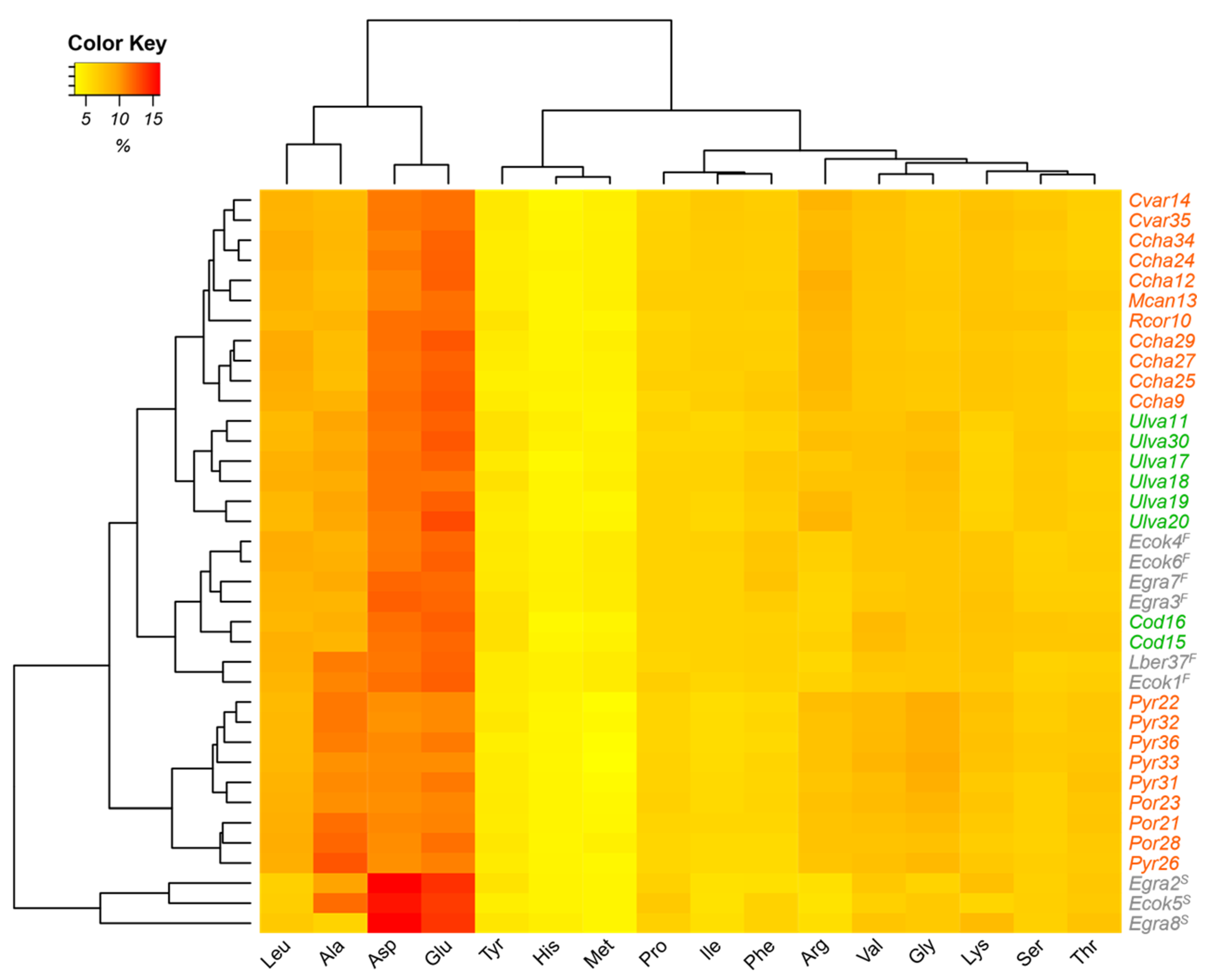
Furthermore, as can be seen from the heat map (Figure 1), a distinction within the algal groups based on amino acid composition was observed. In the case of red algae, there were two groups. The first group, the upper group, had higher concentrations of glutamic acid and aspartic acid; a predominance of the genera Callophyllis, Mazzaella, Chondracanthus, and Rhodymenia; and an identical profile. The second group, the lower group, had a higher concentration of alanine; lower amounts of glutamic acid and aspartic acid; and, in it, the genera Porphyra and Pyropia were predominant. In green algae, Ulva formed a separate group from Codium specimens. The brown algae were grouped into two sets according to their morphological compartment, i.e., stipe or frond. E. cokeri and E. gracilis were analyzed as stipes and had high relative percentages of glutamic acid and aspartic acid, while the species corresponding to fronds, Eisenia and Lessonia, presented lower relative percentages of these amino acids. According to Hernández-Carmona et al. [44], the nonessential amino acids most present in seaweeds are glutamic acid, aspartic acid, and alanine, and the most frequently present essential amino acid is leucine [33]. Several studies have indicated higher concentrations of these amino acids in green algae, followed by red and, to a lesser extent, brown algae [9,10,23,39,41,50,51,52,53]. In particular, the large amounts of amino acids such as glutamic acid are responsible for the special salty or umami taste of various algae [54].
Regarding the genera studied, the high and low amounts of amino acids reported were similar to those reported by other authors for the green algae Ulva fasciata from Brazil [55]; U. capensis, U. rigida, and U. lactuca from South Africa [56]; U. lactuca from Norway [7]; U. lactuca and Ulva intestinalis from Norway [10]; U. lactuca from China [21]; U. rigida from Portugal [39]; and Codium decorticatum, C. spongiosum, and C. taylorii from Brazil [55]. They were also similar to those reported for species of red algae including Porphyra sp. from Japan [57]; P. acanthophora from Brazil [55]; Porphyra sp. from Japan, Korea, and China [34]; P. columbina from Argentina [36]; P. dioica, P. purpurea, and P. umbilicalis from Norway [10]; Porphyra spp. from Portugal [58]; P. umbilicalis from Portugal [39]; and C. chamissoi from southern Peru [26]. With respect to brown algae, E. cokeri and E. gracilis from Peru presented the same ranges as were reported for E. arborea from Mexico [44] for glutamic acid (0.55–4.27 g/100 g), aspartic acid (0.40–3.67 g/100 g), alanine (0.22–4.30 g/100 g), and leucine (0.29–3.89 g/100 g).
Concerning the essential amino acids ratio (EAA%), the highest average was found in red algae from Gigartinales, with 48.65% (p = 0.000). The other orders did not show significant differences among them, with averages being between 43.71 and 45.98%. These values are higher than those reported for soy protein (39%) and very close to those for egg protein (47%) [59]. In addition, our results show that the red alga C. chamissoi collected from different localities in Peru had a higher amount of EAA% (average 48.41%) than that reported for C. chamissoi collected in Moquegua, in southern Peru (31.07%) [26]. Similarly, Porphyra sp./Pyropia sp. in our study had an EAA% of 44.48–46.65%, while the published values are in the range of 31.07–44.44% for Porphyra sp. from Japan, Korea, and China [34]; for Porphyra spp. from Portugal [58]; and for P. umbilicalis from Portugal [39]. This was the same for the green alga Ulva sp., which in our study had an EAA% of 44.54–46.22%, which is higher than the range determined (40.30–40.79%) for U. lactuca from Norway [7] and for U. rigida from Portugal [39].
Additionally, in terms of amino acid score (AAS), considering all of the taxa analyzed, the essential amino acids with the highest average scores were lysine, threonine, valine, and leucine (AAS > 0.96), followed by phenylalanine + tyrosine (0.91), and finally, isoleucine and histidine (0.82 > AAS > 0.80, p = 0.000). Numerically, the lowest average AAS was that of histidine from the Bryopsidales green algae (0.65). The latter result is in agreement with data previously reported in studies suggesting that sulfur-containing amino acids such as histidine are the major limiting amino acids in the proteins of some seaweeds [60].
Regarding the essential amino acid index (EAAI), the highest average percentages corresponded to Ulvales, Laminariales, Gigartinales, and Rhodymeniales (EAAI > 0.92), while the lowest percentages corresponded to Bryopsidales and Bangiales (0.90 > EAAI > 0.88, p = 0.000). Furthermore, in our study, the EAAI of Porphyra sp./Pyropia sp. was in the range of 0.87–0.90, which is slightly lower than the results obtained for Porphyra sp. from Japan, Korea, and China [34], and for P. umbilicalis and P. dioica from Portugal [39] was between 0.89 and 0.96, which is in agreement with those authors who have stated that the limiting essential amino acids are methionine, tryptophan, leucine, and isoleucine. Additionally, our EAAI results for the green alga Ulva sp. were in the range of 0.91–0.95, which is similar to the value reported by other authors indicating an EAAI between 0.92 and 1.23, with methionine as the limiting essential amino acid, in U. lactuca from Norway [7] and U. rigida from Portugal [39]. According to our results, most of the analyzed samples presented high and moderate quality with an EAAI value of >0.80 [61,62,63]. In addition, they had excellent profiles, with EAAI near that of egg protein and AASs higher than those of other plant proteins [33].

2.3. Fatty Acid Composition

Table 5, Table 6 and Table 7 show the fatty acid compositions of brown, red, and green algae. The highest average content of fatty acids corresponded to polyunsaturated fatty acids (PUFAs, 44.06%), followed by saturated fatty acids (SFAs, 34.56%), and finally monounsaturated fatty acids (MUFAs, 21.17%) (p = 0.000). The percentages of PUFAs in the references consulted for all seaweed groups were in the range of 30–60% [7,16,34,37,43]. However, values below 22% were also found [26,41,50,64]. The PUFAs found in the highest proportion were linolenic acid (C18:3n − 3) in the green algae group, arachidonic acid (ARA, C20:4n − 6) in the brown and red algae groups, and EPA (C20:5n − 3) in the red algae group.
In addition, a significant difference was observed in the MUFAs when orders were compared, with the stipe of the brown algae Eisenia (Laminariales) having the highest median (31.27%), while red algae from Bangiales had the lowest (11.40%) (p = 0.006). Previous studies have reported that Bangiales had a range of 14–21% [34,37,64], which is higher than our results. The MUFA present in the highest quantity was oleic acid (C18:1n − 9) in the brown algae from Laminariales and in the red algae from Gigartinales and Rhodymeniales.
As for SFAs, the fatty acid present in the highest quantity was palmitic acid (C16:0) for the three groups of algae, with the highest content being found in the red algae C. chamissoi (Gigartinales), with a median of 46.98%, and the lowest in the frond of the brown algae Eisenia (Laminariales), with 25.51%. These results agree with those reported by Sohrabipour et al. [65], wherein palmitic acid was the fatty acid with the highest amount, regardless of the species of seaweed studied.
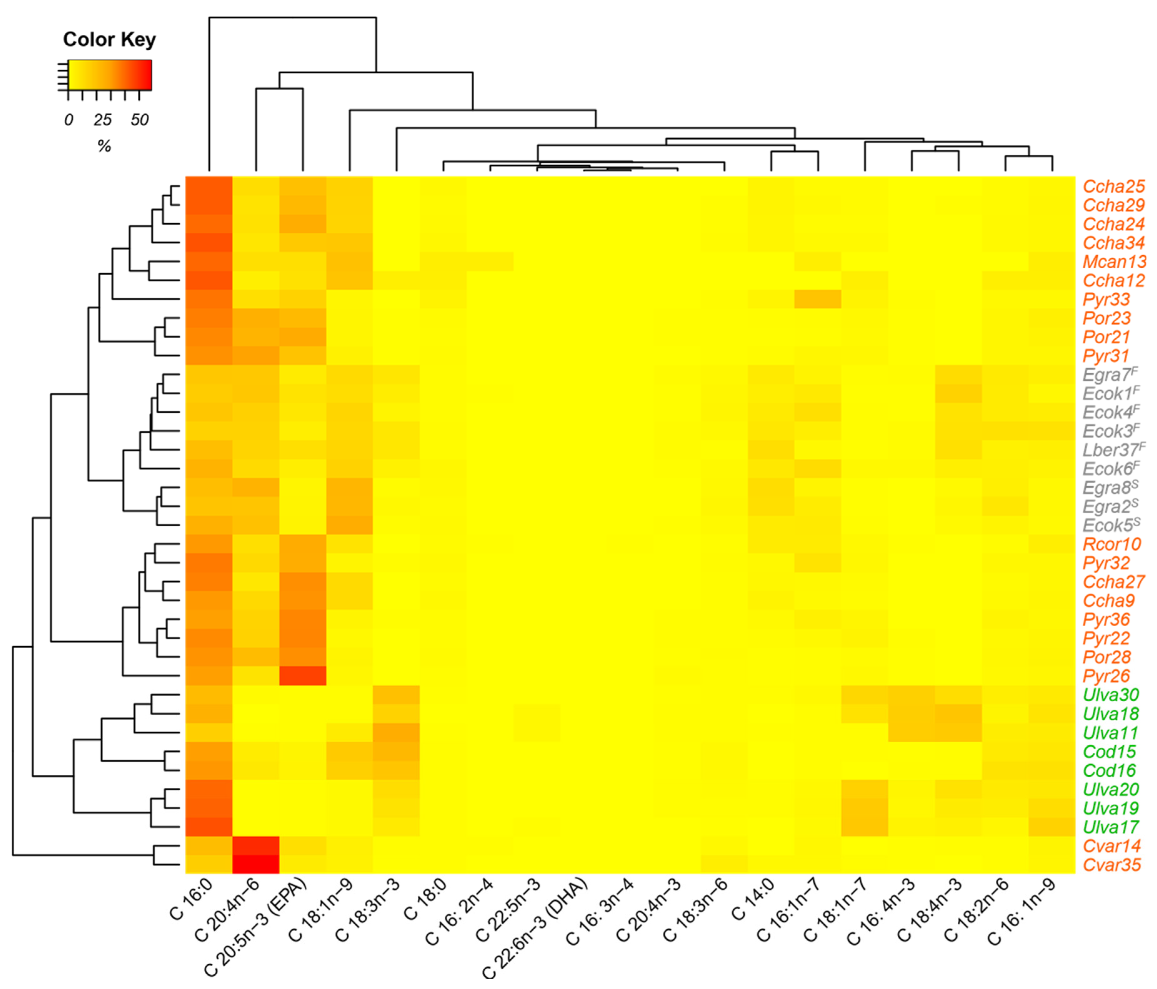
In the heat map (Figure 2), the three groups of algae are organized according to the evaluated composition of the fatty acids. There are two clearly separate groups. One belongs to the green algae, which have a cluster related to C18:3n − 3 acid. The brown algae form another cluster, which is related to the fatty acids C20:4n − 6 and C18:1n − 9, whereby a higher percentage of the C18:1n − 9 fatty acid is presented in the stipe. This allows separation from the frond. Elsewhere, red algae form three clusters with three discriminating fatty acids, C16:0, EPA, and C20:4n − 6. The top cluster, formed by C. chamissoi and M. canaliculata (Gigartinales) and Pyropia and Porphyra (Bangiales), is distinguished by its higher percentage of C16:0; the central cluster, consisting of the red algae C. chamissoi (Gigartinales), Pyropia and Porphyra (Bangiales), and R. corallina (Rhodymeniales), presents higher percentages of EPA; and the lowest cluster, formed by the red alga C. variegata (Gigartinales), has a higher percentage of C20:4n − 6. The characteristic fatty acids found in the heat map for the algal clusters have also been reported in different publications: Green algae from Ulvales and Bryopsidales had a significant relative percentage of C18:3n − 3 between 7 and 27% for U. lactuca from the USA [66] and Norway [7]; for Ulva sp. from southern Chile [67]; for Codium fragile from northern Chile [43]; and for C. tomentosum from Portugal [68]. Similarly, brown algae from Laminariales present significant relative percentages of C20:4n − 6 (9–34%) and C18:1n − 9 (9–17%) for Lessonia flavicans from southern Chile [67]. Finally, red algae from Gigartinales and Bangiales have significant relative percentages of C16:0 (30–64%), EPA (6–42%), and C20:4n − 6 (1–17%) for P. tenera from Japan [57]; for Porphyra sp. from Japan, Korea, and China [34]; for C. variegata from Chile [37]; for P. columbina from Argentina [36]; for M. canaliculata from the central coast of Peru [22]; and for Py. columbina from southern Chile [67].
With respect to omega-6 content (ω6), arachidonic acid (C20:4n − 6) was the compound with the highest amount, with 54.73% for the red alga C. variegata (Gigartinales), followed by the stipe (20. 06%) and the frond (15.21%) of Eisenia (Laminariales) and Bangiales (14.13%). The lowest median of C20:4n − 6 was found for the green algae from Bryopsidales (6.56%) and Ulvales (0.37%) (p = 0.001). The relative percentage of ω6 for C. variegata from Peru (58.81%) was three times higher than the value reported for C. variegata from southern Chile, which was 19.15% [37]. In the case of the Laminariales algae studied, L. berteroana, E. cokeri, and E. gracilis, our results (18.03–31.04%) were similar to those reported for L. flavicans from southern Chile, with a relative percentage between 15 and 43% [67]. The green algae of the genus Ulva had a relative percentage of ω6 between 3.61 and 8.19%, which is similar to the reference values (3 and 12%) for U. lactuca from the USA [66], northern Chile [41], and Norway [7]; U. armoricana from France [16]; and Ulva sp. from southern Chile [67].
In terms of omega-3 (ω3) content, at the group level, using the Kruskal–Wallis test, the main fatty acids were found to be EPA (C20:5n − 3) in red algae, with a median of 24.19%, and C18:3n − 3 in green algae, with a median of 16.11%. In brown algae, EPA (C20:5n − 3) (5.28%) and stearidonic acid (C18:4n − 3) (8.73%) were important. Fatty acid ω3 content showed no significant differences when comparing orders (p = 0.180).
At the order level, using the Kruskal–Wallis test, the highest median ω6/ω3 ratio was found in the Gigartinales red alga C. variegata (6.59), followed by stipes of Eisenia (Laminariales) (3.78). For the rest of the algae, the ratio did not exceed the median value of 1 (p = 0.001). The value of the ω6/ω3 ratio for the alga C. variegata from the central coast of Peru was remarkable, with none of the references consulted approaching it in value. The ω6/ω3 ratios reported for the orders Ulvales, Bangiales, and Laminariales from southern Chile [67] had the same tendencies, as shown in our results; the Laminariales had a ratio higher than 1, and the rest of the orders had a ratio lower than 1. Healthy diets have ω6/ω3 ratios with values lower than 10 [69] or with a maximum of 4 [70,71] since higher ratios favor the risk of developing diseases [72]. Based on these results, the red alga C. variegata from two localities in Peru (Paracas and Marcona in Ica) could be used in food because of its high content of ω6, which increases the ratio ω6/ω3.
This study showed that Bangiales (cochayuyos; Porphyra sp. and Pyropia sp.) had the highest average percentage of protein and carbohydrates and the lowest values of ash. Additionally, Ulvales specimens (sea lettuce, Ulva sp.) were remarkable because of their protein content, and Bryopsidales (Codium sp.) were remarkable because of their lipid contents. The highest amounts of essential amino acids were found in Gigartinales (yuyo, C. chamissoi; alga flor, M. canaliculata; and carola, C. variegata), while the highest essential amino acid index score was found for Ulvales (Ulva sp.), Laminariales (“kelps” E. cokeri, E. gracilis, and L. berteroana), Gigartinales (C. chamissoi, M. canaliculata, and C. variegata), and Rhodymeniales (R. corallina). The major PUFAs were linolenic acid in the group of green algae, arachidonic acid in the group of brown and red algae, and EPA in the group of red algae. A major MUFA was oleic acid in Laminariales, Gigartinales, and Rhodymeniales. With respect to the SFAs, palmitic acid had the highest quantity in the Gigartinales specimens. With regard to the content of omega-6, ARA was predominantly found in in the red alga C. variegata (Gigartinales), while for omega-3, it was EPA in red and green algae.
Knowledge regarding edible algae is scarce in Peru, so it was considered necessary to provide detailed and complete information on the chemical compositions of seaweeds that are consumed and that have been consumed since pre-Inca times in Peru, such as yuyos and cochayuyos (red algae), as well as to include data on other potentially edible species. From here, it is possible to revalue these edible algae and emphasize the consumption of other groups (orders) of seaweeds that are nutritionally promising, such as green and brown algae, which are not commonly consumed but could be introduced into the diet not only as a protein source but also for their important contents of essential amino acids, polyunsaturated fatty acids, and carbohydrates. These findings provide, for the first time in Peru, relevant information on the chemical and nutritional composition of seaweeds that could potentially be used directly as food or as ingredients in human or animal diets. It is also a first step in connecting the differences and similarities in the chemical compounds of these taxa with their taxonomic positions since it is important to understand these entities at a specific level in order for them to be commercialized internally or externally. Despite the amount of information generated, the grouping of the different taxa did not reflect a well-defined taxonomic classification, although it allowed the chemical characterization of the edible species analyzed. With that, we reinforce the definition of integrative taxonomy [73], in which species should be revised, analyzing different characteristics, using other methods, and applying other criteria to delimit species.
3. Materials and Methods

3.1. Sample Collection and Preparation

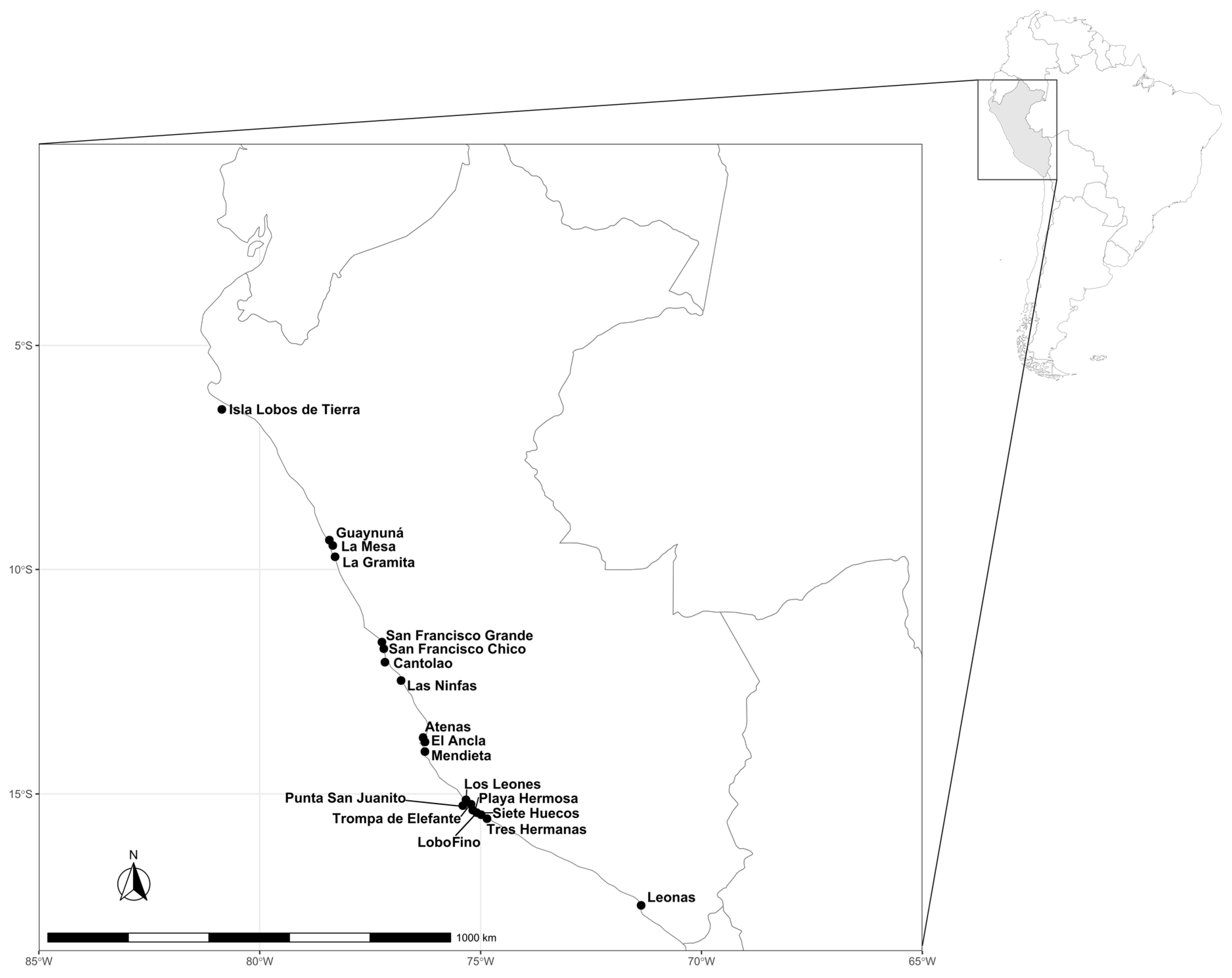
During 2018 and 2019, algal samples were collected and identified in the rocky intertidal and subtidal zone along the Peruvian coast (6°–17° S): Lobos de Tierra Island (Piura), Casma (Ancash), Ancón, Callao and Pucusana (Lima), Paracas and San Juan de Marcona (Ica), and Ilo (Moquegua) (Table 8). A total of 37 specimens were analyzed, belonging to the 3 groups of seaweed (Table 1)—21 Rhodophyta (red), 8 Chlorophyta (green), and 9 Ochrophyta-Phaeophyceae (brown)—and were collected from 19 localities on the Peruvian coast (6°–17° S) (Figure 3). Among the red algae, there were six genera, four of which were identified at the species level (C. variegata, M. canaliculata, C. chamissoi, and R. corallina), while for Porphyra and Pyropia the species were not determined. Among the green algae, Ulva and Codium were identified at the generic level. With respect to brown algae, three species, E. cokeri, E. gracilis, and L. berteroana, were identified. In the kelp Eisenia (Laminariales) alone, two different parts of the thallus were analyzed, the stipe and the frond.
Fresh material was selected and transported to IMARPE (Marine Institute of Peru), hermetically sealed in plastic bags, and kept in refrigerated containers. Epiphytes and contaminants were removed from the collected material via repeated washing in fresh water. The cleaned algae were then frozen at −20 °C. Drying was performed in a Labconco freeze-dryer, model 18 L (Kansas, KS, USA) [74]. The conditions were as follows: 0.022 and 0.070 mbar (vacuum pressure), −56 °C (collection temperature), and −15 °C for 8 h, followed by an increase of 0.5 °C/min up to 5 °C for 15 h, and a further increase of 0.5 °C/min up to 25 °C for 7 h (ramp temperature). Finally, pulverized lyophilized sample was obtained using a mortar and stored in hermetically sealed plastic bags at −20 °C.

3.2. Proximate Analysis

To measure the moisture percentage, 0.1 g of lyophilized sample was weighed and dried in a Vacucell vacuum oven (MMM, Germany) at 105 °C for 16 h at a pressure below 0.1 bar [75]. Additionally, to analyze the ashes, 0.1 g of lyophilized sample was weighed and incinerated in a Thermolyne muffle furnace (F6010, USA) at 550 °C for 16 h [76].
Proteins were quantified from 5 mg of lyophilized sample using the Hartree method [77]. Hydrolysis was carried out with 5 mL of 0.5 N sodium hydroxide at 85 °C for 45 min. A solution containing Folin–Ciocalteu’s reagent, sodium carbonate, copper sulfate, and potassium and sodium tartrate was then added. The absorbance was measured at 650 nm using a UV spectrophotometer (Varian, Australia), and the percentage of proteins was measured using a calibration curve with bovine serum albumin (300 μg/mL) in a concentration range of 0–150 μg/mL.
The modified Folch method was used for the analysis of lipids [78]. Lipids were extracted from 25 mg of lyophilized seaweed with 3 mL of a chloroform–methanol (2:1) solvent mixture. The extraction was performed twice in a sonicator at 4 °C for 30 min, following which 2 mL of 0.88% (w/v) potassium chloride was added. The organic phase was dried in an evaporator–concentrator with nitrogen gas. The lipid extracts were stored under vacuum and in the dark for 14 h and then weighed to obtain the percentage of total lipids.
Carbohydrates were quantified using the following equation: Carbohydrate% = 100% − Protein% − Lipid% − Ash% − Moisture% [22,35,37].
Proximate assays were performed in triplicate.

3.3. Fatty Acid Analysis

The determination of fatty acids was performed by adding toluene, methanol, and hydrochloric acid to 1 mg of the lipids and 0.1 mg of methyl tricosanoate (C23:0, internal standard) and incubating the mixture at 45 °C for 14 h for the derivatization [79]. The produced methylesters were extracted with hexane for gas chromatography analysis on a Varian GC-FID CP-3800 (Germany) under the following conditions: 30 m × 0.25 mm × 0.25 µm fused silica Restek Stabilwax® WCOT column; splitless injection method (0.50 min); injection volume of 1 μL; injector temperature at 250 °C; helium carrier gas at a flow rate of 1 mL/min; ramp 120 °C per minute; 30 °C/min to 160 °C; 160 °C per minute; 4 °C/min to 240 °C; 240 °C for 7 min. The total duration of the temperature program was between 30 and 33 min, and the programmed temperature of detection was 260 °C. Fatty acids assays were performed in triplicate.

3.4. Amino Acid Analysis

Between 30 and 50 mg of lyophilized sample was hydrolyzed with 2 mL of 6 N HCl at 112 °C for 24 h [80]. Next, following the AccQ-Fluor reagent kit manual (Waters Corporation, Milford, MA, USA), a solution containing 50 µL of the hydrolyzed sample, 100 µL of the 2.5 mM 2-L-aminobutyric acid internal standard, and 4850 µL of ultrapure water was prepared, filtered on 0.45 µm PTFE filters, and derivatized. After that, 5 µL of the derivatized amino acid solution was injected in the HPLC-FL Elite LaChrom (Hitachi, Japan). The equipment conditions were as follows: Hypersil GOLD C18 column 5 µm × 4.6 mm × 150 mm; mobile phase flow rate 1 mL/min at 37 °C (column); 250 nm (excitation); 395 nm (emission); 5 µL (injection); mobile phase A: ultrapure water; mobile phase B: sodium acetate pH 5.1; mobile phase C: acetonitrile; one mobile phase ramp: 0 min (0, 100, 0), 1 min (0, 100, 0), 25 min (0, 83, 27), 33 min (50, 0, 50), 36 min (0, 100, 0), and 40 min (0, 100, 0). The amino acids were quantified by a calibration curve with a concentration ranging from 5 to 50 pmol/µL. The amino acids identified were alanine, arginine, aspartic acid, glutamic acid, glycine histidine, isoleucine, leucine, lysine, methionine, phenylalanine, proline, serine, threonine, tyrosine, and valine (Ala, Arg, Asp, Glu, Gly, His, Ile, Leu, Lys, Met, Phe, Pro, Ser, Thr, Tyr, and Val). The applied analytical technique was not able to identify the amino acids cysteine and tryptophan (Cys, Trp). Amino acids assays were performed in triplicate.

3.5. Essential Amino Acid Index (EAAI)

The amino acid score (AASi) was calculated for each essential amino acid i as the quotient of the relative percentage of the essential amino acid i in the sample to the relative percentage of amino acid i in relation to a standard protein [81,82]. In this study, egg protein was used as the standard protein in accordance with FAO [59], which is applicable for measuring protein quality in human nutrition. Chemical scores greater than 1 were replaced with 1. The proposed essential amino acid index is presented as Equation (1) [82].
E A A I = A A S 1 × A A S 2 × A A S n n
Moreover, the essential amino acids ratio (EAA%) was determined as the sum of each essential amino acid among the total amino acids.

3.6. Statistical Analysis

Comparisons of the concentrations of biochemical compounds were made at the taxonomic level of order. For this, analysis of variance (ANOVA) was used at a significance level of α = 0.05, followed by Tukey’s post hoc test. If the ANOVA assumptions were not met, the Kruskal–Wallis test was used. The multivariate analysis was performed using a heat map to obtain the relationship between the concentrations of biochemical compounds and the analyzed seaweed species. The data were analyzed using the statistical programs Minitab 19 and the package heatmap.2 of R-studio v 1.2.1335.

Author Contributions

Conceptualization and resources, N.A. and L.F.R.; methodology, investigation, and writing—original draft preparation, N.A., L.F.R., A.I.O.H. and A.R.S.; formal analysis, L.F.R., A.I.O.H. and A.R.S.; writing—review and editing, N.A., L.F.R., A.I.O.H., A.R.S. and M.E.R. All authors have read and agreed to the published version of the manuscript.

Funding

This research was funded by Presupuesto por Resultados PpR, Ordenamiento y Desarrollo de la Acuicultura (DGIA-IMARPE).

Data Availability Statement

Not applicable.

Acknowledgments

The authors would like to thank the Convenio de Subvención No. 129-2015-FONDECYT, CONCYTEC-Peru for providing samples from Lobos de Tierra Island. We thank Diego Márquez-Corigliano for their generous assistance with the map design and Melissa Perez for her help in the sample collection in Marcona.

Conflicts of Interest

The authors declare no conflict of interest.

References

  1. Murata, M.; Nakasoe, J. Production and use of marine algae in Japan. Jpn. Agric. Res. Q. 2001, 35, 281–290. [Google Scholar] [CrossRef] [Green Version]
  2. Kılınç, B.; Cirik, S.; Turan, G.; Tekogul, H.; Koru, E. Seaweeds for Food and Industrial Applications. In Food Industry; Muzzalupo, I., Ed.; IntechOpen: London, UK, 2013; Chapter 31; pp. 735–748. [Google Scholar] [CrossRef] [Green Version]
  3. Torres, M.D.; Flórez-Fernández, N.; Domínguez, H. Integral Utilization of red seaweed for bioactive production. Mar. Drugs 2019, 17, 314. [Google Scholar] [CrossRef] [PubMed] [Green Version]
  4. Hayashi, L.; Bulboa, C.; Kradolfer, P.; Soriano, G.; Robledo, D. Cultivation of red seaweeds: A Latin American perspective. J. Appl. Phycol. 2013, 26, 719–727. [Google Scholar] [CrossRef]
  5. Mouritsen, O.G. The emerging science of gastrophysics and its application to the algal cuisine. Flavour 2012, 1, 6. [Google Scholar] [CrossRef] [Green Version]
  6. Gressler, V.; Yokoya, N.S.; Fujii, M.T.; Colepicolo, P.; Filho, J.M.; Torres, R.P.; Pinto, E. Lipid, fatty acid, protein, amino acid and ash contents in four Brazilian red algae species. Food Chem. 2010, 120, 585–590. [Google Scholar] [CrossRef]
  7. Mæhre, H.K.; Malde, M.K.; Eilertsen, K.E.; Elvevoll, E.O. Characterization of protein, lipid and mineral contents in common Norwegian seaweeds and evaluation of their potential as food and feed. J. Sci. Food Agric. 2014, 94, 3281–3290. [Google Scholar] [CrossRef]
  8. Olsson, J.; Toth, G.B.; Albers, E. Biochemical composition of red, green and brown seaweeds on the Swedish west coast. J. Appl. Phycol. 2020, 32, 3305–3317. [Google Scholar] [CrossRef]
  9. Gamero-Vega, G.; Palacios-Palacios, M.; Quitral, V. Nutritional composition and bioactive compounds of red seaweed: A mini-review. J. Food Nutr. Res. 2020, 8, 431–440. [Google Scholar] [CrossRef]
  10. Biancarosa, I.; Espe, M.; Bruckner, C.G.; Heesch, S.; Liland, N.; Waagbø, R.; Torstensen, B.; Lock, E.J. Amino acid composition, protein content, and nitrogen-to-protein conversion factors of 21 seaweed species from Norwegian waters. J. Appl. Phycol. 2016, 29, 1001–1009. [Google Scholar] [CrossRef]
  11. Salehi, B.; Sharifi-Rad, J.; Seca, A.M.L.; Pinto, D.C.G.A.; Michalak, I.; Trincone, A.; Mishra, A.P.; Nigam, M.; Zam, W.; Martins, N. Current trends on seaweeds: Looking at chemical composition, phytopharmacology, and cosmetic applications. Molecules 2019, 24, 4182. [Google Scholar] [CrossRef] [Green Version]
  12. Kumar, M.; Kumari, P.; Trivedi, N.; Shukla, M.K.; Gupta, V.; Reddy, C.R.K.; Jha, B. Minerals, PUFAs and antioxidant properties of some tropical seaweeds from Saurashtra coast of India. J. Appl. Phycol. 2011, 23, 797–810. [Google Scholar] [CrossRef]
  13. Cofrades, S.; Serdaroğlu, M.; Jiménez-Colmenero, F. Design of healthier foods and beverages containing whole algae. In Functional Ingredients from Algae for Foods and Nutraceuticals; Domínguez, H., Ed.; Woodhead Publishing Series in Food Science, Technology and Nutrition, Functional Ingredients from Algae for Foods and Nutraceuticals; Woodhead Publishing: Cambridge, UK, 2013; pp. 609–633. [Google Scholar] [CrossRef]
  14. Stengel, D.B.; Connan, S.; Popper, Z.A. Algal chemodiversity and bioactivity: Sources of natural variability and implications for commercial application. Biotechnol. Adv. 2011, 29, 483–501. [Google Scholar] [CrossRef] [PubMed]
  15. Belghit, I.; Rasinger, J.D.; Heesch, S.; Biancarosa, I.; Liland, N.; Torstensen, B.; Waagbø, R.; Lock, E.-J.; Bruckner, C.G. In-depth metabolic profiling of marine macroalgae confirms strong biochemical differences between brown, red and green algae. Algal Res. 2017, 26, 240–249. [Google Scholar] [CrossRef]
  16. Kendel, M.; Wielgosz-Collin, G.; Bertrand, S.; Roussakis, C.; Bourgougnon, N.; Bedoux, G. Lipid composition, fatty acids and sterols in the seaweeds Ulva armoricana, and Solieria chordalis from Brittany (France): Analysis from nutritional, chemotaxonomic, and antiproliferative activity perspectives. Mar. Drugs 2015, 13, 5606–5628. [Google Scholar] [CrossRef]
  17. Fleury, B.G.; Figueiredo, L.; Marconi, M.I.; Teixeira, V.L.; Ferreira, A.B.B.; Pinto, A.C. Fatty acids as chemotaxonomic markers of marine macrophytes from Rio de Janeiro State, Brazil. Nat. Prod. Commun. 2011, 6, 667–672. [Google Scholar] [CrossRef] [PubMed] [Green Version]
  18. Herbreteau, F.; Coiffard, L.J.M.; Derrien, A.; De Roeck-Holtzhauer, Y. The fatty acid composition of five species of macroalgae. Bot. Mar. 1997, 40, 25–27. [Google Scholar] [CrossRef]
  19. Schubert, N.; García-Mendoza, E.; Pacheco-Ruiz, I. Carotenoid composition of marine red algae. J. Phycol. 2006, 42, 1208–1216. [Google Scholar] [CrossRef]
  20. Cardoso, C.; Ripol, A.; Afonso, C.; Freire, M.; Varela, J.; Quental-Ferreira, H.; Pousao-Ferreira, P.; Bandarra, N. Fatty acid profiles of the main lipid classes of green seaweeds from fish pond aquaculture. Food Sci. Nutr. 2017, 5, 1186–1194. [Google Scholar] [CrossRef]
  21. Wong, K.H.; Cheung, P.C.K. Nutritional evaluation of some subtropical red and green seaweeds Part I—Proximate composition, amino acid profiles and some physico-chemical properties. Food Chem. 2000, 71, 475–482. [Google Scholar] [CrossRef]
  22. Velásquez, R.O. Caracterización Químico-Nutricional y Actividad Antioxidante del Alga Chondrus canaliculatus (C. Agardh) Greville (Rhodophyta) Proveniente de Paracas. Bachelor’s Thesis, Universidad Peruana Cayetano Heredia, Lima, Perú, 2019. [Google Scholar]
  23. Rojas, N.C.; Valdivieso, R.; Arnao, I. Composición nutricional del alga roja Rodhymenia howeana de la Bahía de Ancón, Perú. Rev. Soc. Quím. Perú 2018, 84, 488–498. [Google Scholar]
  24. Tabacchi, P.; García, F. Caracterizacion bioquímica del alga Gigartina chamissoi (Bahía de Ancón). Rev. Química 1994, 8, 21–28. [Google Scholar]
  25. Sumarriva, L. Caracterización y evaluación biológica de las algas: Gigartina chamissoi, Porphyra columbina y Ulva lactuca. Infinitum 2013, 3, 40–44. [Google Scholar]
  26. Vilcanqui, Y.; Mamani-Apaza, L.O.; Flores, M.; Ortiz-Viedma, J.; Romero, N.; Mariotti-Celis, M.S.; Huamán-Castilla, N.L. Chemical characterization of brown and red seaweed from Southern Peru, a sustainable source of bioactive and nutraceutical compounds. Agronomy 2021, 11, 1669. [Google Scholar] [CrossRef]
  27. Márquez-Corigliano, D.; Arakaki, N.; Gil, P.; Tellier, F. Diversidad de especies de Porphyra y Pyropia (Bangiaceae, Rhodophyta) de Marcona (Ica, Perú) bajo la evidencia molecular. Arnaldoa 2019, 26, 623–642. [Google Scholar]
  28. Arakaki, N.; Carbajal, P.; Márquez-Corigliano, D.; Suárez, S.; Gil-Kodaka, P.; Perez-Araneda, K.; Tellier, F. Genética de macroalgas en el Perú: Diagnóstico, guía metodológica y casos de estudio. Inf. Inst. Mar Perú 2021, 48, 594–609. [Google Scholar]
  29. Roleda, M.Y.; Heesch, S. Chemical profiling of Ulva species for food applications: What is in a name? Food Chem. 2021, 361, 130084. [Google Scholar] [CrossRef]
  30. Acleto, C. Algas Marinas del Perú de Importancia Económica; Serie de Divulgación 5; UNMSM-Museo de Historia Natural Javier Prado, Departamento de Botánica: Lima, Perú, 1986. [Google Scholar]
  31. Masuda, S. Cochayuyo, macha, camarón, y higos charqueados. In Estudios Etnográficos del Perú Meridional; Masuda, S., Ed.; Universidad de Tokio: Tokyo, Japan, 1981; pp. 173–192. [Google Scholar]
  32. Ávila, M.; Alcapán, A.; Piel, M.I.; Ramírez, M.E.; Cortés, M. Manual de Cultivo del Alga Comestible Callophyllis variegata (Bory) Kützing (“carola”) en el sur de Chile; Serie Programa educativo para el desarrollo de la acuicultura de especies nativas; Universidad Arturo Prat: Iquique, Chile, 2012. [Google Scholar]
  33. Thiviya, P.; Gamage, A.; Gama-Arachchige, N.S.; Merah, O.; Madhujith, T. Seaweeds as a source of functional proteins. Phycology 2022, 2, 216–243. [Google Scholar] [CrossRef]
  34. Dawczynski, C.; Schubert, R.; Jahreis, G. Amino acids, fatty acids, and dietary fibre in edible seaweed products. Food Chem. 2007, 103, 891–899. [Google Scholar] [CrossRef]
  35. Smith, J.L.; Summers, G.; Wong, R. Nutrient and heavy metal content of edible seaweeds in New Zealand. N. Z. J. Crop Hortic. Sci. 2010, 38, 19–28. [Google Scholar] [CrossRef]
  36. Cian, R.E.; Fajardo, M.A.; Alaiz, M.; Vioque, J.; González, R.J.; Drago, S.R. Chemical composition, nutritional and antioxidant properties of the red edible seaweed Porphyra columbina. Int. J. Food Sci. Nutr. 2014, 65, 299–305. [Google Scholar] [CrossRef] [Green Version]
  37. Astorga-España, M.S.; Mansilla, A. Sub-Antarctic macroalgae: Opportunities for gastronomic tourism and local fisheries in the Region of Magallanes and Chilean Antarctic Territory. J. Appl. Phycol. 2013, 26, 973–978. [Google Scholar] [CrossRef]
  38. Patarra, R.F.; Paiva, L.; Neto, A.I.; Lima, E.; Baptista, J. Nutritional value of selected macroalgae. J. Appl. Phycol. 2011, 23, 205–208. [Google Scholar] [CrossRef] [Green Version]
  39. Machado, M.; Machado, S.; Pimentel, F.B.; Freitas, V.; Alves, R.C.; Oliveira, M. Amino acid profile and protein quality assessment of macroalgae produced in an integrated multi-trophic aquaculture system. Foods 2020, 9, 1382. [Google Scholar] [CrossRef] [PubMed]
  40. Westermeier, R.; Murúa, P.; Robles, M.; Barría, M.; Patiño, D.J.; Muñoz, L.; Müller, D.G. Population biology and chemical composition of the red alga Callophyllis variegata (Rhodophyta; Cryptonemiales) in southern Chile. J. Appl. Phycol. 2019, 32, 2505–2513. [Google Scholar] [CrossRef]
  41. Ortiz, J.; Romero, N.; Robert, P.; Araya, J.; López-Hernández, J.; Bozzo, C.; Navarrete, E.; Osorio, A.; Ríos, A. Dietary fiber, amino acid, fatty acid and tocopherol contents of the edible seaweeds Ulva lactuca and Durvillaea antarctica. Food Chem. 2006, 99, 98–104. [Google Scholar] [CrossRef]
  42. Rohani-Ghadikolaei, K.; Abdulalian, E.; Ng, W.K. Evaluation of the proximate, fatty acid and mineral composition of representative green, brown and red seaweeds from the Persian Gulf of Iran as potential food and feed resources. J. Food Sci. Technol. 2012, 49, 774–780. [Google Scholar] [CrossRef] [Green Version]
  43. Ortiz, J.; Uquiche, E.; Robert, P.; Romero, N.; Quitral, V.; Llantén, C. Functional and nutritional value of the Chilean seaweeds Codium fragile, Gracilaria chilensis and Macrocystis pyrifera. Eur. J. Lipid Sci. Technol. 2009, 111, 320–327. [Google Scholar] [CrossRef] [Green Version]
  44. Hernández-Carmona, G.; Carrillo-Domínguez, S.; Arvizu-Higuera, D.L.; Rodríguez-Montesinos, Y.E.; Murillo-Álvarez, J.I.; Muñoz-Ochoa, M.; Castillo-Domínguez, R.M. Monthly variation in the chemical composition of Eisenia arborea J.E. Areschoug. J. Appl. Phycol. 2009, 21, 607–616. [Google Scholar] [CrossRef]
  45. Vega, J.M.A.; Toledo, P.H. The chemical composition of Lessonia berteroana (ex L. nigrescens) in kelp harvest management and open access areas near Coquimbo, Chile. Lat. Am. J. Aquat. Res. 2018, 46, 258–267. [Google Scholar] [CrossRef]
  46. Kavale, M.G.; Kasi, M.A.; Bagal, P.U.; Singh, V.V.; Behera, D.P. Food value of Pyropia vietnamensis (Bangiales, Rhodophyta) from India. Indian J. Mar. Sci. 2018, 47, 402–408. [Google Scholar]
  47. Leandro, A.; Pacheco, D.; Cotas, J.; Marques, J.C.; Pereira, L.; Goncalves, A.M.M. Seaweed's bioactive candidate compounds to food industry and global food security. Life 2020, 10, 140. [Google Scholar] [CrossRef] [PubMed]
  48. Food and Nutrition Board (FNB); Institute of Medicine (IOM). Dietary Reference Intakes: The Essential Guide to Nutrient Requirements; National Academy Press: Washington, DC, USA, 2006. [Google Scholar] [CrossRef]
  49. Véliz, K.; Toledo, P.; Araya, M.; Gómez, M.F.; Villalobos, V.; Tala, F. Chemical composition and heavy metal content of Chilean seaweeds: Potential applications of seaweed meal as food and feed ingredients. Food Chem. 2022, 398, 133866. [Google Scholar] [CrossRef] [PubMed]
  50. Ortiz, J. Composición Nutricional y Funcional de las Algas Clorofíceas Chilenas: Codium fragile y Ulva lactuca; Monografía; Laboratorio de Química y Análisis de Alimentos, Departamento de Ciencia de los Alimentos y Tecnología Química, Facultad de Ciencias Químicas y Farmacéuticas, Universidad de Chile: Santiago, Chile, 2011. [Google Scholar]
  51. Angell, A.R.; Angell, S.F.; de Nys, R.; Paul, N.A. Seaweed as a protein source for mono-gastric livestock. Trends Food Sci. Technol. 2016, 54, 74–84. [Google Scholar] [CrossRef]
  52. Astorga-España, M.S.; Rodríguez-Galdón, B.; Rodríguez-Rodríguez, E.M.; Díaz-Romero, C. Amino acid content in seaweeds from the Magellan Straits (Chile). J. Food Compos. Anal. 2016, 53, 77–84. [Google Scholar] [CrossRef]
  53. Sumarriva, L.; Castro, A.; Sotelo, A.; Chávez, N. Evaluación biológica de proteína, vitaminas, minerales y aminoácidos del alga comestible Ulva lactuca "lechuga de mar" del litoral peruano. Rev. Soc. Quím. Perú 2019, 85, 34–42. [Google Scholar] [CrossRef]
  54. Ummat, V.; Garcia-Vaquero, M.; Poojary, M.M.; Lund, M.N.; O'Donnell, C.; Zhang, Z.; Tiwari, B.K. Green extraction of proteins, umami and other free amino acids from brown macroalgae Ascophyllum nodosum and Fucus vesiculosus. J. Appl. Phycol. 2021, 33, 4083–4091. [Google Scholar] [CrossRef] [PubMed]
  55. Lourenço, S.O.; Barbarino, E.; De-Paula, J.C.; Pereira, L.O.S.; Marquez, U.M.L. Amino acid composition, protein content and calculation of nitrogen-to-protein conversion factors for 19 tropical seaweeds. Phycol. Res. 2002, 50, 233–241. [Google Scholar] [CrossRef]
  56. Shuuluka, D.; Bolton, J.J.; Anderson, R.J. Protein content, amino acid composition and nitrogen-to-protein conversion factors of Ulva rigida and Ulva capensis from natural populations and Ulva lactuca from an aquaculture system, in South Africa. J. Appl. Phycol. 2013, 25, 677–685. [Google Scholar] [CrossRef]
  57. Nisizawa, K.; Noda, H.; Kikuchi, R.; Watanabe, T. The main seaweed foods in Japan. Hydrobiologia 1987, 151–152, 5–29. [Google Scholar] [CrossRef]
  58. Vieira, E.F.; Soares, C.; Machado, S.; Correia, M.; Ramalhosa, M.J.; Oliva-teles, M.T.; Carvalho, A.P.; Domingues, V.F.; Antunes, F.; Oliveira, T.A.C.; et al. Seaweeds from the Portuguese coast as a source of proteinaceous material: Total and free amino acid composition profile. Food Chem. 2018, 269, 264–275. [Google Scholar] [CrossRef] [Green Version]
  59. FAO/WHO/UN. Protein and Amino Acid Requirements in Human Nutrition; Report of a Joint FAO/WHO/UNU. Expert Consultation; Technical Report Series 935; World Health Organization: Geneva, Switzerland, 2007; Available online: https://apps.who.int/iris/handle/10665/43411 (accessed on 22 July 2022).
  60. Parjikolaei, B.R.; Bruhn, A.; Eybye, K.L.; Larsen, M.M.; Rasmussen, M.B.; Christensen, K.V.; Fretté, X.C. Valuable biomolecules from nine North Atlantic red macroalgae: Amino acids, fatty acids, carotenoids, minerals and metals. Nat. Resour. 2016, 7, 157–183. [Google Scholar] [CrossRef] [Green Version]
  61. Brown, M.R.; Jeffrey, S. Biochemical composition of microalgae from the green algal classes Chlorophyceae and Prasinophyceae. 1. Amino acids, sugars and pigments. J. Exp. Mar. Biol. Ecol. 1992, 161, 91–113. [Google Scholar] [CrossRef]
  62. Ju, Z.Y.; Forster, I.; Conquest, L.; Dominy, W. Enhanced growth effects on shrimp (Litopenaeus vannamei) from inclusion of whole shrimp floc or floc fractions to a formulated diet. Aquac. Nutr. 2008, 14, 533–543. [Google Scholar] [CrossRef]
  63. Zhang, T.; Chi, Z.; Sheng, J. A highly thermosensitive and permeable mutant of the marine yeast Cryptococcus aureus G7a potentially useful for single-cell protein production and its nutritive components. Mar. Biotechnol. 2009, 11, 280–286. [Google Scholar] [CrossRef] [PubMed]
  64. Sánchez-Machado, D.I.; López-Cervantes, J.; López-Hernández, J.; Paseiro-Losada, P. Fatty acids, total lipid, protein and ash contents of processed edible seaweeds. Food Chem. 2004, 85, 439–444. [Google Scholar] [CrossRef]
  65. Sohrabipour, J.; Rabiei, R.; Pirian, K. Fatty acids composition of marine macroalgae. J. Phycol. Res. 2019, 3, 348–374. [Google Scholar] [CrossRef]
  66. Khotimchenko, S.V.; Vaskovsky, V.E.; Titlyanova, T.V. Fatty acids of marine algae from the Pacific Coast of North California. Bot. Mar. 2002, 45, 17–22. [Google Scholar] [CrossRef]
  67. dos Santos, M.A.Z.; Berneira, L.M.; Goulart, N.L.; Mansilla, A.; Astorga-España, M.S.; de Pereira, C.M.P. Rhodophyta, Ochrophyta and Chlorophyta macroalgae from different sub-Antarctic regions (Chile) and their potential for polyunsaturated fatty acids. Rev. Bras. Bot. 2021, 44, 429–438. [Google Scholar] [CrossRef]
  68. Rodrigues, D.; Freitas, A.C.; Pereira, L.; Rocha-Santos, T.A.P.; Vasconcelos, M.W.; Roriz, M.; Rodríguez-Alcalá, L.M.; Gomes, A.M.P.; Duarte, A.C. Chemical composition of red, brown and green macroalgae from Buarcos bay in Central West Coast of Portugal. Food Chem. 2015, 183, 197–207. [Google Scholar] [CrossRef]
  69. WHO. Population nutrient intake goals for preventing diet-related chronic diseases. Recommendations for preventing dental diseases. In Diet, Nutrition and the Prevention of Chronic Diseases; Report of a joint WHO/FAO expert consulta; WHO Technical Report Series, 916; WHO: Geneva, Switzerland, 2008. [Google Scholar]
  70. HMSO. Nutritional aspects of cardiovascular disease. Report of the Cardiovascular Review Group Committee on Medical Aspects of Food Policy. Rep. Health Soc. Subj. 1994, 46, 1–186. [Google Scholar]
  71. Simopoulos, A.P. The importance of the ratio of omega-6/omega-3 essential fatty acids. Biomed. Pharmacoth. 2002, 56, 365–379. [Google Scholar] [CrossRef] [PubMed]
  72. Rocha, C.P.; Pacheco, D.; Cotas, J.; Marques, J.C.; Pereira, L.; Gonçalves, A.M.M. Seaweeds as valuable sources of essential fatty acids for human nutrition. Int. J. Environ. Res. Public Health 2021, 18, 4968. [Google Scholar] [CrossRef] [PubMed]
  73. Pante, E.; Puillandre, N.; Viricel, A.; Arnaud-Haond, S.; Aurelle, D.; Castelin, M.; Chenuil, A.; Destombe, C.; Forcioli, D.; Valero, M.; et al. Species are hypotheses: Avoid connectivity assessments based on pillars of sand. Mol. Ecol. 2015, 24, 525–544. [Google Scholar] [CrossRef] [Green Version]
  74. Oscanoa, A.; Cervantes, M.; Febrero, P. Manual para la producción de biomasa microalgal en condiciones de invernadero. Inf. Inst. Mar Perú 2020, 47, 332–356. [Google Scholar]
  75. Kolar, K. Gravimetric determination of moisture and ash in meat and meat products: NMKL Interlaboratory Study. J. AOAC Int. 1992, 75, 1016–1022. [Google Scholar] [CrossRef]
  76. AOAC. Official Methods of Analysis, 13th ed.; Association Official Analytical Chemists: Washington, DC, USA, 1980. [Google Scholar]
  77. Hartree, E.F. Determination of protein: A modification of the Lowry method that gives a linear photometric response. Anal. Biochem. 1972, 48, 422–427. [Google Scholar] [CrossRef]
  78. Folch, J.; Lees, M.; Stanley, G.H.S. A simple method for the isolation and purification of total lipides from animal tissues. J. Biol. Chem. 1957, 226, 497–509. [Google Scholar] [CrossRef]
  79. Ichihara, K.; Fukubayashi, Y. Preparation of fatty acid methyl esters for gas-liquid chromatography. J. Lipid Res. 2010, 51, 635–640. [Google Scholar] [CrossRef] [Green Version]
  80. AOAC. Official Methods of Analysis, 16th ed.; Association Official Analytical Chemists: Washington, DC, USA, 1997. [Google Scholar]
  81. Oser, B.L. An integrated essential amino acid index for predicting the biological value of proteins. In Protein and Amino Acid Nutrition; Albanese, A.A., Ed.; Academic Press: New York, NY, USA, 1959; Chapter 10; pp. 295–311. [Google Scholar] [CrossRef]
  82. Peñaflorida, V.D. An evaluation of indigenous protein sources as potential component in the diet formulation for tiger prawn, Penaeus monodon, using essential amino acid index (EAAI). Aquaculture 1989, 83, 319–330. [Google Scholar] [CrossRef]
Figure 1. Heat map for classification of red, green, and brown macroalgae characterized by amino acid composition. The amino acid concentrations are expressed as relative percentages. F Frond; S stipe.
Plants 12 01795 g001
Figure 2. Heat map of classification of red, green, and brown macroalgae from the Peruvian coast characterized by fatty acid composition. Fatty acid concentrations are expressed as relative percentages. F Frond; S stipe.
Plants 12 01795 g002
Figure 3. Map of the Peruvian coast showing the collection localities of edible seaweeds.
Plants 12 01795 g003
Table 1. Proximate composition of lyophilized Peruvian seaweeds.
OrderCodeProtein (%)Carbohydrate (%)Lipid (%)Ash (%)Moisture (%)
Brown AlgaeLaminarialesEcok1 F11.50 ± 0.0946.99 ± 0.231.43 ± 0.0430.18 ± 0.259.90 ± 0.11
Egra3 F15.34 ± 0.0860.19 ± 0.650.36 ± 0.0610.56 ± 0.2813.55 ± 1.07
Ecok4 F16.12 ± 0.0559.11 ± 1.420.91 ± 0.069.50 ± 0.0014.36 ± 1.53
Ecok6 F14.40 ± 0.2360.88 ± 0.380.50 ± 0.0311.58 ± 0.0312.64 ± 0.09
Egra7 F13.18 ± 0.9048.45 ± 0.430.46 ± 0.1427.22 ± 0.3310.69 ± 0.66
mean 14.11 y54.310.7317.8112.23
Ecok5 S4.81 ± 0.13 *49.54 ± 0.090.14 ± 0.0336.45 ± 0.369.06 ± 0.34
Egra2 S8.78 ± 0.3250.20 ± 0.620.36 ± 0.0530.32 ± 0.7110.34 ± 0.35
Egra8 S10.13 ± 0.3950.93 ± 0.330.36 ± 0.0625.52 ± 0.3813.06 ± 1.04
mean 9.45 z50.220.2930.7610.82
Lber37 F8.65 ± 0.2250.23 ± 0.440.85 ± 0.1727.38 ± 0.1412.89 ± 0.09
mean or median 12.26 c50.230.60 c27.2212.64
Red AlgaeGigartinalesCcha1211.32 ± 0.2162.25 ± 1.160.01 ± 0.1018.19 ± 0.218.23 ± 1.06
Ccha2410.29 ± 0.1559.28 ± 0.240.53 ± 0.0220.83 ± 0.139.06 ± 0.21
Ccha2714.54 ± 0.4258.48 ± 0.140.29 ± 0.1016.65 ± 0.4410.04 ± 0.06
Ccha2915.59 ± 0.4656.49 ± 0.170.31 ± 0.0717.73 ± 0.569.88 ± 0.20
Ccha3410.36 ± 0.5958.97 ± 0.380.18 ± 0.0919.82 ± 0.2110.66 ± 0.09
Ccha910.52 ± 0.4955.02 ± 0.780.28 ± 0.2320.41 ± 0.0113.76 ± 0.08
Ccha2511.50 ± 0.8757.36 ± 1.200.26 ± 0.2220.53 ± 0.3610.35 ± 0.19
Cvar1414.34 ± 0.1559.18 ± 0.500.75 ± 0.0419.90 ± 0.295.84 ± 0.02
Cvar3518.67 ± 1.2152.48 ± 0.151.24 ± 0.3919.36 ± 0.048.24 ± 0.72
Mcan1311.25 ± 1.4763.05 ± 1.440.02 ± 0.0017.86 ± 0.097.81 ± 0.12
mean or median 12.84 c58.730.39 c19.599.47
BangialesPor2127.40 ± 0.6952.87 ± 1.390.72 ± 0.068.49 ± 0.0810.52 ± 0.68
Por2327.21 ± 0.8258.74 ± 0.730.73 ± 0.057.09 ± 0.256.23 ± 0.11
Por2823.09 ± 0.1056.48 ± 0.610.33 ± 0.157.64 ± 0.126.86 ± 0.90
Pyr2223.95 ± 0.6859.85 ± 0.900.39 ± 0.017.95 ± 0.397.86 ± 0.16
Pyr2628.91 ± 1.1562.08 ± 1.030.87 ± 0.116.87 ± 0.466.86 ± 0.03
Pyr3121.09 ± 0.6764.64 ± 1.160.54 ± 0.009.01 ± 0.234.72 ± 0.26
Pyr3219.02 ± 0.7559.18 ± 0.830.41 ± 0.159.07 ± 0.1012.31 ± 0.13
Pyr3316.71 ± 1.40 *62.03 ± 1.750.36 ± 0.148.19 ± 0.1912.71 ± 0.30
Pyr3622.16 ± 1.2662.70 ± 0.580.65 ± 0.146.96 ± 0.287.53 ± 0.27
mean or median 24.10 a59.85 0.56 c7.95 ††7.53
RhodymenialesRcor1014.58 ± 0.1052.52 ± 0.340.14 ± 0.0320.44 ± 0.1412.32 ± 0.26
Green AlgaeBryopsidalesCod1516.51 ± 1.0558.11 ± 1.465.92 ± 0.3512.25 ± 0.177.22 ± 0.24
Cod1614.35 ± 0.2542.81 ± 1.024.84 ± 0.5030.82 ± 0.197.18 ± 0.08
mean or median 15.43 bc50.465.38 a21.547.20
UlvalesUlva1117.48 ± 0.4952.31 ± 0.921.51 ± 0.1113.49 ± 0.1415.21 ± 0.18
Ulva1818.58 ± 0.4261.77 ± 0.102.39 ± 0.2311.21 ± 0.176.05 ± 0.46
Ulva1917.56 ± 1.6952.31 ± 2.521.08 ± 0.1518.06 ± 0.8610.99 ± 0.11
Ulva2020.09 ± 0.1255.43 ± 2.571.85 ± 0.1812.16 ± 0.509.38 ± 0.25
Ulva3017.82 ± 0.2056.52 ± 0.451.74 ± 0.2426.63 ± 0.097.82 ± 0.05
Ulva1720.85 ± 2.5745.99 ± 0.110.90 ± 0.1013.07 ± 0.149.75 ± 0.24
mean or median 18.73 b53.871.58 b13.289.57
The results are means of triplicate determination ± standard deviation. The comparisons of protein percentage by orders showed significant differences indicated by letters a–c, according to the Tukey test. The same applies to the lipid percentage, where letters a–c indicate significant differences according to the Tukey test. In the case of letters y and z, they indicate significant differences when comparing the protein percentage of the frond and stipe of the Laminariales order. Means that do not share a letter are significantly different. Medians were compared by Kruskal–Wallis test for carbohydrates (%), ash (%), and moisture (%). F Frond; S stipe; * outliers;  z-value = 2.41 (p = 0.029); †† z-value = −4.44 (p = 0.000).
Table 2. Amino acid concentration (g/100 g), essential amino acid ratio (EAA ratio), amino acid score (AAS), and essential amino acid index (EAAI) of lyophilized species of brown macroalgae.
Laminariales
Ecok1 FEgra3 FEcok4 FEcok6 FEcok5 SEgra7 FEgra2 SEgra8 SLber37 FMean
Essential amino acids
Arginine0.47 ± 0.010.72 ± 0.010.9 ± 0.080.74 ± 0.010.16 ± 0.010.55 ± 0.000.23 ± 0.010.29 ± 0.000.35 ± 0.010.49
Histidine0.19 ± 0.000.31 ± 0.010.35 ± 0.020.29 ± 0.000.09 ± 0.000.24 ± 0.010.12 ± 0.000.17 ± 0.000.15 ± 0.000.21
Isoleucine0.42 ± 0.020.79 ± 0.010.88 ± 0.070.72 ± 0.010.16 ± 0.010.57 ± 0.010.23 ± 0.010.29 ± 0.000.39 ± 0.000.50
Leucine 0.72 ± 0.031.24 ± 0.021.47 ± 0.111.21 ± 0.020.26 ± 0.020.94 ± 0.020.34 ± 0.010.45 ± 0.000.64 ± 0.000.81
Lysine 0.55 ± 0.021.02 ± 0.011.04 ± 0.080.87 ± 0.010.22 ± 0.020.72 ± 0.010.44 ± 0.000.55 ± 0.020.50 ± 0.000.66
Methionine 0.24 ± 0.000.39 ± 0.010.45 ± 0.020.38 ± 0.000.08 ± 0.000.29 ± 0.000.10 ± 0.000.12 ± 0.000.19 ± 0.000.25
Phenylalanine 0.46 ± 0.010.87 ± 0.091.06 ± 0.090.87 ± 0.010.24 ± 0.030.75 ± 0.010.24 ± 0.010.38 ± 0.000.42 ± 0.000.59
Threonine 0.52 ± 0.010.86 ± 0.020.94 ± 0.070.8 ± 0.010.28 ± 0.020.63 ± 0.010.4 ± 0.010.50 ± 0.010.41 ± 0.000.60
Valine 0.53 ± 0.030.93 ± 0.011.07 ± 0.090.89 ± 0.010.26 ± 0.010.67 ± 0.000.38 ± 0.010.48 ± 0.010.49 ± 0.000.63
Non-essential amino acids
Alanine 1.03 ± 0.051.21 ± 0.011.36 ± 0.121.18 ± 0.000.61 ± 0.011.04 ± 0.040.62 ± 0.000.37 ± 0.520.91 ± 0.000.93
Aspartic acid 1.13 ± 0.042.05 ± 0.021.96 ± 0.131.68 ± 0.000.84 ± 0.011.50 ± 0.031.19 ± 0.011.40 ± 0.030.94 ± 0.001.41
Glutamic acid 1.21 ± 0.051.99 ± 0.032.17 ± 0.171.87 ± 0.010.73 ± 0.021.47 ± 0.021.02 ± 0.001.19 ± 0.021.02 ± 0.021.41
Glycine 0.55 ± 0.050.96 ± 0.031.07 ± 0.080.90 ± 0.000.28 ± 0.010.73 ± 0.020.31 ± 0.010.45 ± 0.000.48 ± 0.000.64
Proline 0.50 ± 0.010.79 ± 0.010.85 ± 0.070.73 ± 0.010.28 ± 0.020.58 ± 0.010.34 ± 0.000.40 ± 0.000.36 ± 0.010.54
Serine 0.48 ± 0.010.86 ± 0.030.87 ± 0.060.74 ± 0.000.26 ± 0.020.62 ± 0.010.34 ± 0.010.40 ± 0.000.40 ± 0.000.55
Tyrosine 0.25 ± 0.000.54 ± 0.020.49 ± 0.030.40 ± 0.000.10 ± 0.010.34 ± 0.000.22 ± 0.000.24 ± 0.020.21 ± 0.000.31
EAA ratio (%)44.3245.9148.2347.4436.1446.0538.0442.2844.9843.71
AAS
Histidine 0.980.90.90.9110.930.9510.890.94
Isoleucine 0.790.830.820.810.70.80.660.640.840.77
Leucine 10.97110.8410.750.7510.92
Lysine 1111111111
Phenylalanine + Tyrosine 0.880.970.960.950.9710.870.910.880.93
Threonine1111111111
Valine0.920.890.920.920.990.8710.990.960.94
EAAI0.940.930.940.940.920.940.880.890.940.92
The results are means of triplicate determination ± standard deviation. F Frond; S stipe.
Table 3. Amino acid concentration (g/100 g), essential amino acid ratio (EAA ratio), amino acid score (AAS), and essential amino acid index (EAAI) of lyophilized species of red macroalgae.
Gigartinales
Ccha9Ccha12Ccha24Ccha27Ccha29Ccha34Ccha25Cvar14Cvar35Mcan13Mean
Essential amino acids
Arginine0.85 ± 0.061.03 ± 0.010.88 ± 0.011.05 ± 0.001.13 ± 0.070.76 ± 0.000.90 ± 0.041.37 ± 0.001.44 ± 0.010.80 ± 0.011.08
Histidine0.22 ± 0.020.21 ± 0.000.22 ± 0.000.25 ± 0.000.28 ± 0.020.17 ± 0.000.22 ± 0.010.25 ± 0.000.34 ± 0.000.17 ± 0.000.25
Isoleucine0.65 ± 0.040.67 ± 0.010.66 ± 0.010.77 ± 0.000.83 ± 0.050.56 ± 0.010.63 ± 0.040.99 ± 0.011.14 ± 0.000.53 ± 0.010.79
Leucine 0.99 ± 0.070.99 ± 0.010.99 ± 0.001.25 ± 0.001.34 ± 0.070.85 ± 0.001.01 ± 0.061.39 ± 0.021.59 ± 0.000.79 ± 0.001.18
Lysine 0.74 ± 0.050.76 ± 0.010.75 ± 0.000.89 ± 0.000.93 ± 0.040.63 ± 0.010.75 ± 0.041.13 ± 0.021.35 ± 0.010.63 ± 0.000.92
Methionine 0.21 ± 0.010.25 ± 0.010.24 ± 0.010.27 ± 0.000.33 ± 0.020.21 ± 0.000.23 ± 0.010.36 ± 0.000.40 ± 0.000.20 ± 0.000.28
Phenylalanine 0.70 ± 0.050.66 ± 0.010.64 ± 0.000.75 ± 0.000.82 ± 0.040.55 ± 0.010.68 ± 0.040.96 ± 0.011.12 ± 0.000.56 ± 0.010.79
Threonine 0.60 ± 0.040.68 ± 0.000.60 ± 0.000.73 ± 0.000.78 ± 0.030.52 ± 0.000.63 ± 0.040.92 ± 0.011.04 ± 0.010.57 ± 0.000.75
Valine 0.73 ± 0.050.78 ± 0.010.76 ± 0.010.88 ± 0.000.93 ± 0.050.64 ± 0.000.73 ± 0.041.15 ± 0.011.31 ± 0.000.63 ± 0.010.91
Non-essential amino acids
Alanine 0.97 ± 0.060.85 ± 0.010.87 ± 0.000.99 ± 0.001.06 ± 0.020.76 ± 0.000.82 ± 0.051.25 ± 0.021.49 ± 0.000.71 ± 0.011.06
Aspartic acid 1.48 ± 0.081.37 ± 0.031.38 ± 0.011.68 ± 0.001.86 ± 0.071.11 ± 0.011.44 ± 0.072.02 ± 0.022.36 ± 0.001.11 ± 0.011.71
Glutamic acid 1.62 ± 0.101.62 ± 0.041.53 ± 0.011.82 ± 0.002.07 ± 0.081.28 ± 0.011.58 ± 0.092.1 ± 0.022.43 ± 0.001.21 ± 0.011.83
Glycine 0.69 ± 0.050.73 ± 0.000.69 ± 0.000.85 ± 0.000.87 ± 0.030.57 ± 0.000.70 ± 0.041.00 ± 0.011.14 ± 0.000.59 ± 0.000.83
Proline 0.54 ± 0.040.66 ± 0.000.59 ± 0.010.69 ± 0.000.74 ± 0.040.51 ± 0.010.63 ± 0.030.83 ± 0.010.98 ± 0.000.54 ± 0.000.71
Serine 0.69 ± 0.050.74 ± 0.020.66 ± 0.010.83 ± 0.000.87 ± 0.040.56 ± 0.000.70 ± 0.041.01 ± 0.011.23 ± 0.000.58 ± 0.010.85
Tyrosine 0.31 ± 0.010.31 ± 0.000.27 ± 0.000.34 ± 0.000.41 ± 0.000.24 ± 0.000.24 ± 0.020.50 ± 0.010.58 ± 0.000.25 ± 0.010.38
EAA ratio (%)47.547.4148.9848.7348.3349.3148.6349.4548.849.3448.65
AAS
Histidine 0.830.780.840.810.830.790.860.660.770.790.80
Isoleucine 0.900.910.930.900.900.920.880.930.920.880.91
Leucine 11111110.980.960.980.99
Lysine 11111111111
Phenylalanine + Tyrosine 0.930.860.840.840.880.860.850.900.910.890.88
Threonine0.931111111110.99
Valine10.980.980.940.930.960.940.990.970.950.96
EAAI0.940.930.940.930.930.930.930.920.930.930.93
BangialesRhodymeniales
Por21Por23Por28Pyr22Pyr26Pyr31Pyr32Pyr33Pyr36MeanRcor10
Essential amino acids
Arginine1.63 ± 0.061.53 ± 0.111.89 ± 0.071.41 ± 0.001.42 ± 0.101.18 ± 0.031.09 ± 0.110.48 ± 0.561.36 ± 0.001.331.66 ± 0.06
Histidine0.43 ± 0.020.38 ± 0.020.48 ± 0.010.34 ± 0.000.38 ± 0.030.31 ± 0.010.26 ± 0.030.12 ± 0.130.34 ± 0.000.340.38 ± 0.00
Isoleucine1.13 ± 0.041.00 ± 0.061.26 ± 0.000.82 ± 0.000.94 ± 0.070.81 ± 0.000.67 ± 0.080.30 ± 0.350.84 ± 0.010.861.14 ± 0.01
Leucine 2.09 ± 0.081.87 ± 0.112.51 ± 0.021.49 ± 0.001.89 ± 0.131.5 ± 0.011.24 ± 0.140.56 ± 0.641.54 ± 0.011.631.62 ± 0.04
Lysine 1.45 ± 0.061.41 ± 0.091.64 ± 0.011.38 ± 0.001.34 ± 0.101.09 ± 0.001.06 ± 0.130.48 ± 0.551.37 ± 0.021.251.40 ± 0.03
Methionine 0.38 ± 0.000.33 ± 0.050.57 ± 0.040.17 ± 0.000.31 ± 0.030.2 ± 0.050.19 ± 0.020.05 ± 0.050.15 ± 0.000.260.34 ± 0.01
Phenylalanine 1.14 ± 0.061.09 ± 0.071.25 ± 0.020.89 ± 0.000.96 ± 0.070.86 ± 0.000.74 ± 0.080.34 ± 0.390.9 ± 0.010.911.13 ± 0.04
Threonine 1.56 ± 0.071.39 ± 0.081.70 ± 0.051.24 ± 0.001.28 ± 0.081.23 ± 0.000.98 ± 0.100.46 ± 0.531.22 ± 0.001.231.15 ± 0.05
Valine 1.72 ± 0.061.64 ± 0.101.89 ± 0.031.48 ± 0.001.52 ± 0.121.29 ± 0.011.2 ± 0.130.57 ± 0.661.52 ± 0.011.431.34 ± 0.01
Non-essential amino acids
Alanine 3.10 ± 0.122.43 ± 0.133.75 ± 0.092.42 ± 0.013.09 ± 0.262.02 ± 0.011.95 ± 0.200.76 ± 0.882.37 ± 0.012.431.69 ± 0.02
Aspartic acid 2.75 ± 0.132.43 ± 0.153.13 ± 0.032.17 ± 0.002.38 ± 0.182.00 ± 0.031.70 ± 0.180.78 ± 0.902.24 ± 0.002.182.61 ± 0.08
Glutamic acid 2.83 ± 0.142.52 ± 0.163.57 ± 0.042.21 ± 0.012.57 ± 0.192.20 ± 0.001.78 ± 0.190.78 ± 0.902.41 ± 0.012.322.64 ± 0.08
Glycine 1.80 ± 0.081.78 ± 0.101.96 ± 0.031.72 ± 0.001.67 ± 0.121.55 ± 0.041.36 ± 0.170.64 ± 0.751.71 ± 0.021.581.23 ± 0.04
Proline 1.21 ± 0.051.20 ± 0.071.33 ± 0.030.98 ± 0.001.02 ± 0.070.95 ± 0.000.78 ± 0.080.37 ± 0.430.98 ± 0.000.980.99 ± 0.03
Serine 1.32 ± 0.081.20 ± 0.081.51 ± 0.031.14 ± 0.001.23 ± 0.090.97 ± 0.000.91 ± 0.090.41 ± 0.481.19 ± 0.001.101.40 ± 0.05
Tyrosine 0.62 ± 0.050.60 ± 0.120.87 ± 0.110.52 ± 0.000.57 ± 0.070.45 ± 0.080.48 ± 0.010.18 ± 0.210.44 ± 0.000.530.70 ± 0.00
EAA ratio (%)45.8346.6545.0245.2244.4845.4645.3246.1644.8845.4545.32
AAS
Histidine 0.810.770.770.780.80.770.750.730.780.770.81
Isoleucine 0.760.730.740.690.730.730.690.690.710.720.89
Leucine 1110.94110.960.950.970.980.94
Lysine 11111111111
Phenylalanine + Tyrosine 0.780.820.830.790.780.790.830.780.740.790.93
Threonine11111111111
Valine11111111110.95
EAAI0.900.900.900.880.890.890.880.870.880.890.93
The results are means of triplicate determination ± standard deviation.
Table 4. Amino acid concentration (g/100 g), essential amino acid ratio (EAA ratio), amino acid score (AAS), and essential amino acid index (EAAI) of lyophilized species of green macroalgae.
BryopsidalesUlvales
Cod15Cod16MeanUlva11Ulva18Ulva19Ulva20Ulva30Ulva17Mean
Essential amino acids
Arginine0.95 ± 0.030.66 ± 0.020.811.35 ± 0.031.41 ± 0.011.69 ± 0.022.07 ± 0.041.73 ± 0.000.67 ± 0.001.49
Histidine0.30 ± 0.000.19 ± 0.000.250.45 ± 0.020.37 ± 0.000.40 ± 0.010.44 ± 0.010.46 ± 0.010.15 ± 0.000.38
Isoleucine0.92 ± 0.020.70 ± 0.020.810.94 ± 0.021.07 ± 0.001.02 ± 0.021.18 ± 0.021.16 ± 0.030.57 ± 0.010.99
Leucine 1.51 ± 0.041.06 ± 0.041.291.53 ± 0.041.82 ± 0.011.70 ± 0.021.96 ± 0.041.86 ± 0.050.92 ± 0.021.64
Lysine 1.14 ± 0.000.89 ± 0.011.021.11 ± 0.031.08 ± 0.031.12 ± 0.011.36 ± 0.001.20 ± 0.040.55 ± 0.021.07
Methionine 0.35 ± 0.010.24 ± 0.000.300.36 ± 0.010.47 ± 0.000.36 ± 0.010.46 ± 0.020.49 ± 0.000.21 ± 0.000.39
Phenylalanine 0.96 ± 0.030.71 ± 0.030.841.12 ± 0.031.28 ± 0.011.29 ± 0.021.43 ± 0.021.27 ± 0.030.68 ± 0.011.18
Threonine 1.10 ± 0.000.82 ± 0.000.961.17 ± 0.041.17 ± 0.001.25 ± 0.061.42 ± 0.001.44 ± 0.040.61 ± 0.011.18
Valine 1.29 ± 0.001.00 ± 0.021.151.35 ± 0.021.42 ± 0.001.47 ± 0.021.65 ± 0.031.66 ± 0.050.76 ± 0.021.39
Non-essential amino acids
Alanine 1.45 ± 0.011.14 ± 0.021.301.96 ± 0.051.86 ± 0.032.14 ± 0.052.38 ± 0.012.20 ± 0.061.05 ± 0.021.93
Aspartic acid 2.20 ± 0.031.70 ± 0.001.952.55 ± 0.092.63 ± 0.012.75 ± 0.043.07 ± 0.012.96 ± 0.061.38 ± 0.032.56
Glutamic acid 2.33 ± 0.051.83 ± 0.002.082.74 ± 0.082.61 ± 0.013.02 ± 0.023.77 ± 0.033.38 ± 0.111.47 ± 0.042.83
Glycine 1.14 ± 0.020.88 ± 0.011.021.47 ± 0.031.53 ± 0.001.54 ± 0.031.76 ± 0.011.70 ± 0.080.83 ± 0.021.47
Proline 0.89 ± 0.020.67 ± 0.030.781.04 ± 0.021.08 ± 0.031.14 ± 0.021.35 ± 0.021.13 ± 0.030.55 ± 0.011.05
Serine 1.10 ± 0.010.84 ± 0.000.981.21 ± 0.061.28 ± 0.041.33 ± 0.071.53 ± 0.021.49 ± 0.030.66 ± 0.021.25
Tyrosine 0.65 ± 0.000.49 ± 0.030.570.72 ± 0.020.75 ± 0.000.64 ± 0.010.68 ± 0.020.86 ± 0.000.30 ± 0.000.66
EAA ratio (%)46.5845.3845.9844.5446.2245.0645.1445.1245.1245.20
AAS
Histidine 0.710.600.6510.780.860.810.880.610.82
Isoleucine 0.80.820.810.770.820.790.800.800.860.81
Leucine 0.990.930.960.9410.9910.9710.98
Lysine 1111111111
Phenylalanine + Tyrosine 0.930.930.930.9910.980.950.970.980.98
Threonine1111111111
Valine1111111111
EAAI0.910.880.900.950.940.940.930.940.910.94
The results are means of triplicate determination ± standard deviation.
Table 5. Fatty acid composition of lyophilized species of brown macroalgae from the Peruvian coast. Results expressed in relative percentage.
FA (%)Laminariales
Ecok1 FEgra3 FEcok4 FEcok6 FEgra7 FMedianEgra2 SEcok5 SEgra8 SMedianLber37 F
C 14:04.94 ± 0.018.02 ± 0.316.78 ± 0.137.37 ± 1.046.61 ± 0.52 10.49 ± 0.456.31 ± 0.3910.78 ± 1.53 10.47 ± 0.38
C 16:016.41 ± 0.3614.34 ± 0.5118.13 ± 0.0224.78 ± 3.3317.68 ± 1.17 19.30 ± 0.3425.06 ± 0.7220.65 ± 1.36 20.86 ± 0.52
C 18:00.71 ± 0.191.27 ± 0.170.69 ± 0.012.49 ± 1.151.00 ± 0.35 1.28 ± 0.091.51 ± 0.071.77 ± 0.69 0.91 ± 0.08
Ʃ SFAs22.0923.4325.6034.6125.5125.5130.8632.8233.3332.8232.30
C 16:1n − 92.66 ± 0.038.98 ± 1.005.52 ± 0.023.85 ± 0.954.90 ± 0.39 1.88 ± 0.031.46 ± 0.192.34 ± 0.05 5.06 ± 0.01
C 16:1n − 77.02 ± 0.225.42 ± 0.1910.07 ± 0.3411.31 ± 0.163.73 ± 0.47 5.63 ± 0.257.04 ± 0.103.31 ± 0.90 2.06 ± 0.03
C 18:1n − 70.39 ± 0.060.70 ± 0.010.62 ± 0.021.66 ± 0.420.64 ± 0.15 0.42 ± 0.040.49 ± 0.140.31 ± 0.13 12.81 ± 0.63
C 18:1n − 911.05 ± 0.8212.52 ± 0.8713.34 ± 0.2614.28 ± 0.2711.75 ± 0.63 23.25 ± 0.3426.33 ± 0.7123.73 ± 1.64 0.55 ± 0.38
Ʃ MUFAs21.1827.3929.5131.2521.1027.3931.2735.2629.6931.27 20.35
C 16:4n − 30.39 ± 0.101.2 ± 0.140.47 ± 0.011.83 ± 0.531.04 ± 0.2 0.24 ± 0.01ND0.29 ± 0.04 0.82 ± 0.49
C 18:2n − 66.41 ± 0.129.13 ± 0.136.01 ± 0.064.57 ± 0.326.85 ± 0.24 7.70 ± 0.283.74 ± 0.024.74 ± 0.29 4.07 ± 0.00
C 18:3n − 62.94 ± 0.101.58 ± 0.083.19 ± 0.131.54 ± 0.121.79 ± 0.10 2.97 ± 0.031.46 ± 0.081.88 ± 0.12 0.38 ± 0.02
C 18:3n − 35.52 ± 0.557.54 ± 0.493.27 ± 0.104.14 ± 1.278.02 ± 0.575.501.81 ± 0.220.87 ± 0.020.93 ± 0.070.967.41 ± 0.70
C 18:4n − 314.28 ± 0.878.89 ± 0.418.74 ± 0.454.79 ± 0.911.38 ± 0.718.912.90 ± 0.671.51 ± 0.361.44 ± 0.131.539.5 ± 0.77
C 20:4n − 30.32 ± 0.000.89 ± 0.040.38 ± 0.020.30 ± 0.010.78 ± 0.05 0.28 ± 0.000.16 ± 0.010.21 ± 0.00 1.09 ± 0.00
C 20:4n − 617.85 ± 0.3614.23 ± 0.4815.24 ± 0.2412.03 ± 1.7317.4 ± 0.6515.2118.45 ± 0.120.1 ± 1.0924.53 ± 2.5220.0613.91 ± 0.44
C 20:5n − 3 8.74 ± 0.295.3 ± 0.37.56 ± 0.225.05 ± 0.536.43 ± 0.376.463.39 ± 0.313.96 ± 0.063.08 ± 0.233.4010.09 ± 0.28
C 22:6n − 3NDNDNDNDND NDNDND ND
Ʃ PUFAs56.3749.1744.8634.1453.3848.2537.8631.9136.9836.9847.33
ɷ329.1524.0920.4716.1127.5624.098.716.685.946.6829.19
ɷ627.2225.0824.3918.0325.8225.0829.1525.2331.0429.1518.14
ɷ6/ɷ30.931.041.191.120.940.993.353.785.233.78 ††0.62
The results are means of triplicate determination ± standard deviation. Medians were compared by Kruskal–Wallis test. F Frond; S stipe;  z-value = 2.50 (p = 0.006); †† z-value = 2.56 (p = 0.001). ND: Not detected.
Table 6. Fatty acid composition of lyophilized species of red macroalgae from the Peruvian coast. Results expressed in relative percentage.
FA (%)Gigartinales
Ccha12Ccha24Ccha27Ccha29Ccha34Ccha9Ccha25MedianCvar14Cvar35MedianMcan13
C 14:0ND3.21 ± 0.862.52 ± 0.383.40 ± 0.643.52 ± 0.243.74 ± 0.123.21 ± 0.16 ND1.92 ± 0.09 ND
C 16:042.24 ± 1.1139.20 ± 0.8235.42 ± 0.3342.13 ± 0.8943.27 ± 1.0631.15 ± 0.0242.15 ± 0.86 20.9 ± 0.0815.89 ± 0.32 41.83 ± 1.17
C 18:04.67 ± 0.161.54 ± 0.011.15 ± 0.071.31 ± 0.082.04 ± 0.222.05 ± 0.581.75 ± 0.11 0.69 ± 0.010.6 ± 0.22 5.46 ± 0.31
Ʃ SFAs47.6244.3339.6146.9849.236.7947.6246.98 21.6118.4620.0445.00
C 16:1n − 95.67 ± 1.302.56 ± 0.132.50 ± 0.093.14 ± 0.092.30 ± 0.082.75 ± 0.162.78 ± 0.01 3.61 ± 0.133.23 ± 0.05 4.73 ± 0.15
C 16:1n − 72.29 ± 0.311.23 ± 0.011.66 ± 0.091.79 ± 0.031.24 ± 0.031.58 ± 0.311.56 ± 0.12 2.86 ± 0.251.81 ± 0.02 3.35 ± 0.33
C 18:1n − 74.06 ± 1.261.12 ± 0.091.17 ± 0.071.12 ± 0.031.47 ± 0.2011.61 ± 0.141.16 ± 0.01 ND1.05 ± 0.05 ND
C 18:1n − 920.62 ± 1.2013.29 ± 0.0611.49 ± 0.0613.64 ± 0.0617.97 ± 0.261.10 ± 0.2514.17 ± 0.10 4.75 ± 0.124.22 ± 0.06 19.64 ± 0.91
Ʃ MUFAs28.5718.0417.5419.8823.3816.9819.7319.7311.2710.2710.7730.00
C 16:4n − 3ND0.23 ± 0.020.18 ± 0.000.26 ± 0.010.17 ± 0.01ND0.27 ± 0.02 0.35 ± 0.040.28 ± 0.02 ND
C 18:2n − 63.55 ± 0.461.54 ± 0.121.59 ± 0.051.19 ± 0.042.01 ± 0.061.47 ± 0.161.31 ± 0.04 0.72 ± 0.030.62 ± 0.01 2.28 ± 0.13
C 18:3n − 6ND0.39 ± 0.010.62 ± 0.070.59 ± 0.000.73 ± 0.050.51 ± 0.050.31 ± 0.02 1.89 ± 0.034.96 ± 0.14 ND
C 18:3n − 32.98 ± 2.540.15 ± 0.050.14 ± 0.030.12 ± 0.02NDNDND0.150.95 ± 0.170.39 ± 0.000.62ND
C 18:4n − 3ND0.15 ± 0.020.16 ± 0.010.35 ± 0.08NDND0.32 ± 0.070.240.79 ± 0.120.21 ± 0.000.42ND
C 20:4n − 3ND0.16 ± 0.02NDNDNDNDND 0.12 ± 0.000.06 ± 0.01 ND
C 20:4n − 65.36 ± 1.479.37 ± 0.628.06 ± 0.158.59 ± 0.138.13 ± 0.2012.25 ± 0.6510.86 ± 0.338.4351.11 ± 0.4658.3 ± 0.5354.73 ‡‡‡8.69 ± 0.73
C 20:5n − 3 8.55 ± 1.6325.87 ± 0.8233.36 ± 0.1722.37 ± 0.3017.17 ± 0.7431.78 ± 0.1320.15 ± 1.1022.299.91 ± 0.326.46 ± 0.128.1610.77 ± 0.03
C 22:6n − 3 NDNDNDNDNDNDND 0.19 ± 0.04ND ND
Ʃ PUFAs23.837.6442.8633.1227.4246.2332.6533.1265.9871.2768.2320.00
ɷ314.2826.2933.1222.8916.9432.0820.4122.8912.427.229.8210.00
ɷ69.5211.359.7410.2310.4814.1512.2410.4853.5664.0558.8110.00
ɷ6/ɷ30.670.430.290.450.620.440.600.454.318.876.59 ‡‡1.00
The results are means of triplicate determination ± standard deviation. Medians were compared by Kruskal–Wallis test.  z-value = 3.41 (p = 0.008); ‡‡ z-value = 2.28 (p = 0.001); ‡‡‡ z-value = 2.35 (p = 0.001). ND: Not detected.
FA (%)BangialesRhodymeniales
Por21Por23Por28Pyr22Pyr26Pyr31Pyr32Pyr33Pyr36MedianRcor10
C 14:00.38 ± 0.020.71 ± 0.080.28 ± 0.020.82 ± 0.020.38 ± 0.030.69 ± 0.041.71 ± 0.113.24 ± 0.011.33 ± 0.13 6.50 ± 0.15
C 16:033.89 ± 0.1835.96 ± 0.0531.98 ± 0.6333.87 ± 0.0430.36 ± 1.2832.87 ± 0.1036.63 ± 0.2437.27 ± 0.8029.85 ± 0.4 31.08 ± 0.04
C 18:01.10 ± 0.031.29 ± 0.031.12 ± 0.011.27 ± 0.051.02 ± 0.031.41 ± 0.021.43 ± 0.043.72 ± 2.711.29 ± 0.09 1.25 ± 0.26
Ʃ SFAs35.4637.9033.5935.8831.6234.7140.0044.9432.4831.4638.96
C 16:1n − 93.75 ± 0.054.42 ± 0.013.47 ± 0.073.23 ± 0.172.99 ± 0.012.89 ± 0.012.71 ± 0.061.85 ± 0.132.45 ± 0.02 5.03 ± 0.22
C 16:1n − 70.59 ± 0.020.97 ± 0.090.54 ± 0.012.27 ± 0.100.89 ± 0.012.92 ± 0.188.19 ± 1.5518.82 ± 0.364.46 ± 0.24 6.01 ± 0.02
C 18:1n − 73.13 ± 0.113.29 ± 0.073.4 ± 0.032.36 ± 0.033.15 ± 0.054.00 ± 0.063.37 ± 0.203.01 ± 0.353.42 ± 0.05 2.03 ± 0.01
C 18:1n − 91.48 ± 0.212.29 ± 0.051.25 ± 0.023.54 ± 0.091.75 ± 0.042.60 ± 0.222.93 ± 0.124.00 ± 0.163.06 ± 0.02 8.45 ± 0.47
Ʃ MUFAs9.0110.838.5811.48.5412.3917.427.5313.9211.4021.52
C 16:4n − 30.35 ± 0.040.36 ± 0.050.31 ± 0.010.28 ± 0.040.24 ± 0.010.26 ± 0.010.31 ± 0.060.36 ± 0.000.23 ± 0.01 0.36 ± 0.08
C 18:2n − 62.93 ± 0.102.81 ± 0.012.52 ± 0.032.41 ± 0.002.12 ± 0.122.77 ± 0.072.54 ± 0.082.16 ± 0.023.56 ± 0.42 1.14 ± 0.09
C 18:3n − 60.30 ± 0.010.31 ± 0.010.68 ± 0.020.42 ± 0.000.73 ± 0.000.38 ± 0.020.54 ± 0.040.46 ± 0.010.52 ± 0.02 0.62 ± 0.03
C 18:3n − 30.30 ± 0.170.18 ± 0.02ND0.23 ± 0.01ND0.34 ± 0.020.47 ± 0.060.21 ± 0.040.36 ± 0.000.41 ‡‡‡0.15 ± 0.00
C 18:4n − 30.15 ± 0.140.05 ± 0.000.08 ± 0.01ND0.15 ± 0.00ND0.43 ± 0.060.21 ± 0.000.09 ± 0.02 0.25 ± 0.00
C 20:4n − 30.29 ± 0.010.25 ± 0.010.29 ± 0.000.41 ± 0.021.36 ± 0.020.09 ± 0.000.35 ± 0.000.19 ± 0.010.45 ± 0.06 ND
C 20:4n − 624.03 ± 0.5225.13 ± 0.2021.13 ± 0.4414.07 ± 0.148.39 ± 0.3029.46 ± 0.5612.11 ± 0.1110.26 ± 0.5214.17 ± 0.3914.139.85 ± 0.24
C 20:5n − 3 27.31 ± 0.5821.99 ± 0.0432.95 ± 0.2834.83 ± 0.1046.46 ± 1.1619.32 ± 0.0826.27 ± 1.6114.24 ± 1.1134.75 ± 1.6527.33 26.42 ± 0.54
C 22:6n − 3 NDNDNDNDNDNDNDNDND ND
Ʃ PUFAs55.5351.2857.8652.7059.8352.8842.6127.52 *53.6352.88 ‡‡38.78
ɷ328.222.9333.5935.8648.2920.2426.9614.635.5828.2027.28
ɷ627.3328.3524.2716.8411.5432.6415.6512.9218.0518.0511.5
ɷ6/ɷ30.971.240.720.470.241.610.580.880.510.720.42
The results are means of triplicate determination ± standard deviation. Medians were compared by Kruskal–Wallis test. * Outlier;  z-value = 3.58 (p = 0.000); ‡‡ z-value = 2.32 (p = 0.031); ‡‡‡ z-value = −2.86 (p = 0.004). ND: Not detected.
Table 7. Fatty acid composition of lyophilized species of green macroalgae from the Peruvian coast. Results expressed in relative percentage.
FA (%)BryopsidalesUlvales
Cod15Cod16MedianUlva11Ulva18Ulva19Ulva20Ulva30Ulva17Median
C 14:0NDND 0.37 ± 0.05ND0.79 ± 0.030.63 ± 0.020.84 ± 0.01ND
C 16:029.8 ± 0.2931.57 ± 0.69 15.48 ± 0.0924.87 ± 0.540.71 ± 0.2039.6 ± 0.7521.53 ± 0.1044.11 ± 0.03
C 18:00.89 ± 0.021.03 ± 0.01 0.29 ± 0.110.37 ± 0.000.84 ± 0.010.86 ± 0.060.27 ± 0.010.88 ± 0.02
Ʃ SFAs30.7032.5031.6016.1324.7942.2341.2322.6944.9833.01
C 16:1n − 97.83 ± 0.419.19 ± 0.43 5.8 ± 0.238.37 ± 0.6311.12 ± 0.27.36 ± 0.106.66 ± 0.5214.36 ± 0.22
C 16:1n − 71.11 ± 0.031.02 ± 1.18 0.82 ± 0.060.78 ± 0.012.54 ± 0.172.13 ± 0.042.18 ± 0.401.68 ± 0.09
C 18:1n − 70.56 ± 0.05ND 1.10 ± 0.109.40 ± 0.1816.83 ± 0.0714.87 ± 0.1112.63 ± 0.0217.56 ± 0.43
C 18:1n − 916.65 ± 0.2615.21 ± 0.09 6.16 ± 0.020.75 ± 0.041.46 ± 0.041.48 ± 0.021.04 ± 0.061.66 ± 0.52
Ʃ MUFAs26.1625.4425.8013.9019.0132.0625.7322.4434.9524.09
C 16:4n − 30.37 ± 0.020.49 ± 0.03 16.35 ± 0.1815.65 ± 0.192.69 ± 0.013.14 ± 0.0515.1 ± 0.343.92 ± 0.44
C 18:2n − 66.84 ± 0.028.75 ± 0.06 5.42 ± 0.093.37 ± 0.295.65 ± 0.096.83 ± 0.085.11 ± 0.002.71 ± 0.22
C 18:3n − 62.15 ± 0.022.06 ± 0.03 0.86 ± 0.050.60 ± 0.050.73 ± 0.011.10 ± 0.030.67 ± 0.020.31 ± 0.06
C 18:3n − 322.88 ± 0.8918.14 ± 0.0820.54 ‡‡‡26.17 ± 0.1213.99 ± 0.548.72 ± 0.0611.13 ± 0.1120.42 ± 0.186.57 ± 0.3112.58 ‡‡‡‡
C 18:4n − 30.97 ± 0.000.98 ± 0.030.9516.76 ± 0.3717.9 ± 0.106.48 ± 0.109.20 ± 0.1411.15 ± 0.244.15 ± 0.410.08 ‡‡‡‡‡
C 20:4n − 35.83 ± 0.607.36 ± 0.13 0.40 ± 0.000.32 ± 0.030.72 ± 0.070.74 ± 0.030.43 ± 0.000.40 ± 0.33
C 20:4n − 60.30 ± 0.010.24 ± 0.006.560.51 ± 0.060.49 ± 0.030.28 ± 0.020.41 ± 0.031.25 ± 0.810.12 ± 0.010.37
C 20:5n − 33.67 ± 0.083.74 ± 0.073.720.79 ± 0.150.54 ± 0.020.45 ± 0.030.52 ± 0.080.72 ± 0.090.70 ± 0.080.68 ‡‡
C 22:5n − 30.05 ± 0.000.05 ± 0.01 2.72 ± 0.082.60 ± 0.23NDNDND0.75 ± 0.11
C 22:6n − 3 NDND NDNDNDNDNDND
Ʃ PUFAs43.0541.8742.4669.9856.2125.6933.0454.8619.8843.95
ɷ328.2723.6925.9863.3252.0719.0824.8547.816.2736.33
ɷ614.7818.1816.486.664.146.618.197.063.616.64
ɷ6/ɷ30.520.770.650.110.080.350.330.150.220.19
The results are means of triplicate determination ± standard deviation. Medians were compared by Kruskal–Wallis test.  z-value = −3.55 (p = 0.001); ‡‡ z-value = −3.83 (p = 0.000); ‡‡‡ z-value = 2.02 (p = 0.004); ‡‡‡‡ z-value = 2.92 (p = 0.004); ‡‡‡‡‡ z-value = 2.26 (p = 0.024). ND: Not detected.
Table 8. Taxonomic position and collection data of Peruvian seaweed included in this study.
OrderSpeciesCodeLocalityGPS Coordinates
Brown AlgaeLaminarialesEisenia cokeri M.Howe Ecok1 FCabeza de Leon, Isla Lobos de Tierra06°25′31.4′′, 80°51′59.2′′
Eisenia cokeriEcok4 FLa Gramita, Casma09°42′24.2′′, 78°17′48.4′′
Eisenia cokeriEcok5 SEl Ancla, Laguna Grande, Paracas14°10′29.5′′, 76°15′24.8′′
Eisenia cokeriEcok6 FEl Ancla, Laguna Grande, Paracas14°10′29.5′′, 76°15′24.8′′
Eisenia gracilis E.Y.Dawson, Acleto & FoldvikEgra2 SPlaya Tres Hermanas, Marcona15°26′29.1′′, 75°04′32.5′′
Eisenia gracilisEgra3 FPlaya Tres Hermanas, Marcona15°26′29.1′′, 75°04′32.5′′
Eisenia gracilisEgra7 FLeonas, Ilo17°40′39.3”, 71°22′15.9”
Eisenia gracilisEgra8 SLeonas, Ilo17°40′39.3”, 71°22′15.9”
Lessonia berteroana MontagneLber37 FPlaya Punta San Juanito, Marcona15°16′18.9”, 75°14′26.9”
Red AlgaeGigartinalesChondracanthus chamissoi (C. Agardh) Kützing Ccha12Playa Mendieta, Paracas14°02′48”, 76°15′51.2”
Chondracanthus chamissoiCcha24Playa Guaynuná, Casma09°21′01”, 78°25′33.1”
Chondracanthus chamissoiCcha27Playa San Francisco Grande, Ancón11°46′12.45”, 77°11′27.08”
Chondracanthus chamissoiCcha29Playa Las Ninfas, Pucusana12°28′, 76°48′
Chondracanthus chamissoiCcha34Playa Siete Huecos, Marcona15°24′21.2”, 75°07′54”
Chondracanthus chamissoiCcha9Playa Cantolao, Callao14°02′48”, 76°15′51.2”
Chondracanthus chamissoiCcha25Playa La Mesa, Casma09°46′33.8”, 78°14′42.5”
Callophyllis variegata (Bory) KützingCvar14Playa Mendieta, Paracas14°02′48”, 76°15′51.2”
Callophyllis variegataCvar35Playa Hermosa, Marcona15°21′13.9”, 75°10′3.7”
Mazzaella canaliculata (C.Agardh) Arakaki & M.E.Ramírez Mcan13Playa Mendieta, Paracas14°02′48”, 76°15′51.2”
BangialesPorphyra sp.Por21Playa La Mesa, Casma09°46′33.8”, 78°14′42.5”
Porphyra sp.Por23Playa La Mesa, Casma09°46′36.8”, 78°14′32.4”
Porphyra sp.Por28Playa Las Ninfas, Pucusana12°28′, 76°48′
Pyropia sp.Pyr22Playa La Mesa, Casma09°46′33.8”, 78°14′42.5”
Pyropia sp.Pyr26San Francisco Chico, Ancón11°46′15.65”, 77°11′19.64”
Pyropia sp.Pyr31Playa Los Leones, Marcona15°23′09” S, 75°09′30.1”
Pyropia sp.Pyr32Playa Trompa de Elefante, Marcona15°23′34.3”, 75°09′32.1”
Pyropia sp.Pyr33Playa Lobo Fino, Marcona15°24′25.3”, 75°08′24”
Pyropia sp.Pyr36Playa Punta San Juanito, Marcona15°16′18.9”, 75°14′26.9”
RhodymenialesRhodymenia corallina (Bory) Greville Rcor10Playa Cantolao, Callao14°02′48”, 76°15′51.2”
Green AlgaeBryopsidalesCodium sp.Cod15Playa Mendieta, Paracas14°02′48”, 76°15′51.2”
Codium sp. Cod16Atenas, Bahía de Paracas13°49′52.1′′, 78°17′52.4′′
UlvalesUlva sp. Ulva11Playa Cantolao, Callao14°02′48”, 76°15′51.2”
Ulva sp. Ulva18Playa Mendieta, Paracas14°02′48”, 76°15′51.2”
Ulva sp. Ulva19Playa Guaynuná, Casma09°21′01”, 78°25′33.1”
Ulva sp. Ulva20Playa Guaynuná, Casma09°21′01”, 78°25′33.1”
Ulva sp. Ulva30Playa Las Ninfas, Pucusana12°28′, 76°48′
Ulva papenfussii Pham-Hoang Hô Ulva17Atenas, Bahía de Paracas13°49′52.1′′, 78°17′52.4′′
F Frond; S stipe.
Disclaimer/Publisher’s Note: The statements, opinions and data contained in all publications are solely those of the individual author(s) and contributor(s) and not of MDPI and/or the editor(s). MDPI and/or the editor(s) disclaim responsibility for any injury to people or property resulting from any ideas, methods, instructions or products referred to in the content.