


Cytotoxic and Antibacterial Prenylated Acylphloroglucinols from Hypericum olympicum L.

 ,
,  ,
,  ,
,  ,
,  and
and
Abstract

1. Introduction
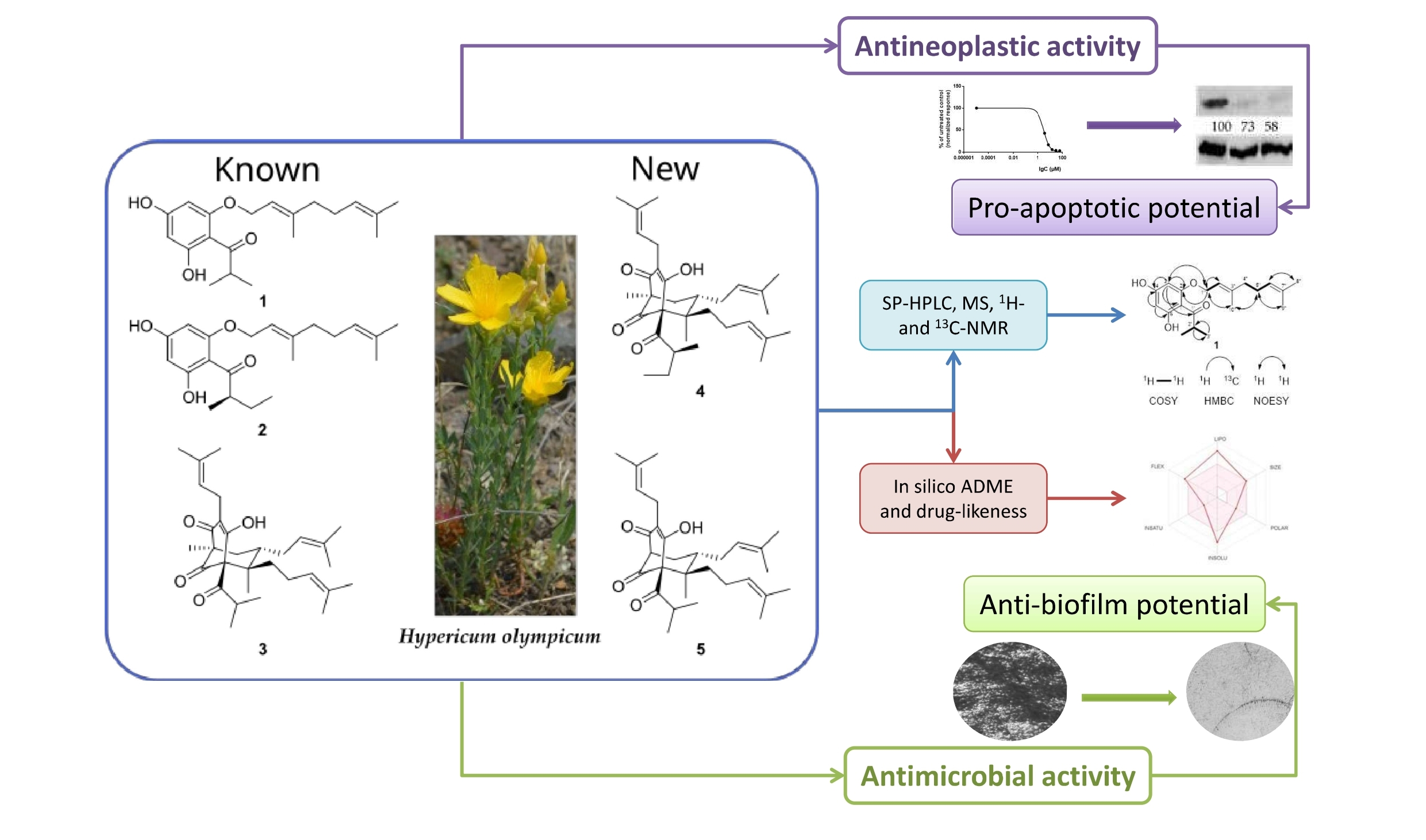
2. Results
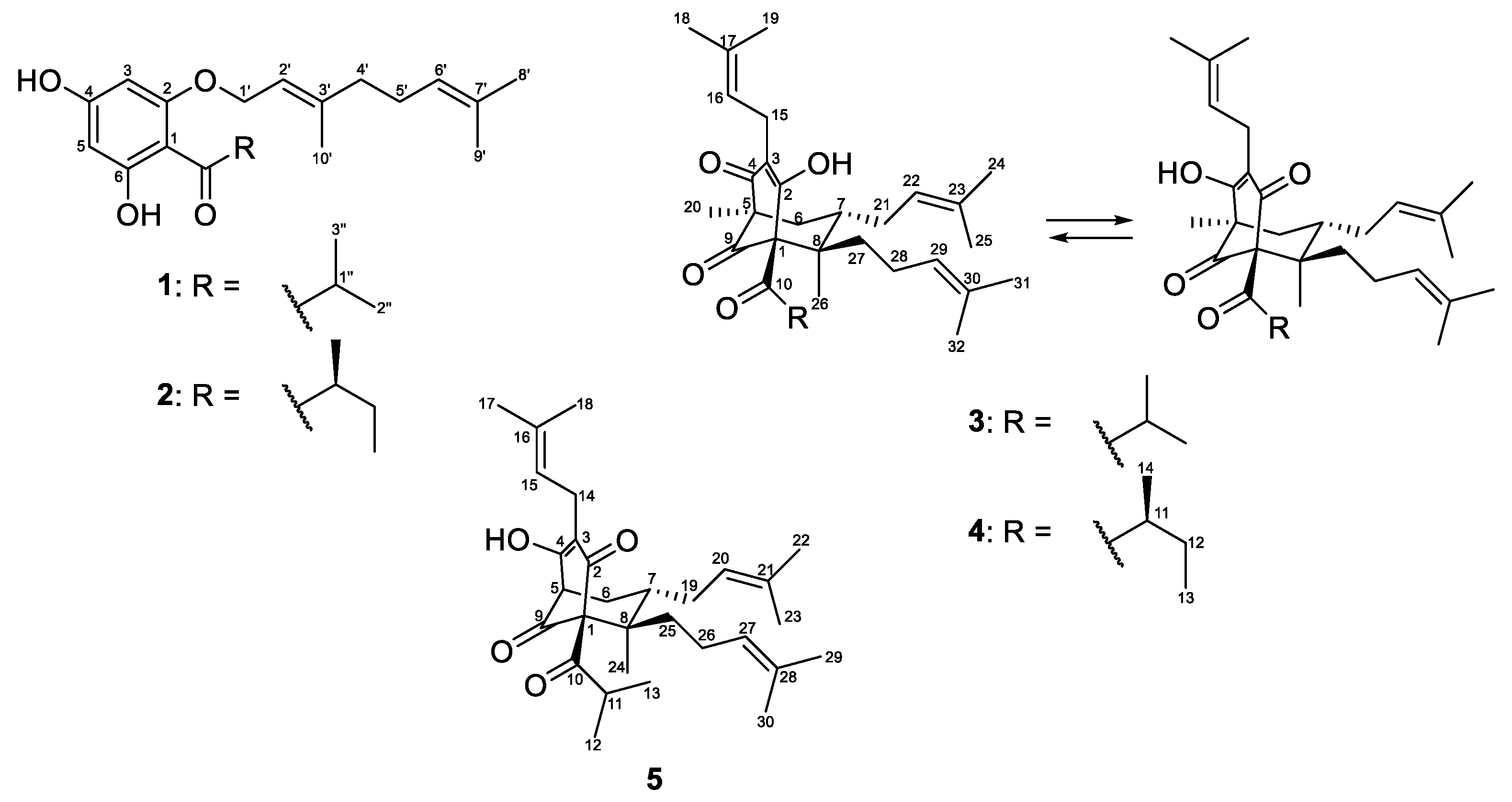
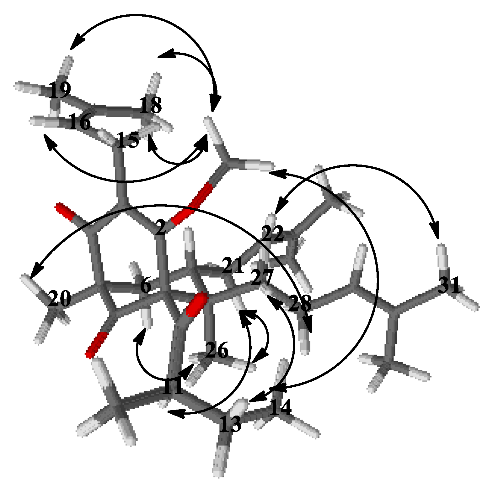
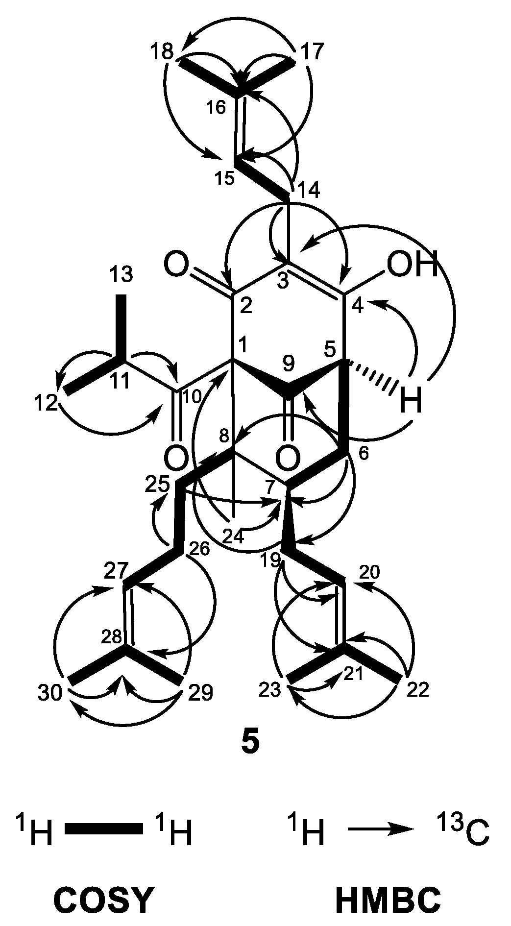
2.1. Identification of the Isolated Compounds
2.2. Cytotoxic Activity of the Isolated Compounds
2.3. Antiproliferative Activity of the Isolated Compounds
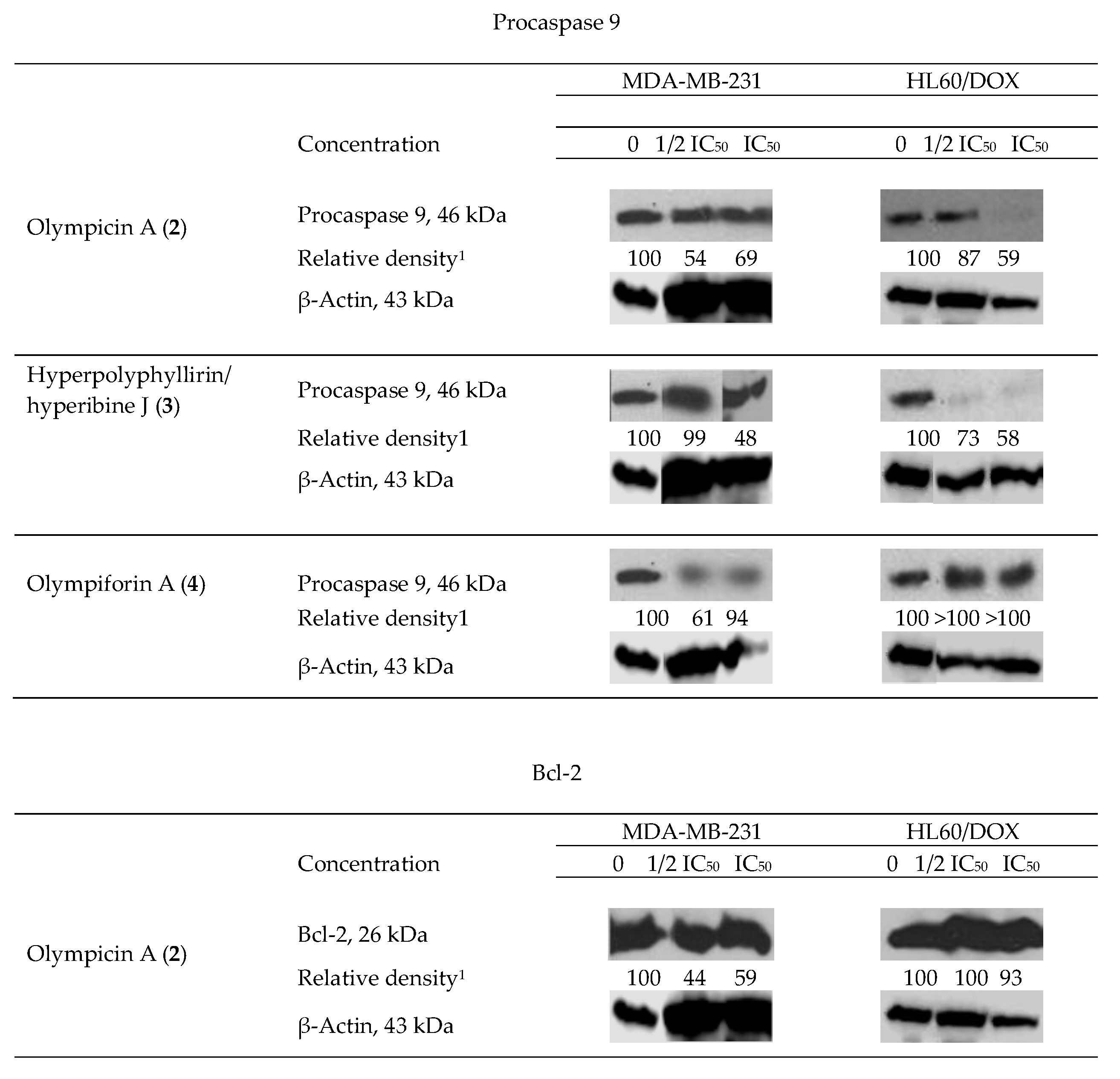
2.4. Influence of the Isolated Compounds on the Apoptosis-Related Proteins Procaspase 9 and Bcl-2
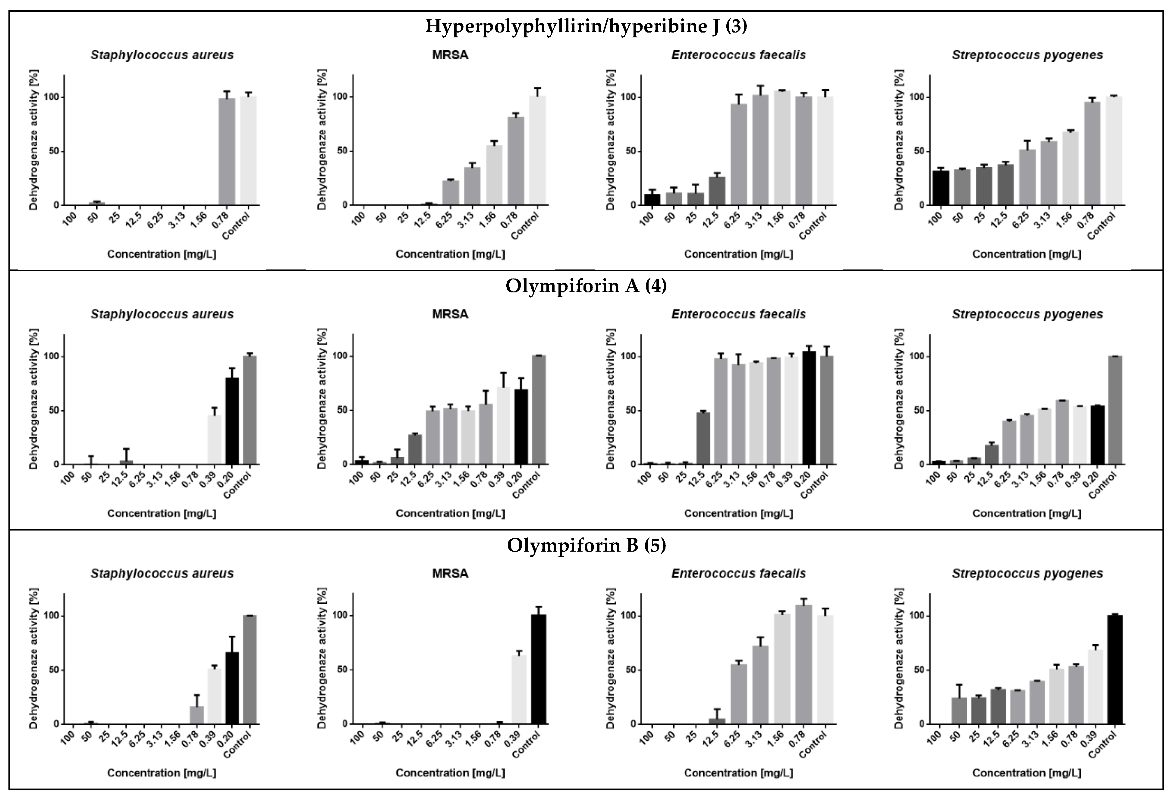
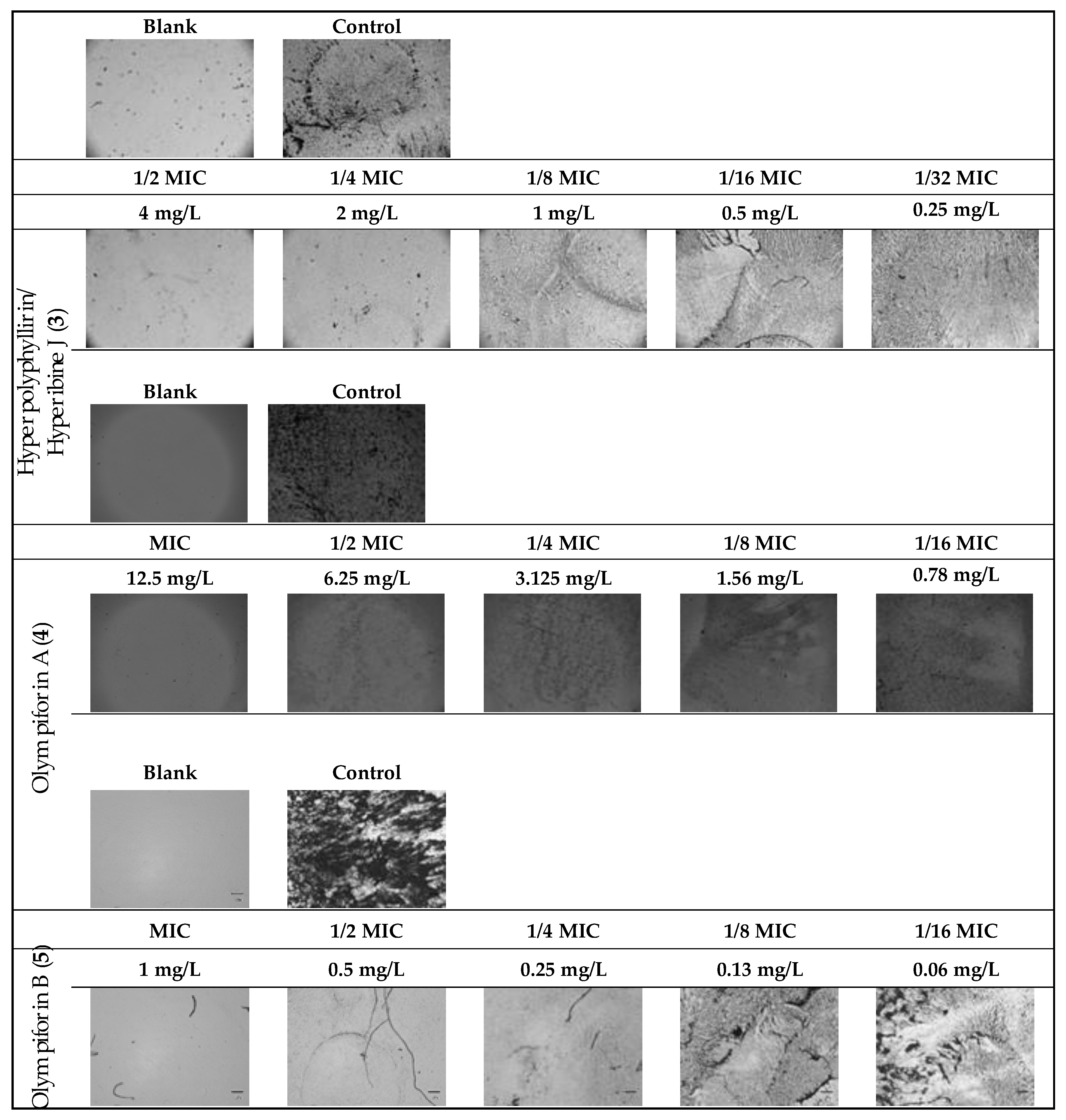
2.5. Antibacterial Activity
2.6. In Silico ADME and Drug-Likeness Evaluation
3. Discussion
4. Materials and Methods
4.1. General Experimental Procedures
4.2. Plant Material
4.3. Extraction and Isolation
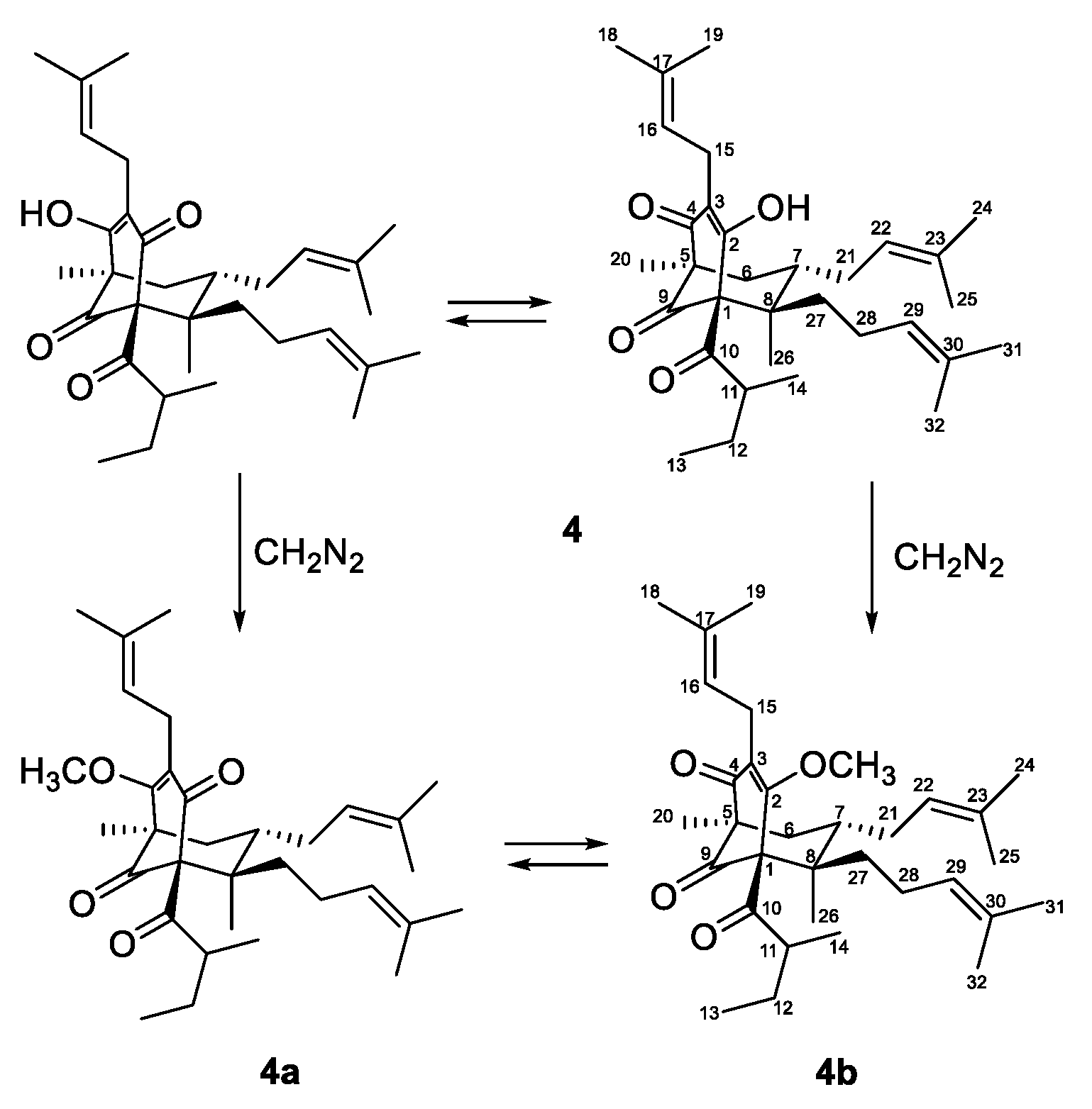
4.4. Methylation of Compound 4
4.5. New Compound Data
4.5.1. (E)-1-[2-{(3,7-dimethylocta-2,6-dien-1-yl)oxy}-4,6-dihydroxyphenyl]-2-methylpropan-1-one (1)
4.5.2. Olympiforin A or Mixtures of Keto-Enol Tautomers (1R,5R,7S,8R)-4-hydroxy-5,8-dimethyl-3,7-bis(3-methylbut-2-en-1-yl)-1-(2-methylbutanoyl)-8-(4-methylpent-3-en-1-yl)bicyclo[3.3.1]non-3-ene-2,9-dione and (1R,5S,6R,7S)-4-hydroxy-1,6-dimethyl-3,7-bis(3-methylbut-2-en-1-yl)-5-(2-methylbutanoyl)-6-(4-methylpent-3-en-1-yl)bicyclo[3.3.1]non-3-ene-2,9-dione (4)
4.5.3. (1R,5R,7S,8R)-4-methoxy-5,8-dimethyl-3,7-bis(3-methylbut-2-en-1-yl)-1-(2-methylbutanoyl)-8-(4-methylpent-3-en-1-yl)bicyclo[3.3.1]non-3-ene-2,9-dione (4a)
4.5.4. (1R,5S,6R,7S)-4-methoxy-1,6-dimethyl-3,7-bis(3-methylbut-2-en-1-yl)-5-(2-methylbutanoyl)-6-(4-methylpent-3-en-1-yl)bicyclo[3.3.1]non-3-ene-2,9-dione (4b)
4.5.5. Olympiforin B or (1R,5S,6R,7S)-4-hydroxy-5-isobutyryl-6-methyl-3,7-bis(3-methylbut-2-en-1-yl)-6-(4-methylpent-3-en-1-yl)bicyclo[3.3.1]non-3-ene-2,9-dione (5)
4.6. Cell Lines and Culture Conditions
4.7. In Vitro Cytotoxicity Determination
4.8. Antiproliferative Test
4.9. Western Blot
4.10. Bacterial Strains and Growth Conditions
4.11. MIC Determination by Broth Microdilution Method (BMD)
4.12. Assessment of the Cell Redox (Dehydrogenase) Activity
4.13. Biofilm Assay of MRSA
4.14. Physiochemical Properties and General Computational Methodology, ADME and Drug-Likeness Estimation
4.15. Statistics
5. Conclusions
Supplementary Materials
Author Contributions
Funding
Data Availability Statement
Acknowledgments
Conflicts of Interest
References
- Crockett, S.; Robson, N. Taxonomy and Chemotaxonomy of the Genus Hypericum. Med. Aromat. Plant Sci. Biotechnol. 2011, 5, 1–13. [Google Scholar] [CrossRef]
- Zhao, J.; Liu, W.; Wang, J.C. Recent Advances Regarding Constituents and Bioactivities of Plants from the Genus Hypericum. Chem. Biodivers. 2015, 12, 309–349. [Google Scholar] [CrossRef]
- Li, D.; Zhu, H.; Qi, C.; Xue, Y.; Yao, G.; Luo, Z.; Wang, J.; Zhang, J.; Du, G.; Zhang, Y. Two New Adamantyl-like Polyprenylated Acylphloroglucinols from Hypericum attenuatum Choisy. Tetrahedron Lett. 2015, 56, 1953–1955. [Google Scholar] [CrossRef]
- Yang, X.-W.; Grossman, R.B.; Xu, G. Research Progress of Polycyclic Polyprenylated Acylphloroglucinols. Chem. Rev. 2018, 118, 3508–3558. [Google Scholar] [CrossRef] [PubMed]
- Gurevich, A.I.; Dobrynin, V.N.; Kolosov, M.N.; Popravko, S.A.; Ryabova, I.D.; Chernov, B.K.; Derbents, N.A.; Aizenman, B.E.; Garaguly, A.D. Hyperforin, an Antibiotic from Hypericum perforatum L. Antibiot. Khimioterapiya 1971, 16, 510. [Google Scholar]
- Schempp, C.M.; Pelz, K.; Wittmer, A.; Schöpf, E.; Simon, J.C. Antibacterial Activity of Hyperforin from St John’s Wort, against Multiresistant staphylococcus Aureus and Gram-Positive Bacteria. Lancet 1999, 353, 2129. [Google Scholar] [CrossRef]
- Schiavone, B.I.P.; Rosato, A.; Marilena, M.; Gibbons, S.; Bombardelli, E.; Verotta, L.; Franchini, C.; Corbo, F. Biological Evaluation of Hyperforin and Its Hydrogenated Analogue on Bacterial Growth and Biofilm Production. J. Nat. Prod. 2013, 76, 1819–1823. [Google Scholar] [CrossRef]
- Billard, C.; Merhi, F.; Bauvois, B. Mechanistic Insights into the Antileukemic Activity of Hyperforin. Curr. Cancer Drug Targets 2013, 13, 1–10. [Google Scholar] [CrossRef] [PubMed]
- Quiney, C.; Billard, C.; Salanoubat, C.; Fourneron, J.D.; Kolb, J.P. Hyperforin, a New Lead Compound against the Progression of Cancer and Leukemia? Leukemia 2006, 20, 1519–1525. [Google Scholar] [CrossRef] [PubMed]
- Zaher, M.; Tang, R.; Bombarda, I.; Merhi, F.; Bauvois, B.; Billard, C. Hyperforin Induces Apoptosis of Chronic Lymphocytic Leukemia Cells through Upregulation of the BH3-Only Protein Noxa. Int. J. Oncol. 2012, 40, 269–276. [Google Scholar] [CrossRef] [PubMed]
- Zhu, H.-C.; Chen, C.-M.; Zhang, J.-W.; Guo, Y.; Tan, D.-D.; Wei, G.-Z.; Yang, J.; Wang, J.-P.; Luo, Z.-W.; Xue, Y.-B.; et al. Hyperisampsins N and O, Two New Benzoylated Phloroglucinol Derivatives from Hypericum Sampsonii. Chin. Chem. Lett. 2017, 28, 986–990. [Google Scholar] [CrossRef]
- Rui, D.-Y.; Chen, X.-Q.; Li, Z.; Tang, L.-Y.; Li, F. Chemical Constituents of Hypericum Petiolulatum. Chem. Nat. Compd. 2017, 53, 457–462. [Google Scholar] [CrossRef]
- Zhang, H.-B.; Zhang, X.; Jiang, K.; Qu, S.-J.; Meng, L.-H.; Lu, Q.; Tan, C.-H. Polycyclic Polyprenylated Acylphloroglucinols from Hypericum choisianum. Nat. Prod. Res. 2021, 35, 195–202. [Google Scholar] [CrossRef] [PubMed]
- Guo, Y.; Tong, Q.; Zhang, N.; Duan, X.; Cao, Y.; Zhu, H.; Xie, S.; Yang, J.; Zhang, J.; Liu, Y.; et al. Highly Functionalized Cyclohexanone-Monocyclic Polyprenylated Acylphloroglucinols from Hypericum perforatum Induce Leukemia Cell Apoptosis. Org. Chem. Front. 2019, 6, 817–824. [Google Scholar] [CrossRef]
- Qiu, D.-R.; Zhou, M.; Liu, X.-Z.; Chen, J.-J.; Wang, G.-H.; Lin, T.; Yu, F.-R.; Ding, R.; Sun, C.-L.; Tian, W.-J.; et al. Cytotoxic Polyprenylated Phloroglucinol Derivatives from Hypericum Elodeoides Choisy Modulating the Transactivation of RXRα. Bioorg. Chem. 2021, 107, 104578. [Google Scholar] [CrossRef]
- Zhang, X.-W.; Fan, S.-Q.; Xia, F.; Ye, Y.-S.; Yang, X.-W.; Yang, X.-W.; Xu, G. Prenylated Acylphloroglucinols from Hypericum faberi. J. Nat. Prod. 2019, 82, 1367–1371. [Google Scholar] [CrossRef]
- Zhang, N.; Shi, Z.; Guo, Y.; Xie, S.; Qiao, Y.; Li, X.-N.; Xue, Y.; Luo, Z.; Zhu, H.; Chen, C.; et al. The Absolute Configurations of Hyperilongenols A–C: Rare 12,13-Seco-Spirocyclic Polycyclic Polyprenylated Acylphloroglucinols with Enolizable β,Β′-Tricarbonyl Systems from Hypericum longistylum Oliv. Org. Chem. Front. 2019, 6, 1491–1502. [Google Scholar] [CrossRef]
- Li, Y.-P.; Hu, K.; Yang, X.-W.; Xu, G. Antibacterial Dimeric Acylphloroglucinols from Hypericum japonicum. J. Nat. Prod. 2018, 81, 1098–1102. [Google Scholar] [CrossRef]
- Niwa, K.; Tanaka, N.; Tatano, Y.; Yagi, H.; Kashiwada, Y. Hypascyrins A–E, Prenylated Acylphloroglucinols from Hypericum Ascyron. J. Nat. Prod. 2019, 82, 2754–2760. [Google Scholar] [CrossRef]
- Li, Q.-J.; Tang, P.-F.; Zhou, X.; Lu, W.-J.; Xu, W.-J.; Luo, J.; Kong, L.-Y. Dimethylated Acylphloroglucinol Meroterpenoids with Anti-Oral-Bacterial and Anti-Inflammatory Activities from Hypericum Elodeoides. Bioorg. Chem. 2020, 104, 104275. [Google Scholar] [CrossRef]
- Bridi, H.; Pustay, A.P.; Bordignon, S.A.D.L.; Picoli, S.U.; von Poser, G.L.; Ferraz, A.B.D.F. Antimicrobial Activity of Dimeric Acylphloroglucinols Isolated from Southern Brazilian Hypericum Species against to Resistant Bacterial. Nat. Prod. Res. 2022, 36, 6448–6452. [Google Scholar] [CrossRef]
- Song, P.; Hao, J.; Wang, Y.; Yang, X.-Z. Polycyclic polyprenylated acylphloroglucinols from Hypericum species and their biological activities. Zhongguo Zhong Yao Za Zhi 2021, 46, 4881–4890. [Google Scholar] [CrossRef] [PubMed]
- Peron, G.; López, A.M.; Cabada-Aquirre, P.; Garay Buenrosto, K.D.; Ostos Mendoza, K.C.; Mahady, G.B.; Seidel, V.; Sytar, O.; Koirala, N.; Gurung, R.; et al. Antiviral and Antibacterial Properties of Phloroglucinols: A Review on Naturally Occurring and (Semi)Synthetic Derivatives with Potential Therapeutic Interest. Crit. Rev. Biotechnol. 2023; online ahead of print. [Google Scholar] [CrossRef]
- Bridi, H.; de Meirelles, G.C.; von Poser, G.L. Structural Diversity and Biological Activities of Phloroglucinol Derivatives from Hypericum Species. Phytochemistry 2018, 155, 203–232. [Google Scholar] [CrossRef] [PubMed]
- Marrelli, M.; Statti, G.; Conforti, F. Hypericum Spp.: An Update on the Biological Activities and Metabolic Profiles. Mini-Rev. Med. Chem. 2020, 20, 66–87. [Google Scholar] [CrossRef] [PubMed]
- Ciochina, R.; Grossman, R.B. Polycyclic Polyprenylated Acylphloroglucinols. Chem. Rev. 2006, 106, 3963–3986. [Google Scholar] [CrossRef]
- Xiao, C.-Y.; Mu, Q.; Gibbons, S. The Phytochemistry and Pharmacology of Hypericum. In Progress in the Chemistry of Organic Natural Products; Springer: New York, NY, USA, 2020; pp. 85–182. [Google Scholar]
- WHO Cancer. Available online: https://www.who.int/health-topics/cancer#tab=tab_1 (accessed on 2 June 2021).
- Gayatri, K.V.; Soundhari, C.; Pavithra, B.P. Biofilm Inhibitory Effect of Chlorella Extracts on Pseudomonas Aeruginosa. Int. J. Pharm. Sci. Res. 2019, 10, 1966–1971. [Google Scholar] [CrossRef]
- Piechota, M.; Kot, B.; Frankowska-Maciejewska, A.; Grużewska, A.; Woźniak-Kosek, A. Biofilm Formation by Methicillin-Resistant and Methicillin-Sensitive Staphylococcus aureus Strains from Hospitalized Patients in Poland. BioMed Res. Int. 2018, 2018, 4657396. [Google Scholar] [CrossRef] [PubMed]
- Sarkisian, S.A.; Janssen, M.J.; Matta, H.; Henry, G.E.; LaPlante, K.L.; Rowley, D.C. Inhibition of Bacterial Growth and Biofilm Production by Constituents from Hypericum Spp. Phytother. Res. 2012, 26, 1012–1016. [Google Scholar] [CrossRef]
- Schiavone, B.; Verotta, L.; Rosato, A.; Marilena, M.; Gibbons, S.; Bombardelli, E.; Franchini, C.; Corbo, F. Anticancer and Antibacterial Activity of Hyperforin and Its Derivatives. Anti-Cancer Agents Med. Chem. 2014, 14, 1397–1401. [Google Scholar] [CrossRef]
- Yan, X.-T.; An, Z.; Huangfu, Y.; Zhang, Y.-T.; Li, C.-H.; Chen, X.; Liu, P.-L.; Gao, J.-M. Polycyclic Polyprenylated Acylphloroglucinol and Phenolic Metabolites from the Aerial Parts of Hypericum Elatoides and Their Neuroprotective and Anti-Neuroinflammatory Activities. Phytochemistry 2019, 159, 65–74. [Google Scholar] [CrossRef]
- Hu, L.; Liu, Y.; Wang, Y.; Wang, Z.; Huang, J.; Xue, Y.; Liu, J.; Liu, Z.; Chen, Y.; Zhang, Y. Discovery of Acylphloroglucinol-Based Meroterpenoid Enantiomers as KSHV Inhibitors from Hypericum japonicum. RSC Adv. 2018, 8, 24101–24109. [Google Scholar] [CrossRef] [PubMed]
- Centurião, F.B.; Sakamoto, S.; Stein, A.C.; Müller, L.G.; Chagas, P.; von Poser, G.; Nogueira, C.W.; Rates, S.M.K. The Antidepressant-like Effect of Hyperbrasilol B, A Natural Dimeric Phloroglucinol Derivative Is Prevented by Veratrine, a Sensitive-Voltage Na+ Channel Opener. Eur. J. Med. Plants 2014, 4, 1268–1281. [Google Scholar] [CrossRef]
- Tuzlacı, E.; Aymaz, P.E. Turkish Folk Medicinal Plants, Part IV: Gönen (Balıkesir). Fitoterapia 2001, 72, 323–343. [Google Scholar] [CrossRef] [PubMed]
- Osborn, E.M. On the Occurrence of Antibacterial Substances in Green Plants. Br. J. Exp. Pathol. 1943, 24, 227–231. [Google Scholar]
- Smelcerovic, A.; Spiteller, M. Phytochemical Analysis of Nine Hypericum L. Species from Serbia and the F.Y.R. Macedonia. Die Pharm.—Int. J. Pharm. Sci. 2006, 61, 251–252. [Google Scholar]
- Shiu, W.K.P.; Rahman, M.M.; Curry, J.; Stapleton, P.; Zloh, M.; Malkinson, J.P.; Gibbons, S. Antibacterial Acylphloroglucinols from Hypericum Olympicum. J. Nat. Prod. 2012, 75, 336–343. [Google Scholar] [CrossRef]
- Nedialkov, P.T.; Momekov, G.; Kokanova-Nedialkova, Z.K.; Heilmann, J. Polyprenylated Phloroglucinols from Hypericum maculatum. Nat. Prod. Commun. 2015, 10, 1934578X1501000. [Google Scholar] [CrossRef]
- Nedialkov, P.T.; Zheleva-Dimitrova, D.; Momekov, G.; Karlov, K.; Girreser, U.; Kitanov, G.M. Elegaphenone and 7-Epi-Clusianone, the Major Cytotoxic Constituents of Hypericum elegans. Nat. Prod. Res. 2011, 25, 1743–1750. [Google Scholar] [CrossRef] [PubMed]
- Henry, G.E.; Campbell, M.S.; Zelinsky, A.A.; Liu, Y.; Bowen-Forbes, C.S.; Li, L.; Nair, M.G.; Rowley, D.C.; Seeram, N.P. Bioactive Acylphloroglucinols from Hypericum densiflorum. Phytother. Res. 2009, 23, 1759–1762, Erratum in Phytother. Res. 2010, 24, 1264. [Google Scholar] [CrossRef] [PubMed]
- Nedialkov, P.T.; Ilieva, Y.; Momekov, G.; Kokanova-Nedialkova, Z. Cytotoxic Prenylated Acylphloroglucinols from Hypericum annulatum. Fitoterapia 2018, 127, 375–382. [Google Scholar] [CrossRef]
- Rios, M.Y.; Delgado, G. Polyprenols and Acylphloroglucinols from Esenbeckia Nesiotica. Phytochemistry 1992, 31, 3491–3494. [Google Scholar] [CrossRef]
- Bohlmann, F.; Zdero, C. Neue Phloroglucin-Derivate Aus Helichrysum Natalitium Und Helichrysum Bellum. Phytochemistry 1979, 18, 641–644. [Google Scholar] [CrossRef]
- Bittrich, V.; do Amaral, M.C.E.; Machado, S.M.F.; Marsaioli, A.J. Floral Resin of Tovomitopsis Saldanhae (Guttiferae) and 7-Epi-Nemorosone: Structural Revision. Z. Nat. C 2003, 58, 643–648. [Google Scholar] [CrossRef] [PubMed]
- Verotta, L.; Appendino, G.; Jakupovic, J.; Bombardelli, E. Hyperforin Analogues from St. John’s Wort (Hypericum perforatum). J. Nat. Prod. 2000, 63, 412–415. [Google Scholar] [CrossRef] [PubMed]
- Tatsis, E.C.; Boeren, S.; Exarchou, V.; Troganis, A.N.; Vervoort, J.; Gerothanassis, I.P. Identification of the Major Constituents of Hypericum Perforatum by LC/SPE/NMR and/or LC/MS. Phytochemistry 2007, 68, 383–393. [Google Scholar] [CrossRef]
- Charchoglyan, A.; Abrahamyan, A.; Fujii, I.; Boubakir, Z.; Gulder, T.A.M.; Kutchan, T.M.; Vardapetyan, H.; Bringmann, G.; Ebizuka, Y.; Beerhues, L. Differential Accumulation of Hyperforin and Secohyperforin in Hypericum perforatum Tissue Cultures. Phytochemistry 2007, 68, 2670–2677. [Google Scholar] [CrossRef]
- Decosterd, L.A.; Stoeckli-Evans, H.; Chapuis, J.-C.; Msonthi, J.D.; Sordat, B.; Hostettmann, K. New Hyperforin Derivatives from Hypericum Revolutum VAHL with Growth-Inhibitory Activity against a Human Colon Carcinoma Cell Line. Helv. Chim. Acta 1989, 72, 464–471. [Google Scholar] [CrossRef]
- Lu, Z.J.; Ren, Y.Q.; Wang, G.P.; Song, Q.; Li, M.; Jiang, S.S.; Ning, T.; Guan, Y.S.; Yang, J.L.; Luo, F. Biological Behaviors and Proteomics Analysis of Hybrid Cell Line EAhy926 and Its Parent Cell Line A549. J. Exp. Clin. Cancer Res. 2009, 28, 16. [Google Scholar] [CrossRef]
- Guo, Y.; Zhang, N.; Chen, C.; Huang, J.; Li, X.-N.; Liu, J.; Zhu, H.; Tong, Q.; Zhang, J.; Luo, Z.; et al. Tricyclic Polyprenylated Acylphloroglucinols from St John’s Wort, Hypericum perforatum. J. Nat. Prod. 2017, 80, 1493–1504. [Google Scholar] [CrossRef] [PubMed]
- Zhu, H.; Chen, C.; Tong, Q.; Chen, X.; Yang, J.; Liu, J.; Sun, B.; Wang, J.; Yao, G.; Luo, Z.; et al. Hyperisampsins H–M, Cytotoxic Polycyclic Polyprenylated Acylphloroglucinols from Hypericum sampsonii. Sci. Rep. 2015, 5, 14772. [Google Scholar] [CrossRef]
- Zhu, H.; Chen, C.; Yang, J.; Li, D.; Zhang, J.; Guo, Y.; Wang, J.; Luo, Z.; Xue, Y.; Zhang, Y. Hyperhexanone A, a Crucial Intermediate from Bicyclo [3.3.1]- to Cyclohexanone Monocyclic-Polycyclic Polyprenylated Acylphloroglucinols. Tetrahedron 2016, 72, 4655–4659. [Google Scholar] [CrossRef]
- Shiu, W.K.P.; Malkinson, J.P.; Rahman, M.M.; Curry, J.; Stapleton, P.; Gunaratnam, M.; Neidle, S.; Mushtaq, S.; Warner, M.; Livermore, D.M.; et al. A New Plant-Derived Antibacterial Is an Inhibitor of Efflux Pumps in Staphylococcus Aureus. Int. J. Antimicrob. Agents 2013, 42, 513–518. [Google Scholar] [CrossRef] [PubMed]
- Ilieva, Y.; Marinov, T.; Trayanov, I.; Kaleva, M.; Zaharieva, M.M.; Yocheva, L.; Kokanova-Nedialkova, Z.; Najdenski, H.; Nedialkov, P. Outstanding Antibacterial Activity of Hypericum Rochelii—Comparison of the Antimicrobial Effects of Extracts and Fractions from Four Hypericum Species Growing in Bulgaria with a Focus on Prenylated Phloroglucinols. Life 2023, 13, 274. [Google Scholar] [CrossRef]
- Ilieva, Y.; Kokanova-Nedialkova, Z.; Nedialkov, P.; Momekov, G. In Silico ADME and Drug-Likeness Evaluation of a Series of Cytotoxic Polyprenylated Acylphloroglucinols, Isolated from Hypericum annulatum Morris Subsp. Annulatum. Bulg. Chem. Commun. 2018, 50, 193–199. [Google Scholar]
- Wang, E.; Barecki-Roach, M.; Johnson, W.W. Quantitative Characterization of Direct P-Glycoprotein Inhibition by St John’s Wort Constituents Hypericin and Hyperforin. J. Pharm. Pharmacol. 2004, 56, 123–128. [Google Scholar] [CrossRef] [PubMed]
- Weber, C.C.; Kressmann, S.; Fricker, G.; Müller, W.E. Modulation of P-Glycoprotein Function by St John’s Wort Extract and Its Major Constituents. Pharmacopsychiatry 2004, 37, 292–298. [Google Scholar] [CrossRef] [PubMed]
- Quiney, C.; Billard, C.; Faussat, A.-M.; Salanoubat, C.; Kolb, J.-P. Hyperforin Inhibits P-Gp and BCRP Activities in Chronic Lymphocytic Leukaemia Cells and Myeloid Cells. Leuk. Lymphoma 2007, 48, 1587–1599. [Google Scholar] [CrossRef]
- Šemeláková, M.; Jendželovský, R.; Fedoročko, P. Drug Membrane Transporters and CYP3A4 Are Affected by Hypericin, Hyperforin or Aristoforin in Colon Adenocarcinoma Cells. Biomed. Pharmacother. 2016, 81, 38–47. [Google Scholar] [CrossRef]
- Winkelmann, K.; Heilmann, J.; Zerbe, O.; Rali, T.; Sticher, O. New Prenylated Bi- and Tricyclic Phloroglucinol Derivatives from Hypericum papuanum. J. Nat. Prod. 2001, 64, 701–706. [Google Scholar] [CrossRef]
- Winkelmann, K.; Heilmann, J.; Zerbe, O.; Rali, T.; Sticher, O. Further Prenylated Bi- and Tricyclic Phloroglucinol Derivatives from Hypericum papuanum. Helv. Chim. Acta 2001, 84, 3380–3392. [Google Scholar] [CrossRef]
- Fobofou, S.A.T.; Harmon, C.R.; Lonfouo, A.H.N.; Franke, K.; Wright, S.M.; Wessjohann, L.A. Prenylated Phenyl Polyketides and Acylphloroglucinols from Hypericum peplidifolium. Phytochemistry 2016, 124, 108–113. [Google Scholar] [CrossRef] [PubMed]
- Gao, W.; Hou, W.-Z.; Zhao, J.; Xu, F.; Li, L.; Xu, F.; Sun, H.; Xing, J.-G.; Peng, Y.; Wang, X.-L.; et al. Polycyclic Polyprenylated Acylphloroglucinol Congeners from Hypericum scabrum. J. Nat. Prod. 2016, 79, 1538–1547. [Google Scholar] [CrossRef] [PubMed]
- Merhi, F.; Tang, R.; Piedfer, M.; Mathieu, J.; Bombarda, I.; Zaher, M.; Kolb, J.-P.; Billard, C.; Bauvois, B. Hyperforin Inhibits Akt1 Kinase Activity and Promotes Caspase-Mediated Apoptosis Involving Bad and Noxa Activation in Human Myeloid Tumor Cells. PLoS ONE 2011, 6, e25963. [Google Scholar] [CrossRef]
- Porzel, A.; Farag, M.A.; Mülbradt, J.; Wessjohann, L.A. Metabolite Profiling and Fingerprinting of Hypericum Species: A Comparison of MS and NMR Metabolomics. Metabolomics 2014, 10, 574–588. [Google Scholar] [CrossRef]
- Zhou, Z.-B.; Mou, P.-Y.; Huang, Y.-Y.; Zeng, H.; Huang, Z.-L.; Wei, X. Bioactive Polycyclic Polyprenylated Acylphloroglucinols from Hypericum scabrum. Fitoterapia 2022, 161, 105249. [Google Scholar] [CrossRef] [PubMed]
- Biljali, S.; Momekov, G.; Nedialkov, P.; Zheleva-Dimitrova, D.; Kitanov, G.; Momekova, D.; Stoyanov, N.; Guenova, M.; Michova, A.; Karaivanova, M. In Vitro Investigation of the Antiproliferative and Proapoptotic Effects of Hyperatomarin—A Bicyclic Prenylated Acylphloroglucinol from Hypericum Annulatum Moris Subsp. Annulatum against Human Tumor and Endothelial Cells. J. Pharm. Technol. Drug Res. 2012, 1, 6. [Google Scholar] [CrossRef]
- Martínez-Poveda, B.; Quesada, A.R.; Medina, M.Á. Hyperforin, a Bio-Active Compound of St. John’s Wort, Is a New Inhibitor of Angiogenesis Targeting Several Key Steps of the Process. Int. J. Cancer 2005, 117, 775–780. [Google Scholar] [CrossRef]
- Liu, J.-Y.; Liu, Z.; Wang, D.-M.; Li, M.-M.; Wang, S.-X.; Wang, R.; Chen, J.-P.; Wang, Y.-F.; Yang, D.-P. Induction of Apoptosis in K562 Cells by Dicyclohexylammonium Salt of Hyperforin through a Mitochondrial-Related Pathway. Chem.-Biol. Interact. 2011, 190, 91–101. [Google Scholar] [CrossRef] [PubMed]
- Lorusso, G.; Vannini, N.; Sogno, I.; Generoso, L.; Garbisa, S.; Noonan, D.M.; Albini, A. Mechanisms of Hyperforin as an Anti-Angiogenic Angioprevention Agent. Eur. J. Cancer 2009, 45, 1474–1484. [Google Scholar] [CrossRef]
- Chen, X.-Q.; Li, Y.; Li, K.-Z.; Peng, L.-Y.; He, J.; Wang, K.; Pan, Z.-H.; Cheng, X.; Li, M.-M.; Zhao, Q.-S.; et al. Spirocyclic Acylphloroglucinol Derivatives from Hypericum beanii. Chem. Pharm. Bull. 2011, 59, 1250–1253. [Google Scholar] [CrossRef]
- Tanaka, N.; Kashiwada, Y.; Kim, S.-Y.; Sekiya, M.; Ikeshiro, Y.; Takaishi, Y. Xanthones from Hypericum Chinense and Their Cytotoxicity Evaluation. Phytochemistry 2009, 70, 1456–1461. [Google Scholar] [CrossRef] [PubMed]
- Chen, X.-Q.; Li, Y.; Cheng, X.; Wang, K.; He, J.; Pan, Z.-H.; Li, M.-M.; Peng, L.-Y.; Xu, G.; Zhao, Q.-S. Polycyclic Polyprenylated Acylphloroglucinols and Chromone O-Glucosides from Hypericum Henryi Subsp. Uraloides. Chem. Biodivers. 2010, 7, 196–204. [Google Scholar] [CrossRef] [PubMed]
- Hashida, W.; Tanaka, N.; Kashiwada, Y.; Sekiya, M.; Ikeshiro, Y.; Takaishi, Y. Tomoeones A–H, Cytotoxic Phloroglucinol Derivatives from Hypericum Ascyron. Phytochemistry 2008, 69, 2225–2230. [Google Scholar] [CrossRef]
- Tanaka, N.; Kashiwada, Y.; Kim, S.Y.; Hashida, W.; Sekiya, M.; Ikeshiro, Y.; Takaishi, Y. Acylphloroglucinol, Biyouyanagiol, Biyouyanagin B, and Related Spiro-Lactones from Hypericum chinense. J. Nat. Prod. 2009, 72, 1447–1452. [Google Scholar] [CrossRef]
- Biljali, S.; Nedialkov, P.; Zheleva-Dimitrova, D.; Kitanov, G.; Momekova, D.; Momekov, G. Cytotoxic Effects and Multidrug Resistance Modulation by Five Benzophenones and a Xanthone Isolated from Hypericum annulatum Moris Subsp. Annulatum. Biotechnol. Biotechnol. Equip. 2013, 27, 3561–3568. [Google Scholar] [CrossRef]
- Yang, X.-W.; Li, M.-M.; Liu, X.; Ferreira, D.; Ding, Y.; Zhang, J.-J.; Liao, Y.; Qin, H.-B.; Xu, G. Polycyclic Polyprenylated Acylphloroglucinol Congeners Possessing Diverse Structures from Hypericum henryi. J. Nat. Prod. 2015, 78, 885–895. [Google Scholar] [CrossRef]
- Liu, X.; Yang, X.-W.; Chen, C.-Q.; Wu, C.-Y.; Zhang, J.-J.; Ma, J.-Z.; Wang, H.; Zhao, Q.-S.; Yang, L.-X.; Xu, G. Hypercohones A–C, Acylphloroglucinol Derivatives with Homo-Adamantane Cores from Hypericum cohaerens. Nat. Prod. Bioprospect. 2013, 3, 233–237. [Google Scholar] [CrossRef]
- Ye, Y.; Yang, X.-W.; Xu, G. Unusual Adamantane Type Polyprenylated Acylphloroglucinols with an Oxirane Unit and Their Structural Transformation from Hypericum Hookerianum. Tetrahedron 2016, 72, 3057–3062. [Google Scholar] [CrossRef]
- Zhang, J.-J.; Yang, J.; Liao, Y.; Yang, X.-W.; Ma, J.-Z.; Xiao, Q.-L.; Yang, L.-X.; Xu, G. Hyperuralones A and B, New Acylphloroglucinol Derivatives with Intricately Caged Cores from Hypericum uralum. Org. Lett. 2014, 16, 4912–4915. [Google Scholar] [CrossRef]
- Zhu, H.; Chen, C.; Yang, J.; Li, X.-N.; Liu, J.; Sun, B.; Huang, S.-X.; Li, D.; Yao, G.; Luo, Z.; et al. Bioactive Acylphloroglucinols with Adamantyl Skeleton from Hypericum sampsonii. Org. Lett. 2014, 16, 6322–6325. [Google Scholar] [CrossRef]
- Yang, X.W.; Yang, J.; Liao, Y.; Ye, Y.; Li, Y.P.; Yang, S.Y.; Xia, F.; Xu, G. Hypercohin K, a Polycyclic Polyprenylated Acylphloroglucinol with an Unusual Spiro-Fused Cyclopropane Ring from Hypericum cohaerens. Tetrahedron Lett. 2015, 56, 5537–5540. [Google Scholar] [CrossRef]
- Yang, X.-W.; Deng, X.; Liu, X.; Wu, C.-Y.; Li, X.-N.; Wu, B.; Luo, H.-R.; Li, Y.; Xu, H.-X.; Zhao, Q.-S.; et al. Hypercohin A, a New Polycyclic Polyprenylated Acylphloroglucinol Possessing an Unusual Bicyclo[5.3.1]Hendecane Core from Hypericum cohaerens. Chem. Commun. 2012, 48, 5998–6000. [Google Scholar] [CrossRef] [PubMed]
- Liu, X.; Yang, X.-W.; Chen, C.-Q.; Wu, C.-Y.; Zhang, J.-J.; Ma, J.-Z.; Wang, H.; Yang, L.-X.; Xu, G. Bioactive Polyprenylated Acylphloroglucinol Derivatives from Hypericum Cohaerens. J. Nat. Prod. 2013, 76, 1612–1618. [Google Scholar] [CrossRef] [PubMed]
- Li, D.; Du, G.; Gong, X.; Guo, J.; Zhang, J.; Chen, C.; Xue, Y.; Zhu, H.; Zhang, Y. Hyperattenins L and M, Two New Polyprenylated Acylphloroglucinols with Adamantyl and Homoadamantyl Core Structures from Hypericum attenuatum. Fitoterapia 2018, 125, 130–134. [Google Scholar] [CrossRef]
- Kong, L.M.; Long, X.W.; Yang, X.W.; Xia, F.; Khan, A.; Yan, H.; Deng, J.; Li, X.; Xu, G. Seco-Polycyclic Polyprenylated Acylphloroglucinols with Unusual Carbon Skeletons from Hypericum ascyron. Tetrahedron Lett. 2017, 58, 2113–2117. [Google Scholar] [CrossRef]
- Zhu, H.; Chen, C.; Liu, J.; Sun, B.; Wei, G.; Li, Y.; Zhang, J.; Yao, G.; Luo, Z.; Xue, Y.; et al. Hyperascyrones A–H, Polyprenylated Spirocyclic Acylphloroglucinol Derivatives from Hypericum ascyron Linn. Phytochemistry 2015, 115, 222–230. [Google Scholar] [CrossRef] [PubMed]
- Yang, X.-W.; Ding, Y.; Zhang, J.-J.; Liu, X.; Yang, L.-X.; Li, X.-N.; Ferreira, D.; Walker, L.A.; Xu, G. New Acylphloroglucinol Derivatives with Diverse Architectures from Hypericum Henryi. Org. Lett. 2014, 16, 2434–2437. [Google Scholar] [CrossRef]
- Liao, Y.; Yang, S.-Y.; Li, X.-N.; Yang, X.-W.; Xu, G. Polyprenylated Acylphloroglucinols from the Fruits of Hypericum Henryi. Sci. China Chem. 2016, 59, 1216–1223. [Google Scholar] [CrossRef]
- Li, D.; Xue, Y.; Zhu, H.; Li, Y.; Sun, B.; Liu, J.; Yao, G.; Zhang, J.; Du, G.; Zhang, Y. Hyperattenins A–I, Bioactive Polyprenylated Acylphloroglucinols from Hypericum Attenuatum Choisy. RSC Adv. 2015, 5, 5277–5287. [Google Scholar] [CrossRef]
- Zhu, H.; Chen, C.; Tan, D.; Li, D.; Guo, Y.; Wei, G.; Zhang, J.; Wang, J.; Luo, Z.; Xue, Y.; et al. Sampbenzophenones A–G, Prenylated Benzoylphloroglucinol Derivatives from Hypericum sampsonii. RSC Adv. 2016, 6, 86710–86716. [Google Scholar] [CrossRef]
- El Gaafary, M.; Saber, F.R.; Mahrous, E.A.; Ashour, R.M.; Okba, M.M.; Jin, L.; Lang, S.J.; Schmiech, M.; Simmet, T.; Syrovets, T. The Phloroglucinol Calcitrinone A, a Novel Mitochondria-Targeting Agent, Induces Cell Death in Breast Cancer Cells. Food Chem. Toxicol. 2022, 162, 112896. [Google Scholar] [CrossRef] [PubMed]
- Guo, C.; Wang, P.; Lin, X.; Salendra, L.; Kong, F.; Liao, S.; Yang, B.; Zhou, X.; Wang, J.; Liu, Y. Phloroglucinol Heterodimers and Bis-Indolyl Alkaloids from the Sponge-Derived Fungus Aspergillus Sp. SCSIO 41018. Org. Chem. Front. 2019, 6, 3053–3059. [Google Scholar] [CrossRef]
- Nishiwaki, H.; Fujiwara, S.; Wukirsari, T.; Iwamoto, H.; Mori, S.; Nishi, K.; Sugahara, T.; Yamauchi, S.; Shuto, Y. Revised Stereochemistry of Ficifolidione and Its Biological Activities against Insects and Cells. J. Nat. Prod. 2015, 78, 43–49. [Google Scholar] [CrossRef]
- Zhang, R.; Ji, Y.; Zhang, X.; Kennelly, E.J.; Long, C. Ethnopharmacology of Hypericum Species in China: A Comprehensive Review on Ethnobotany, Phytochemistry and Pharmacology. J. Ethnopharmacol. 2020, 254, 112686. [Google Scholar] [CrossRef] [PubMed]
- Jayasuriya, H.; Clark, A.M.; McChesney, J.D. New Antimicrobial Filicinic Acid Derivatives from Hypericum Drummondii. J. Nat. Prod. 1991, 54, 1314–1320. [Google Scholar] [CrossRef] [PubMed]
- Šavikin-Fodulović, K.; Aljančić, I.; Vajs, V.; Menković, N.; Macura, S.; Gojgić, G.; Milosavljević, S. Hyperatomarin, an Antibacterial Prenylated Phloroglucinol from Hypericum atomarium Ssp. Degenii. J. Nat. Prod. 2003, 66, 1236–1238. [Google Scholar] [CrossRef]
- Ueda, Y.; Mashima, K.; Miyazaki, M.; Hara, S.; Takata, T.; Kamimura, H.; Takagi, S.; Jimi, S. Inhibitory Effects of Polysorbate 80 on MRSA Biofilm Formed on Different Substrates Including Dermal Tissue. Sci. Rep. 2019, 9, 3128. [Google Scholar] [CrossRef] [PubMed]
- Antimicrobial Resistance Division. Global Antimicrobial Resistance Surveillance System (GLASS) Antimicrobial Resistance: Global Report on Surveillance; World Health Organization: Geneva, Switzerland, 2014; ISBN 978-92-4-156474-8. [Google Scholar]
- Stewart, P.S. Mechanisms of Antibiotic Resistance in Bacterial Biofilms. Int. J. Med. Microbiol. 2002, 292, 107–113. [Google Scholar] [CrossRef]
- Neopane, P.; Nepal, H.P.; Shrestha, R.; Uehara, O.; Abiko, Y. In Vitro Biofilm Formation by Staphylococcus Aureus Isolated from Wounds of Hospital-Admitted Patients and Their Association with Antimicrobial Resistance. Int. J. Gen. Med. 2018, 11, 25–32. [Google Scholar] [CrossRef]
- Lipinski, C.A. Rule of Five in 2015 and beyond: Target and Ligand Structural Limitations, Ligand Chemistry Structure and Drug Discovery Project Decisions. Adv. Drug Deliv. Rev. 2016, 101, 34–41. [Google Scholar] [CrossRef]
- Cragg, G.M.; Grothaus, P.G.; Newman, D.J. Impact of Natural Products on Developing New Anti-Cancer Agents. Chem. Rev. 2009, 109, 3012–3043. [Google Scholar] [CrossRef]
- Demain, A.L.; Vaishnav, P. Natural Products for Cancer Chemotherapy. Microb. Biotechnol. 2011, 4, 687–699. [Google Scholar] [CrossRef] [PubMed]
- Newman, D.J.; Cragg, G.M. Natural Products as Sources of New Drugs from 1981 to 2014. J. Nat. Prod. 2016, 79, 629–661. [Google Scholar] [CrossRef]
- Butler, M.S.; Robertson, A.A.B.; Cooper, M.A. Natural Product and Natural Product Derived Drugs in Clinical Trials. Nat. Prod. Rep. 2014, 31, 1612–1661. [Google Scholar] [CrossRef]
- Mitsopoulou, K.P.; Vidali, V.P.; Maranti, A.; Couladouros, E.A. Isolation and Structure Elucidation of Hyperibine J [Revised Structure of Adhyperfirin (7-Deprenyl-13-methylhyperforin)]: Synthesis of Hyperibone J. Eur. J. Org. Chem. 2015, 2015, 287–290. [Google Scholar] [CrossRef]
- Saddiqe, Z.; Naeem, I.; Abbas, G.; Shahzad, M. Aerial Parts of Hypericum Olympicum Possess Antioxidant, Anti-Lipid Peroxidation and Antiglycation Activity. Int. J. Phytomed. 2014, 6, 248–255. [Google Scholar]
- Sun, Q.; Schmidt, S.; Tremmel, M.; Heilmann, J.; König, B. Synthesis of Natural-like Acylphloroglucinols with Anti-Proliferative, Anti-Oxidative and Tube-Formation Inhibitory Activity. Eur. J. Med. Chem. 2014, 85, 621–628. [Google Scholar] [CrossRef] [PubMed]
- Cargnin, S.T.; de Vieira, P.B.; Cibulski, S.; Cassel, E.; Vargas, R.M.F.; Montanha, J.; Roehe, P.; Tasca, T.; von Poser, G.L. Anti-Trichomonas Vaginalis Activity of Hypericum Polyanthemum Extract Obtained by Supercritical Fluid Extraction and Isolated Compounds. Parasitol. Int. 2013, 62, 112–117. [Google Scholar] [CrossRef] [PubMed]
- Appendino, G.; Gibbons, S.; Giana, A.; Pagani, A.; Grassi, G.; Stavri, M.; Smith, E.; Rahman, M.M. Antibacterial Cannabinoids from Cannabis Sativa: A Structure−Activity Study. J. Nat. Prod. 2008, 71, 1427–1430. [Google Scholar] [CrossRef] [PubMed]
- Rahman, M.M.; Shiu, W.K.P.; Gibbons, S.; Malkinson, J.P. Total Synthesis of Acylphloroglucinols and Their Antibacterial Activities against Clinical Isolates of Multi-Drug Resistant (MDR) and Methicillin-Resistant Strains of Staphylococcus Aureus. Eur. J. Med. Chem. 2018, 155, 255–262. [Google Scholar] [CrossRef] [PubMed]
- Momekov, G.; Ferdinandov, D.; Zheleva-Dimitrova, D.; Nedialkov, P.; Girreser, U.; Kitanov, G. Cytotoxic Effects of Hyperatomarin, a Prenylated Phloroglucinol from Hypericum annulatum Moris Subsp. Annulatum, in a Panel of Malignant Cell Lines. Phytomedicine 2008, 15, 1010–1015. [Google Scholar] [CrossRef] [PubMed]
- National Cancer Institute NCI-60 Screening Methodology. NCI 60 Cell One-Dose Screen. Available online: https://dtp.cancer.gov/discovery_development/nci-60/methodology.htm (accessed on 26 February 2023).
- ISO 20776-1:2019; Susceptibility Testing of Infectious Agents and Evaluation of Performance of Antimicrobial Susceptibility Test Devices—Part 1: Broth Micro-Dilution Reference Method for Testing the in Vitro Activity of Antimicrobial Agents against Rapidly Growing Aerobic Bacteria Involved in Infectious Diseases. ISO: Geneva, Switzerland, 2019.
- EUCAST Clinical Breakpoints—Breakpoints and Guidance. Available online: https://www.eucast.org/clinical_breakpoints (accessed on 13 March 2023).
- Wang, H.; Cheng, H.; Wang, F.; Wei, D.; Wang, X. An Improved 3-(4,5-Dimethylthiazol-2-Yl)-2,5-Diphenyl Tetrazolium Bromide (MTT) Reduction Assay for Evaluating the Viability of Escherichia Coli Cells. J. Microbiol. Methods 2010, 82, 330–333. [Google Scholar] [CrossRef] [PubMed]
- Stepanović, S.; Vuković, D.; Hola, V.; Bonaventura, G.D.; Djukić, S.; Ćirković, I.; Ruzicka, F. Quantification of Biofilm in Microtiter Plates: Overview of Testing Conditions and Practical Recommendations for Assessment of Biofilm Production by Staphylococci. APMIS 2007, 115, 891–899. [Google Scholar] [CrossRef] [PubMed]
- Zaharieva, M.M.; Kroumov, A.; Dimitrova, L.; Tsvetkova, I.; Trochopoulos, A.; Konstantinov, S.M.; Berger, M.R.; Momchilova, M.; Yoncheva, K.; Najdenski, H.M. Micellar Curcumin Improves the Antibacterial Activity of the Alkylphosphocholines Erufosine and Miltefosine against Pathogenic Staphyloccocus Aureus Strains. Biotechnol. Biotechnol. Equip. 2019, 33, 38–53. [Google Scholar] [CrossRef]


| No. | δC, Mult. 1 | δH, (J in Hz) |
|---|---|---|
| 1 | 105.2, C | |
| 2 | 162.5, C | |
| 3 | 91.7, CH | 5.93 d (2.3) |
| 4 | 162.55, C | |
| 5 | 96.5, CH | 6.00 d (2.3) |
| 6 | 167.4, C | |
| 1′ | 210.6, C | |
| 2′ | 39.5, CH | 3.80 sept (6.8) |
| 3′ and 4′ | 19.3, CH3 | 1.15 d (6.8) |
| 1″ | 65.6, CH2 | 4.56 d (6.7) |
| 2″ | 118.1, CH | 5.50 m |
| 3″ | 142.3, C | |
| 4″ | 39.4, CH2 | 2.10 m |
| 5″ | 26.3, CH2 | 2.13 m |
| 6″ | 123.6, CH | 5.10 m |
| 7″ | 132.0, C | |
| 8″ | 17.7, CH3 | 1.62 s |
| 9″ | 25.7, CH3 | 1.69 s |
| 10″ | 16.6, CH3 | 1.74 s |
| 6-OH | 14.14 br. s |
| Carbon No. | δC, Mult. 1 | ||
|---|---|---|---|
| 4 | 4a | 4b | |
| 1 | 82.7 2, C | 84.0, C | 78.5, C |
| 2 | ND 3 | 194.3, C | 170.2, C |
| 3 | 121.3, C | 128.0, C | 123.3, C |
| 4 | ND 3 | 173.2, C | 196.6, C |
| 5 | ND 3 | 54.5, C | 60.3, C |
| 6 | 41.9, CH2 | 40.4, CH2 | 42.1, CH2 |
| 7 | 43.8, CH | 43.4, CH | 44.3, CH |
| 8 | 48.7, C | 48.6, C | 60.3, C |
| 9 | 209.2, C | 207.8, C | 208.1, C |
| 10 | 211.6, C | 209.0, C | 209.3, C |
| 11 | 49.8, CH | 49.2, CH | 47.4, CH |
| 12 | 28.7, CH2 | 27.4, CH2 | 27.6, CH2 |
| 13 | 12.1, CH3 | 11.6, CH3 | 11.8, CH3 |
| 14 | 17.3, CH3 | 16.6, CH3 | 17.5, CH3 |
| 15 | 22.6, CH2 | 23.4, CH2 | 23.5, CH2 |
| 16 | 122.6, CH | 121.6, CH | 122.3 4, CH |
| 17 | 133.5, C | 133.0, C | 132.9, C |
| 18 | 18.1 4, CH3 | 18.0 4, CH3 | 18,1, CH3 |
| 19 | 26.0, CH3 | 25.7 4, CH3 | 25.7, CH3 |
| 20 | 16.9, CH3 | 16.9, CH3 | 16.5, CH3 |
| 21 | 25.8, CH2 | 27.0, CH2 | 27.8, CH2 |
| 22 | 126.0, CH | 122.4, CH | 122.3 4, CH |
| 23 | 131.9, C | 133.4, C | 133.4, C |
| 24 | 25.9, CH3 | 25.8, CH3 | 25.8, CH3 |
| 25 | 17.9, CH3 | 18.04, CH3 | 17.0, CH3 |
| 26 | 14.9, CH3 | 13.4, CH3 | 13.6, CH3 |
| 27 | 38.0, CH2 | 36.4, CH2 | 25.3, CH2 |
| 28 | 28.5, CH2 | 25.1, CH2 | 37.7, CH2 |
| 29 | 123.8, CH | 124.8, CH | 124.7, CH |
| 30 | 134.2, C | 131.2, C | 131.4, C |
| 31 | 18.1 4, CH3 | 17.7, CH3 | 17.8, CH3 |
| 32 | 26.1, CH3 | 25.7 4, CH3 | 25.9, CH3 |
| OCH3 | - | 62.3, CH3 | 60.6, CH3 |
| Position | δH, Mult. (J in Hz) | ||
|---|---|---|---|
| 4 | 4a | 4b | |
| 6 | 1.94 dd (13.7, 4.3, Heq); 1.38 dd (12.2, 13.7, Hax) | 1.95 m (Heq); 1.38 dd (13.7, 12.7, Hax) | 1.88 dd (14.0, 4.3, Heq); 1.31 dd (14.0, 12.0, Hax) |
| 7 | 1.68 m | 1.56 m | 1.69 m |
| 11 | 1.82 m | 1.74 m | 2.12 m |
| 12 | 1.76 m; 1.25 m | 1.71 m; 1.28 m | 2.13 m |
| 13 | 0.79 t (7.5) | 0.78 t (7.4) | 0.87 t (7.5) |
| 14 | 1.08 d (6.5) | 1.12 d (6.50) | 1.17 d (6.5) |
| 15 | 3.10 m | 3.21 dd (15.0, 6.9); 3.11 dd (15.0, 6.3) | 3.23 dd (16.4, 5.8); 3.42 dd (16.6, 6.3) |
| 16 | 5.06 m | 5.01 m | 5.00 m |
| 18 | 1.72 s | 1.70 s | 1.69 s |
| 19 | 1.64 s | 1.66 s | 1.69 s |
| 20 | 1.25 s | 1.30 s | 1.21 s |
| 21 | 2.03 m; 1.95 m | 2.12 m; 1.75 m | 2.14 m; 1.71 m |
| 22 | 5.02 m | 4.95 m | 4.93 m |
| 24 | 1.66 s | 1.68 s | 1.67 s |
| 25 | 1.60 s | 1.54 s | 1.56 s |
| 26 | 0.99 s | 1.01 s | 1.05 s |
| 27 | 1.53 m; 1.80 m | 1.96 m; 1.32 m | 1.96 m; 2.18 m |
| 28 | 2.09 m; 1.76 m | 2.12 m; 1.80 m | 1.37 m |
| 29 | 4.99 m | 5.05 m | 5.02 m |
| 31 | 1.58 s | 1.60 s | 1.61 s |
| 32 | 1.68 s | 1.65 s | 1.67 s |
| OCH3 | 3.92 s | 4.03 s | |
| No. | δC, Mult. 1 | δH (J in Hz) |
|---|---|---|
| 1 | 83.7, C | |
| 2 | 192.2, C | |
| 3 | 117.9, C | |
| 4 | 169.2, C | |
| 5 | 53.4, CH | 3.24 m 2 |
| 6 | 36.5, CH2 | 1.96 dd (4.4, 14, Heq); 1.36 m (Hax) |
| 7 | 42.5, CH | 1.63 m |
| 8 | 48.6, C | |
| 9 | 206.2, C | |
| 10 | 209,5, C | |
| 11 | 42,2, CH | 2.08 sept (6.5) |
| 12 | 20.3, CH3 | 1.11 d (6.5) |
| 13 | 21.4, CH3 | 1.03 d (6.5) |
| 14 | 22.1, CH2 | 3.19 m, 3.24 m 2 |
| 15 | 120.4, CH | 5.25 m |
| 16 | 137.8, C | |
| 17 | 18.0, CH3 | 1.78 s 2 |
| 18 | 25.9, CH3 | 1.78 s 2 |
| 19 | 25.1, CH2 | 1.90 m; 2.13 m |
| 20 | 124.7, CH | 5.06 m |
| 21 | 131.2, C | |
| 22 | 17.7, CH3 | 1.60 s |
| 23 | 25.7, CH3 | 1.65 s |
| 24 | 13.6, CH3 | 1.05 s |
| 25 | 31.8, CH2 | 1.73 m; 2.04 m |
| 26 | 27.2, CH2 | 1.80 s; 2.14 m |
| 27 | 122.4, CH | 4.96 m |
| 28 | 133.4, C | |
| 29 | 17.9, CH3 | 1.57 s |
| 30 | 25.8, CH3 | 1.69 s |
| Compound | Cell Line | ||||||
|---|---|---|---|---|---|---|---|
| MDA-MB-231 | EJ | K-562 | HL-60 | HL-60/DOX | HEK-293 | EA.hy926 | |
| 1 | 24.94 ± 3.2 | 7.01 ± 1.3 | 9.05 ± 0.8 | 16.64 ± 3.3 | 5.59 ± 0.4 | 14.74 ± 2.2 | 6.73 ± 0.9 |
| 2 | 2.95 ± 0.4 | 5.61 ± 0.5 | 22.87 ± 4.0 | 13.77 ± 1.8 | 1.96 ± 0.2 | 22.47 ± 3.8 | 33.97 ± 6.5 |
| 3 | 2.63 ± 0.3 | 8.02 ± 1.6 | 3.17 ± 0.3 | 10.90 ± 1.9 | 1.28 ± 0.2 | 10.68 ± 2.1 | 19.86 ± 3.1 |
| 4 | 12.51 ± 2.1 | 8.06 ± 1.7 | 17.15 ± 3.0 | 22.25 ± 3.4 | 12.19 ± 3.2 | 35.77 ± 4.4 | 5.34 ± 1.9 |
| 5 | 1.31 ± 0.2 | 1.21 ± 0.3 | 1.58 ± 0.1 | 1.16 ± 0.1 | 1.71 ± 0.2 | 0.89 ± 0.1 | 9.43 ± 2.0 |
| Etoposide | 8.42 ± 1.3 | 5.4 ± 1.3 | 2.34 ± 0.7 | 1.27 ± 0.4 | 42.34 ± 2.7 | N.T. 1 | N.T. 1 |
| Compound | SIHEK-293/x | SIEA.hy926/x | RI | x |
|---|---|---|---|---|
| 1 | 1.02 | 0.46 | 0.34 | 14.48 |
| 2 | 1.99 | 3.00 | 0.14 | 11.30 |
| 3 | 1.73 | 3.21 | 0.12 | 6.18 |
| 4 | 2.39 | 0.35 | 0.55 | 14.99 |
| 5 | 0.68 | 7.14 | 1.47 | 1.32 |
| Compound | Cell Line | |||||
|---|---|---|---|---|---|---|
| MDA-MB-231 | K-562 | HL-60 | ||||
| GI50 | TGI | GI50 | TGI | GI50 | TGI | |
| 1 | 10.41 | 18.56 | 5.20 | 11.90 | 3.79 | 9.39 |
| 2 | 1.07 | 2.54 | 20.29 | 23.49 | 72.62 | 81.63 |
| 3 | 1.93 | 2.96 | 0.44 | 0.77 | 41.07 | 47.43 |
| 4 | 0.52 | 0.81 | 15.98 | 18.38 | 41.07 | 55.55 |
| Compound | MIC | Compound | MIC | ||
|---|---|---|---|---|---|
| [mg/L] | [μM] | [mg/L] | [μM] | ||
| Staphylococcus aureus | Enterococcus faecalis | ||||
| 3 | 2.00 | 4.14 | 3 | 8.00 | 16.57 |
| 4 | 0.78 | 1.57 | 4 | 12.5 | 25.17 |
| 5 | 1.00 | 2.14 | 5 | 4.00 | 8.53 |
| Gentamicin | 0.25 | 0.52 | Gentamicin | 8.00 | 16.75 |
| Penicillin | 2.50 | 7.48 | |||
| Ciprofloxacin | 0.50 | 1.51 | |||
| MRSA | Streptococcus pyogenes | ||||
| 3 | 8.00 | 16.57 | 3 | 31.00 | 64.22 |
| 4 | 12.5 | 25.17 | 4 | 12.50 | 25.17 |
| 5 | 1.00 | 2.14 | 5 | 4.00 | 8.53 |
| Gentamicin | 0.25 | 0.52 | Penicillin | 0.08 | 0.24 |
| Compound | MBIC 1 | MBIC50 2 | ||
|---|---|---|---|---|
| [mg/L] | [μM] | [mg/L] | [μM] | |
| 3 | 4.0 (= ½ MIC) | 8.29 | 0.17 (0.07 to 0.46) | 0.35 (0.14 to 0.95) |
| 4 | 12.5 (= MIC) | 25.17 | 0.67 (0.39 to 1.17) | 1.35 (0.78 to 2.36) |
| 5 | 0.5 (= ½ MIC) | 1.07 | 0.05 (0.03 to 0.07) | 0.12 (0.09 to 0.17) |
| Properties | 1 | 2 | 3 | 4 | 5 |
|---|---|---|---|---|---|
| Molecular weight (g/mol) | 332.43 | 346.46 | 482.69 | 496.72 | 468.67 |
| No. heavy atoms | 24 | 25 | 35 | 36 | 34 |
| No. aromatic heavy atoms | 6 | 6 | 0 | 0 | 0 |
| Fraction Csp3 | 0.45 | 0.48 | 0.65 | 0.66 | 0.63 |
| No. rotatable bonds | 8 | 9 | 9 | 10 | 9 |
| No. H-bond acceptors | 4 | 4 | 4 | 4 | 4 |
| No. H-bond donors | 2 | 2 | 1 | 1 | 1 |
| Molar Refractivity | 99.10 | 103.91 | 146.40 | 151.21 | 141.85 |
| Topological polar surface area (Ų) | 66.76 | 66.76 | 71.44 | 71.44 | 71.44 |
| Properties | 1 | 2 | 3a 1 | 3b 1 | 4a 2 | 4b 2 | 5 |
|---|---|---|---|---|---|---|---|
| Log Po/w (iLOGP) | 3.24 | 3.58 | 5.11 | 4.71 | 4.53 | 4.06 | 4.55 |
| Log Po/w (XLOGP3) | 5.82 | 6.17 | 8.75 | 8.20 | 9.11 | 8.56 | 8.39 |
| Log Po/w (WLOGP) | 5.01 | 5.40 | 7.65 | 7.65 | 8.04 | 8.04 | 7.26 |
| Log Po/w (MLOGP) | 2.93 | 3.16 | 3.91 | 3.91 | 4.10 | 4.10 | 3.72 |
| Log Po/w (SILICOS-IT) | 4.75 | 5.17 | 8.24 | 8.24 | 8.67 | 8.67 | 7.70 |
| Consensus Log Po/w | 4.35 | 4.69 | 6.73 | 6.54 | 6.89 | 6.69 | 6.33 |
| Properties | 1 | 2 | 3a 1 | 3b 1 | 4a 2 | 4b 2 | 5 |
|---|---|---|---|---|---|---|---|
| Log S (ESOL) | −5.22 | −5.46 | −7.75 | −7.40 | −8.00 | −7.65 | −7.44 |
| Solubility | 1.98 × 10⁻³ mg/mL; 5.96 × 10⁻⁶ mol/L | 1.20 × 10⁻³ mg/mL; 3.48 × 10⁻⁶ mol/L | 8.56 × 10⁻⁶ mg/mL; 1.77 × 10⁻⁸ mol/L | 1.90 × 10⁻⁵ mg/mL; 3.94 × 10⁻⁸ mol/L | 4.98 × 10⁻⁶ mg/mL; 1.00 × 10⁻⁸ mol/L | 1.11 × 10⁻⁵ mg/mL; 2.23 × 10⁻⁸ mol/L | 1.71 × 10⁻⁵ mg/mL; 3.65 × 10⁻⁸ mol/L |
| Class | Moderately soluble | Moderately soluble | Poorly soluble | Poorly soluble | Poorly soluble | Poorly soluble | Poorly soluble |
| Log S (Ali) | −6.99 | −7.36 | −10.13 | −9.56 | −10.50 | −9.93 | −9.76 |
| Solubility | 3.38 × 10⁻⁵ mg/mL; 1.02 × 10⁻⁷ mol/L | 1.53 × 10⁻⁵ mg/mL; 4.41 × 10⁻⁸ mol/L | 3.57 × 10⁻⁸ mg/mL; 7.39 × 10⁻¹¹ mol/L | 1.33 × 10⁻⁷ mg/mL; 2.75 × 10⁻¹⁰ mol/L | 1.55 × 10⁻⁸ mg/mL; 3.13 × 10⁻¹¹ mol/L | 5.78 × 10⁻⁸ mg/mL; 1.16 × 10⁻¹⁰ mol/L | 8.19 × 10⁻⁸ mg/mL; 1.75 × 10⁻¹⁰ mol/L |
| Class | Poorly soluble | Poorly soluble | Insoluble | Poorly soluble | Insoluble | Poorly soluble | Poorly soluble |
| Log S (SILICOS-IT) | −4.29 | −4.68 | −6.88 | −6.88 | −7.27 | −7.27 | −6.29 |
| Solubility | 1.72 × 10⁻² mg/mL; 5.16 × 10⁻⁷ mol/L | 7.19 × 10⁻³ mg/mL; 2.07 × 10⁻⁵ mol/L | 6.29 × 10⁻⁵ mg/mL; 1.30 × 10⁻⁷ mol/L | 6.29 × 10⁻⁵ mg/mL; 1.30 × 10⁻⁷ mol/L | 2.64 × 10⁻⁵ mg/mL; 5.32 × 10⁻⁸ mol/L | 2.64 × 10⁻⁵ mg/mL; 5.32 × 10⁻⁸ mol/L | 2.38 × 10⁻⁴ mg/mL; 5.08 × 10⁻⁷ mol/L |
| Class | Moderately soluble | Moderately soluble | Poorly soluble | Poorly soluble | Poorly soluble | Poorly soluble | Poorly soluble |
| Properties | 1 | 2 | 3a | 4a | 5 |
|---|---|---|---|---|---|
| GI absorption | High | High | Low | Low | Low |
| BBB permeant | Yes | No | No | No | No |
| P-gp substrate | No | Yes | Yes | Yes | Yes |
| CYP1A2 inhibitor | Yes | Yes | No | No | No |
| CYP2C19 inhibitor | No | No | No | No | No |
| CYP2C9 inhibitor | Yes | Yes | No | No | Yes |
| CYP2D6 inhibitor | No | No | No | No | No |
| CYP3A4 inhibitor | Yes | Yes | Yes | Yes | Yes |
| Log Kp (skin permeation) | −4.20 cm/s | −4.03 cm/s | −3.03 cm/s 1 | −2.86 cm/s 2 | −3.20 cm/s |
| Properties | 1 | 2 | 3a | 4a | 5 |
|---|---|---|---|---|---|
| Lipinski | Yes; 0 violations | Yes; 0 violations | Yes; 0 violations | Yes; 0 violations | Yes; 0 violations |
| Ghose | Yes | Yes | No; 4 violations: MW > 480, WLOGP > 5.6, MR > 130, No. atoms > 70 | No; 4 violations: MW > 480, WLOGP > 5.6, MR > 130, No. atoms > 70 | No; 3 violations: WLOGP > 5.6, MR > 130, No. atoms > 70 |
| Veber | Yes | Yes | Yes | Yes | Yes |
| Egan | Yes | Yes | No; 1 violation: WLOGP > 5.88 | No; 1 violation: WLOGP > 5.88 | No; 1 violation: WLOGP > 5.88 |
| Muegge | No; 1 violation: XLOGP3 > 5 | No; 1 violation: XLOGP3 > 5 | No; 1 violation: XLOGP3 > 5 | No; 1 violation: XLOGP3 > 5 | No; 1 violation: XLOGP3 > 5 |
| Bioavailability Score | 0.55 | 0.55 | 0.56 | 0.56 | 0.56 |
| PAINS | 0 alert | 0 alert | 0 alert | 0 alert | 0 alert |
| Brenk | 1 alert: isolated alkene | 1 alert: isolated alkene | 2 alerts: β- keto anhydride, isolated alkene | 2 alerts: β-keto anhydride, isolated alkene | 2 alerts: β-keto anhydride, isolated alkene |
| Leadlikeness | No; 2 violations: Rotors 1 > 7, XLOGP3 > 3.5 | No; 2 violations: Rotors 1 > 7, XLOGP3 > 3.5 | No; 3 violations: MW > 350, Rotors 1 > 7, XLOGP3 > 3.5 | No; 3 violations: MW > 350, Rotors 1 > 7, XLOGP3 > 3.5 | No; 3 violations: MW > 350, Rotors 1 > 7, XLOGP3 > 3.5 |
| Synthetic Accessibility | 3.11 | 3.70 | 6.86 | 7.10 | 6.74 |
Disclaimer/Publisher’s Note: The statements, opinions and data contained in all publications are solely those of the individual author(s) and contributor(s) and not of MDPI and/or the editor(s). MDPI and/or the editor(s) disclaim responsibility for any injury to people or property resulting from any ideas, methods, instructions or products referred to in the content. |
© 2023 by the authors. Licensee MDPI, Basel, Switzerland. This article is an open access article distributed under the terms and conditions of the Creative Commons Attribution (CC BY) license (https://creativecommons.org/licenses/by/4.0/).
Share and Cite
Ilieva, Y.; Momekov, G.; Zaharieva, M.M.; Marinov, T.; Kokanova-Nedialkova, Z.; Najdenski, H.; Nedialkov, P.T. Cytotoxic and Antibacterial Prenylated Acylphloroglucinols from Hypericum olympicum L. Plants 2023, 12, 1500. https://doi.org/10.3390/plants12071500
Ilieva Y, Momekov G, Zaharieva MM, Marinov T, Kokanova-Nedialkova Z, Najdenski H, Nedialkov PT. Cytotoxic and Antibacterial Prenylated Acylphloroglucinols from Hypericum olympicum L. Plants. 2023; 12(7):1500. https://doi.org/10.3390/plants12071500
Chicago/Turabian StyleIlieva, Yana, Georgi Momekov, Maya Margaritova Zaharieva, Teodor Marinov, Zlatina Kokanova-Nedialkova, Hristo Najdenski, and Paraskev T. Nedialkov. 2023. "Cytotoxic and Antibacterial Prenylated Acylphloroglucinols from Hypericum olympicum L." Plants 12, no. 7: 1500. https://doi.org/10.3390/plants12071500
APA StyleIlieva, Y., Momekov, G., Zaharieva, M. M., Marinov, T., Kokanova-Nedialkova, Z., Najdenski, H., & Nedialkov, P. T. (2023). Cytotoxic and Antibacterial Prenylated Acylphloroglucinols from Hypericum olympicum L. Plants, 12(7), 1500. https://doi.org/10.3390/plants12071500