Article

The Content of Phenolic Compounds in Stevia rebaudiana (Bertoni) Plants Derived from Melatonin and NaCl Treated Seeds

1
Department of Plant Breeding, Physiology and Seed Science, University of Agriculture in Krakow, Łobzowska 24, 31-140 Krakow, Poland
2
Department of Pharmaceutical Botany, Faculty of Pharmacy, Jagiellonian University Medical College, Medyczna 9, 30-688 Krakow, Poland
*
Author to whom correspondence should be addressed.
Plants 2023, 12(4), 780; https://doi.org/10.3390/plants12040780
Submission received: 18 January 2023 / Revised: 4 February 2023 / Accepted: 7 February 2023 / Published: 9 February 2023
(This article belongs to the Special Issue Multifunctionality of Phenolic Compounds in Plants)

Abstract

:
Stevia is a plant with many beneficial properties. It contains not only steviol glycosides, which are used as non-caloric natural sweeteners, but also a number of metabolites with antioxidant properties. This study examined the content of both phenolic acids and flavonoids in stevia leaves as an effect of treating seeds with melatonin and conducting germination in NaCl conditions. The results of our research indicated higher amounts of phenolic acids compared to flavonoids in stevia leaves. Among these acids, isochlorogenic, rosmarinic, and chlorogenic acids were accumulated in the largest amounts, regardless of the germination conditions. For 5 and 100 µM of melatonin treatments, the content of both phenolic acids and flavonoids increased. However, in salinity conditions (50 mM NaCl), 500 µM of melatonin had the most favorable effect on the synthesis of phenolic acids. The phenolic acids in that case reached a level three-times higher than that in the samples with the same melatonin concentration but without NaCl. We also found that the content of phenolic compounds varied depending on the age of the leaves. To the best of our knowledge, this is the first study to describe the effect of melatonin and NaCl on the synthesis on phenolic acids and flavonoids in stevia.

1. Introduction

Stevia rebaudiana (Bertoni) is a perennial herb belonging to the Asteraceae family. The natural areas of occurrence of stevia are Paraguay and Brazil. Currently, the population of stevia in natural sites is significantly limited, mainly due to the excessive grazing of animals and plant exploitation [1]. Interest in stevia results mainly from the content of steviol glycosides (SGs), which are sweet, zero-calorie metabolites that can successfully replace traditional sugar in human diets. These compounds are derivatives of tetracyclic diterpenes, and the main part of their molecules is steviol linked by glycosidic bonds with the sugar residue [2]. About 30 SGs have been identified in stevia; these differ in the number of sugar elements, with the main sweet-tasting compounds being stevioside and rebaudioside A [3,4,5,6,7,8]. Steviol glycosides are found mainly in leaves, and their synthesis begins in plastids along the methylerythritol phosphate pathway (MEP). In leaves, the estimated concentrations of stevioside is 6.5–9.1% and rebaudioside A is 2.3–3.8% [9,10]. Smaller amounts are also found in the stem and flowers. The SG content varies depending on the stage of plant development. Angelini et al. [8] showed that the greatest amounts of SGs accumulate in the leaves during the flowering period. Steviol glycosides are not only natural sweeteners, they also have proven health-promoting properties; since they lower the glycemic index, they are especially recommended for people with diabetes. The sweetening properties of stevia have been known for a long time, but it was not until the 20th century that SGs were approved for use in the human diet by the EU Commission [11].
In recent years, there has been an increase in the number of studies and published works on stevia. This interest has resulted not only from the plant’s sweetening properties but also from its antibacterial [12,13,14,15], antiviral [16], anticancer [17,18], and antioxidant [19,20] abilities. As in other plants, these antioxidant effects are partly due to the presence of phenolic acids and flavonoids [20,21]. To date, more than 30 phenolic compounds have been reported in stevia plants [22,23,24,25,26,27,28,29,30]. The major phenolic compounds from stevia leaves are chlorogenic acid, isochlorogenic acid, and other hydroxycinnamic acids [31].
Melatonin (MEL; N-acetyl-5-methoxytryptamine) is a multiple-function molecule that was first identified in animals and later in plants [32]. Melatonin in plants regulates versatile processes involved in growth and development, including seed germination, root architecture, flowering time, leaf senescence, fruit ripening, and biomass production. There is already evidence that treating seeds in MEL solutions prior to sowing increases not only germination but also plant growth [33,34,35,36,37,38,39,40,41,42,43,44,45,46]. The effects of melatonin under various abiotic and biotic stress conditions have also been observed [41,47,48,49,50]. Recently published studies have also shown that MEL affects secondary metabolism in plants [46,51,52].
Although various approaches for applying MEL have been tested, there is no research related to phenolic acid and flavonoid contents in S. rebaudiana as a result of seed treatment. Therefore, this study is the first to describe the content of phenolic compounds in stevia plants derived from seeds treated with various concentrations of MEL and additionally germinated on NaCl.

2. Results and Discussion

2.1. Phenolic Acids and Flavonoids Identified in Stevia Leaves

Using reverse-phase high performance liquid chromatography (RP-HPLC) of methanolic extract of stevia leaves, regardless of how the seeds were treated, we were able to identify seven phenolic acids—chlorogenic, neochlorogenic, protocatechuic, cryptochlorogenic, isoferulic, isochlorogenic, and rosmarinic—and three flavonoids—hyperoside, isoquercetin, and quercitrin (Figure 1; Table 1). However, we did not identify any catechins. Previously, Karaköse et al. [22] reported 24 chlorogenic acids in a chloroform-methanolic extract of S. rebaudiana leaves. These chlorogenic acids were mainly presented as hydroxycinnamic acid derivatives of quinic and shikimic acid. Rajbhandari and Roberts [53] described six flavonoid glycosides in Stevia nepetifolia: apigenin-4′-O-glucoside, luteolin-7-O-glucoside, kaempferol-3-O-rhamnoside, quercitrin, quercitin-3-O-glucoside, and quercetin-3-O-arabinoside. Marchyshyn et al. [54] indicated the presence of five flavonoids in S. rebaudiana leaves: rutin, hyperoside, luteolin, quercetin-3-D-glycoside, and kaempferol. Carrera-Lanestosa [55] also found luteolin, quercetin, and apigenin in S. rebaudiana. The differences in the qualitative composition of the identified compounds can be explained by the source of the analyzed plant material and the growth condition. There is also evidence that the method of extraction also affects the pool of identified compounds [31,56,57].

2.2. The Effect of Melatonin on Phenolic Acid and Flavonoid Content

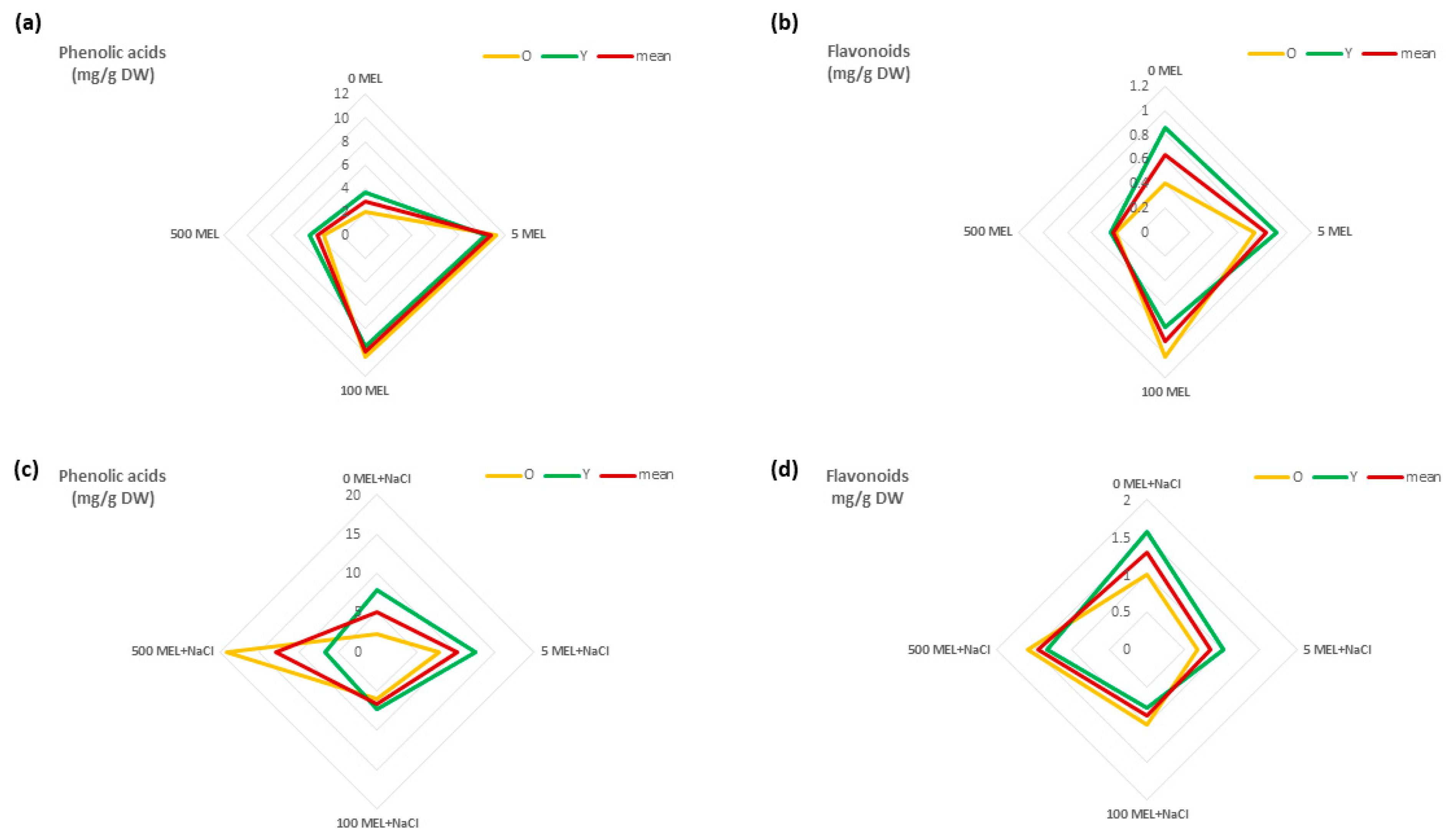
In previous research, we indicated the effect of MEL treatment of seeds on the development and growth of stevia plants as well as on the synthesis of SGs [46]. For this study, we investigated its effects on the contents of phenolic acids and flavonoids. As previously indicated, MEL could enhance the level of flavonoids and different forms of isoflavone during soybean germination [58]. Xu et al. [59] demonstrated that melatonin treatment increased the contents of total phenols (for 18 of 22 phenolic compounds), flavonoids, anthocyanins, and proanthocyanidins in berries. Ptak et al. [52] found that MEL enhanced the galanthamine biosynthesis in the in vitro culture of Leucojum aestivum, with the most promising effect shown for the concentration of 5 µM of MEL. In the experiment presented here, treatment of seeds before germination with 5 μM and 100 μM of MEL also increased the contents of total phenolic acids (5.5-fold and 5.2-fold, respectively) and total flavonoids (1.9-fold and 2.6-fold, respectively) in stevia leaves compared to those in the control. A higher concentration of MEL (500 μM) had no such beneficial effects (Figure 2a,b). Regardless of the concentration of MEL, the total content of phenolic acids was higher than that of flavonoids, which is consistent with the results of other studies [23,60].
In this study, the content of phenolic compounds were analyzed separately in older and younger stevia leaves. Nevertheless, we did not observe any effect of the leaf maturity stage on the total phenolic acid content. Despite this, Liu et al. [61] previously showed that younger leaves of tea plants contained twice as much phenolic acid as older ones. However, we did find that the stage of leaf development had an effect on the content of flavonoids in relation to the concentration of MEL. In older leaves, we observed the highest combined amounts of the three flavonoids (hyperoside, isoquercetin, and quercitrin) for 100 μM of MEL. In younger and control leaves, these compounds accumulated in the highest amounts for plants derived from seeds treated with 5 µM of melatonin. The highest concentration of melatonin (500 μM) used for seed treatment had no effect on the synthesis of the determined flavonoids in stevia leaves (Figure 2a,b).
Despite no differences in the qualitative composition of phenolic acids and flavonoids, on the basis of the peak area of each compound, we found that melatonin concentrations and the maturity stage of the leaves significantly affected the content of individual phenolic compounds (Table 2).
Padda and Picha [62] also described the different levels of certain phenolic acids in sweet potato leaves at different stages of maturity. However, no studies have shown the effect of melatonin on the content of individual phenolic compounds and flavonoids. In our research, the highest phenolic acid concentrations were found for isochlorogenic, rosmarinic, and chlorogenic acids, and the highest flavonoid concentration was for quercitrin. The average content of each of these compounds was 3.18, 2.38, and 0.62 of dry weight (DW) for isochlorogenic, rosmarinic, and chlorogenic acids, respectively, and 0.58 mg/g for quercitrin (Table 3).
Regarding isochlorogenic and chlorogenic acids, their concentrations were significantly higher in younger leaves compared to older ones in all tested samples with 5 μM of MEL. The contents of chlorogenic acid was 1.9-fold and isochlorogenic acid was 3.3-fold higher compared to those in the control. With an increase in MEL concentration, the content of both acids decreased slightly for both older and younger leaves. Our research confirms the literature indicating that the main phenolic acids in stevia leaves are isochlorogenic and chlorogenic acid [22,31]. However, this is the first study to show the influence of MEL on the biosynthesis of these acids. Regarding rosmarinic acid, when MEL was used for the seed treatment before germination, it significantly increased the content in older leaves compared to that in younger leaves at all tested concentrations; however, this effect was best seen with lower melatonin concentrations (Table 3). Among the identified flavonoids, quercitrin was accumulated in the greatest amount. In that case, the highest concentration (about 2.7-times higher than that in the control) was detected in older leaves under 100 μM of MEL (0.87 mg/g of DW). However, 500 μM of MEL significantly decreased the quercitrin concentration, specifically in younger leaves (Table 3).
The effect of MEL on plants seems to be long-term [63]. Therefore, a specific concentration of MEL used for seed treatment before germination may be a good stimulator of the biosynthesis of phenolic compounds in S. rebaudiana leaves. For the extraction of specific compounds, attention should be paid to the use of leaves in the appropriate maturity stage.

2.3. The Effect of Melatonin on Phenolic Acid and Flavonoid Content under NaCl

Salinity is one of the most significant abiotic factors that negatively influences plant efficiency and the production of many crops. Around 19.5% of irrigated agricultural land is considered to be saline [64]. Stevia plantlet growth decreases as salinity increases [65,66,67]. Our previous study also showed that stevia germination and growth is impacted by salt stress [46]. Although a low level of salinity (50 mM) had a positive impact on the morphological parameters, 150 mM of sodium chloride (NaCl) exerted an unfavorable effect on seedling development, resulting in growth inhibition and even death [46]. Our previous study also indicated that 50 mM of NaCl increased the stevioside content but decreased rebaudioside A [46]. This finding was in agreement with other research showing that low levels of NaCl increased SG, chlorophyll a, carotenoids, and total sugars [68,69]. However, no previous studies have shown the effect of NaCl on the content of individual phenolic compounds in stevia. In this study, we found that NaCl at a concentration of 50 mM increased the contents of both phenolic acids and total flavonoids (Figure 2c,d). Similarly, Zhu et al. [70] found that in Taraxacum officinale, the contents of chlorogenic and isochlorogenic acids were higher under salt stress conditions (≤1 g kg−1) compared to those in the control, with the opposite effect observed at higher concentrations of NaCl. An interesting solution limiting the negative impact of NaCl is the use of MEL. Melatonin is a molecule with a wide spectrum of activity, and in many species, it has been proven to have a beneficial effect on growth under stress conditions, including salt stress. In Malus hupehensis L., exogenous melatonin modulated the flavonoid content in response [71]. Bistgani et al. [72] found that foliar application of MEL improved the total phenolic compounds in Thymus daenensis L. leaves under salinity stress. In this study, we observed that the treatment of seeds with 5 or 500 μM of MEL before germination on 50 mM of NaCl affected the total phenolic acid concentration in stevia leaves. However, 5 μM of MEL had the best effect on younger leaves, while 500 μM of MEL had the best effect on older leaves (Figure 2c). Additionally, the highest sum of flavonoids was observed for 500 μM of MEL. We observed that 500 μM of MEL increased the total flavonoid content in older leaves, while 5 µM and no melatonin (0 MEL) increased the content in younger leaves (Figure 2d). In a previous study [73,74], melatonin affected the concentrations of the flavonoids and total phenolic acids. In our research, MEL also had a significant effect on the content of individual phenolic compounds under NaCl conditions (Table 2). The highest accumulations were recorded for isochlorogenic, rosmarinic, and chlorogenic acids (average contents of 3.46, 2.78, and 0.81 mg/g of DW, respectively) as well as quercitrin (0.72 mg/g of DW). The highest contents of rosmarinic and isochlorogenic acids were recorded in older leaves obtained from seeds exposed to 500 µM of MEL (9.28 and 7.74 mg/g of the DW, respectively), which were about 13-times and 10-times higher, respectively, than their amounts in the older leaves of control plants (Table 3). The results described above indicate that in the presence of NaCl, melatonin regulates the content of individual phenolic compounds in a slightly different way compared to conditions without NaCl.

2.4. The Effect of Melatonin and NaCl on the Accumulation of Phenolic Compounds

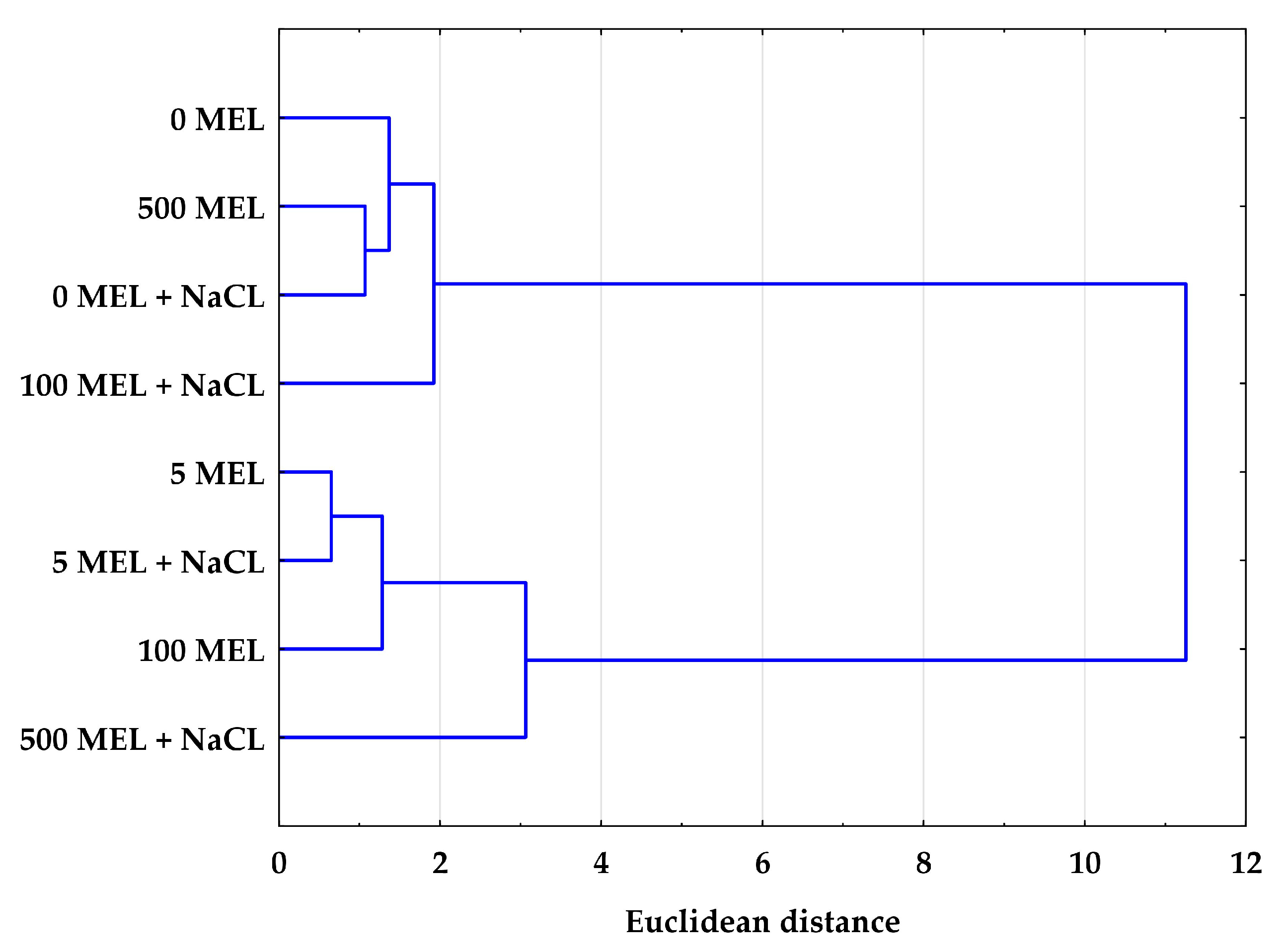
To examine the linkage between the effects of melatonin and NaCl in terms of the studied biochemical compounds, Ward’s [75] hierarchical clustering was applied. Based on the average content of the identified phenolic compounds, two clusters formed on the dendrogram (Figure 3). The similarities shown in both clusters indicate the important roles of both melatonin and NaCl in inducing a specific response in secondary metabolism. At a lower melatonin concentration (5 μM) and in the absence of melatonin (0 MEL), the presence of NaCl had no significant effect on the content of phenolic compounds in stevia plants. However, the presence of NaCl in the germination medium had an effect on seeds treated with melatonin at higher concentrations (100 and 500 μM); samples that germinated in the presence and absence of NaCl in the medium were located in different clusters (Figure 3). This may indicate that in addition to the adverse effects of NaCl observed in many plant species, higher concentrations of melatonin may also induce a stress response in stevia plants [76].
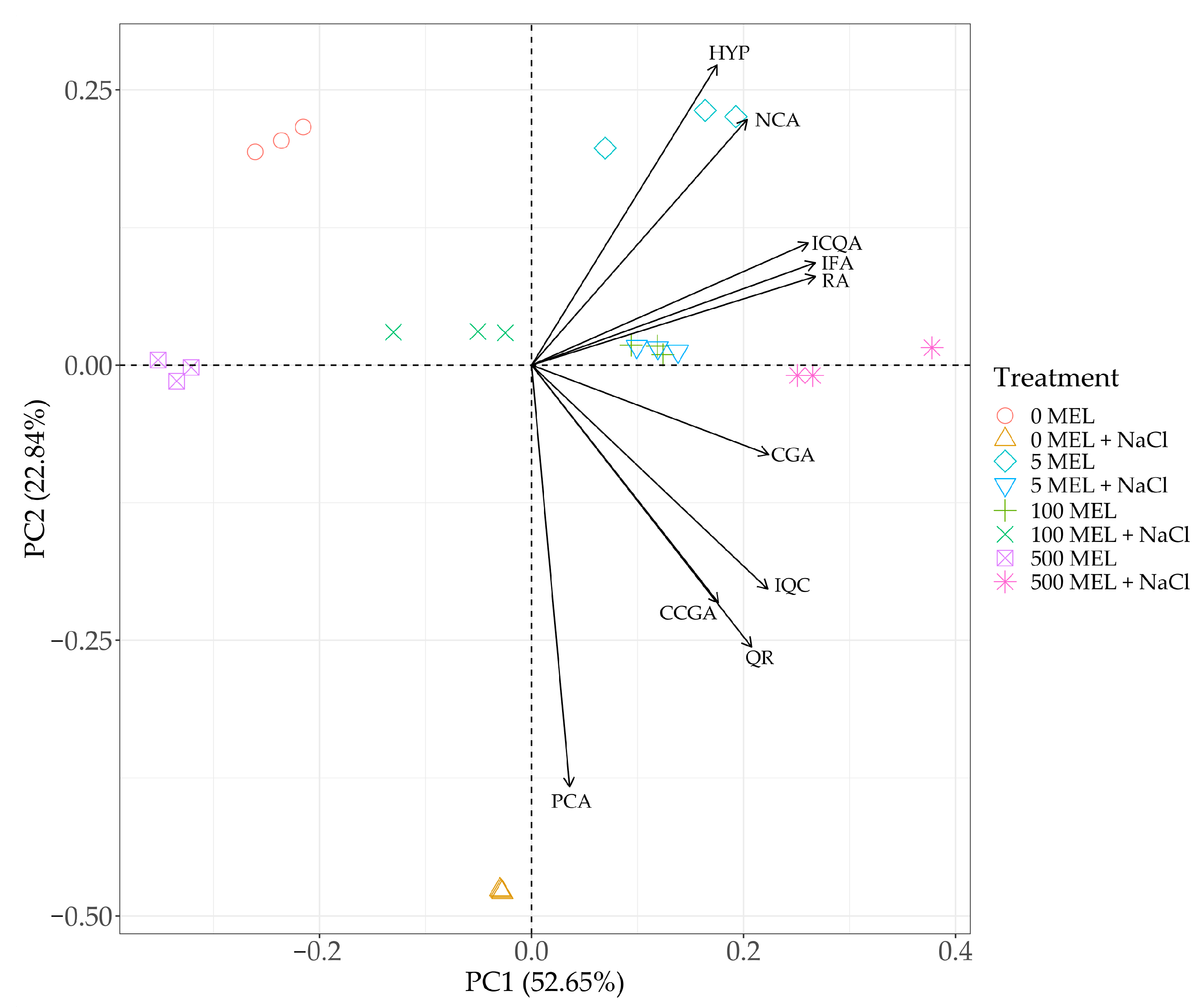
Principal component analysis (PCA) showed that the first component (PC1) explained nearly 53% of the variation, while the second component (PC2) explained about 23% of the variation. PC1 was significantly affected by almost all variables except the protocatechuic acid content. In contrast, PC2 was strongly negatively influenced by the content of protocatechuic acid, cryptochlorogenic acid, quercitrin, and isoquercetin. However, the contents of neochlorogenic acid and hyperoside had a significant positive effect on PC2 (Figure 4). The position of the loading vectors for the individual variables made it possible to indicate several relationships. A significant positive correlation was found between the contents of neochlorogenic acid and hyperoside. Additionally, a significant relationship was found between the contents of isochlorogenic, isoferulic, and rosmarinic acids. A significant relationship was also seen between the contents of cryptochlorogenic acid and quercitrin and the content of isoquercetin. The biplot chart indicates the relationship between the studied treatments and the content of the analyzed phenolics. From the distribution of the data, it can be concluded that the control, characterized by the absence of melatonin treatment and NaCl, was not related to the variation in any of the analyzed acids. In contrast, the plants treated only with NaCl were characterized by a particular increase in protocatechuic acid content not observed in other treatments. Melatonin at a dose of 5 μM was specifically associated with an increase in the content of neochlorogenic acid and a higher content of hyperoside. A similar distribution of phenolic compound contents was found in the plants obtained from seeds treated with MEL at a dose of 100 μM and at a dose of 500 μM in combination with NaCl, especially for chlorogenic, isochlorogenic, isoferulic, and rosmarinic acids (Figure 4).

3. Materials and Methods

3.1. Plant Material

The stevia plants were obtained from seeds incubated in an aqueous solution of MEL prior to germination.
The seeds were surface-sterilized according to Simlat et al. [77] before treatment. Three concentrations of MEL were used: 5 μM (5 MEL), 100 μM (100 MEL), and 500 μM (500 MEL). For the control, the seeds were incubated in water (0 MEL). After incubation, half of the seeds were germinated in in vitro conditions on agar gel and the other half on agar gel supplemented with 50 mM of NaCl. After three weeks of germination experiment, seedlings were transferred to the Murashige and Skoog (MS) [78] medium for one month and then well-developed plantlets were transplanted into pots (15 × 15 cm) containing soil, peat, and sand in a 3:1:1 ratio. The plants used for analysis were grown for six months in controlled conditions (25 °C, fluorescent light with intensity expressed as Photosynthetic Photon Flux Density of 320 μmol m2s1 for 16 h/day, and 70% ± 5% relative humidity; Adaptis-A1000AR, Conviron, Winnipeg, MB, Canada).

3.2. RP-HPLC Analysis

The leaves of six-month-old plants were used for the RP-HPLC analysis. Phenolic acids, flavonoids, and catechins were quantified in methanol extracts obtained by sonification (30 °C, 1 h) of 250 mg of dry biomass in 2 mL of methanol. RP-HPLC analyses were conducted according to the method described by Ellnain-Wojtaszek and Zgórka [79] with modifications using a Merck–Hitachi liquid chromatograph (LaChrom Elite, Hitachi, Tokyo, Japan) equipped with a DAD detector L-2455 and Purospher ® RP-18e (250 × 4 mm/5 mm) column (Merck, Darmstadt, Germany). Analyses were carried out at 25°C with a mobile phase consisting of A—methanol and B—methanol: 0.5% acetic acid; 1:4 (v/v). The gradients were as follows: 100% B for 0–20 min, 100–80% B for 20–35 min, 80–60% B for 35–55 min, 60–0% B for 55–70 min, 0% B for 70–75 min, 0–100% B for 75–80 min, and 100% B for 80–90 min at a flow rate of 1 mL min-1 and λ = 254 nm for phenolic acids and catechins and λ = 370 nm for flavonoids. Identification was done by comparison of the retention times of the peaks with authentic reference compounds and co-chromatography with standards. Quantification was done by measurement of the peak area with reference to the standard curve derived from five concentrations (0.03125 to 0.5 mg ml1). Caffeic, chlorogenic, cinnamic, ellagic, gallic, gentisic, isoferulic, neochlorogenic, o-coumaric, protocatechuic, rosmarinic, salicylic, sinapic, syringic acid, apigetrin (apigenin 7-glucoside), hyperoside (quercetin 3-O-galactoside), isoquercetin (quercetin 3-O-glucoside), isorhamnetin, kaempferol, luteolin, populin (kaempferol 7-O-glucoside), quercetin, quercitrin (quercetin 3-O-rhamnoside), rhamnetin, rutin, and vitexin standards were purchased from Sigma-Aldrich (St Louis, MO, USA). Vanillic, p-coumaric, ferulic, and p-hydroxybenzoic acid standards were purchased from Fluka (Bucha, Switzerland). Cryptochlorogenic acid, isochlorogenic acid, catechin, epigallocatechin, epicatechin gallate, epicatechin, epigallocatechin gallate, and cynaroside (luteolin 7-O-glucoside) standards were purchased from ChromaDex (Irvine, CA, USA).

3.3. Statistical Analyses

The data were reported as mean ± SD. To study the effect of MEL and NaCl on the phenolic acid and flavonoid content, factorial ANOVA (TIBCO Statistica, Palo Alto, CA, USA) was applied. To estimate the significance of differences between the means, we performed Duncan’s multiple range test at a significance level of p < 0.05. To examine the linkage between the effects of MEL and NaCl in terms of the studied biochemical compounds, Ward’s [75] hierarchical clustering was applied. The dendrogram was generated based on the phenolic acid and flavonoid contents. To determine the relationship between treatment and the phenolic content, the data were analyzed with PCA using R v. 3.6.1 [80].

4. Conclusions

Taking into account the present results as well as previously published studies on S. rebaudiana, it is reasonable to conclude that stevia is an abundant source of secondary metabolites with health benefits. The presence of bioactive compounds in stevia leaves belonging to phenolic and flavonoid groups tend to justify their medicinal properties and their application in both the food and pharmaceutical industries.
Furthermore, our studies pointed out that MEL used at the stage of seed germination seems to be a good stimulator of the biosynthesis of phenolic compounds in stevia plants, and low dose of NaCl may be an additional stimulating factor. This has a very practical aspect: MEL treated seeds can be used in the establishment of plantations for obtaining high quality plants useful for the food and pharmaceutical industries.
Although significant progress has been made in understanding the role of MEL in plants, the MEL signaling pathway under salt stress remains unclear. Melatonin’s mitigation of salt stress damages can be achieved through multiple mechanisms: (1) MEL reduces excessive production of reactive oxygen species (ROS) by facilitating electron leakage as a result of increasing the efficiency of the mitochondrial electron transport chain; (2) MEL regulates ion homeostasis; (3) MEL modulates the activity of transcription factors; and (4) MEL may also mitigate cell damage and reduce the increase in osmotic pressure caused by salt stress by increasing the accumulation of the phenolic compounds and all phenolic acids.
Comprehensive and well-designed future research on the role of MEL in biosynthesis of phenolic compounds under salt stress in S. rebaudiana would be valuable. Our further research will focus on the expression of genes involved in the metabolism of phenylpropanoids and the analysis of the activity of relevant enzymes. We believe that these analyses will by valuable for understanding these mechanisms. Such knowledge could create new opportunities to optimize the biosynthesis of these medicinally important metabolites.

Author Contributions

Conceptualization, M.S. and A.P.; methodology, M.S., T.W. and A.S.; software, M.S., T.W. and A.S.; validation, M.S., A.P., T.W. and A.S.; formal analysis, M.S., A.P., T.W. and A.S.; investigation, M.S.; resources, M.S and A.S.; data curation, M.S. and A.S.; writing—original draft preparation, M.S.; writing—review and editing, M.S., A.P., T.W. and A.S.; visualization, M.S. and T.W. All authors have read and agreed to the published version of the manuscript.

Funding

This research received no external funding.

Data Availability Statement

The data presented in this study are available on request from the corresponding author.

Acknowledgments

The research was financed from the subsidy of the Ministry of Science and Higher Education of the Republic of Poland awarded to the University of Agriculture in Krakow, Poland.

Conflicts of Interest

The authors declare no conflict of interest. The funders had no role in the design of the study; in the collection, analyses, or interpretation of data; in the writing of the manuscript; or in the decision to publish the results.

References

  1. Soejarto, D.D. Botany of Stevia and Stevia rebaudiana. In Stevia: The Genus Stevia; Kinghorn, A.D., Ed.; Taylor and Francis: London, UK, 2001; Volume 2, pp. 18–39. [Google Scholar]
  2. Brandle, J.E.; Telmer, P.G. Steviol glycoside biosynthesis. Phytochemistry 2007, 68, 1855–1863. [Google Scholar] [CrossRef] [PubMed]
  3. Ohta, M.; Sasa, S.; Inoue, A.; Tamai, T.; Fujita, I.; Morita, K.; Matsuura, F. Characterization of novel steviol glycosides from leaves of Stevia rebaudiana Morita. J. Appl. Glycosci. 2010, 57, 199–209. [Google Scholar]
  4. Chaturvedula, V.S.; Prakash, I. Structures of the novel diterpene glycosides from Stevia rebaudiana. Carbohydr. Res. 2011, 346, 1057–1060. [Google Scholar]
  5. Chaturvedula, V.S.; Prakash, I. Additional minor diterpene glycosides from Stevia rebaudiana. Nat. Prod. Commun. 2011, 6, 1059–1062. [Google Scholar]
  6. Chaturvedula, V.S.; Rhea, J.; Milanowski, D.; Mocek, U.; Prakash, I. Two minor diterpene glycosides from the leaves of Stevia rebaudiana. Nat. Prod. Commun. 2011, 6, 175–178. [Google Scholar] [PubMed]
  7. Ceunen, S.; Geuns, J.M.C. Steviol glycosides: Chemical diversity, metabolism, and function. J. Nat. Prod. 2013, 76, 1201–1228. [Google Scholar]
  8. Angelini, L.G.; Martini, A.; Passera, B.; Tavarini, S. Cultivation of Stevia rebaudiana Bertoni and associated challenges. In Sweeteners. Reference Series in Phytochemistry; Mérillon, J.M., Ramawat, K., Eds.; Springer: Cham, Switzerland, 2018; pp. 1–52. [Google Scholar]
  9. Atteh, J.; Onagbesan, O.; Tona, K.; Buyse, J.; Decuypere, E.; Geuns, J. Potential use of Stevia rebaudiana in animal feeds. Arch. Zootec. 2011, 60, 133–136. [Google Scholar] [CrossRef]
  10. Goyal, S.K.; Samsher; Goyal, R.K. Stevia (Stevia rebaudiana) a bio-sweetener: A review. Int. J. Food Sci. Nutr. 2010, 61, 1–10. [Google Scholar] [CrossRef]
  11. European Food Safety Authority (EFSA). Revised exposure assessment for steviol glycosides for the proposed uses as a food additive. EFSA J. 2011, 9, 1972. [Google Scholar]
  12. Jeong, Y.; Lee, H.J.; Jin, G.H.; Park, Y.D.; Choi, D.S.; Kang, M.A. Anti-inflammatory activity of Stevia rebaudiana in LPS-induced RAW 264.7 cells. J. Food Sci. Nutr. 2010, 15, 14–18. [Google Scholar] [CrossRef]
  13. Preethi, D.; Sridhar, T.M.; Josthna, P.; Naidu, C.V. Studies on antibacterial activity, phytochemical analysis of Stevia rebaudiana (Bert.). An important calorie free biosweetner. J. Ecobiotech. 2011, 3, 5–10. [Google Scholar]
  14. Arya, A.; Kumar, S.; Kasana, M.S. Anti-inflammatory activity of in vitro regenerated calli and in vivo plant of Stevia rebaudiana (Bert.) Bertoni. J. Sci. Ind. Res. 2012, 2, 435–439. [Google Scholar]
  15. Gamboa, F.; Chaves, M. Antimicrobial potential of ex-tracts from Stevia rebaudiana leaves against bacteria of importance in dental caries. Acta Odontol. Latinoam. 2012, 25, 171–175. [Google Scholar]
  16. Kedik, S.A.; Yartsev, E.I.; Stanishevskaya, I.E. Antiviral activity of dried extract of Stevia. Pharm. Chem. J. 2009, 43, 198–199. [Google Scholar] [CrossRef]
  17. Takasaki, M.; Konoshima, T.; Kozuka, M.; Tokuda, H.; Takayasu, J.; Nishino, H.; Miyakoshi, M.; Mizutani, K.; Lee, K.-H. Cancer preventive agents. Part 8: Chemopreventive effects of stevioside and related compounds. Bioorg. Med. Chem. 2009, 17, 600–605. [Google Scholar] [PubMed]
  18. Chen, J.; Xia, Y.; Sui, X.; Peng, Q.; Zhang, T.; Li, J.; Zhang, J. Steviol, a natural product inhibits proliferation of the gastrointestinal cancer cells intensively. Oncotarget 2018, 9, 26299–26308. [Google Scholar] [CrossRef]
  19. Ghanta, S.; Banerjee, A.; Poddar, A.; Chattopadhyay, S. Oxidative DNA damage preventive activity and anti-oxidant potential of Stevia rebaudiana (Bertoni) Bertoni, a natural sweetener. J. Agric. Food Chem. 2007, 55, 10962–10967. [Google Scholar] [CrossRef]
  20. Shukla, S.; Mehta, A.; Mehta, P.; Bajpai, V.K. Antioxidant ability and total phenolic content of aqueous leaf extract of Stevia rebaudiana Bert. Exp. Toxicol. Pathol. 2012, 64, 807–811. [Google Scholar] [CrossRef]
  21. Muandam, F.; Soulimani, R.; Diop, B.; Dicko, A. Study on chemical composition and biological activities of essential oil and extracts from Stevia rebaudiana Bertoni leaves. LWT-Food Sci. Technol. 2011, 44, 1865–1872. [Google Scholar] [CrossRef]
  22. Karaköse, H.; Jaiswal, R.; Kuhnert, N. Characterization and quantification of hydroxycinnamate derivatives in Stevia rebaudiana leaves by LC-MSn. J. Agric. Food Chem. 2011, 59, 10143–10150. [Google Scholar] [CrossRef]
  23. Kim, I.S.; Yang, M.; Lee, O.H.; Kang, S.N. The antioxidant activity and the bioactive compound content of Stevia rebaudiana water extracts LWT. Food Sci. Technol. 2011, 44, 1328–1332. [Google Scholar]
  24. Lemus-Mondaca, R.; Vega-Galvez, A.; Rojas, P.; Stucken, K.; Delporte, C.; Valenzuela-Barra, G.; Jagus, J.J.; Agüero, M.V.; Pasten, A. Antioxidant, antimicrobial and anti-inflammatory potential of Stevia rebaudiana leaves: Effect of different drying methods. J. Appl. Res. Med. Aromat. Plants 2018, 11, 37–46. [Google Scholar] [CrossRef]
  25. Madan, S.; Ahmad, S.; Singh, G.N.; Kohli, K.; Kumar, Y.; Singh, R.; Garg, M. Stevia rebaudiana (Bert.) Bertoni—A review. Indian J. Nat. Prod. Resour. 2010, 1, 267–286. [Google Scholar]
  26. Pacifico, S.; Piccolella, S.; Nocera, P.; Tranquillo, E.; Dal Poggetto, F.; Catauro, M. New insights into phenol and polyphenol composition of Stevia rebaudiana leaves. J. Pharm. Biomed. Anal. 2019, 163, 45–57. [Google Scholar] [PubMed]
  27. Wolwer-Rieck, U. The leaves of Stevia rebaudiana (Bertoni), their constituents and the analyses thereof: A review. J. Agric. Food Chem. 2012, 60, 886–895. [Google Scholar] [PubMed]
  28. Yu, H.; Yang, G.Q.; Sato, M.; Yamaguchi, T.; Nakano, T.; Xi, Y.C. Antioxidant activities of aqueous extract from Stevia rebaudiana stem waste to inhibit fish oil oxidation and identification of its phenolic compounds. Food Chem. 2017, 232, 379–386. [Google Scholar]
  29. Zhang, Q.N.; Yang, H.; Li, Y.N.; Liu, H.B.; Jia, X.D. Toxicological evaluation of ethanolic extract from Stevia rebaudiana Bertoni leaves: Genotoxicity and subchronic oral toxicity. Regul. Toxicol. Pharmacol. 2017, 86, 253–259. [Google Scholar]
  30. Amriteswori, R.; Margaret, F.R. The flavonoids of Stevia rebaudiana. J. Nat. Prod. 1983, 46, 194–195. [Google Scholar]
  31. Myint, K.Z.; Wu, K.; Xia, Y.; Fan, Y.; Shen, J.; Zhang, P.; Gu, J. Polyphenols from Stevia rebaudiana (Bertoni) leaves and their functional properties. J. Food Sci. 2020, 85, 240–248. [Google Scholar]
  32. Hattori, A.; Migitaka, H.; Iigo, M.; Itoh, M.; Yamamoto, K.; Ohtani-Kaneko, R.; Hara, M.; Suzuki, T.; Reiter, R.J. Identification of melatonin in plants and its effects on plasma melatonin levels and binding to melatonin receptors in vertebrates. Biochem. Mol. Biol. Int. 1995, 35, 627–634. [Google Scholar]
  33. Posmyk, M.M.; Kuran, H.; Marciniak, K.; Janas, K.M. Presowing seed treatment with melatonin protects red cabbage seedlings against toxic copper ion concentrations. J. Pineal Res. 2008, 45, 24–31. [Google Scholar] [CrossRef] [PubMed]
  34. Posmyk, M.; Bałabusta, M.; Wieczorek, M.; Śliwińska, E.; Janas, K.M. Melatonin applied to cucumber (Cucumis sativus L.) seeds improves germination during chilling stress. J. Pineal Res. 2009, 46, 214–223. [Google Scholar] [CrossRef] [PubMed]
  35. Tiryaki, I.; Keles, H. Reversal of the inhibitory effect of light and high temperature on germination of Phacelia tanacetifolia seeds by melatonin. J. Pineal Res. 2012, 52, 332–339. [Google Scholar] [CrossRef] [PubMed]
  36. Wei, W.; Li, Q.-T.; Chu, Y.-N.; Reiter, R.J.; Yu, X.M.; Zhu, D.H.; Zhang, W.K.; Ma, B.; Lin, Q.; Zhang, J.S.; et al. Melatonin enhances plant growth and abiotic stress tolerance in soybean plants. J. Exp. Bot. 2015, 66, 695–707. [Google Scholar]
  37. Hernandez-Ruiz, J.; Cano, A.; Arnao, M.B. Melatonin: A growth-stimulating compound present in lupin tissues. Planta 2004, 220, 140–144. [Google Scholar]
  38. Hernandez-Ruiz, J.; Cano, A.; Arnao, M.B. Melatonin acts as a growth-stimulating compound in some monocot species. J. Pineal Res. 2005, 39, 137–142. [Google Scholar] [CrossRef] [PubMed]
  39. Hernandez-Ruiz, J.; Arnao, M.B. Melatonin stimulates the expansion of etiolated lupin cotyledons. Plant Growth Regul. 2008, 55, 29–34. [Google Scholar] [CrossRef]
  40. Arnao, M.B.; Hernandez-Ruiz, J. Melatonin promotes adventitious- and lateral root regeneration in etiolated hypocotyls of Lupinus albus L. J. Pineal Res. 2007, 42, 147–152. [Google Scholar] [CrossRef]
  41. Zhang, N.; Zhao, B.; Zhang, H.-J.; Weeda, S.; Yang, C.; Yang, Z.C.; Ren, S.; Guo, Y.D. Melatonin promotes water-stress tolerance, lateral root formation, and seed germination in cucumber (Cucumis sativus L.). J. Pineal Res. 2013, 54, 15–23. [Google Scholar] [CrossRef]
  42. Park, S.; Back, K. Melatonin promotes seminal root elongation and root growth in transgenic rice after germination. J. Pineal Res. 2013, 53, 385–389. [Google Scholar] [CrossRef]
  43. Sarrou, E.; Therios, I.; Dimassi-Theriou, K. Melatonin and other factors that promote rooting and sprouting of shoot cuttings in Punica granatum cv. Wonderful. Turk. J. Botany 2014, 38, 293–301. [Google Scholar]
  44. Chen, Q.; Qi, W.B.; Reiter, R.J.; Wei, W.; Wang, B.M. Exogenously applied melatonin stimulates root growth and raises endogenous indoleacetic acid in roots of etiolated seedlings of Brassica juncea. J. Plant Physiol. 2009, 166, 324–328. [Google Scholar] [CrossRef] [PubMed]
  45. Simlat, M.; Ptak, A.; Skrzypek, E.; Warchoł, M.; Morańska, E.; Piórkowska, E. Melatonin significantly influences seed germination and seedling growth of Stevia rebaudiana Bertoni. PeerJ 2018, 6, e5009. [Google Scholar] [CrossRef] [PubMed]
  46. Simlat, M.; Szewczyk, A.; Ptak, A. Melatonin promotes seed germination under salinity and enhances the biosynthesis of steviol glycosides in Stevia rebaudiana Bertoni leaves. PLoS ONE 2020, 15, e0230755. [Google Scholar]
  47. Rodriguez, C.; Mayo, J.C.; Sainz, R.M.; Antolín, I.; Herrera, F.; Martín, V.; Reiter, R.J. Regulation of antioxidant enzymes: A significant role for melatonin. J. Pineal Res. 2004, 36, 1–9. [Google Scholar]
  48. Bałabusta, M.; Szafrańska, K.; Posmyk, M.M. Exogenous melatonin improves antioxidant defense in cucumber seeds (Cucumis sativus L.) germinated under chilling stress. Front. Plant Sci. 2016, 7, 575. [Google Scholar]
  49. Wang, L.Y.; Liu, J.L.; Wang, W.X.; Sun, Y. Exogenous melatonin improves growth and photosynthetic capacity of cucumber under salinity-induced stress. Photosynthetica 2016, 54, 19–27. [Google Scholar] [CrossRef]
  50. Wang, Y.; Reiter, R.J.; Chan, Z. Phytomelatonin: A universal abiotic stress regulator. J. Exp. Bot. 2018, 69, 963–974. [Google Scholar] [CrossRef]
  51. Coskun, Y.; Duran, R.E.; Kilic, S. Striking effects of melatonin on secondary metabolites produced by callus culture of rosemary (Rosmarinus officinalis L.). Plant Cell Tiss. Organ Cult. 2019, 138, 89–95. [Google Scholar] [CrossRef]
  52. Ptak, A.; Simlat, M.; Morańska, E.; Skrzypek, E.; Warchoł, M.; Tarakemeh, A.; Laurain-Mattar, D. Exogenous melatonin stimulated Amaryllidaceae alkaloid biosynthesis in in vitro cultures of Leucojum aestivum L. Ind. Crops Prod. 2019, 138, 111458. [Google Scholar] [CrossRef]
  53. Rajbhandari, A.; Roberts, M.F. Flavonoids of Stevia nepetifolia. J. Nat. Prod. 1984, 47, 559–560. [Google Scholar] [CrossRef]
  54. Marchyshyn, S.; Hudz, N.A.; Dakhym, I.; Husak, L.V.; Demydyak, O.L. HPLC analysis of phenolic compounds from Stevia rabaudiana Bertoni leaves. Pharma Innov. 2018, 7, 515–517. [Google Scholar]
  55. Carrera-Lanestosa, A.; Coral-Martínez, T.; Ruíz-Ciau, D.; Moguel-Ordoñez, Y.; Rubí Segura-Campos, M. Phenolic compounds and major steviol glucosides by HPLC-DAD-RP and in vitro evaluation of the biological activity of aqueous and ethanolic extracts of leaves and stems: S. rebaudiana Bertoni (creole variety INIFAP C01). Int. J. Food Prop. 2020, 23, 199–212. [Google Scholar] [CrossRef]
  56. Khiraoui, A.; Al Faiz, C.; Hasib, A.; Bakha, M.; Benhmimou, A.; Amchra, F.Z.; Boulli, A. Antioxidant ability, total phenolic and flavonoid contents of leaf extract of Stevia rebaudiana Bertoni cultivated in Morocco. Int. J. Sci. Eng. Res. 2018, 9, 1585–1590. [Google Scholar]
  57. Dos Santos Szewczyk, K.; Pietrzak, W.; Klimek, K.; Grzywa-Celińska, A.; Celiński, R.; Gogacz, M. LC-ESI-MS/MS identification of biologically active phenolics in different extracts of Alchemilla acutiloba Opiz. Molecules 2022, 27, 621. [Google Scholar]
  58. Yin, Y.; Tian, X.; He, X.; Yang, J.; Yang, Z.; Fang, W. Exogenous melatonin stimulated isoflavone biosynthesis in NaCl-stressed germinating soybean (Glycine max L.). Plant Physiol. Biochem. 2022, 185, 123–131. [Google Scholar]
  59. Xu, L.; Yue, Q.; Bian, F.; Sun, H.; Zhai, H.; Yao, Y. Melatonin enhances phenolics accumulation partially via ethylene signaling and resulted in high antioxidant capacity in grape berries. Front. Plant Sci. 2017, 18, 1426. [Google Scholar]
  60. Gaweł-Bęben, K.; Bujak, T.; Nizioł-Łukaszewska, Z.; Antosiewicz, B.; Jakubczyk, A.; Karaś, M.; Rybczyńska, K. Stevia rebaudiana Bert. leaf extracts as a multifunctional source of natural antioxidants. Molecules 2015, 20, 468–486. [Google Scholar]
  61. Liu, Z.; Bruins, M.E.; de Bruijn, W.J.C.; Vincken, J.-P. A comparison of the phenolic composition of old and young tea leaves reveals a decrease in flavanols and phenolic acids and an increase in flavonols upon tea leaf maturation. J. Food Compos. Anal. 2020, 86, 103385. [Google Scholar]
  62. Padda, M.; Picha, D.H. Antioxidant activity and phenolic composition in ‘Beauregard’ sweetpotato are affected by root size and leaf age. J. Am. Soc. Hortic. Sci. 2007, 132, 447–451. [Google Scholar] [CrossRef]
  63. Liang, B.; Ma, C.; Zhang, Z.; Wei, Z.; Gao, T.; Zhao, Q.; Ma, F.; Li, C. Long-term exogenous application of melatonin improves nutrient uptake fluxes in apple plants under moderate drought stress. Environ. Exp. Bot. 2018, 155, 650–661. [Google Scholar]
  64. Flowers, T.J. Improving crop salt tolerance. Am. J. Bot. 2004, 55, 307–319. [Google Scholar]
  65. Mubarak, M.H.; Belal, A.H.; El-Dein, T.N.; El-Sarag, E.I. In vitro response growth Stevia rebaudiana to salinity and drought. Sinai J. Appl. Sci. 2012, 1, 13–20. [Google Scholar] [CrossRef]
  66. Zeng, J.; Cheng, A.; Lim, D.; Yi, B.; Wu, W. Effects of salt stress on the growth, physiological responses, and glycoside contents of Stevia rebaudiana Bertoni. J. Agric. Food Chem. 2013, 61, 5720–5726. [Google Scholar] [CrossRef] [PubMed]
  67. Pandey, M.; Chikara, S.K. In vitro regeneration and effect of abiotic stress on physiology and biochemical content of Stevia rebaudiana ‘Bertoni’. J. Plant Sci. Res. 2014, 1, 113. [Google Scholar]
  68. Shahverdi, M.A.; Omidi, H.; Tabatabaei, S.J. Stevia (Stevia rebaudiana Bertoni) responses to NaCl stress: Growth, photosynthetic pigments, diterpene glycosides and ion content in root and shoot. J. Saudi Soc. Agric. Sci. 2019, 18, 355–360. [Google Scholar]
  69. Gerami, M.; Majidian, P.; Ghorbanpour, A.; Alipour, Z. Stevia rebaudiana Bertoni responses to salt stress and chitosan elicitor. Physiol. Mol. Biol. Plants 2020, 26, 965–974. [Google Scholar] [CrossRef]
  70. Zhu, Y.; Gu, W.; Tian, R.; Li, C.; Ji, Y.; Li, T.; Wei, C.; Chen, Z. Morphological, physiological, and secondary metabolic responses of Taraxacum officinale to salt stress. Plant Physiol. Biochem. 2022, 189, 71–82. [Google Scholar] [CrossRef]
  71. Wei, Z.; Li, C.; Gao, T.; Zhang, Z.; Liang, B.; Lv, Z.; Zou, Y.; Ma, F. Melatonin increases the performance of Malus hupehensis after UV-B exposure. Plant Physiol. Biochem. 2019, 139, 630–641. [Google Scholar]
  72. Bistgani, Z.E.; Hashemi, M.; DaCosta, M.; Craker, L.; Maggi, F.; Morshedloo, M.R. Effect of salinity stress on the physiological characteristics, phenolic compounds and antioxidant activity of Thymus vulgaris L. and Thymus daenensis Celak. Ind. Crops Prod. 2019, 135, 311–320. [Google Scholar] [CrossRef]
  73. Nazir, M.; Asad Ullah, M.; Mumtaz, S.; Siddiquah, A.; Shah, M.; Drouet, S.; Hano, C.; Abbasi, B.H. Interactive effect of melatonin and UV-C on phenylpropanoid metabolite production and antioxidant potential in callus cultures of purple basil (Ocimum basilicum L. var.s purpurascens). Molecules 2020, 25, 1072. [Google Scholar] [PubMed]
  74. Wei, L.; Liu, C.; Wang, J.; Younas, S.; Zheng, H.; Zheng, L. Melatonin immersion affects the quality of fresh-cut broccoli (Brassica oleracea L.) during cold storage: Focus on the antioxidant system. J. Food Process. Preserv. 2020, 44, e14691. [Google Scholar]
  75. Ward, J.H., Jr. Hierarchical grouping to optimize an objective function. J. Am. Stat. Assoc. 1963, 58, 236–244. [Google Scholar] [CrossRef]
  76. Ghasemi-Omran, V.O.; Ghorbani, A.; Sajjadi-Otaghsara, S.A. Melatonin alleviates NaCl-induced damage by regulating ionic homeostasis, antioxidant system, redox homeostasis, and expression of steviol glycosides-related biosynthetic genes in in vitro cultured Stevia rebaudiana Bertoni. In Vitro Cell. Dev. Biol.-Plant 2021, 57, 319–331. [Google Scholar] [CrossRef]
  77. Simlat, M.; Ślęzak, P.; Moś, M.; Warchoł, M.; Skrzypek, E.; Ptak, A. The effect of light quality on seed germination, seedling growth and selected biochemical properties of Stevia rebaudiana Bertoni. Sci. Hortic. 2016, 211, 295–304. [Google Scholar]
  78. Murashige, T.; Skoog, F. A revised medium for rapid growth and bio assays with tobacco tissue cultures. Plant Physiol. 1962, 15, 473–497. [Google Scholar]
  79. Ellnain-Wojtaszek, M.; Zgórka, G. High-performance liquid chromatography and thin-layer chromatography of phenolic acids from Ginkgo biloba L. leaves collected within vegetative period. J. Liq. Chromatogr. Relat. Technol. 1999, 22, 1457–1471. [Google Scholar] [CrossRef]
  80. R Core Team, R. A Language and Environment for Statistical Computing. In R Foundation for Statistical Computing; R Core Team R: Vienna, Austria, 2019; Available online: https://www.R-project.org (accessed on 2 November 2022).
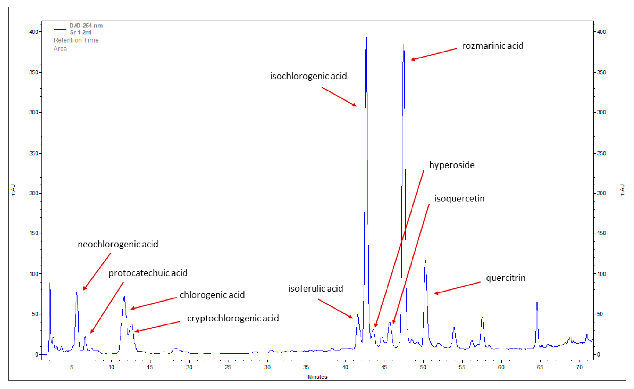
Figure 1. Reverse-phase high performance liquid chromatography (RP-HPLC) chromatogram of phenolic acids and flavonoids identified in methanolic extract of stevia leaves (the leaves were from plants derived from control seeds—incubated in water before germination experiment).
Plants 12 00780 g001
Figure 2. The content (mg/g of dry weight [DW]) of the sum of phenolic acids (a,c) and flavonoids (b,d) in methanolic extracts of stevia leaves under melatonin (a,b) and melatonin/NaCl (c,d) treatments. 0 MEL, 5 MEL, 100 MEL, 500 MEL—different concentrations of melatonin: 0 μM, 5 μM, 100 μM, and 500 μM, respectively.
Plants 12 00780 g002
Figure 3. The dendrogram based on Ward’s hierarchical clustering based on the average content of the identified phenolic compounds in stevia leaves. Stevia leaves were obtained from plants derived from melatonin treated seeds and germinated in control or in 50 mM NaCl conditions (+NaCl) (0 MEL, 5 MEL, 100 MEL, 500 MEL—different concentrations of melatonin used for seed treatment: 0 μM, 5 μM, 100 μM, and 500 μM, respectively).
Plants 12 00780 g003
Figure 4. Principal component analysis (PCA) biplot showing relationships between the melatonin and NaCl treatment in the context of phenolic compound contents in stevia leaves: neochlorogenic acid (NCA), protocatechuic acid (PCA), chlorogenic acid (CGA), cryptochlorogenic acid (CCGA), isoferulic acid (IFA), isochlorogenic acid (ICQA), hyperoside (HYP), isoquercetin (IQC), rosmarinic acid (RA), and quercitrin (QR). Stevia leaves were obtained from plants derived from melatonin treated seeds and germinated in control or in 50 mM NaCl conditions (+NaCl) (0 MEL, 5 MEL, 100 MEL, 500 MEL—different concentrations of melatonin used for seed treatment: 0 μM, 5 μM, 100 μM, and 500 μM, respectively).
Plants 12 00780 g004
Table 1. Phenolic compounds detected in stevia leaves by LC/UV, LC/MS, and LC/ESI/MS.
Compound Retention Time
(tr min)
FormulaMolecular Mass
(Rr)
neochlorogenic acid5.88C16H18O9354.31
protocatechuic acid6.98C7H6O4154.12
chlorogenic acid11.12C16H18O9354.31
cryptochlorogenic acid12.63C16H18O9354.31
isoferulic acid39.32C10H10O4194.18
isochlorogenic acid42.33C16H18O9354.31
hyperoside43.94C21H20O12464.38
isoquercetin45.43C21H20O12464.38
rosmarinic acid47.16C18H16O8360.31
quercitrin49.9C21H20O11448.38
Table 2. Variance analysis for the content of phenolic compounds with regard to melatonin concentration and the maturity of stevia leaves. The analyses were carried out separately for plants obtained from seeds treated with melatonin and germinated under control conditions (−NaCl) and for plants obtained from melatonin treated seeds and germinated on a medium with the addition of NaCl (+NaCl).
CompoundSourcedfMSF–ValueMSF–Value
–NaCL+NaCl
neochlorogenic acidMEL concentration (M)30.277576223.510 ***0.04049745.423 ***
Type of leaves (T)10.01539512.397 **0.01798020.167 ***
M × T30.0097427.844 **0.101441113.781 ***
Error160.001242 0.000892
protocatechuic acidM30.001284162.482 ***0.0062661140.207 ***
T10.00044255.952 ***0.00043278.547 ***
M × T30.0000668.398 **0.00008715.741 ***
Error160.000008 0.000005
chlorogenic acidMEL concentration (M)30.583775181.817 ***0.2067933.541 ***
Type of leaves (T)10.779998242.931 ***4.09826664.724 ***
M × T30.04990615.543 ***0.5424387.981 ***
Error160.003211 0.00617
cryptochlorogenic acidMEL concentration (M)30.112743364.286 *0.04086577.858 ***
Type of leaves (T)10.0003181.029 ns0.01617630.819 ***
M × T30.0008632.789 ns0.05112597.408 ***
Error160.000309 0.000525
isoferulic acidMEL concentration (M)318.7137323.605 ***0.079112116.3412 ***
Type of leaves (T)14.978186.083 ***0.087028127.9835 ***
M × T30.10691.849 ***0.092553136.1074 ***
Error160.0578 0.000680
isochlorogenic acidMEL concentration (M)30.060263216.828 ***15.7731113.472 ***
Type of leaves (T)10.033797121.603 ***3.419724.601 ***
M × T30.00910532.762 ns19.6256141.186 ***
Error160.000278 0.1390
rosmarinic acidMEL concentration (M)315.7940292.521 ***20.4823104.381 ***
Type of leaves (T)15.5199102.234 ***18.981696.732 ***
M × T31.675931.040 ***21.1283107.672 ***
Error160.0540 0.1962
hyperosideMEL concentration (M)30.00244177.082 ***0.00140131.794 ***
Type of leaves (T)10.0000010.025 ns0.00261659.368 ***
M × T30.00084226.589 ***0.00252357.248 ***
Error160.000032 0.000044
isoquercetinMEL concentration (M)30.005569286.379 ***0.030799243.874 ***
Type of leaves (T)10.008554439.882 ***0.020344161.088 ***
M × T30.00072737.401 ***0.00206516.355 ***
Error160.000019 0.000126
quercitrinMEL concentration (M)30.175491199.646 ***0.3296081.646 ***
Type of leaves (T)10.02842332.335 ***0.032668.089 *
M × T30.113650129.292 ***0.1971548.838 ***
Error160.000879 0.00404
ns, not significant; * significant at the 0.05 probability level; ** significant at the 0.01 probability level; *** significant at the 0.001 probability level.
Table 3. The content of individual phenolic acids and flavonoids (mg/g of DW) in stevia leaves obtained from melatonin and NaCl treated seeds. Results are presented as a mean of three replications (n = 3) ± SD. Different letters in each column (separately for −NaCl and +NaCl) indicate a significant difference of p < 0.05 according to Duncan’s test; for mean value of each compound, homogeneous groups were designated separately and marked with capital letters. 0 MEL, 5 MEL, 100 MEL, 500 MEL—different concentrations of melatonin used for seed treatment: 0 μM, 5 μM, 100 μM, and 500 μM, respectively; O—older leaves, Y—younger leaves. Monitored compounds: neochlorogenic acid (NCA); protocatechuic acid (PCA); chlorogenic acid (CGA); cryptochlorogenic acid (CCGA); isoferulic acid (IFA); isochlorogenic acid (ICQA); rosmarinic acid (RA); hyperoside (HYP); isoquercetin (IQC); quercitrin (QR).
MEL ConcentrationType of LeavesPhenolic AcidsFlavonoids
NCAPCAGCACCGAICQAIFARAHYPIQCQR
NaCl
0 MELO0.209 c ± 0.050.018 e ± 0.000.238 f ± 0.060.128 c ± 0.030.716 f ± 0.150.037 de ± 0.010.664 f ± 0.170.059 d ± 0.010.031 f ± 0.010.313 e ± 0.04
Y0.367 b ± 0.010.018 e ± 0.000.640 cd ± 0.010.138 c ± 0.001.694 e ± 0.040.035 e ± 0.000.770 ef ± 0.010.094 a ± 0.000.086 c ± 0.000.685 c ± 0.03
mean0.288 C0.018 C0.440 B0.133C1.205 D0.306 B0.717 B0.076 A0.059 B0.499 BC
5 MELO0.609 a ± 0.080.043 bc ± 0.000.628 d ± 0.090.274 b ± 0.034.722 b ± 0.520.307 a ± 0.044.555 a ± 0.580.082 b ± 0.010.065 d ± 0.010.588 d ± 0.04
Y0.581 a ± 0.040.033 d ± 0.001.229 a ± 0.110.247 b ± 0.025.542 a ± 0.340.139 c ± 0.012.481 c ± 0.020.067 cd ± 0.000.118 b ± 0.010.738 b ± 0.03
mean0.595 A0.038 B0.929 A0.261 B5.132 A0.223 A3.518 A0.075 A0.092 A0.663 AB
100 MELO0.337 b ± 0.000.055 a ± 0.000.738 c ± 0.020.368 a ± 0.013.799 c ± 0.010.271 b ± 0.014.819 a ± 0.030.071 c ± 0.000.092 c ± 0.000.868 c ± 0.02
Y0.397 b ± 0.010.045 b ± 0.000.939 b ± 0.020.396 a ± 0.014.404 b ± 0.100.160 c ± 0.013.188 b ± 0.100.068 cd ± 0.000.129 a ± 0.000.578 a± 0.01
mean0.367 B0.050 A0.838 A0.382A4.101 B0.215 A4.003 A0.070 A0.110 A0.723 A
500 MELO0.068 d ± 0.010.055 a ± 0.010.160 f ± 0.020.066 d ± 0.001.668 e ± 0.180.066 d ± 0.011.402 d ± 0.100.041 e ± 0.000.040 e ± 0.010.322 e ± 0.03
Y0.081d ± 0.000.040 c ± 0.000.398 e ± 0.030.083 d ± 0.002.907 d ± 0.080.047 de ± 0.001.165 de ± 0.060.026 f ± 0.000.047 e ± 0.000.365 e± 0.01
mean0.074 D0.048 A0.279 B0.075 D2.287 C0.057 B1.284 B0.034 B0.044 B0.343 C
+NaCl (50 mM)
0 MELO0.017 e ± 0.000.091 a ± 0.000.301 f ± 0.000.234 c ± 0.000.780 e ± 0.000.039 e ± 0.000.694 e ± 0.000.037 d ± 0.000.143 d ± 0.000.821 c ± 0.00
Y0.377 b ± 0.000.092 a ± 0.001.530 b ± 0.000.548 a ± 0.003.303 c ± 0.000.097 d ± 0.001.982 d ± 0.000.045 cd ± 0.000.215 b ± 0.001.323 a ± 0.00
mean0.197 B0.091 A0.915 A0.391 A2.041 C0.068 C1.338 B0.041 B0.179 B1.072 A
5 MELO0.331 b ± 0.020.040 c ± 0.000.619 e ± 0.040.367 b ± 0.032.932 c ± 0.210.164 b ± 0.013.394 b ± 0.250.052 c ± 0.000.094 e ± 0.010.531 d ± 0.03
Y0.446 a ± 0.020.029 e ± 0.001.942 a ± 0.120.365 b ± 0.026.514 b ± 0.310.150 bc ± 0.013.108 b ± 0.150.069 b ± 0.000.178 c ± 0.010.777 c ± 0.03
mean0.389 A0.035 C1.281 A0.366 AB4.723 AB0.157 AB3.251 AB0.061 AB0.136 BC0.653 B
100 MELO0.265 c ± 0.050.019 f ± 0.000.580 e ± 0.130.258 c ± 0.052.034 d ± 0.370.106 cd ± 0.022.801 bc ± 0.610.069 b ± 0.010.094 e ± 0.010.824 c ± 0.11
Y0.275 c ± 0.010.012 g ± 0.001.327 c ± 0.060.271 c ± 0.013.456 c ± 0.180.069 de ± 0.001.923 d ± 0.090.035 d ± 0.000.097 e ± 0.000.639 d ± 0.03
mean0.270 AB0.015 D0.953 A0.264 BC2.745 BC0.088 C2.362 B0.052 AB0.095 C0.732 B
500 MELO0.464 a ± 0.050.051 b ± 0.010.872 d ± 0.100.276 c ± 0.027.739 a ± 0.890.565 a ± 0.079.285 a ± 1.050.113 a ± 0.020.226 b ± 0.031.247 a ± 0.13
Y0.197 d ± 0.000.034 d ± 0.000.880 d ± 0.000.158 d ± 0.003.231 c ± 0.000.077 de ± 0.002.046 cd ± 0.000.041 cd ± 0.000.300 a ± 0.000.981 b ± 0.00
mean0.330 AB0.043 B0.876 A0.217 C5.485 A0.321 A5.665 A0.077 A0.263 A1.114 A
Disclaimer/Publisher’s Note: The statements, opinions and data contained in all publications are solely those of the individual author(s) and contributor(s) and not of MDPI and/or the editor(s). MDPI and/or the editor(s) disclaim responsibility for any injury to people or property resulting from any ideas, methods, instructions or products referred to in the content.